For individuals experiencing eye problems, can acupuncture treatment help and benefit overall eye health?
Contents
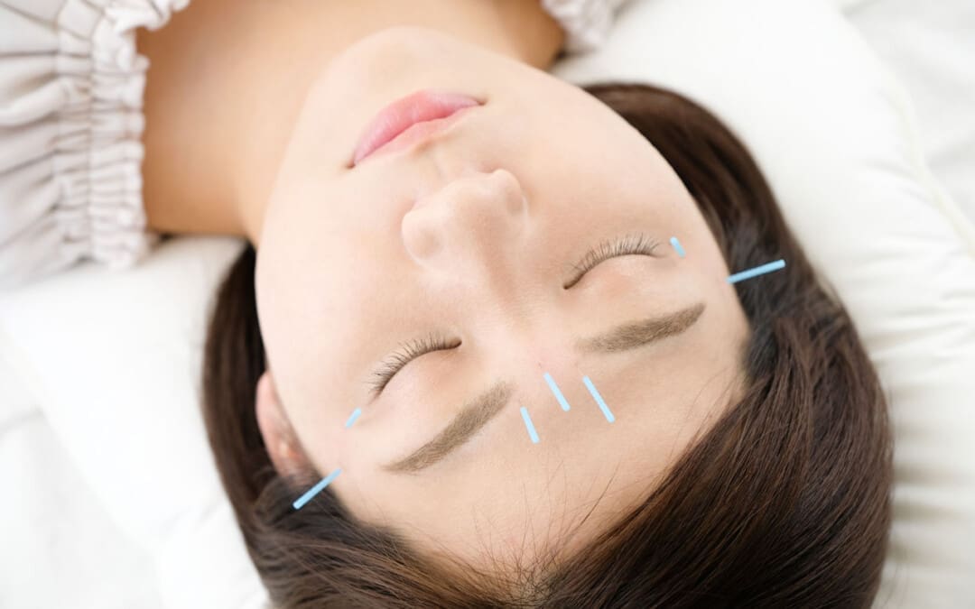
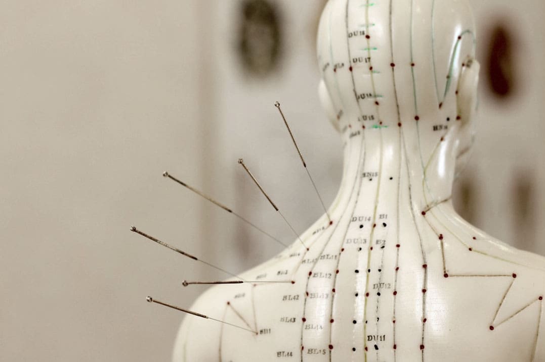
Acupuncture For Eye Health
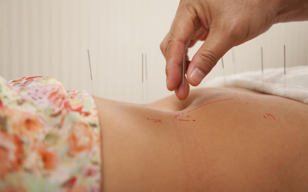
Acupuncture is an alternative medical practice that involves inserting thin needles at specific points on the body. The objective is to restore balance and health by restoring and balancing energy circulation through pathways throughout the body. These pathways, known as meridians, are separate from nerve and blood pathways.
Studies have shown that the insertion of needles manipulates accumulations of certain neurotransmitters by nearby nerves and may be what causes beneficial health effects. (Heming Zhu 2014)
Scientists are not sure exactly how acupuncture works, but it has been shown to provide pain relief and alleviation of cancer treatment nausea. (Weidong Lu, David S. Rosenthal 2013)
Studies have shown that acupuncture can help treat eye conditions like dry eye syndrome. (Tae-Hun Kim et al., 2012)
Eye Problems
For some individuals, a body imbalance can be caused by eye problems or disease. With acupuncture, imbalance-causing symptoms are addressed. Acupuncture promotes the circulation of energy and blood around the eyes.
Acupuncture has been used as an alternative treatment for chronic dry eye syndrome. (Tae-Hun Kim et al., 2012)
Studies have shown acupuncture helps reduce the eye surface’s temperature to reduce the evaporation of tears.
The procedure is also sometimes used to treat glaucoma.
Glaucoma is an optic nerve disease usually caused by above-normal eye pressure levels.
Another study showed successful reduced allergic and inflammatory eye disease symptoms. (Justine R. Smith et al., 2004)
Eye Acupoints
The following acupoints are for eye health.
Jingming
Jingming – UB-1 is located in the inner corner of the eye.
This point is thought to increase energy and blood and to help with problems such as blurry vision, cataracts, glaucoma, night blindness, and conjunctivitis. (Tilo Blechschmidt et al., 2017)
Zanzhu
The Zanzhu point – UB-2 is in the crease at the inner end of the eyebrow.
This acupoint is used when individuals complain of headaches, blurred vision, pain, tearing, redness, twitching, and glaucoma. (Gerhard Litscher 2012)
Yuyao
Yuyao is in the middle of the eyebrow, above the pupil.
This point is used for treating eye strain, eyelid twitching, ptosis, or when the upper eyelid droops over, cloudiness of the cornea, redness, and swelling. (Xiao-yan Tao et al., 2008)
Sizhukong
The Sizhukog – SJ 23 area is in the hollow area outside the eyebrow.
It is thought to be a point where acupuncture can help with eye and facial pain, including headaches, redness, pain, blurred vision, toothache, and facial paralysis. (Hongjie Ma et al., 2018)
Tongzilia
The Tongzilia – GB 1 is located on the outside corner of the eye.
The point helps brighten the eyes.
Acupuncture also helps treat headaches, redness, eye pain, light sensitivity, dry eyes, cataracts, and conjunctivitis. (GladGirl 2013)
Early studies with acupuncture have shown promise for improving eye health. Individuals considering acupuncture are recommended to consult their primary healthcare provider to see if it can be an option for those who have not found a resolution by traditional means.
Neck Injuries
References
Zhu H. (2014). Acupoints Initiate the Healing Process. Medical acupuncture, 26(5), 264–270. https://doi.org/10.1089/acu.2014.1057
Lu, W., & Rosenthal, D. S. (2013). Acupuncture for cancer pain and related symptoms. Current pain and headache reports, 17(3), 321. https://doi.org/10.1007/s11916-013-0321-3
Kim, T. H., Kang, J. W., Kim, K. H., Kang, K. W., Shin, M. S., Jung, S. Y., Kim, A. R., Jung, H. J., Choi, J. B., Hong, K. E., Lee, S. D., & Choi, S. M. (2012). Acupuncture for the treatment of dry eye: a multicenter randomised controlled trial with active comparison intervention (artificial teardrops). PloS one, 7(5), e36638. https://doi.org/10.1371/journal.pone.0036638
Law, S. K., & Li, T. (2013). Acupuncture for glaucoma. The Cochrane database of systematic reviews, 5(5), CD006030. https://doi.org/10.1002/14651858.CD006030.pub3
Smith, J. R., Spurrier, N. J., Martin, J. T., & Rosenbaum, J. T. (2004). Prevalent use of complementary and alternative medicine by patients with inflammatory eye disease. Ocular immunology and inflammation, 12(3), 203–214. https://doi.org/10.1080/092739490500200
Blechschmidt, T., Krumsiek, M., & Todorova, M. G. (2017). The Effect of Acupuncture on Visual Function in Patients with Congenital and Acquired Nystagmus. Medicines (Basel, Switzerland), 4(2), 33. https://doi.org/10.3390/medicines4020033
Litscher G. (2012). Integrative laser medicine and high-tech acupuncture at the medical university of graz, austria, europe. Evidence-based complementary and alternative medicine : eCAM, 2012, 103109. https://doi.org/10.1155/2012/103109
Tao, X. Y., Sun, C. X., Yang, J. L., Mao, M., Liao, C. C., Meng, J. G., Fan, W. B., Zhang, Y. F., Ren, X. R., & Yu, H. F. (2008). Zhongguo zhen jiu = Chinese acupuncture & moxibustion, 28(3), 191–193.
Can physical therapy treatment protocols aimed at improving range of motion and flexibility around the hip and relieving inflammation around the sciatic nerve help individuals experiencing deep buttock pain or piriformis syndrome?
Contents
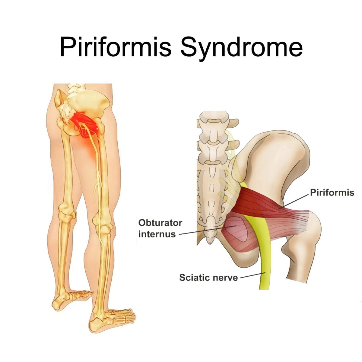
Deep Buttock Pain
Piriformis syndrome, a.k .a. deep buttock pain, is described as sciatic nerve irritation from the piriformis muscle.
The piriformis is a small muscle behind the hip joint in the buttocks.
It is about one centimeter in diameter and functions in the hip joint’s external rotation or turning outward.
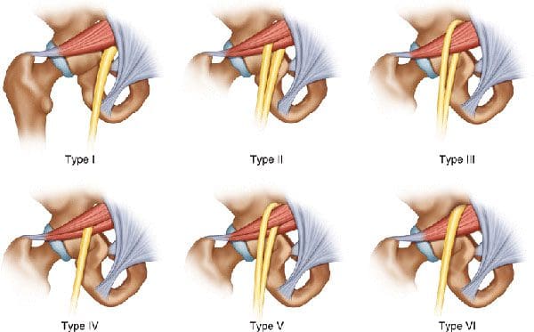
The piriformis muscle and tendon are close to the sciatic nerve, which supplies the lower extremities with motor and sensory functions.
Depending on an individual’s anatomic variation of the muscle and tendon:
The two cross over, under, or through each other behind the hip joint in the deep buttock.
This relationship is thought to irritate the nerve, leading to sciatica symptoms.
Piriformis Syndrome
When diagnosed with piriformis syndrome, it is thought that the muscle and tendon bind to and/or spasm around the nerve, causing irritation and pain symptoms.
The theory supported is that when the piriformis muscle and its tendon tighten, the sciatic nerve becomes compressed or pinched. This decreases blood circulation and irritates the nerve from the pressure. (Shane P. Cass 2015)
Tenderness with pressure on the piriformis muscle.
Discomfort in the back of the thigh.
Deep buttock pain behind the hip.
Electric sensations, shocks, and pains travel down the back of the lower extremity.
Numbness in the lower extremity.
Some individuals develop symptoms abruptly, while others go through a gradual increase.
Diagnosis
Doctors will order X-rays, MRIs, and nerve conduction studies, which is normal.
Because piriformis syndrome can be challenging to diagnose, some individuals with minor hip pain may receive a piriformis syndrome diagnosis even if they don’t have the condition. (Shane P. Cass 2015)
It is sometimes referred to as deep buttock pain. Other causes of this type of pain include back and spinal problems like:
Herniated discs
Spinal stenosis
Radiculopathy – sciatica
Hip bursitis
A piriformis syndrome diagnosis is usually given when these other causes are eliminated.
When the diagnosis is uncertain, an injection is administered in the area of the piriformis muscle. (Danilo Jankovic et al., 2013)
Different medications can be used, but the injection itself is used to help determine the specific location of the discomfort.
When an injection is given into the piriformis muscle or tendon, it is often administered by ultrasound guidance to ensure the needle delivers the medication to the correct location. (Elizabeth A. Bardowski, J. W. Thomas Byrd 2019)
Avoiding activities that cause symptoms for at least a few weeks.
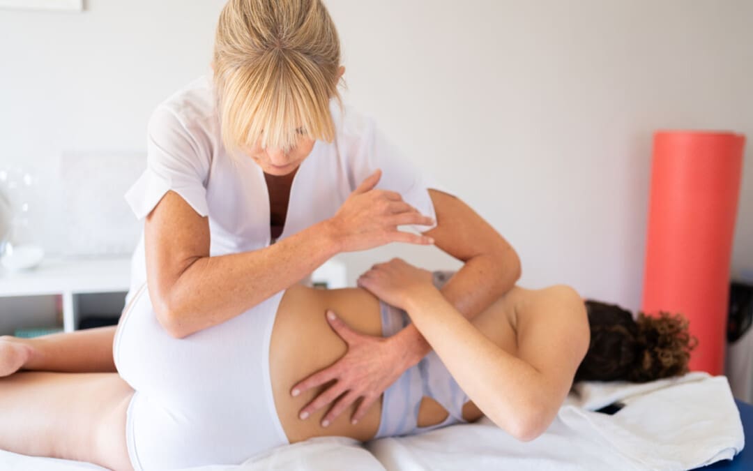
Physical Therapy
Emphasize stretching and strengthening the hip rotator muscles.
Non-Surgical Decompression
Gently pulls the spine to release any compression, allowing optimal rehydration and circulation and taking the pressure off the sciatic nerve.
Therapeutic Massage Techniques
To relax and release muscle tension and increase circulation.
Acupuncture
To help relax the piriformis muscle, sciatic nerve, and surrounding area.
Relieve pain.
Chiropractic Adjustments
Realignment rebalances the spine and musculoskeletal system to alleviate pain.
Anti-Inflammatory Medication
To decrease inflammation around the tendon.
Cortisone Injections
Injections are used to decrease inflammation and swelling.
Botulinum Toxin Injection
Injections of botulinum toxin paralyze the muscle to relieve pain.
Surgery
Surgery can be performed in rare cases to loosen the piriformis tendon, known as a piriformis release. (Shane P. Cass 2015)
Surgery is a last resort when conservative treatments have been tried for at least 6 months with little to no relief.
Recovery can take several months.
Sciatica Causes and Treatment
References
Cass S. P. (2015). Piriformis syndrome: a cause of nondiscogenic sciatica. Current sports medicine reports, 14(1), 41–44. https://doi.org/10.1249/JSR.0000000000000110
Jankovic, D., Peng, P., & van Zundert, A. (2013). Brief review: piriformis syndrome: etiology, diagnosis, and management. Canadian journal of anaesthesia = Journal canadien d’anesthesie, 60(10), 1003–1012. https://doi.org/10.1007/s12630-013-0009-5
Bardowski, E. A., & Byrd, J. W. T. (2019). Piriformis Injection: An Ultrasound-Guided Technique. Arthroscopy techniques, 8(12), e1457–e1461. https://doi.org/10.1016/j.eats.2019.07.033
Can acupuncture treatment help individuals dealing with or experiencing insomnia and sleep issues and/or disorders?
Contents
Acupuncture For Insomnia
Acupuncture is a type of holistic medicine that involves inserting sterile, disposable, thin needles at specific points known as acupoints on the body. Each needle is inserted into a different area to stimulate symptom relief of various conditions, like chronic pain and nausea. (Johns Hopkins Medicine. 2024) Recent research has looked into acupuncture for insomnia and found that it may be an effective alternative. (Mingming Zhang et al., 2019)
Insomnia
Insomnia causes individuals to have trouble falling or staying asleep. Individuals who have insomnia tend to wake up earlier than they intend to and find it difficult to impossible to get back to sleep once they are awake. The sleep disorder is quite common, with around 10% of individuals experiencing it at some point. (Andrew D. Krystal et al., 2019)
There are three categories, all characterized by the duration of the disorder. They include: (Andrew D. Krystal et al., 2019)
Acute/Short-Term
Lasting less than three months.
Episodic
Happens once in a while for less than three months.
Chronic
Lasting more than three months.
Health Issues
Insomnia can cause various health issues, and individuals can develop mood changes, irritability, fatigue, and problems with memory, impulse control, and concentration. (Andrew D. Krystal et al., 2019)
Insomnia has also been shown to increase the risk of heart failure, heart attack, and other chronic health conditions. (Mingming Zhang et al., 2019)
Benefits
Studies on the use of acupuncture for insomnia have found that it may improve sleep because of its influence on certain neurotransmitters. One review noted that specific neurotransmitters involved in the sleep-wake cycle are positively affected by acupuncture. (Kaicun Zhao 2013) The neurotransmitters include:
Norepinephrine
Helps with waking up and staying alert.
Melatonin
A hormone that helps the body calm down and prepare for sleep.
Gamma-aminobutyric acid – GABA
Helps the body fall asleep and stay asleep.
However, more research is needed to confirm the benefits of acupuncture for insomnia further.
Conditions
Certain conditions can contribute to insomnia, including:
Mood disorders
Chronic pain
Other sleep disorders
Acupuncture can help lower the effects of these disorders.
Pain
Because of the way acupuncture affects certain chemicals, it is a proven complementary treatment for pain.
The needles enhance chemicals like endorphins, dynorphins, and encephalins.
Acupuncture also releases corticosteroids, which are stress hormones.
Each of these chemicals has a role in pain symptoms.
Studies have found that individuals with anxiety can also benefit from acupuncture to help reduce symptoms. (Meixuan Li et al., 2019)
Sleep Apnea
Sleep apnea is a sleep-breathing disorder that causes an individual to stop breathing during the night temporarily.
The muscles in the nasal cavity, nose, mouth, or throat become overly relaxed.
Acupuncture can help stimulate the muscles and prevent over-relaxation, preventing apneas.
Data suggests that acupuncture may affect the apnea-hypopnea index, the number of times an individual stops and starts breathing during sleep. (Liaoyao Wang et al., 2020)
Session
Individuals should not feel pain and just a small amount of pressure in the needles’ insertion area.
If pain is present, it could be because the needles are not inserted in the right spot.
Bleeding or bruising where the needle was inserted.
Nausea
Fainting
Pins and needles sensation
Feeling more pain treatment
Prior to getting acupuncture, individuals are recommended to speak to their healthcare provider. They can advise on how it can help and any side effects that may occur due to the individual’s health, underlying conditions, and medical history. Once cleared, they can recommend a licensed acupuncturist.
Zhang, M., Zhao, J., Li, X., Chen, X., Xie, J., Meng, L., & Gao, X. (2019). Effectiveness and safety of acupuncture for insomnia: Protocol for a systematic review. Medicine, 98(45), e17842. https://doi.org/10.1097/MD.0000000000017842
Krystal, A. D., Prather, A. A., & Ashbrook, L. H. (2019). The assessment and management of insomnia: an update. World psychiatry: official journal of the World Psychiatric Association (WPA), 18(3), 337–352. https://doi.org/10.1002/wps.20674
Zhao K. (2013). Acupuncture for the treatment of insomnia. International review of neurobiology, 111, 217–234. https://doi.org/10.1016/B978-0-12-411545-3.00011-0
Patil, S., Sen, S., Bral, M., Reddy, S., Bradley, K. K., Cornett, E. M., Fox, C. J., & Kaye, A. D. (2016). The Role of Acupuncture in Pain Management. Current pain and headache reports, 20(4), 22. https://doi.org/10.1007/s11916-016-0552-1
Li, M., Xing, X., Yao, L., Li, X., He, W., Wang, M., Li, H., Wang, X., Xun, Y., Yan, P., Lu, Z., Zhou, B., Yang, X., & Yang, K. (2019). Acupuncture for treatment of anxiety, an overview of systematic reviews. Complementary therapies in medicine, 43, 247–252. https://doi.org/10.1016/j.ctim.2019.02.013
Wang, L., Xu, J., Zhan, Y., & Pei, J. (2020). Acupuncture for Obstructive Sleep Apnea (OSA) in Adults: A Systematic Review and Meta-Analysis. BioMed research international, 2020, 6972327. https://doi.org/10.1155/2020/6972327
Chan, M. W. C., Wu, X. Y., Wu, J. C. Y., Wong, S. Y. S., & Chung, V. C. H. (2017). Safety of Acupuncture: Overview of Systematic Reviews. Scientific reports, 7(1), 3369. https://doi.org/10.1038/s41598-017-03272-0
Ernst, G., Strzyz, H., & Hagmeister, H. (2003). Incidence of adverse effects during acupuncture therapy-a multicentre survey. Complementary therapies in medicine, 11(2), 93–97. https://doi.org/10.1016/s0965-2299(03)00004-9
“For individuals that have difficulty getting plenty of fruits and vegetables, can incorporating green powder supplements increase nutritional levels for a balanced diet?”
Contents
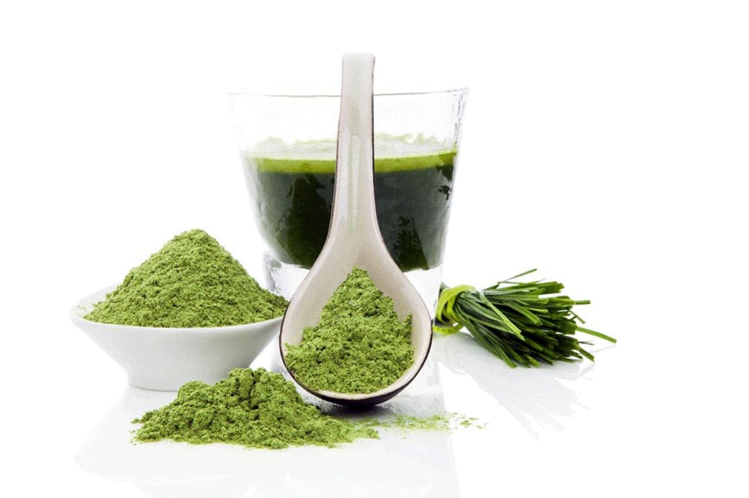
Green Powder Supplements
Meeting daily nutrient needs through whole, unprocessed foods can’t always be met when access is limited or for other reasons. A green powder supplement is a great way to fill in the gaps. Green powder supplements are a daily supplement that helps increase vitamin, mineral, and fiber intake and enhances overall health. Green powders are easy to mix in water with a favorite beverage or smoothie or bake into a recipe. They can help:
Increase energy
Nourish the immune system
Improve digestion
Promote mental clarity
Contribute to healthy blood sugar levels
Reduce the risk of chronic disease
Promote optimal liver and kidney function
What Are They?
Green powder supplements are forms of vitamins, minerals, fiber, antioxidants, phytochemicals, and other bioactive compounds.
They are derived from fruits, vegetables, herbs, and algae to combine ingredients into a convenient supplement. (Giulia Lorenzoni et al., 2019)
Nutrients
Because most green powders comprise a combination of ingredients, the nutrient density is high. Green powder supplements can be considered a vitamin and mineral product. They typically contain:
Vitamins A, C, and K
Iron
Magnesium
Calcium
Antioxidants
The recommended daily intake of vitamins and minerals can be helpful for individuals with limited access to produce or who want to supplement their diet with additional nutrients.
Energy
The phytochemicals found in fruits and vegetables have been shown to improve energy levels. Studies on their effects on physical performance and endurance have resulted in positive outcomes. Researchers found that phytonutrients like those in green powders helped to increase energy, improve agility, reduce fatigue perception, improve memory, and decrease recovery time. (Nicolas Monjotin et al., 2022)
Digestive Health
Green powders are rich in soluble and insoluble fiber, which contribute to feeling full and satisfied after a meal and are important for healthy digestion and regular bowel movements. Eating fiber-rich foods is associated with optimal blood sugar control and improved gut microbiota diversity. These factors are important for maintaining a healthy body weight and decreasing the risk of chronic disease, for example, type 2 diabetes. (Thomas M. Barber et al., 2020) Phytochemicals, including flavonoids, have been shown to have therapeutic effects on gas, bloating, constipation, and diarrhea associated with IBS. Other phytonutrients have been shown to reduce certain symptoms of ulcerative colitis. (Nicolas Monjotin et al., 2022)
Immune System Function
Supplemental green powder supplements have shown the ability to maintain a healthy immune system and reduce inflammation by their antioxidant content. Green powders containing seaweed or algae are rich in phytochemical and poly-unsaturated fatty acids that have antioxidant properties to reduce inflammation and prevent oxidative damage to cells. (Agnieszka Jaworowska, Aliza Murtaza 2022) A randomized trial found that a fruit, berry, and vegetable powder concentrate blend decreased oxidation and reduced inflammation, attributed to the phytochemicals found in fruits and vegetables.(Manfred Lamprecht et al., 2013)
Detoxification
The liver and kidneys are the main organs of natural detoxification. The liver helps the body absorb nutrients from consumed foods and removes waste and toxins through the kidneys. (National Library of Medicine. 2016) Plants are packed with antioxidants and phytochemicals that protect the liver and kidneys from free radical damage and oxidative stress. (Yong-Song Guan et al., 2015) The green powder supplements are made from these plants. When drinking green powders, fluid intake naturally increases as a standard serving of green powder is mixed with 8 to 12 ounces of water.
Whether mixed, blended, or made into a shake, powdered greens are a convenient and efficient way to get the daily dose of antioxidants, vitamins, minerals, and other nutrients.
The Healing Diet: Combat Inflammation, Embrace Wellness
References
Lorenzoni, G., Minto, C., Vecchio, M. G., Zec, S., Paolin, I., Lamprecht, M., Mestroni, L., & Gregori, D. (2019). Fruit and Vegetable Concentrate Supplementation and Cardiovascular Health: A Systematic Review from a Public Health Perspective. Journal of clinical medicine, 8(11), 1914. https://doi.org/10.3390/jcm8111914
Monjotin, N., Amiot, M. J., Fleurentin, J., Morel, J. M., & Raynal, S. (2022). Clinical Evidence of the Benefits of Phytonutrients in Human Healthcare. Nutrients, 14(9), 1712. https://doi.org/10.3390/nu14091712
Barber, T. M., Kabisch, S., Pfeiffer, A. F. H., & Weickert, M. O. (2020). The Health Benefits of Dietary Fibre. Nutrients, 12(10), 3209. https://doi.org/10.3390/nu12103209
Jaworowska, A., & Murtaza, A. (2022). Seaweed Derived Lipids Are a Potential Anti-Inflammatory Agent: A Review. International journal of environmental research and public health, 20(1), 730. https://doi.org/10.3390/ijerph20010730
Lamprecht, M., Obermayer, G., Steinbauer, K., Cvirn, G., Hofmann, L., Ledinski, G., Greilberger, J. F., & Hallstroem, S. (2013). Supplementation with a juice powder concentrate and exercise decrease oxidation and inflammation, and improve the microcirculation in obese women: randomised controlled trial data. The British journal of nutrition, 110(9), 1685–1695. https://doi.org/10.1017/S0007114513001001
InformedHealth.org [Internet]. Cologne, Germany: Institute for Quality and Efficiency in Health Care (IQWiG); 2006-. How does the liver work? 2009 Sep 17 [Updated 2016 Aug 22]. Available from: https://www.ncbi.nlm.nih.gov/books/NBK279393/
Guan, Y. S., He, Q., & Ahmad Al-Shatouri, M. (2015). Complementary and Alternative Therapies for Liver Diseases 2014. Evidence-based complementary and alternative medicine : eCAM, 2015, 476431. https://doi.org/10.1155/2015/476431
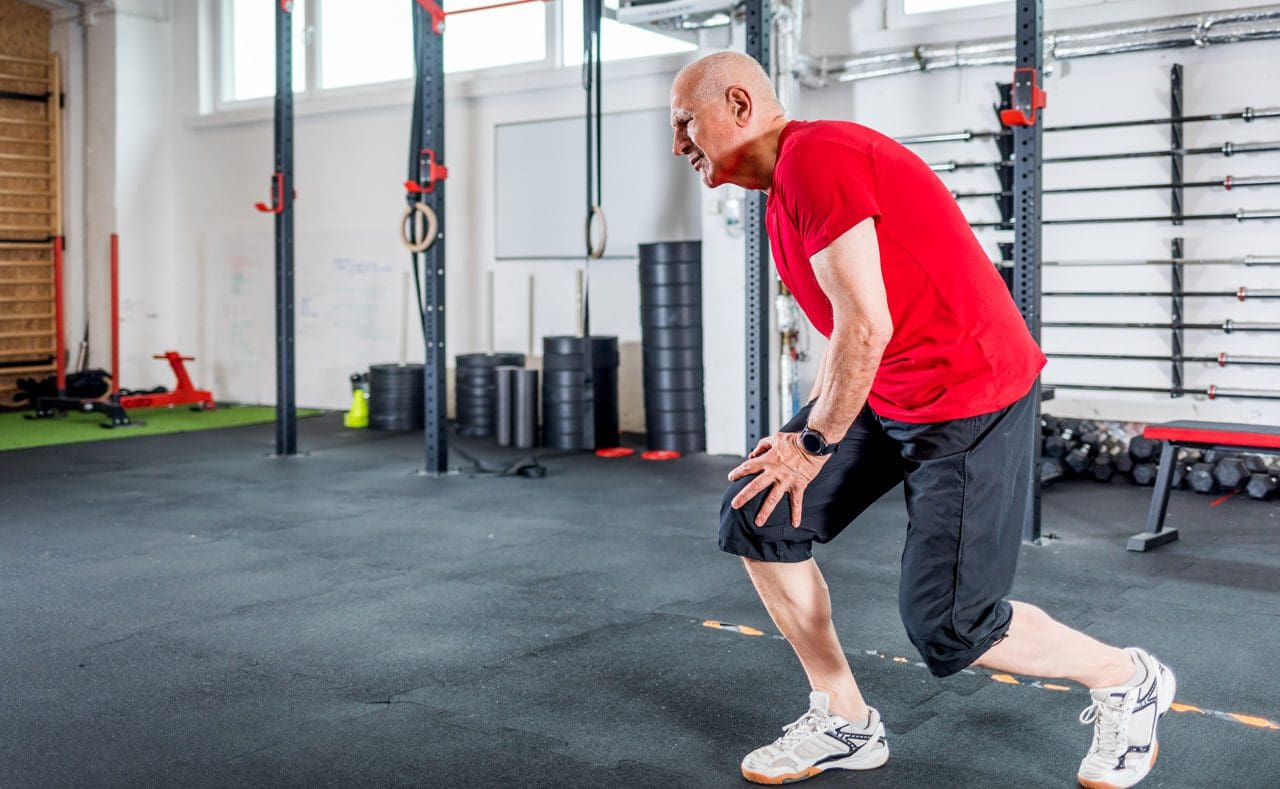
For individuals training for long distance walking marathons and/or events, can focusing on building a walking foundation, then increasing mileage progressively help condition the body for overall readiness?
Contents
Long Distance Walking Training
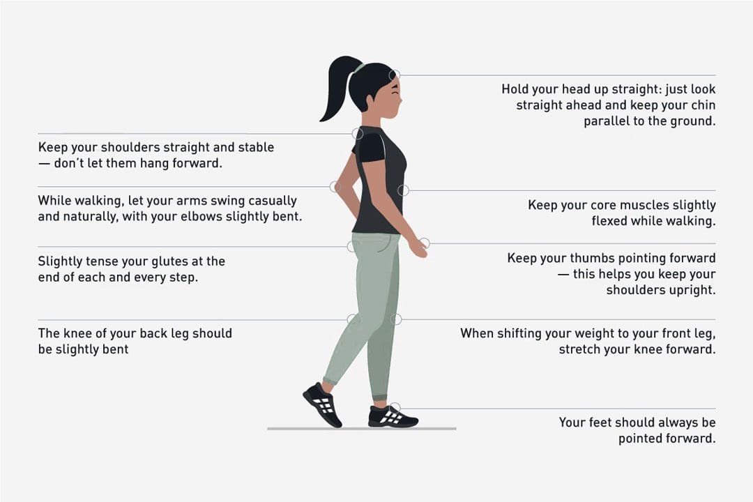
Training helps individuals be comfortable and safe for long-distance walking and events.
Training should focus on building a walking pace and increasing mileage gradually.
Individuals need endurance, not speed, and want to build mental stamina for walking for hours at a steady pace.
To reduce the risk of training injuries, increasing the total mileage per week/the distance of the longest walk per week to no more than 10% is recommended.
Individuals should also train to wear the gear worn during long-distance walks.
Training could last a few months.
Being methodical allows the body time to repair and build new muscle, blood supplies, and endurance.
Example Training Plans
Following a marathon training plan for mileage building and determining the proper hydration, nutrition, and gear for multi-day walks and treks is recommended. However, individuals must build back-to-back long days into their training sessions to assess any issues or problems resulting from walking long distances on back-to-back days.
Example Walking Training Plans
Multi-Day Walks/Treks Training Schedule
13 miles per day/21 kilometers
Use this plan for marathons or other multi-day walks with hills and natural surfaces requiring a backpack.
Training to Walk a Marathon
26.2 miles/42 kilometers
This will condition the body to go longer distances.
When training for distances of 31 to 100 miles/50 to 161 kilometers, the longest distance to train should not need to exceed 20 to 25 miles,
These should be performed at least twice two months before the marathon or event.
Taper down the month before the event to a 12.4-mile/20-kilometer distance.
Gear
All clothing, shoes, sunscreen, backpacks, etc., must be tested on the longer training days before the event.
Given the climate and terrain, plan for what will be needed and removed.
Try things out, as individuals don’t want to be surprised with something unfamiliar at the event. From head to toe, test the gear, including:
Shoes/boots, socks, underwear, bra, shirt, pants, hat, jacket, and rain gear.
Choose shoes or walking boots and wear them on long training days to break them in and ensure they perform.
Backpacks should be tested on longer training days to ensure they can be carried comfortably over long distances and have the necessary capacity.
Choose wicking fabrics that allow the skin to breathe and cool, especially under layers. (Justin De Sousa et al., 2014)
Individuals will want to wear gear similar to marathon walkers if the walk will mostly be on pavement or asphalt.
Individuals can modify their gear if the route is off-road or during different seasons. Find out what other long-distance walkers have worn on the same route or event.
Individuals can connect with fellow walkers via social media or find answers to frequently asked questions on the event’s or destination’s website.
Individuals can also contact the event director via the website or social media.
Nutrition
Proper sports nutrition will prepare the body for endurance activity.
For example, individuals are recommended to follow a diet comprising 70% carbohydrates, 20% protein, and 10% fat.
Avoid high-protein diets, as they can cause hydration problems and strain your kidneys under endurance walking conditions. (Marta Cuenca-Sánchez et al., 2015)
Train with the water, sports drinks, food, and snacks taken to the event, and do not deviate from them during the event.
Water is needed for 20 kilometers and under events, but an electrolyte replacement sports drink may be better for longer walks.
Diluting or leaving out some sugar can be easier on the stomach.
Have snacks pre-packaged and labeled for the times to be eaten.
Individuals need to eat fat and protein for ultramarathon distances – this can come from trail mix, peanut butter sandwiches, and chocolate bars with nuts.
Carbohydrates can be provided by sports gels or energy bars.
It is recommended to avoid products made for short distances and power sports as they can cause digestive problems when walking longer distances.
Planning a Walk
Planning begins by setting goals. Considerations include:
Time of year
Distance
Transportation to the event
Event pace requirements
Altitude and hill profile
Climate
Individuals are recommended to:
Prepare by researching routes and trails.
Study the course maps to know what services are provided along the way and what individuals must bring.
Walk a long distance without a supporting event.
Contact individuals who have walked the course.
Know the terrain and areas of total sun, hills, pavement, natural trails, and shade.
If possible, drive the course to become familiar with it.
Individuals may be able to find apps designed for their route.
Taking Breaks and Resting
Regular breaks should be short – using the bathroom, eating a snack, rehydrating, tying shoes, or bandaging blisters.
The body can stiffen up quickly during breaks and take several minutes to regain walking pace after a long break.
Recommendations could be taking a walking break instead, which means continuing to walk but at a very slow pace.
Foot Care
Individuals will have found what works for them concerning shoes, boots, socks, etc., on the long training days to prevent blisters and injuries. It is recommended to try different strategies, which include:
Sports tape
Blister block pads
Sprays
Lubricants
Wicking and/or double-layered socks
Moleskin
Stop at the first sign of irritation along the walk and doctor the foot with tape, blister bandages, or whatever method works best.
The body was built for walking. Planning and training properly before taking a long-distance or multi-day walk will ensure a safe and enjoyable marathon.
Move Better, Live Better
References
De Sousa, J., Cheatham, C., & Wittbrodt, M. (2014). The effects of a moisture-wicking fabric shirt on the physiological and perceptual responses during acute exercise in the heat. Applied ergonomics, 45(6), 1447–1453. https://doi.org/10.1016/j.apergo.2014.04.006
Cuenca-Sánchez, M., Navas-Carrillo, D., & Orenes-Piñero, E. (2015). Controversies surrounding high-protein diet intake: satiating effect and kidney and bone health. Advances in nutrition (Bethesda, Md.), 6(3), 260–266. https://doi.org/10.3945/an.114.007716
For individuals dealing with knee pain symptoms from injury and/or arthritis, can incorporating an acupuncture and/or electroacupuncture treatment plan help in pain relief and management?
Contents
Acupuncture For Knee Pain
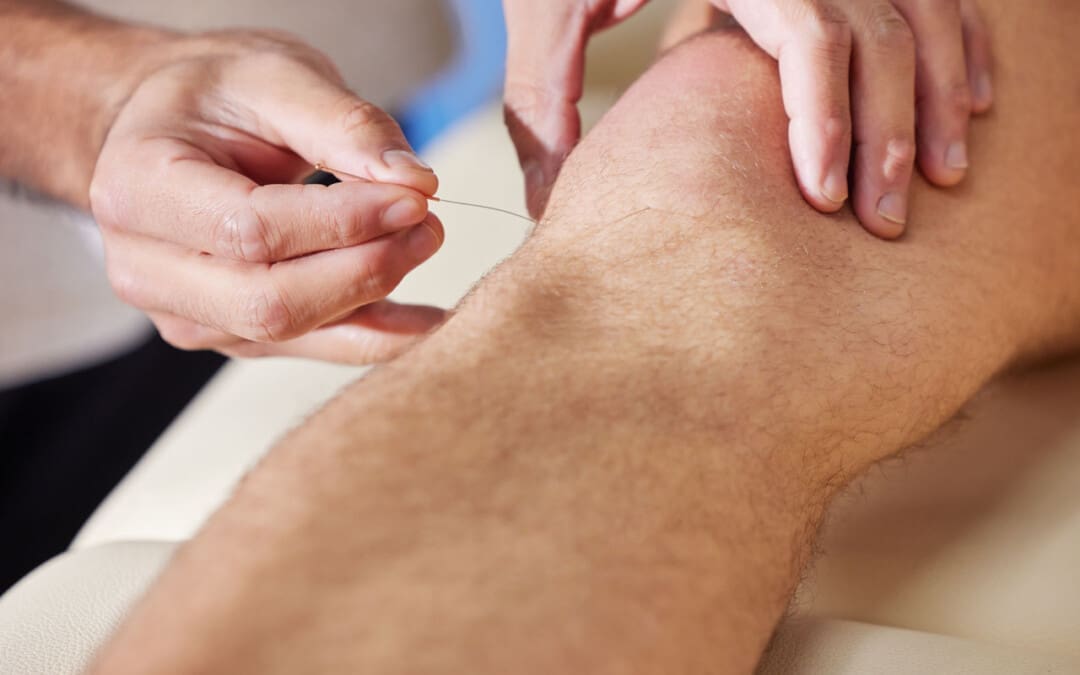
Acupuncture involves inserting very thin needles into the skin at specific acupoints on the body. It is based on the premise that the needles restore the flow of the body’s energy to activate and promote healing, relieve pain, and help the body relax.
Acupuncture can help address various health conditions, including knee pain caused by arthritis or injury.
Depending on the type and severity of pain, treatments can help reduce the pain for days or weeks.
Acupuncture is often used as a complementary therapy – treatment in addition to other treatment or therapy strategies like massage and chiropractic.
Acupuncture Benefits
Knee pain caused by osteoarthritis or injury can reduce flexibility, mobility, and quality of life. Acupuncture can help provide relief.
When the acupuncture needles are placed on the body, a signal is sent along the spinal cord to the brain, which triggers a release of endorphins/pain hormones. Medical researchers believe this helps reduce pain. (Qian-Qian Li et al., 2013) Acupuncture also helps decrease the production of cortisol, a hormone that helps control inflammation. (Qian-Qian Li et al., 2013) With reduced pain sensations and less inflammation after acupuncture treatments, knee function and mobility can be improved.
Various factors play a role in the pain relief experienced from acupuncture. Some evidence suggests that an individual’s expectations may impact the results of acupuncture treatment. (Stephanie L. Prady et al., 2015)
Researchers are currently assessing whether the expectation that acupuncture is beneficial contributes to a better outcome after treatment. (Zuoqin Yang et al., 2021)
In 2019, acupuncture was recommended in treating knee osteoarthritis in the American College of Rheumatology/Arthritis Foundation guidelines for hand, hip, and knee osteoarthritis pain management. (Sharon L. Kolasinski et al., 2020)
Research
Different clinical studies support acupuncture’s ability to help in knee pain relief and management.
One study found that acupuncture helps manage various conditions that cause chronic pain. (Andrew J. Vickers et al., 2012)
A scientific review analyzed previous studies on pain management interventions after knee surgery and found supporting evidence that the treatments delayed and reduced the use of medications for pain relief post-surgery. (Dario Tedesco et al., 2017)
Osteoarthritis
A systematic review analyzed randomized control studies to determine whether or not acupuncture reduced pain and improved joint function in individuals with chronic osteoarthritis knee pain. (Xianfeng Lin et al., 2016)
Individuals received six to twenty-three weekly acupuncture sessions for three to 36 weeks.
The analysis determined that acupuncture can improve short and long-term physical function and mobility and provide up to 13 weeks of pain relief in individuals with chronic knee pain caused by osteoarthritis.
Rheumatoid Arthritis
Rheumatoid arthritis is a chronic disease that affects joints, including the knee joint, causing pain and stiffness.
Acupuncture is beneficial in treating rheumatoid arthritis/RA.
A review found that acupuncture alone and in combination with other treatment modalities benefits individuals with RA. (Pei-Chi, Chou Heng-Yi Chu 2018)
Acupuncture is believed to have anti-inflammatory and antioxidant effects to help regulate immune system function.
Chronic Knee Pain
Various conditions and injuries can cause chronic knee pain, making mobility difficult.
Individuals with joint pain often turn to complementary therapies for pain relief management, with acupuncture being one of the popular modalities. (Michael Frass et al., 2012)
Working with a licensed, professional acupuncture practitioner can reduce the risk of unwanted side effects and complications.
Types
Other acupuncture options that may be offered include:
Electroacupuncture
A modified form of acupuncture where a mild electrical current passes through the needles, providing additional stimulation to the acupoints.
In one research study, individuals with knee osteoarthritis reported significant improvements in their pain, stiffness, and physical function after electroacupuncture treatment. (Ziyong Ju et al., 2015)
Auricular
Auricular or ear acupuncture works on acupoints in the ear corresponding to the body’s different parts.
A research review analyzed several studies on auricular acupuncture for pain relief and found that it can provide relief within 48 hours of pain onset. (M. Murakami et al., 2017)
Battlefield Acupuncture
The military and veteran healthcare facilities use a unique form of auricular acupuncture for pain management.
Studies show that it is effective at providing immediate pain relief, but more research is necessary to determine long-term pain relief effectiveness. (Anna Denee Montgomery, Ronovan Ottenbacher 2020)
Before trying acupuncture, consult a healthcare professional for guidance, as it may be integrated with other therapies and lifestyle adjustments.
Overcoming an ACL Injury
References
Li, Q. Q., Shi, G. X., Xu, Q., Wang, J., Liu, C. Z., & Wang, L. P. (2013). Acupuncture effect and central autonomic regulation. Evidence-based complementary and alternative medicine : eCAM, 2013, 267959. https://doi.org/10.1155/2013/267959
Prady, S. L., Burch, J., Vanderbloemen, L., Crouch, S., & MacPherson, H. (2015). Measuring expectations of benefit from treatment in acupuncture trials: a systematic review. Complementary therapies in medicine, 23(2), 185–199. https://doi.org/10.1016/j.ctim.2015.01.007
Yang, Z., Li, Y., Zou, Z., Zhao, Y., Zhang, W., Jiang, H., Hou, Y., Li, Y., & Zheng, Q. (2021). Does patient’s expectation benefit acupuncture treatment?: A protocol for systematic review and meta-analysis. Medicine, 100(1), e24178. https://doi.org/10.1097/MD.0000000000024178
Kolasinski, S. L., Neogi, T., Hochberg, M. C., Oatis, C., Guyatt, G., Block, J., Callahan, L., Copenhaver, C., Dodge, C., Felson, D., Gellar, K., Harvey, W. F., Hawker, G., Herzig, E., Kwoh, C. K., Nelson, A. E., Samuels, J., Scanzello, C., White, D., Wise, B., … Reston, J. (2020). 2019 American College of Rheumatology/Arthritis Foundation Guideline for the Management of Osteoarthritis of the Hand, Hip, and Knee. Arthritis care & research, 72(2), 149–162. https://doi.org/10.1002/acr.24131
Vickers, A. J., Cronin, A. M., Maschino, A. C., Lewith, G., MacPherson, H., Foster, N. E., Sherman, K. J., Witt, C. M., Linde, K., & Acupuncture Trialists’ Collaboration (2012). Acupuncture for chronic pain: individual patient data meta-analysis. Archives of internal medicine, 172(19), 1444–1453. https://doi.org/10.1001/archinternmed.2012.3654
Tedesco, D., Gori, D., Desai, K. R., Asch, S., Carroll, I. R., Curtin, C., McDonald, K. M., Fantini, M. P., & Hernandez-Boussard, T. (2017). Drug-Free Interventions to Reduce Pain or Opioid Consumption After Total Knee Arthroplasty: A Systematic Review and Meta-analysis. JAMA surgery, 152(10), e172872. https://doi.org/10.1001/jamasurg.2017.2872
Lin, X., Huang, K., Zhu, G., Huang, Z., Qin, A., & Fan, S. (2016). The Effects of Acupuncture on Chronic Knee Pain Due to Osteoarthritis: A Meta-Analysis. The Journal of bone and joint surgery. American volume, 98(18), 1578–1585. https://doi.org/10.2106/JBJS.15.00620
Chou, P. C., & Chu, H. Y. (2018). Clinical Efficacy of Acupuncture on Rheumatoid Arthritis and Associated Mechanisms: A Systemic Review. Evidence-based complementary and alternative medicine : eCAM, 2018, 8596918. https://doi.org/10.1155/2018/8596918
Frass, M., Strassl, R. P., Friehs, H., Müllner, M., Kundi, M., & Kaye, A. D. (2012). Use and acceptance of complementary and alternative medicine among the general population and medical personnel: a systematic review. Ochsner journal, 12(1), 45–56.
Hinman, R. S., McCrory, P., Pirotta, M., Relf, I., Forbes, A., Crossley, K. M., Williamson, E., Kyriakides, M., Novy, K., Metcalf, B. R., Harris, A., Reddy, P., Conaghan, P. G., & Bennell, K. L. (2014). Acupuncture for chronic knee pain: a randomized clinical trial. JAMA, 312(13), 1313–1322. https://doi.org/10.1001/jama.2014.12660
National Center for Complementary and Integrative Health. (2022). Acupuncture in depth. National Center for Complementary and Integrative Health. https://www.nccih.nih.gov/health/acupuncture-what-you-need-to-know
Harvard Medical School. (2023). Acupuncture: what is it? Harvard Health Publishing Harvard Medical School Blog. https://www.health.harvard.edu/a_to_z/acupuncture-a-to-z#:~:text=The%20most%20common%20side%20effects,injury%20to%20an%20internal%20organ.
Ju, Z., Guo, X., Jiang, X., Wang, X., Liu, S., He, J., Cui, H., & Wang, K. (2015). Electroacupuncture with different current intensities to treat knee osteoarthritis: a single-blinded controlled study. International journal of clinical and experimental medicine, 8(10), 18981–18989.
Murakami, M., Fox, L., & Dijkers, M. P. (2017). Ear Acupuncture for Immediate Pain Relief-A Systematic Review and Meta-Analysis of Randomized Controlled Trials. Pain medicine (Malden, Mass.), 18(3), 551–564. https://doi.org/10.1093/pm/pnw215
Montgomery, A. D., & Ottenbacher, R. (2020). Battlefield Acupuncture for Chronic Pain Management in Patients on Long-Term Opioid Therapy. Medical acupuncture, 32(1), 38–44. https://doi.org/10.1089/acu.2019.1382
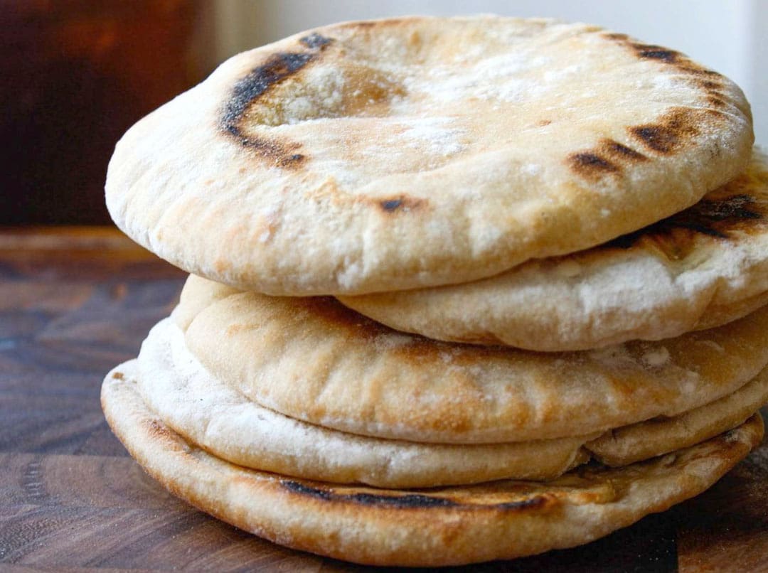
Could pita bread be a possible option for individuals trying to eat healthier?
Contents
Pita Bread
Pita bread is a yeast-leavened, round flatbread made with wheat flour. When baked, the dough turns into two layers. These layers create a pocket that can be filled with vegetables, meats, or vegetarian proteins. Pita bread offers health benefits because of its low carbohydrate count, the amount of nutrients in one serving, and the use of wheat flour.
The carbohydrate count for pita bread is 17 grams per serving or a little more than one carb count – 15 grams, used in meal planning for individuals with diabetes.
Non-keto bread is around 20 grams of carbohydrates per serving or slice.
Pita bread has a lower carbohydrate count than most breads.
Fats
Pita breads are relatively low in fat content.
The total lipid fat is under 2 grams, only 2% of the recommended daily amount or RDA.
The bread contains no fatty acids or trans or saturated fat.
Protein
Four grams of protein are in one serving of pita bread.
The protein content is found in the wheat flour.
Vitamins and Minerals
Other minerals in pita bread include:
Calcium, with 60.1 milligrams per serving.
Iron with 1.08 milligrams per serving – helps the body create hemoglobin, a protein in red blood cells that carries oxygen from the lungs. (National Institute of Health, 2023)
Sodium with 120 milligrams.
According to the Federal Drug Administration, this is a low amount of sodium. However, individuals should stay aware of sodium intake and limit it to no more than 2,300 milligrams per day.
Pita bread for a sandwich contains fewer calories than two slices of regular bread.
Benefits
Potential health benefits include the following:
Glucose Levels Lowered
Whole wheat can be beneficial to glucose levels.
The American Diabetes Association suggests that choosing bread with whole wheat grains, like pita bread, instead of white bread, can work to keep blood sugar levels from spiking. (American Diabetes Association 2024)
Digestion Support
Whole-grain pita bread fiber content can benefit the digestive system by regulating bowel movements.
Complex carbohydrates are digested slower than simple carbohydrates, keeping the body fuller for longer and assisting in weight management. (Harvard Health 2022)
Protein Source
Pita bread provides a healthy amount of protein.
A serving contains around 8% of protein.
Consuming the proper amount of protein helps in muscle repair. (Harvard Health 2024)
Allergies
Major allergies or intolerances can cause individuals to pass on the bread. What individuals need to know.
Celiac Disease
Celiac disease is a heredity autoimmune disease that occurs in genetically predisposed individuals.
Individuals with the disease cannot ingest gluten – a protein found in wheat – which can lead to small intestinal damage.
Individuals who experience gastrointestinal distress when eating wheat should consult a healthcare professional to get tested. (Celiac Disease Foundation 2023)
Wheat Allergy
A wheat allergy may mimic celiac disease symptoms, but they are different allergies.
The allergy occurs when the body produces antibodies to wheat proteins.
USDA. Pita Bread. (2021). Pita Bread. Retrieved from https://fdc.nal.usda.gov/fdc-app.html#/food-details/2134834/nutrients
National Institute of Health, Office of Dietary Supplements. (2023). Iron. Retrieved from https://ods.od.nih.gov/factsheets/Iron-HealthProfessional/
Food and Drug Administration. (2022). Sodium in your diet. Retrieved from https://www.fda.gov/food/nutrition-education-resources-materials/sodium-your-diet
American Diabetes Association. (2024). Types of carbohydrates (Food and Nutrition, Issue. https://diabetes.org/food-nutrition/understanding-carbs/types-carbohydrates
Harvard Health. (2022). Fiber (The Nutrition Source, Issue. https://www.hsph.harvard.edu/nutritionsource/carbohydrates/fiber/
Harvard Health. (2024). Protein (The Nutrition Source, Issue. https://www.hsph.harvard.edu/nutritionsource/what-should-you-eat/protein/
Celiac Disease Foundation. (2023). What is celiac disease? (About Celiac Disease, Issue. https://celiac.org/about-celiac-disease/what-is-celiac-disease/
American College of Allergy, Asthma, and Immunology. (2024). Wheat (Allergic Conditions, Issue. https://acaai.org/allergies/allergic-conditions/food/wheat-gluten/
Can individuals dealing with joint pain incorporate acupuncture therapy to manage lupus symptoms and restore body mobility?
Contents
Introduction
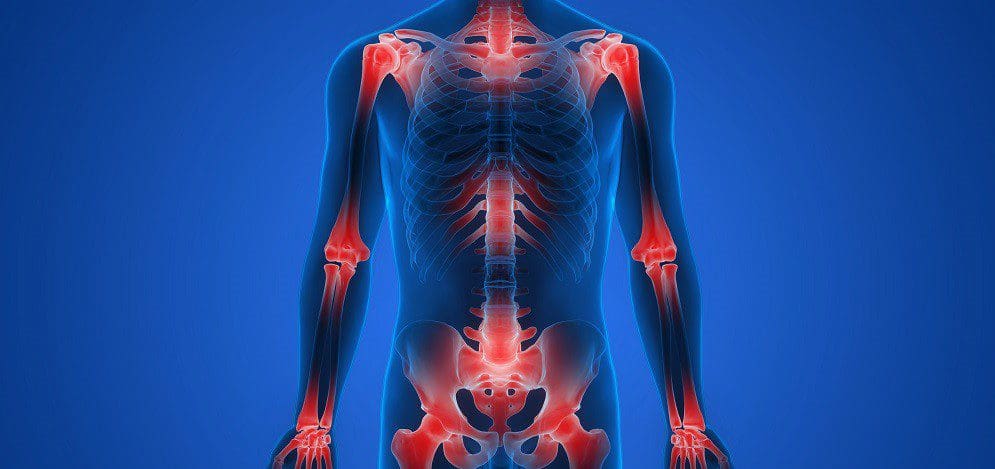
The immune system is highly important to the body as its main job is to protect vital structures from foreign invaders that can cause pain-like issues and discomfort. The immune system has a healthy relationship with the different body systems, including the musculoskeletal system, as the inflammatory cytokines help heal muscle and tissue damage when the body is injured. Over time, however, when normal environmental and genetic factors start to develop in the body, the immune system will begin to send out these cytokines to healthy, normal cells. To that point, the body starts at risk of developing autoimmune diseases. Now, autoimmune diseases in the body can cause havoc over time when they are not managed, leading to chronic disorders that can cause overlapping symptoms in the musculoskeletal system. One of the most common autoimmune diseases is systemic lupus erythematosus or lupus, and it can cause a person to be in consistent pain and discomfort while correlating with muscle and joint pain. Today’s article looks at the factors and effects of lupus, the burden of joint pain in lupus, and how holistic approaches like acupuncture can help manage lupus while restoring body mobility. We talk with certified medical providers who consolidate our patients’ information to assess how to minimize the pain effects caused by lupus on the joints. We also inform and guide patients on how acupuncture can help manage lupus and combine other therapies to reduce its pain-like symptoms affecting the musculoskeletal system. We encourage our patients to ask their associated medical providers intricate and important questions about incorporating acupuncture therapy to relieve the inflammatory effects of lupus while finding natural ways to restore mobility. Dr. Jimenez, D.C., includes this information as an academic service. Disclaimer.
The Factors & Effects Of Lupus
Have you been experiencing joint pain in your upper or lower extremities, making it difficult to function throughout the day? Have you been feeling the constant effects of fatigue? Many individuals experiencing these pain-like issues could risk developing systemic lupus erythematosus. In this autoimmune disease, the body’s own immune system mistakenly starts to attack its tissues, thus leading to inflammation and a range of pain-like symptoms. Lupis is tricky to diagnose because of its complex immune dysregulation that can lead to an overproduction of cytokines that can affect the body. (Lazar & Kahlenberg, 2023) At the same time, lupus can affect a diverse population, with symptoms and severity varying depending on how mild or severe the factors affect the body. Lupus can impact various body parts, including the joints, skin, kidneys, blood cells, and other vital body parts and organs, as environmental and hormonal factors can influence its development. (Tsang & Bultink, 2021) Additionally, lupus can be closely associated with other comorbidities that are causing overlapping risk profiles with inflammation that can affect the joints in the musculoskeletal system.
The Burden of Joint Pain In Lupus
Lupus is tricky to diagnose since it often mimics other ailments; the most common pain symptom that lupus affects is the joints. Individuals with lupus experience joint pain, which can cause inflammatory effects and structural damage to the joints, tendons, muscles, and bones, causing pathological abnormalities. (Di Matteo et al., 2021) Since lupus causes inflammatory effects in the joints, many individuals will think that they are experiencing inflammatory arthritis, and it can cause overlapping risk profiles as it is accompanied by lupus, thus causing localized pain in the joints regardless of its origin. (Senthelal et al., 2024) Joint pain in lupus individuals can significantly hinder daily activities, reducing mobility and overall quality of life as they are trying to find relief.
Unlocking The Secrets of Inflammation-Video
A Holistic Approach to Managing Lupus
While standard treatments for lupus involve medication and immunosuppressants to reduce the inflammation caused by lupus, many people want to seek out holistic approaches to manage lupus and reduce the inflammatory effects from affecting their joints by making small changes in their lives. Many people incorporate anti-inflammatory foods rich in antioxidants to dampen the inflammatory effects. Various supplements, like vitamin D, calcium, zinc, etc., can help reduce inflammation caused by lupus and strengthen bone health. Additionally, non-surgical treatments can even improve cardiorespiratory capacity and decrease fatigue while improving psychological function, which can help improve a person’s quality of life by managing the symptoms caused by lupus. (Fangtham et al., 2019)
How Acupuncture Could Help Lupus & Restore Mobility
One of the oldest forms of non-surgical and holistic approaches to reducing inflammation and managing lupus is acupuncture. Acupuncture involves solid, thin needles used by highly trained professionals to be inserted into specific body points to balance the body’s qi (energy) by stimulating the nervous system and releasing beneficial chemicals into the affected muscles, spinal cord, and brain. Additionally, acupuncture, with its minimal side effects and holistic approach, can help manage lupus. This is because when acupuncture needles are placed at the acupoints of the body, it can disrupt the pain signals that are causing pain in the affected area and regulate the inflammatory cytokines from lupus to provide relief. (Wang et al., 2023) This is due to its philosophy of addressing not only the physical pain but also the emotional and psychological symptoms of living with a chronic condition like lupus.
Additionally, acupuncture can help restore joint mobility while managing lupus through consecutive treatments, as many people notice that their joint mobility is improved and their pain is diminished. This is because the insertion and manipulation of the needles in the body’s acupoints cause alterations in afferent sensory input to the central nervous system, which increases alpha motoneuron excitability and reduces inflammation. (Kim et al., 2020) When individuals are dealing with lupus and are trying to find alternative holistic methods to relieve inflammation and joint pain caused by lupus, acupuncture, and non-surgical treatments can offer a ray of hope in managing the daily challenges of lupus.
References
Di Matteo, A., Smerilli, G., Cipolletta, E., Salaffi, F., De Angelis, R., Di Carlo, M., Filippucci, E., & Grassi, W. (2021). Imaging of Joint and Soft Tissue Involvement in Systemic Lupus Erythematosus. Curr Rheumatol Rep, 23(9), 73. https://doi.org/10.1007/s11926-021-01040-8
Fangtham, M., Kasturi, S., Bannuru, R. R., Nash, J. L., & Wang, C. (2019). Non-pharmacologic therapies for systemic lupus erythematosus. Lupus, 28(6), 703-712. https://doi.org/10.1177/0961203319841435
Kim, D., Jang, S., & Park, J. (2020). Electroacupuncture and Manual Acupuncture Increase Joint Flexibility but Reduce Muscle Strength. Healthcare (Basel), 8(4). https://doi.org/10.3390/healthcare8040414
Tsang, A. S. M. W. P., & Bultink, I. E. M. (2021). New developments in systemic lupus erythematosus. Rheumatology (Oxford), 60(Suppl 6), vi21-vi28. https://doi.org/10.1093/rheumatology/keab498
Wang, H., Wang, B., Huang, J., Yang, Z., Song, Z., Zhu, Q., Xie, Z., Sun, Q., & Zhao, T. (2023). Efficacy and safety of acupuncture therapy combined with conventional pharmacotherapy in the treatment of systemic lupus erythematosus: A systematic review and meta-analysis. Medicine (Baltimore), 102(40), e35418. https://doi.org/10.1097/MD.0000000000035418
For individuals considering acupuncture for sciatica relief and management, can knowing how it works and what to expect during a session help in making the decision?
Contents
Acupuncture Sciatica Treatment Session
Acupuncture for sciatica is a safe and effective medical treatment to relieve and manage pain symptoms. Studies suggest it is as effective as other treatment strategies and causes fewer side effects. (Zhihui Zhang et al., 2023) The frequency of acupuncture to relieve sciatica pain depends on the severity of the condition and injury, but many report improvement within two to three weeks. (Fang-Ting Yu et al., 2022)
Needle Placement
Circulation problems can cause the body’s energy to stagnate in one or more meridians/channels, leading to pain in and around the surrounding area. (Wei-Bo Zhang et al., 2018)
The objective of acupuncture is to restore optimal circulation by stimulating specific points in the body called acupoints.
Thin, sterile needles stimulate the acupoints to activate the body’s natural healing abilities and relieve pain. (Heming Zhu 2014)
Some practitioners use electroacupuncture – a gentle, mild electrical current is applied to the needles and passes through the tissues to activate the nervous system. (Ruixin Zhang et al., 2014)
Acupoints
Acupuncture sciatica treatment involves specific acupoints along the bladder and gallbladder meridians.
Bladder Meridian – BL
The bladder meridian/BL runs down the back along the spine, hips, and legs. The acupoints within the meridian for sciatica include: (Fang-Ting Yu et al., 2022)
BL 23 -Shenshu – Location on the lower back, near the kidney.
BL 25 – Dachangshu – Location on the lower back.
BL 36 – Chengfu – Location on the back of the thigh, just below the buttocks.
GB 30 – Huantiao – Location on the back, where the buttocks meet the hips.
GB 34 – Yanglingquan – Location on the outside of the leg, below the knee.
GB 33 – Xiyangguan – Location lateral to the knee, on the side.
Stimulating acupoints in these meridians increases blood flow to the area, reduces inflammation, and releases endorphins and other pain-relieving neurochemicals to relieve symptoms. (Ningcen Li et al., 2021) The specific acupoints vary depending on symptoms and the root cause. (Tiaw-Kee Lim et al., 2018)
Example Patient
An example of acupuncture sciatica treatment session: A patient with persistent shooting pain extending down the back and side of the leg. A standard treatment consists of the following:
The acupuncturist thoroughly goes over the patient’s medical history and symptoms and has the patient point to where the pain is located.
Then, they palpate on and around the area to find where the pain worsens and lessens, communicating with the patient as they go along.
Depending on the site and severity, they may start placing needles at the lower back, focusing on the site of the injury.
Sometimes, the sacrum is involved, so the acupuncturist will place needles on those acupoints.
They then move to the back of the leg and insert needles.
The needles are retained for 20-30 minutes.
The acupuncturist leaves the room or treatment area but regularly checks in.
The patient may feel a warmth, tingling, or mild heaviness, which is a normal response. This is where patients report a calming effect. (Shilpadevi Patil et al., 2016)
The needles are carefully removed.
The patient may feel deeply relaxed and will be advised to get up slowly to avoid dizziness.
There may be soreness, redness, or bruising at the needle insertion site, which is normal and should resolve quickly.
The patient will be given recommendations as to avoiding strenuous activity, properly hydrating, and performing gentle stretches.
Acupuncture Benefits
Acupuncture has been shown to be a complementary therapy for pain relief and management. The benefits of acupuncture:
Improves Circulation
Acupuncture stimulates blood circulation, which nourishes damaged or irritated nerves and promotes healing.
This helps relieve sciatica symptoms, like numbness, tingling, and pain. (Song-Yi Kim et al., 2016)
Releases Endorphins
Acupuncture triggers the release of endorphins and other natural pain-relieving chemicals, which help relieve pain. (Shilpadevi Patil et al., 2016)
Regulates the Nervous System
Acupuncture rebalances the sympathetic and parasympathetic responses, which reduces stress, tension, and pain. (Xin Ma et al., 2022)
Relaxes the Muscles
Nerve pain often accompanies muscle tension and spasms.
Acupuncture relaxes tight muscles, reducing pressure and providing relief. (Zhihui Zhang et al., 2023)
From Symptoms to Solutions
References
Zhang, Z., Hu, T., Huang, P., Yang, M., Huang, Z., Xia, Y., Zhang, X., Zhang, X., & Ni, G. (2023). The efficacy and safety of acupuncture therapy for sciatica: A systematic review and meta-analysis of randomized controlled trails. Frontiers in neuroscience, 17, 1097830. https://doi.org/10.3389/fnins.2023.1097830
Yu, F. T., Liu, C. Z., Ni, G. X., Cai, G. W., Liu, Z. S., Zhou, X. Q., Ma, C. Y., Meng, X. L., Tu, J. F., Li, H. W., Yang, J. W., Yan, S. Y., Fu, H. Y., Xu, W. T., Li, J., Xiang, H. C., Sun, T. H., Zhang, B., Li, M. H., Wan, W. J., … Wang, L. Q. (2022). Acupuncture for chronic sciatica: protocol for a multicenter randomised controlled trial. BMJ open, 12(5), e054566. https://doi.org/10.1136/bmjopen-2021-054566
Zhang, W. B., Jia, D. X., Li, H. Y., Wei, Y. L., Yan, H., Zhao, P. N., Gu, F. F., Wang, G. J., & Wang, Y. P. (2018). Understanding Qi Running in the Meridians as Interstitial Fluid Flowing via Interstitial Space of Low Hydraulic Resistance. Chinese journal of integrative medicine, 24(4), 304–307. https://doi.org/10.1007/s11655-017-2791-3
Zhu H. (2014). Acupoints Initiate the Healing Process. Medical acupuncture, 26(5), 264–270. https://doi.org/10.1089/acu.2014.1057
Zhang, R., Lao, L., Ren, K., & Berman, B. M. (2014). Mechanisms of acupuncture-electroacupuncture on persistent pain. Anesthesiology, 120(2), 482–503. https://doi.org/10.1097/ALN.0000000000000101
Perreault, T., Fernández-de-Las-Peñas, C., Cummings, M., & Gendron, B. C. (2021). Needling Interventions for Sciatica: Choosing Methods Based on Neuropathic Pain Mechanisms-A Scoping Review. Journal of clinical medicine, 10(10), 2189. https://doi.org/10.3390/jcm10102189
Li, N., Guo, Y., Gong, Y., Zhang, Y., Fan, W., Yao, K., Chen, Z., Dou, B., Lin, X., Chen, B., Chen, Z., Xu, Z., & Lyu, Z. (2021). The Anti-Inflammatory Actions and Mechanisms of Acupuncture from Acupoint to Target Organs via Neuro-Immune Regulation. Journal of inflammation research, 14, 7191–7224. https://doi.org/10.2147/JIR.S341581
Lim, T. K., Ma, Y., Berger, F., & Litscher, G. (2018). Acupuncture and Neural Mechanism in the Management of Low Back Pain-An Update. Medicines (Basel, Switzerland), 5(3), 63. https://doi.org/10.3390/medicines5030063
Kim, S. Y., Min, S., Lee, H., Cheon, S., Zhang, X., Park, J. Y., Song, T. J., & Park, H. J. (2016). Changes of Local Blood Flow in Response to Acupuncture Stimulation: A Systematic Review. Evidence-based complementary and alternative medicine : eCAM, 2016, 9874207. https://doi.org/10.1155/2016/9874207
Patil, S., Sen, S., Bral, M., Reddy, S., Bradley, K. K., Cornett, E. M., Fox, C. J., & Kaye, A. D. (2016). The Role of Acupuncture in Pain Management. Current pain and headache reports, 20(4), 22. https://doi.org/10.1007/s11916-016-0552-1
Ma, X., Chen, W., Yang, N. N., Wang, L., Hao, X. W., Tan, C. X., Li, H. P., & Liu, C. Z. (2022). Potential mechanisms of acupuncture for neuropathic pain based on somatosensory system. Frontiers in neuroscience, 16, 940343. https://doi.org/10.3389/fnins.2022.940343
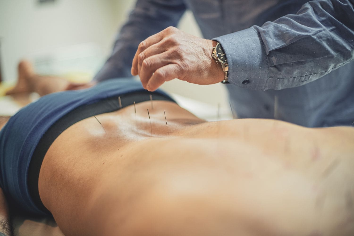
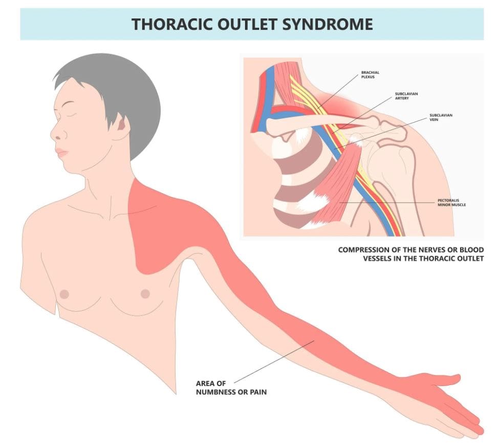
Can individuals with thoracic outlet syndrome incorporate electroacupuncture to reduce neck pain and restore proper posture?
Contents
Introduction
More times throughout the world, many individuals have experienced pain around their necks, which can lead to pain and discomfort. Many environmental factors, like being in a hunched position while looking at the computer or phone, traumatic injuries, poor posture, or spinal issues, can cause pain-like symptoms and complications to the body. Since neck pain is a common complaint many people suffer, symptoms like tingling, numbness, or muscle weakness in the upper extremities can lead to comorbidities. When this happens, it can lead to the development of a complex condition known as thoracic outlet syndrome or TOS. Today’s article looks at the link between thoracic outlet syndrome and neck pain, how to manage TOS while alleviating neck pain, and how electroacupuncture can help with TOS. We talk with certified medical providers who consolidate our patients’ information to assess how to minimize the effects of TOS while reducing neck pain. We also inform and guide patients on how electroacupuncture can help manage TOS. We encourage our patients to ask their associated medical providers intricate and important questions about incorporating electroacupuncture to alleviate TOS associated with the neck. Dr. Jimenez, D.C., includes this information as an academic service. Disclaimer.
The Link Between Thoracic Outlet Syndrome & Neck Pain
Have you been noticing how you are hunched over more than usual? Do you experience symptoms of tingling or numbness down from your arms to your hands? Or do you feel muscle tension in your neck? Thoracic outlet syndrome, or TOS, is a challenging condition resulting in the compression of neurovascular structures between the clavicle and the first rib. (Masocatto et al., 2019) These neurovascular structures are near the neck and shoulders. When environmental structures affect the upper extremities, it can lead to referred neck pain, which can cause overlapping risk profiles. Some of the factors that TOS can contribute to neck pain include:
Atomical variations
Poor posture
Repetitive motions
Traumatic injuries
At the same time, people with neck pain can develop TOS, as neck pain is a multifactorial musculoskeletal condition that can be associated with overlapping risk profiles that contribute to TOS. (Kazeminasab et al., 2022) As stated earlier, factors like poor posture can overstretch the neck muscles and the neurovascular structures, leading to neuropathic pain symptoms that can cause deep aching referred pain to the neck and muscle weakness. (Childress & Stuek, 2020) When this happens, many people will begin to feel miserable and start to seek treatment to not only reduce TOS but also alleviate neck pain.
What Is Thoracic Outlet Syndrome- Video
Managing TOS & Alleviating Neck Pain
When it comes to treating TOS, especially when neck pain is a significant component, many individuals will try to seek out non-surgical treatments to reduce the symptoms. Many individuals may try physical therapy to stretch and strengthen their shoulder, chest, and neck muscles to relieve compression. Others might try a manual treatment that is joint-oriented for the neck while neural-tissue-oriented for TOS to improve mobilization on the upper extremities and even improve poor posture. (Kuligowski et al., 2021) Additionally, non-surgical treatments can be combined with other therapies to reduce the chances of TOS from returning as they can further increase sensory-motor function back to the neck and upper extremities. (Borrella-Andres et al., 2021)
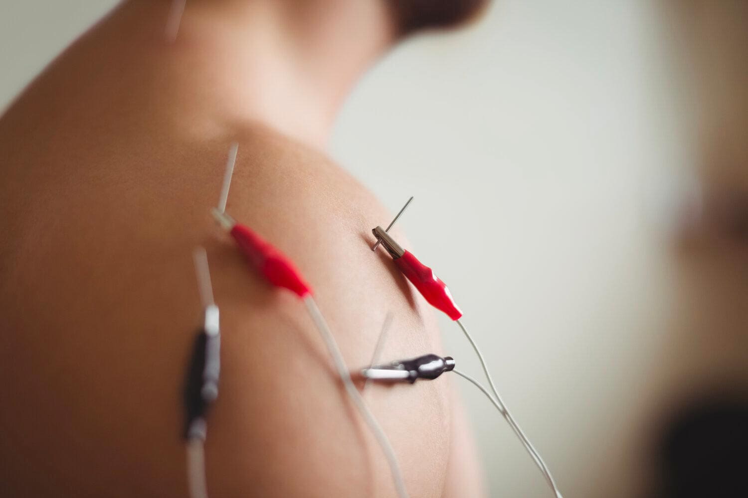
How Electroacupuncture Can Help With TOS
Electroacupuncture is a modern form of traditional acupuncture that is part of the non-surgical treatments that can help manage TOS while alleviating neck pain. Electroacupuncture is a modification of inserting needles into the body’s acupoints while incorporating electric stimulation to deliver a pulsed electrical current to the affected area gently. (Zhang et al., 2022) Some of the beneficial properties that electrostimulation can provide for TOS include:
Pain reduction by stimulating the release of endorphins to decrease inflammation.
Help relax the affected muscles in the chest and neck to alleviate the pressure on the nerves of the thoracic outlet.
Help enhance the blood flow to reduce vascular compression of TOS.
Help stimulate the nerve pathway to promote healthy nerve function and reduce pain-like symptoms.
By incorporating electroacupuncture and non-surgical treatments to reduce TOS, many individuals can make modifications to their lifestyle habits and prevent issues from affecting their upper body extremities. By utilizing these treatments, many people can listen to their bodies and focus on their health and well-being by addressing the pain-like symptoms they are experiencing from TOS correlating with neck pain. At the same time, they have a positive relationship with their primary doctors to develop a personalized treatment plan that can manage their TOS symptoms to the best outcomes.
References
Borrella-Andres, S., Marques-Garcia, I., Lucha-Lopez, M. O., Fanlo-Mazas, P., Hernandez-Secorun, M., Perez-Bellmunt, A., Tricas-Moreno, J. M., & Hidalgo-Garcia, C. (2021). Manual Therapy as a Management of Cervical Radiculopathy: A Systematic Review. Biomed Res Int, 2021, 9936981. https://doi.org/10.1155/2021/9936981
Kazeminasab, S., Nejadghaderi, S. A., Amiri, P., Pourfathi, H., Araj-Khodaei, M., Sullman, M. J. M., Kolahi, A. A., & Safiri, S. (2022). Neck pain: global epidemiology, trends and risk factors. BMC Musculoskelet Disord, 23(1), 26. https://doi.org/10.1186/s12891-021-04957-4
Kuligowski, T., Skrzek, A., & Cieslik, B. (2021). Manual Therapy in Cervical and Lumbar Radiculopathy: A Systematic Review of the Literature. Int J Environ Res Public Health, 18(11). https://doi.org/10.3390/ijerph18116176
Masocatto, N. O., Da-Matta, T., Prozzo, T. G., Couto, W. J., & Porfirio, G. (2019). Thoracic outlet syndrome: a narrative review. Rev Col Bras Cir, 46(5), e20192243. https://doi.org/10.1590/0100-6991e-20192243 (Sindrome do desfiladeiro toracico: uma revisao narrativa.)
Zhang, B., Shi, H., Cao, S., Xie, L., Ren, P., Wang, J., & Shi, B. (2022). Revealing the magic of acupuncture based on biological mechanisms: A literature review. Biosci Trends, 16(1), 73-90. https://doi.org/10.5582/bst.2022.01039
IFM's Find A Practitioner tool is the largest referral network in Functional Medicine, created to help patients locate Functional Medicine practitioners anywhere in the world. IFM Certified Practitioners are listed first in the search results, given their extensive education in Functional Medicine