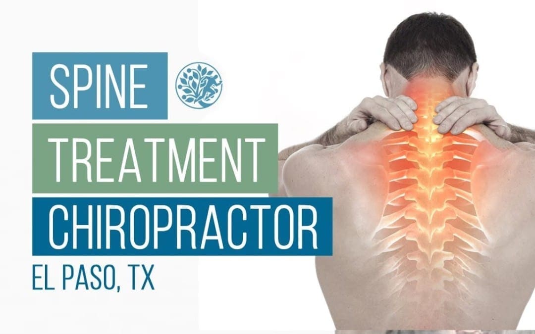
ClickCease
+1-915-850-0900 spinedoctors@gmail.com
Select Page
Epley Maneuver Used by Chiropractors for BPPV in El Paso, TX

Epley Maneuver Used by Chiropractors for BPPV in El Paso, TX

Benign paroxysmal positional vertigo is a common type of vertigo, a sensation of spinning or whirling and loss of balance, which has been reported to account for as many as 17 percent of all cases of dizziness. Benign paroxysmal positional vertigo, or BPPV, is believed to be caused by a health issue in the inner ear. While it is typically associated with aging, head injuries have also been found to cause BPPV.


BPPV occurs when several of the small crystals found in the inner ear, known as otoconia, become loose and wind up in one or more of the three fluid-filled semicircular canals of the ear. Whenever these crystals move around the inner ear, they can cause the fluid in the semicircular canals to become displaced. This ultimately results in a spinning or whirling sensation, otherwise referred to as vertigo. The symptoms of BPPV can often come on suddenly when an individual with benign paroxysmal positional vertigo moves their head in a certain position. By way of instance, symptoms may trigger when turning over in bed during night time. Symptoms of BPPV can last anywhere from several seconds to several minutes, and may include:


  • Dizziness;
  • A feeling that surroundings are spinning or moving (vertigo);
  • A loss of equilibrium or balance;
  • Nausea; and
  • Vomiting.


BPPV Treatment


Although many healthcare professionals often prescribe drugs and/or medications for BPPV, there is not enough evidence to support their use as treatment for this condition. In other, very rare cases, surgical interventions are considered. However, in the majority of instances, BPPV can safely and effectively be adjusted mechanically.


Once a healthcare professional specializing in vestibular disorders, such as a vestibular rehabilitation therapist, a chiropractor, a specially trained physical therapist, an occupational therapist or audiologist, or an ENT (ear, nose & throat specialist who specializes on vestibular disorders), has properly diagnosed the individual’s type of benign paroxysmal positional vertigo by performing tests like the Dix-Hallpike Test, then they’ll have the ability to understand which of the semicircular canal(s) the crystals are in, and whether it is canalithiasis, where the loose crystals can move freely in the fluid of the tube, or cupulolithiasis, where the crystals are believed to be ‘hung up’ on the bundle of nerves that feel the fluid motion, then they can recommend you the appropriate therapy maneuver.


Other Auditory & Vestibular Function Tests


The Dix-Hallpike Test is commonly used to diagnose BPPV, however, if the diagnosis is negative, healthcare professionals may utilize a variety of other auditory and vestibular function tests in order to properly diagnose the patient’s source of their symptoms.


Dix-Hallpike Test to Diagnose BPPV


The most common treatment for benign paroxysmal positional vertigo, or BPPV, is called the Epley maneuver. The Epley maneuver, sometimes referred to as canalith repositioning, is a procedure which involves a succession of head movements, normally performed by a healthcare professional who is qualified and experienced in the treatment of vestibular disorders, in order to relieve the symptoms associated with BPPV.


Research studies have demonstrated that the Epley maneuver is a safe and effective treatment for the condition, offering both immediate and long-term relief. The Epley maneuver, named after Dr. John Epley, has been named the canalith repositioning maneuver because it�helps reposition the small crystals in a individual’s ear, which may be causing the sensation of dizziness. Repositioning these small crystals, also known as otoconia, ultimately helps to relieve BPPV symptoms.


Epley Maneuver to Treat BPPV BPPV


The Epley maneuver is performed by placing the patient’s head at an angle from where gravity can help alleviate the symptoms. Tilting the head can move the crystals from the semicircular canals of the inner ear. This means that they will stop displacing the fluid, relieving the dizziness and nausea they may have been causing. In this way, the Epley maneuver alleviates the symptoms of BPPV. But, it may need to be repeated more than once, as occasionally, some head movements can once again displace the small crystals of the inner ear, once they had already been repositions after the initial treatment.


Epley Maneuver Diagram | El Paso, TX Chiropractor


When a healthcare professional carries out the Epley maneuver, they’ll perform the following measures:


  • Ask the patient to sit upright in an examination table, completely extending their legs out in front of them.
  • Rotate the patient’s head in a 45-degree angle to the side they’re experiencing the worst vertigo.
  • Instantly push the patient back, so they are lying with their shoulders touching the table. The patient’s head is retained facing the side most negatively affected by vertigo but at a 30-degree angle, so that it is lifted slightly off the table. The healthcare professional holds the patient in this position for between 30 seconds and two minutes, until their symptoms stop.
  • Rotate the patient’s head 90 degrees from the opposite direction, stopping when the other ear is 30 degrees away from the table. Again, the doctor holds the patient in this position for between 30 minutes and two minutes, until their symptoms cease.
  • Next, the healthcare professional will roll the patient in precisely the same direction that they are facing, onto their side. The moment they encounter the worst vertigo on will be facing upward. The physician holds the patient in this position for between 30 minutes and 2 minutes, until their symptoms stop.
  • Eventually, the healthcare professional will bring the patient back up into a sitting position.
  • The whole process is repeated up to three times, until the patients’s symptoms have been completely relieved.


A healthcare professional specializing in vestibular disorders, such as a chiropractor or physical therapist, will utilize the Epley maneuver to help alleviate an individual’s dizziness and nausea, among other symptoms, when they have decided that BPPV is the cause. As mentioned before, the Epley maneuver isn’t suitable to treat vertigo brought on by another health issue aside from BPPV. If the individual is unsure of what is causing their vertigo, they ought to talk to a doctor and ask to be properly diagnosed. Other causes of vertigo may include:


  • Migraine headaches
  • Ear infections
  • Anemia
  • Cerebellar stroke


After performing the Epley maneuver, a doctor will advise the patient who has BPPV to prevent specific movements that may dislodge the crystals. These movements include:


  • Bending quickly
  • Lying down fast
  • Leaning the head
  • Moving the head back and forth


Many research studies have been done on the safety and effectiveness of therapy maneuvers for BPPV, such as the Epley maneuver, together with results and outcome measures demonstrating that the rates of recovery are well into the 90 percent range by 1 to 3 treatments. The more infrequent cupulolithiasis, or ‘hung-up’ version of BPPV, can be a little more stubborn to resolve, as this type of BPPV is generally the consequence of trauma or injury.


Dr-Jimenez_White-Coat_01.png

Dr. Alex Jimenez’s Insight

If you’ve ever experienced a sudden spinning or whirling sensation, dizziness and nausea when you make certain head movements, especially while rolling over in bed at night or when getting out of bed in the morning, you may be suffering from a common condition called benign paroxysmal positional vertigo, or BPPV. This type of vertigo can be frustrating to deal with and it can tremendously affect an individual’s quality of life. A healthcare professional who specializes in vestibular disorders, including chiropractors and physical therapists, frequently diagnose BPPV using the Dix-Hallpike Test before following up treatment for benign paroxysmal positional vertigo using the Epley maneuver.


Benign Paroxysmal Positional Vertigo, or BPPV, is a frequent health issue, and will be encountered more and more as our population ages. The effect can vary from a mild annoyance to a highly debilitating condition, and can affect function and safety as well as increase the risk of falls. Fortunately, symptoms tend to decrease in intensity over time as the brain gradually adjusts to the strange signals it is receiving, or as the condition resolves on its own. However, with a healthcare professional who’s suitably qualified and experienced in the diagnosis and treatment of BPPV, most patients will find overall relief of their problem once it readily corrected and their world stops spinning or whirling. The scope of our information is limited to chiropractic as well as to spinal injuries and conditions. To discuss the subject matter, please feel free to ask Dr. Jimenez or contact us at 915-850-0900 .


Curated by Dr. Alex Jimenez


Green-Call-Now-Button-24H-150x150-2-3.png


Additional Topics: Sciatica

Sciatica is medically referred to as a collection of symptoms, rather than a single injury and/or condition. Symptoms of sciatic nerve pain, or sciatica, can vary in frequency and intensity, however, it is most commonly described as a sudden, sharp (knife-like) or electrical pain that radiates from the low back down the buttocks, hips, thighs and legs into the foot. Other symptoms of sciatica may include, tingling or burning sensations, numbness and weakness along the length of the sciatic nerve. Sciatica most frequently affects individuals between the ages of 30 and 50 years. It may often develop as a result of the degeneration of the spine due to age, however, the compression and irritation of the sciatic nerve caused by a bulging or herniated disc, among other spinal health issues, may also cause sciatic nerve pain.


blog picture of cartoon paperboy big news


EXTRA IMPORTANT TOPIC: Chiropractor Sciatica Symptoms


MORE TOPICS: EXTRA EXTRA: El Paso Back Clinic | Back Pain Care & Treatments

Skateboarding Injury Treatment | El Paso, TX. | Video

Skateboarding Injury Treatment | El Paso, TX. | Video

Skateboarding: Javier Mata has entrusted Dr. Alex Jimenez for many years now, receiving trustworthy chiropractic care after experiencing several low back pain injuries throughout his life. Javier Mata first considered the non-surgical treatment choice following his own father’s recommendation to seek chiropractic care after he himself received treatment with Dr. Alex Jimenez. Javier Mata has found tremendous relief through spinal adjustments and manual manipulations and he highly recommends chiropractic care with Dr. Alex Jimenez.

Skateboarding Injury Treatment

Skateboarding is a popular recreational activity among children and teenagers. Although it is an enjoyable and exciting activity, skateboarding carries with it a serious risk for injury. Roughly 70,000 injuries requiring a trip to the emergency department occur each year. There are many things that parents and children can do to help prevent skateboarding injuries, such as carefully selecting safe places to ride and wearing protective gear, especially helmets. Many accidents happen when a child loses balance, falls off the skateboard and lands on an outstretched arm. Skateboarding accidents often involve the wrist, ankle, or face and sprains or strains may occur.

skateboarding el paso tx.

We are blessed to present to you�El Paso�s Premier Wellness & Injury Care Clinic.

Our services are specialized and focused on injuries and the complete recovery process.�Our areas of practice include:Wellness & Nutrition, Chronic Pain,�Personal Injury,�Auto Accident Care, Work Injuries, Back Injury, Low�Back Pain, Neck Pain, Migraine Headaches, Sport Injuries,�Severe Sciatica, Scoliosis, Complex Herniated Discs,�Fibromyalgia, Chronic Pain, Stress Management, and Complex Injuries.

As El Paso�s Chiropractic Rehabilitation Clinic & Integrated Medicine Center,�we passionately are focused treating patients after frustrating injuries and chronic pain syndromes. We focus on improving your ability through flexibility, mobility and agility programs tailored for all age groups and disabilities.

If you have enjoyed this video and/or we have helped you in any way please feel free to subscribe and share us.

Thank You & God Bless.

Dr. Alex Jimenez DC, C.C.S.T

Facebook Clinical Page: www.facebook.com/dralexjimenez/

Facebook Sports Page: www.facebook.com/pushasrx/

Facebook Injuries Page: www.facebook.com/elpasochiropractor/

Facebook Neuropathy Page: www.facebook.com/ElPasoNeuropathyCenter/

Facebook Fitness Center Page: www.facebook.com/PUSHftinessathletictraining/

Yelp: El Paso Rehabilitation Center: goo.gl/pwY2n2

Yelp: El Paso Clinical Center: Treatment: goo.gl/r2QPuZ

Clinical Testimonies: www.dralexjimenez.com/category/testimonies/

Information:

LinkedIn: www.linkedin.com/in/dralexjimenez

Clinical Site: www.dralexjimenez.com

Injury Site: personalinjurydoctorgroup.com

Sports Injury Site: chiropracticscientist.com

Back Injury Site: elpasobackclinic.com

Rehabilitation Center: www.pushasrx.com

Fitness & Nutrition: www.push4fitness.com/team/

Pinterest: www.pinterest.com/dralexjimenez/

Twitter: twitter.com/dralexjimenez

Twitter: twitter.com/crossfitdoctor

Injury Medical Chiropractic Clinic: Accident Treatment & Recovery

Dix-Hallpike Test Used by Chiropractors for BPPV in El Paso, TX

Dix-Hallpike Test Used by Chiropractors for BPPV in El Paso, TX

Benign Paroxysmal Positional Vertigo, or BPPV, is the most common vestibular disorder and it is by far the most common cause of vertigo, a false sensation of rotational movement or spinning. BPPV isn’t life-threatening, it can come in unexpectedly in brief spells and it can trigger with certain head positions or motions. This might frequently occur when you tip your head down or up, when you lie down, or when you flip over or sit up in bed.


BPPV is a mechanical problem in the inner ear. It occurs when some of the calcium carbonate crystals, known as otoconia, that are typically embedded in gel at the utricle, become dislodged and migrate into at least one of the 3 fluid-filled semicircular canals, in which they are not supposed to be. When enough of these particles collect among the canals, they interfere with the fluid movement that these canals use to sense head motion, causing the internal ear to send false signals to the brain.


BPPV Diagram | El Paso, TX Chiropractor


Fluid from the canals does not normally respond to gravity. On the other hand, the crystals do interact with gravity, thereby shifting the fluid when it normally would remain still. After the fluid moves, nerve endings in the canal are triggered and send a message to the brain that the head is moving, even though it is not. This false information does not match what the other ear may be sensing, together with what the eyes are seeing, or using what the muscles and joints do, and this mismatched information is sensed by the brain as a spinning sensation, or vertigo, which normally lasts less than one minute. Between vertigo spells some people may feel symptom-free, while others feel a mild sense of imbalance or disequilibrium.


Symptoms of BPPV


The signs and symptoms of benign paroxysmal positional vertigo, or BPPV, may include:


  • Dizziness
  • A feeling that you or your surroundings are spinning or moving (vertigo)
  • A loss of equilibrium or balance
  • Nausea
  • Vomiting


The signs and symptoms of BPPV can come and go, with these generally lasting less than one minute. Episodes of benign paroxysmal positional vertigo can disappear for a while and then return. Activities that cause the signs and symptoms of BPPV may vary from person to person, but are nearly always brought on by a change in the placement of the head. Some people also feel out of balance when standing or walking. Abnormal rhythmic eye movements, known as nystagmus, usually follow the outward signs of benign paroxysmal positional vertigo, or BPPV.


It’s essential, however, to understand that BPPV will not give you continuous dizziness that is unaffected by motion or even a change in position. Also, it will not affect your hearing or produce fainting, headache or neurological signs, such as numbness, a sensation of “pins and needles,” difficulty speaking or difficulty coordinating your movements. If you have one or more of these additional symptoms, tell a healthcare professional immediately. Other disorders could be originally misdiagnosed as BPPV. By alerting a healthcare professional about any signs and symptoms you may be experiencing along with vertigo, they could reevaluate your illness and think about whether you might have another kind of disorder, instead of or in addition to BPPV.


BPPV is rather common, with an estimated prevalence of 107 per 100,000 annually plus a lifetime prevalence of 2.4 percent. It is thought to be quite rare in children but can affect adults of any age, particularly seniors. The wide majority of cases happen for no apparent reason, with many individuals describing how they simply went to get out of bed and the room began to spin. Nevertheless, associations have been made with injury, migraine headaches, inner ear infection or disease, diabetes, osteoporosis, intubation, presumably due to protracted time lying in bed, and reduced blood flow. There might also be a correlation with a person’s favorite sleeping side.


Diagnosis for BPPV


General practitioners normally refer patients to a healthcare professional specifically trained to take care of vestibular disorders, most commonly a vestibular rehabilitation therapist, such as a chiropractor, a specially trained physical therapist, or sometimes an occupational therapist or audiologist. An ENT (ear, nose & throat specialist) who specializes on vestibular disorders can also diagnose BPPV.


Normal medical imaging (e.g. an MRI) isn’t effective in diagnosing BPPV, because it doesn’t show the crystals that have moved to the semi-circular canals. However, when someone with BPPV has their own head moved into a position that makes the dislodged crystals go within a canal, the error signals have been known to cause the eyes to move in a very specific pattern, known as “nystagmus”.


The association between the internal ears and the eye muscles are what generally permit us to remain focused on our environment while the head is moving. Since the dislodged crystals make the brain think a person is moving when they are not, it causes the eyes to move, making it seem like the room is spinning. The eye movement is the indication that something is happening automatically in order to move the fluid in the inner ear canals when it shouldn’t be.


The nystagmus will have different characteristics that allow a healthcare professional to recognize which ear the displaced crystals are inside, as well as which canal(s) they have moved into. Evaluations like the Dix-Hallpike test involves moving the head into specific orientations, allowing gravity to move the dislodged crystals and activate the vertigo while the healthcare professional watches for the recognizable eye movements, or nystagmus.


Dix-Hallpike Test Diagram | El Paso, TX Chiropractor


Dix-Hallpike Test for BPPV


Healthcare professionals, such as chiropractors specializing in vestibular diseases, typically utilize the Dix-Hallpike test, sometimes called the Dix-Hallpike maneuver, to test for benign paroxysmal positional vertigo, or BPPV. To execute the Dix-Hallpike test, your doctor will ask you to sit on the test table with your legs stretched out. He’ll turn your head 45 degrees to one side, which contrasts the right posterior semicircular canal with the sagittal plane of the body, then they are going to allow you to lie back quickly, while the eyes are open, so that your head hangs slightly over the edge of the desk.


Dix-Hallpike Test to Diagnose BPPV


This motion may cause the loose crystals to move inside your semicircular canals. The healthcare professional will ask if you are feeling symptoms of vertigo and observe your eyes to find out how they move. As soon as you’ve got a few minutes to recover, your doctor may do the test on the opposite side of your head.


The latency, length and direction of nystagmus, if present, along with the latency and duration of vertigo, if present, should be noted. If the test is negative, it will demonstrate that�benign paroxysmal positional vertigo is a less probable diagnosis and central nervous system involvement ought to be considered. There are two sorts of BPPV: One at which loose crystals can move freely in the fluid of the canal (canalithiasis), and, more infrequently, one where the crystals are believed to be ‘wrapped up’ on the bundle of nerves that feel the fluid motion, or cupulolithiasis.


With canalithiasis, it requires less than a moment for those crystals to stop moving after a particular change in head position has triggered a twist. Once the crystals quit shifting, the fluid motion settles and the nystagmus and vertigo cease. With cupulolithiasis, the crystals trapped on the package of sensory nerves will make the nystagmus and vertigo last longer, until the head is moved out of the offending position. It is necessary to make the proper diagnosis, since the treatment is different for every variant. BPPV can be treated using various treatment methods, one of the most common being the Epley Maneuver.


Dr-Jimenez_White-Coat_01.png

Dr. Alex Jimenez’s Insight

Chiropractic care is an alternative treatment option commonly utilized to help treat a variety of injuries and conditions associated with the proper alignment of the spine. Occasionally, a spinal misalignment, or subluxation, can develop into numerous health issues, causing a wide array of symptoms if left untreated for an extended period of time. However, many chiropractors can treat many other ailments not closely associated with the spine. In a clinical setting, chiropractic care has been used for the management of benign paroxysmal positional vertigo, or BPPV. Chiropractors will utilize the Dix-Hallpike test to diagnose a patient followed by the Epley maneuver to help treat patients with BPPV. Many patients have reported a reduction in symptoms.


The scope of our information is limited to chiropractic as well as to spinal injuries and conditions. To discuss the subject matter, please feel free to ask Dr. Jimenez or contact us at 915-850-0900 .


Curated by Dr. Alex Jimenez


Green-Call-Now-Button-24H-150x150-2-3.png


Additional Topics: Sciatica

Sciatica is medically referred to as a collection of symptoms, rather than a single injury and/or condition. Symptoms of sciatic nerve pain, or sciatica, can vary in frequency and intensity, however, it is most commonly described as a sudden, sharp (knife-like) or electrical pain that radiates from the low back down the buttocks, hips, thighs and legs into the foot. Other symptoms of sciatica may include, tingling or burning sensations, numbness and weakness along the length of the sciatic nerve. Sciatica most frequently affects individuals between the ages of 30 and 50 years. It may often develop as a result of the degeneration of the spine due to age, however, the compression and irritation of the sciatic nerve caused by a bulging or herniated disc, among other spinal health issues, may also cause sciatic nerve pain.


blog picture of cartoon paperboy big news


EXTRA IMPORTANT TOPIC: Chiropractor Sciatica Symptoms


MORE TOPICS: EXTRA EXTRA: El Paso Back Clinic | Back Pain Care & Treatments

Neuropathy Presentation | El Paso, TX. | Part II

Neuropathy Presentation | El Paso, TX. | Part II

Neuropathy Presentation II:�El Paso, TX. Chiropractor, Dr. Alexander Jimenez�continues the overview with neuropathy part II. Continued are the most common neuropathies to be seen in practice. Because the human body is composed of many different kinds of nerves which perform different functions, nerve damage is classified into several types. Neuropathy can also be classified according to the location of the nerves being affected and according to the disease causing it. For instance, neuropathy caused by diabetes is called diabetic neuropathy. Furthermore, depending on which nerves are affected will depend on the symptoms that will manifest. The complications which follow neuropathy depends on the type of nerves that are damaged. According to Dr. Jimenez, different neuropathies can cause numbness and/or tingling sensations, increased pain or the loss of ability to feel pain, muscle weakness along with twitching and cramps, even dizziness and/or loss of bladder control function.

Sciatic Nerve Entrapment

  • Piriformis Syndrome
  • Peroneal Nerve Entrapment
  • Tarsal Tunnel Syndrome

neuropathy presentation el paso tx.

Sciatic N. Piriformis Syndrome

neuropathy presentation el paso tx.Causes

  • Anatomic variation
  • Piriformis overuse/tension

Exam

  • Positive Lase?gue test possible
  • Doctor extends patient�s leg passively, while patient is lying supine positive test if maneuver is limited by pain
  • Tenderness and palpable tension in piriformis muscle which elicits symptoms

Sciatic N. Peroneal Nerve Entrapment

  • Peroneal or Fibular branch of Sciatic nerve entrapped at the fibular head
  • Tinel�s sign may be present at fibular head/neck
  • Usually affects common peroneal nerve, therefore motor and sensory symptoms can be seen
  • Weakness of ankle dorsiflexion and eversion (tibialis anterior m.)
  • Sensory disruption on the dorsum of the foot and lateral aspect of the calf

Sciatic N. Tarsal Tunnel Syndrome

  • Tibial nerve impinged in the tarsal tunnel
  • Sensory changes in the sole of the foot
  • Tinel�s sign may be present with percussion posterior to the medial malleolus

neuropathy presentation el paso tx.Radiculopathy

  • A mononeuropathy � located in one specific area
  • Neuropathy involving spinal nerve roots
  • Presents as changes in sensory and/or motor function affecting a single or a few nerve root level(s)
  • Most commonly seen radiculopathies include:
  • Sciatica
  • Cervical radiculopathy

Common Causes Of Radiculopathy

  • Disc herniation
  • Osteophytes
  • Spinal Stenosis
  • Trauma
  • Diabetes
  • Epidural abscess or metastasis
  • Nerve sheath tumors (schwannomas and neurofibromas)
  • Guillain-Barre? syndrome
  • Herpes Zoster (shingles)
  • Lyme disease
  • Cytomegalovirus
  • Myxedema/Thyroid disorder
  • Idiopathic neuritis

Narrowing Down Common Causes Of Radiculopathy

  • Disc Herniation

  • Most commonly affected nerve roots are C6, C7, L5 & S1
  • Spinal Stenosis

  • Lumbar stenosis may produce neurogenic claudication
  • Pain & weakness with ambulation
  • Cervical stenosis may present with mixed picture of radiculopathy and myelopathy due to long tract involvement
  • Trauma

  • May cause compression, trauma or avulsion of the nerve roots
  • Diabetes

  • More likely to cause a polyneuropathy, but mononeuropathy is possible
  • Herpes Zoster (Shingles)

  • Most often on the trunk, accompanied by vesicular lesions in a single dermatome
  • If pain persits past vesicular regression = post-herpetic neuralgia

Patient History Of Radiculopathy

  • The patient will often complain of burning pain or tingling that radiates or shoots down an affected area in a dermatomal pattern.
  • Sometimes patient will complain of motor weakness, however if onset is recent, there is often no motor involvement

Exam Of Radiculopathy

  • Most often hypoesthesia in the affected dermatome level
  • Best to evaluate for pain, as light touch can be difficult for these patient�s to distinguish
  • Fasciculations and/or atrophy may be seen if radiculopathy is chronic, due to lower motor neuron being impinged
  • Motor weakness may be seen in muscles innervated by the same root level

Orthopedic tests:

  • Straight-leg raise test (SLR)
  • Pain between 10-60 degrees likely indicates nerve root compression
  • Well-leg raise/Crossed straight-leg raise test (WLR)
  • If positive, 90% specificity for L/S nerve root compression
  • Valsalva Maneuver
  • Positive if increase in radicular symptoms
  • Spinal Percussion
  • Pain may indicate metastatic disease, abscess or osteomyelitis

Examinations: Merck Manual Professional

How To Test Reflexes

How To Do A Sensory Exam

How To Do A Motor Examination

Dermatomes

neuropathy presentation el paso tx.Testing Cervical Nerve Roots

neuropathy presentation el paso tx.Testing Lumbosacral Nerve Roots

neuropathy presentation el paso tx.

Specific Radiculopathy Patterns

  • T1 radiculopathy can cause Horner�s syndrome
  • This is due to affect on cervical sympathetic ganglia
  • Ptosis, miosis, anhidrosis
  • Below L1, radiculopathies can cause Cauda Equina syndrome
  • Saddle anesthesia (sensory loss in S2-S5 distribution)
  • Urinary retention or overflow incontinence
  • Constipation, decreased rectal tone or fecal incontinence
  • Loss of erectile function
  • Must be referred for emergency care immediately to prevent permanent dysfunction

Other Patterns Of Neuropathy

  • Cape/Shawl distribution of symptoms
  • Intramedullary lesion
  • Syringomyelia
  • Intramedullary tumor
  • Central cord damage
  • Stocking and Glove Distribution of Symptoms
  • Diabetes mellitus
  • B12 deficiency
  • Alcoholism/hepatitis
  • HIV
  • Thyroid dysfunction/myxedema

Cape/Shawl Pattern

  • Intramedullary lesion such as tumor, syringomyelia or hyperextension injury in patient with C/S spondylosis

neuropathy presentation el paso tx.

  • Loss of pain and temp sensation in C/T dermatomes because of arrangement of lateral spinothalamic tract

neuropathy presentation el paso tx.Stocking & Glove Pattern

neuropathy presentation el paso tx.

  • Symmetrical polyneuropathy
  • Feet/legs usually affected first, followed by hands/arms
  • Vibration sensation in the smallest toes is usually the first thing lost and neuropathy progresses across foot to great toe and then upward through the ankle and leg, then hands, arms and finally trunk if sever
  • Most likely cause of this distribution is diabetes mellitus, but other possible causes include B12 deficiency, alcoholism, HIV, chemotherapy treatment, thyroid dysfunction and multiple other causes

Diabetic Neuropathy

  • Diabetic neuropathy often presents as a polyneuropathy but can also present as a mononeuropathy, usually with acute onset
  • Most common in CN III, femoral and sciatic nerves

Demyelinating Neuropathies

  • Acute inflammatory demyelinating polyneuropathy (Guillain-Barre? syndrome)
  • Chronic inflammatory demyelinating polyneuropathy

Guillain-Barre? Syndrome (AIDP)

  • Onset 1-2 weeks post viral infection
  • Progressive weakness
  • Loss of DTRs/areflexia
  • Paresthesia in hands and feet
  • More motor involvement than sensory
  • Potential autonomic fiber involvement
  • Elevated CSF protein
  • EMG/NCV studies indicate demyelination
  • May require treatment with plasmapheresis or IV Ig therapy

Chronic Inflammatory Demyelinating Polyneuropathy

  • Appears similar to AIDP but does not follow infection
  • Symptoms must be present for at least 8 weeks for this diagnosis to be considered
  • Anti-inflammatory treatments may help

By�Rachel Klein, ND, DC, DACNB

National University of Health Sciences Master of Science (MS) – Advanced Clinical Practice (ACP) MS ACP 551: Clinical Neurology � 2018

Sources

Blumenfeld, Hal. Neuroanatomy through Clinical Cases. Sinauer, 2002.

Evans, Ronald C. Illustrated Orthopedic Physical Assessment. Mosby/Elsevier, 2009.

�Radial Nerve Entrapment: Background, Anatomy, Pathophysiology.� Medscape, 25 Oct. 2017, emedicine.medscape.com/article/1244110- overview#a8.

Neuropathy Presentation | El Paso, TX. | Part I

Neuropathy Presentation | El Paso, TX. | Part I

Neuropathy Presentation: El Paso, TX. Chiropractor, Dr. Alexander Jimenez�presents an overview of neuropathy. These are the most common neuropathies to be seen in practice.�Neuropathy is a medical term used to characterize damage or injury to the nerves, which refers to the peripheral nerves as opposed to the central nervous system. The complications which follow neuropathy depends largely on the type of nerves that are affected. According to Dr. Alex Jimenez, different neuropathies can cause numbness and tingling sensations, increased pain or the loss of ability to feel pain, muscle weakness along with twitching and cramps, even dizziness and/or loss of control over bladder function.

Neuropathy

Three primary classifications based on location of symptoms:

  • Mononeuropathy (focal)
  • Mononeuropathy multiplex (multifocal)
  • Polyneuropathy (generalized)
  • Can involve CNS and/or PNS
  • Can affect large and/or small diameter fibers
  • Can affect both sensory and motor fibers
  • Sometimes one more than the other, but often both
  • May be permanent or reversible

neuropathy presentation el paso tx.

*https://neupsykey.com/muscle-weakness-cramps-and-stiffness/

Assessment Of Neuropathy

Sensory Exam:

  • Determine What Sensory Modalities Are Involved

  • If sensory disruption is limited to certain modalities, it implies CNS is involved
  • If all sensation is affected in the area, implies PNS is involved
  • Determine Pattern Of Symptoms

  • Mononeuropathy (focal)?
  • Mononeuropathy multiplex (multifocal)?
  • Polyneuropathy (generalized)?
  • Motor Exam

  • Determine if there is change to muscle strength
  • Determine if there is a change in muscle tone
  • Determine which muscles are affected
  • Determine if there has been a change in reflexes
  • This information can help determine the level(s) of involvement
  • Check For Autonomic�Signs

  • Auscultate heart
  • Palpate palms
  • Auscultate abdomen
  • Assess autonomic history
  • For example, is patient complaining about sweating more on one side than another? Complaining of stress levels?
  • Suggest ANS involvement

Exams: Merck Manual Professional Version

How To Test Reflexes

How To Do The Sensory Exam

How To Do The Motor Examination

Classification Of Nerve Injuries Resulting In Neuropathy

  • Neurapraxia – This is a transient episode of motor paralysis with little or no sensory or autonomic dysfunction; no disruption of the nerve or its sheath occurs; with removal of the compressing force, recovery should be complete
  • Axonotmesis – This is a more severe nerve injury, in which the axon is disrupted but the Schwann sheath is maintained; motor, sensory, and autonomic paralysis results; recovery can occur if the compressing force is removed in a timely fashion and if the axon regenerates
  • Neurotmesis – This is the most serious injury, in which both the nerve and its sheath are disrupted; although recovery may occur, it is always incomplete, secondary to loss of nerve continuity

Brachial Plexopathies

  • Erb�s Palsy
  • Klumke�s Palsy

neuropathy presentation el paso tx.Erb�s Palsy

  • AKA Erb�Duchenne palsy or Waiter’s tip palsy
  • Most common mechanism of injury in adults is a patient who fall forward while holding onto something behind them
  • Can also happen to an infant during childbirth ? Results from damage to C5-6 nerve roots in the brachial plexus
  • Dermatomal distribution of sensory disruption
  • Weakness or paralysis in deltoid, biceps, and brachialis muscles resulting in �waiter�s tip� position

neuropathy presentation el paso tx.Klumke�s Palsy

  • AKA Dejerine�Klumpke palsy
  • Happens to infants during childbirth if arm is pulled overhead
  • Can also happen to adults with overhead traction injuries
  • Results from damage to C8-T1 nerve roots in the brachial plexus
  • Dermatomal distribution of sensory disruption
  • Weakness or paralysis in wrist flexors and pronators as well as muscles of the hand
  • May produce Horner�s syndrome due to T1 involvement
  • Results in a �claw hand� appearance
  • Forearm supinated with wrist hyperextended, with finger flexion

neuropathy presentation el paso tx.Entrapment Neuropathies

  • Thoracic Outlet Syndromes
  • Median Nerve Entrapment
  • Pronator Teres syndrome
  • Carpal Tunnel syndrome
  • Ulnar Nerve Entrapment
  • Cubital Tunnel syndrome
  • Tunnel of Guyon syndrome
  • Radial Nerve Entrapment
  • Spiral Groove Entrapment
  • Supinator Syndrome
  • Posterior Interosseous Syndrome (Radial Tunnel Syndrome)
  • Sciatic Nerve Entrapment
  • Piriformis syndrome
  • Fibular head entrapment
  • Tarsal tunnel syndrome

Thoracic Outlet Syndromes

neuropathy presentation el paso tx.

  • Neurogenic thoracic outlet syndrome

  • Compression of the brachial plexus
  • Vascular thoracic outlet syndrome

  • Veins (venous thoracic outlet syndrome) or arteries (arterial thoracic outlet syndrome) are compressed
  • Nonspecific-type thoracic outlet syndrome

  • Worsens with activity
  • Idiopathic

Causes

  • Cervical rib
  • Subclavius muscle tension
  • Postural – excessive thoracic kyphosis
  • Trauma
  • Repetitive activity
  • Obesity
  • Pregnancy

Tests

  • Adsons test
  • Allen maneuver
  • Costoclavicular maneuver
  • Halstead maneuver
  • Reverse bakody maneuver
  • Roos test
  • Shoulder compression test
  • Wright test

neuropathy presentation el paso tx.

Median Nerve Entrapment

  • Sensory disruption in the lateral portion (3.5 fingers) of the palmar aspect of the hand, and dorsal finger tips of the same fingers
  • Motor fibers affected in forearm if applicable, muscles of the thenar eminence
  • Abductor pollicis brevis
  • Opponens pollicis
  • Flexor pollicis brevis

neuropathy presentation el paso tx.Median N. Pronator Teres Syndrome

Etiology

  • Compression of the median nerve at the elbow

Causes

  • Repetitive movement
  • Pronator teres muscle inflammation
  • Thickened bicipital aponeurosis

Findings

  • Tenderness with palpation of the pronator teres muscle
  • Pain with resisted pronation of the arm
  • Flexor pollicus longus and flexor digitorum profundus involvement
  • Otherwise may appear similar to carpal tunnel syndrome in symptoms, but without positive wrist orthopedics

Median N. Carpal Tunnel Syndrome

neuropathy presentation el paso tx.

Etiology

  • Compression of the median nerve at the wrist

Causes

  • Repetitive motions
  • Hypothyroidism
  • Obesity
  • Rheumatoid Arthritis
  • Diabetes
  • Pregnancy


Orthopedic Tests

neuropathy presentation el paso tx.Tinel�s Sign

  • Positive if tapping over the median nerve reproduces/exacerbates symptoms

Phalen�s Maneuver/Prayer Sign

  • Hands together with wrists flexed
  • Repeat in reverse with wrists extended
  • Hold each for at least 60 seconds
  • Positive if tests reproduce/exacerbate symptoms

Wringing Test

  • Wringing a towel produces paresthesia


Ulnar Nerve Entrapment

  • Sensory disruption in the medial two digits of the palmar & dorsal aspects of the hand
  • Motor fibers affected in hand, all fingers besides the thumb weakened general hand weakness

Ulnar N. Cubital Tunnel Syndrome

neuropathy presentation el paso tx.Etiology

  • Compression of the ulnar nerve at the elbow

Causes

  • Repetitive motions
  • Hypothyroidism
  • Obesity
  • Diabetes
  • Trauma to the cubital tunnel
  • Prolonged sitting with pressure on bent elbow

Ulnar N. Tunnel Of Guyon Syndrome

neuropathy presentation el paso tx.

Etiology

  • Compression of the ulnar nerve at the wrist

Causes

Repetitive motions

  • Long term crutch use
  • Break of the hamate
  • Ganglion cyst
  • Hypothyroidism
  • Obesity
  • RheumatoidArthritis
  • Diabetes


Orthopedic Tests

neuropathy presentation el paso tx.

Tinel�s Sign

  • Present if test over the ulnar nerve at the wrist elicits symptoms

Wartenberg Sign

  • 5th digit abducts when patient performs hard grip strength test or attempts to squeeze fingers together
  • Reduced two-point discrimination in the hand


Radial Nerve Entrapment

neuropathy presentation el paso tx.

  • Sensory disruption in the lateral 3.5 digits of the dorsal aspect of the hand
  • Motor fibers affected in posterior arm and extensor compartment of the forearm
  • Wrist drop may be seen

Spiral Groove Entrapment

  • All radial nerve innervated muscles below entrapment are affected
  • �Saturday night palsy� (from sleeping on own arm)
  • Brachioradialis & triceps reflexes both diminished

Supinator Syndrome

  • Compression at the arcade of Frohse
  • No change in reflexes

Posterior Interosseous Syndrome (Radial Tunnel Syndrome)

  • No change in reflexes

By Rachel Klein, ND, DC, DACNB

National University of Health Sciences Master of Science (MS) – Advanced Clinical Practice (ACP) MS ACP 551: Clinical Neurology � 2018

Sources

Blumenfeld, Hal. Neuroanatomy through Clinical Cases. Sinauer, 2002.

Evans, Ronald C. Illustrated Orthopedic Physical Assessment. Mosby/Elsevier, 2009.

�Radial Nerve Entrapment: Background, Anatomy, Pathophysiology.� Medscape, 25 Oct. 2017, emedicine.medscape.com/article/1244110- overview#a8.

Common Clinical Neuropathies in El Paso, TX

Common Clinical Neuropathies in El Paso, TX

Neuropathy is a medical term used to describe a collection of general diseases or malfunctions which affect the nerves. The causes of neuropathy, or nerve damage, can vary greatly among each individual and these may be caused by a number of different diseases, injuries, infections and even vitamin deficiency states. However, neuropathy can most commonly affect the nerves that control the motor and sensory nerves. Because the human body is composed of many different kinds of nerves which perform different functions, nerve damage is classified into several types. Neuropathy can also be classified according to the location of the nerves being affected and according to the disease causing it. For instance, neuropathy caused by diabetes is called diabetic neuropathy. Furthermore, depending on which nerves are affected will depend on the symptoms that will manifest as a result. Below we will discuss several specific types of neuropathies clinically treated by chiropractors, physical therapists and physical medicine doctors alike, as well as briefly describing their causes and their symptoms.


Brachial Plexopathies


Brachial plexopathy is a type of peripheral neuropathy, which affects the nerves that transmit messages from the brain and the spinal cord to the rest of the body. This kind of nerve damage occurs when harm affects the brachial plexus, a region found on each side of the neck where nerve roots from the spinal cord branch out into each arm’s nerves. Damage, injury or a condition that impacts these nerve roots can result in pain, decreased mobility and reduced sensation in the arm and shoulder. In some cases, no cause can be identified.


Erb’s Palsy


Erb’s Palsy, also known as�Erb�Duchenne palsy or Waiter’s tip palsy, is identified as a paralysis of the arm caused by damage or injury to the nerves in the neck which form part of the brachial plexus. The most common mechanism of injury in adults with Erb’s Palsy is a patient who fell forward while holding onto something behind them. Erb�Duchenne palsy can also happen to an infant during childbirth, most commonly, but not exclusively, from shoulder dystocia during a difficult birth. To be more precise, this type of brachial plexopathy results from damage to the C5-C6 nerve roots along the brachial plexus in the neck. Symptoms of Erb’s Palsy include dermatomal distribution of sensory disruption followed by weakness or paralysis in the deltoid, biceps, and brachialis muscles, leading to the �waiter�s tip� position associated with this type of neuropathy. While many infants can recover on their own from this type of brachial plexopathy, some may require rehabilitation.


Erb's Palsy Image | El Paso, TX Chiropractor


Klumpke’s Palsy


Klumpke’s Palsy, also known as Klumpke’s paralysis or�Dejerine�Klumpke palsy, is a partial palsy in the nerve roots of the brachial plexus located along the cervical spine, or neck. It is named after�Augusta D�jerine-Klumpke, an American-born French medical doctor acknowledged for her work in neuroanatomy. Klumpke’s Palsy is characterized as a form of paralysis involving the muscles of the forearm and hand, which occurs to�infants during childbirth if their arm is pulled overhead.�Dejerine�Klumpke palsy can also occur to adults with overhead traction injuries caused by harm to the C8-T1 nerve roots in the brachial plexus and upper thoracic region of the spine. Symptoms of Klumpke’s paralysis include dermatomal distribution of sensory disruption, weakness or paralysis, in the wrist flexors and pronators as well as in the muscles of the hand. This type of brachial plexopathy may often lead to Horner�s syndrome, a collection of symptoms which manifest when a set of nerves, known as the sympathetic trunk, are damaged or injured due to T1 involvement. This form of neuropathy is identified by resulting�in a �claw hand� appearance, where the forearm is supinated with the wrist hyperextended, together with finger flexion.


Klumpke's Paralysis Image | El Paso, TX Chiropractor


Entrapment Neuropathies


Entrapment neuropathy, also known as nerve compression syndrome or compression neuropathy, is best-known as nerve damage or a type of neuropathy caused by direct pressure on a nerve. Common symptoms include pain and discomfort, tingling or burning sensations, numbness and muscle weakness which affects only a particular part of the human body, depending on which nerve is affected. A nerve can become compressed as a result of a constant external force or due to a lesion, such as a tumor. Additionally, some conditions can make the nerves more susceptible to compression, including diabetes, where the nerves are rendered more sensitive to minor degrees of compression due to their already compromised supply of blood. Nerve damage caused by a single episode of harm can be considered an entrapment neuropathy, however, it is generally not classified under this group of compression neuropathy or nerve compression syndrome.


Thoracic Outlet Syndromes


Thoracic outlet syndromes are a group of disorders which develop when the nerves or blood vessels between the collarbone and the thoracic outlet, located in the region of the first rib, are compressed. As a result, this can cause pain and discomfort in the neck and shoulders as well as numbness in the fingers. There are a number of types of thoracic outlet syndromes, including neurogenic, or neurological, thoracic outlet syndrome, specifically caused by the compression of the brachial plexus, vascular thoracic outlet syndrome, which is caused specifically by the compression of the veins, known as venous thoracic outlet syndrome, or arteries, known as arterial thoracic outlet syndrome, and nonspecific-type thoracic outlet syndrome, which is considered to be idiopathic and has been described to worsen with activity. Several healthcare professionals believe that nonspecific-type thoracic outlet syndrome doesn’t exist, while others claim it to be a common disorders. However, the majority of thoracic outlet syndromes are often classified as neurogenic.


Thoracic outlet syndromes are caused by the compression of the cervical rib, an extra “rib” in the seventh cervical vertebra, subclavius muscle tension, improper posture or�excessive thoracic kyphosis, physical trauma, repetitive activity, obesity and pregnancy. Thoracic outlet syndromes can vary depending on which structures are compressed. Thoracic outlet syndromes can be diagnosed using tests, such as the Adsons test, the Allen maneuver, the Costoclavicular maneuver, the Halstead maneuver, the�Reverse bakody maneuver, the Roos test, the Shoulder compression test and the Wright test. Thoracic outlet syndromes can cause permanent neurological damage if not diagnosed and treated early.


Thoracic Outlet Syndrome | El Paso, TX Chiropractor


Median Nerve Entrapment


Median nerve entrapment or median nerve entrapment syndrome, is a mononeuropathy, a condition that impacts only a single nerve or nerve group outside the brain and spinal cord, which affects the movement of or sensation in the hand. Median nerve entrapment is caused by the compression of the median nerve found in the elbow or distally in the forearm or wrist. Symptoms include sensory disruption in the lateral portion of the palmar aspect of the hand and dorsal finger tips of the same fingers. In addition, motor fibers may also be affected in the forearm, if applicable, including the muscles of the thenar eminence, such as the abductor pollicis brevis, the opponens pollicis, and the flexor pollicis brevis. Other forms of median nerve entrapment syndromes include: pronator teres syndrome and carpal tunnel syndrome.


Pronator teres syndrome is characterized as the compression of the median nerve at the elbow. It is considered rare compared to carpal tunnel syndrome. Pronator teres syndrome is caused by repetitive movement, pronator teres muscle inflammation and thickened bicipital aponeurosis. Clinical findings for this type of neuropathy include, tenderness with palpation of the pronator teres muscle, pain with resisted pronation of the arm, flexor pollicus longus and flexor digitorum profundus involvement, otherwise, symptoms manifestations for pronator teres syndrome may appear similar to carpal tunnel syndrome but without positive wrist orthopedics.


Carpal tunnel syndrome is characterized as the compression of the median nerve at the wrist. Carpal tunnel syndrome is identified by symptoms of pain and discomfort, tingling sensations in the thumb, index finger, middle finger and the thumb side of the ring fingers, and numbness. These can generally start gradually and may extend up the arm. Advanced instances of carpal tunnel syndrome may cause weakened grip strength where the muscles at the base of the thumb may waste away if left untreated for an extended period of time. In many cases, carpal tunnel syndrome may affect both hands or arms. Carpal tunnel syndrome is caused by repetitive movements, hypothyroidism, obesity, rheumatoid arthritis, diabetes and pregnancy. Orthopedic tests utilized to diagnose carpal tunnel syndrome include the use of the Tinel�s Sign, positive if tapping over the median nerve reproduces/exacerbates symptoms, the�Phalen�s Maneuver/Prayer Sign, performed by bringing the hands together, with wrists flexed, and is repeated in reverse with the wrists extended, for at least 60 seconds, and is considered positive if tests reproduce/exacerbate symptoms, and the�Wringing Test, if wringing a towel produces paresthesia.


Carpal Tunnel Syndrome | El Paso, TX Chiropractor


Ulnar Nerve Entrapment


Ulnar nerve entrapment is a condition where the ulnar nerve itself becomes physically trapped or pinched, resulting in symptoms of pain, numbness and weakness which extends throughout the little finger, the ulnar half of the ring finger and throughout the intrinsic muscles of the hand. Symptoms or ulnar nerve entrapment ultimately involve sensory disruption in the medial two digits of the palmar and dorsal aspects of the hand. Symptoms of ulnar nerve entrapment may vary depending on the specific location of the ulnar nerve compression or impingement. These may also be classified as motor, sensory or both, depending on the location of the injury. If motor fibers are affected in the hand, all fingers, besides the thumb, may become weakened, described as general hand weakness. The most common location of ulnar nerve entrapment is within the cubital tunnel. Other forms of ulnar nerve entrapment include: cubital tunnel syndrome and tunnel of Guyon syndrome.


Ulnar Nerve Entrapment | El Paso, TX Chiropractor


Cubital tunnel syndrome is identified by the compression or impingement of the ulnar nerve in the cubital tunnel at the elbow. It is considered to be the second most common entrapment neuropathy which affects the upper extremities, following carpal tunnel syndrome. Symptoms of cubital tunnel syndrome are characterized by pain and discomfort along the region of the ulnar nerve entrapment, along with sensory impairment, paresis and paresthesia.�Causes of cubital tunnel syndrome include, repetitive movements, hypothyroidism, obesity, diabetes, physical trauma or injury to the cubital tunnel, and prolonged sitting with pressure on bent elbow.


Tunnel of Guyon syndrome, or Guyon’s canal syndrome, is identified by the compression or impingement of the ulnar nerve at the wrist, particularly along an anatomical space in the wrist known as Guyon’s canal. Guyon’s canal syndrome may also be referred to as ulnar tunnel syndrome. Symptoms of tunnel of Guyon syndrome are similar to those of cubital tunnel syndrome with slight variations depending on the region of ulnar nerve entrapment.�Causes of tunnel of Guyon syndrome include, repetitive movements, long term crutch use, fracture of the hamate, a carpal bone, due to a ganglion cyst, hypothyroidism, obesity, rheumatoid arthritis and diabetes.�Orthopedic tests utilized to diagnose Guyon’s canal syndrome include the use of the�Tinel�s Sign, positive if test over the ulnar nerve at the wrist elicits symptoms, the Wartenberg Sign, positive if the 5th digit abducts when patient performs hard grip strength test or attempts to squeeze fingers together and reduced two-point discrimination in the hand.


Radial Nerve Entrapment


Radial nerve entrapment, also known as radial tunnel syndrome, is a condition caused by the compression of the radial nerve, which travels from the brachial plexus, to the hand and wrist. Healthcare professionals believe that radial tunnel syndrome occurs because the radial nerve becomes irritated or inflamed due to the friction caused by the impingement of the muscles in the forearm. Radial nerve entrapment manifests symptoms of sensory disruption in the lateral three and a half digits of the dorsal aspect of the hand. Motor�fibers may also be affected along the�posterior arm and extensor compartment of the forearm, and wrist drop may be seen. Other forms of radial tunnel syndrome include: spiral groove entrapment, where all radial nerve innervated muscles below entrapment are affected,�Saturday night palsy caused due to sleeping on your own arm and the brachioradialis & triceps reflexes are both diminished, supinator syndrome, caused by the compression at the arcade of Frohse with no change in reflexes. Posterior interosseous syndrome, or radial tunnel syndrome, also elicits no change in reflexes.


Sciatic Nerve Entrapment


Sciatic nerve entrapment is a condition caused by the compression of the sciatic nerve, the longest and largest nerve in the human body, which travels from the low back, down through the buttocks, thighs, legs and into the foot. The collection of symptoms which manifest as a result of sciatic nerve entrapment, including pain and discomfort, tingling and burning sensations, and numbness as well as weakness in the lower extremitites, is commonly known as sciatica. Sciatic nerve entrapment, or sciatica, can be caused by a variety of injuries and/or aggravated conditions which can lead to the compression of the sciatic nerve, including, but not limited to, disc herniation and spinal stenosis. However, symptoms of sciatic nerve entrapment may vary depending on the location of the compression of the sciatic nerve. Other conditions caused by the compression of the sciatic nerve include: piriformis syndrome, peroneal nerve entrapment and tarsal tunnel syndrome.


Sciatica | El Paso, TX Chiropractor


Piriformis syndrome is a condition which occurs due to the compression of the sciatic nerve as a result of the irritation or inflammation of the piriformis muscle. Symptoms of piriformis syndrome may include pain and discomfort, followed by numbness in the buttocks and down the leg. Symptoms may worsen with regular activities, such as sitting and running. Piriformis syndrome is caused by anatomic variation or due to piriformis overuse/tension. Piriformis syndrome diagnosis exams include, a positive Lase?gue test, where the healthcare professional�extends the patient�s leg passively, while the patient is lying supine,�test is positive if the maneuver is limited by pain, and through the use of tenderness and palpable tension in piriformis muscle which elicits symptoms.


Peroneal nerve entrapment is a condition which occurs when the peroneal or the fibular branch of the sciatic nerve are compressed at the fibular head. Tinel�s sign may be present at the fibular region of the head and/or neck. Peroneal nerve entrapment generally affects the common peroneal nerve, therefore, motor and sensory symptoms may manifest, including, weakness of the ankle dorsiflexion and eversion, or the tibialis anterior. Other symptoms of peroneal nerve entrapment may include sensory disruption on the dorsum of the foot and lateral aspect of the calf. Common peroneal nerve entrapement at the fibular head is the most common nerve entrapment syndrome in the lower extremities.


Tarsal tunnel syndrome, also known as posterior tibial neuralgia, is a condition caused by the compression of the tibial nerve as it travels through the tarsal tunnel, found along the region of the inner leg, posterior to the medial malleolus, or the bump on the inside of the ankle. Tarsal tunnel syndrome can manifest symptoms of pain and discomfort, burning or tingling sensations, and numbness along the big toe and the first three toes. However, symptoms may vary slightly depending on the area of compression, where the entire foot may manifest the symptoms previously described. Other symptoms associated with posterior tibial neuralgia include sensory changes in the sole of the foot. Tinel�s sign may be present with percussion posterior to the medial malleolus. The exact cause of tarsal tunnel syndrome may be difficult to determine and it is essential to receive a proper diagnosis to determine the source of the symptoms.


Radiculopathy


Radiculopathy is a mononeuropathy,�a condition that impacts only a single nerve or nerve group outside the brain and spinal cord, which affects the movement of or sensation in one specific area. It is often associated with neuropathy involving spinal nerve roots and presents as changes in sensory and/or motor function affecting a single or a few nerve root level(s). The most common types of radiculopathies include: sciatica and cervical radiculopathy. The most prevalent causes of radiculopathy include, disc herniation, osteophytes, spinal stenosis, trauma, diabetes, epidural abscess or metastasis, nerve sheath tumors, such as schwannomas and neurofibromas, Guillain-Barre? syndrome, Herpes Zoster, or shingles, Lyme disease, cytomegalovirus, myxedema and/or thyroid disorder, and idiopathic neuritis.


Narrowing down some of the most common causes of radiculopathy, symptoms can manifest due to disc herniation which most commonly affects the nerve roots along the C6, C7, L5 & S1 vertebrae of the spine, spinal stenosis and lumbar stenosis which may produce neurogenic claudication, and pain and weakness with ambulation. Cervical stenosis may present with mixed radiculopathy and myelopathy due to long tract involvement. Symptoms may also manifest due to trauma, because it may lead to compression, trauma or avulsion of the nerve roots, diabetes, which is most�likely to cause a polyneuropathy, but mononeuropathy is possible, and Herpes Zoster, or shingles, most often on the trunk, accompanied by vesicular lesions in a single dermatome. If pain persists past vesicular regression, radiculopathy may instead be considered post-herpetic neuralgia.


Patients with a history of radiculopathy will often complain of burning pain or tingling sensations which radiates or shoots down an affected area in a “dermatomal” pattern. Occasionally, patients will complain of motor weakness, however if onset is recent, there is often no motor involvement. The diagnosis of radiculopathy can depend on a variety of exams.�Most often, hypoesthesia may be present in the affected dermatome level. It’s recommended to�evaluate for pain, as light touch can be difficult for these patient�s to distinguish. Fasciculations and/or atrophy may be seen if radiculopathy is chronic, due to the lower motor neuron being compressed or impinged. Motor weakness may be seen in muscles innervated by the same root level. Orthopedic tests for the diagnosis of radiculopathy may include: the straight-leg raise test (SLR), where pain between 10 to 60 degrees likely indicates nerve root compression, the�Well-leg raise/Crossed straight-leg raise test (WLR), where if positive, 90 percent specificity for L/S nerve root compression may be present, the Valsalva Maneuver, where its considered positive if there is an increase in radicular symptoms, and spinal percussion, where pain may indicate metastatic disease, abscess or osteomyelitis.


How to Do the Motor Examination for Neuropathy


How to Do the Sensory Exam for Neuropathy


How to Test Reflexes


Specific radiculopathy patterns may also develop as a result of different regions being affected. Radiculopathy along the T1 can cause Horner�s syndrome, a combination of symptoms caused by the disruption of a nerve pathway from the brain to the face and eye on one side of the body. This is due to its effect on cervical sympathetic ganglia, includind ptosis, miosis, anhidrosis. Radiculopathy below the L1, can cause Cauda Equina syndrome, a condition caused by damage or injury to the bundle of nerves found below the end of the spinal cord, known as the cauda equina. This type of radiculopathy may manifest symptoms of saddle anesthesia, sensory loss in the S2-S5 distribution, urinary retention or overflow incontinence, constipation, decreased rectal tone or fecal incontinence, and loss of erectile function. Individuals with these signs and symptoms must be referred for emergency care immediately to prevent permanent dysfunction.


Other patterns of neuropathy can include the cape/shawl distribution of symptoms, identified by an intramedullary lesion, such as syringomyeli, intramedullary tumor and central cord damage. Stocking and glove distribution of symptoms may manifest as a result of diabetes mellitus,�B12 deficiency, alcoholism and/or hepatitis,�HIV, and thyroid dysfunction and/or myxedema.


The cape/shawl pattern of neuropathy is characterized by symptoms occurring due to an intramedullary lesion, such as a tumor, syringomyelia or a hyperextension injury in patient with C/S spondylosis. It can also be characterized by loss of pain and temperature sensation in C/T dermatomes because of the arrangement of the lateral spinothalamic tract. The stocking and glove pattern may progress gradually depending on its specific stage. It can also be characterized as a symmetrical polyneuropathy, where the feet and legs are generally affected first, followed by the hands and arms. A vibration-like sensation in the smallest toes are also typically the first to go and the neuropathy symptoms may progress across the foot to the big toe and then upward through the ankle and leg, then hands, arms and finally to the trunk if the condition becomes severe. The most likely cause of this pattern may be attributed to diabetes mellitus, but other possible causes include, B12 deficiency, alcoholism, HIV, chemotherapy treatment, thyroid dysfunction and multiple other causes.


Cape and Shawl Pattern of Neuropathy | El Paso, TX Chiropractor
Stocking and Glove Pattern of Neuropathy | El Paso, TX Chiropractor


Diabetic Neuropathy


Diabetic neuropathy is medically defined as a collection of nerve damaging disorders associated with diabetes. These conditions are believed to occur as a result of a diabetic microvascular injury involving the small blood vessels, known as the vasa nervorum, which supply the nerves. Additionally, macrovascular conditions have also been considered to accumulate and cause diabetic neuropathy.�Diabetic neuropathy often presents as a polyneuropathy, or the simultaneous damage or disease of many peripheral nerves throughout the body, but it can also present as a mononeuropathy, usually with acute onset. Diabetic neuropathy most commonly affects the CN III, femoral and sciatic nerves. Diabetic neuropathy can affect all peripheral nerves, including the sensory neurons, motor neurons and, although rarely, the autonomic nervous system. As a result, diabetic neuropathy can affect all organs and systems, as these are all innervated. Diabetic neuropathy can manifest into a wide array of symptoms, including, but not limited to, pain, burning or tingling sensations, numbness, dizziness and trouble with balance.


Demyelinating Neuropathies


Demyelinating neuropathies can be individually defined by its two types: Acute inflammatory demyelinating polyneuropathy, best known as�Guillain-Barre? syndrome, or Chronic inflammatory demyelinating polyneuropathy.�Guillain-Barre? syndrome, abbreviated as AIDP, is identified as a rapid-onset muscle weakness caused when the immune system damages, harms or destroys the peripheral nervous system. Onset has been reported by around one to two weeks following viral infection with progressive weakness, loss of DTRs/areflexia, paresthesia in the hands and feet, more motor involvement than sensory, potential autonomic fiber involvement, elevated CSF protein, and EMG/NCV studies indicating demyelination.�Guillain-Barre? syndrome may require treatment with plasmapheresis or IV Ig therapy.�Chronic inflammatory demyelinating polyneuropathy, abbreviated as CIDP,�is identified as an acquired immune-mediated inflammatory disorder of the peripheral nervous system which appears similar to AIDP but does not follow infection. Symptoms must be present for at least 8 weeks for this diagnosis to be considered positive.�Anti-inflammatory treatments may help treat CIDP.


The scope of our information is limited to chiropractic as well as to spinal injuries and conditions. To discuss the subject matter, please feel free to ask Dr. Jimenez or contact us at 915-850-0900 .


Curated by Dr. Alex Jimenez


Green-Call-Now-Button-24H-150x150-2-3.png


Additional Topics: Sciatica

Sciatica is medically referred to as a collection of symptoms, rather than a single injury and/or condition. Symptoms of sciatic nerve pain, or sciatica, can vary in frequency and intensity, however, it is most commonly described as a sudden, sharp (knife-like) or electrical pain that radiates from the low back down the buttocks, hips, thighs and legs into the foot. Other symptoms of sciatica may include, tingling or burning sensations, numbness and weakness along the length of the sciatic nerve. Sciatica most frequently affects individuals between the ages of 30 and 50 years. It may often develop as a result of the degeneration of the spine due to age, however, the compression and irritation of the sciatic nerve caused by a bulging or herniated disc, among other spinal health issues, may also cause sciatic nerve pain.


blog picture of cartoon paperboy big news


EXTRA IMPORTANT TOPIC: Chiropractor Sciatica Symptoms


MORE TOPICS: EXTRA EXTRA: El Paso Back Clinic | Back Pain Care & Treatments

Vitamin D Supports The Skeletal System | El  Paso, TX.

Vitamin D Supports The Skeletal System | El Paso, TX.

The human body needs vitamin D in order to build strong muscles and bones. When the body does not get enough vitamin D, it is not able to absorb calcium effectively. This makes it very important for good bone health. Children who do not get adequate vitamin D develop rickets, a condition that causes weak bones, deformities in the skeleton, and a stooped posture.

What Is Vitamin D?

Most people believe that vitamin D is a vitamin; it isn�t. It is actually a hormone. A vitamin is a nutrient that the human body requires but is unable to produce. This means that it can only be obtained through supplements and food.

However, the body is able to manufacture this vitamin. When the skin is exposed to the appropriate sunlight, the body begins a process that produces vitamin D. It should also be noted that vitamin D plays a part in a strong immune system and can prevent certain chronic diseases in older adults.

There are supplements for people who do not produce enough vitamin D or who do not get adequate amounts in the food that they eat (fish liver oils, certain fish, and egg yolks are good sources). Children and adults typically do not get enough from their foods and activities. People spend a great deal of time indoors with adults at work and children at school. The emergence of digital devices and video games has managed to keep children inside as they engage in these activities.

This vitamin is not measured in milligrams like many other supplements, but in International Units (IU) instead. Research has caused the daily recommended allowance for vitamin D to increase over the years, and the current recommendation is 400 IU. It has been determined that this amount is the most beneficial in promoting healthy bones.

The Role Of The Skeletal System

The skeletal system has several functions. The first and most obvious is that it is the structural support for the body. It also protects vital organs.

For instance, the skull protects the brain and the rib cage protects the lungs and heart. It is also an anchor point for muscle so it helps with mobility. The red bone marrow provides illness fighting white blood cells, as well as red blood cells. Calcium is stored in the marrow, as is phosphorous. Certain minerals and fats are stored in the yellow marrow which is found in the long bones of the body.

vitamin d el paso tx.

How Vitamin D Benefits The Skeletal System

Getting enough vitamin D helps the body absorb calcium which is a bone building mineral. In addition to that task, it also promotes good muscle health.

Stronger muscles can work much more effectively in protecting the muscles and supporting the skeletal system. This can help with joint health as well. Older people who have adequate vitamin D do not fall down as often and people of all ages have fewer broken bones.

Results Of Vitamin D Deficiency

Vitamin D deficiency is a serious condition mainly due to its essential role in the body�s ability to absorb calcium. Muscle weakness and bone pain are two common symptoms, but are typically seen in more severe cases.

A simple blood test can check for vitamin D levels � and it should be checked regularly. Even �minor� cases of low vitamin (those without any symptoms) have been linked to some serious health conditions including:

  • Certain cancers
  • Asthma � often severe, especially in children
  • Cognitive impairment, especially in older adults
  • Increased risk of death due to cardiovascular disease

Vitamin D is generally very accessible. You can get it through foods and sunlight or via a supplement. People who may have some trouble getting adequate amounts are those who have limited exposure to sunlight, fail to consume the necessary levels of the vitamin, and people who have dark skin. If you think that you may have low vitamin D, it is best to talk to your doctor so you can come up with a plan to get your body back in balance.

Chiropractic Clinic Extra: Athletic Recovery & Rehabilitation

Physical Therapy for Sciatica in El Paso, TX

Physical Therapy for Sciatica in El Paso, TX

A healthcare professional may refer you to a physical therapist to help you relieve your sciatica. Physical therapy includes both passive and active treatments. Passive treatments help unwind you and your body. These ultimately prepare your body for therapeutic exercises, which are the active treatments commonly utilized as a part of physical therapy.


Your physical therapist can give you passive treatments such as:


  • Deep tissue massage: This procedure targets chronic muscle stress which can be compressing or irritating your sciatic nerve and its associated nerve roots. The physical therapist uses direct friction and pressure to attempt to release the tension in your soft tissues, such as the tendons, ligaments and muscles.
  • Hot and cold therapies: By employing heat, the physical therapist seeks to get more blood to the target region because an increased blood circulation brings more oxygen and nutrients to the affected area. By way of instance, a heating pack placed on your piriformis muscle may help to reduce muscle spasms that could be causing your sciatica. Cold therapy, on the other hand, slows down blood flow, helping to decrease inflammation, muscle spasms, and pain. Your physical therapist will alternate between hot and cold therapies in order to achieve the desired results.
  • TENS (transcutaneous electric nerve stimulation): It may even be used at home, if your physical therapist thinks it is necessary. A machine stimulates your muscles through a variety of safe intensities of electric current. TENS helps decrease muscle spasms, and it might increase your body’s production of endorphins, the body’s natural painkillers. The TENS equipment your physical therapist utilizes is larger than the “at-home” usage system. Whether big or small, a TENS device may be an essential treatment for sciatica and its associated symptoms.
  • Ultrasound: Ultrasound sends sound waves deep in your muscle tissues and makes a gentle heat that enhances circulation and helps to speed up recovery. Greater circulation can help to reduce muscle spasms, cramping, swelling, stiffness, and pain.


In the active part of physical therapy, your physical therapist will teach you various exercises to help treat your sciatica. Your physical therapy’s treatment program is individualized, taking into account your overall health and wellness as well as your medical history. It might consist of strengthening exercises, aerobic conditioning, and movements to increase endurance and range of movement.


Physical therapy might be part of a comprehensive sciatica treatment plan prescribed by your healthcare professional. Aside from receiving physical therapy for your sciatica, or sciatic nerve pain, the following list includes other treatment options which are often considered when discussing the best treatment options for your source of sciatica symptoms. These treatments include:


  • Alternative treatments, such as acupuncture;
  • Chiropractic care;
  • Drugs and/or medications; and
  • Surgery


If necessary, your physical therapist may teach you how to fix your posture and integrate ergonomic principles into your everyday activities. This will be done in order to work on preventing future episodes of sciatica. Of the list of treatment options to help treat sciatica, chiropractic care and physical therapy are similar to each other and may involve common treatment modalities. However, a chiropractor uses spinal adjustments and manual manipulations to relieve symptoms of sciatica by carefully restoring the original alignment of the spine and reducing spinal stress and tension.


Dr-Jimenez_White-Coat_01.png

Dr. Alex Jimenez’s Insight

As its previously been addressed, because sciatica can occur due to a variety of injuries and/or aggravated conditions, a proper diagnosis followed by the best treatment option for the patient’s source of their symptoms is key for overall improvement and prevention of further episodes of sciatic nerve pain. Among the various types of treatment, chiropractic care and physical therapy are popular alternative treatment approaches which help treat sciatica, without the need for drugs and/or medications or surgical interventions. Chiropractic care focuses on correcting the alignment of the spine through the use of spinal adjustments and manual manipulations in order to release tension on the spine and improve sciatic nerve pain.


Chiropractic Care for Sciatica


Chiropractic care is a well-known alternative treatment option which is regularly used to treat sciatica. Proper diagnosis of sciatica Is essential before considering any of the above treatment modalities. Because there are lots of disorders that cause sciatica, the chiropractor’s first step towards treating sciatica would be to determine what’s causing the patient’s relapse. Forming a diagnosis involves a thoughtful review of the patient’s medical history as well as a physical and neurological evaluation.


Diagnostic testing includes x-rays, MRI, CT scans and/or electrodiagnostic tests (nerve conduction speed, electromyography). These examinations and evaluations help to detect potential contraindications to spinal adjustments and manual manipulations along with other chiropractic alternative treatment options. The aim of chiropractic care is to help the human body heal itself. Chiropractic care is noninvasive (non-surgical) and drug-free.


The type of chiropractic care provided depends on the reason for the individual’s sciatica. A sciatica treatment program might include several distinct therapies like ice/cold treatments, ultrasound, TENS, (similar to those used in physical therapy) and spinal adjustments and manual manipulations. Spinal adjustments and manual manipulations differ from a swift high velocity push to those that combine minimal pressure and gentle force. Mastery of every method is an art which requires great precision and skill. Spinal adjustments and manual manipulations are the treatment modalities that differentiate chiropractic care from other medical disciplines.


However, sciatica can be brought on by other disorders beyond the scope of chiropractic care. If the chiropractor determines that the patient’s source of their symptoms requires treatment from a different type of doctor, then the patient is referred to another healthcare professional. In some instances, the chiropractor may continue to treat the patient and also co-manage the patient’s care with another healthcare professional.


The scope of our information is limited to chiropractic as well as to spinal injuries and conditions. To discuss the subject matter, please feel free to ask Dr. Jimenez or contact us at 915-850-0900 .


Curated by Dr. Alex Jimenez


Green-Call-Now-Button-24H-150x150-2-3.png


Additional Topics: Sciatica

Sciatica is medically referred to as a collection of symptoms, rather than a single injury and/or condition. Symptoms of sciatic nerve pain, or sciatica, can vary in frequency and intensity, however, it is most commonly described as a sudden, sharp (knife-like) or electrical pain that radiates from the low back down the buttocks, hips, thighs and legs into the foot. Other symptoms of sciatica may include, tingling or burning sensations, numbness and weakness along the length of the sciatic nerve. Sciatica most frequently affects individuals between the ages of 30 and 50 years. It may often develop as a result of the degeneration of the spine due to age, however, the compression and irritation of the sciatic nerve caused by a bulging or herniated disc, among other spinal health issues, may also cause sciatic nerve pain.


blog picture of cartoon paperboy big news


EXTRA IMPORTANT TOPIC: Chiropractor Sciatica Symptoms


MORE TOPICS: EXTRA EXTRA: El Paso Back Clinic | Back Pain Care & Treatments

Receptors, Brainstem Pathways And Spinal Cord Tracts | El Paso, TX. | Part II

Receptors, Brainstem Pathways And Spinal Cord Tracts | El Paso, TX. | Part II

El Paso, TX. Chiropractor, Dr. Alexander Jimenez continues the discussion on the anatomy of nerve fibers, receptors, spinal tracts and brain pathway/s. As the spinal nerve nears the spinal cord, it splits into the dorsal and ventral roots. The dorsal root only contains the axons of sensory neurons. While the ventral roots contain only the axons of motor neurons. Some of the branches synapse with local neurons in the dorsal root ganglion, posterior (dorsal) horn, and even the anterior (ventral) horn, at the spine where they enter.

Other branches travel short distances up or down the spine to interact with neurons at other levels of the spinal cord. A branch can also turn into the posterior (dorsal) column white matter to connect with the brain. Spinal nerve systems that connect to the brain are contralateral, in that the right side of the body is connected to the left side of the brain and the left side of the body is connected to the right side of the brain.

Cranial nerves convey specific sense information from the head and neck directly to the brain. Whereas spinal information is contralateral, cranial nerve systems are for the most part�ipsilateral, meaning that a cranial nerve on the right side of the head is connected to the right side of the brain. Some cranial nerves contain only sensory axons. Other cranial nerves have both sensory and motor axons, including the trigeminal, facial and glossopharyngeal. General senses of somatosensation for the face travel through the trigeminal system.

PATHWAYS

THE POSTERIOR COLUMN� MEDIAL LEMNISCUS SYSTEM CONVEYS INFORMATION ABOUT TOUCH AND LIMB POSITION

POSTERIOR COLUMN MEDIAL LEMNISCAL PATHWAY

  • pathway el paso tx.The term posterior column refers to the entire contents of a posterior funiculus, exclusive of its share of the propriospinal tract. The posterior columns consist mainly of ascending collaterals of large myelinated primary afferents carrying impulses from various kinds of mechanoreceptors (although substantial numbers of second-order fibers and unmyelinated fibers are also included). This has traditionally been considered the major pathway by which information from low-threshold cutaneous, joint, and muscle receptors reaches the cerebral cortex.


pathway el paso tx.


2-Minute Neuroscience: Touch & The Dorsal Columns-Medial Lemniscus

DAMAGE TO THE POSTERIOR COLUMN�MEDIAL LEMNISCUS SYSTEM CAUSES IMPAIRMENT OF PROPRIOCEPTION AND DISCRIMINATIVE TACTILE FUNCTIONS

�As might be expected from the types of afferents contained in the posterior columns, this pathway carries information important for the conscious appreciation of touch, pressure, and vibration and of joint position and movement. However, because input from cutaneous receptors also reaches the cortex by other routes, damage to the posterior columns causes impairment, but not abolition, of tactile perception. Complex discrimination tasks are more severely affected than is the simple detection of stimuli. Other functions, such as proprioception and kinesthesia, are classically considered to be totally lost after posterior column destruction. The result is a distinctive type of ataxia (incoordination of movement); the brain is unable to direct motor activity properly without sensory feedback about the current position of parts of the body. This ataxia is particularly pronounced when the patient�s eyes are closed, preventing visual compensation.�

Given the role of the posterior column, the patient should be screened for any abnormalities regarding their sense of fine touch, vibration, barognosis, graphesthesia, stereognosis, kinaesthesia, two-point discrimination and conscious proprioception:

  • A common way of testing for fine touch is to ask the patient to recognize common objects placed within a cloth using their touch.
  • Vibration sense can be tested using a low pitched C128 tuning fork placed along a bony prominence of the desired corresponding spinal level(s) to be tested.
  • Barognosis refers to the ability to determine the approximate weight of an object.
  • Graphesthesia refers to the ability to recognize writing on the skin by touch. The practitioner can draw out a letter on the patients skin as a way of testing.
  • Kinaesthesia refers to ones own sense of body motion (excluding equilibrium which is controlled in part by the inner ear) and is commonly tested using the subject�s ability to detect an externally imposed passive movement, or the ability to reposition a joint to a predetermined position.
  • Proprioception is often assessed using the Rombergs test. This examination is based on the notion that a person requires at least two of the three following senses to maintain balance while standing: proprioception; vestibular function and vision. A patient who has a defect within their proprioceptive mechanism can still maintain balance by using vestibular function and vision. In the Romberg test, the patient is stood up and asked to close their eyes. A loss of balance is interpreted as a positive Romberg sign.

THE SPINOTHALAMIC TRACT CONVEYS INFORMATION ABOUT PAIN AND TEMPERATURE

A GOOD BRAIN CAN MODULATE PAIN

pathway el paso tx.
SPINOTHALAMIC TRACT

  • pathway el paso tx.Pain is a complex sensation, in that a noxious stimulus leads not only to the perception of where it occurred but also to things such as a rapid increase in level of attention, emotional reactions, autonomic responses, and a greater likelihood that the event and its circumstances will be remembered. Corresponding to this complexity, multiple pathways convey nociceptive information rostrally from the spinal cord. One of them (the spinothalamic tract) is analogous to the posterior column�medial lemniscus pathway.


SPINOTHALAMIC TRACTS

Two main parts of the Spinothalamic Tract (STT)

  • Lateral Spinothalamic Tract
  • Transmission of pain and temperature
  • Anterior Spinothalamic Tract
  • Transmission of crude touch and firm pressure

pathway el paso tx.

DAMAGE TO THE ANTEROLATERAL SYSTEM CAUSES DIMINUTION OF PAIN AND TEMPERATURE SENSATIONS

Examination:

Given the role of the spinothalamic tract, the patient should be screened for any abnormalities regarding their sense of touch, pain, temperature, and pressure sensation.

Screening for such abnormalities is commonly done using gentle pin pricks and cotton wool, to contrast between sharp and soft, following cutaneous sensory nerve root distributions. Hot and cold discrimination can be ascertained using the cold metal arm of a tuning fork, and a warm palm or heated object.

2 Minute Neuroscience: Pain & The Anterolateral System

HAUSER ET AL. FIBROMYALGIA, 2015

pathway el paso tx.

  • �Pain processing and its modulation: Activation of peripheral pain receptors (also called nociceptors) by noxious stimuli generates signals that travel to the dorsal horn of the spinal cord via the dorsal root ganglion. From the dorsal horn, the signals are carried along the ascending pain pathway or the spinothalamic tract to the thalamus and the cortex. Pain can be controlled by nociception- inhibiting and nociception-facilitating neurons. Descending signals originating in the supraspinal centers can modulate activity in the dorsal horn by controlling spinal pain transmission. CNS, central nervous system.�

pathway el paso tx.

SPINAL INFORMATION REACHES THE CEREBELLUM BOTH DIRECTLY AND INDIRECTLY

The spinal cord is an important source of information used by the cerebellum in the coordination of movement. This information reaches the cerebellar cortex and nuclei both directly, by way of spinocerebellar tracts, and indirectly, by way of relays in brainstem nuclei. A number of spinocerebellar tracts have been described, some representing the upper extremity and others the lower extremity. Only three have been well characterized.

Ascending Tracts | Spinocerebellar Tract

pathway el paso tx.

pathway el paso tx.

pathway el paso tx.

DESCENDING PATHWAYS INFLUENCE THE ACTIVITY OF LOWER MOTOR NEURONS

pathway el paso tx.

pathway el paso tx.

pathway el paso tx.

By RYAN CEDERMARK, DC DACNB RN BSN MSN

Neuropathy Signs and Symptoms Diagnosis in El Paso, TX

Neuropathy Signs and Symptoms Diagnosis in El Paso, TX

The vast array of symptoms caused by neuropathy, also known as peripheral neuropathy, reflect the fact that it may be caused by an equally broad range of ailments involving disease and damage to peripheral nerves.


Signs and Symptoms of Neuropathy


Depending on the reason and unique to each patient, signs and symptoms of neuropathy can include:�pain; tingling, burning or prickling sensations; increased sensitivity to touch; muscle weakness or wasting;�temporary or permanent numbness; paralysis; dysfunction in glands or organs; or impairment in urination and sexual functioning.


Such signs and symptoms are dependent on whether autonomic, sensory, or motor nerves, as well as a combination of them, are ultimately affected. Autonomic nerve damage can influence physiological functions like blood pressure or create gastrointestinal problems and issues. Damage or dysfunction in the sensory nerves may impact sensations and sense of equilibrium or balance, while harm to motor nerves may affect movement and reflexes. When both sensory and motor nerves are involved, the condition is known as sensorimotor polyneuropathy.


Diabetic Neuropathy Symptoms


Diabetic peripheral neuropathy, which affects between 12 and 50 percent of individuals with diabetes, is one of the most common types of neuropathy. Many times, symptoms include a gradual change in sensation, as well as pain and weakness in the feet and, although less commonly, the hands. As the neuropathy develops further, it can lead to a loss of sensation in the affected regions.


This lack of feeling raises the odds of harm to the affected areas, explains Matthew Villani, doctor of podiatric medicine at Central Florida Regional Hospital at Lake Mary. Without the pain to signal when there’s an issue, individuals with diabetic neuropathy may allow modest abrasions or blisters on their feet, for instance, to fester as sores or ulcers. “The ulcers can become infected since they are open wounds, which can also progress to bone infection. Unfortunately, it frequently requires amputations if it does progress to that point”, states Dr. Matthew Villani.


Chemotherapy-Associated Neuropathy Symptoms


Cancer patients may suffer with neuropathy induced by chemotherapy as well as by other drugs and/or medications used to treat the disease. Symptoms can include intense pain, impaired movement, changes in heart rate and blood pressure, issues with balance, difficulty breathing, paralysis, and even organ failure. After chemotherapy is done, the symptoms frequently abate swiftly, but occasionally they last more, or these may not go away at all.


HIV- and AIDS-Associated Neuropathy Symptoms


Individuals being treated for HIV or AIDS can develop neuropathy from effects of the virus and the drugs and/or medications used to treat it as well. Common symptoms include stiffness, burning, prickling, tingling, and loss of feeling in the toes and soles of their feet. Sometimes the nerves in the fingers, hands, and wrists are also affected. The drugs Videx (didanosine), Hivid (zalcitabine), and Zerit (stavudine) have been most commonly associated with neuropathic symptoms.


Inflammation-Associated Neuropathy Symptoms


Inflammation caused by infections, like herpes zoster (also known as shingles), Lyme disease, or hepatitis B and hepatitis C, may lead to neuropathy, as may inflammation caused by autoimmune disorders, such as vasculitis, sarcoidosis, or autoimmune disease. In such situations, the signs and symptoms generally include burning and tingling sensations or numbness.


Other Causes of Neuropathy Symptoms


Additional causes of neuropathy and associated signs and symptoms include metabolic disorders, such as hypoglycemia or kidney failure; autoimmune disorders, such as rheumatoid arthritis, lupus, Sjogren’s syndrome, and Guillain-Barr� syndrome; toxicity; hereditary disorders, such as Charcot-Marie-Tooth disorder; hormonal disorders; alcoholism; vitamin deficiencies; physical trauma or injury; compression; and repetitive stress. In addition, many individuals may experience idiopathic neuropathy signs and symptoms, meaning that healthcare professionals may not know the reason for their neuropathy.


Dr-Jimenez_White-Coat_01.png

Dr. Alex Jimenez’s Insight

Neuropathy can be caused by a variety of injuries and/or aggravated conditions, often manifesting into a plethora of associated signs and symptoms. While every type of neuropathy, such as diabetic neuropathy or autoimmune disease-associated neuropathy, develops its own unique group of signs and symptoms, many patients will often report common complaints. Individuals with neuropathy generally describe their pain as stabbing, burning or tingling in character. If you experience unusual or abnormal tingling or burning sensations, weakness and/or pain in your hands and feet, it’s essential to seek immediate medical attention in order to receive a proper diagnosis of the cause of your specific signs and symptoms. Early diagnosis may help prevent further nerve injury.


What are the Common Signs and Symptoms of Neuropathy?


“Although there’s a wide array of signs and symptoms associated with neuropathy, the type of pain that people encounter may be common in many aspects of the disorder”, notes Vernon Williams, MD, a sports neurologist and director of the Center for Sports Neurology and Pain Medicine at Cedars-Sini Kerlan-Jobe Institute in Los Angeles. “The character and quality of neuropathic pain will often be pain that is burning or electric in character.” Furthermore, he describes that the pain will frequently be associated with different symptoms, like paresthesia, or a lack of normal sensation associated with pain; allodynia, or a painful reaction to a stimulus that wouldn’t normally trigger pain signals; and hyperalgesia, or a striking or severe pain in response to a stimulus that normally causes moderate pain.


How is Neuropathy Diagnosed?


If you think you’re having any of the above neuropathy signs and symptoms, consult a healthcare professional. A number of tests can be done to diagnose neuropathy. “There are certain patterns of complaints that indicate neuropathy,” stated Dr. Williams, “so taking down a patient’s history which includes a description of these complaints is an important first step.”


“After that, your healthcare professional can perform a physical evaluation, including checking motor and sensory function, assessing deep tendon reflexes, as well as looking for signs and symptoms like allodynia and hyperalgesia,” Williams says. “Then we can even perform electrodiagnostic testing; the most common being electromyography and nerve conduction testing, where we can stimulate nerves and document responses, calculate the rate at which signals are being transmitted and see whether there are some areas where nerves are not transmitting signals normally,” Williams continues.


How to Do the Motor Examination for Neuropathy


How to Do the Sensory Exam for Neuropathy


How to Test Reflexes


With needle tests, Williams states, “We can put modest needles into human muscles, and, according to what we see and listen together with all the needle in the muscle, we get details about the way the nerves supplying those muscle tissues are functioning. There are a number of unique tests that could be handy to identifying neuropathy, in addition to localizing where the abnormality is the most likely to be coming from”, concluded Dr.�Vernon Williams.


Often, blood tests may test for elevated blood glucose to see whether your neuropathy signs and symptoms could possibly be associated to type 2 diabetes, nutritional deficiencies, toxic elements, hereditary disorders, and evidence of an abnormal immune response. Your healthcare professional may also do a nerve biopsy, which normally involves removing a small segment of a sensory nerve to search for abnormalities, or even a skin biopsy to see if there’s a reduction in nerve endings.


To give yourself the best chance of an accurate diagnosis as well as relief from your neuropathy signs and symptoms, be prepared to describe everything you are experiencing in detail, even when you experience them, how long an episode persists, and the amount of pain, discomfort or loss of sensation or movement you experience. The more specific you are on the signs and symptoms you’re experiencing, the easier it’ll be for your doctor to understand what’s happening. The scope of our information is limited to chiropractic as well as to spinal injuries and conditions. To discuss the subject matter, please feel free to ask Dr. Jimenez or contact us at 915-850-0900 .


Curated by Dr. Alex Jimenez


Green-Call-Now-Button-24H-150x150-2-3.png


Additional Topics: Sciatica

Sciatica is medically referred to as a collection of symptoms, rather than a single injury and/or condition. Symptoms of sciatic nerve pain, or sciatica, can vary in frequency and intensity, however, it is most commonly described as a sudden, sharp (knife-like) or electrical pain that radiates from the low back down the buttocks, hips, thighs and legs into the foot. Other symptoms of sciatica may include, tingling or burning sensations, numbness and weakness along the length of the sciatic nerve. Sciatica most frequently affects individuals between the ages of 30 and 50 years. It may often develop as a result of the degeneration of the spine due to age, however, the compression and irritation of the sciatic nerve caused by a bulging or herniated disc, among other spinal health issues, may also cause sciatic nerve pain.


blog picture of cartoon paperboy big news


EXTRA IMPORTANT TOPIC: Chiropractor Sciatica Symptoms


MORE TOPICS: EXTRA EXTRA: El Paso Back Clinic | Back Pain Care & Treatments