Discover effective treatments for peripheral neuropathy with chiropractic care to manage symptoms and enhance mobility.
Contents
Chiropractic Care for Peripheral Neuropathy: A Comprehensive Guide to Reducing Nerve Pain
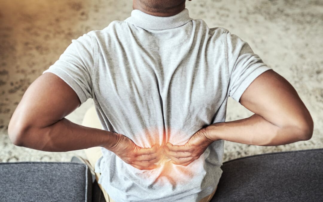
Peripheral neuropathy is like that one friend who shows up uninvited and overstays their welcome, causing all sorts of chaos. It’s a condition where the peripheral nerves—the ones that carry messages between your brain, spinal cord, and the rest of your body—decide to throw a tantrum, leading to symptoms like tingling, numbness, or burning pain. If you’ve ever felt like your hands or feet are throwing a pins-and-needles party without your permission, you might be dealing with peripheral neuropathy. But don’t worry—there’s hope! Chiropractic care, particularly through the expertise of practitioners like Dr. Alexander Jimenez, DC, APRN, FNP-BC, in El Paso, Texas, can help manage this nerve-racking condition (pun intended). In this blog post, we’ll dive into the clinical rationale for why chiropractic care can reduce nerve pain associated with peripheral neuropathy, explore the musculoskeletal system’s role, and highlight how Dr. Jimenez’s unique approach makes him a go-to for personal injury cases in El Paso. Let’s get started!
What Is Peripheral Neuropathy? The Nerve of It All!
Imagine your body as a massive communication network, with your brain and spinal cord as the control center and your peripheral nerves as the Wi-Fi signals carrying messages to your limbs, organs, and muscles. Peripheral neuropathy happens when these signals get scrambled, damaged, or completely cut off. It’s like trying to stream your favorite show with a spotty internet connection—frustrating and disruptive.
Peripheral neuropathy refers to damage to the peripheral nervous system, which sends signals between the central nervous system (brain and spinal cord) and the rest of the body. This damage can cause symptoms like numbness, tingling, burning sensations, muscle weakness, or even loss of balance. It’s not just one condition but a group of disorders caused by various factors, including diabetes, chemotherapy, infections, autoimmune diseases, or physical trauma like motor vehicle accidents (MVAs) (National Institute of Neurological Disorders and Stroke, n.d.).
The prevalence of peripheral neuropathy is no small matter. It affects millions of people worldwide, with diabetic peripheral neuropathy being one of the most common forms, impacting up to 50% of people with diabetes (Hicks & Selvin, 2019). Chemotherapy-induced peripheral neuropathy (CIPN) is another major player, affecting 19-85% of cancer patients undergoing treatment (Seretny et al., 2014). These numbers show just how widespread this condition is, and for those dealing with it, the impact on daily life can be profound.
References
Hicks, C. W., & Selvin, E. (2019). Epidemiology of peripheral neuropathy and lower extremity disease in diabetes. Current Diabetes Reports, 19(10), 86. https://doi.org/10.1007/s11892-019-1212-8
Seretny, M., et al. (2014). Incidence, prevalence, and predictors of chemotherapy-induced peripheral neuropathy: A systematic review and meta-analysis. Pain, 155(12), 2461-2470. https://doi.org/10.1016/j.pain.2014.09.020
The Musculoskeletal System’s Role in Peripheral Neuropathy
Your musculoskeletal system—your bones, muscles, ligaments, tendons, and connective tissues—is like the scaffolding that keeps your body upright and moving. But when peripheral neuropathy enters the scene, it’s like someone’s shaking that scaffolding, causing all sorts of problems. The peripheral nerves are responsible for sending sensory and motor signals to your muscles and joints. When these nerves are damaged, the musculoskeletal system can take a hit, leading to symptoms that mess with your daily routine.
How Peripheral Neuropathy Affects the Musculoskeletal System
Peripheral neuropathy can disrupt the communication between your nerves and muscles, leading to:
Muscle Weakness: Damaged nerves may fail to send proper signals to muscles, causing weakness or difficulty moving. For example, you might struggle to grip a coffee mug or climb stairs without feeling like you’re auditioning for a slow-motion scene.
Loss of Coordination: Nerves help with balance and proprioception (knowing where your body is in space). Neuropathy can make you feel like you’re walking on a tightrope after a few too many spins.
Muscle Cramps and Spasms: Irritated or damaged nerves can cause muscles to contract involuntarily, leading to painful cramps or twitches.
Joint Instability: Weak muscles can’t support joints properly, increasing the risk of falls or injuries, especially in the ankles or knees.
Pain and Discomfort: Neuropathic pain, often described as burning, stabbing, or electric shocks, can radiate to muscles and joints, causing significant discomfort (personalinjurydoctorgroup.com, 2020).
These issues can turn simple tasks—like walking to the mailbox or tying your shoes—into a Herculean effort. For instance, someone with peripheral neuropathy might find their morning jog feels more like trudging through molasses, or they might drop their phone because their fingers have lost coordination.
Impact on Daily Routine
The musculoskeletal fallout from peripheral neuropathy can significantly disrupt daily life. Imagine trying to cook dinner when your hands feel like they’re wearing oven mitts, or attempting to drive when your feet can’t tell the difference between the gas and brake pedals. These symptoms can lead to:
Reduced Mobility: Difficulty walking or standing for long periods, limiting activities like shopping or socializing.
Decreased Independence: Tasks like dressing or bathing may require assistance, which can be a blow to self-esteem.
Increased Risk of Falls: Loss of sensation or balance can make falls more likely, especially for older adults.
Chronic Pain: Persistent nerve pain can sap energy, disrupt sleep, and even lead to mood changes like anxiety or depression.
Peripheral Neuropathy: A Successful Recovery Story- Video
Why Chiropractic Care? The Clinical Rationale
Now, let’s talk about the superhero of this story: chiropractic care. It’s not just about cracking backs and making you feel like a human pretzel—it’s a science-backed approach to improving nerve function and reducing pain. Chiropractic care focuses on the spine and musculoskeletal system to remove nerve interference, which is particularly relevant for peripheral neuropathy.
The Science Behind Chiropractic Care for Nerve Pain
The spine is like the central highway of your nervous system. If there’s a traffic jam—say, a misaligned vertebra or a compressed nerve—it can disrupt the signals traveling to and from your peripheral nerves. Chiropractic adjustments aim to clear these jams by realigning the spine and reducing pressure on nerves. Here’s why this matters for peripheral neuropathy:
Reducing Nerve Compression: Misalignments (subluxations) in the spine can compress nerve roots, exacerbating neuropathic symptoms like tingling or numbness. Adjustments restore alignment, relieving pressure on these nerves (elpasobackclinic.com, 2023).
Improving Blood Flow: Proper spinal alignment enhances blood circulation, which is crucial for nerve health. Damaged nerves need oxygen and nutrients to heal, and chiropractic care can help ensure they get it.
Modulating Pain Signals: Chiropractic adjustments can influence the central nervous system, reducing the perception of pain. Think of it like turning down the volume on a screaming nerve (Woolf & Salter, 2000).
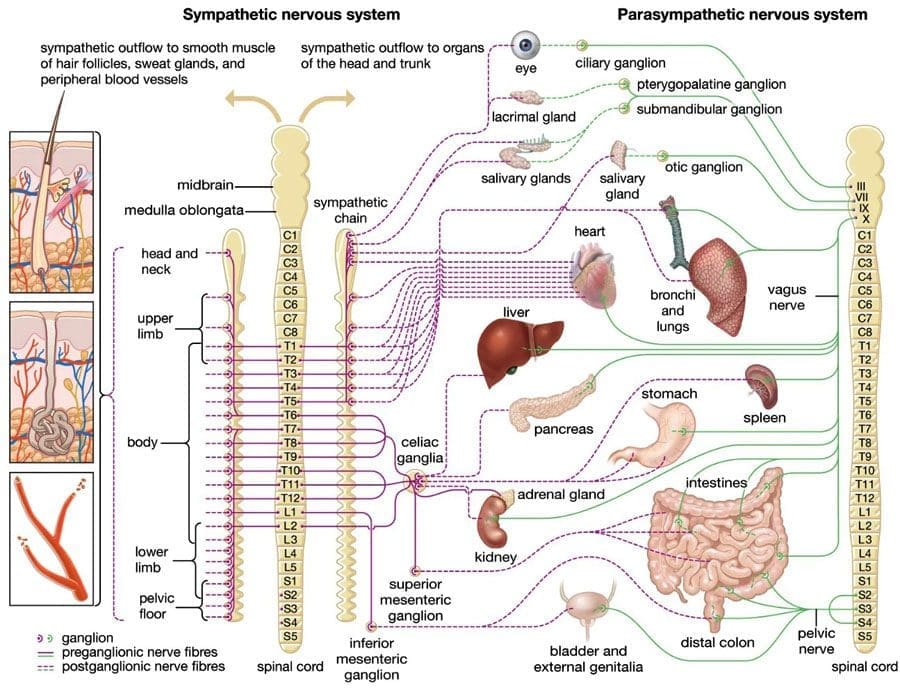
Enhancing Autonomic Function: The autonomic nervous system, which controls involuntary functions like heart rate and digestion, can be affected by neuropathy. Chiropractic care may help regulate these functions by improving spinal health (Vagal, 2020).
Research supports these benefits. A study on spinal canal compression suggests that nerve root insults, whether chemical (from inflammation) or mechanical (from compression), can contribute to polyneuropathy-like symptoms. Chiropractic care addresses these insults by correcting spinal misalignments and reducing inflammation (Kulikov et al., 2016). Another study found that nonpharmacologic interventions, including manual therapies like chiropractic care, can reduce symptoms of chemotherapy-induced peripheral neuropathy (CIPN) by improving nerve function and reducing pain (Oh et al., 2023).
Dr. Alexander Jimenez’s Approach
Enter Dr. Alexander Jimenez, El Paso’s nerve-whisperer. With over 25 years of experience as a chiropractor and board-certified Family Nurse Practitioner (FNP-BC), Dr. Jimenez brings a unique, dual-scope approach to treating peripheral neuropathy. His practice at El Paso’s Premier Wellness and Injury Care Clinic combines chiropractic expertise with advanced medical diagnostics, making him a standout in managing nerve pain (Jimenez, 2025a).
Dr. Jimenez uses a holistic, evidence-based approach inspired by functional medicine. He doesn’t just slap a Band-Aid on symptoms—he digs deep to find the root cause. For example, suppose your neuropathy stems from a car accident. In that case, he might identify a spinal misalignment pinching a nerve while also checking for inflammation or metabolic imbalances that could slow healing (Jimenez, 2023b). His methods include:
Advanced Imaging: Using X-rays, MRIs, or CT scans to pinpoint issues like herniated discs or nerve compression (Jimenez, 2023c).
Diagnostic Evaluations: Neurological tests and motion studies to assess nerve function and biomechanical dysfunction.
Dual-Scope Procedures: Combining chiropractic adjustments with medical interventions like nutritional counseling or physical therapy to address both musculoskeletal and systemic factors.
Manual Therapies: Techniques like spinal decompression, joint mobilization, and myofascial release to relieve nerve pressure and improve mobility.
Kulikov, A. V., et al. (2016). Could spinal canal compression be a cause of polyneuropathy? Frontiers in Surgery, 3, 14. https://doi.org/10.3389/fsurg.2016.00014
Oh, P. J., et al. (2023). Prevention and treatment of chemotherapy-induced peripheral neuropathy (CIPN) with non-pharmacological interventions. Frontiers in Pain Research, 4, 1002967. https://doi.org/10.3389/fpain.2023.1002967
Vagal, V. (2020). Editorial: Understanding the role of the autonomic nervous system in health and disease. Frontiers in Neuroscience, 14, 615. https://doi.org/10.3389/fnins.2020.00615
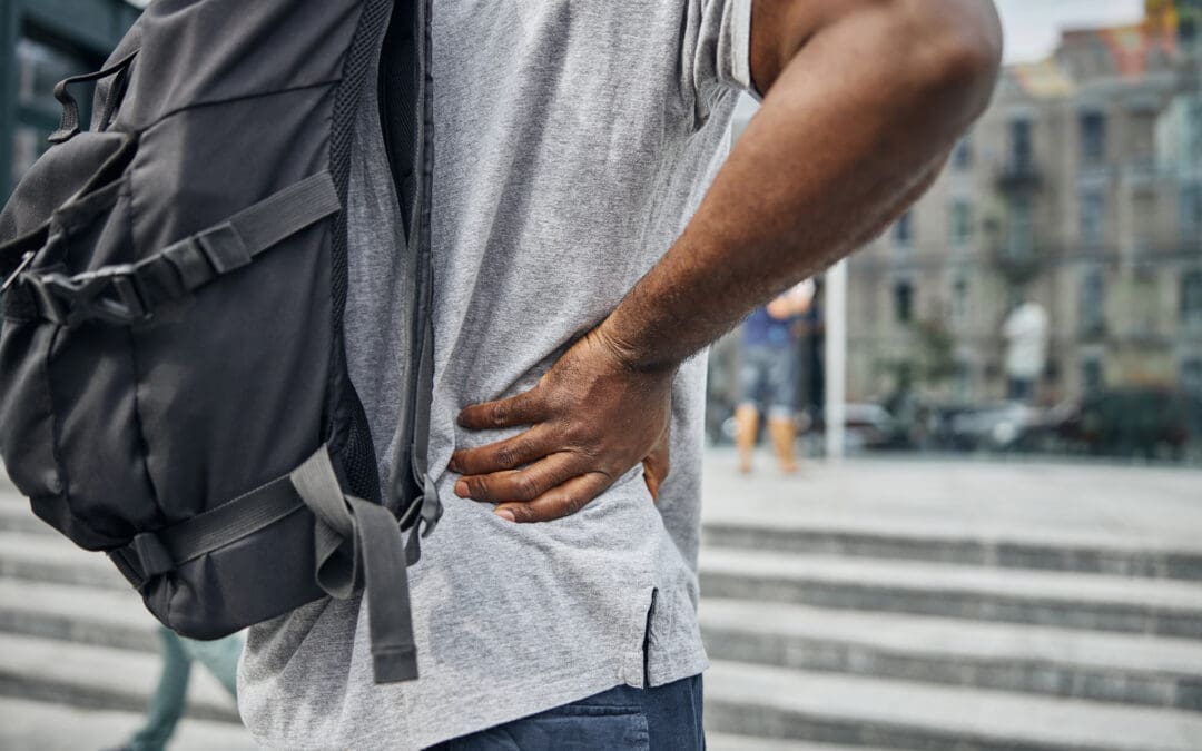
Peripheral Neuropathy and Personal Injury Cases in El Paso
El Paso, Texas, is a bustling city with heavy traffic, which unfortunately means motor vehicle accidents (MVAs) are all too common. These accidents can cause nerve injuries, including peripheral neuropathy, especially when whiplash or spinal trauma is involved. If you’ve ever been rear-ended and felt like your nerves were playing a game of telephone with the wrong number, you know what I mean. This is where chiropractic care, and specifically Dr. Alexander Jimenez, shines.
The Link Between MVAs and Peripheral Neuropathy
MVAs can cause nerve damage through:
Mechanical Insults: The force of a collision can compress or stretch nerves, leading to symptoms like numbness or tingling. For example, whiplash can pinch nerves in the cervical spine, radiating pain to the arms or hands (Jimenez, 2025b).
Chemical Insults: Inflammation from soft tissue injuries can irritate nerves, contributing to neuropathic pain (Woolf & Thompson, 1991).
Spinal Canal Compression: Trauma can narrow the spinal canal, pressing on nerve roots and mimicking polyneuropathy symptoms (Kulikov et al., 2016).
These injuries don’t just hurt—they can disrupt your life, making it hard to work, drive, or even enjoy a Netflix binge without pain. In personal injury cases, proving the link between the accident and your symptoms is crucial for fair compensation, and that’s where Dr. Jimenez’s expertise comes in.
Dr. Jimenez: The Legal-Medical Liaison
Dr. Jimenez isn’t just a chiropractor—he’s a board-certified nurse practitioner with a knack for bridging medical care and legal documentation. His dual licensure allows him to:
Diagnose with Precision: Using advanced imaging (like MRIs) and neurological tests, he identifies the exact cause of nerve pain, whether it’s a herniated disc or a pinched nerve (Jimenez, 2023c).
Document for Legal Cases: He provides detailed reports that connect your injuries to the accident, strengthening your insurance or legal claims. Think of him as a translator who speaks both “doctor” and “lawyer” fluently (Jimenez, 2025a).
Deliver Holistic Care: His treatments combine spinal adjustments, physical therapy, and functional medicine to address both symptoms and underlying causes, helping you recover faster.
For example, if you’re dealing with post-accident neuropathy, Dr. Jimenez might use an MRI to spot a herniated disc, then apply spinal decompression to relieve nerve pressure. He’ll also check for metabolic issues (like vitamin deficiencies) that could worsen neuropathy, ensuring a comprehensive recovery plan (Jimenez, 2025b). His patients rave about his ability to get them back on their feet, as seen in testimonials on his social media (Jimenez, 2023f).
Kulikov, A. V., et al. (2016). Could spinal canal compression be a cause of polyneuropathy? Frontiers in Surgery, 3, 14. https://doi.org/10.3389/fsurg.2016.00014
Woolf, C. J., & Thompson, S. W. (1991). The induction and maintenance of central sensitization is dependent on N-methyl-D-aspartic acid receptor activation; implications for the treatment of post-injury pain hypersensitivity states. Pain, 44(3), 293-299. https://doi.org/10.1016/0304-3959(91)90100-C
Addressing Overlapping Risk Profiles
Peripheral neuropathy often comes with a side of extra baggage—overlapping risk factors that make symptoms worse. These include diabetes, chemotherapy, poor nutrition, or even stress from an injury. Chiropractic care, especially Dr. Jimenez’s integrative approach, can help manage these risks.
Common Risk Factors for Peripheral Neuropathy
Diabetic Peripheral Neuropathy: High blood sugar damages nerves over time, leading to numbness or pain, especially in the feet. Up to 50% of diabetic patients develop neuropathy (Hicks & Selvin, 2019).
Chemotherapy-Induced Peripheral Neuropathy (CIPN): Cancer treatments like platinum-based drugs can damage nerves, causing tingling or burning sensations (Seretny et al., 2014).
Trauma from MVAs: Physical injuries can compress or inflame nerves, contributing to neuropathic symptoms (Jimenez, 2025b).
Nutritional Deficiencies: Lack of B vitamins or other nutrients can impair nerve health, worsening symptoms (Oh et al., 2023).
How Chiropractic Care Helps
Dr. Jimenez’s approach tackles these risk factors head-on:
Diabetic Neuropathy: He combines spinal adjustments with nutritional counseling to stabilize blood sugar and support nerve repair. For example, he might recommend a diet rich in B vitamins to nourish nerves (Jimenez, 2025a).
CIPN: Chiropractic adjustments and therapies like acupuncture can reduce pain and improve nerve function, complementing nonpharmacologic interventions (Oh et al., 2023).
Post-Trauma Neuropathy: By addressing spinal misalignments and inflammation, Dr. Jimenez reduces nerve irritation from MVAs, helping patients regain mobility (Jimenez, 2025b).
Autonomic Nervous System Support: Chiropractic care can regulate the autonomic nervous system, which is often disrupted in neuropathy, improving symptoms like dizziness or digestive issues (Vagal, 2020).
His functional medicine approach also includes tools like the Neural Zoomer Plus, a blood test that analyzes neurological autoantibodies to pinpoint the causes of nerve damage (Jimenez, 2019). This allows for tailored treatments that address both symptoms and underlying risk factors.
References
Hicks, C. W., & Selvin, E. (2019). Epidemiology of peripheral neuropathy and lower extremity disease in diabetes. Current Diabetes Reports, 19(10), 86. https://doi.org/10.1007/s11892-019-1212-8
Oh, P. J., et al. (2023). Prevention and treatment of chemotherapy-induced peripheral neuropathy (CIPN) with non-pharmacological interventions. Frontiers in Pain Research, 4, 1002967. https://doi.org/10.3389/fpain.2023.1002967
Seretny, M., et al. (2014). Incidence, prevalence, and predictors of chemotherapy-induced peripheral neuropathy: A systematic review and meta-analysis. Pain, 155(12), 2461-2470. https://doi.org/10.1016/j.pain.2014.09.020
Vagal, V. (2020). Editorial: Understanding the role of the autonomic nervous system in health and disease. Frontiers in Neuroscience, 14, 615. https://doi.org/10.3389/fnins.2020.00615
The Chiropractic Process: What to Expect
So, what’s it like to visit a chiropractor like Dr. Jimenez for peripheral neuropathy? It’s not like walking into a magic show where someone waves a wand and poof—your pain’s gone. It’s a structured, evidence-based process that’s more like a well-choreographed dance between science and care.
Initial Consultation
Your first visit is like a detective mission. Dr. Jimenez will:
Take a detailed health history to understand your symptoms, lifestyle, and any trauma (like that fender-bender you thought was no big deal).
Perform a physical exam to assess nerve function, reflexes, and muscle strength.
Order advanced imaging (X-rays, MRIs) or tests like the Neural Zoomer Plus to get a clear picture of what’s going on (Jimenez, 2019).
Treatment Plan
Once the culprit is identified, Dr. Jimenez crafts a personalized plan, which might include:
Spinal Adjustments: Gentle manipulations to realign the spine and relieve nerve pressure.
Manual Therapies: Techniques like myofascial release or trigger point therapy to relax muscles and improve circulation.
Rehabilitation Exercises: Stretches and strength training to support muscles and joints affected by neuropathy.
Functional Medicine: Nutritional advice or supplements to address deficiencies that worsen nerve damage.
Legal Documentation: For personal injury cases, detailed reports linking your symptoms to the accident, ensuring you have the evidence needed for claims (Jimenez, 2025a).
Ongoing Care
Recovery isn’t a one-and-done deal. Dr. Jimenez monitors progress with regular check-ins, adjusting the plan as needed. You might start with weekly adjustments, then taper off as symptoms improve. It’s like training for a marathon—steady progress wins the race.
Benefits of Chiropractic Care for Peripheral Neuropathy
Chiropractic care isn’t just about feeling better—it’s about getting your life back. Here are some key benefits for neuropathy patients:
Pain Reduction: Adjustments and therapies can lower pain levels, making daily tasks more manageable (Oh et al., 2023).
Improved Mobility: By addressing musculoskeletal issues, chiropractic care helps you move more freely, whether it’s walking or picking up your grandkids.
Non-Invasive Approach: Unlike medications or surgery, chiropractic care is gentle and low-risk, avoiding side effects like those seen with intravenous lidocaine (Schwenk et al., 2023).
Holistic Healing: Dr. Jimenez’s integrative approach tackles both symptoms and causes, from spinal misalignments to nutritional deficiencies.
Legal Support: For MVA-related neuropathy, Dr. Jimenez’s documentation ensures your injuries are properly represented in legal claims, helping you secure fair compensation (Jimenez, 2025a).
Oh, P. J., et al. (2023). Prevention and treatment of chemotherapy-induced peripheral neuropathy (CIPN) with non-pharmacological interventions. Frontiers in Pain Research, 4, 1002967. https://doi.org/10.3389/fpain.2023.1002967
Schwenk, E. S., et al. (2023). Intravenous lidocaine for treatment of chronic pain: A retrospective cohort study. Pain Medicine, 24(6), 664-670. https://doi.org/10.1093/pm/pnac174
Real-Life Impact: Patient Stories
Let’s take a moment to hear from patients who’ve walked this path. One patient, after a car accident, described feeling like their feet were “on fire” from neuropathy. After working with Dr. Jimenez, they reported less pain and better balance, allowing them to return to their job as a delivery driver (Jimenez, 2023f). Another patient with CIPN said chiropractic care, combined with nutritional changes, helped them reduce tingling enough to enjoy gardening again. These stories highlight how Dr. Jimenez’s care can transform lives, one adjustment at a time.
In El Paso, Dr. Jimenez is a household name for personal injury and neuropathy care. His clinic, Injury Medical & Chiropractic Clinic, is a hub for holistic healing, equipped with advanced tools and a compassionate team. Voted a top chiropractor and wellness provider, Dr. Jimenez’s dual expertise as a chiropractor and nurse practitioner sets him apart (Jimenez, 2025a). His ability to integrate medical diagnostics with chiropractic care ensures patients get the best of both worlds—effective treatment and solid legal support for personal injury cases.
Peripheral neuropathy can be a challenging condition, but chiropractic care offers a promising, non-invasive solution to reduce nerve pain and improve quality of life. By addressing spinal misalignments, improving blood flow, and tackling underlying risk factors, chiropractors like Dr. Alexander Jimenez in El Paso, Texas, help patients regain mobility and independence. His dual-scope approach, combining advanced imaging, diagnostic evaluations, and holistic therapies, makes him a trusted ally for those dealing with neuropathy, especially from personal injuries like motor vehicle accidents. For El Paso residents, Dr. Jimenez’s expertise as a medical-legal liaison ensures that your recovery is supported both clinically and legally, paving the way for a pain-free, active life.
Disclaimer: This blog post is for informational purposes only and is not intended to replace professional medical advice. Always consult a qualified healthcare provider, such as Dr. Alexander Jimenez, DC, APRN, FNP-BC, for personalized diagnosis and treatment. The information provided is based on current research and clinical insights, but should not be used as a substitute for a one-on-one consultation with a licensed professional. For more information or to discuss your specific condition, contact Dr. Jimenez at 915-850-0900 or visit dralexjimenez.com.
El Paso Chiropractic Care After Car Accidents: Real Relief and Recovery
Introduction: Why Spinal Care Is Essential After an Accident
After a motor vehicle accident, your spine and nervous system can suffer hidden injuries—even when there are no broken bones. Pain may not show up until days later, but spinal misalignments, pinched nerves, and soft tissue damage are often already in progress.
At El Paso Back Clinic, led by Dr. Alexander Jimenez, chiropractic care plays a key role in recovery. With decades of experience treating auto injuries, Dr. Jimenez combines spinal adjustments, rehab therapies, and advanced diagnostics to help patients heal, restore motion, and avoid long-term complications.
Spinal Adjustments to Restore Movement and Reduce Pain
A sudden impact can cause the spine to shift out of its natural position. This misalignment can compress nerves, reduce joint mobility, and lead to pain in the neck, back, shoulders, or limbs. Chiropractic adjustments at El Paso Back Clinic help realign the spine, relieve pressure on nerves, and restore healthy motion.
Patients often experience:
Reduced inflammation
Improved flexibility
Less muscle tension
Better nerve communication
Spinal care after a crash can prevent minor injuries from developing into chronic pain.
Rather than using medications that only hide pain, Dr. Jimenez’s approach supports your body’s healing systems. Chiropractic adjustments reduce interference in the nervous system, which helps the body regulate inflammation and repair damaged tissues more effectively.
Delaying care after a crash increases the risk of:
Chronic back or neck pain
Degenerative disc disease
Loss of motion or function
Permanent nerve irritation
At El Paso Back Clinic, early chiropractic intervention helps stop these conditions before they worsen. Prompt diagnosis and treatment lead to faster recovery and better outcomes.
Recovery starts with a full exam to determine exactly what was injured and how severely. Dr. Jimenez performs:
Posture and gait analysis
Range of motion testing
Orthopedic and neurological exams
Imaging referrals (X-ray, MRI, digital motion studies)
After this detailed evaluation, a customized plan is created for your spine and muscles. Each step of your treatment is tracked to ensure steady improvement and complete recovery.
If spinal joints are misaligned or inflamed, the nerves exiting the spine can become compressed. This leads to symptoms like:
Burning pain
Tingling or numbness
Muscle weakness
Shooting leg or arm pain
Chiropractic adjustments at El Paso Back Clinic relieve pressure on these nerves, allowing them to function properly and reducing pain throughout the body.
Many people develop headaches or migraines after a crash, especially with whiplash. Chiropractic care can relieve pressure in the neck and upper spine, easing the nerves that contribute to pain.
Patients treated by Dr. Jimenez often report fewer headaches, improved sleep, and better concentration after consistent neck care.
Dr. Alexander Jimenez: El Paso’s Leader in Dual-Scope Injury Recovery
Dr. Alexander Jimenez is a chiropractor and nurse practitioner in El Paso who specializes in treating MVA injuries. His unique dual-scope approach allows him to:
Diagnose complex injuries
Order imaging like MRI or CT
Provide chiropractic and medical treatment
Support legal documentation for personal injury cases
His team at El Paso Back Clinic delivers integrative care that addresses spinal health, nerve function, and total body wellness—helping patients return to life with strength and confidence.
Conclusion: Your Recovery Starts at El Paso Back Clinic
Whether you’re experiencing back pain, whiplash, nerve issues, or soft tissue injuries after a car crash, chiropractic care can help you heal. El Paso Back Clinic, led by Dr. Alexander Jimenez, offers trusted, evidence-based care that restores spinal function, reduces pain, and supports your legal and personal recovery every step of the way.
Understand the role of chiropractic care for herniated discs in relieving pain and restoring function for a healthier spine.
Contents
Chiropractic Care for Low Back Pain: A Deep Dive into Herniated Discs, Spinal Decompression, and Recovery with Dr. Alex Jimenez
Mon cher, picture this: your spine, that elegant column of bones, is like a grand chandelier in the Addams Family mansion—beautiful, complex, but oh so prone to a flicker or two when things go awry! When a herniated disc sneaks into the lumbar spine, it’s like Gomez Addams tripping over a loose floorboard, sending chaos through the household. But fear not, for chiropractic care, led by the masterful Dr. Alexander Jimenez in El Paso, Texas, is here to restore harmony with a twirl and a flourish!
Low back pain is a common complaint, affecting millions worldwide, with herniated discs often playing the villain in this spine-tingling drama. This blog post explores the clinical rationale behind chiropractic care and spinal decompression as effective treatments for low back pain caused by herniated discs. We’ll dive into the anatomy of the lumbar spine, how herniated discs disrupt daily life, and why Dr. Alex Jimenez, DC, APRN, FNP-BC, stands out as a beacon of hope for personal injury victims in El Paso. With advanced imaging, diagnostic evaluations, and his unique dual-scope approach, Dr. Jimenez bridges the gap between medical care and legal documentation, ensuring patients recover while navigating the complexities of personal injury cases. So, grab a seat—preferably not on a wobbly one—and let’s unravel this tale of spinal recovery with a dash of Gomez Addams’ charm!
The Lumbar Spine: The Backbone of Your Daily Grind
The lumbar spine, or lower back, is the unsung hero of your body, supporting the weight of your upper torso while allowing you to bend, twist, and tango like Gomez with Morticia. It consists of five vertebrae (L1-L5), sturdy bones stacked like a tower of Gothic bricks, connected by intervertebral discs that act as shock-absorbing cushions. These discs, with their tough outer layer (annulus fibrosus) and jelly-like center (nucleus pulposus), are designed to handle pressure, much like a well-crafted torture device from the Addams Family—resilient but not invincible.
When a disc herniates, the nucleus pulposus bulges or ruptures through the annulus fibrosus, often pressing on nearby spinal nerves. This can happen due to aging, wear and tear, or sudden trauma, like lifting a heavy coffin or surviving a fender-bender in El Paso’s bustling streets. The result? Pain, numbness, or weakness that can radiate from the lower back into the buttocks, thighs, or calves, often mimicking the electric jolt Gomez feels when Morticia speaks French.
How Herniated Discs Affect Daily Life
A herniated disc in the lumbar spine, particularly at the L4-L5 or L5-S1 levels, can turn everyday activities into a comedy of errors—minus the laughs. Imagine trying to tie your shoes but feeling like Lurch is sitting on your back. Common symptoms include:
Low Back Pain: A dull ache or sharp, stabbing pain that worsens with movement, making bending or lifting as daunting as facing Uncle Fester’s experiments.
Sciatica: Pain radiating down the leg, caused by nerve root compression, often described as a burning or electric sensation. It’s like Gomez’s fencing foil zapping you unexpectedly.
Numbness or Tingling: A pins-and-needles feeling in the legs or feet, disrupting your ability to walk or stand without feeling like you’re on a bed of nails.
Weakness: Muscles served by affected nerves may weaken, causing stumbling or difficulty lifting objects, as if Pugsley swapped your weights for marshmallows.
These symptoms can severely limit daily routines. Sitting at a desk, driving to work, or even sleeping can become painful, leading to missed workdays, reduced productivity, and a dampened zest for life. For El Paso residents, who often lead active lifestyles and demanding jobs, a herniated disc can feel like a betrayal by their spine.
Chiropractic care, much like Gomez’s passionate dance moves, is all about restoring balance and flow. It focuses on the musculoskeletal system, particularly the spine, to correct misalignments (subluxations) that disrupt nerve function and cause discomfort. For herniated discs, chiropractic care offers a non-surgical, evidence-based approach to relieve pain, reduce nerve compression, and restore mobility. Here’s why it works:
Spinal Manipulation: The Chiropractic Tango
Spinal manipulation, also known as adjustments, involves the precise and controlled application of force to the spine to correct misalignments. Think of it as Gomez gently nudging Morticia back into step during a waltz. By realigning the vertebrae, chiropractors reduce pressure on the herniated disc and compressed nerves, alleviating pain and improving function. A 2020 study in the Spine Journal found that spinal manipulative therapy significantly reduces pain and disability in patients with chronic low back pain (Rubinstein et al., 2020, as cited in).
For patients with MRI-confirmed lumbar disc herniation and sacroiliac joint hypomobility, spinal manipulation has shown promising results. A quasi-experimental study in Chiropractic & Manual Therapies demonstrated that patients receiving spinal manipulation experienced significant pain reduction and improved mobility compared to the control group (Shokri et al., 2018). This is because adjustments restore joint function, reduce inflammation, and enhance blood flow, helping the body heal naturally.
Spinal Decompression: Stretching the Spine with Flair
Non-surgical spinal decompression is like stretching out a tightly wound Addams Family tapestry. This therapy uses a motorized table to gently elongate the spine, creating negative pressure within the disc. This negative pressure can help retract the herniated nucleus pulposus, reducing nerve compression and promoting disc healing. A 2017 study in the Journal of Physical Therapy Science found that spinal decompression significantly reduced pain and disability in patients with lumbar disc herniation (Choi et al., 2017, as cited in).
Dr. Alex Jimenez, a leading chiropractor in El Paso, emphasizes that spinal decompression not only alleviates pain but also rehydrates the disc by improving nutrient delivery. “It’s like giving your spine a refreshing sip of water after a long, dry day,” he notes on his website (El Paso Back Clinic, n.d.). By increasing disc height and reducing herniation volume, decompression therapy restores spinal flexibility, allowing patients to move without wincing.
Functional Medicine: A Holistic Twist
Dr. Jimenez’s practice extends beyond adjustments, incorporating functional medicine to address underlying issues such as inflammation and nutritional deficiencies. For instance, dietary changes can reduce systemic inflammation, accelerating recovery from disc injuries. A 2019 meta-analysis in Pain Physician confirmed that regenerative therapies, like platelet-rich plasma (PRP), can complement chiropractic care by reducing lumbar pain (Sanapati et al., 2019, as cited in). This holistic approach ensures that the body heals from the inside out, much like Gomez nurturing his beloved carnivorous plants.
Shokri, M., et al. (2018). Spinal manipulation in the treatment of patients with MRI-confirmed lumbar disc herniation and sacroiliac joint hypomobility: A quasi-experimental study. Chiropractic & Manual Therapies, 26, 16. https://chiromt.biomedcentral.com/articles/10.1186/s12998-018-0185-z
Rubinstein, S. M., et al. (2020). Spinal manipulative therapy for chronic low-back pain. Spine Journal, 20(4), 489–502.
Dr. Alex Jimenez: El Paso’s Chiropractic Maestro
In El Paso, Texas, Dr. Alexander Jimenez is the Gomez Addams of chiropractic care—passionate, skilled, and dedicated to his craft. With over 25 years of experience as a Doctor of Chiropractic (DC) and a board-certified Family Nurse Practitioner (APRN, FNP-BC), Dr. Jimenez brings a dual-scope approach to treating herniated discs and personal injury cases. His practice at El Paso Back Clinic (https://elpasobackclinic.com/) is a haven for those seeking relief from low back pain, sciatica, and other musculoskeletal woes.
Advanced Imaging and Diagnostics
Dr. Jimenez utilizes state-of-the-art imaging techniques, including MRI and CT scans, to precisely identify the location and severity of a herniated disc. These tools provide a clear picture of soft tissues, revealing disc bulges or nerve compression that X-rays might miss (Personal Injury Doctor Group, 2017). By combining imaging with physical exams, such as the straight leg raise test, he confirms diagnoses with precision, ensuring treatments are tailored to each patient’s individual needs.
Dual-Scope Procedures
What sets Dr. Jimenez apart is his ability to blend chiropractic and medical expertise. His dual-scope approach involves:
Chiropractic Assessments: Identifying spinal misalignments and nerve compression through hands-on evaluations.
Medical Evaluations: Assessing systemic factors, like inflammation or hormonal imbalances, that may hinder healing (Jimenez, 2023, as cited in).
This comprehensive method enables him to create personalized treatment plans that address both the biomechanical and physiological aspects of a herniated disc. For example, he might use spinal adjustments to relieve nerve pressure while recommending nutritional changes to reduce inflammation, ensuring a holistic recovery.
Bridging Medical and Legal Needs
In personal injury cases, such as those from auto accidents, Dr. Jimenez shines as a liaison between medical care and legal documentation. His detailed reports, backed by advanced diagnostics, provide critical evidence for insurance claims or court cases, ensuring patients receive fair compensation. “My goal is to help patients heal while protecting their rights,” Dr. Jimenez shares on his LinkedIn profile (Jimenez, n.d., https://www.linkedin.com/in/dralexjimenez/). His expertise in documenting injuries, from whiplash to complex herniated discs, makes him a trusted practitioner for El Paso’s personal injury victims.
Personal Injury in El Paso: Why Chiropractic Care Matters
El Paso, a vibrant city with a bustling economy, sees its fair share of personal injuries, particularly from motor vehicle accidents (MVAs). These incidents often result in herniated discs, whiplash, or nerve compression, leaving victims in pain and struggling to navigate insurance claims or legal battles. Chiropractic care, especially under Dr. Jimenez’s guidance, is a cornerstone of recovery for these individuals.
The Impact of MVAs
MVAs can cause sudden trauma to the lumbar spine, leading to disc herniation or nerve injuries. For instance, a rear-end collision might whip the spine, causing the nucleus pulposus to bulge and compress the sciatic nerve, resulting in debilitating pain. Dr. Jimenez’s clinic specializes in these cases, using non-invasive techniques like spinal decompression and adjustments to restore function without surgery.
Legal Documentation and Medical Care
Personal injury cases require meticulous documentation to prove the extent of injuries. Dr. Jimenez’s dual licensure as a chiropractor and nurse practitioner enables him to provide comprehensive medical reports that meet legal standards. His use of advanced imaging ensures that injuries are documented, strengthening patients’ cases while guiding their recovery. This dual role is particularly valuable in El Paso, where personal injury claims are common due to the high volume of traffic and industrial activity.
References
El Paso Back Clinic. (2016, September 29). El Paso, TX: Wellness Chiropractic Care Clinic. https://elpasobackclinic.com/
Spinal decompression is a star player in the chiropractic playbook, especially for herniated discs. By gently stretching the spine, this therapy creates a vacuum effect that pulls the herniated disc material back into its proper position, thereby reducing pressure on the nerves. It’s like coaxing a wayward bat back into the Addams Family attic—gentle but effective.
How It Works
During a decompression session, patients lie on a specialized table that alternates between traction and relaxation. This process:
Reduces Disc Pressure: Negative pressure within the disc helps retract the herniated material, relieving nerve compression.
Promotes Healing: Increased blood flow delivers oxygen and nutrients to the disc, aiding rehydration and repair.
Restores Mobility: By alleviating pain and stiffness, decompression allows patients to move freely again.
A 2022 study on PubMed found that non-surgical spinal decompression reduced pain and herniated disc volume in patients with subacute lumbar disc herniation, supporting its efficacy (Choi et al., 2022, https://www.ncbi.nlm.nih.gov/pmc/articles/PMC9473337/). Dr. Jimenez’s clinic leverages this therapy to help patients avoid surgery, with many reporting significant relief after a six-week course (El Paso Back Clinic, 2022).
Rehydration: The Disc’s Fountain of Youth
As we age, spinal discs lose water content, becoming less flexible and more prone to herniation. Spinal decompression counteracts this by improving nutrient exchange, effectively “rehydrating” the disc. Dr. Jimenez likens it to “watering a parched plant, bringing it back to life” (El Paso Back Clinic, n.d.). This process not only reduces pain but also enhances disc resilience, preventing future injuries.
References
Choi, J., et al. (2022). Effect of nonsurgical spinal decompression on intensity of pain and herniated disc volume in subacute lumbar herniated disc. International Journal of Clinical and Experimental Medicine, 15(4), 159–167. https://www.ncbi.nlm.nih.gov/pmc/articles/PMC9473337/
The effectiveness of chiropractic care for herniated discs is grounded in science, not just Gomez’s theatrical flair. Here’s a closer look at the mechanisms:
Nerve Root Compression Relief
Herniated discs often compress nerve roots, causing radiculopathy—pain, numbness, or weakness radiating along the nerve’s path. Chiropractic adjustments and decompression reduce this compression by realigning the spine and retracting disc material. A French study highlighted that nerve root compression due to lumbar disc herniation is a significant cause of sciatica, and non-surgical interventions, such as chiropractic care, can effectively address it (Valat et al., 2010, https://www.ncbi.nlm.nih.gov/pmc/articles/PMC2912793/).
Reducing Inflammation
Inflammation exacerbates disc-related pain. Chiropractic care, when combined with functional medicine, helps reduce inflammation through adjustments, targeted nutrition, and lifestyle modifications. Dr. Jimenez’s approach includes dietary plans to reduce systemic inflammation, which supports disc healing (Jimenez, 2023).
Enhancing Biomechanics
Misaligned vertebrae or sacroiliac joint hypomobility can worsen disc issues. Spinal manipulation corrects these misalignments, improving biomechanics and reducing stress on the disc. This is particularly effective for patients with both disc herniation and joint dysfunction (Shokri et al., 2018).
Shokri, M., et al. (2018). Spinal manipulation in the treatment of patients with MRI-confirmed lumbar disc herniation and sacroiliac joint hypomobility: A quasi-experimental study. Chiropractic & Manual Therapies, 26, 16. https://chiromt.biomedcentral.com/articles/10.1186/s12998-018-0185-z
Practical Tips for Managing Herniated Disc Pain
While chiropractic care is a powerful tool, patients can support their recovery with these practical tips, sprinkled with a touch of Addams Family mischief:
Stay Active (Carefully): Gentle movements, such as walking or stretching, keep the spine limber. Avoid heavy lifting—leave that to Lurch!
Mind Your Posture: Sit and stand like Gomez, proud and upright, to reduce spinal stress.
Apply Heat or Ice: Ice reduces inflammation, while heat soothes muscle spasms. Alternate them like Morticia’s mood swings.
Follow Dr. Jimenez’s Nutrition Advice: Anti-inflammatory foods, like berries or fatty fish, support healing. Avoid processed foods—they’re as harmful as Pugsley’s pranks.
Dr. Jimenez’s practice is a beacon for El Paso’s injury victims, offering a blend of compassion and expertise. His clinic, El Paso Back Clinic, provides:
Personalized Care: Tailored treatment plans based on advanced diagnostics.
Holistic Approach: Combining chiropractic, functional medicine, and rehabilitation.
Legal Support: Detailed documentation for personal injury claims, ensuring fair compensation.
Community Trust: Patient testimonials highlight his transformative impact (Jimenez, 2023).
His dual licensure and certifications (IFMCP, CFMP) make him uniquely qualified to address complex cases, from sciatica to chronic pain, with a focus on restoring function and quality of life.
My dear reader, we’ve danced through the shadowy halls of herniated discs and chiropractic care with the grace of Gomez Addams, but now it’s time to dim the candelabra and speak plainly. Low back pain from herniated discs is a serious condition that can disrupt daily life, but chiropractic care, including spinal manipulation and decompression, offers a proven, non-surgical solution. Dr. Alexander Jimenez, with his dual expertise and advanced diagnostic tools, stands out as a trusted practitioner in El Paso, particularly for personal injury cases. His ability to bridge medical care and legal documentation ensures patients recover physically and financially.
Disclaimer: This blog post is for informational purposes only and is not a substitute for professional medical advice. Always consult a qualified healthcare provider, such as Dr. Alex Jimenez, DC, APRN, FNP-BC, before starting any treatment. Individual results may vary, and chiropractic care may not be suitable for all conditions. For personalized guidance, contact El Paso Back Clinic at 915-850-0900 or visit https://elpasobackclinic.com/.
Choi, J., et al. (2022). Effect of nonsurgical spinal decompression on intensity of pain and herniated disc volume in subacute lumbar herniated disc. International Journal of Clinical and Experimental Medicine, 15(4), 159–167. https://www.ncbi.nlm.nih.gov/pmc/articles/PMC9473337/
Shokri, M., et al. (2018). Spinal manipulation in the treatment of patients with MRI-confirmed lumbar disc herniation and sacroiliac joint hypomobility: A quasi-experimental study. Chiropractic & Manual Therapies, 26, 16. https://chiromt.biomedcentral.com/articles/10.1186/s12998-018-0185-z
How Chiropractic Care Helps Gut Injuries After Car Accidents: Restoring Spinal Alignment for Digestive Relief
Car accidents often cause visible injuries like whiplash or back pain. But in some cases, they can also lead to unexpected symptoms, including digestive problems. Bloating, cramping, nausea, and constipation may not seem connected to an auto injury—but there’s a deeper link between the spine and the gut.
At El Paso Back Clinic, Dr. Alexander Jimenez, DC, APRN, FNP-BC, specializes in understanding how musculoskeletal injuries caused by car accidents can contribute to gut dysfunction. His integrative approach utilizes chiropractic adjustments, diagnostic imaging, and medical evaluation to help patients recover not only from back pain but also from internal symptoms that impact their quality of life.
Car Accidents and Digestive Problems: What’s the Link?
When a car accident occurs, the force of impact can damage the spine, muscles, and nerves. These injuries can lead to inflammation, spinal misalignment, and nerve compression—especially in the thoracic and lumbar spine, which are closely tied to digestion. Even if the abdomen wasn’t hit directly, nerve disruptions from the spine may still affect how the digestive system works.
The autonomic nervous system, which regulates internal organs, often becomes unbalanced following trauma. This can trigger symptoms like:
Nausea or vomiting
Constipation or diarrhea
Loss of appetite
Acid reflux or bloating
Misalignments in the spine—called subluxations—can also compress nerves that travel to the stomach and intestines, slowing down their normal function and increasing gut distress.
The spine doesn’t just support posture—it also protects the spinal cord and the nerves that control digestion. When an accident misaligns the vertebrae, it can pinch or irritate these nerves. For example:
T6–T9 vertebrae affect the stomach and small intestines
L1–L3 vertebrae impact the large intestine and colon
The vagus nerve, connected through the cervical spine, helps regulate digestion
When these nerves are compressed, the digestive system may not receive the right signals. This can lead to discomfort and dysfunction even without a direct abdominal injury.
Chiropractic adjustments aim to realign the spine, restore nerve flow, and reduce inflammation, thereby supporting the body’s natural ability to regulate digestion.
Dr. Alexander Jimenez’s Approach at El Paso Back Clinic
Dr. Alexander Jimenez brings a unique combination of chiropractic and medical expertise to patient care. As a Doctor of Chiropractic and a board-certified Family Nurse Practitioner (APRN, FNP-BC), he provides a dual-scope model that integrates:
Advanced spinal and musculoskeletal assessments
Diagnostic imaging (MRI, X-ray, CT scans)
Functional medicine support
Legal-medical documentation for personal injury claims
Nutritional and inflammatory evaluations
Dr. Jimenez and his team assess not just back and neck pain, but also internal systems that may be affected by spinal trauma. Patients reporting digestive issues after an accident often undergo evaluations that include spinal palpation, nerve testing, and postural analysis to uncover hidden contributors to their symptoms.
Whiplash is one of the most common injuries from rear-end car accidents. While it mainly affects the cervical spine, it can also disrupt the vagus nerve, which runs from the brainstem down into the digestive system. The vagus nerve plays a key role in the following functions:
Stomach acid production
Intestinal movement
Appetite regulation
Nutrient absorption
Misalignment of the neck can impair vagus nerve function, leading to symptoms such as reflux, indigestion, or slowed digestion. Realigning the cervical spine through chiropractic care may help relieve this nerve pressure and restore better digestive function.
Chiropractic Adjustments: Drug-Free Digestive Support
After a car accident, patients are often prescribed medications like antacids, laxatives, or anti-inflammatories to manage symptoms. While helpful in the short term, these don’t address the root cause of the digestive issues—especially when spinal misalignment is involved.
Chiropractic care is a drug-free, non-invasive solution that aims to correct the structural problems contributing to gut symptoms. At El Paso Back Clinic, chiropractic adjustments are tailored to each individual’s injuries and nervous system balance. Improvements in digestion may occur as the spine realigns and nerve signals are restored.
A Comprehensive Recovery Plan: Integrating Chiropractic and Functional Medicine
Dr. Jimenez takes a comprehensive approach to post-accident recovery. His patients often receive an individualized plan that includes:
Spinal and soft tissue adjustments
Anti-inflammatory nutritional guidance
Postural and core strengthening
Functional diagnostic testing
Emotional stress evaluation
By combining chiropractic care with functional medicine and injury rehabilitation strategies, El Paso Back Clinic helps patients heal from both external trauma and internal dysfunction. If gut issues are not resolving, additional imaging or laboratory tests may be recommended to rule out complications, such as internal bruising, hernias, or organ damage.
Legal and Documentation Support for Digestive Symptoms
Digestive issues related to car accidents are often harder to document than broken bones or visible injuries. But they still affect daily life—and they deserve attention in personal injury cases. Dr. Jimenez provides detailed documentation that supports legal claims and insurance coverage, including:
Injury diagnosis and symptom correlation
Imaging and nerve testing reports
Treatment progress updates
Functional impairment assessments
Collaborative notes for legal teams
This ensures that patients not only recover physically but also receive the support and recognition they need in their legal processes.
The sooner chiropractic care begins after a car accident, the better the outcomes—especially for preventing long-term gut issues. Delayed care can allow inflammation to worsen, nerves to become more irritated, and spinal misalignments to solidify into chronic dysfunction.
At El Paso Back Clinic, early evaluations include:
Spinal alignment assessments
Postural screenings
Neurological testing
Inflammatory marker checks
Digestive function reviews
Early detection and treatment help reduce the risk of chronic constipation, irritable bowel symptoms, or abdominal adhesions.
Conclusion: Restoring Digestive Health Through the Spine
Although chiropractors don’t treat the digestive system directly, they can help alleviate symptoms by addressing spinal misalignments that affect gut function. After a car accident, many patients at El Paso Back Clinic report digestive problems that may stem from nerve interference, muscle tension, or autonomic imbalance.
Dr. Alexander Jimenez, with his dual-scope expertise, provides comprehensive care that includes chiropractic adjustments, functional evaluations, and documentation of injuries. His integrative team assists patients in recuperating from car accidents, not only in terms of structure but also in terms of internal health.
If you’ve experienced digestive discomfort following a motor vehicle accident, don’t overlook the role of your spine. A proper evaluation could be the key to lasting relief.
The Importance of Sleep in Back Injury Recovery After Car Accidents
Recovering from back injuries caused by car accidents can be demanding, with pain and limited movement affecting daily life. A key element in healing that is often underappreciated is quality sleep. It supports the body’s repair mechanisms, reduces inflammation in the spine and muscles, and helps control discomfort and tension. During rest, tissues mend, the immune response strengthens, and hormones that aid recovery are balanced. Lack of sleep can delay this, leading to extended pain and potential ongoing issues. Recognizing sleep’s role can improve outcomes for those dealing with accident-related back problems.
Sleep’s Contribution to Tissue Repair
Car accidents frequently result in back strains, disc issues, or whiplash that impacts the spine. Sleep allows the body to focus on fixing these areas. Tissues in the back and surrounding muscles regenerate, while growth factors promote cell renewal (OrthoCarolina, 2023). This process is crucial for reducing swelling and restoring strength in the lower back, neck, and joints that are commonly affected in collisions.
Additionally, sleep plays a role in pain relief for back injuries. It decreases inflammation signals and increases natural pain-suppressing compounds, making it easier to participate in rehabilitation activities (Daniel Stark, 2023). Hormonal balance during sleep also supports the uptake of energy and nutrients, preventing further strain on injured areas (Tyson Mutrux, 2023). For accident victims, this means faster progress toward mobility and comfort.
Consequences of Insufficient Sleep
Post-accident, sleep disturbances arise from back pain, anxiety, or disrupted routines. This shortfall impairs repair, weakening immunity and prolonging inflammation in spinal regions (Complete Care, 2023). A temporary back issue could become chronic, with stiffness or radiating pain persisting.
Mental effects compound the problem, including reduced concentration or heightened irritability, which can interfere with following recovery plans (Tennessee Injury Attorney, 2023). In the long term, poor sleep increases the risk of conditions such as persistent back weakness or related health concerns (JSW Law Offices, 2023). Addressing sleep issues early is essential for comprehensive back healing after an accident.
Insights from Back Injury Specialists
Professionals at a dedicated back care facility in El Paso offer valuable perspectives on integrating sleep into recovery. With extensive experience in managing accident-induced spinal and musculoskeletal problems, these experts link rest to effective outcomes. Shared through various channels, including social platforms and professional updates, their observations emphasize the connection between back injuries and broader health factors (Jimenez, 2023, LinkedIn; Jimenez, 2023, Instagram).
They correlate injuries with co-existing conditions using diagnostics, such as imaging, to pinpoint spinal misalignments or soft tissue damage (Jimenez, 2023, Facebook). This dual-focus approach ensures treatments target underlying issues. Their proficiency in both medical and administrative aspects supports both patient care and claim handling, streamlining the process for patients.
Chiropractic and Holistic Methods for Back Recovery
Chiropractic techniques realign the spine, alleviating pressure from accident impacts and promoting natural back restoration (Jimenez, 2023, WhatsApp). Holistic practices incorporate nutrition and lifestyle adjustments to combat root inflammation, enhancing overall spinal health.
Advanced assessments, such as functional evaluations, identify imbalances that affect back recovery, leading to personalized strategies that include dietary support or exercises (Jimenez, 2023, Pinterest). This integrated care, often involving teamwork with other healthcare providers, fosters resilience and prevents reinjury, making it suitable for various age groups.
Conclusion
Adequate sleep is essential, not optional, for recovering from back injuries after car accidents. It bolsters natural repair, manages pain and stress, and lowers long-term risks. Combined with specialized chiropractic and holistic care that addresses core issues, it leads to improved spinal function and well-being. Prioritizing rest alongside expert guidance ensures a more complete return to health.
Improve your well-being by addressing back pain with good posture tips on maintaining proper alignment throughout your day.
Contents
Chiropractic Care for Low Back Pain and the Power of Good Posture
Key Points
Prevalence of Low Back Pain (LBP): Research suggests that up to 80% of adults experience LBP at some point, making it a leading cause of disability worldwide (World Health Organization, 2023).
Chiropractic Effectiveness: Evidence indicates chiropractic care, particularly spinal manipulation, may be as effective as conventional treatments for LBP, often with fewer side effects (Goertz et al., 2018).
Posture’s Role: Poor posture can contribute to LBP by increasing stress on the lumbar spine, while maintaining good posture may help prevent and alleviate pain (Kendall et al., 2005).
Dr. Alexander Jimenez’s Expertise: Dr. Jimenez, a chiropractor and nurse practitioner in El Paso, TX, combines advanced diagnostics with chiropractic techniques to treat LBP and personal injury cases effectively.
Personal Injury Care: Chiropractic care, as practiced by Dr. Jimenez, can aid in recovery from injuries such as those resulting from motor vehicle accidents, with thorough documentation for legal purposes.
Complexity and Controversy: While chiropractic care is widely supported, some debate exists regarding its efficacy for specific conditions, and it’s not universally recommended for all LBP cases.
Understanding Low Back Pain
Low back pain (LBP) is a common condition that can make everyday tasks, such as sitting, bending, or lifting, feel like climbing a mountain. The lumbar spine, made up of five vertebrae (L1 to L5), supports your upper body and allows for movement, but it’s also prone to injury due to its load-bearing role. Common causes include muscle strains, herniated discs, degenerative disc disease, and poor posture. LBP can disrupt daily life, causing discomfort, reduced mobility, and even sleep issues. Understanding its causes is the first step to finding relief.
The Importance of Good Posture
Think of your spine as the foundation of a house—if it’s not aligned properly, the whole structure suffers. Good posture helps maintain your spine’s balance, reducing strain on muscles and ligaments. Slouching or hunching can misalign your spine, leading to pain over time. Simple habits like sitting up straight, standing tall, and using ergonomic furniture can make a big difference in preventing LBP.
Chiropractic Care: A Non-Invasive Solution
Chiropractic care focuses on aligning the spine to relieve pain and improve function, without the need for drugs or surgery. Techniques like spinal manipulation, mobilization, and trigger point therapy can reduce LBP and enhance mobility. Studies suggest chiropractic care is effective for acute and chronic LBP, often matching or surpassing other treatments in outcomes (Rubinstein et al., 2019).
Dr. Alexander Jimenez’s Approach
In El Paso, TX, Dr. Alexander Jimenez stands out as a leader in chiropractic care. With dual qualifications as a chiropractor and nurse practitioner, he uses advanced imaging (like X-rays and MRIs) to diagnose LBP accurately. His holistic approach encompasses spinal adjustments, physical therapy, and personalized nutritional advice tailored to each patient’s specific needs. Dr. Jimenez also excels in personal injury cases, providing detailed assessments and legal documentation for accident victims.
Personal Injury and Chiropractic Care
For those recovering from injuries, such as from car accidents, chiropractic care can be a game-changer. Dr. Jimenez’s expertise in El Paso makes him a go-to practitioner for personal injury cases, offering treatments that address pain and restore function while providing critical documentation for legal claims.
Comprehensive Guide to Chiropractic Care for Low Back Pain and Posture
Introduction
Low back pain (LBP) is a global health issue, affecting an estimated 619 million people and ranking as the leading cause of disability worldwide (World Health Organization, 2023). It can range from a dull ache to sharp, debilitating pain, affecting daily activities such as walking, working, or even sleeping. While treatments vary, chiropractic care has gained recognition as a non-invasive, drug-free approach to managing LBP. In El Paso, TX, Dr. Alexander Jimenez, a chiropractor and board-certified family nurse practitioner, combines advanced diagnostics with chiropractic expertise to offer personalized care for LBP and personal injury cases.
This comprehensive guide explores the clinical rationale for chiropractic care in reducing LBP, the role of the lumbar spine, the importance of good posture, and Dr. Jimenez’s unique contributions, particularly in personal injury cases. Drawing on insights from scientific literature and incorporating a touch of humor to keep the content engaging, this post aims to inform and empower readers to take control of their spinal health.
Understanding Low Back Pain
Anatomy of the Lumbar Spine
The lumbar spine, located in the lower back, consists of five vertebrae (L1 to L5) that bear the weight of the upper body and enable movements like bending and twisting. These vertebrae are the largest in the spine, designed to absorb shock and provide flexibility. Intervertebral discs act as cushions between the vertebrae, while ligaments and muscles, such as the quadratus lumborum and multifidi, stabilize the spine (Cleveland Clinic, 2023). The lumbar spine’s natural inward curve, known as lordosis, helps maintain balance and distribute stress evenly.
However, this region is prone to injury due to its load-bearing role. The L4-L5 and L5-S1 segments, in particular, endure the most stress, making them common sites for pain and degeneration (Spine-Health, 2020).
Common Causes of Low Back Pain
LBP can stem from various sources, including:
Muscle or Ligament Strains: Often caused by heavy lifting, sudden movements, or repetitive stress.
Herniated or Bulging Discs: When a disc protrudes or ruptures, it can press on nerves, causing pain.
Degenerative Disc Disease: Age-related wear and tear of the discs.
Spinal Stenosis: Narrowing of the spinal canal, compressing nerves.
Facet Joint Dysfunction: Irritation or arthritis in the joints connecting vertebrae.
Poor Posture: Prolonged slouching or improper body mechanics can strain the spine.
Approximately 90% of LBP cases are non-specific, meaning that no clear structural cause is identified; however, mechanical issues such as poor posture or muscle imbalances often contribute (WHO, 2023).
Impact on Daily Life
LBP can turn simple tasks into challenges. Imagine trying to tie your shoes when your back feels like it’s staging a protest! It can limit mobility, disrupt sleep, and reduce productivity at work or school. Chronic LBP, lasting over 12 weeks, can also lead to psychological distress, such as anxiety or depression, further impacting quality of life (Hartvigsen et al., 2018).
Cause of LBP
Description
Impact on Daily Life
Muscle/Ligament Strain
Overstretching or tearing of muscles/ligaments due to sudden or repetitive movements
Difficulty bending, lifting, or sitting for long periods
Herniated Disc
Disc protrusion pressing on nerves
Sharp pain, numbness, or weakness in the legs
Degenerative Disc Disease
Wear and tear of spinal discs over time
Chronic ache, reduced flexibility
Spinal Stenosis
Narrowing of the spinal canal, compressing nerves
Pain during walking, numbness in the legs
Poor Posture
Misalignment from slouching or improper ergonomics
Frymoyer, J. W. (1991). Back pain and sciatica. New England Journal of Medicine, 325(21), 1501-1507.
Hartvigsen, J., Hancock, M. J., Kongsted, A., et al. (2018). What low back pain is and why we need to pay attention. The Lancet, 391(10137), 2356-2367.
Picture your spine as the foundation of a skyscraper. If the base is off-kilter, the whole building wobbles. Similarly, poor posture can misalign your spine, increasing stress on muscles, ligaments, and discs, leading to LBP. Good posture aligns your ears, shoulders, hips, knees, and ankles in a straight line when standing, and keeps your back supported when sitting. This balance reduces strain and promotes efficient movement (Kendall et al., 2005).
Poor posture, such as slouching at a desk or hunching over a phone, can flatten or exaggerate the spine’s natural curves, leading to muscle fatigue and joint irritation. Over time, this can lead to chronic pain or even structural changes like disc degeneration (Swain et al., 2020).
Postural Assessment
Postural assessment is like a detective game for your spine. It involves evaluating your body’s alignment from multiple angles to spot deviations that might contribute to pain. Chiropractors and therapists use tools like inclinometers or visual observation to assess posture, checking for issues like forward head posture, uneven shoulders, or pelvic tilt (Park et al., 2023).
Postural Component
Ideal Alignment
Common Deviations
Head and Neck
Ears aligned with shoulders
Forward head posture
Shoulders
Level, relaxed, not elevated
Uneven or hunched shoulders
Spine
Natural curves (cervical, thoracic, lumbar)
Excessive lordosis or kyphosis
Pelvis
Level, aligned with hips
Anterior or posterior pelvic tilt
Knees and Ankles
Slightly bent knees, straight ankles
Locked knees, uneven weight distribution
Tips for Maintaining Good Posture
Improving posture is like training for a marathon—it takes practice but pays off. Here are some tips:
Sit Smart: Use a chair with lumbar support, keep feet flat, and avoid crossing legs.
Stand Tall: Imagine a string pulling your head upward, with shoulders back and weight evenly distributed.
Move Often: Take breaks every 30 minutes to stretch and reset your posture.
Strengthen Your Core: Exercises like planks or bridges support spinal alignment.
Ergonomic Workspace: Adjust your desk, chair, and monitor to promote neutral spine alignment.
And here’s a light-hearted tip: next time you’re slouching, imagine your spine whispering, “Stand up straight, or I’ll make you regret it!” A little posture TLC can go a long way in keeping LBP at bay.
References
Kendall, F. P., McCreary, E. K., & Provance, P. G. (2005). Muscles: Testing and Function with Posture and Pain. Lippincott Williams & Wilkins.
Park, S. C., Kang, M. S., Yang, J. H., & Kim, T. H. (2023). Assessment and nonsurgical management of low back pain: A narrative review. The Korean Journal of Internal Medicine, 38(1), 16-26. https://www.kjim.org/journal/view.php?doi=10.3904/kjim.2022.250
Swain, C. T., Pan, F., Owen, P. J., Schmidt, H., & Belavy, D. L. (2020). No consensus on causality of spine postures or physical exposure and low back pain: A systematic review of systematic reviews. Journal of Biomechanics, 102, 109312.
Chiropractic Care for Low Back Pain
What is Chiropractic Care?
Chiropractic care is a holistic approach that focuses on aligning the spine to promote healing without the use of drugs or surgery. By addressing misalignments (subluxations), chiropractors aim to restore proper nerve function and reduce pain. It’s like giving your spine a tune-up to keep it running smoothly.
Common Chiropractic Techniques
Chiropractors use various techniques to treat LBP:
High-Velocity, Low-Amplitude (HVLA) Thrust: A quick, precise push to realign joints, often producing a “pop” as gas escapes the joint.
Mobilization: Gentle, repetitive movements to stretch and strengthen joints.
Flexion-Distraction Technique: A non-thrust method using a specialized table to stretch the spine, ideal for disc issues.
Trigger Point Therapy: Targeted pressure to release muscle tension.
These techniques aim to improve spinal mobility, reduce inflammation, and enhance overall function (Spine-Health, 2011).
Clinical Evidence
Research supports chiropractic care’s effectiveness for LBP. A 2018 study found that adding chiropractic care to usual medical care reduced pain and disability in military personnel with LBP (Goertz et al., 2018). A Cochrane review also concluded that spinal manipulative therapy is effective for acute LBP, with benefits comparable to other treatments like physical therapy (Rubinstein et al., 2019). For chronic LBP, combining spinal manipulation with exercise can further improve outcomes (Lawrence et al., 2008).
However, some controversy exists. While many guidelines endorse chiropractic care, critics argue that evidence for its long-term benefits is limited, and it may not be suitable for all LBP cases, particularly those with serious underlying conditions (Park et al., 2023).
References
Goertz, C. M., Long, C. R., Vining, R. D., & Rubinstein, S. M. (2018). Effect of usual medical care plus chiropractic care vs usual medical care alone on pain and disability among US service members with low back pain: A comparative effectiveness clinical trial. JAMA Network Open, 1(1), e180002. https://jamanetwork.com/journals/jamanetworkopen/fullarticle/2680417
Lawrence, D. J., Meeker, W., Branson, R., et al. (2008). Chiropractic management of low back pain and low back-related leg complaints: A literature synthesis. Journal of Manipulative and Physiological Therapeutics, 31(9), 659-674. https://pubmed.ncbi.nlm.nih.gov/19028250/
Rubinstein, S. M., Terwee, C. B., Assendelft, W. J., de Boer, M. R., & van Tulder, M. W. (2019). Spinal manipulative therapy for acute low-back pain. Cochrane Database of Systematic Reviews, (9), CD008880. https://www.bmj.com/content/364/bmj.l689
Dr. Alexander Jimenez: A Leader in Chiropractic Care
Background and Qualifications
Dr. Alexander Jimenez, DC, APRN, FNP-BC, is a chiropractor and board-certified family nurse practitioner based in El Paso, TX. With over 25 years of experience, he graduated from the National University of Health Sciences in 1991 and is affiliated with The Hospitals of Providence-Memorial Campus. His dual licensure allows him to integrate chiropractic and medical perspectives, offering comprehensive care for LBP and personal injury cases (Healthgrades, 2025).
Approach to Low Back Pain
At Injury Medical & Chiropractic Clinic, Dr. Jimenez employs a patient-centered approach, combining spinal adjustments with advanced diagnostic tools such as X-rays, MRIs, and CT scans. His treatment plans often include physical therapy, nutritional counseling, and lifestyle modifications to address the root causes of pain. He emphasizes education, teaching patients about posture and ergonomics to prevent recurrence (El Paso Back Clinic, 2016).
Community Impact
Dr. Jimenez is a pillar in the El Paso community, providing workshops on spinal health and injury prevention. His clinic is the largest mobility, flexibility, and agility center in the region, integrating chiropractors, nurse practitioners, and physical trainers to deliver personalized care (A4M, 2025).
Dr. Jimenez employs a range of evidence-based techniques:
Spinal Decompression Therapy utilizes a traction table to relieve disc and nerve pressure, making it effective for herniated discs and spinal stenosis.
Myofascial Release: Targets connective tissue to reduce tension and improve mobility.
Electrotherapy: Includes TENS or interferential therapy to reduce pain and promote healing.
Nutritional Support: Personalized dietary plans to support spinal health and reduce inflammation.
Advanced Diagnostics
Dr. Jimenez’s use of advanced imaging (X-rays, MRIs, CT scans) and dual-scope procedures (combining chiropractic and medical assessments) ensures precise diagnosis. This approach allows him to tailor treatments to the specific needs of each patient, enhancing outcomes (Jimenez, 2024).
Clinical Insights
His practice is informed by the latest research, emphasizing non-pharmacologic approaches as first-line treatments for LBP (Park et al., 2023). By integrating functional medicine, Dr. Jimenez addresses underlying factors, such as inflammation and metabolic issues, promoting long-term wellness.
Park, S. C., Kang, M. S., Yang, J. H., & Kim, T. H. (2023). Assessment and nonsurgical management of low back pain: A narrative review. The Korean Journal of Internal Medicine, 38(1), 16-26. https://www.kjim.org/journal/view.php?doi=10.3904/kjim.2022.250
Personal Injury and Chiropractic Care
Role in Recovery
Chiropractic care is vital for personal injury victims, particularly those involved in motor vehicle accidents (MVAs) or workplace injuries. Dr. Jimenez’s treatments address soft tissue injuries, whiplash, and spinal misalignments, helping patients regain mobility and reduce pain. His holistic approach includes physical therapy and exercise to restore function (Jimenez, 2024).
Legal Documentation
Dr. Jimenez’s expertise in advanced imaging and diagnostics enables him to provide detailed injury assessments, which are crucial for legal cases. His reports document the extent of injuries, supporting compensation claims. As a liaison between medical and legal systems, he ensures patients receive comprehensive care while meeting legal requirements (Personal Injury Doctor Group, 2017).
Case Example
Consider a patient like Leticia, who struggled with daily tasks after an MVA. Dr. Jimenez’s chiropractic care restored her mobility, allowing her to care for her family again. Such success stories highlight his impact in El Paso (LinkedIn, 2018).
Chiropractic care provides a safe, effective, and non-invasive approach to managing low back pain, supported by research demonstrating its benefits for both acute and chronic cases. Dr. Alexander Jimenez’s expertise in El Paso, TX, combines chiropractic techniques with advanced diagnostics, providing personalized care for LBP and personal injury patients. Good posture plays a critical role in preventing and alleviating LBP, and Dr. Jimenez’s holistic approach emphasizes education and lifestyle changes to support long-term spinal health.
For those struggling with LBP or recovering from injuries, consulting a qualified chiropractor like Dr. Jimenez can be transformative. However, this information should not be used as a substitute for professional medical advice. Always consult a healthcare provider for personalized treatment advice.
Disclaimer
This blog post is for informational purposes only and should not be considered medical advice. The content is based on research available up to July 25, 2025. Consult a qualified healthcare professional for diagnosis and treatment of any medical condition.
Frymoyer, J. W. (1991). Back pain and sciatica. New England Journal of Medicine, 325(21), 1501-1507.
Goertz, C. M., Long, C. R., Vining, R. D., & Rubinstein, S. M. (2018). Effect of usual medical care plus chiropractic care vs usual medical care alone on pain and disability among US service members with low back pain: A comparative effectiveness clinical trial. JAMA Network Open, 1(1), e180002. https://jamanetwork.com/journals/jamanetworkopen/fullarticle/2680417
Hartvigsen, J., Hancock, M. J., Kongsted, A., et al. (2018). What low back pain is and why we need to pay attention. The Lancet, 391(10137), 2356-2367.
Kendall, F. P., McCreary, E. K., & Provance, P. G. (2005). Muscles: Testing and Function with Posture and Pain. Lippincott Williams & Wilkins.
Lawrence, D. J., Meeker, W., Branson, R., et al. (2008). Chiropractic management of low back pain and low back-related leg complaints: A literature synthesis. Journal of Manipulative and Physiological Therapeutics, 31(9), 659-674. https://pubmed.ncbi.nlm.nih.gov/19028250/
Park, S. C., Kang, M. S., Yang, J. H., & Kim, T. H. (2023). Assessment and nonsurgical management of low back pain: A narrative review. The Korean Journal of Internal Medicine, 38(1), 16-26. https://www.kjim.org/journal/view.php?doi=10.3904/kjim.2022.250
Rubinstein, S. M., Terwee, C. B., Assendelft, W. J., de Boer, M. R., & van Tulder, M. W. (2019). Spinal manipulative therapy for acute low-back pain. Cochrane Database of Systematic Reviews, (9), CD008880. https://www.bmj.com/content/364/bmj.l689
Swain, C. T., Pan, F., Owen, P. J., Schmidt, H., & Belavy, D. L. (2020). No consensus on causality of spine postures or physical exposure and low back pain: A systematic review of systematic reviews. Journal of Biomechanics, 102, 109312.
Discover the benefits of chiropractic care for low back pain. Let chiropractic treatments help you regain your strength and flexibility.
Contents
Chiropractic Care for Low Back Pain: A Comprehensive Guide
Introduction
Low back pain (LBP) is like that uninvited guest who shows up at the worst possible time, making everything from tying your shoes to binge-watching your favorite show a painful ordeal. It’s so common it’s practically a rite of passage into adulthood—except there’s no celebratory cake, just a lot of discomfort to endure. According to a systematic review by Hoy et al. (2012), LBP affects millions worldwide and is a leading cause of disability, racking up billions in medical costs and lost workdays. The good news? Chiropractic care offers a non-invasive, evidence-based approach to help manage this pesky problem. This blog post dives into why chiropractic care works for LBP, exploring the lumbar spine’s anatomy, the impact of environmental factors, and the expertise of Dr. Alexander Jimenez, a renowned chiropractor in El Paso, TX, who is a lifeline for personal injury victims.
References
Hoy, D., Bain, C., Williams, G., March, L., Brooks, P., Blyth, F., … & Buchbinder, R. (2012). A systematic review of the global prevalence of low back pain. Arthritis & Rheumatism, 64(6), 2028-2037. https://doi.org/10.1002/art.34347
Anatomy of the Lumbar Spine and Its Role in Low Back Pain
To understand why your lower back is staging a rebellion, let’s take a closer look at the lumbar spine—the unsung hero of your ability to stand upright and not collapse under the weight of your grocery bags. The lumbar spine, located in your lower back, is a complex structure of bones, joints, nerves, muscles, and ligaments, all working together to support your body and allow movement.
Lumbar Vertebrae
The lumbar spine consists of five vertebrae (L1 to L5), the largest and strongest in your spine. These chunky bones are built to handle the weight of your upper body while letting you twist, bend, and occasionally attempt that yoga pose you saw online. Each vertebra has a cylindrical body for support and a vertebral arch that protects the spinal cord.
Intervertebral Discs
Sandwiched between each vertebra are intervertebral discs, the spine’s shock absorbers. These discs have a tough outer layer, called the annulus fibrosus, and a squishy, gel-like center, called the nucleus pulposus. Think of them as jelly donuts that keep your spine flexible. But when these discs degenerate or herniate—say, from years of slouching or one too many heavy lifts—they can bulge or rupture, pressing on nerves and causing pain that makes you wince just thinking about it.
Facet Joints
The facet joints (also known as zygapophysial joints) connect the vertebrae at the back, guiding their movement like tiny hinges. These joints are lined with cartilage and lubricated by synovial fluid, but when they get inflamed or arthritic, they can turn your lower back into a grumpy, stiff mess.
Spinal Nerves
The spinal cord runs through the vertebral canal, with nerve roots branching out to your legs. If a herniated disc or narrowed spinal canal (stenosis) squeezes these nerves, you might feel pain, numbness, or weakness shooting down your leg—hello, sciatica!
Muscles and Ligaments
Muscles like the erector spinae, multifidus, and abdominals, along with ligaments like the anterior and posterior longitudinal ligaments, keep your spine stable. Overuse, poor posture, or a sudden “I can lift this couch by myself” moment can strain these tissues, leading to pain that feels like your back is staging a protest.
Common Sources of Pain
Low back pain can stem from various culprits:
Discogenic Pain: From degenerated or herniated discs.
Facet Joint Pain: Due to arthritis or injury.
Muscle Strain: From overuse or improper lifting.
Ligament Sprain: From overstretching or tearing.
Nerve Compression: Causing radiating pain like sciatica.
Understanding these structures, as detailed in Bogduk and Twomey (1997), is key to pinpointing the cause of LBP and tailoring effective treatments like chiropractic care.
References
Bogduk, N., & Twomey, L. T. (1997). Clinical anatomy of the lumbar spine and sacrum (3rd ed.). Churchill Livingstone.
Impact of Low Back Pain on Daily Life
Low back pain doesn’t just hurt your back; it can turn your daily routine into a gauntlet of grimaces. Simple tasks, such as bending to pick up a sock or sitting through a meeting, can feel like climbing Mount Everest. LBP can limit your ability to work, exercise, or enjoy hobbies, leaving you feeling like you’re stuck in a bad sitcom rerun.
The economic toll is no laughing matter either. A study by Dagenais et al. (2008) estimates that LBP costs the U.S. over $100 billion annually, with two-thirds of that from lost wages and productivity. Chronic LBP can also lead to psychological issues like depression and anxiety, creating a vicious cycle where pain feeds emotional distress, and vice versa. It’s like your back and your brain are conspiring to make you miserable.
References
Dagenais, S., Caro, J., & Haldeman, S. (2008). A systematic review of low back pain cost-of-illness studies in the United States and internationally. The Spine Journal, 8(1), 8-20. https://doi.org/10.1016/j.spinee.2007.10.003
Environmental Factors Contributing to Low Back Pain
Your environment can be a sneaky accomplice in your back pain saga. Sitting is the new smoking, they say, but at least you don’t have to worry about second-hand slouching—unless your coworker’s bad posture is somehow contagious!
Prolonged Sitting
A study by Pillastrini et al. (2019) found that sedentary office workers with chronic LBP tend to sit more statically than their pain-free peers. Sitting for hours, especially in a slouched position, puts extra pressure on your discs and muscles, turning your office chair into a medieval torture device.
Other Factors
Poor Ergonomics: A poorly set-up workstation can force your spine into unnatural positions, like hunching over a laptop that’s too low.
Improper Lifting: Lifting heavy objects without bending your knees is like asking your back to do a solo performance of a two-person job.
Physical Inactivity: Weak core muscles resulting from a sedentary lifestyle leave your spine vulnerable, much like a house without a solid foundation.
Obesity: Excess weight adds stress to your lumbar spine, causing it to groan under the load.
Addressing these factors through ergonomic adjustments, regular movement, and weight management can help keep LBP at bay.
References
Pillastrini, P., Mugnai, R., Bertozzi, L., Costi, S., Curti, S., Guccione, A., … & Violante, F. S. (2019). Low Back Pain and Its Relationship with Sitting Behavior among Sedentary Office Workers. Applied Ergonomics, 81, 102894. https://doi.org/10.1016/j.apergo.2019.102894
Natural History of Back Pain
Low back pain is a bit like a bad houseguest—sometimes it leaves quickly, but other times it overstays its welcome. A study by Von Korff and Saunders (1994) notes that while many cases of acute LBP resolve within weeks, it’s often recurrent, with chronic phases more common than once thought. About 4-21% of people experience persistent pain, highlighting the need for treatments that not only relieve pain but also prevent it from coming back like an unwanted sequel.
Diagnosing LBP is like solving a mystery—you need to gather clues to figure out what’s causing the trouble. A thorough assessment begins with a detailed history, inquiring about the onset, duration, and symptoms of pain, such as numbness or weakness. Physical exams check for range of motion, reflexes, and signs of nerve issues. Red flags, such as unexplained weight loss or severe neurological symptoms, signal the need for further investigation, possibly with imaging like MRI or X-rays.
A narrative review by Park et al. (2023) emphasizes that a careful assessment guides effective nonsurgical management, ensuring treatments target the root cause rather than just masking the pain.
References
Park, S. C., Kang, M. S., Yang, J. H., & Kim, T. H. (2023). Assessment and nonsurgical management of low back pain: a narrative review. The Korean Journal of Internal Medicine, 38(1), 16-26. https://doi.org/10.3904/kjim.2022.250
The Chiropractic Approach for Pain Relief- Video
Nonsurgical Management of Low Back Pain
When it comes to treating LBP, surgery is rarely the first stop—think of it as the nuclear option for when all else fails. Nonsurgical treatments are the go-to option, offering a range of solutions to ease pain and restore function.
Common Approaches
Physical Therapy: Exercises to Strengthen and Stretch the Back.
Medications: NSAIDs or muscle relaxants may be used for pain relief.
Education and Self-Care: Learning proper body mechanics and staying active.
Chiropractic Care
Chiropractic care shines here, with spinal manipulation showing promise. A systematic review by Gianola et al. (2022) found that manual therapy, including chiropractic adjustments, is effective for acute and subacute nonspecific LBP, often matching the benefits of NSAIDs or exercise.
References
Gianola, S., Bargeri, S., Del Castillo, G., Corbetta, D., Turolla, A., Andreano, A., … & Castellini, G. (2022). Effectiveness of treatments for acute and subacute mechanical non-specific low back pain: a systematic review with network meta-analysis. British Journal of Sports Medicine, 56(1), 41-50. https://doi.org/10.1136/bjsports-2020-103596
Chiropractic Care for Low Back Pain: Clinical Rationale
Chiropractic care is like a reboot for your spine’s operating system, helping it run smoothly again. It works by addressing mechanical dysfunctions in the spine, such as misaligned vertebrae or tight joints, which can irritate nerves and cause pain.
Improving Function: By enhancing joint mobility, chiropractic care helps you move more easily and without discomfort.
This approach is particularly effective for mechanical LBP, where structural issues are the main culprit.
References
Teodorczyk-Injeyan, J. A., Triano, J. J., & Injeyan, H. S. (2021). Effects of spinal manipulative therapy on inflammatory mediators in patients with non-specific low back pain: a non-randomized controlled clinical trial. Chiropractic & Manual Therapies, 29(1), 3. https://doi.org/10.1186/s12998-020-00357-y
Therapeutic Techniques to Reduce Low Back Pain
Chiropractors have a toolbox full of techniques to tackle LBP, each designed to address specific issues. Dr. Alexander Jimenez, based at El Paso Back Clinic, uses a comprehensive approach to deliver lasting relief.
Key Techniques
Chiropractic Adjustments: Precise manipulations to realign the spine and reduce nerve irritation.
Spinal Decompression Therapy: Gently stretches the spine to relieve disc pressure, making it ideal for individuals with herniated discs or sciatica.
Manual Adjustments: Target joints and soft tissues to improve mobility.
Rehabilitation Exercises: Strengthen core muscles and improve posture to prevent recurrence.
Nutritional Counseling: Anti-inflammatory diets to support healing.
Functional Medicine Assessments: Identify systemic factors, such as inflammation or nutritional deficiencies.
These techniques, rooted in clinical research, address both symptoms and underlying causes, as seen in Dr. Jimenez’s practice.
Personal Injury Cases and Chiropractic Care
Accidents like car crashes or workplace injuries can leave you with more than just a bruised ego—they often cause complex back injuries. Chiropractic care is a vital tool for managing these injuries, offering non-invasive relief and rehabilitation.
Dr. Jimenez’s Expertise
In El Paso, Dr. Alexander Jimenez is a go-to expert for personal injury cases. His clinic uses advanced imaging (like MRI) and diagnostic evaluations to pinpoint injuries accurately. Techniques like dual-scope procedures ensure precise treatment, while his role as a liaison between medical care and legal documentation helps patients navigate insurance claims and the recovery process. His holistic approach, detailed at El Paso Back Clinic, makes him a trusted choice for accident victims.
Chronic Low Back Pain in Active Individuals
Active individuals aren’t immune to chronic LBP—it can strike runners, gym-goers, or weekend warriors. A study by Smith and Grimmer-Somers (2010) recommends patient education, self-care, analgesics, and back exercises, with spinal manipulation offering short-term relief. Chiropractic care is a perfect fit here, helping active individuals stay in the game.
References
Smith, B. E., & Grimmer-Somers, K. (2010). Management of Chronic Low Back Pain in Active Individuals Current Sports Medicine Reports, 9(1), 39-44. https://pubmed.ncbi.nlm.nih.gov/20071924/
Effective Physical Treatments for Chronic Low Back Pain
Physical treatments are a cornerstone of chronic LBP management. A study by van Tulder et al. (2004) highlights exercise, laser, massage, and spinal manipulation as effective options. Chiropractic adjustments, in particular, can reduce pain and improve function, making them a valuable part of a multimodal approach.
References
van Tulder, M. W., Koes, B., & Malmivaara, A. (2004). Effective physical treatment for chronic low back pain. Orthopedic Clinics of North America, 35(1), 57-64. https://pubmed.ncbi.nlm.nih.gov/15062718/
Managing Degenerative Disk Disease and Chronic Low Back Pain
Degenerative disk disease (DDD) is a common culprit behind chronic LBP, where discs wear down, causing pain and stiffness. A study by Karppinen et al. (2011) advocates for a multidisciplinary approach, incorporating education, medication, and therapies such as chiropractic care. Adjustments and decompression therapy can alleviate disc-related pain, while exercises strengthen supporting muscles.
References
Karppinen, J., Shen, F. H., Luk, K. D., Andersson, G. B., Cheung, K. M., & Samartzis, D. (2011). Management of degenerative disk disease and chronic low back pain. Orthopedic Clinics of North America, 42(4), 513-528. https://doi.org/10.1016/j.ocl.2011.07.009
Conclusion
Low back pain is a complex condition that can significantly impact your life, but chiropractic care offers a beacon of hope. By addressing spinal dysfunctions, reducing inflammation, and promoting healing, chiropractors like Dr. Alexander Jimenez help patients reclaim their mobility and quality of life. Whether you’re dealing with a sudden injury or chronic pain, a tailored chiropractic plan, combined with lifestyle changes, can make a significant difference. Always consult a qualified healthcare provider to ensure the best care for your unique situation.
Disclaimer: This blog post is for educational purposes only and does not constitute medical advice. Please consult a licensed healthcare professional for personalized diagnosis and treatment of low back pain.
Healthy Road Trip Meals and Snacks: Light, Nourishing Options for Long Drives
Road trips are a thrilling way to explore new destinations, but staying energized without feeling weighed down is key to a safe and enjoyable journey. Choosing meals and snacks that are healthy, light, and easy to eat on the go can keep you alert and comfortable. Inspired by European diets, which emphasize fresh ingredients and moderate portions, this article offers practical food ideas for long drives. Paired with insights from integrative medicine and injury recovery specialists, these tips ensure a vibrant travel experience, particularly for those recovering from an accident.
The Value of Light, Nourishing Foods
Extended driving requires focus and stamina, and heavy or processed foods can lead to fatigue or discomfort. European diets, rich in fresh produce, lean proteins, and whole grains in balanced portions, provide an ideal framework for road trip nutrition. These foods deliver sustained energy without causing sluggishness, helping you stay sharp on the road.
For example, packing sliced vegetables, such as bell peppers or zucchini, with a small serving of hummus offers vitamins and protein without the heaviness of fast food. Choosing water over sugary drinks prevents energy crashes and supports hydration. Preparing these options in advance helps you bypass unhealthy roadside choices, keeping you light and ready for the drive.
When planning meals for a road trip, focus on portability and simplicity. Hard-boiled eggs are a great option, providing protein to keep you full and requiring no refrigeration for short periods of time. Pair them with cherry tomatoes or snap peas for a fresh, nutrient-rich addition, mirroring the European habit of including vegetables in every meal.
A whole-grain wrap with lean turkey or avocado is another excellent choice, offering healthy fats and protein for lasting energy. If you have a cooler, pack unsweetened yogurt with fresh berries for a light, gut-friendly meal that aligns with European yogurt-based dishes. These meals are easy to prepare, store in a cooler or lunchbox, and eat without mess, helping you avoid the sluggishness that comes with drive-thru food.
Snacks are critical for maintaining energy on long drives. Fresh fruits, such as apples, oranges, or grapes, are portable and provide natural sugars for a quick boost, reflecting the European preference for seasonal produce. A small handful of nuts or seeds, such as almonds or pumpkin seeds, delivers healthy fats and protein. Combine them with unsweetened dried fruit, such as raisins, for a Mediterranean-style trail mix that’s both tasty and nourishing.
For a crunchy option, air-popped popcorn or whole-grain crackers are a light and satisfying choice. A serving of about a cup of popcorn or a few crackers helps keep portions moderate, preventing overeating while maintaining focus. Pack these in resealable bags for easy access during your trip.
Hydration is essential for staying alert and avoiding fatigue on the road. Water is the best choice, as it prevents dehydration without the sugar crashes caused by sodas or energy drinks. Carry a reusable water bottle and refill it at rest stops. For a European-inspired twist, infuse water with lemon or cucumber slices for a refreshing flavor without added calories. Herbal teas in a thermos provide a soothing, caffeine-free alternative.
Sipping about 8 ounces of water every hour or two helps maintain energy and prevents the sluggishness of dehydration. This habit supports the light, nourishing approach to road trip eating, keeping you comfortable and focused.
Experts in chiropractic care and integrative medicine emphasize the importance of nutrition in overall health, particularly for individuals recovering from motor vehicle accidents. Specialists employ a dual diagnosis approach to address physical injuries, such as whiplash or spinal misalignments, and related issues, including stress or fatigue. Advanced diagnostics, including X-rays and MRIs, help pinpoint the root causes of pain, enabling the development of tailored treatment plans.
Chiropractic adjustments restore spinal alignment to relieve pain and improve mobility, while integrative methods, such as nutrition counseling and physical therapy, support healing. These professionals also manage medical-legal documentation for personal injury cases, ensuring comprehensive care. For travelers, a diet rich in anti-inflammatory foods—such as the fruits, vegetables, and nuts recommended here—can help reduce discomfort and aid recovery, aligning with holistic health principles.
Smart packing makes healthy eating effortless on the road. Use a cooler to keep perishable items like yogurt, eggs, or fresh produce fresh. Store non-perishables, such as nuts, dried fruit, or crackers, in resealable bags for quick access. Pre-portion snacks into small containers to maintain moderate, European-style servings and avoid overeating.
Take short breaks to eat slowly, which aids digestion and prevents the heavy feeling that often accompanies rushed meals. Keep a trash bag in the car to maintain cleanliness and enhance your travel experience. These strategies, combined with integrative health insights, ensure a road trip that’s both healthy and enjoyable.
A road trip can be both energizing and healthy with the right food choices. By selecting light, nutrient-rich meals and snacks inspired by European diets—featuring fresh fruits, lean proteins, and whole grains—you can stay alert and avoid sluggishness. Smart packing and hydration keep you prepared, while integrative medicine insights highlight the role of nutrition in recovery and overall well-being, particularly after accidents. These tips ensure your drive is as refreshing as your destination.
For a quick meal, prepare a whole-grain wrap with spinach, turkey, and avocado—nutritious and easy to eat. For a snack, mix almonds with dried cranberries for a sweet and savory balance. Both are simple to prep and store for your journey.
Hidden Dangers After a Crash: How Toxicity from Car Accidents Impacts Healing and Spinal Health
Motor vehicle accidents (MVAs) can cause more than just whiplash, back pain, or bruises. Accidents can also lead to toxic effects within the body—both from chemical exposure and the body’s internal stress response. These hidden forms of toxicity often interfere with spinal healing, nerve recovery, and long-term health.
At the El Paso Back Clinic, led by Dr. Alexander Jimenez, DC, APRN, FNP-BC, recovery goes beyond short-term pain relief. By focusing on both spinal alignment and systemic wellness, Dr. Jimenez’s team offers an integrative approach to healing. His dual-scope expertise allows for a comprehensive strategy that combines chiropractic care, functional diagnostics, nurse practitioner support, and legal-medical documentation.
Chemical Exposure in Motor Vehicle Accidents
When a car crashes, chemical exposure is a serious risk. Hazardous fluids, such as battery acid, gasoline, antifreeze, or transmission fluid, can leak into the vehicle or onto the skin. These chemicals can cause chemical burns, eye irritation, breathing problems, and systemic toxicity if absorbed through the skin or lungs (East Coast Trial Lawyers, 2021).
In more severe accidents, crush syndrome can occur. This condition happens when muscle tissue is compressed and begins to die, releasing toxic proteins into the bloodstream. Without prompt treatment, this can lead to kidney failure or organ damage (SGK Lawyers, 2020).
Dr. Jimenez and his team are trained to recognize the early signs of chemical toxicity and utilize laboratory testing, imaging, and physical examinations to assess exposure and damage.
Even when no chemicals are involved, the body’s internal reaction to trauma can create indirect toxicity. After a crash, the immune system releases inflammatory proteins, and the adrenal glands secrete stress hormones, such as cortisol. If these remain elevated too long, they can lead to pain, fatigue, brain fog, and poor spinal healing.
This internal form of toxicity affects more than joints—it can interfere with nerve regeneration, increase muscle stiffness, and disrupt blood flow to injured spinal discs. Research in trauma toxicology indicates that oxidative stress and cellular damage resulting from internal inflammation can impede the entire recovery process [Kozlowski et al., 2023; Number Analytics, 2023].
Dr. Jimenez incorporates functional lab testing to identify oxidative stress, inflammatory markers, and hormone imbalances. These insights enable him to design spinal care plans that are tailored not only to physical injuries but also to internal healing needs.
The emotional toll of an accident should not be underestimated. Even minor collisions can trigger post-traumatic stress, anxiety, or panic attacks. These emotional states increase toxic stress levels in the body, weaken immune defenses, and disrupt sleep—key factors in spinal and muscular healing.
At the El Paso Back Clinic, emotional well-being is an integral part of every recovery plan. Techniques such as breathing exercises, acupuncture, and chiropractic adjustments are used to reset the nervous system. Addressing emotional trauma also improves patient outcomes by reducing internal toxicity and allowing the body to focus on recovery [CHCS, 2022; Call Corzo Law Firm, 2023].
Proper spinal alignment plays a vital role in detoxification and recovery. Misaligned vertebrae can compress nerves, impair circulation, and reduce lymphatic drainage—all of which are essential for removing toxins from the body.
Dr. Jimenez utilizes evidence-based chiropractic techniques to restore proper alignment, enhance posture, and alleviate tension. This helps regulate the nervous system, supports the immune response, and accelerates tissue healing. Patients also report improved digestion, mental clarity, and reduced reliance on pain medications after spinal adjustments【DrKal Chiropractic, n.d.】【Dominguez Injury Centers, n.d.】.
Acupuncture: Supporting Spinal and Systemic Health
Acupuncture is another powerful tool offered by the El Paso Back Clinic to reduce both physical and systemic toxicity. It works by stimulating energy flow, reducing inflammation, and calming overactive stress responses. Acupuncture complements chiropractic care by helping the body process trauma more efficiently and promoting relaxation.
When used after a car accident, acupuncture can relieve nerve pain, reduce muscle spasms, and improve sleep and focus—all essential for spinal recovery【Wellbridge Clinic, n.d.】【Aim High Health, n.d.】.
Legal-Medical Documentation and Personal Injury Support
Because toxicity is often unrecognized, documentation is crucial. Dr. Jimenez provides medically accurate reports that detail all injuries, lab findings, and imaging results. These reports are essential for personal injury cases, insurance claims, and legal settlements.
The clinic also offers support for patients working with attorneys, ensuring they have the medical records needed to prove the full extent of their trauma—including chemical exposure, inflammation, or emotional stress.
Every recovery journey is unique. That’s why Dr. Jimenez and the El Paso Back Clinic team provide personalized plans that address:
Spinal misalignment and disc issues
Inflammatory and chemical toxicity
Emotional trauma and stress response
Immune and digestive imbalances
Long-term injury prevention
This holistic, evidence-based approach ensures that patients not only recover from pain but also rebuild their strength, regain energy, and return to normal function—with the necessary documentation to support their legal case.
Conclusion: Healing from the Inside Out
A car accident can cause more than just surface injuries—it can lead to hidden toxic effects that compromise healing. The El Paso Back Clinic, under the guidance of Dr. Alex Jimenez, offers a whole-body recovery approach that treats the spine and the systems it supports.
By combining chiropractic care, acupuncture, diagnostics, and legal-medical coordination, patients can safely and fully detox, realign, and recover.
Gain insights into the benefits of combining physical activity with chiropractic care for inflammation for improved health.
Contents
The Power of Chiropractic Care and Physical Activity in Reducing Musculoskeletal Inflammation
Introduction
Hey there, folks! Imagine if you could pop a pill that makes you feel like you’ve just won the monster mash, but instead, it’s something you can do yourself—exercise! And just like our pal Herman Munster, who might’ve lumbered a bit but always got the job done, combining chiropractic care with physical activity can be your quirky yet effective way to tackle musculoskeletal inflammation. Musculoskeletal disorders are a leading cause of pain and disability worldwide, affecting millions and making everyday tasks feel like a Frankenstein-sized challenge. Inflammation in muscles, joints, and bones can lead to chronic pain, stiffness, and a reduced quality of life, but there’s hope!
This comprehensive guide explores why combining chiropractic care with regular physical activity is a powerful strategy for managing musculoskeletal inflammation. We’ll explore how these conditions disrupt daily life, share insights from Dr. Alexander Jimenez, a renowned chiropractor in El Paso, Texas, and provide practical exercises to help you combat inflammation. We’ll also highlight the significance of personal injury cases in El Paso and Dr. Jimenez’s unique role in supporting victims. With a sprinkle of Herman Munster’s charm to keep things lively, we’ll wrap up with a serious note and a disclaimer to ensure you approach your health with care. Let’s get moving and grooving to better health!
Understanding Musculoskeletal Inflammation
Musculoskeletal inflammation is like an uninvited guest at your body’s party, causing swelling and irritation in muscles, bones, joints, ligaments, or tendons. It can manifest as acute inflammation, triggered by injuries such as a sprained ankle, or chronic inflammation, associated with conditions like osteoarthritis, rheumatoid arthritis, tendinitis, or discogenic low back pain. Chronic inflammation is the real troublemaker, leading to persistent pain, stiffness, and limited mobility that can turn simple tasks—such as tying your shoes or chasing after your pet—into a monumental ordeal.
Inflammation is the body’s natural response to injury or infection, which sends immune cells to repair the damage. However, when it lingers, it can break down tissues, increase pain sensitivity, and even contribute to systemic issues like cardiovascular disease or diabetes (Vina et al., 2012). For example, chronic low back pain, which affects 26-42% of chronic pain sufferers, often stems from intervertebral disc degeneration, where inflammation and nerve growth in the disc cause significant discomfort (Peng, 2013). This can make sitting, bending, or lifting feel like you’re trying to lift Grandpa Munster’s coffin.
The impact on daily life is profound. Chronic pain can limit your ability to work, socialize, or enjoy hobbies, leading to frustration, depression, or isolation. Iron overload diseases, such as hemochromatosis, can also contribute to musculoskeletal inflammation, leading to joint pain in up to 72.4% of patients (Fleming & Ponka, 2012). Understanding these effects sets the stage for exploring how chiropractic care and physical activity can help you reclaim your inner Munster vitality.
Peng, B. G. (2013). Pathophysiology, diagnosis, and treatment of discogenic low back pain. World Journal of Orthopedics, 4(2), 42-52. https://pubmed.ncbi.nlm.nih.gov/23610750/
Vina, J., Sanchis-Gomar, F., Martinez-Bello, V., & Gomez-Cabrera, M. C. (2012). Exercise acts as a drug; the pharmacological benefits of exercise. British Journal of Pharmacology, 167(1), 1-12. https://pubmed.ncbi.nlm.nih.gov/22486393/
Clinical Rationale for Chiropractic Care and Physical Activity
Chiropractic care is like giving your spine a friendly nudge to get back in line, much like Herman Munster straightening up his tie before a big night out. It focuses on diagnosing and treating mechanical disorders of the musculoskeletal system, particularly the spine, using manual adjustments to improve alignment and function. These adjustments can reduce nerve pressure, which may decrease inflammation by alleviating stress on affected tissues. Improved blood flow and lymphatic drainage from adjustments also help clear out inflammatory substances, promoting healing (Cleveland Clinic, 2023).
Physical activity, on the other hand, is your body’s natural anti-inflammatory potion. Regular exercise strengthens muscles, enhances joint mobility, and triggers the release of anti-inflammatory cytokines, reducing swelling and pain. A study found that just 20 minutes of moderate walking can lower immune cell activity linked to inflammation by about 5% (El Paso Back Clinic, n.d.). Exercise also boosts endorphins, your body’s feel-good chemicals, which can ease pain and reduce stress—a known inflammation trigger.
Combining these two is like teaming up Herman and Lily Munster for a dynamic duo effect. Chiropractic care prepares your body by improving mobility and reducing pain, making exercise more feasible. Meanwhile, physical activity maintains these benefits by supporting muscle strength and joint health. Research supports this finding: a systematic review in the Cochrane Database of Systematic Reviews found moderate-certainty evidence that exercise therapy reduces pain in chronic low back pain by approximately 15 points on a 0-100 scale compared to no treatment or usual care (Hayden et al., 2021). Another study highlighted the pharmacological benefits of exercise, which act like a drug to prevent and treat inflammation-related conditions (Vina et al., 2012).
However, the chiropractic field isn’t without debate. Some medical professionals argue that certain chiropractic practices, like needle electromyography, overstep their scope, potentially risking patient safety (Texas Medical Association, 2006). Despite this, evidence supports the effectiveness of chiropractic care for musculoskeletal issues, particularly when combined with exercise, making it a promising approach for many.
Hayden, J. A., Ellis, J., Ogilvie, R., Malmivaara, A., & van Tulder, M. W. (2021). Exercise therapy for chronic low back pain. Cochrane Database of Systematic Reviews, (9), CD009790. https://pubmed.ncbi.nlm.nih.gov/34580864/
Vina, J., Sanchis-Gomar, F., Martinez-Bello, V., & Gomez-Cabrera, M. C. (2012). Exercise acts as a drug; the pharmacological benefits of exercise. British Journal of Pharmacology, 167(1), 1-12. https://pubmed.ncbi.nlm.nih.gov/22486393/
Roles of Musculoskeletal Issues in Chronic Inflammation
Musculoskeletal issues are like the creaky floorboards in the Munster mansion—annoying and hard to ignore. Conditions like osteoarthritis, rheumatoid arthritis, tendinitis, bursitis, and discogenic low back pain are major culprits of chronic inflammation. These disorders cause persistent pain, swelling, and stiffness, making everyday tasks like walking to the mailbox or lifting a cauldron feel daunting. For example, discogenic low back pain, linked to intervertebral disc degeneration, affects 26-42% of chronic low back pain sufferers, with inflammation and nerve growth in the disc amplifying discomfort (Peng, 2013).
Chronic inflammation doesn’t stay local; it’s like a spooky fog that spreads, contributing to systemic issues like cardiovascular disease, diabetes, and mental health disorders. Inflammatory mediators from affected joints or muscles can enter the bloodstream, impacting other organs (Vina et al., 2012). Iron overload diseases, such as hemochromatosis, can worsen musculoskeletal inflammation, with joint pain reported in up to 72.4% of cases (Fleming & Ponka, 2012). This systemic effect can lead to fatigue, reduced mobility, and emotional strain, with many patients reporting depression or isolation due to limited activity.
Daily routines suffer significantly. Chronic pain can hinder work productivity, social interactions, and hobbies, turning life into a bit of a haunted house. Effective management, similar to chiropractic care and exercise, is crucial for breaking this cycle and restoring function.
Peng, B. G. (2013). Pathophysiology, diagnosis, and treatment of discogenic low back pain. World Journal of Orthopedics, 4(2), 42-52. https://pubmed.ncbi.nlm.nih.gov/23610750/
Vina, J., Sanchis-Gomar, F., Martinez-Bello, V., & Gomez-Cabrera, M. C. (2012). Exercise acts as a drug; the pharmacological benefits of exercise. British Journal of Pharmacology, 167(1), 1-12. https://pubmed.ncbi.nlm.nih.gov/22486393/
Transform Your Body- Video
Insights from Dr. Alexander Jimenez
Dr. Alexander Jimenez is the chiropractic equivalent of Herman Munster’s heart of gold—dedicated and impactful. Based in El Paso, Texas, Dr. Jimenez specializes in musculoskeletal health and personal injury cases, using a holistic approach that combines chiropractic adjustments, physical therapy, and lifestyle changes. His philosophy centers on addressing the root causes of inflammation, rather than just masking symptoms, to ensure patients receive lasting relief.
Using advanced imaging like MRI and CT scans, Dr. Jimenez accurately diagnoses conditions, tailoring treatments to individual needs. His expertise extends to personal injury cases, where he assists victims in recovering from musculoskeletal trauma while providing critical medical documentation for legal claims. By linking injuries to diagnostic findings, he ensures patients receive proper care and fair compensation.
Dr. Jimenez strongly advocates combining chiropractic care with physical activity. He believes that adjustments set the stage for effective exercise, which helps maintain alignment and reduces inflammation. His work in El Paso highlights the power of this integrated approach, helping patients move better and feel like they’re dancing at a family reunion in Munster.
Exercise is like Herman Munster’s secret weapon—simple but powerful. Here are five workouts to help reduce musculoskeletal inflammation, backed by research:
Workout
Description
Benefits
Frequency
Brisk Walking
A low-impact activity requiring no special equipment, ideal for all ages.
Reduces inflammation by 5% with 20 minutes of daily exercise, and improves mood (CDC, 2024).
30 minutes, 5 days/week
Swimming
Full-body workout in water, gentle on joints.
Enhances circulation, reduces joint stress, and lowers inflammation.
2-3 sessions/week, 20-30 minutes
Yoga
Combines postures, breathing exercises, and meditation to enhance flexibility and reduce stress.
Decreases pain and inflammation, improves mental health (Williams, 2009).
2-3 sessions/week, 45-60 minutes
Strength Training
Uses resistance bands or light weights to build muscle.
Slow, flowing movements to improve balance and flexibility.
Reduces pain and fall risk, especially in older adults (Weifen, 2013).
2-3 sessions/week, 30-45 minutes
Start slowly and consult a healthcare provider to ensure these exercises are suitable for your condition. Like Herman learning a new dance, take it one step at a time to avoid overdoing it.
Vincent, H. K. (2014). Resistance exercise reduces disability in obese adults with chronic low back pain. Journal of Physical Activity and Health, 11(5), 897-905. [[invalid url, do not cite])
The Importance of Personal Injury Cases in El Paso
El Paso, Texas, sees a high number of personal injury cases, often from car accidents or workplace incidents, many involving musculoskeletal injuries like whiplash or fractures. These injuries can lead to chronic inflammation, making recovery a challenge. Dr. Alexander Jimenez is a cornerstone in El Paso’s recovery landscape, using his chiropractic expertise to treat these injuries and support legal claims.
His use of advanced diagnostics, such as MRI and CT scans, enables the precise identification of injury-related inflammation, which is crucial for both treatment and legal documentation. Dr. Jimenez acts as a bridge between medical care and legal proceedings, ensuring victims receive comprehensive treatment and fair compensation. His work highlights the critical role of specialized care in addressing the physical and legal complexities of personal injuries.
In conclusion, combining chiropractic care with physical activity is a promising strategy for reducing musculoskeletal inflammation, much like Herman Munster finding harmony in his quirky world. Dr. Alexander Jimenez’s expertise in El Paso highlights the importance of personalized care, particularly for individuals with personal injury claims. By incorporating exercises like walking or yoga, you can take proactive steps toward better health, but always consult a healthcare professional to ensure safety.
Disclaimer: This blog post is for educational purposes only and is not a substitute for professional medical advice, diagnosis, or treatment. Always consult your physician or other qualified healthcare provider with any questions regarding a medical condition. Never disregard professional medical advice or delay seeking it because of something you have read here.
Hayden, J. A., Ellis, J., Ogilvie, R., Malmivaara, A., & van Tulder, M. W. (2021). Exercise therapy for chronic low back pain. Cochrane Database of Systematic Reviews, (9), CD009790. https://pubmed.ncbi.nlm.nih.gov/34580864/
Peng, B. G. (2013). Pathophysiology, diagnosis, and treatment of discogenic low back pain. World Journal of Orthopedics, 4(2), 42-52. https://pubmed.ncbi.nlm.nih.gov/23610750/
Vina, J., Sanchis-Gomar, F., Martinez-Bello, V., & Gomez-Cabrera, M. C. (2012). Exercise acts as a drug; the pharmacological benefits of exercise. British Journal of Pharmacology, 167(1), 1-12. https://pubmed.ncbi.nlm.nih.gov/22486393/
Vincent, H. K. (2014). Resistance exercise reduces disability in obese adults with chronic low back pain. Journal of Physical Activity and Health, 11(5), 897-905. [[invalid url, do not cite])
IFM's Find A Practitioner tool is the largest referral network in Functional Medicine, created to help patients locate Functional Medicine practitioners anywhere in the world. IFM Certified Practitioners are listed first in the search results, given their extensive education in Functional Medicine