If you are experiencing any of these situations, then you might want to try astaxanthin.
The body needs specific vitamins, minerals, and supplements from food, in order to function correctly. The variety of these nutrients can be found in healthy foods like fruits, vegetables, lean meats, and whole grains are precisely what the body needs. One of the essential nutrients that the body needs is antioxidants. Antioxidants help the body get rid of free radicals that can cause the body to become overly stressed and leading it to develop chronic illnesses. There is an antioxidant that can help the body and can be found in berries and pomegranates, and it is called astaxanthin.
Astaxanthin
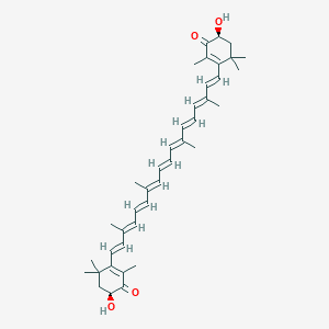
Astaxanthin is a xanthophyll carotenoid that can be found in various microorganisms and marine animals. Astaxanthin is common for humans to apply and consume into the body while also being different. This red, fat-soluble pigment is quite different from the other kinds of food that contain carotenoids. Astaxanthin surprising does not contain vitamin A like all the other food containing carotenoids, and astaxanthin is an impressive antioxidant. Studies have shown that astaxanthin can not only be beneficial for the eyes but can provide nutritional support as well as having potential health-promoting effects in preventing and treating various diseases that can harm the body. Some of the various diseases that can harm the body when there is an excessive amount of free radicals can include:
Various cancers
Chronic inflammatory diseases
Metabolic syndrome
Diabetes
Gastrointestinal diseases
Another study found that astaxanthin was superior to fish oil due to astaxanthin having the ability to enhance the body’s immune response and thus lowering the risk of vascular and infectious diseases that can harm the body, causing it to dysfunction.
A Powerful Antioxidant
There are some fantastic beneficial properties that astaxanthin can provide for the body and help improve the body�s systems as well.
Astaxanthin is a powerful antioxidant since various chronic diseases are rooted in a disproportionate balance of reactive oxygen and nitrogen species to antioxidants. Studies have shown�that astaxanthin has been known to scavenge free radicals more effectively out of the body than beta-carotene. There was another study showing how the body�s DNA was damage due to low plasma 8 -OHdG (8-hydroxy-2′-deoxyguanosine) levels.
Boosts the Immune System
The publication of the immunomodulatory effects of astaxanthin is not getting enough attention as they should be. A test study has reported that dietary astaxanthin was able to stimulate mitogen-induced lymphocyte proliferation. This will help increase the natural killer cell cytotoxicity and even delay the hypersensitivity response in the body while also increasing the numbers of total T and B cells in the peripheral blood in the body. Another study showed how astaxanthin could help significantly enhanced lymphocyte proliferation in vitro and ex vivo. The studies also found that astaxanthin can be consumed in high concentrations without the risk of cytotoxicity.
Controls Glucose and Lipids
Surprisingly there has been new research that has been revealing about another unique but vital role that astaxanthin has. The studies show that it can modulate peroxisome proliferator-activated receptors or PPARs. What this function does is that it may have various applications in human health, including producing glucose and lipid homeostasis. Since PPARs are members of the nuclear hormone receptors in the body, they are a superfamily that plays roles in the expression of many genes that are regulating cellular differentiation and many other functions in the body.
There are at least three subtypes of PPARs that helps the major organs and help the metabolism of glucose and lipids. PPAR? can primarily be expressed in the liver, kidney, heart, and skeletal muscle, where it can be involved in lipid metabolism and insulin sensitivity to the body. Another subtype of PPARs is PPAR?, which plays a role in glucose and lipid homeostasis but also is the site of action in the adipose tissue in the body. When astaxanthin is being involved, astaxanthin is a PPAR? agonist but can act as either an agonist or antagonist to PPAR? receptors. Studies have found that PPAR? agonist and PPAR? antagonist in astaxanthin can decrease cholesterol and triglycerides in loaded HepG2 cells, while changing several enzymes expressions that are being involved in lipid and glucose metabolism pathways, thus resulting in a hypolipidemic effect in the body.
Exercise Enhancement
Surprisingly astaxanthin can be used to prevent exercise-induced free radical production and is a lesser-known application. Astaxanthin can enhance exercise performance and even improve the recovery process. The increase in the reactive oxygen and nitrogen species or RONS are being produced during an exercise regime is deleterious to the health. It is often combated with a matching increase in the endogenous antioxidant enzymes. However, when a person is doing excessive exercises, it can cause RONS to rise above the body’s natural capacity to eliminate them. This will cause an increased risk of oxidative damage in lipids, protein, and DNA molecules. In a review study, it showed the ability of astaxanthin to squelch the RONS generating during exercising. It reported that the antioxidant effects of astaxanthin could provide a variety of benefits to athletes.
Conclusion
Astaxanthin is a powerful immunomodulatory antioxidant that can support numerous biological pathways that are in the body. It can dampen the effects of a variety of chronic diseases and illnesses that can harm the body. Astaxanthin is useful for being a therapeutic and powerful nutraceutical while also being an excellent addition for someone who needs supplements to support their general health and well-being. Some of the products here are beneficial to the body as they help support the immune system while providing more excellent stability.
The scope of our information is limited to chiropractic, musculoskeletal, and nervous health issues or functional medicine articles, topics, and discussions. We use functional health protocols to treat injuries or disorders of the musculoskeletal system. Our office has made a reasonable attempt to provide supportive citations and has identified the relevant research study or studies supporting our posts. We also make copies of supporting research studies available to the board and or the public upon request. To further discuss the subject matter above, please feel free to ask Dr. Alex Jimenez or contact us at 915-850-0900.
References:
Ambati, Ranga Rao, et al. �Astaxanthin: Sources, Extraction, Stability, Biological Activities and Its Commercial Applications–a Review.� Marine Drugs, MDPI, 7 Jan. 2014, www.ncbi.nlm.nih.gov/pmc/articles/PMC3917265/.
Brown, Daniel R, et al. �Astaxanthin in Exercise Metabolism, Performance and Recovery: A Review.� Frontiers in Nutrition, Frontiers Media S.A., 18 Jan. 2018, www.ncbi.nlm.nih.gov/pmc/articles/PMC5778137/.
Brown, Daniel R, et al. �Astaxanthin in Exercise Metabolism, Performance and Recovery: A Review.� Frontiers in Nutrition, Frontiers Media S.A., 18 Jan. 2018, www.ncbi.nlm.nih.gov/pmc/articles/PMC5778137/.
Choi, Chang-Ik. �Astaxanthin as a Peroxisome Proliferator-Activated Receptor (PPAR) Modulator: Its Therapeutic Implications.� Marine Drugs, MDPI, 23 Apr. 2019, www.ncbi.nlm.nih.gov/pmc/articles/PMC6521084/.
Lin, Kuan-Hung, et al. �Astaxanthin, a Carotenoid, Stimulates Immune Responses by Enhancing IFN-? and IL-2 Secretion in Primary Cultured Lymphocytes in Vitro and Ex Vivo.� International Journal of Molecular Sciences, MDPI, 29 Dec. 2015, www.ncbi.nlm.nih.gov/pmc/articles/PMC4730289/.
Park, Jean Soon, et al. �Astaxanthin Decreased Oxidative Stress and Inflammation and Enhanced Immune Response in Humans.� Nutrition & Metabolism, BioMed Central, 5 Mar. 2010, www.ncbi.nlm.nih.gov/pmc/articles/PMC2845588/?report=reader.
Team, DFH. �Applications of the Antioxidant, Astaxanthin.� Designs for Health, 27 June 2019, blog.designsforhealth.com/node/1047.
Yuan, Jian-Ping, et al. �Potential Health-Promoting Effects of Astaxanthin: a High-Value Carotenoid Mostly from Microalgae.� Molecular Nutrition & Food Research, U.S. National Library of Medicine, Jan. 2011, www.ncbi.nlm.nih.gov/pubmed/21207519.
The University offers a wide variety of medical professions for functional and integrative medicine. Their goal is to inform individuals who want to make a difference in the functional medical fields with knowledgeable information that they can provide.
Chiropractic is more than spinal manipulation. More people are choosing chiropractic for their pain symptoms, conditions, and injuries. Many are exchanging pills and surgery for all-natural non-invasive medicine known as chiropractic. Chiropractors do not prescribe medications and do not perform surgery. But they are trained to recognize when an injury is outside the scope of their practice and will refer an individual to the appropriate medical specialist.
Here are a few reasons why individuals are trying out chiropractic:
Doctors recommend chiropractic
The American College of Physicians recommends chiropractic and other non-drug treatments, as a first-line treatment for chronic and acute low back pain. Spinal manipulation is strongly recommended as a natural remedy. It is highly effective when combined with gentle exercises like Pilates, yoga, and tai chi.
Studies show spinal manipulation highly effective
The Journal of the American Medical Association cited spinal manipulation to be highly beneficial in treating lower back pain. Results are consistently showing that chiropractic treatment, specifically spinal manipulation, is effective in helping low back pain and improving function while minimizing pain.
95% Return for treatment
In the 2016 Gallup-Palmer College of Chiropractic Annual Report, 95% of people who used chiropractic say that it is an effective treatment. 97% said that if back or neck pain returned they would seek chiropractic treatment again. More than half of adults that have never seen a chiropractor said they would seek chiropractic treatment if they developed neck or back pain.
Chiropractic rated higher than All other treatments for back pain
A Consumer Reports survey ranked chiropractic as a top treatment compared to medications for back pain, osteoarthritis, and neck pain relief.
Individuals prefer a whole-body approach that includes:
Lifestyle recommendations
Dietary advice
Supplements for pain, immobility, and range of motion problems.
Many said they opted for natural treatments like chiropractic to avoid harmful and undesirable side effects of prescription medication.
Opioids
With the Centers for Disease Control declaring prescription abuse an epidemic, individuals are searching for safer options. Natural treatments are becoming more popular to avoid the potential for addiction. And not just addiction but the often side effects that come with prescription meds, like constipation, brain fog, nausea, dry mouth, etc.
Spine surgery rising Individuals seeking the less invasive option
Spinal fusion surgery has been on a steep rise, increasing by about 500%. Therefore many are going for natural treatment like chiropractic to avoid surgery. Chiropractic looks at the whole person along with advice on lifestyle habits, dietary adjustments, and exercise programs. The bottom line is it works. It is less invasive, and yes, it is drug-free, but most of all, it is effective.
How adjustments work
When the vertebrae become misaligned, they place pressure on the nerves and cause pain, swelling, awkward movement/s, etc. This is called a subluxation. Chiropractors use their repertoire of techniques that equal a form of spinal adjustment to treat the subluxations. An adjustment means that the vertebra is returned to the proper position in the spine. When the vertebrae are adjusted, tiny pockets of gas are released from the joints, sometimes make a popping/releasing noise. It is the same sound when cracking knuckles.
The benefits
Individuals with the following conditions have found chiropractic to be helpful:
Arthritis
Bursitis and
Fibromyalgia
Headaches
Leg pain
Pain or stiffness in the neck, shoulders, back, arms, hands, chest, abdomen, hips, legs, feet.
Sciatica
Scoliosis
Sports injuries
Tendonitis
Trauma injuries like auto accidents
Other Services
The main method of treatment is spinal adjustment, many chiropractors use a variety of therapies to treat patients, including:
Health Coaching
Exercise
Diet
Massage
Weight loss
Stress management
Chiropractic medicine is more than just spinal manipulation. Chiropractors use a variety of treatments to help the body heal itself and return individuals to a pain-free and healthy life.
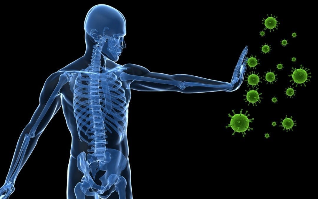
With everything that is going on in today’s world immunity is especially important. Without a properly functioning immune system, our bodies can become inflamed and more susceptible to viruses. Inflammation can cause a weakened immune system, joint pain, headaches, fatigue and more!
So what can we do to build up our immunity and help give our bodies a fighting chance? First off, washing your hands is highly important. Not just now, but always. Be sure to wash your hands with warm water and scrub everywhere. Second, get plenty of sleep. Rest is how the body recovers. If you do not give your body adequate sleep, the strength you’re cells have to fight off infection lessens. Third, eat healthy food, hydrate, and exercise. Finally, last but not least help kick up your immune system by supplementing the body with all-natural supplements.
There are many supplements that will be beneficial to the body. However, two of the most important are NAC and Glutamine.
What Are They?
NAC stands for N-acetyl-Cystine. NAC is an amino acid that the body can produce but the body can also greatly benefit from taking additional NAC in supplemental form. NAC plays an important role in helping the liver to detox. In addition to this, NAC helps to replenish the glutathione levels in the lungs and can help to reduce the inflammation. This is highly beneficial in helping to relieve the symptoms of a respiratory infection.
NAC is also greatly beneficial in boosting brain health. NAC helps to regulate glutamate levels and replenish glutathione. However, one of the most important factors of NAC is its ability to boost Glutathione levels.
Glutamine is an amino acid that helps the body perform many functions. Glutamine plays a crucial part in the immune system.
The Connection & How It Impacts Immunity
However, one of the most important factors of NAC is its ability to booze Glutathione levels. NAC and glutathione can help to boost an individual’s immune health. In research studies shown, NAC has been shown to lessen the effects of a virus and its ability to replicate. When it comes to immunity NAC and Glutamine are powerful molecules. Stoping the replication of a virus can help reduce the spread and the length of the virus in an individual.
Many infections and diseases have been linked to low glutathione levels. When the glutathione levels are low this is typically due to enhanced oxygen radicals. Studies have been done and show that when supplementing NAC to those who have low glutathione levels, it directly boosts their levels and helps with infection.
Especially with everything happening today, we want to increase our immunity and decrease the inflammation in the body.� Essentially, think of the body as a road trip. For this trip we need two main things: the gas for the car, and the car to take you to the end destination.� NAC is the gas that drives the car. We need the gas to get to our end destination. Our end destination is being healthy and giving our body the best chance to fight off infection (increased Glutathione). So by giving our body gas (NAC) we provide it with what it needs to take us to where we want to go (increased Glutathione, leading to increased immunity).
How Can I Benefit?
Overall, NAC is great to decrease inflammation. Inflammation is an extremely common underlying issue relating to other health conditions individuals suffer from. By providing your body with additional supplements, you can help increase your immunity and decrease your chances of contracting a virus and/or the length of the virus. Always discuss supplements with your primary care doctor before you begin them, but consider adding these into your daily routine!
I always recommend talking to your primary care provider and taking supplements daily. Supplements, in general, are a great way to help provide the body with the essential vitamins and minerals you may be missing. However, now more than ever supplementation is key. By building up and providing the body with the nutrients it needs for proper function, it will help prepare your body to fight off an infection. Supplementation like NAC is great to have already running in your system to help combat an infection if you were to catch one. Remember to be smart, talk to a primary care doctor before beginning supplementation, and keep in mind that not all supplements are created equal.� -Kenna Vaughn, Senior Health Coach��
The scope of our information is limited to chiropractic, musculoskeletal, and nervous health issues or functional medicine articles, topics, and discussions. We use functional health protocols to treat injuries or disorders of the musculoskeletal system. Our office has made a reasonable attempt to provide supportive citations and has identified the relevant research study or studies supporting our posts. We also make copies of supporting research studies available to the board and or the public upon request. To further discuss the subject matter above, please feel free to ask Dr. Alex Jimenez or contact us at 915-850-0900.�
References:
Dinicola S, De Grazia S, Carlomagno G, Pintucci JP. N-acetylcysteine as powerful molecule to destroy bacterial biofilms. A systematic review.�Eur Rev Med Pharmacol Sci. 2014;18(19):2942�2948.
Wessner B, Strasser EM, Spittler A, Roth E. Effect of single and combined supply of glutamine, glycine, N-acetylcysteine, and R,S-alpha-lipoic acid on glutathione content of myelomonocytic cells.�Clin Nutr. 2003;22(6):515�522. doi:10.1016/s0261-5614(03)00053-0
Dr. Alex Jimenez, a chiropractor in El Paso, TX, Kenna Vaughn, Truide Torres, and Astrid Ornelas discuss what it is that they do and why they do it. Chiropractic care is a safe and effective, alternative treatment option that focuses on the diagnosis, treatment, and prevention of a variety of health issues associated with the musculoskeletal and nervous system, including neck pain, back pain, low back pain, and sciatica, among other health issues within the scope of chiropractic care. Dr. Alex Jimenez utilizes spinal adjustments and manual manipulations, among other well-known chiropractic care treatment methods and techniques, to help provide pain relief and promote overall health and wellness. Dr. Alex Jimenez, Kenna Vaughn, Truide Torres, and Astrid Ornelas discuss how they patients, from chiropractic care to educating them on diet and lifestyle changes, to help patients achieve their nutrition and fitness goals. According to Dr. Alex Jimenez, Kenna Vaughn, Truide Torres, and Astrid Ornelas, chiropractic care and functional medicine are treatment options that can naturally support well-being. – Podcast Insight
If you have enjoyed this video and/or we have helped you in any way
please feel free to subscribe and share us.
Thank You & God Bless.
Dr. Alex Jimenez RN, DC, MSACP, CCST
Dr. Alex Jimenez, a chiropractor in El Paso, TX, Kenna Vaughn, Truide Torres, Alexander Jimenez, and Astrid Ornelas, discuss how chiropractic care can ultimately help treat sciatica or sciatic nerve pain. The sciatic nerve is the largest and longest nerve in the human body. It runs from the lower back, down the buttocks and hips, and into the legs, knees, and feet. Sciatica can be caused by a variety of health issues which result in the compression or impingement of the sciatic nerve. Dr. Alex Jimenez, Kenna Vaughn, Truide Torres, Alexander Jimenez, and Astrid Ornelas discuss sciatica or sciatic nerve pain in further detail to ultimately help educate patients on their symptoms. Diet and lifestyle modifications, including nutraceuticals and exercise or physical activity, can be beneficial for patients with sciatic nerve pain. Furthermore, sciatica or sciatic nerve pain is a collection of symptoms rather than a single injury or underlying condition. Dr. Alex Jimenez, Kenna Vaughn, Truide Torres, Alexander Jimenez, and Astrid Ornelas conclude the podcast by describing how they each can help patients achieve overall health and wellness. – Podcast Insight
If you have enjoyed this video and/or we have helped you in any way
please feel free to subscribe and share us.
Thank You & God Bless.
Dr. Alex Jimenez RN, DC, MSACP, CCST
Reducing stress is important for emotional well being and physical health. Rheumatoid arthritis is a complex condition with no cure and can cause intense chronic pain. Stress only exacerbates the symptoms, affects pain perception, and weakens the body. Stress management is highly important for reducing pain. In a weakened state, an individual is more vulnerable to arthritis symptoms, like flare-ups, weakness, and fatigue.�Chiropractic can help.
There are non-surgical treatments for arthritis, like medications, physical, and massage therapy that address the physical nature of the condition. By eliminating stressful triggers and making healthy behavioral/lifestyle changes a better sense of well being can be achieved.
Different emotions can run rampant:
Confusion
Frustration
Anger
Sadness
Helplessness
And all of these feelings can generate intense stress on an individual. Chiropractic excels in wellness and is becoming more common for individuals to visit chiropractic clinics for treating a variety of different kinds of pain symptoms and conditions. Chiropractic adjustments provide countless benefits to those with arthritis. We�ll explore how chiropractic can help those with arthritis and give additional information along with suggestions on how to alleviate the pain.
What Chiropractic Does
A doctor of chiropractic is a health professional that focuses on wellness and optimal health instead of ailment/sickness symptoms. Their specialty aims at adjusting the spine to correct misalignments that could be pressing down on nerves and causing major disruption in the body. Regular chiropractic not only restores health throughout the body but helps alleviate back pain and any other symptoms associated with an out of alignment spine.
They also work in setting up exercise programs specific to the condition being treated along with the proper diet to utilize in assisting with the management of inflammation and pain.
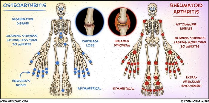
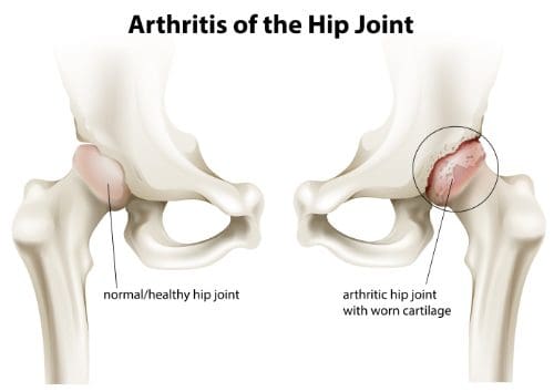
Arthritis
Arthritis is inflammation in the body’s joints which results in pain, stiffness and limited range of movement. There are over 200 different varieties of arthritis. Generally associated with age, it can affect young people. It can strike pretty much any area of the body. Arthritis can cause damage to soft tissues and muscles.
Osteoarthritis also known as degenerative joint disease, is the most common type. It comes from repeated trauma to the joints and occurs more often in the elderly.
Other forms include:
Rheumatoid arthritis is the second common type in which the body�s immune system attacks the joint/s.
Psoriatic arthritis, an autoimmune form of arthritis.
Ankylosing spondylitis is a type of arthritis where the body attacks itself.
Septic arthritis is caused by a viral or bacterial infection of the joint/s.
Diagnosis
Diagnosing arthritis involves a thorough physical examination. Rheumatologists often need help with these cases, and so a medical work-up can be done and a chiropractor could be recommended. This includes X-rays or MRIs, urine, blood analysis, and physical examinations. Having the condition properly diagnosed will help to more effectively treat the symptoms.
Chiropractic
The most common treatment is medication, which takes down the inflammation, the swelling and reduces pain. Chiropractors can be of great help in managing arthritis. Medications work but as we’ve seen they can have long-term health risks like impaired healing, damage to the stomach lining and internal bleeding.
A chiropractor can reduce stress, and reliance on medications, all the while managing the pain and symptoms in a natural way. Chiropractic can:
Improve range of motion
Keeps the spine properly aligned
Improve endurance
Improve flexibility
Increase strength
Increase muscle tone
Develop a dietary and nutritional plan to reduce inflammation
Recommend an exercise regimen conducive to arthritis symptoms
According to the American Chiropractic Association, this is vital in managing arthritis symptoms.
Treatment
Understand that chiropractic cannot cure arthritis. They can help alleviate symptoms, slow the progression and help to reduce stress levels. They will use adjustments in combination with other treatments. This can include:
Hot and cold treatments
Ultrasound treatments
Massage
Electronic muscle stimulation
Physical rehabilitation
Magnet therapy
Reduce Stress
Exercise
Water aerobics or make walking around the park/neighborhood part of a daily routine, as it promotes a healthy mind by reducing stress and anxiety. Gentle exercises like aerobic exercise are perfect because it improves mobility and helps shed a few pounds taking pressure off the joints. Exercise creates endorphins, which reduces pain and uplifts the mood.
Support groups
With any type of painful condition, it’s easy to feel alone. Joining a support group can connect you with people who understand what’s going on and the emotions you’re experiencing. The community helps diminish the sense of isolation.
Relaxation therapy
This focuses on calming the body and mind by making a conscious effort to relax. Even for only a few moments, you might find this technique effective at controlling the stress response. Begin by focusing on one part of the body like the hands, feet, etc.
Concentrate until the area you’re focusing on is completely free of stress or tension. Then imagine weightlessness flow through the body. Close your eyes, lie down, turn off the lights, and think of something soothing. There are no strict guidelines for relaxation. Whatever puts you in a relaxed frame of mind is the way to do it.
Warm bath
Warm moist heat from a shower, bath, or steam room can decrease the secretion of stress hormones and raise levels of endorphins, as aforementioned the body’s natural pain killers.
Take time for yourself
When it comes to reducing stress, balance is key. Staying active, and regular rest/sleep is vital to successful treatment. A balance needs to be in everything you do. Therefore, make time for the things you want to do.
Results
Inflammatory diseases like arthritis have shown the best results are achieved from combating it from all angles. Working with a chiropractor and rheumatologist to combine treatments can make all the difference. A healthy diet and active exercise program will get you in the right direction toward a healthy active lifestyle. If you or a loved one are suffering from arthritis, don�t hesitate to call. We�re here to help in any way possible!
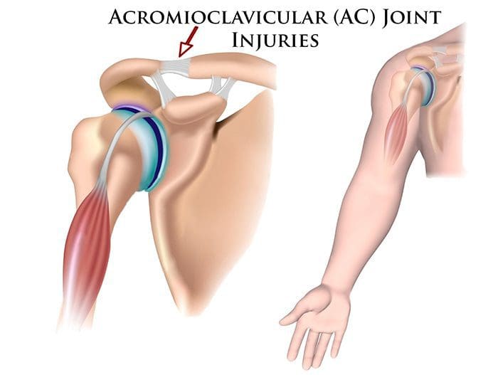
Shoulder pain along with the kinds of shoulder problems like sprains and strains that chiropractors regularly treat often involves a form of rotator dysfunction. The shoulder has the greatest mobility of any joint in the body. However, there is little stability when in certain positions the soft tissues in the shoulder area can get injured through sports injury/s, recreational activities, at work or in a fall.�
This usually begins with dysfunction of the rotator cuff muscles and progresses to:
In cases like this, there is no direct, acute injury.
Every sprain and strain injury to the shoulder must be treated and rehabilitated�properly�to avoid future injuries and pain. There is a higher probability of shoulder instability that becomes chronic after an injury. This is because the surrounding muscles and connective tissues are what create shoulder joint stability.
Shoulder Pain
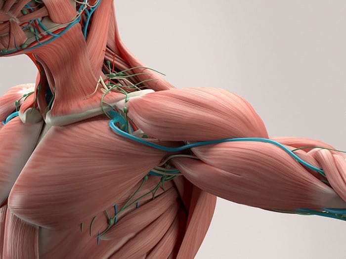
The shoulder goes through a great deal that we do not realize. It is made up of an intricate network of ligaments and muscles, with the rotator cuff taking most of the load/weight during movement and exertion. As aforementioned, the shoulder is the most flexible joint in the human body, but it is one of the most unstable. For athletes, shoulder injuries are pretty common, as the shoulder sustains more injuries than almost any other part of the body.
The shoulder is unstable because of its unique construction. Other joints like the ankle or elbow are limited in their range of motion. The shoulder, on the other hand, can move pretty widely, which leaves it open to stress, injury, and pain. The soft tissues in the shoulder, muscles, tendons, and ligaments can get injured from overuse, excessive strain, falls, and improper motion.
Treating shoulder pain can be a challenge because it is used so much daily. A Chiropractor can bring relief to shoulder pain and speed up the healing process.
Injury and pain causes
Shoulder pain can be caused by a variety of conditions. Injury is one of the most common causes and is seen in athletes, like swimmers, gymnasts, golfers, baseball players, bowlers, and tennis players, etc, who use their arms and shoulders regularly, place added stress on the joint.
Jobs that involve repetitive shoulder movements can also cause injury. People whose job requires repetitive motions or strenuous upper bodywork are at higher risk. Truck drivers, construction workers, warehouse and grocery store employees commonly experience shoulder injury and pain.
Prevention
Preventing shoulder pain and injury can be a simple process. Workers and athletes whose jobs require repetitive arm work or rotation should warm-up, and take stretching and shake out the hand/s and arm/s breaks. Pain should be addressed, as soon as it presents with ice/heat and a possible chiropractic appointment. Ignoring the pain could lead to a more serious/chronic condition.
Shoulder rest is always a must. The joint needs time off to heal and regular chores should not be resumed until the doctor or chiropractor clears the patient. Chiropractors usually recommend exercises and stretches geared toward the type of injury/s or condition/s.
Chiropractor
Chiropractic is highly beneficial for shoulder injury/s and shoulder pain.
It will:
Reduce pain
Relieve pain
Improve range of motion
Increase flexibility
Restore function to the joint
Different techniques are used depending on the type of injury or condition. The patient will be assessed by the chiropractor and will determine a treatment plan. Compression techniques have been found to be very effective for shoulder pain. It is favored because it is not strenuous for the doctor, is very safe, and is handled well by the patient. Chiropractic is the treatment of choice for shoulder pain because it is minimally invasive and it works.
Chiropractic addresses the entire body with its aim to relieve pain without drugs or surgery and keep the individual healthy and functioning at an optimal level. Are you, a family member, or friend suffering from shoulder stiffness/pain? If so, give us a call. Dr. Jimenez and his outstanding physical therapy team are here to help!
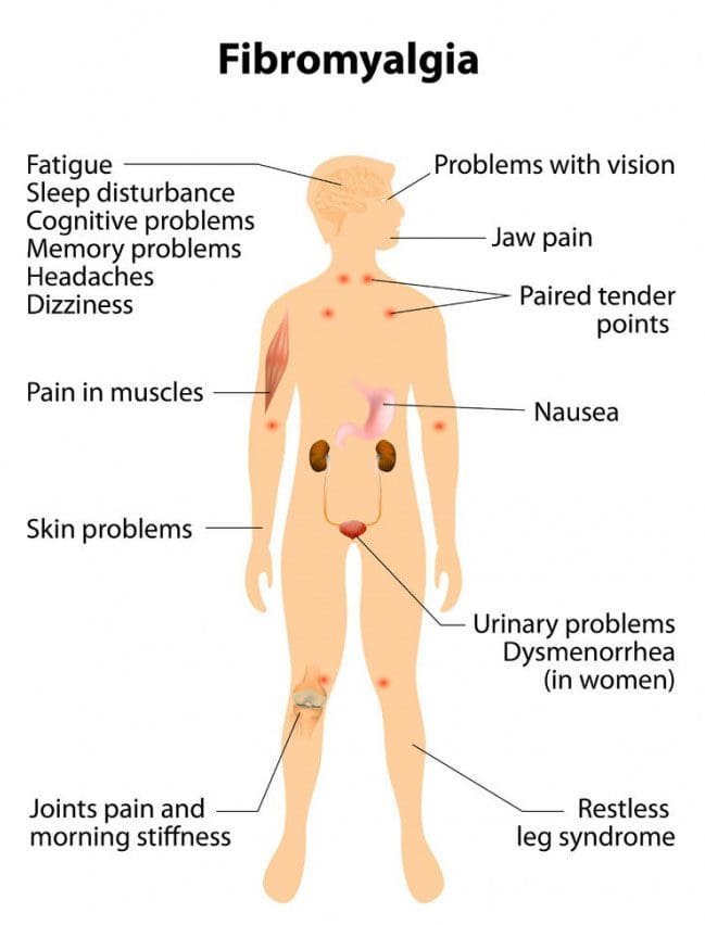
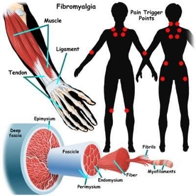
Fibromyalgia is a chronic pain disorder affecting millions and mostly women. It is physically and emotionally distressing. Those with the condition experience widespread chronic muscle pain. Research has shown that individuals with fibromyalgia could have a lower threshold for pain. This can come from injury, emotional distress, or abnormal levels of substances/chemicals in the brain and spine linked to pain sensitivity. One of the most common treatments is chiropractic medicine.
Common symptoms/conditions individuals report:
Chronic fatigue syndrome
Irritable bladder
Irritable bowel syndrome
Migraines
Sleep disorders
Restless legs syndrome
TMJ or Temporomandibular joint disorder
Raynaud’s Syndrome�-�a�rare blood vessel disorder causing the toes and hands to feel cold or numb.
Doctors are still trying to figure out the relationship between these conditions and fibromyalgia.
Causes
Doctors have yet to determine the exact cause, however, research is ongoing and beginning to shed light on the condition. Some possible causes include:
Abnormalities in the endocrine system
Abnormalities in the autonomic nervous system
Genetics
Muscle tissue abnormalities
Abnormal blood flow
As research has discovered many conditions/disorders do not have one cause but rather, several factors that impact the probability of developing the condition.
Questions
It has become one of the most common chronic pain conditions. 1 in 50 Americans are dealing with fibromyalgia. The condition can be difficult to diagnose, and, because of its chronic nature, it can linger for months and even years. Typically it causes pain throughout the body and creates areas that become tender to the slightest touch. There are both traditional and alternative treatments available.
Traditional approaches to manage the pain:
Anti-inflammatory’s
Over-the-counter pain relievers
Sleep medications
Muscle relaxants
Fibromyalgia medications include:
Lyrica – pregabalin, which is a nerve pain medication
Cymbalta – duloxetine hydrochloride, which is an antidepressant that can also help manage pain
Savella – milnacipran HCI, which is an antidepressant and nerve pain medicine
The type of treatment depends on the symptoms. For example, a doctor could prescribe an antidepressant to reduce pain and depression. If stress, anxiety, and trouble sleeping are presenting,�a therapeutic exercise program could be the answer. Individuals prefer natural remedies/therapies instead of more medications like vitamin therapy, acupuncture, and meditation.
Other treatment options include alternative treatments like:
The most common issue is constant and consistent pain, which can affect the entire body for weeks and even months. Individuals realize that chiropractic helps restore overall health and aids the body to heal itself. Adjustments to the spine bring alignment and balance back to the body. Also incorporated is soft tissue work that can relieve and reduce painful pressure/trigger points and decrease pain in tender spots.
Range of motion is increased
Chiropractic medicine also adjusts the body’s joints and helps loosen them up. This increases the range of motion and allows the individual to move more freely and easily. Depending on how long the individual has been dealing with the condition, it can take a few treatments to achieve optimal results, so it does take a commitment from the individual patient. However, in the long run, it is well worth the time.
Sleep is improved
The pain associated with fibromyalgia often affects an individual’s ability to sleep well. Being unable to sleep normally will leave you exhausted, foggy, unable to accomplish things and irritable to downright angry. A chiropractor’s ability to loosen the body’s joints, massage tender points, and kickstart the body’s self-healing mechanisms means individuals with this condition can enjoy deep sleep, and stay asleep.
Complements other therapies
Medicines/treatments/therapies can counteract with each other, or get mixed up and cause side effects. Chiropractic medicine can be utilized in combination with medications/treatments, either traditional or natural. Individuals diagnosed with this condition should speak to their chiropractor about the different treatments available. Customized treatment programs are created case-by-case and are tailored to the specific needs of that individual. Remember there is not a one-stop solution.
Empowers the individual
Individuals that have to deal with painful, chronic conditions can exhaust themselves with the varying treatment options and can feel as if they have no control over the situation. This causes stress, anxiety, and depression, which works against achieving overall wellness. With chiropractic, individuals are more in charge of their treatment plan, which leads to an optimistic outlook in their recovery.�
Chiropractic medicine treats not only the symptoms of fibromyalgia but attempts to get to the root cause to alleviate the condition or to activate the body’s self-healing response. Patients that commit will see the benefits along with reduced pain, better mobility, and sound sleep.
The best benefit is being able to take control of the circumstances and playing a vital role in the managing of an individual’s well-being. Understand that there are options available for fibromyalgia pain management. If you or a loved one has been diagnosed with fibromyalgia, don�t go it alone. Dr. Alex Jimenez is passionate about helping those who are injured or struggling with a condition get relief. Contact us today to schedule an appointment.
We are familiar with neck stiffness or a crick. This can prevent us from comfortably moving the head all around. A crick can cause the spine, and shoulders to feel rigid and stressed from not being able to turn around and could cause an upper or low-back strain from having to turn the whole body to look back or even just to the side. Chiropractic treatment is available and will help, along with some self-care therapies that can be done at home.
Crick in the Neck vs. Neck Stiffness
A crick in the neck is the same as a stiff neck. It develops when the neck muscles, tendons, and ligaments become strained/sprained. Most strains and sprains are minor but do cause inflammation/swelling of the neck�s soft tissues, which results in stiffness and, at times muscle spasms.
The symptoms
Cricks in the neck are uncomfortable, but not necessarily painful. If there is a pre-existing neck condition or injuries like whiplash the crick and stiffness could increase the uncomfortableness and generate pain.
The most common symptoms include:
Neck stiffness
Muscle stiffness
Reduced mobility affecting the neck�s range of motion
A popping sensation when trying to turn or tilt the head
Causes of a stiff neck or crick
There are different causes of neck stiffness. It can be a combination of things you can control and some you can�t.
Possible causes that you can control:
Poor posture working either sitting or standing for several hours without breaks or stretching.
Sleeping in a position that puts the neck in an awkward position or using a pillow that does not support the neck when sleeping.
Constantly looking down at a cell phone or tablet.
Stress and emotional tension can cause involuntarily tightening of the neck muscles and shoulders.
Heavy labor along with incorrect lifting techniques.
Reaching or having to look up/overhead for several hours like when painting a ceiling.
Possible causes that are out of your control:
Whiplash injury
Sports-injuries like a football stinger
Aging muscles and bones
Around 13% of cases the stiffness, and pain are caused by separate cervical spinal conditions, like:
Cold therapy reduces the swelling of soft tissues, like muscles and ligaments, while heat soothes the tightness by boosting blood circulation to the affected area. There are different products available that can deliver cold or heat to the neck and upper back.
Apply ice for 15 minutes each hour.
Apply heat therapy like a heating pad for 15 minutes every 2 or 3 hours.
Over-the-counter anti-inflammation medicines
Non-steroidal anti-inflammatory medications like ibuprofen and naproxen can help relieve inflammation and pain.
Because neck stiffness can be linked to lifestyle choices, individuals may find that they occur repeatedly. Simple neck stretches, chiropractic treatment, using a supportive pillow, and taking frequent breaks at your job can help prevent neck stiffness and keep you moving. These professionals have undergone extensive training in their field and are capable of treating neck pain effectively. So if you or a loved one are experiencing neck pain, give us a call. We�re ready to help!
In the following podcast video article, Dr. Alex Jimenez, a chiropractor in El Paso, TX, and Dr. Mario Ruja, another chiropractor in El Paso, TX, discuss chiropractic care and why it works. Chiropractic care is a safe and effective, alternative treatment option that focuses on the diagnosis, treatment, and prevention, of injuries and underlying conditions associated with the musculoskeletal and nervous system. Chiropractic care is a healthcare profession that has existed for many years throughout many civilizations and it focuses on the use of spinal adjustments and manual manipulations to carefully restore the original alignment of the spine and the human body as a whole. Dr. Alex Jimenez and Dr. Mario Ruja describe how they were first interested in becoming chiropractors, or doctors of chiropractic, as they also describe how they feel when they are able to provide pain relief to their patients. Dr. Jimenez and Dr. Ruja will focus on discussing why chiropractic care works and how it is different from other healthcare professions in the way it helps treat a variety of health issues associated with the spine, from neck pain to low back pain and sciatica. Chiropractic care can help promote overall health and wellness. � Podcast Insight
If you have enjoyed this video and/or we have helped you in any way
please feel free to subscribe and share us.
Thank You & God Bless.
Dr. Alex Jimenez RN, DC, MSACP, CCST
IFM's Find A Practitioner tool is the largest referral network in Functional Medicine, created to help patients locate Functional Medicine practitioners anywhere in the world. IFM Certified Practitioners are listed first in the search results, given their extensive education in Functional Medicine