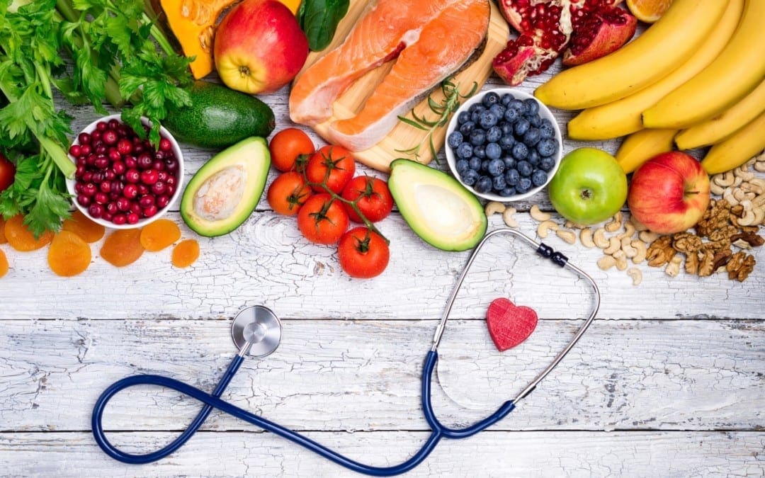
The foods we eat can have the potential to be beneficial or harmful to our health. Poor nutrition can cause a variety of health issues, including obesity, cardiovascular disease, and type 2 diabetes. Meanwhile, proper nutrition can make you feel energized, reduce your risk of health issues, as well as help maintain and regulate a healthy weight. If you want to promote longevity, you have to fuel your body with good foods. In the following article, we will list several good foods that can ultimately help promote longevity by also helping to improve overall health and wellness.
Contents
Cruciferous Vegetables
Cruciferous vegetables have the unique ability to change our hormones, trigger the body�s natural detoxification system, and even reduce the growth of cancerous cells. These must be chewed thoroughly or eaten shredded, chopped, juiced, or blended in order to release their beneficial properties. Sulforaphane, found in cruciferous vegetables, has also been found to help protect the blood vessel wall from inflammation that can cause heart disease. Cruciferous vegetables, such as kale, cabbage, Brussels sprouts, cauliflower, and broccoli are several of the most nutrient-dense foods in the world.
Salad Greens
Raw leafy greens have less than 100 calories per pound, which makes them the perfect food for weight loss. Eating more salad greens has also been associated with the reduced risk of heart attack, stroke, diabetes, and several types of cancers. Raw leafy greens are also rich in the essential B-vitamin folate, plus lutein and zeaxanthin, carotenoids that can help protect the eyes. Fat-soluble phytochemicals, such as carotenoids, found in salad greens like lettuce, spinach, kale, collard greens, and mustard greens also have antioxidant and anti-inflammatory effects in the body.
Nuts
Nuts are a low-glycemic food and a great source of healthy fats, plant protein, fiber, antioxidants, phytosterols, and minerals, which also helps to reduce the glycemic load of an entire meal, making them an essential part of an anti-diabetes diet. Regardless of their caloric density, eating nuts can help promote weight loss. Nuts can also reduce cholesterol and help reduce the risk of heart disease.
Seeds
Seeds, much like nuts, also provide healthy fats, antioxidants, and minerals, however, these have more protein and are rich in trace minerals. Chia, flax, and hemp seeds are rich in omega-3 fats. Chia, flax, and sesame seeds are also rich lignans or breast cancer-fighting phytoestrogens. Moreover, sesame seeds are rich in calcium and vitamin E, and pumpkin seeds are rich in zinc.
Berries
Berries are antioxidant-rich fruits that can help promote heart health. Research studies where participants ate strawberries or blueberries daily for several weeks reported improvements in blood pressure, total and LDL cholesterol, and even signs of oxidative stress. Berries also have anti-cancer properties and have been shown to help prevent cognitive decline associated with aging.
Pomegranate
The most well-known phytochemical in pomegranates, punicalagin, is responsible for more than half of the fruit’s antioxidant activity. Pomegranate phytochemicals have anti-cancer, cardioprotective, and brain-healthy benefits. In one research study, older adults who drank pomegranate juice daily for 28 days performed better on a memory test compared to those who drank a placebo beverage.
Beans
Eating beans and other legumes can help balance blood sugar, reduce your appetite, and protect against colon cancer. Beans are an anti-diabetes food that can help promote weight loss because they are digested slowly, which slows down the increase of blood sugar after a meal and helps prevent food cravings by promoting satiety. Eating beans and other legumes twice a week has been found to decrease the risk of colon cancer. Eating beans and other legumes, such as red beans, black beans, chickpeas, lentils, and split peas, also provides significant protection against other cancers.
Mushrooms
Eating mushrooms regularly is associated with a reduced risk of breast cancer. White and Portobello mushrooms are especially beneficial against breast cancer because they have aromatase inhibitors or compounds that inhibit the production of estrogen. Mushrooms have shown to have anti-inflammatory effects as well as provide enhanced immune cell activity, prevention of DNA damage, slowed cancer cell growth, and angiogenesis inhibition. Mushrooms should always be cooked as raw mushrooms have a potentially carcinogenic chemical known as agaritine that is significantly reduced by cooking.
Onions and Garlic
Onions and garlic provide cardiovascular and immune system benefits as well as provide anti-diabetic and anti-cancer effects. These have also been associated with a lower risk of gastric and prostate cancers. Onions and garlic are known for their organosulfur compounds which help to prevent the development of cancers by detoxifying carcinogens, decreasing cancer cell growth, and blocking angiogenesis. Onions and garlic also have high concentrations of health-promoting flavonoid antioxidants, which have anti-inflammatory effects that may help provide cancer prevention.
Tomatoes
Tomatoes are rich in a variety of nutrients, such as lycopene, vitamin C and E, beta-carotene, and flavonol antioxidants. Lycopene can help protect against prostate cancer, UV skin damage, and? cardiovascular disease. Lycopene is better absorbed when tomatoes are cooked. One cup of tomato sauce has about 10 times the amount of lycopene as a cup of raw, chopped tomatoes. Also keep in mind that carotenoids, like lycopene, are best absorbed when accompanied by healthy fats, so enjoy your tomatoes in a salad with nuts or a nut-based dressing for extra nutritional benefits.
The foods we eat can have the potential to be beneficial or harmful to our health. Poor nutrition can cause a variety of health issues, including obesity, cardiovascular disease, and type 2 diabetes. Meanwhile, proper nutrition can make you feel energized, reduce your risk of health issues, as well as help maintain and regulate a healthy weight. If you want to promote longevity, you have to fuel your body with good foods. Good foods can also help reduce inflammation associated with a variety of health issues, including joint pain and arthritis. Healthcare professionals, such as chiropractors, can offer diet and lifestyle advice to help promote health and wellness. In the following article, we will list several good foods that can ultimately help promote longevity. – Dr. Alex Jimenez D.C., C.C.S.T. Insight
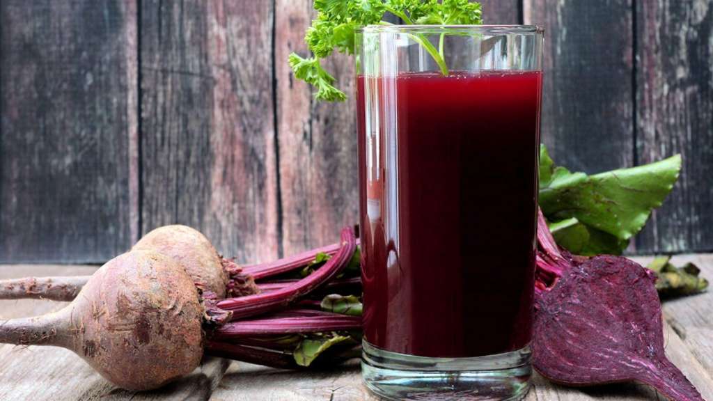
Zesty Beet Juice
Servings: 1 Cook time: 5-10 minutes
� 1 grapefruit, peeled and sliced
� 1 apple, washed and sliced
� 1 whole beet, and leaves if you have them, washed and sliced
� 1-inch knob of ginger, rinsed, peeled and chopped
Juice all ingredients in a high-quality juicer. Best served immediately.
Just one carrot gives you all of your daily vitamin A intake
Yes, eating just one boiled 80g (2�oz) carrot gives you enough beta carotene for your body to produce 1,480 micrograms (mcg) of vitamin A (necessary for skin cell renewal). That’s more than the recommended daily intake of vitamin A in the United States, which is about 900mcg. It’s best to eat carrots cooked, as this softens the cell walls allowing more beta carotene to be absorbed. Adding healthier foods into your diet is a great way to improve your overall health.
The scope of our information is limited to chiropractic, musculoskeletal, physical medicines, wellness, and sensitive health issues and/or functional medicine articles, topics, and discussions. We use functional health & wellness protocols to treat and support care for injuries or disorders of the musculoskeletal system. Our posts, topics, subjects, and insights cover clinical matters, issues, and topics that relate and support directly or indirectly our clinical scope of practice.* Our office has made a reasonable attempt to provide supportive citations and has identified the relevant research study or studies supporting our posts. We also make copies of supporting research studies available to the board and or the public upon request. We understand that we cover matters that require an additional explanation as to how it may assist in a particular care plan or treatment protocol; therefore, to further discuss the subject matter above, please feel free to ask Dr. Alex Jimenez or contact us at 915-850-0900. The provider(s) Licensed in Texas*& New Mexico*�
Curated by Dr. Alex Jimenez D.C., C.C.S.T.
References:
Joel Fuhrman, MD. �10 Best Foods You Can Eat to Live Longer and Stay Healthy.� Verywell Health, 6 June 2020, www.verywellhealth.com/best-foods-for-longevity-4005852.
Dowden, Angela. �Coffee Is a Fruit and Other Unbelievably True Food Facts.� MSN Lifestyle, 4 June 2020, www.msn.com/en-us/foodanddrink/did-you-know/coffee-is-a-fruit-and-other-unbelievably-true-food-facts/ss-BB152Q5q?li=BBnb7Kz&ocid=mailsignout#image=24.
There are various chronic treatment/management options available. Chronic pain treatment focuses on treating and managing the root cause and underlying condition that is causing the pain. The physical and psychological aspects of chronic pain need to be balanced in order for a treatment plan to work.
�
�
That is why a complete treatment plan can sometimes be necessary to address both the physical and psychological factors generating the pain. Because of this treatment plans often involve different pain specialists working in conjunction with a customized treatment/management plan according to the individual’s needs. This can include a combination of treatment protocols, like:
Health coaching
Psychological therapy
Chiropractic
Physical therapy
Medication
Acupuncture
Yoga, Pilates
Contents
�
Treatment/Management
The focus of chronic pain treatment is to:
Lessen pain frequency and intensity
Help individuals get back to work
Improve mobility and flexibility
Maintain quality of life
Reduce or eliminate reliance on pain meds
Reduce possible re-injury or new injury
Reduce mental and emotional symptoms like anxiety and depression
�
Pain Meds
�
Non-Opioids
Nonsteroidal anti-inflammatory medications are usually the first treatment for chronic mild to moderate pain. Examples are ibuprofen, aspirin, and naproxen. These medications work by blocking enzymes and reduce prostaglandinsthroughout the body that cause pain and swelling. Acetaminophen used in Tylenol is similar to these medications but works differently. Instead, these meds block the production of inflammatory chemicals in the brain.
�
Opioids
Opioids are narcotics and can be extremely powerful pain relievers. These are used to relieve severe pain symptoms temporarily. Narcotics work by blocking the pain signals before they get to the brain. However, these meds are highly addictive and can lead to abuse. Doctors prescribe narcotics when non-opioids and all forms of non-pharmacological treatment/s fail or don’t work in providing sufficient pain relief. Examples include:
Anticonvulsants or anti-epileptics are used to treat seizures. They can also help in relieving pain that is associated with nerve injury/damage and fibromyalgia. Examples include:
Muscle relaxants can be used for chronic pain but there is division among medical experts as to how effective they are and of their addictiveness. Plus there are few studies supporting their use in individuals with chronic pain.
�
Corticosteroids
Corticosteroids are hormone-based medications that help reduce inflammation. They are generated naturally in the body while some are synthesized in a laboratory. Injectable steroids can help relieve pain brought on from pinched nerves or joint disorders.
�
Antirheumatics
Antirheumatic meds are used to control and manage rheumatoid arthritis symptoms. They prevent or inhibit the immune system and help reduce joint damage. Examples include:
Methotrexate
Leflunomide
Hydroxychloroquine
Sulfasalazine
�
Antidepressants
Antidepressants are used to treat anxiety disorders and depression disorders but are also used to relieve chronic pain. They are used to treat pain caused by:
Arthritis
Migraine
Nerve damage
Fibromyalgia
These medications increase the brain’s chemical levels like serotonin, dopamine, and norepinephrine. They can also be used even when an individual has no depression symptoms. Examples include:
Amitriptyline
Venlafaxine
Paroxetine.
�
Alternative Treatment
Alternative treatment/management can also help with the pain. It’s recommended to discuss any type of alternative treatment with a doctor or medical professional. Doctors encourage alternative treatments along with keeping a journal of how an individual feels after a series of treatment sessions. If the individual feels better, and the treatment is working, then consider continuing for an extended period. Here are some alternative treatments/therapies to think about.
Acupuncture: Works by releasing endorphins, the natural pain-relieving chemicals, and affects the brain region that controls serotonin, the chemical that regulates mood.
Massage: Helps relieve pain by keeping muscles, ligaments loose and proper blood flow throughout the body
Meditation: Has been shown to help improve pain perception and reducing depressive symptoms
Hypnosis: Has been found to be useful in treating cancer and back pain
�
Psychological Therapy
Psychotherapy, also known as talking therapy could be part of a chronic pain treatment plan. What it does is to help improve the associated symptoms/conditions which include:
Depression
Anxiety
Fear of pain
Psychotherapy has shown promising results and has various forms. They are:
�
Acceptance/Commitment Therapy
Acceptance commitment therapy is short-term psychotherapy. There are two approaches to pain perception. One, it teaches the individual to accept things beyond what they control. Second, it encourages the individual to feel things the way they are, work towards relief instead of questioning and being skeptical. It opens an individual’s psychological perspective. It can be used to treat low back, leg, and neck pain.
�
Cognitive-Behavioral Therapy
This therapy educates individuals on pain, mood, behavior, and how they all relate to each other. It also trains an individual on relaxation strategies. Individuals learn techniques to replace negative thoughts concerning their pain with positive thoughts. Cognitive-behavioral therapy has been shown to be effective in treating pain caused by:
The scope of our information is limited to chiropractic, musculoskeletal, physical medicines, wellness, and sensitive health issues and/or functional medicine articles, topics, and discussions. We use functional health & wellness protocols to treat and support care for injuries or disorders of the musculoskeletal system. Our posts, topics, subjects, and insights cover clinical matters, issues, and topics that relate and support directly or indirectly our clinical scope of practice.*
Our office has made a reasonable attempt to provide supportive citations and has identified the relevant research study or studies supporting our posts. We also make copies of supporting research studies available to the board and or the public upon request. We understand that we cover matters that require an additional explanation as to how it may assist in a particular care plan or treatment protocol; therefore, to further discuss the subject matter above, please feel free to ask Dr. Alex Jimenez or contact us at 915-850-0900. The provider(s) Licensed in Texas& New Mexico*
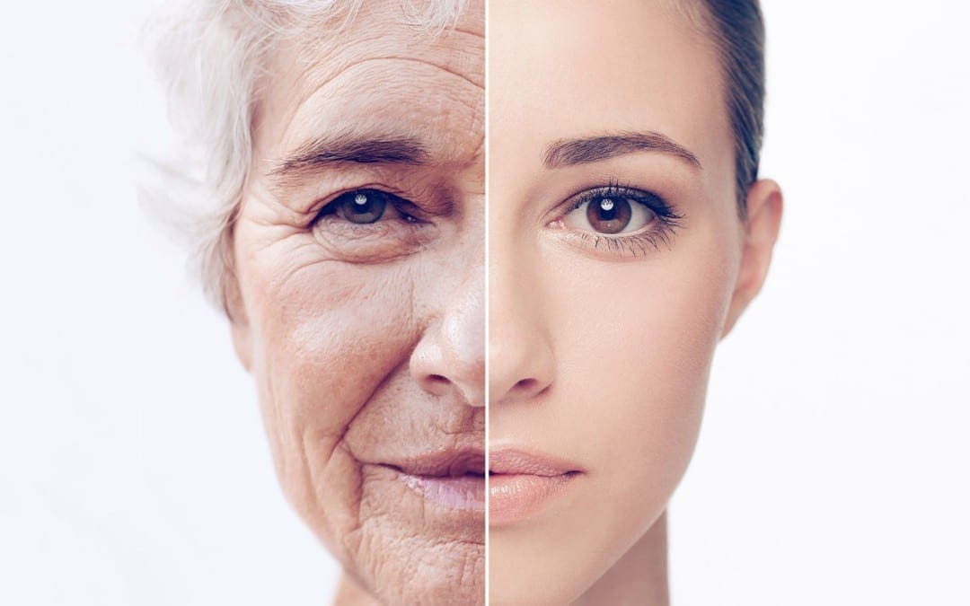
Aging is a natural part of life and it can’t be stopped. Or at least, that’s what we used to think. Researchers at Intervene Immune, Stanford, the University of British Columbia, and UCLA believe that our epigenetic clock can be changed, suggesting that there may still be ways for humans to live longer. In the following article, we will discuss the findings associated with epigenetics and aging.
Contents
What is the Epigenetic Clock?
The epigenetic clock is a measurement of biological age that can be used to estimate the chronological age of humans or other organisms by testing several patterns of DNA methylation. Although the age estimated by the epigenetic clock frequently correlates with chronological age, it is not fully understood if DNA methylation profiles in the epigenetic clock are directly associated with aging.
For many years, researchers have observed age-related changes in gene expression and DNA methylation. However, the idea of using an “epigenetic clock” to be able to estimate chronological age by testing several patterns of DNA methylation was first proposed by Steve Horvath where it gained popularity after his 2013 research study was published in the journal Genome Biology.
Epigenetic clocks are used in forensic studies to determine the age of an unknown person through blood or other biological samples at the scene of a crime and in diagnostic screens to determine increased risks for diseases associated with aging, including a variety of cancers. Epigenetic clocks can also highlight whether several behaviors or treatments can affect epigenetic age.
Does Epigenetic Age Correlate with Chronological Age?
The main reason that epigenetic clocks and DNA methylation are used to estimate the chronological age of humans or other organisms is that they correlate very well with the chronological age in the subjects tested. The first research study on the epigenetic clock that Steve Horvath published in 2013 included 353 individual CpG sites identified from previous research studies.
Of these sites, 193 become more methylated with age and 160 become less methylated, which leads to the DNA methylation age estimate that is used to determine the epigenetic clock. Throughout all outcome measures, including all ages of subjects, Horvath observed a 0.96 correlation between the epigenetic age he calculated and the true chronological age, with an error rate of 3.6 years.
Current epigenetic clocks are also being evaluated to help further improve age prediction as well as the diagnostic and/or prognostic abilities of these tests. Further evaluations using NGS approaches ultimately have the potential to improve epigenetic clocks, making them more comprehensive by extending the evaluation of DNA methylation sites to all CpG sites in the genome.
Can We Change Our Epigenetic Clocks?
Research studies have demonstrated that cancer can change the epigenetic clock. These observations suggest that the epigenetic clock can change under certain conditions. Therefore, it is possible that the epigenetic clock can be manipulated through changes in behavior or treatment strategies to slow it down or potentially reverse it, allowing humans to live longer and healthier lives.
Researchers believe that our epigenetic clock can be changed. In the following article, we discussed the findings associated with epigenetics and aging. The epigenetic clock is a measurement of biological age that can be used to estimate the chronological age of humans or other organisms by testing several patterns of DNA methylation. The main reason that epigenetic clocks and DNA methylation are used to estimate the chronological age of humans or other organisms is that they correlate very well with the chronological age in the subjects tested. Current epigenetic clocks are also being evaluated to help further improve age prediction as well as the diagnostic and/or prognostic abilities of these tests. Research studies have demonstrated that cancer can change the epigenetic clock. Therefore, it is possible that the epigenetic clock can be manipulated through changes in behavior or treatment strategies to slow it down or potentially reverse it, allowing humans to live longer and healthier lives. By changing our epigenetic clocks, healthcare professionals may also be able to regulate age-related health issues, such as inflammation and joint pain. These could potentially be helpful for chiropractic care, an alternative treatment option that uses spinal adjustments to carefully restore the alignment of the spine.�- Dr. Alex Jimenez D.C., C.C.S.T. Insight
Zesty Beet Juice
Servings: 1Cook time: 5-10 minutes
� 1 grapefruit, peeled and sliced
� 1 apple, washed and sliced
� 1 whole beet, and leaves if you have them, washed and sliced
� 1-inch knob of ginger, rinsed, peeled and chopped
Juice all ingredients in a high-quality juicer. Best served immediately.
Just one carrot gives you all of your daily vitamin A intake
Yes, eating just one boiled 80g (2�oz) carrot gives you enough beta carotene for your body to produce 1,480 micrograms (mcg) of vitamin A (necessary for skin cell renewal). That’s more than the recommended daily intake of vitamin A in the United States, which is about 900mcg. It’s best to eat carrots cooked, as this softens the cell walls allowing more beta carotene to be absorbed. Adding healthier foods into your diet is a great way to improve your overall health.
The scope of our information is limited to chiropractic, musculoskeletal, physical medicines, wellness, and sensitive health issues and/or functional medicine articles, topics, and discussions. We use functional health & wellness protocols to treat and support care for injuries or disorders of the musculoskeletal system. Our posts, topics, subjects, and insights cover clinical matters, issues, and topics that relate and support directly or indirectly our clinical scope of practice.* Our office has made a reasonable attempt to provide supportive citations and has identified the relevant research study or studies supporting our posts. We also make copies of supporting research studies available to the board and or the public upon request. We understand that we cover matters that require an additional explanation as to how it may assist in a particular care plan or treatment protocol; therefore, to further discuss the subject matter above, please feel free to ask Dr. Alex Jimenez or contact us at 915-850-0900. The provider(s) Licensed in Texas*& New Mexico*�
Curated by Dr. Alex Jimenez D.C., C.C.S.T.
References:
Active Motif Staff. �Can You Really Reverse Your Epigenetic Age?� Active Motif, 1 Oct. 2019, www.activemotif.com/blog-reversing-epigenetic-age#:~:text=Epigenetic%20clocks%20are%20a%20measure,certain%20patterns%20of%20DNA%20methylation.
Pal, Sangita, and Jessica K Tyler. �Epigenetics and Aging.� Science Advances, American Association for the Advancement of Science, 29 July 2016, www.ncbi.nlm.nih.gov/pmc/articles/PMC4966880/.
Matloff, Ellen. �Mirror, Mirror, On The Wall: The Epigenetics Of Aging.� Forbes, Forbes Magazine, 25 Jan. 2020, www.forbes.com/sites/ellenmatloff/2020/01/24/mirror-mirror-on-the-wall-the-epigenetics-of-aging/#75af95734033.
Dowden, Angela. �Coffee Is a Fruit and Other Unbelievably True Food Facts.� MSN Lifestyle, 4 June 2020, www.msn.com/en-us/foodanddrink/did-you-know/coffee-is-a-fruit-and-other-unbelievably-true-food-facts/ss-BB152Q5q?li=BBnb7Kz&ocid=mailsignout#image=24.
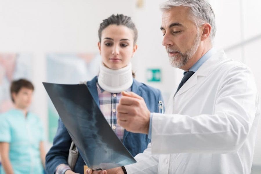
Getting an accurate chronic pain diagnosis is essential to creating the most optimal, highly customized treatment plan for the individual. Depending on the severity and cause of pain, individuals could require various pain specialists/therapists combined with a primary physician. These could include:
Chiropractor
Physical therapist
Neurosurgeon
Pain medicine specialist
Physiatrist
Rheumatologist
Orthopedic spine surgeon
Contents
Chronic Pain Diagnosis
�
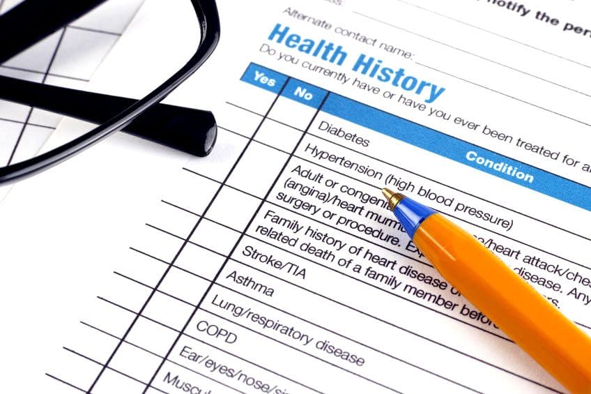
� Over time chronic pain symptoms can change or alter and need reevaluation. This could mean having to adjust treatment and management but that is exactly what it is, an adjustment to the treatment plan flowing with the symptoms as they come and go while keeping to the objective of. Chronic pain diagnosis entails a series of tests, as well as, a full review of symptoms and medical history. A doctor will ask a series of questions concerning symptoms and pain triggers. These questions could include:
When did the pain begin?
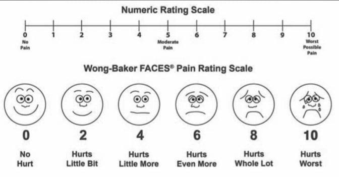
Describe and rate the pain, is it shooting, electrical, burning, throbbing, dull, or sharp?
�
Has there ever been an injury at or around the problem area?
What activities/actions/movements relieve and worsen the pain?
Is there a history of mental illness, like depression or anxiety?
�
Labs
Tests will be ordered to identify physical/non-physical causes that could be the cause or contributor. Possible tests include:
�
Blood
Blood tests are used in the diagnosis of infections and inflammation. Individuals with infection/s or inflammatory disorders have high levels of white blood cells and inflammatory reactive substances like C-reactive protein. Blood tests also help determine the presence of rheumatoid arthritis, gout, or cancer. If rheumatoid arthritis is present, the blood analysis will show positive results for proteins known as rheumatoid factor.
�
Urine
Urinalysis is commonly used to check for gout. This is a type of arthritis that causes high blood levels of uric acid. A doctor may order a urine test for a patient using prescription pain meds.
�
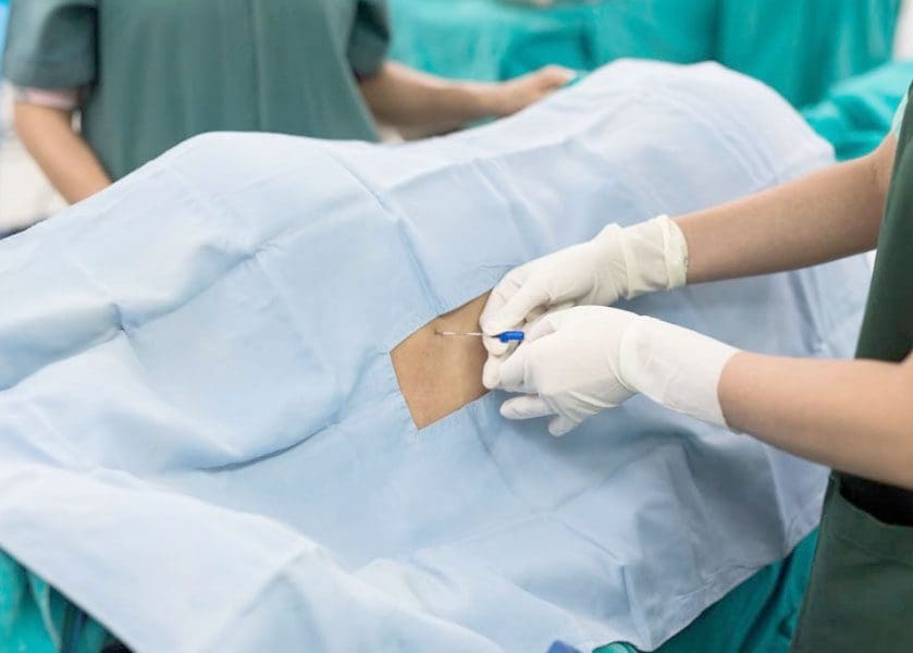
Spinal tap
A doctor inserts a needle into the lower back and a sample of cerebrospinal fluid is collected. Cerebrospinal fluid is clear and protects the brain and spinal cord. A cerebrospinal fluid analysis helps to diagnose disorders of the central nervous system and certain cancers. �
�
Musculoskeletal/Neurological tests
A musculoskeletal exam looks at posture, joint mobility, muscle stiffness, tightness, and swelling in or around the area, as well as the rest of the body. An example is a diagnosis of carpal tunnel syndrome. A detailed spine examination is done to identify deformities and moving/walking posture. A neurological examination is used to check:
Muscle strength
Touch reaction
Balance
Overall sensation
A neurological exam can also be used to test:
Memory
Alertness
Mood
Behavior
�
Imaging
Imaging provides detailed images of the body’s organs and bones. Doctors use these to:
Spot fractures or inflammatory alterations in the bone/s
Focus on details of a bone and surrounding structures
Differentiate between growths, infections, or fractures
Identify nerve/s injury or damage
�
X-Rays
X-rays are standard in the diagnosis of fractures. An arthrogram is an x-ray that uses a contrasting agent to check and identify joint disorders.
�
MRI
Magnetic resonance imaging uses a magnetic field and radio waves to create detailed images. Magnetic resonance imaging helps in diagnosing:
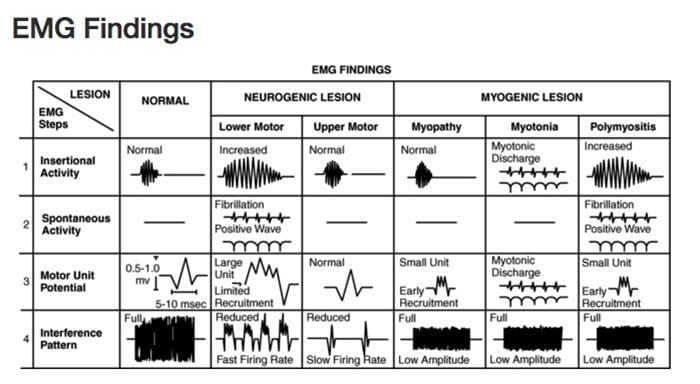
EMG’s are used to diagnose disorders of the muscles and nerves. Electrical activity in the muscles is recorded to see how the impulses/electrical signals are transmitting from the nerves to muscles. �
�
An EMG could be required if an individual has:
Numbness
Muscle weakness
Muscle pain
Tics
Electromyography is also used to identify conditions that can cause chronic pain like:
A nerve conduction study measures the speed of electrical signals passing through a nerve. It can identify:
Carpal tunnel syndrome
Herniated disk disease
Sciatic nerve injury/damage/abnormality
A doctor can order both an EMG and NCS in combination.
�
Back Pain Chiropractic Care
Dr. Alex Jimenez�s Blog Post Disclaimer
The scope of our information is limited to chiropractic, musculoskeletal, physical medicines, wellness, and sensitive health issues and/or functional medicine articles, topics, and discussions. We use functional health & wellness protocols to treat and support care for injuries or disorders of the musculoskeletal system. Our posts, topics, subjects, and insights cover clinical matters, issues, and topics that relate and support directly or indirectly our clinical scope of practice.*
Our office has made a reasonable attempt to provide supportive citations and has identified the relevant research study or studies supporting our posts. We also make copies of supporting research studies available to the board and or the public upon request. We understand that we cover matters that require an additional explanation as to how it may assist in a particular care plan or treatment protocol; therefore, to further discuss the subject matter above, please feel free to ask Dr. Alex Jimenez or contact us at 915-850-0900. The provider(s) Licensed in Texas& New Mexico*
Research studies have demonstrated the fundamental role of nutrition in health and longevity. The standard American diet, which is generally high in fat and sugar, has been associated with a variety of health issues, including obesity, high cholesterol, hypertension, and type 2 diabetes. Moreover, these health issues can lead to kidney disease, heart disease, Alzheimer’s disease, and cancer. �Unfortunately, the type 2 diabetes curve is going in the wrong direction, and we�re living longer as well,� stated Gary Gibbons, director of the National Heart, Lung, and Blood Institute. �So we have an aging population that�s more and more obese, and has more and more hypertension.� In the following article, we will discuss the effects of good nutrition on overall health, wellness, and longevity.
A healthy diet ultimately includes:
Fruits and vegetables
Low-fat dairy products, such as yogurt and cheese
Skinless poultry
Salmon and other fish, such as trout and herring
Nuts and beans
Whole grains
Non-tropical vegetable oils, such as olive, corn, peanut, and safflower oils
Contents
Calorie Restriction and Longevity
According to several research studies, nutrition, and specifically restricting calories, has been associated with aging itself. In the 1930s, research studies in a wide variety of research models, including yeast, drosophila and c. elegans (laboratory fruit flies and nematodes), rats, and inbred mice, demonstrated a connection between a limited-calorie diet and extended life span. Researchers today are starting to take these research studies to the next level by evaluating how different individuals respond to different calorie intakes in order to demonstrate the physiological and genetic variations associated with health and longevity. However, because it’s difficult for humans to follow any type of calorie-restricted diet, it’s impossible to determine lifelong results and further research studies are still required.
On the other hand, mice can ultimately provide further evidence due to their significantly short life span (average two years), as well as due to the ability to control every aspect of their laboratory environment, including diet. JAX Professor Gary Churchill�is one of the architects of a special type of mouse colony known as Diversity Outbred (DO). As a result of the careful, cross-breeding of genetically defined inbred strains, these mice demonstrate the type of random-looking genetic variation you�d find in the general human population. �Several calorie-restricted mice in the DO population have lived incredibly long life spans,� stated Churchill, �several have even reached almost five years of age,� which is the equivalent of a human living about 160 years, according to research studies.
Churchill has also separated DO mice into several groups given different diets and calorie restrictions throughout their life span. Control animals are typically on an ad libitum (�all-you-can-eat�) diet. Several mice are given food daily but at a reduced amount. Fasting animals are given food ad libitum on most days but spend a period of time each week with no food access. All mice receive frequent and extensive physical evaluations to collect data that can later be associated with how long they live. And, because the genomic sequence of every mouse is well-known, overlaying the physiological data can ultimately help provide further unprecedented insights into the genetic impact of nutrition, diet, and calorie restriction on overall health, wellness, and longevity, among further evidence.
�Although it is understood that several animal models, like the inbred C57BL6/J mouse strain, can benefit from caloric restriction, there is also evidence which demonstrates that the effects can be different depending on the genetic makeup of the animal,� stated Churchill. �The same will probably be true for most people: caloric restriction may be beneficial for one person but not for another. Until researchers understand these individual differences, healthcare professionals must be very cautious about recommending nutritional and dietary changes to people.� Understanding how nutrition affects the genetic components of health and longevity can eventually lead to treatments that may ultimately help reverse the negative effects of poor nutrition, including health issues like heart disease and diabetes.
Research studies have found the important role of nutrition in longevity. The standard American diet, which is high in fat and sugar, is associated with many health issues, including obesity and type 2 diabetes which may lead to heart disease, Alzheimer’s disease, and even cancer. Furthermore, several research studies have also found that nutrition, and specifically calorie restriction, is associated with aging. In the article above, we discussed the evidence showing the effects of good nutrition on health and longevity. – Dr. Alex Jimenez D.C., C.C.S.T. Insight
Zesty Beet Juice
Servings: 1Cook time: 5-10 minutes
� 1 grapefruit, peeled and sliced
� 1 apple, washed and sliced
� 1 whole beet, and leaves if you have them, washed and sliced
� 1-inch knob of ginger, rinsed, peeled and chopped
Juice all ingredients in a high-quality juicer. Best served immediately.
Just one carrot gives you all of your daily vitamin A intake
Yes, eating just one boiled 80g (2�oz) carrot gives you enough beta carotene for your body to produce 1,480 micrograms (mcg) of vitamin A (necessary for skin cell renewal). That’s more than the recommended daily intake of vitamin A in the United States, which is about 900mcg. It’s best to eat carrots cooked, as this softens the cell walls allowing more beta carotene to be absorbed. Adding healthier foods into your diet is a great way to improve your overall health.
The scope of our information is limited to chiropractic, musculoskeletal, physical medicines, wellness, and sensitive health issues and/or functional medicine articles, topics, and discussions. We use functional health & wellness protocols to treat and support care for injuries or disorders of the musculoskeletal system. Our posts, topics, subjects, and insights cover clinical matters, issues, and topics that relate and support directly or indirectly our clinical scope of practice.* Our office has made a reasonable attempt to provide supportive citations and has identified the relevant research study or studies supporting our posts. We also make copies of supporting research studies available to the board and or the public upon request. We understand that we cover matters that require an additional explanation as to how it may assist in a particular care plan or treatment protocol; therefore, to further discuss the subject matter above, please feel free to ask Dr. Alex Jimenez or contact us at 915-850-0900. The provider(s) Licensed in Texas*& New Mexico*�
Curated by Dr. Alex Jimenez D.C., C.C.S.T.
References:
Peterson, Joyce Dall’Acqua. �Exploring the Diet-Life Span Connection.� The Jackson Laboratory, 15 Nov. 2017, www.jax.org/news-and-insights/2017/november/diet-and-longevity#.
Donovan, John. �Eating for Longevity: Foods for a Long, Healthy Life.� WebMD, WebMD, 13 Sept. 2017, www.webmd.com/healthy-aging/features/longevity-foods#1.
Fontana, Luigi, and Linda Partridge. �Promoting Health and Longevity through Diet: From Model Organisms to Humans.� Cell, U.S. National Library of Medicine, 26 Mar. 2015, www.ncbi.nlm.nih.gov/pmc/articles/PMC4547605/.
Dowden, Angela. �Coffee Is a Fruit and Other Unbelievably True Food Facts.� MSN Lifestyle, 4 June 2020, www.msn.com/en-us/foodanddrink/did-you-know/coffee-is-a-fruit-and-other-unbelievably-true-food-facts/ss-BB152Q5q?li=BBnb7Kz&ocid=mailsignout#image=24.
Anybody can have chronic pain. Adults typically complain of joint pain, low back pain, and neurogenic pain. While children and teenagers are more likely to have more headaches, abdominal pain, leg, and hand pain. Regardless there are individuals that have a higher risk because of their age, gender, and job. It isn’t always clear what causes chronic pain. There are several possibilities: �
�
Injury – Even after the injury has healed, the nerves keep sending pain signals to and from the brain. Doctors are still not sure why this occurs.
Disease – Conditions can cause chronic pain like fibromyalgia and osteoarthritis.
Nerve problems – Part of the nervous system can be injured, the nerves themselves. This is called neuropathic pain.
Unknown Cause/s – Pain that presents with no obvious injury, disease, or nerve problem.
Contents
�
Military Veterans
Chronic pain is quite common in veterans according to a National Veterans Affairs Study. Around one in five veterans receiving primary care have chronic pain. While one in ten has chronic pain syndrome. Veterans recently served in a war, tend to report a variety of causes for their pain. This includes:
Multiple injuries
Brain trauma
Muscle injuries
Bone/s injuries
�
Athletes
Most sports require a certain level of fitness. Athletes train with all types of activities to help maintain their body’s. Unfortunately, they are still not immune to chronic pain. Chronic pain is common with:
Spinal stenosis is a narrowed spinal canal, which creates added pressure on the nerves that travel through the low spine into the legs
�
Seniors
Age is a high-risk factor for chronic pain. Around 30-40% of individuals older than sixty-five have or are beginning to deal with chronic pain. The severity in anybody forty-five to sixty-five is the greatest. Common conditions that cause chronic pain in older adults are:
Cancer
Arthritis and gout
Heart disease
Kidney disease
Damaged nerves
Stroke
Shingles
�
Women
Men and women experience pain differently. Several factors contribute to this. These include:
Hormones
Menstruation
Puberty
Reproductive health
Women have a higher risk of developing disorders that cause chronic pain. Examples include:
Arthritis
Brittle bones
Migraines
Irritable bowel syndrome
�
Anybody dealing with chronic pain, finding relief can be difficult and time-consuming. Individuals are often sent back and forth between primary care, specialists, and therapists for a solution.
�
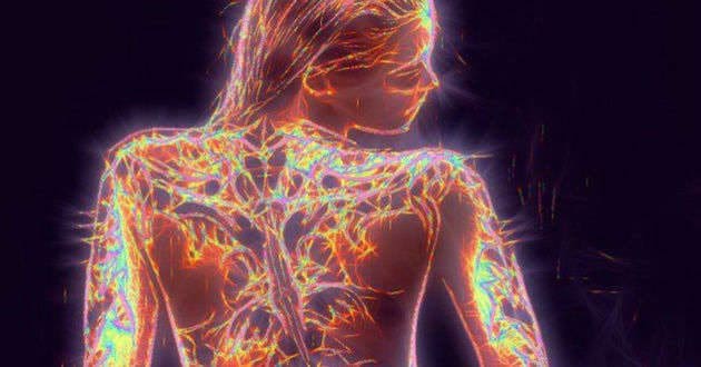
Improved Nervous System
When there is a communication breakdown between the brain and the body�s tissues, organs, and cells it can lead to a variety of health problems. There are manychronic and even degenerative health conditions that are impacted by the nervous system. Studies have shown that chiropractic is a highly effective treatment for numerous neurological conditions which include:
Ataxia
Autism
Cerebral palsy
Epilepsy
Multiple sclerosis
Parkinson�s
Tourette�s Syndrome
Vertigo
Anybody can seek treatment and they will experience the benefits. Chiropractic provides a safe, effective treatment for an improved nervous system function. The type, frequency, and intensity of treatment depend on the patient and condition. Chiropractic positively affects the nervous system and as a result, positively affects the whole body.
�
Chronic Body Pain Treatment
Dr. Alex Jimenez�s Blog Post Disclaimer
The scope of our information is limited to chiropractic, musculoskeletal, physical medicines, wellness, and sensitive health issues and/or functional medicine articles, topics, and discussions. We use functional health & wellness protocols to treat and support care for injuries or disorders of the musculoskeletal system. Our posts, topics, subjects, and insights cover clinical matters, issues, and topics that relate and support directly or indirectly our clinical scope of practice.*
Our office has made a reasonable attempt to provide supportive citations and has identified the relevant research study or studies supporting our posts. We also make copies of supporting research studies available to the board and or the public upon request. We understand that we cover matters that require an additional explanation as to how it may assist in a particular care plan or treatment protocol; therefore, to further discuss the subject matter above, please feel free to ask Dr. Alex Jimenez or contact us at 915-850-0900. The provider(s) Licensed in Texas& New Mexico*
Several conditions and factors can cause chronic pain. Usually, these are conditions that accompany normal aging, which affect bones and joints. The top three are osteoarthritis, rheumatoid arthritis, and fibromyalgia. Other common causes are nerve damage and injuries that fail to heal properly.
Contents
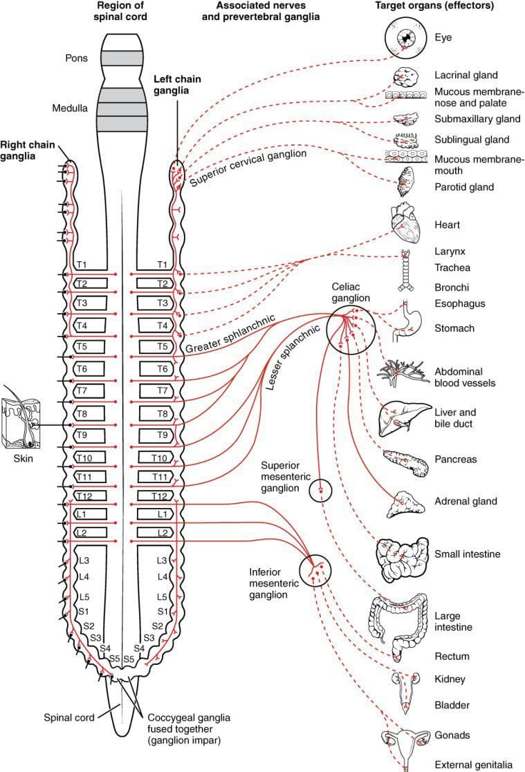
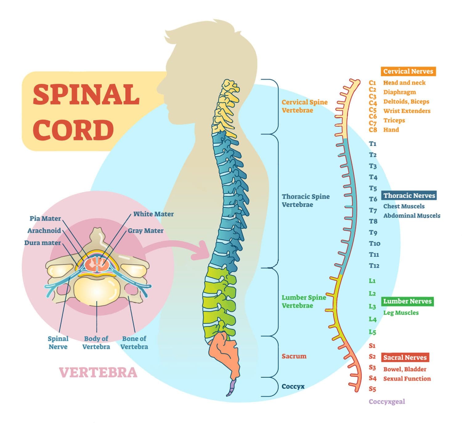
Spinal Cord and the Nerves of the Corresponding Organs
�
Fibromyalgia
Individuals with fibromyalgia experience unexplained pain in almost every part of their bodies. Doctors and scientists are still trying to figure out what causes fibromyalgia. Currently, scientists think a part of the condition comes from an imbalance of certain chemicals in the brain. They believe the imbalances play a critical role. Fibromyalgia can create:
Tender areas
Muscle pain
Headaches
Long-term back pain
Long-term neck pain
�
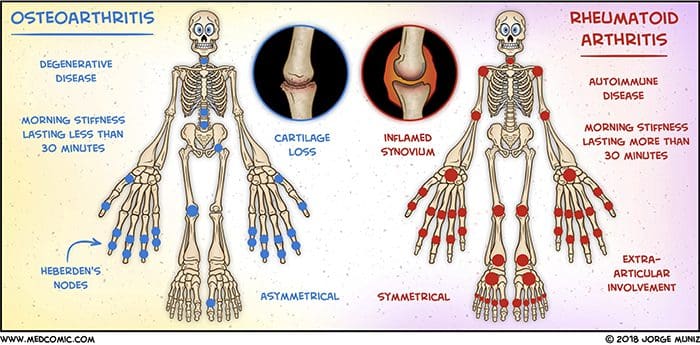
Osteoarthritis
Osteoarthritis causes severe sporadic or non-stop aches and pain in the knees, hips, spine, and feet. Associated symptoms include joint stiffness, swelling, and limited joint mobility. Individuals with osteoarthritis could have some pain throughout their lives. According to the CDC, around fifteen million adults with arthritis have severe pain in their joints. �
�
Rheumatoid Arthritis
Rheumatoid arthritiscauses continual aching that affects multiple joints. The hands, wrists, and knees are the most affected joints. Individuals with rheumatoid arthritis can present alternate symptoms, like joint stiffness, swelling, and fever.
�
Multiple Sclerosis
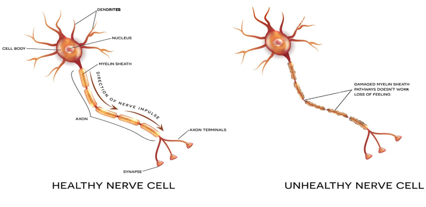
Multiple sclerosis is a disease of the brain and the spinal cord. What happens is the immune system targets and damages the protective covering of the nerves themselves. The brain can’t properly and effectively communicate with the body. Multiple sclerosis causes pain in the legs, feet, arms, and hands. Associated symptoms include burning, prickling, or stabbing pain just about every day. �
�
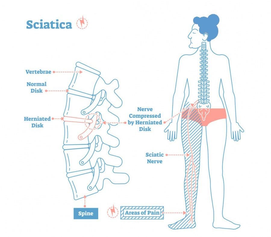
Sciatica
Sciatica can cause mild to sharp, electrical burning pain that travels from the lower back through the buttocks to the back of the leg and even into the foot. Chronic sciatica lasts for three months or more. The condition is more common in adults age 40 and older.
�
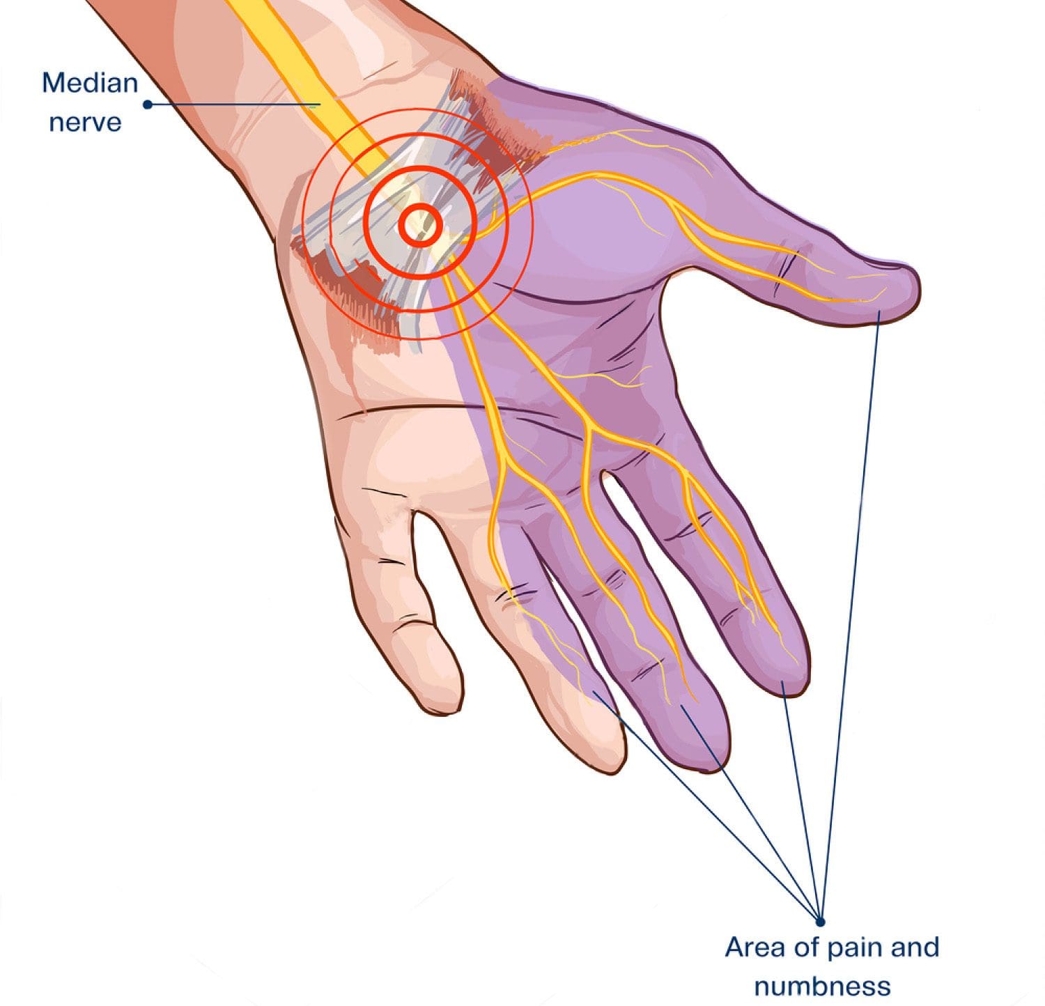
Carpal Tunnel Syndrome
Carpal tunnel syndrome causes pain and numbness in the:
Around half of the cases involving chronic pain are linked to physical trauma and injury. Individuals hospitalized after a serious injury often report chronic pain symptoms within the first year. Scientists are still unsure of how injuries lead to chronic pain. They believe several factors increase the risk. These include:
Pre-injury depression
Anxiety
Alcohol use
Family history of chronic pain
Individuals that have sustained multiple injuries are at higher risk for chronic pain. �
�
Spinal Injuries
One of the most common causes of chronic back pain. The lower back is the area likely to be affected. Certain types of chronic pain can have more than one cause. For example, general back pain could be caused by a single factor or a combination of factors like:
Poor posture
Improper lifting of heavy objects
Improper carrying of heavy objects
Being overweight places added strain on the back and knees
Abnormal curvature of the spine
Wearing high heels too often
Sleeping on a worn-out mattress
Degenerative disc changes
�
Combat Injuries
More than half of combat-related injuries are the result of explosions, from landmines, and shrapnel. Nearly all injured soldiers have to deal with some type of pain and many have a traumatic brain injury. A traumatic brain injury can cause chronic headaches. Delayed treatment and repeated injuries in injured soldiers make up for most chronic pain cases. �
�
Sports Injuries
Sports injuries and chronic pain is nothing new. Studies found that 1 in 2 football players deal with chronic pain in their retirement. This along with sleep problems and mild-severe depression. Both can contribute to chronic pain. Athletes are continuously exposed to high-risk injury situations. Having the pressure of performing optimally and winning can take a toll on an athlete’s health. �
�
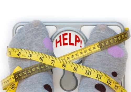
Weight
Obesity does not directly cause chronic pain, but it does raise the risk. Around 40% of individuals that are obese also experience mild to severe chronic pain. Plus, individuals that are severely overweight are more likely to develop a condition that can cause chronic pain like diabetes, arthritis, and fibromyalgia. �
�
The source of chronic pain can be very complex. It can start with an injury or illness and develop slowly without the individual realizing it until it has become a full-blown chronic condition. This fact alone makes recommending a single course of treatment risky and is why health care providers recommend a number of different types of treatment options.
Chiropractic Care on Personal Injury
Dr. Alex Jimenez�s Blog Post Disclaimer
The scope of our information is limited to chiropractic, musculoskeletal, physical medicines, wellness, and sensitive health issues and/or functional medicine articles, topics, and discussions. We use functional health & wellness protocols to treat and support care for injuries or disorders of the musculoskeletal system. Our posts, topics, subjects, and insights cover clinical matters, issues, and topics that relate and support directly or indirectly our clinical scope of practice.*
Our office has made a reasonable attempt to provide supportive citations and has identified the relevant research study or studies supporting our posts. We also make copies of supporting research studies available to the board and or the public upon request. We understand that we cover matters that require an additional explanation as to how it may assist in a particular care plan or treatment protocol; therefore, to further discuss the subject matter above, please feel free to ask Dr. Alex Jimenez or contact us at 915-850-0900. The provider(s) Licensed in Texas& New Mexico*
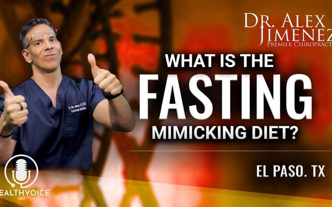
[embedyt] https://www.youtube.com/watch?v=P5joK7TqIok[/embedyt]
PODCAST: Dr. Alex Jimenez and Kenna Vaughn introduce Sonja Schoonenberg to discuss epigenetics and nutrition. Our diet can affect our gene expression. Therefore, eating unhealthy foods can ultimately increase our predisposition to develop a variety of health issues, such as diabetes, stroke, and cardiovascular disease. Sonja Schoonenbert describes the benefits of fasting and how the Regenerate program can help provide people with similar benefits to fasting in order to promote overall health and wellness. The purpose of the following podcast is to emphasize the connection between dietary changes and gene expression as well as focus on natural regenerative treatment protocols. – Podcast Insight
[00:06:16] We’re all excited today. We’ve got an unbelievable guest to discuss some really great technologies that are out there, as you guys know. [00:06:41][24.8][00:06:41] El Paso is my home and Kenna’s. And for all our patients, we try to bring in different technologies out there that are informative, that help us with our health design. Today, we got a very, very special guest, Sonja Schoonenberg. Who comes to us from a good distance and is gonna be discussing today regenerative or regenerate in its theories and its ideas behind an amazingly complete package of health products that assist us to have more personalized medicine. Personalized wellness is the new mantra of many of the health providers. We understand that health care is not a process that can just be, you know, one size fits all. This particular company that I’m very much proud to be part of is really interesting in its design because it takes all the way from what our philosophy, as you guys know, is from the kitchen to the genes, vice versa, and specifically formulates a treatment, not a treatment protocol, but a regimen that is focused on making all the best protocols that assist an individual based on their particular genetics. And then what it does, it follows through with a good dietary adaptive process that finds holes and vulnerabilities in our metabolic processes that can assist us. So what I’d like to do is I’d like to introduce her and Kenna is here, she’s my super-duper health coach. And so as we go through this process, like you guys, you’ll see the number pop up every so often. Give us a call so we can talk about this particular wellness solution. Sonja, tell us a little bit about the product because we’re a little excited to hear from you. [00:08:51][129.1][00:08:53] I am happy to. I’m so excited to be here, so thank you for inviting me today and I am really excited about how the evolution of health care is changing. So I thought I’d kind of dive into my background a little bit and why I’m excited to be here because I think that’ll help to bring it all full circle. So I am a registered dietitian, nutritionist, and certified diabetes educator by trade. And I’ve also been involved not only in the pharmaceutical, but the nutraceutical experience. And I’m now the V.P. of product experience for regenerate. And what really led me down this road of personalized wellness, personalized nutrition, personalized medicine, was looking at the individual and realizing that there were so many differences between people and results they would get from different programs, myself included. And I’ll share my story a little bit further in this journey. But what I saw was a huge gap in outcomes for patients. And so when I learned more and more about epigenetics and customized wellness, customized nutrition, it really resonated with me. And I saw this as a solution to really fill many of the gaps that patients were seeing today. So that’s a little bit about my background and how I got here. What I’d like to do is take you all through a journey of regenerate. We have a very, very simple system that is something that I think you’ll find is very, very unique and different than everything out there. So it all starts with what we refer to as our epigenetic biomarker profile test. So without getting too complicated, really what this is, is an at-home test kit. That is a first-morning void urine sample that allows us to really look upstream within the body to see metabolically what is happening epigenetically, how our genes are behaving so that we can make some realistic changes in terms of a lifestyle plan. So the epigenetic biomarker profile test, when it’s done at home. Once that occurs and the sample is set off, sent off to the lab, we actually create for each individual an approximately 19-page custom nutrition report. And here’s what the test is looking at. And as a result, the report that comes reflects these different things. And these are very, very key areas as it relates to health, wellness, and nutrition. So we look at B vitamins and methylation co-factor. [00:11:33][159.5][00:11:34] And when you read anything these days, you hear all about methylation and people that are poor methylators. And, you know, there’s all kinds of buzz around that word methylation, although most people don’t fully understand what it means. So we expose what’s going on and really look at that area as something that if we can correct those nutritional imbalances, a person can really have optimal health. We also look at cellular energy assessment or the health of the mitochondria. So the little cellular powerhouses that give us energy, we look at muscle assessment and gut assessment. This is a big one, of course, as we know that much of our immune system in many of the things that we experience health-wise are somewhat related to what’s going on in our gut. And I know we had this conversation the other day. Yes. It’s so important to look at what’s going on inside of the gut. We look at amino acids and inflammation and oxidative stress. So these are what I refer to as kind of the launching pad of things that we can expose. Peek behind the curtain and see what’s happening with this individual and then be able to formulate some solutions based on that. So I know when we were talking the other day, you got really excited when you saw how much information that this test actually provided. [00:12:56][82.1][00:12:57] I got to tell you, one of the things that as you delve in this and you become really into it, typically we have a lot of mothers, wives that really do a lot of research and you get peace meals somewhere down the road. It becomes absolutely frustrating because you try to figure out the full whole process yourself. [00:13:19][22.0][00:13:19] What I was very amazed about Regenerate was that it really puts the whole thing together in the sense of from understanding the deep, real, real benefits of the genome all the way to the packaging of nutraceuticals and nutrition all into one, but really can explain that whole process. So when I saw it, I was excited. Kenna and I were going like, this is really failing a monster void because of the lack of organized information that’s out there. So when we see this in the visual. We knew about the mitochondria. I understood about the mitochondria back in the 80s. I knew that it was a powerhouse, but we didn’t realize that it was so connected to so many pathologies. We knew that the gut had a lot to do with many issues. Now we know that the gut is one of the very first places we look for diseases and disorders. And in the world of functional medicine, this is what we talk about. And putting this together to make sense for our patients was really complex. This is awesome because it allows us to simplify the dynamics and really make it really consumable and effective for our patients. [00:14:29][70.4][00:14:30] So, yeah, we were very excited. And that you hit on something that was so key. We knew about these things years ago. But now, regardless of where you turn around and look, this information is everywhere. Yet there’s so much confusion around what to do with the information. So we try to really make it simple. But really, the whole premise behind doing that test is to give an individual what we refer to as a nutrition prescription or a road map to the nutrition prescription for what we refer to as our Optim8, whole food plant-based organic nutrition. So I want to stop here for a moment and just talk a little bit about what this is and what this is not? There are so many supplements out there and there are a lot of good ones. But most of them are made in a laboratory from some form of a synthetic ingredient. There are combinations and, of course, singular nutrients. There are all types of things out there. But when we get back to the roots of who we are on the inside and what our medical care as it relates to nutrition should come from, that’s food. Unfortunately, our food supply, we know today is a real problem. It’s malnourished when we have malnourished food. We have malnourished humans. And what I loved most about this particular product is it’s a bioactive superfood. So it’s not truly a supplement. If you think about it as the perfect food for every individual. [00:15:59][89.3][00:16:01] Sonja, if I may. Because that’s a really good point. What does bioactive in? It’s used a lot and versus non-bioactive. What does it mean when you hear that word bioactive, what does that represent? [00:16:15][14.0][00:16:17] So in this case, we’re talking about live active superfoods. So foods that are truly foods, essentially everything in that green powder. Comes from food that goes from goes through very minimal processing and let me explain what that looks like. We have a partnership with a cooperative farm in central Florida called Grow More Farms. They’re based in Indian town, Florida. [00:16:43][26.4][00:17:03] But these soil conditions in Florida are really optimal for growing year-round. And what’s interesting about the farm that we have a partnership with more farms is that they were soil farmers before they got into formulating nutraceutical products. And what they’ve really done is they’ve crafted the healthiest soil conditions for each and every plant. Which means that if you have healthy soil and then you grow, let’s say, asparagus or cucumbers or tomatoes, whatever it is that you’re growing, if it’s grown in optimal conditions and optimal soil, you have much healthier food. And so the only processing the Optim8 custom superfood goes through is the plants are grown to their peak ripeness picked at their peak ripeness. They go through a dehydration process at a very low temperature and then they’re ground into a fine powder, which then becomes the basis for our custom superfood. Now, this food is actually if you think about going back to the task we were just talking about, that test gives us essentially what’s off-balance with an individual’s nutritional health. So anything that is rated on a score of fair or poor would be things that that individual really has some imbalances in. And so what we want to do, based on the findings of that report, is to formulate this custom nutrition with foods. And there’s the key foods that are high in those nutrients that an individual needs. So instead of a supplement pack containing a bunch of supplements that, you know, meet the individual’s needs, these are actually foods that are high concentrations of the nutrients that an individual needs. So it’s a really simple thing you can use every single day. You can put it just in a shaker cup. I actually have it right here in a shaker cup, blend it up, and drink it in water. You can mash it an avocado, put it in a protein shake, and you only have to take it once a day. So as a dietitian, that was music to my ears that I didn’t have to remind people to take things 20 times a day or at like me, at one point I was taking 30 different supplements. What’s your outlook on that? [00:19:22][139.5][00:19:23] My outlook on that is, first of all, let’s go with two points, one you’re in Florida and we live out here in El Paso and El Paso has got great chiles. We got great different foods in this area. But if you’ve ever gone to Florida and just using the example of an avocado. OK. The avocados from Florida, I am not kidding you. These things are like this big. Right. The ones where we are we’ve got these really itty bitty ones that are really dense. Now, avocado is a superfood. [00:19:51][27.9][00:19:52] But my point being is that the soil in Florida is so optimal in terms of what it provides from the earth. So I can see why the first of all, the farm would be there. And if these guys and these specialists that work on soils actually understand that that is an amazing thing to bring the product out as well as the byproduct, which is the vegetable or the food. The second point is that the missing of the components and being able to get the foods bio actively active inside of humans when we are actually showing deficiency, that is huge. And from my point of view is we’re full of a lot of different nutraceuticals. And when you go to a place like GNC or a vitamin X store, you know, you have a wall of stuff. You can’t be taking everything and you don’t know where the hole is. Right. So that’s where personalized medicine is taking a spin. You don’t have to spend a hundred dollars on stuff that you may not even need or some people say, you know, expensive urine. But what we have to do is we have to find the holes in our particular diet and not only in our particular diet but in our genetic predispositions, too. So putting that together is huge. So when you ask me, I’m there with you. I totally get it. And that’s why I’m excited about this product. So I hope I’ve answered your question. [00:21:22][89.6][00:21:22] I’m just gonna hop in and say that I love that you guys know the farm that you work with so well, because, you know, as you said, there’s so much information out there and you try to eat healthily and you try to eat organic, but at the end of the day, you’re still not really sure what that farm is doing, what that farm is feeding their animals. And so the fact that you’re actually partnered with the farm, you know what they’re getting. And you know that this food is made from really good quality ingredients that are going to actually help your body’s system. That’s something that really excites me more. [00:21:57][34.5][00:21:59] And I actually just did a Facebook live on this very topic yesterday. And that is the fact that even when we have healthy food, to your point, A, we don’t always know where it comes from and even if it’s organic. Unfortunately, our food as we know it today compared to 50, 60 years ago, is much less nutritious. Did you know that broccoli contains 50 percent less calcium than it did in 1963? Wow. So we have issues with the soil. So when you can partner with a farm that has really perfected healthy soil, that is so very key to creating healthy food, which becomes our healthy superfood, which creates healthy humans. And so it really is true that you are what you eat, of course. So we’re putting the healthy stuff in. Of course, we’re gonna have the optimal health in people. What I also love about this is what it’s not and what it is. It is USDA organic. So, of course, very important. It is gluten-free, soy-free, non-GMO, and 100 percent plant-based. So when it comes to all of the things that people are typically seeing intolerances and allergies to and some of those common things that people are worried about, you can be rest assured that it’s free from all of those things as well, because there’s a lot of bad stuff out there. [00:23:23][84.2][00:23:24] Mm-hmm. [00:23:24][0.0][00:23:26] I notice that it says gluten-free. I love that, non-GMO and vegan-friendly, tell me a little bit about the vegan-friendly component. [00:23:34][7.4][00:23:35] Sure, it’s 100 percent plant-based, meaning that every single ingredient in the Optim8 custom superfood is made from some form of a plant. So there is nothing that is a derivative of animal anything. And the biggest concentration. And after seeing many different blends, I’ve had four different blends myself, because it is something that you want to retest every now and then every three to six months or so, because as humans, of course, our lives change and the environment changes the seasons. All of these different things change. So, therefore, our genetic expression changes. And so we want to make sure that because our health is a little like a moving target, we want to make sure that we’re addressing the right things. But we can do that through plants. And so when we have a blend, I can guarantee you. And I’ve seen a lot of them that it will be green because that’s typically what most people are missing, is some form of green. But then also, of course, any other plants that fit within the nutritional parameters of what an individual needs. So mine, for example, I’ll read you what’s in mind. I’ve got my formula right here. This is my most recent formula. It’s made from spinach, mushrooms, broccoli, asparagus, carrots, oats, sunflower seeds, pumpkin seeds, echinacea, and potentilla are good too because I have an immune boost that’s added to it because you can add not only a flavor boost but also an immunity boost. And given the world we’re living in right now. Anything I can do to boost my life with some additional immune support? Medicinal herbs are a wonderful way of doing that. And you’d never know they were in here because they’re actually added to the blends. So depending, of course, on what a person’s nutritional requirements are, that will address what’s in the nutrition. [00:25:36][120.3][00:25:37] The other thing I should share, which I think is very, very relevant, is the fact that. Not only does it fit into all these guidelines here, but we use what’s in season. So your blends will look different in the even from month to month. It’s based on a rotation of foods and what’s in season, because we know in-season foods are the most nutritious foods. So I thought you’d find that interesting as well. [00:25:59][22.4][00:26:00] You know what? Totally a great question. I had that question yesterday and someone was talking. One of my patients was speaking to me. What’s the difference between, you know, she eats plantains right. And particularly plantains is a food that is eaten out here in El Paso and then all Latino America in different formats. But she noticed that the green ones did not cause her discomfort. But when they started turning yellow, she had nothing but digestive issues with that. And the reason is obviously the fermentation process. Right. So as each staging of the nutrient, such as a plantain or an avocado goes through its stages, it becomes, it goes through an arc of the most kind of important, I guess, nutrient providing moments, I guess, is a good thing. And the fact that you had mentioned that, that you look for the optimal points of nutrition, for the growth and picking the fruit or the vegetable, that is huge. [00:27:07][67.1][00:27:08] Yeah, definitely so. And along with that, of course, comes a lot of different health benefits. So let’s kind of switch gears a little bit and talk about why this is having such an impact on people’s health. So really, the whole benefit of Optim8 custom nutrition, again, is to individualize the formula, because we know that one size fits all nutrition doesn’t fit anybody. And we also know that there are a lot of people who are spending money on supplements that they don’t know what they need or they’re buying things that somebody else is taking, or they read an article that they said, you know, was good for this particular health condition. But the reality is they’re buying a lot of different things and they may be missing the things that they actually need. So the whole goal and premise behind this is to get the body back into what we refer to as nutritional homeostasis, to get the body back into balance, to improve energy levels, to support the immune system. We talked about gut health being so important. So the gut microbiome to improve energy so that if a person is an athlete, they have better athletic performance. A big one. I’ve noticed personally is the improvements in mental clarity and less brain fog. I know that’s a big one that people struggle with and a lot of that brain fog. And there are many different things that can, of course, play into that picture. But a lot of that is you start to give the body the right building blocks of nutrition. A lot of that begins to resolve itself. [00:28:41][93.5][00:28:42] Sonja I’d like to add in that mental clarity. It’s such a hit or miss. When you take supplements and trying to get that moment when you have a good product and it actually helps you hit that sweet spot of mental clarity, you notice it right away. Your brain is focused, your I.Q. almost soars, your ability to pay attention. Totally changes and you sit there in all. How quickly that you can understand things. And it’s almost like we thirst for that kind of level of awareness and clarity. So I’m totally with you on that mental clarity. That mental clarity is one of those ways that people kind of actually feel the effects of a product. And I think we all strive to have the greatest mental clarity. So kudos to that. [00:29:29][46.8][00:29:30] I think when you talk to people and patients everywhere, you know, a lot of times I can’t even tell you how many conversations I have every week where people report to me that one of the biggest things they struggle with is two o’clock in the afternoon. It’s like their brain is in a fog, literally. And so, therefore, they’re reaching for the caffeine or another coffee or an energy shot or something just to make it from two to five until they can go home for the day. And so if we can provide them with that sustained energy throughout the day, of course, that’s optimal because that means you don’t need to rush for all those different stimulants just to keep your body going throughout the day. So huge, huge benefits. And, you know, I think. [00:30:12][42.0][00:30:28] Yes. Well, I think that the next thing we’re going to talk about, you’re really going to light up when we talk about weight management, but really from this perspective, as it relates to nutritional balance, when an individual is nutritionally out of balance. They’re grabbing foods maybe craving foods like high carbohydrate foods, perhaps. You know, I always say people are either starchy or sweet. They either want the potato chips, the bread, the pasta, or they can’t get enough sugar. Of course, we know all of those things are high carbohydrate foods. Well, those eating too much of those, of course, causes a whole host of things, from insulin resistance to diabetes, obesity, heart problems. I mean, you name it, the list goes on. But a lot of that stuff stems from a nutritional deficiency. So if we can correct the nutritional deficiency. We can stop a lot of those behaviors because a person all of a sudden doesn’t have those cravings. Therefore, they’re not eating all of the high carb foods. Therefore, they’re not gaining weight and having all kinds of problems. So it’s more from the perspective of fixing the underlying problem. Yes. That we can help and facilitate weight loss. [00:31:47][78.1][00:31:47] Sonja I gotta tell you, there’s sometimes when you just nothing fulfills your hunger. And in that process, you eat the whole house. And you’re like, what is it? And once you have it, let’s say you need potassium, for example, and you’re just eating everything. And all of a sudden the last thing you eat is a banana. So to speak. And then you’re all. Feel good. But your belly is about as huge as it can get. Right. So it’s kind of one of those things with as we search for the missing mineral element co-factor vitamin, it’d be nice if we had a cheat sheet. [00:32:19][32.1][00:32:38] Yes. Because now we are able to look inside the cells. Look inside the DNA. Look at genetic expression. All these things that 20 years ago were only concepts. And, you know, I remember when I was in school, the way that I was taught to figure out what somebody was malnourished in was a few lab results, of course, but also sitting down and taking an archaic version of what we called a food recall. [00:33:06][27.7][00:33:07] And I was always thinking to myself, if I really had a snapshot of what was going on with this person rather than trying, rather than them trying to recall what they ate yesterday. And was that white bread or wheat bread or did I put mustard or mayonnaise? I mean, you know, trusting that somebody actually remembered what they eat. And then, you know, really feeling a void of do I have the correct information? So I think to have the ability to test an individual in a non-threatening way. I mean, this is a simple at-home test kit that takes all of five minutes to do and it gives 19 pages of information that when I was practicing full time as a dietitian, I would have loved to have for every single patient. I can’t tell you how much value this provides. [00:34:02][54.5][00:34:02] I deal with a lot of athletes. And in that sense, the athletes would love this information. But more importantly, I also deal with a lot of moms who have little Billy who’s an athlete too, little Saras. Is this also something that we can do for the children as well in order to see their, I guess, areas that we can supplement them properly? [00:34:24][21.1][00:34:25] Great question. So it is approved for children over the age of seven. So between the ages of seven and twelve, they would be able to have a half of a scoop per day of the custom nutrition. So, of course, they go through the tests just like anybody else. Half a scoop a day. Once they’re twelve and older, they can have a full serving. So definitely a good idea to start them young, especially if they’re athletic. Because we want to, of course, give these kiddos the optimal ability to thrive in their sports. The advantage. Because we know they’re not eating it. Right. [00:35:01][36.4][00:35:17] Yeah. So it really does make a difference. So while we have really perfected sort of that roadmap of looking at what’s wrong with an individual or what’s out of balance, what’s wrong is probably not the right word, but what’s out of balance, because I believe that really the whole goal is to get us back into balance again. We have a solution, of course, to put that body back into the optimal state of nutritional homeostasis. We also have the ability here at Regenerate to address another really big problem. We touched on weight loss just a moment ago, but I want to just touch base on some things that are really creating another huge problem here, and that’s toxicity and the air we breathe, the food we eat. We’ve talked a little bit about that, but the things that we’re exposed to, environmental chemicals and skincare products and hair care products, all these different things that are surrounding us. And we’re a world of convenience. So if it’s cheaper, faster, easier, fewer steps, whatever you want to call it, we want it. If it smells better, right? If it’s nice, fruity fragrance, even though it’s an artificial fragrance, we want those things. Unfortunately, our bodies have become burdened with toxins. And so one of the things that I think is equally as important is getting that body into what I refer to as a partially fasted state. So one of the things that we do is we take the concept of fasting and make it easier for people. So if you’ve ever worked with people or perhaps yourself. Well, let me ask you the question. Have you ever tried to do a water fast? [00:37:03][106.2][00:37:04] I have. I have. And you know what I’ve got to tell you. I was a competitive bodybuilder when I was a teenager and I was good at it and I was good. And I was up there and I competed and I felt good. And what I had was very much control of myself. But this was when I was in my teens. I tried doing a fast, a water fast. I literally could not deal with the emotional up and down of that water fast. It was literally mental schizophrenia. And at that point, I didn’t have responsibilities. I didn’t have a job. I was a student. I was a kid. I was in high school. I didn’t have those. I didn’t have a family, so to speak, in the sense where I had my parents. But I didn’t have all the responsibilities I have right now, trying to do a water fast is literally I guess you go into a Pandora’s box of life. It’s like Jumanji every few minutes. And the experience is like, you don’t know what you’re one moment you’re happy, one moment you’re sad. One moment. It really is a dynamic. And what you realize when you go into these water fasts or deep level fast, where you let’s say you’re not having the right nutrients, you realize how out of control your mind can be scattered and how your body doesn’t enjoy it. Literally, Joy goes out the door. Right. So in essence, if you know, when you asked me that question. Oh, yeah, I know that me that goes into a water fast isn’t a pleasant me. So, no, I don’t do it very well. [00:38:36][91.6][00:38:37] Well, here’s my experience with it. I attempted it. And much like you, I used to compete in fitness shows as well. And, you know, there were all kinds of crazy things that you had to do to get ready for the stage, so to speak. But since then, I’ve tried all kinds of cleanses and detoxes. And this one found me. And the reason that it found me, I’ll share a little bit about my story and my experience with this fast before we get into talking about it. But really, this is a fast that essentially is created to give you the health benefits of a five-day water fast without starving for five days and drinking only water. Because we know as health care providers if you do the research on fasting, there’s a ton of benefits. Unfortunately, most people can’t. They’re not strong enough mentally and physically perhaps to do that and to make it through five days. I think I made it through a day, but also to have somebody who has blood sugar or blood pressure issues, it can create some challenging times, of course, to go through that five days. But nonetheless, we know that there are huge, huge benefits. And so what we’ve done is we’ve created a program that gives the same health benefits of that five-day water fast without starving. So it really is the ultimate in terms of cleansing and detoxifying and getting rid of some of those toxins and things that I just talked about on a deeper level. So I myself have done this fast nine times. And I will tell you that three years ago, I was in a really bad place, even though I’m a very healthy person. I’ve been in fitness my whole life. I’m a cross fit athlete. I’m a dietitian. I know how to eat. Three years ago, I was diagnosed with the Epstein Barr virus, and it really resonated at first as Mono, which, of course, most people get in high school. But nonetheless, it felt like the worst flu I ever had. What came after the acute phase was very life-altering for me. I developed a lot of symptoms of chronic fatigue, fibromyalgia, exhaustion. I couldn’t walk more than four or five blocks without having to sit down and just I mean, I was done for the day. And as a result of not being able to exercise, I sat on the couch for two years because I was so lethargic and exhausted and I gained twenty-five pounds. Well, that not only is physically horrible, but it was of course depressing and gave me a lot of reasons to feel icky about myself. So when Regenerate came into my world, I immediately saw the vision of what this could do for other people based on the testing that we just talked about. But really, this one here was a big one for me. The regenerative fast, the regenerative fast was exactly what my body needed to turn on the lights again. And I believe that everybody is toxic. We all struggle with different symptoms, but some level of fatigue. Whether you have chronic fatigue, that’s another story. But most people suffer from some form of fatigue. And one of the biggest benefits of fasting is to essentially remove not only the toxic junk but to remove the brain fog, improve the mental clarity, and really essentially jump-start the body again. And so we’ve made it very, very simple. And after nine times of doing this, and I do it almost every month, it actually ends up being probably every other month. But I have now lost thirty-eight pounds. My energy is back. I’m back into fitness again. And it was like I was never sick. And I did that, starting with this very fast that you see here before you. It’s very different than a lot of things that are out there because it’s actually an eating plan. So we’re fasting and eating at the same time. So are you familiar with that concept? [00:42:31][234.3][00:42:37] OK. So it’s starting to kind of gain some traction because we know that as we talked about, water fasting is hard, but we know there are benefits. So how can we look for something that creates those benefits without starving? So this regenerative fast is a five-day plan. As you can see here, there is a lot of different soups and some teas and what we refer to as a fast start beverage. So you pick any five-day window during the month that you’d like to do. And of course, you want to keep busy and, you know, find a time that works for your schedule and you can really go on with life as normal. In other words, don’t do it while you’re on vacation. But you start your day with what we refer to as our fast start beverage, which is a combination of whole coffee, fruits, and M.C.T. So it’s a very unique morning beverage that really what I see from it is that it gives the person the feeling of having something in the morning. And then it also really crushes the hunger pains and the cravings. [00:43:43][66.2][00:43:44] When you say the MCT, you’re speaking of medium-chain triglycerides, right? Yes. Got you. That makes you calm. It makes you have some energy. The good energy, by the way. [00:43:54][9.1][00:43:54] Yes. So if you’re into the Keto world at all, MCT is probably one of the most one of the things that you talk most about or hear most about. But yes, medium-chain triglycerides, one of the things that they do is they accelerate the fat burning process. But they’re also really key for making the body believe that there’s actual fuel. Of course. So you feel full. So when you’re doing the fast, you don’t want to be hungry constantly. You want to trick the body into believing that you’re actually full. So the day starts with the morning beverage, the fast start beverage. And then we pick any eight-hour window that a person chooses. So let’s take 12 to eight, for example. So during that 12 to eight timeframe, I’m going to consume two soups. And these soups are really hearty, organic plant-based. Again, they’re all vegan friendly, made on that same farm as the farm we talked about earlier with the custom nutrition. They’re actually hand prepared by a five-star chef. Now, it’s not food from the Ritz Carlton, but for a fast, the soups are delicious and you can jazz them up a little bit. There’s an insert that comes in the box that actually gives you a lot of different ideas for how to jazz up your soup. So if you want to add some cauliflower rice or some chopped up peppers, whatever you’d like to do to jazz it up, salsa. I know. Down there in Texas. Lotta salsa. Yeah. Yes. We do have lost perhaps all of those things because, hey, that helps you burn fat. Let’s get more hot in there so that we can turn our fat. That’s how I look at it. And then so during that eight-hour window, you’re consuming two soups per day and you can see there are different flavors. Also, two teas. One is an energizing tea that you have in the afternoon. It’s made of medicinal herbs that help to really give the body a boost of energy because, again, remember, we’re fasting. We still need energy. And then this sleepy time or calming tea gives you, honestly, the best night’s sleep you’ll ever have. So they’re herbs that are put together to help calm the body. And then the part that I love most about this is. Again, we don’t want to feel restricted when we’re fasting or cleansing or detoxing the body. We would have some flexibility and still feel like we can eat and enjoy food. And so there is the ability to have two snacks during the day, during that same eight-hour window that consists of a fat serving, a fruit serving, and a vegetable serving. And the best part is you get to choose what those are within the guidelines that we give you. So you don’t feel restricted. And I think, you know, I know that you’ve worked with patients when. [00:46:38][163.8][00:46:40] You find that you restrict them too much. The compliance goes out the window. They don’t last in whatever it is that they’re doing, so because we give them such great flexibility. Yet guidelines to follow that compliance is really, really high. And the results are absolutely amazing. [00:46:56][16.6][00:46:57] Yeah, I totally get it. Going through a diet and not knowing what to eat is really confusing. A water fast is just a simple water fast. It’s probably inappropriate for most other than just a very short window of time. But this has five days off of supplementation that kind of I guess what as I understand this correctly, it’s like the same as that water fast. Yet you have the physiological benefits of a water fast, but you also have the supplementation that keeps your body healthy. But the benefits of burning the toxins and burning and kind of regenerating the body back to where it should be are achieved as well. And yeah, my patients would not like the water feel of it, and I don’t think anyone does. But if we have some sort of methods, I bet you those soups taste real good during the process, especially with hot sauce on top. [00:47:55][58.0][00:47:56] That’s my favorite. And, you know, there we talked a little bit about flexibility and, you know, snacks and giving people things to choose from. And we really have seen enhance compliance because of that. We even give you recipes to follow. So we take it a step further. And then, of course, we encourage you to drink a lot of water because remember, a lot of what we’re doing here is flushing those toxins out of the body. When you fast you really for the first couple of days, you’re burning stored carbohydrates by about day three. And it’s a little different for everybody. You switch more into that fat burning more you get into that nutritional ketosis and turn up the heat in terms of burning fat, which is what everybody’s looking for. But we also, toward the end of that fast, we get into a state of something that’s more challenging to reach and that’s a state of autophagy or auto eating of old health, unhealthy cells to make way for the good. So a simple analogy that I like to use with patients is if you remember the game of Pacman. Where Pacman is, you know, he’s eating monsters as he goes across the screen and swallowing the fruits and the little dots and all the things on the screen. So think of autophagy as a process of getting rid of old stuff or eating those monsters to make way for new healthy cells. And of course, along with that comes more energy, better sleep, weight loss, fewer problems with blood sugar and cholesterol and all those different things. [00:49:33][96.8][00:49:33] Let me ask you, you mentioned autophagy. At what stage typically does the research show in this process? Does it begin? Is it on the fourth and fifth day? [00:49:43][10.4][00:49:44] Typically, you know, I think it’s a little different for everybody. But I would say, you know, day three, four or five generally fast that only go for one or two days generally, aren’t long enough to get you there. I mean, you can reach minimal levels of autophagy just by intermittent fasting and doing, you know, exercise. I mean, there are all kinds of things that help to kind of kickstart that process. But when we’re talking about the really deep benefits of it, generally something that goes longer than three days, which is why I always tell people, day four and five don’t quit before that, because they let me go back there. [00:50:20][36.7][00:50:20] Go back there. Tell me what happens to your mind on day three-four, because you’ve gone through this nine times. Right. So what do you go through so we can kind of understand? And when this green monster of hunger rages or just the mental-emotional experience go through. How do you work through that? And what do you experience? [00:50:39][18.2][00:50:40] Sure. And you know what? I was I’ll set the table by saying this, that everybody is different in their experience. But I would say that most people, of course, mindset is the key for everything. So this is no different in preparing your body to get ready for this fast. But I would say that there is usually one day and it’s different for everybody. That is the most challenging part for me. Oftentimes it’s day one or two because I’m used to eating my normal foods and I’m withdrawing from carbohydrates. Now I eat a pretty low carb diet to begin with. Not a no-carb or a Keto diet, but a lower carb, healthy carb diet. So I get into ketosis pretty easily. But once I hit day three, a lot of the hunger pains and the sort of lack of energy. And again, this is different for everybody. But for me by day three, I feel amazing. Oh, someone turned on the lights all of a sudden because they’ve gotten into that state of ketosis and it really ketosis when you’re burning ketones as fuel. A lot of people will say that they have better energy than when they’re eating more carbs, because even though carbs might give you a push to get into the gym and have a great workout, if they’re the wrong type of carbs, you’re going to hit the wall and feel exhausted. [00:52:05][85.0][00:52:05] I’ve got to tell you, something else happens. Many and one of my patients and I know that this is not a promotion for what happens here. However, this does happen with my patients. They start feeling their joints relieved, their aches and pains go away. It’s amazing how when we start cleaning our diet, the body through autophagy, through its cleansing process, through its dynamic cleanse. When you take out the bad and the body processes it through, through the intestines, but through the urine, and it just eventually cleans itself out, the body runs extremely efficient and those aches and sleep. You know what’s crazy about it? When we do these things, you would think that, oh my God, I need my cornflakes or my thing at night or whatever. And, you know, it’s no, I don’t actually sleep like a king when I go through these kinds of dynamic diets. And one of the things is, is that it’s a surprise. The mental clarity, it’s a surprise the joint freedom, it’s a surprise the ability to sleep that returns, which means that this is how it’s kind of supposed to be. We weren’t people that always had food, you know, as nomads, as we traveled through whatever kind of world we had back in two thousand years ago. So you can see that it’s all I see right there reduces inflammation. I see it right there. I just saw it there. I’m reading, you know, the benefits, you know. And see, the thing about it is, is that when when you go through that, you think that it’s this is just a diet that is just too sometimes you will lose weight. Of course, however. But the benefits are amazing. So, yeah, I’m with it. [00:53:51][106.2][00:53:53] Yeah. Really. And I think when somebody goes through this and again, I’ll go back to my story in the Epstein Barr. I wasn’t sure what to expect the first time I went through this fast. All I knew was I felt crummy. And this was another thing I was going to try after not only having at one point thirty supplements a day, I had every possible treatment modality under the sun IV’s in my arm that were nutritional cocktails, sauna therapies, all kinds of different things. And all of it helped a little bit. But I think at the end of the day, the thing that helped me the most, at least to kind of kick start better health was going through this fast. And now looking as I look back and I learn more and more about fasting, all of these things that you see here has benefits on the screen. And I won’t read every single one of them. But these are all things that I was suffering with. I had poor immune health. I, of course, mentioned that I had gained weight because I wasn’t active. Most of that weight was around my midsection. I had a lot of brain fog. I didn’t know. I mean, I would do crazy things like put full glasses of water in the cabinet. And I would look around and think, did I just do that? I mean, these are things that we do unknowingly. But I know there’s a lot of people out there that can relate to these types of things. And they’re not things that are really there’s nothing wrong other than the fact that your body is overburdened with toxins. Your body is overburdened with just stuff. Yes. Stress. All these different daily things that make living difficult. So when you take the time to do a five day fast, all of these things just automatically turn themselves around. It’s like flipping a light switch. People often tell me and I notice this myself. People often tell me that one of the first things they notice after completing their fast is that on day six. They get up and they’re like they just spring out of bed. The lights are brighter. Their mind is clear. They’re ready to take on the day. It’s like all of the crummy things that they experienced five days before they started the fast are gone. [00:56:05][132.9][00:56:07] Sonia, let me ask you something I noticed that we were discussing on the sixth day that return. Is there a reason? What is the I guess the logic behind the five days versus, let’s say, a six or seven day or eight-day fast? What’s the logic there? [00:56:20][13.4][00:56:21] So there, of course, a lot of different ways to cleanse and detoxify. And they’re all different stages and types of things that we can do. But for this type of plan. Really, most of the benefits are seen on day four and day five. When they look beyond day five, there really isn’t a huge benefit to keep going further than that. And we also want to be careful that we don’t start to lose muscle mass. That’s a big part of it. So we’re safe and even my bodybuilders my cross fit. Gym owner actually made it through this fast just fine. And is the throwing around heavyweight? And when I told him he was going to eat only soup for five days, he said, Sonja, I can’t do this. And I said, yes, you can. He made it through just fine. And one of the first things that came up was, am I going to start losing muscle mass? And I said, not within a five-day window. You’re not you’re actually going to. One of the benefits of doing this type of fast is actually to, of course, facilitate better hormonal balance. And part of that is a natural and that’s the key natural improvements in human growth hormone, which, of course, we know does a lot of things. And so when it comes to maintaining muscle mass, you want to make sure that you stay within that five day period and not go into a more lengthy fast. Makes perfect sense. Thank you. You’re welcome. So we’ve seen, you know, just to kind of wrap up this whole fasting discussion, we’ve seen a ton of benefits and this is just kind of a sample here. And we’ve talked about a lot of these benefits. But I thought I’d just share some of the things that we’ve seen over the last 15 months since we launched our company. Weight Loss. We’ve talked about less brain fog, better mental clarity, better sleep, better skin. So for us ladies, that’s a very important one. Men too ladies especially know crusty ladies. Yes, it is inches lost around the waist. And of course, we know that’s where the danger zone is. Right. [00:58:23][122.3][00:58:23] When we have all of that body fat metabolic syndrome is the one of the measurements is waist circumference. Yes. [00:58:29][5.4][00:58:30] … [01:03:06][43.4][01:03:07] Sonia, what we have seen on when people fast the triglycerides change, the LDLs change, and we have noticed in our office the HDLs also to change. So there is a magnificent reaction and a result as of when you allow the body to cleanse itself out. You know, we are in a, you know, a mining town. We El Paso, a lot of people don’t know. But we were smelting town for copper, many, many of the early miners. They had a rich diet. And in fats and carbohydrates, you know, during those periods of time, these guys got pretty large. And what we learned is that the fat holds many toxins. Right. So when we are able to diminish body fat. We sometimes spread those toxins and they become free. Going through these periodic fasting protocols allows our body to cleanse those things and methodically allows us to maintain health. Because when you’ve got someone that is really got a BMI of 40 or forty-five and they decide to lose weight when their body starts breaking down these fats in an aggressive way later on, that they don’t have those kinds of diets, they become toxic. So they need periodic cleansing processes to help us. And that’s what I’ve seen in my practice. So it really is very important to have the right nutrients in your body as you go through any type of diet. So this is awesome. [01:04:43][96.7][01:04:45] Yeah, I think you hit on something very key there. And that is I mean, first of all, we talked earlier. We’re all toxic. The air we breathe, the food we eat, there are toxins ever. We cannot get away from them. And yes, there are lots of things that we can do on a daily basis. But I look at this as a little like cleaning house at least four times a year that you can do all these great things on a daily basis to reduce your toxic load. But everybody needs a deep, clean at least once a quarter, and it’s a safe and effective to do every single month, which a lot of our people, especially if their goal is to lose weight or if they have diabetes. I mean, my father is a type 2 diabetic. We dropped his morning fasting blood sugar by 100 points. So there is very high. [01:05:31][45.8][01:05:56] Yeah. And, you know, I think the moral of this story is this. When you cleanse and detoxify, you clean out the bad, you make way for the good. And so when you make way for the good and you now have the right good to put in your body, in addition to, of course, all the healthy things we want people to do on a regular basis, you get the body back into a state of optimal health. And when that happens, we have happier, healthier people. [01:06:28][32.8][01:06:36] You know, let me ask you certain questions, you know because I’m really excited and I want my patients to know that we provide this service. But how do they get involved with the Regenerate process? [01:06:47][10.6][01:06:48] Very simple. Just to contact you with their health care provider. And of course, let you know what their goals are. And of course, everybody’s goals are different, but it all starts with really figuring out what those goals are and then picking a program that works best for them. Now, the majority of people will go through the entire program. And what that looks like is going through that epigenetic biomarker profile test initially. That’s what I refer to as getting the blueprint. So getting the information once. You have that, of course, your custom nutrition will be formulated. And that’s an ongoing thing. So that’s something you continue to use. And I’ve had most people tell me that their need for supplements, additional supplements, has gone way down. And the world is much more simple. So they love that. And so they continue to stay on that formula. Now, it is recommended that they retest every three to six months because our nutritional needs do change. So they will continue to stay then on that formula. And then at least once a quarter, you want to do a fast. So depending on what their goals are as you work with them, you can help them get set up with any of those things. And if the goal is to lose weight, of course, definitely in that first early stages, you want to include a fast as well. So whenever a person’s goals are, you have a solution to help them with that? [01:08:13][84.7][01:08:14] Well, I got to tell you, in our office, when we first see a person or any individual that wants a change in life, one of the things that we do, we run a physical medicine practice here in El Paso. And one of the components of it is painful joints, inflammatory issues. What we do is we further assess the patient and we do a very detailed questionnaire and a functional medicine way in that questionnaire. We ask many components of all the different systems of the body. Once we do that, Kenna and myself, we go over the physical presentation through the. And actually the questionnaires that are very, very complex with the metabolic neurotransmitter assessments. And what we do is once we figure out exactly where an individual is, then we order the or the Regenerate protocols along with any lab tests that we need to do. And what we do is we pair these things and we put information together. Once we do that, we can sit down with the individual and go over the goals because it’s really important to understand what the desires of the individual are, whether it’s sport-related, whether it’s being just feeling better. And those dynamics. So Kenna as my coach, we run in there and we basically wrap around the patient and we figure out what it is that the desire is. And we can basically find out through those questionnaires and through these lab assessments and these genetic profile assessment tests. What is the best diet? So this is a really good compliment to what we do in the practice. And a good, as you indicated, a good beginning to the detoxification process, because of any ongoing process, because it’s not just it’s just not one point. It’s to find out where the holes are. What we’ve learned in the last I’d say decade is that it’s a lot of lip service to kind of look at someone and say, OK, this is what you need. No, we actually have to go in there and physiologically look at these markers to be able to see whether the changes or the metabolic processes remain that have issues or deficiencies. What we’ve learned about epigenetics is that it changes, it alters, and we have the ability to see these processes. So I’m totally for this. And if anyone of my patients want me to kind of elaborate on this, I am, you know me, I love teaching. So when I’m with them, I don’t. That’s why they keep on yelling at me because all I do is sit in the room and talk. So because people want to learn. People won’t understand. People want to take control of their health. So this is a great opportunity for our team here in El Paso to be able to assist our patients. Because the truth we said, and this is something that’s Sonja, you don’t know. El Paso was considered the heaviest, you know, town in the country. At one point, we have probably had the worst nutrition around. That’s not the way it is now. We have a lot of healthy people. The desire there was no such thing as the Internet. Nineteen ninety. It wasn’t there was a prodigy. It was something that made sounds on the phone and people didn’t have this information. So now as my patients percolate and they read and they understand, they have these questions and they in the end as well as I wanted to allude to it initially, is the confusion that this thing gives because there’s so much information all over the place. This package is so that it becomes very easy for someone or their loved ones to be able to kind of take control of their health and prolong life like it should be. And in that sense, that make sense? [01:11:43][209.3][01:11:44] Absolutely. I think that’s great. And I like your approach and how you’re working with patients and, you know, teaching them to be well. Because, much of, unfortunately, health care is reactive medicine. Yes. Well, I think we have common ground here is that we want to keep these people well and we’re giving them solutions to stay healthy instead of treating disease. [01:12:06][22.4][01:12:07] I know that, you know, you should never say something negative, but I got it. As I’m older, I learned to not shut up. And here’s where I don’t shut up. When you find a doctor, that is is your new doctor. They need to be understanding. And they need to sit down and talk to you. And if they don’t want to talk to you, get rid of them, OK? If they don’t want to go over your lab findings, if they don’t want to understand how nutrition factors into your whole health process. Well, maybe it’s time to find a new doctor or do as some of my older patients say. Sit down here, sonny, and explain it to me, OK? Because it’s very important because today’s medicine is one that personalized medicine means personalized and personalized medicine requires a unique approach to every single individual. So in that sense, what I love to bring into El Paso and I got to tell you, we have amazing providers in El Paso, but sometimes you run into an old guard that says, no, not. I don’t have time for that. Nope, nope, nope, nope. You do have time for that. There’s plenty of doctors out there and plenty of great dietitians, great nutritionists, great physical therapists, chiropractors, people that are out there, medical doctors that are physiologists, internists, family practice doctors, all these kind of people that specialize with their love for care need to sit down and talk about the food because ultimately it creates a lot of confusion and we’re lost and we don’t know with a lot of information. Well, where do we start? And we’re going to have to grab them by the hand and show them that it starts from the kitchen to their genes and from their genes to the kitchen. And here is where the question comes up. Well, what are you talking about? That’s where Regenerate begins, the process of dismantling the information and allowing you almost a sharpshooter approach to an individualized issue. So I hope that made sense. And I know that I can go off for a long time. But what I want my patients to know here in El Paso is that the guard has changed. Personalized medicine is here to stay. How we become aware of these informations. Sciences are to sit down with your doctor and. Yeah. Grab by the ear and say, no, no, no, no, no. You know, sit down and go with the list of questions. And if the doctor is willing to sit down and go over your lab results, the line by line, explain him or to have a team in my situation, sometimes I may be with a patient, but that’s why I have my coach at my side to be able to elaborate so that as I, you know, jump in, jump out, because sometimes, you know, the information is needed right away. And if I’m not there and I’m with a patient, we have a team on our side to make sure that that information is really with you immediately. So I hope that makes sense. And the world of new medicine is coming. New health care, new wellness, understanding, and the grips of this is not going to change for a long time. I don’t think it’s ever gonna change once we know about personalized medicine. Oh, my goodness. I’ve got to tell you, once you have a personalized doctor that spends time with you, you will never go back to open your mouth. And here’s a pill and a piece of paper and out the door, you go. Thank you. We are surrounded by highly educated patients, and the more they know, the better it is for them and for their future. You know, ontogeny. So my early zoology teacher told me an old saying it was ontogeny recapitulates phylogeny, indicating that basically we are today what we will represent tomorrow and we are today what was represented yesterday. So what that is basically a neat way of saying epigenetics rules and whether we like it or not, what we do, what we eat as parents, as doctors, we need to know that it influences not only our future children but our future population. So it’s very important that we start taking control of the DNA for the epigenetic response from the education that we’ve learned about in the genome and take it to the next level to the nutrition and make it personalized. So I appreciate so much, Sonja, your time. Kenna and I are here in El Paso ready to help all my patients and we’re sometimes we feel like a one-legged man in a butt-kicking contest if you can imagine that. So we’re out there and we’re gonna stop and we won’t stop till we drop, so to speak. Anything else guys? [01:16:38][270.3][01:16:39] Well, thank you so much for having me today. I look forward to working with you. And by doing so, of course, helping your patients in our path. Oh, yeah. So much for having me today. [01:16:49][10.6][01:16:50] Thank you, Sonja. Thank you, Kenna. And we’ll be back because we could talk for a long time. I know we can. So I want you to know that I don’t speak for everyone, but I do speak for a lot of people in El Paso, specifically El Paso, Texas because there’s a couple of El Pasos in the United States that we appreciate this knowledge. And thank you for taking the time out of your busy schedule. And thank you kind of as well for taking your time to kind of go over and sharing this information. So I look forward to helping you guys and I. Appreciate everything you guys do. So thank you so much. Guys. [01:17:24][34.5][01:17:27] Bye bye. [01:17:27][0.0]
[4488.0]
How does pain become chronic? Pain can be a complex condition, as it involves both physiological and psychological components. When an individual experiences an injury, the damaged tissues trigger and send pain signals to the brain. This is how the body alerts that there is something wrong, and thus prevents further damage. Those pain signals travel to the brain, stimulate the nerves, and cause a sensation known as pain.
Contents
�
Nerves and the brain
Nerves control the body�s functions like the organs, sensation, and movement. The nervous system receives information and generates the correct response. Two major types of nerves are sensory and motor. Sensory nerves relay information like touch, temperature, and pain to the brain and spinal cord. Motor nerves relay signals from the brain back to the muscles, which causes them to contract voluntarily or reflexively.
The peripheral nervous system is the combined millions of nerves throughout the body. The peripheral nervous system’s nerves relay signals/messages to the central nervous system, which is comprised of the brain and spinal cord. �
�
When pain becomes chronic the brain fails to process these signals correctly and the individual continues to feel pain after the injury has healed or improved. As time goes on the nerves that send the pain signals are more methodical in their signaling. This means more signals than normally will be sent and are far more intense. Conditions like cancer and diabetes can cause tissue and nerve damage throughout the body.
�
Acute vs. Chronic
Acute pain has a short duration of around three months or less. The causes are usually clear and not as difficult to diagnose. The underlying factor/s like an injury is resolved the pain also goes away. As the pain is gone, the individual no longer has problems carrying out daily activities. A few causes of acute pain are:
Surgical procedures
Dental procedures
Bone fractures
Burns
Open wounds
Childbirth
Chronic pain is defined as pain that lasts three to six months with some conditions going for years. Individuals can experience chronic pain weeks and months possibly years after the injury took place. And it can happen without any apparent cause. Many cases often involve more than one factor. This causes significant problems with daily activities. When pain becomes chronic it is often associated with conditions like cancer, diabetes, and arthritis.
�
CP and CPS
Chronic pain is defined as any type of pain that persists for six months or longer. Chronic pain syndrome is a collection of conditions that are associated with chronic pain. These conditions can make the pain worse or cause other complications like sleep problems, anger, anxiety, and depression. �
�
Causes
Doctors don’t know exactly what causes chronic pain syndrome. It often starts with an injury or painful condition such as:
A doctor will go over an individual’s medical history, illnesses, or injuries that could have started the pain. They will ask questions to learn more about the type of pain being felt and how long. Questions can include:
When did the pain begin?
Where on the body does it hurt?
What does the pain feel like – Throbbing, Pounding, Shooting, Sharp, Pinching, Stinging, Burning
The severity of the pain on a scale of 1 to 10
What sets the pain off?
What makes it worse?
Have any treatments helped?
Imaging tests can show if there is joint damage or other problems:
Computed tomography is a powerful X-ray that generates detailed images.
Magnetic resonance imaging uses magnets and radio waves to take pictures of the organs and structures.
X-rays use radiation in very low doses to create images of the body’s structures.
Talk with a chiropractor to understand how a customized treatment plan for chronic pain is developed.
Chronic Hand Pain Treatment
Dr. Alex Jimenez�s Blog Post Disclaimer
The scope of our information is limited to chiropractic, musculoskeletal, physical medicines, wellness, and sensitive health issues and/or functional medicine articles, topics, and discussions. We use functional health & wellness protocols to treat and support care for injuries or disorders of the musculoskeletal system. Our posts, topics, subjects, and insights cover clinical matters, issues, and topics that relate and support directly or indirectly our clinical scope of practice.*
Our office has made a reasonable attempt to provide supportive citations and has identified the relevant research study or studies supporting our posts. We also make copies of supporting research studies available to the board and or the public upon request. We understand that we cover matters that require an additional explanation as to how it may assist in a particular care plan or treatment protocol; therefore, to further discuss the subject matter above, please feel free to ask Dr. Alex Jimenez or contact us at 915-850-0900. The provider(s) Licensed in Texas& New Mexico*
https://youtu.be/RwZq8a9_PpM
PODCAST: Dr. Alex Jimenez, chiropractor in El Paso, TX, and Victoria Hahn discuss the fasting-mimicking diet and the ProLon Fasting Mimicking Diet program developed by Dr. Valter D. Longo. The ProLon Fasting Mimicking Diet is a high-fat, low-calorie intermittent fasting diet that may promote fat loss and reduce blood sugar, inflammation, and cholesterol, similar to other fasting methods. Valter D. Longo is an Italian-American biogerontologist and cell biologist known for his research studies on the role of fasting and nutrient response genes on cellular protection aging and diseases as well as for proposing that longevity is regulated by similar genes and mechanisms in many eukaryotes. Only a few research studies have been carried out to date regarding the fasting-mimicking diet and more research is needed to validate its benefits. �- Podcast Insight
[00:00:06] And we are live. Hi El Paso. This is Dr. Alex Jimenez coming back with you. This is a very unique day, a very special day. We are offering some information to you guys as my patients. Today is a very unique moment in time. As you can see, my podcast is empty as you’re gonna be able to notice in this particular area. You can actually see the podcast presentation. Doing this alone. And as we’re doing, we’re following the rules of engagement of today. Now, today is a� moment in time where we’re finding ourselves kind of a little bit on kind of a sandy ground. The flow and the situation of our present state is that we’re all watching CNN we’re establishing things. But one of the concepts that we’ve been looking at in the last couple of weeks prior to this all happening was a disorder that we’re all experiencing right now. And a lot of us are trying to figure out what we can do because we’re all kind of feeling a little thick. We’re feeling a little fat or feeling a little bit displaced from our normal patterns. So one of the aspects that we want to be able to do is to discuss what can we do with our physiology, things that we can do at home, things that we can adapt, and things that we can alter into our metabolic process. Now, as I’ve gone in the past, I’ve talked and discussed heavily on metabolic syndrome. Metabolic syndrome is one disorder that really affects us all. And we have given us options and the options that we have done in terms of working with the sugar issues, the weight issues, the belly fat issues, as well as the underlying issues of the triglycerides that we’ve been discussing along with cholesterol issues. Though we have looked at diets that have different varying components. One diet that is really, really popular is the fasting-mimicking diet. I had an amazing experience when I went to Hollywood, Florida, about a good two years ago where we actually did the Functional Medicine Institute program. We went out there, we saw the program presenting the fasting-mimicking diet and teaching all of the new things that the diet actually can do in the physiological state. But one of the things is when people hear fasting, it’s kind of confusing. So I’m going to bring in an amazing individual. Victoria Hahn, who works with Al Neutra, and she is the medical science liaison for the fasting-mimicking diet with Al Neutra. She’s going to give us some information in terms of things that we can do and about the diet, because it’s a very unique diet and because we live on the border and we have a lot of, I guess, metabolic syndrome just by definition. There are issues with high blood pressure, glucose, triglycerides, HDL issues. We want to take a look at the things that we can do. Now, one of the greatest things we’ve learned about metabolic syndrome is that it has an immediate response as the diet. But not everyone can do a certain type of diet, whether it’s intermittent fasting, ketogenic diet, a different type of water fasting. We’re going to go ahead and go over those things. So I’m going to present to you Victoria Hahn, who is really, really the person who’s going to bring this home to us. Is she going to educate us on the process? So I’m going to bring her in right now and we’re going to go ahead and talk to her. And you’re going to see me in the sideline in the far distance. So as I kind of click that button. Victoria, tell us, how are you doing?
[00:03:38] How’s everything going with you? It’s going well, going well. Staying safe, practicing physical distancing, you know, doing everything I can to ensure that, you know, I’m keeping myself safe as well as everyone else that I’m interacting with, including by hand-washing.
[00:03:54] So I’m doing good. How are you?
[00:03:57] I’m doing very well. I wanted to present to my patients the fasting-mimicking diet. I have a box right around here. I kind of actually took it away, i’ll have to go get it while we’re talking here. Actually, for the products. But I’d like for you to tell us a little bit about what the fasting-mimicking diet is for the public to understand as to what the options are and what we’re actually talking about when we say this unique fasting-mimicking diet because it’s got a really good, unique name to it. So tell us a little bit about it. I know you got a presentation, too, so I think you can share your screen and you can start talking about it. And let us understand a little bit about what fasting, mimicking diet is for the public.
[00:04:36] Yeah, so fasting mimicking diet. So before I get into the presentation, which here, let me just pop this bad boy open. So fasting mimicking diet is this concept that in essence, you know, maybe you don’t actually have to be fasting. So, you know, ommiting food entirely to be fasting. And so that’s really what this diet brings. It brings the same type of physiologic and biological effect of fasting. But with food. And so it’s a really novel and kind of, you know, bizarre type of nutrition program because it takes you a little bit out of the box. You’re thinking, how can you be fasting with food? And I’ll go over, in essence, how you can, because there is a lot of science to explain how this works. And it unfortunately, fortunately, unfortunately, it’s not as black and white as we like to make it. But that’s the beauty is that, you know, you kind of can find these physiologic loopholes to being able to kind of stimulate a similar process without having to go through that absolute fasting process.
[00:05:43] In terms of this is the presentation. So how? OK. So this is the actual product here. And it looks like it’s pretty simple. It all fits in a box.
[00:05:50] Yeah. Absolutely. It’s super straightforward. So in essence, you actually get a rather large box. And within that large box, you get the individual boxes and they’re all evenly listed out for you.
[00:06:02] So it literally says day one all the way to day five, the product is actually a five-day fasting-mimicking diet. So you get everything that you need. So there’s no, you know, having to go to the grocery store, which is kind of convenient during this time, actually, but there’s no having to go out and buying. Figuring out like how much of this do I have to measure or wait, what about this other ingredient or did I get enough of these, you know, plant-based products? None of that, it’s already laid out for you? And that’s really to not only aid in the ease of it, because, again, this diet mimics fasting, but also in the consistency of it, we really want to make sure that that’s what it’s doing. So we got to kind of treat it in this very, almost clinical fashion.
[00:06:46] I’d like to add that in the design of me presenting this, full disclosure, I’ve actually done the fasting-mimicking diet three times. Each time it’s been an experience for me. And the protocol does include three options or three times that you can do it as the most optimal as from if I’m correct, I’m not too sure if I’m correct, but I do know that three times is the times when we test it. So tell us a little bit about the product line. So I’ll let you kind of take it from here.
[00:07:18] Yeah. So here, let me just go straight into the presentation. Absolutely. We’ll start to kind of way out some of these details. So the first question that, of course, typically is being asked is what are your health care goals? So before we even start to step into Prolon and what fasting-mimicking diets are or even how fasting is beneficial, you know, what are we all trying to achieve? And obviously, a lot of these are listed right here. So body weight loss, body composition, even. I would argue that body comp is way more important than body weight. Body weight only tell you so much. But you know, the amount of fat and muscle mass that you have is so much more important in the closet reader. I’ve definitely had times to where I’m going into my closet.
[00:08:05] And, you know, I was wondering what that was.
[00:08:09] Some things don’t fit the way that, you know, they used to. And that’s always a little alarming. And I personally don’t have a scale at home.
[00:08:16] And so that’s really the way that I can determine whether or not I’ve been overindulging. So all of these things, I’d argue, are super pertinent.
[00:08:24] I mean, most of us have, you know, certain weight parameter goals or, you know, medical or balance goals or even just wanting to feel better, well-being goals. And so arguably, you know, I know I’ve definitely said a few of these right here, like I’m struggling to lose body fat. You know, even when I’m working out and when I’m trying to figure out how I want to eat, even as, you know, a dietitian, some of these things are a little you know, they’re hard to figure out.
[00:08:53] You have to just be constant trial and error, trial and error, because unfortunately for some of these issues, we still haven’t necessarily figured out how to resolve them, which is why arguably obesity is such a huge issue that, you know, especially in the United States and any of the Westernized societies we’re facing today.
[00:09:12] So, you know, with any of those goals, we’re obviously going to be talking about, you know, how can we feel better about them? And so that really leads us to kind of a novel, a strategy which is fasting. Now, of course, you know, before we get into fasting, we want to define what it is. So what is fasting now? Arguably, whenever I ask this question, most people remark like, oh, that’s muscle mass loss or oh, that’s, you know, me starving or oh, that’s me feeling hungry or I can’t eat any food or isn’t that juice fasting? And to some extent, most of those would coincide with, you know, the common definition of fasting, which that would be to abstain from food or drink. And maybe that’s for religious observation, for example, Ramadan fasting or Yom Kippur. But, you know, that’s just one way to think of fasting. You could actually think about it from the perspective of what’s happening within your body when you start to fast. So I like to think of fasting from this common definition as the trigger for an outcome. And so the outcome is really what fasting is. And with that outcome is the lack of the triggering or the turning on, if you will, of what we call these food sensing pathways. It seems a little ambiguous, but truth be told, we’ve seen this in many different mouse models as well as clinical trials. There are literally these pathways that our bodies have that respong to food intake, and that makes sense. You know, if you eat food. Something has to happen, right? Yeah. And we call these pathways, these food sensing pathways. And so in the absence of nutrients or rather the right amount of protein, calories, carbohydrates, you can to some extent circumvent their activation. And so that’s really what fasting is. It’s this biological process.
[00:11:09] I like that word of food sensing pathways. How did that, how did that come about? I just kind of just touch on that food sensing pathways.
[00:11:18] It just seems to me like a very understandable way of allowing people to understand that the body is sensing, almost feeling out that the foods, you know, so go deep there.
[00:11:34] Yeah. So, you know, no problem. So I honest to goodness, this is actually my favorite part about fasting. Is this molecular biology surrounding it, the physiology of it. It’s just it’s honestly really beautiful.
[00:11:46] But I’m a little bit of a nerd in that regard.
[00:11:49] But really, what this is and how we came about it. One of the individuals who actually kind of piecemeal all of these individual components, was Dr. Longo and I’ll get into some of what he’s contributed to in regards to this whole area of fasting and the longevity research. But really what these food sensing pathways are, is they are literally these mechanisms that are triggered by certain levels of carbohydrates or sugars or we actually call it glucose. But basically, it’s just a carbohydrate or certain levels of proteins or amino acids and also certain degrees of energy. So calories. All of these things can contribute to this overall biological process that we think of as being fed. Because when you think about it, like when you eat food, you know, well, what happens? Obviously, we know that something happens where we don’t feel as sluggish and, you know, we get some energy and, you know, now we can go and do things throughout the day. And we know that that also helps out with weight gain. Yes. Too much. Yes. Like what is that cellular process with a biologic process? That would be these foods sensing pathways. It simply awesome.
[00:13:05] Awesome keep on going. What misconceptions?
[00:13:09] Yeah. And so in terms of some misconceptions, you know, I highlight this one because I did get to see quite a few of these juice shops that kind of crop up and they talk about juice fasting.
[00:13:20] And it’s arguable because most of those juices are rather high in calories and even in carbohydrates. And so when you think about biological fasting, it’s really not bad because there’s just too much nutrition in these shakes, which is great to some extent. Obviously, you know, you want to get the vitamins, minerals and fats, but not from the standpoint of actual fasting. It’s not 1 to 1. So maybe it’s fasting from the perspective of not eating solid food. So in that sense, it’s fasting. Fasting is so much more than just kind of the composition of your food. It is the actual amounts that you’re getting of each different type of nutrient. And so it’s a little misconceiving. And there’s a lot of these things that are cropping up because fasting is such a novel topic and it’s really popular. I mean, it’s just so popular. It’s gotten really popular in the last couple of years, which, you know, to kind of go a little forward in terms of how it got there. And I can show you some data as far as how popular it is in the US. So part of the reason that it’s kind of gained a lot of traction is because, you know, although fasting has been around for a millennia, I mean, we had to evolve and adapt to these periods of basically food and not food. And so naturally, you know, we know our bodies know what to do when it has that type of environment. But what we’ve seen in clinical trials is that, you know, it’s been shown to support metabolic health. It’s also been shown to support this really interesting concept called cellular cleanup. We like to use the phrase autophagy. It’s just a real fancy word to basically say the cells. So each of your cells, they have their own micro environment and they actually have their own kind of garbage disposal recycling center, which we could call autophagy. So it’s a really interesting process that’s triggered by fasting and that actually can help to lead to cellular renewal. So if your cell is able to kind of take out these less functional components and repurpose them for other types of activities or endpoints, then as a whole the cell can become more efficient. And so that’s this concept of cellular renewal. And ultimately, what we see all these pieces contributing to is this promotion of longevity and health span. So this is how, you know, in essence, fasting can be so beneficial. So to get into, you know, how it’s so popular, it’s really no surprise that it’s becoming really one of the number one dietary patterns in the United States. And in 2019. So this is in 2018. Number one. In 2019. It just took second place to this concept called clean eating. And you know that’s actually not a bad thing obviously, you know, clean eating is one of the better things that we can be doing for ourselves. But the interesting thing to note about fasting is that especially when you compare it to any of these diet patterns. So for example, vegetarian, vegan diet, low carb diet, gluten free diet, Mediterranean diet, all of these are compositional changes, meaning, you know, for example, with low carb, which is very mindful about carbohydrates that you’re getting sometimes in the types of carbs. So aiming for a low glycemic index card. So ones that don’t cause your blood sugar to spike too rapidly. So you have to be mindful about what you’re eating. Whereas with intermittent fasting or just fasting as a whole, you actually don’t need to consider that component. I mean, at Al Nutra for Prolon. We argue that you do, but for fasting as a whole, it’s really, you know, when and how you’re eating is the consideration. You don’t really have to modify what you’re eating. And so that might be part of why it’s been so well received. And I suppose the community as a whole, you know, wouldn’t you like to be able to continue to do what you’re doing with just a slight alteration? Yes, it’s a lot easier. Absolutely. Than completely shifting your diet.
[00:17:29] You know, simplicity has always been a component of trying to come together and making it easy for people if it’s too hard. You know, you can do a fasting. But soon as you make that mistake of going elsewhere and they don’t know what the rules of engagement are. It just goes crazy. This is so cool because it’s all packaged in one kind of dynamic. So as I see that, tell us a little bit about the overall when you say 36 percent. I notice that you say 36 percent. Follow a specific eating pattern. What was that number of versus the 16 percent that I notice there? Intermittent fasting was the top end that patients.
[00:18:09] Yeah.
[00:18:09] So in essence, really what they were trying to say and this was from a larger study. But anyhow, what they were trying to really say is that people, you know, they are following dietary patterns. It may not be, I suppose, too specific in regards to for certain reason that wasn’t well elucidated as to why they’re following these dietary matters. But it is to say that people are starting to follow specific ways of eating. So in essence, that that’s really this categorization of all of these different diets and these are the ones they were following. They want to see, you know, how many people in the United States are doing fasting? How many are doing paleo, gluten free all the way down to the DASH diet and dietary approaches to stopping hypertension and the ketogenic or other high fat diets. They really want to catalog, in essence, what the US is doing in regards to their own nutrition. So that’s really where this number was got.
[00:19:13] That makes perfect sense.
[00:19:16] And so just to kind of continue on here so we can go over different types of fasting because really, you know, even in the previous slide that I was showing you intermittent fasting, you know, that doesn’t really seem like a lot. It’s even how they were categorizing the ketogenic diet/ high fat diet doesn’t really say a lot. There’s many iterations of, you know, these really broad topics. But to hone in on the fasting and, you know, different types of fasting. So I like to think of fasting as a key umbrella term. And then, yes, refine that by way of duration of time. So in essence, fasting can be longer. So we call that prolonged or periodic fasting, typically two or more days or it can be shorter. And we call this intermittent fasting and that’s typically two or fewer days. I will argue that in the literature, when you’re reading about intermittent fasting, you know, unfortunately, though, kind of lumped together all kinds of fasting, under this umbrella.
[00:20:16] It has to do with the fact it’s a buzz word and you know, it’s kind of what gets the clicks and such.
[00:20:21] But unfortunately, with that being said, there’s like a lot of misinformation because unless you’re really reading into what type of intermittent fasting pattern or fasting pattern, they’re really talking about it just all kind of amalgamates together. And it’s real confusing. Like, were they talking about time or shifted eating or were they talking about the 5:2 diet? It’s kind of up in the air unless, you know, you’re doing your due diligence. So it’s unfortunate. But for the sake of, you know, how we define fasting, this is really our main definitional structure. So to get into different types of intermittent fasting patterns, I’m sure everyone is super familiar with time restricted eating.
[00:21:01] Yes. Maybe like the 16:8, yeah. Do you practice that at all?
[00:21:04] As a matter fact, I do. One of the things that I’ve always mentioned is that certain types of body types require certain types of patterns. And I’ve looked at people’s schedules and the ketogenic diet requires a real. It’s almost like the advanced version of process. You know, it takes a certain hardcore individual to do it. So, you know, intermittent fasting allows people to kind of start really controlling that. We start by just simple things of, you know, circadian, just eat during daylight. And don’t eat at night versus then start bringing it down to eight hours and, you know, having 16 hours of just a good non-mechanism of eating. So I have done that. And the better they became the intermittent fasting individuals and patients that I have, I would actually go to the next level of going six hours. Now, some patients can actually I mean, my extreme athletes and the individuals who are just high performers, they can push it down to three. But it takes a level of mental fortitude to be consistent in that. So I have seen that ability to be certain types of people for even different lifestyles. People who work at home can do more ketogenic diet. But, you know, in my particular practice, I physically work on people. If your job is physical, it’s kind of hard to maintain that if you’re expending calories throughout the day. So it depends on the individual. So we need something that is understandable, that it makes sense to the public and to let them understand that they can really attain those high level of physiological and metabolic, you know, advancements such as, you know, the apoptosis, the autophagy, that actually occurs and the reusing of the body with a simple mechanism. So I love this and this is the kind of stuff I enjoy. So no, no. Yes, I have tried, to answer your question. Yes, I have done it in the past.
[00:22:53] Nice. Yeah. And arguably, you know, at least what I have been seeing within just the consumer channels.
[00:23:00] Time restricted eating has got to be like one of the number one forms of intermittent fasting right now. And it’s because of that ease to some extent, because it’s like a daily window of fasting where you don’t have to be doing it for like a couple of days at a time or for days at a time. You’re just doing it for like a day. And it’s only a few hours within that day. And arguably one of the most vital research ones would have to be the 16:8 and the 12:12. So more information is definitely coming out on this type of fasting pattern. It’s all just in the works. Amazing. One type of intermittent fasting pattern. There’s also the 5:2 diet. This one was quite popular like 10 years ago. So five days of normal calorie intake, two days of fasting. Typically, those days are not one right after another. But sometimes they can be, depends on the individual. And then there’s alternate day fasting, quite extreme.
[00:23:51] Although, you know, there are some people who like practice this and this is basically where you’re fasting for one day and then the next day you’re not fasting, regular calorie intake. Then the next day you’re fasting. And then you’re right back to not fasting. You just keep doing that day after day. So that can obviously be quite a long term commitment. So to shift our focus on to the opposite end of the spectrum. So the longer versions of fasting, there’s not really many examples of this, there’s water fasting, it’s very straightforward. Typically, this is done under the supervision of a medical provider and you’re kind of sequestered somewhere like maybe. Like we are right now.
[00:24:27] Like we’re all sequestered right now.
[00:24:30] Yeah, seriously. And it’s because, you know, for some people, this can be actually quite risky. And I’ll get into some of those risks.
[00:24:36] And just as a whole, it can be quite hard because typically this is fasting for days at a time. And then there’s the fasting mimicking diet. So just going back to really, you know, the main focal point, the FMD is a version of a prolonged fast. It’s not a short term fast. It’s technically a longer fast because you’re fasting for days at a time as opposed to just a couple days or a few hours. And this one doesn’t typically have to be in the hospital. Thankfully, the majority of people, they do this at home safely.
[00:25:06] It’s amazing.
[00:25:07] Yes, so to get to some of the ultimate effects of intermittent fasting and just other fasting patterns. I really like this table because it tries to simplify a lot of this. So I put caloric restriction in here because, you know, when you really start to look at the research for fasting, it looks like it actually is coming out of this, you know, umbrella of dietary restrictions and core restrictions.
[00:25:29] So it deserves a space on this chart, although it is vastly different than fasting. So in terms of, you know, caloric restriction, we do see that obviously this leads to weight loss sometimes, though, especially because it’s chronic and depending on how much caloric restriction you’re implementing. Some people, they can actually lose the body mass. So you have to be mindful and careful about how you implement this type of pattern. We haven’t necessarily observed this. So either rejuvenation effects that’s going back to that renewal, that recycling. It’s not well documented, was caloric restriction.
[00:26:06] And of course, you do get food and to some extent it is of course, safe, but shifting our focus on to the different types of fasting patterns, intermittent fasting.
[00:26:17] Again, we get to see the weight loss. It’s been variable in regards to lean body mass protection. Some studies have shown that lean body mass was impaired, but this may just be an artifact. It’s not very well elucidated.
[00:26:29] This likely depends on the kind of fast that you’re doing, is it the 5:2, ADF, TRE, time-restricted eating which you’re doing eating. You know, it’s a little bit up in the air, but this one’s been variable, not necessarily an impact on the cellular rejuvenation. Again, very inconsistently reported, if at all. Sometimes you get food on this diet and then you don’t. Depending on the type of fast that you’re doing. And typically it’s quite a safe diet to do.
[00:26:55] But again, it depends on the iteration. The next type of fasting is periodic or prolonged fasting. And this is really talking about water fasting in specificity.
[00:27:04] So obviously we see weight loss with this pretty dramatically as well. Lean body mass can be sacrificed with this type of fasting pattern. So, again, you really want to make sure that you’re being medically supervised if you’re doing a water fast for days at a time. You know, it’s a very careful balance, especially wherever you were at before you started into that. So don’t try that one at home, please. But this is the only one that we actually get to see the cellular rejuvenation.
[00:27:34] So it appears that the sustained stress of fasting is really what’s dictating this type of recycling and renewal capacity is, you know, fasting for a while. Actually, obviously, with this type of fast, you don’t get food and the safety like I was mentioning, it’s very variable. Don’t do that one at home.
[00:27:57] Fasting mimicking diet is a form of a prolonged fast, obviously.
[00:28:05] And, you know, you do get to see the weight loss very similarly to periodic fasting.
[00:28:11] Fortunately, and I’ll show you some of this data, lean body mass protection, which is interesting, that may be a testament to the fact that there are nutrients in the diets and you see this preservation effect still with the cellular rejuvenation impact. So thankfully, it is stressful enough fasting for days at a time.
[00:28:31] Again that’s really the mantra to triggering that, you do get food and we do check safety regularly. So it has been proven to be safe.
[00:28:41] Guys, if I may, one of the things that I do is we make sure that we use biometric assessment exams, body mass index is BMI, BMR, BIA are ways that we can assess muscle density or how much protein is in the body, how much muscle is about versus bone density. We got to be very careful that we do this in a very safe way. So in essence, as she alluded to it. She was very exact. We have to make sure that we don’t lose muscle mass. And that’s the beauty of this diet plan, because it not only has been shown to preserve muscle mass while you go through this process, and it also helps with cellular rejuvenation. It’s almost like you’re cleaning out your body. So I hate to use those examples, but we can go into the physiology of it. But this is a very attractive mechanism. And it’s almost like a Tarzan effect. We go just to the point and she’s going to allude to that to the point where the body starts shifting and that’s when the diet ends. And so we go through these cycles. So in order to prevent muscle or actually muscle mass loss, it has to be timed at a certain point. So the beautiful thing that Dr. Longo, unique guy. I think he’s from what country is he from?
[00:30:03] Italy, actually.
[00:30:04] He’s got a unique accent. That is amazing. He’s at the University of Southern Cal. Is that where it’s at? Yeah. So it’s amazing that they have these, there’s always people who go to California, the smart people do, and then they come up with these great technologies that today we’re seeing the effects. And I’m not gonna sway too far, but we’re actually seeing the genomics effects or the genes are actually responding. And that’s where you were talking about the food sensing system our whole body is designed with the DNA that is just reacting to everything, whether it’s a drug or food. So as it responds, this diet is in sync, dancing with it. And so to speak, I guess forgive me if I’m wrong, but I’ll let you continue. But I’m very excited about the rejuvenation processes and also the lean muscle mass protection process.
[00:30:50] Yeah, absolutely, and those are honestly the most exciting pieces of fasting is that you, if done in the right way, exactly as you’re saying, done in the right way, you can get benefits out of it without sacrifice. And that’s part of, you know, there’s a whole concept called ormesis. And in essence, it’s this idea that, you know, to one degree, the same thing could be bad.
[00:31:18] But to another degree, it could be beneficial. For example, fasting, it’s a stress response and it’s similar to any other stress response, which obviously, you know, especially in today’s environment, we don’t want to be stressed out. You know, we want to make sure that we’re being healthy about that. But a little bit of stress can actually be good. And so it’s getting it’s fine tuning that it’s figuring out or what degree into a duration.
[00:31:43] That’s really the beauty of this concept, is that a lot of work, a lot of time with 20 years actually went into trying to figure out how this whole mechanism works and then how, if I may, touch on that word again, ormesis.
[00:31:58] So from what I’m gathering, it’s the extent and the and timing of the duration. Is that what it is to do? So I get like. From what I gather, it is up to the what was the word that you said to the extent of it or the amount of.
[00:32:14] But yeah, it is to say it was hormesis it’s in essence like to the simplest concept. And you know, there are many different ways to look at it.
[00:32:25] But in essence, it’s this idea that the degree or the amount of something can be positive or in essence, negative, or just have a different effect at different levels. Makes perfect sense. You know, for example, even with periodic fasting, with water fasting to a very, very high degree. Obviously, that can be very detrimental, damaging, catabolic. Just because it’s starving. If you’re fasting for too long so you don’t want to do that, you need to figure out what’s the degree of fasting that you need to get the benefit. Because we know there are benefits to a certain extent, but then afterwards there probably isn’t. And so that’s part of this whole concept of fasting mimicking diet and why it’s the five days and such.
[00:33:13] So makes perfect sense. Thank you. Yeah, no problem.
[00:33:17] So they get into some of those benefits and I want to add just to recapitulate, weight loss is a benefit of prolonged fasting that supports that cellular cleaning, that ability to recycle and also supports the sense of well-being. We actually did a survey in 2019 and that was a very interesting finding which I’ll show you some of that. So those notes, all of these things, you know, are some of the benefits of basically doing fasting, whether it be, you know, water-only fasting or any other type of prolonged fasting.
[00:33:54] But it does come with a subset of risks, obviously, you know, first things first. It’s very difficult to adhere to. And I would argue that even for myself and for any of the patients that I’ve ever had, you know, to try to get them to do small behavior changes.
[00:34:11] So, for example, you know, just try to incorporate one vegetable per week that can be quite difficult for some people to do. And it’s a behavior change. The other day, it’s a behavior change. You have to modify something that you’re doing and especially well, we’re continuing to do something and we get stuck in a routine. It’s hard to adjust and, you know, not to kind of, you know, integrate too much of what’s happening in society right now.
[00:34:38] But, you know, we’re kind of starting to see that. Whereas even how people are adjusting to today’s society, it’s a disruption of our routine. And it doesn’t feel very nice. Right. So that’s part of this notion of behavior change.
[00:34:51] So if I were to tell somebody, hey, I want you to eat watercress for four or five days at a time, that’s me taking out everything. I’m just completely abruptly altering their lifestyle.
[00:35:03] And that could be nearly impossible for some people. So it’s no surprise that ordered fasting is just very difficult to do. But at the same time, as we were going over that, it could also be at the risk of eating the lean body mass so that you get to the point of wanting to do it. You have someone to supervise you. You know, there’s still an inherent risk.
[00:35:24] And depending on how long you’re doing the water fasting, there’s always the potential to lose lean body mass. And obviously, it’s malnutrition that is what water fasting is as a whole. The lack of nutrients when you’re only consuming water, at least, you know, macro and micronutrients, water is obviously another type of nutrient, but it’s this deprivation of them. And so it can be very dangerous from that. Especially given wherever you’re starting out with going into that. You’d already be insufficient, so be further exacerbated.
[00:35:59] And then interestingly, there is this gallstone risk. Dr. Longo does talk a lot about this one. And there are some studies to show that certain periods of fasting, there’s almost like a period where it’s not so risky and then it gets to a point where it becomes riskier.
[00:36:15] So there are a lot of these things that we have to consider with water fasting. If one is to try to even do it on our own. And then also considering that it may not be appropriate for everybody and for more than just these reasons, listed expense.
[00:36:31] And so, you know that’s part of the reason why Dr. Longo had developed this fasting-mimicking diet. Truth be told, part of the reason was just that this is a picture of him right here.
[00:36:43] This is Valter Longo, Dr. Longo. And yes, he is at UMC and he’s the director of the Longevity Institute there.
[00:36:51] And in essence, you know, originally a lot of his research was on stress response and longevity and oxidative capabilities as well as water fasting. And it’s one thing to water fast yeast or mice. It’s a completely different ballgame to water fast people.
[00:37:14] You know if it doesn’t work out as easily for obvious reasons.
[00:37:18] So that’s part of the reason why he actually had to figure out a way to still be able to study this really interesting process that he was able to, you know, combine and bridge together. He was able to see that we are screwed sensing pathways. And they are so intimately tied to this aging process. And, you know, if you were to elevate them too high, we can see that they accelerate aging in essence. So the idea is maybe we can try to modify or modulate or just this type of aging process by adjusting these smooth sensing pathways. And so that’s part of the pinnacle research that he was trying to figure out. But when you get to the point of studying this with people, you know, they don’t want to water fast.
[00:38:06] And so that really forced him to figure out an alternative to water fasting. And that’s really the bursting of the fasting. Anything that is just it’s out of absolute necessity in terms of wanting to study it.
[00:38:21] And as a researcher, you know, obviously, if you spend so much time on something you really want to see it through, and especially if you think it’s going to be beneficial. So you have to adapt and adjust. And that’s what he did.
[00:38:34] You know, it’s one of those things wherein when we’re looking, as I alluded to initially, we’re sitting at home where we’re trying to figure out with the dynamics of the present-day what we can do for ourselves and how we can kind of benefit from this period time of pause. We’re all in a queue. So this is a great way of looking at changing our metabolism and making it work. Going back to that first moment when we, when I first understood this or was made aware of the fasting-mimicking diet surrounded the top functional medicine doctors in the country land.
[00:39:13] I think there is a blend of who’s the other one?
[00:39:19] There’s a bunch of guys that actually put this thing together and functional medicine, but the onslaught of all the specialists around the world that keen to try to figure out and understand what this new science had been that had been dropped, dropped down at the Functional Medicine Institute. Seminars. We were all very aware as to what it was. And you can see that the thriving doctors around the world from all over the world were trying to figure out how can we incorporate this for patients. So I feel blessed to have you here again. So did I mention that again and to continue with your story, because I just want my patients to know that this is a blessing to have you guys for the people of El Paso.
[00:40:03] Yeah, no problem. You know, I’m really excited to be here. I’m excited about being able to share some of this information. So, yeah, we’re just kind of keep on going. So in terms of the fasting-mimicking diet, you know, it’s really it’s just such an interesting and novel product as a whole. And so it really ticks off some of these really important elements of a breakthrough product. So it is all plant-based. That’s something kind of interesting to note, because even when some people go through it, you know, you’re thinking like, oh, no, I can’t do just plants.
[00:40:36] Like I always eat. I love my meat. You’d be surprised. You can do that for five days. Absolutely. And that’s you know, I’ll go over the kind of emotional effects that happen with FMD and Prolon.
[00:40:52] But it’s interesting that, you know. Yeah, everybody you actually could be eating just a plant-based diet. And this is just one way to do so. And again, it’s only five days. So it’s not a full-time commitment. But part of what built this was NIH and university-based research and development.
[00:41:11] Again, 20 years plus of research went into this type of concept and just a lot of grant funding. Honest to goodness. And foundational research. So pre-clinical, we had to test it out on different animal models first and then we started going into clinical trials to show its safety in humans in essence. And of course, this technology has a patent. And it is very innovative.
[00:41:39] And so that really lends well to why we wanted to go forward with our patent and a lot of the research.
[00:41:46] If you were to ever want to look into it, it’s actually published in some of these top tier medical journals.
[00:41:52] So maybe you’re not familiar with some of these, but these are the journals we use as clinicians to find evidence-based information.
[00:42:03] So it really does speak volumes that any of these publishers, they were like, hey, this does sound really good. Like let’s publish this, let’s get it out there.
[00:42:13] It’s huge to have this happen. So it really adds to the validity of this concept, which is nice. Of course, it is.
[00:42:21] Yes.
[00:42:23] And so then to kind of get into like the biological effects of FMD.
[00:42:27] And this is like a really simplified version of what happens for the most part. You know, it kind of meets all of these little parameters. This does happen. It’s just sometimes we like to simplify. So on day one, I like to call this the equalizer.
[00:42:42] And it’s because everybody gets the same day one. It’s around 100 calories for that day. And it’s really there to equalize and wash you out from whatever you were doing before Prolon.
[00:42:57] So basically, this is, you know, that transition day. So some people by the end of it, you’ll start to really ramp up this fasting state by day 2. This is the fasting state. Now, you’re fasting, the caloric ranges, they do drop down by day two to five and they have a week of dependent factor.
[00:43:13] So you’ll get a little bit of a different caloric amount depending on your weight. But in essence, the calories are anywhere from 700 to 800 calories. And so by day 2, we really like to view this as a fat-burning day, because your body is adapting to fasting.
[00:43:30] Now it has to, you know, utilize different types of substrates in order to fuel itself. So it’s going to start to, you know, in essence, fat burn by day three.
[00:43:41] You really would have wrapped a process called ketosis. Most people or many can actually reach the state on day three, but it’s contingent on the individual.
[00:43:52] Everybody has a different metabolic rate. Everybody has a different body composition. So not everybody will reach it on this day. But, you know, we do observe that many do. And that’s really like an accelerated state of fat burning, in essence.
[00:44:06] And you can perpetuate ketosis by way of fasting or by way of genic that they are a little different. So they’re not really they don’t get there the same way.
[00:44:15] But, you know, the outcome is relatively similar, at least in regards to kind of ramping up to this fat burning process. But day three is also where we like to consider that a tautology is really starting to kick start so that, you know, recycling mechanisms can also be seen on day two as well.
[00:44:33] But day three is like really where it’s kind of taking a full-fledged, you know, running the course by day four more of the cellular recycling, again, sustaining that keep ketogenic state and by now more of the cellular renewal. So enough of that recycling has occurred. Now it’s really optimizing. And by day five, again, the sustained effect of the process tosses recycling and renewal.
[00:45:00] And so that really lends us really well into day six, which is where you kind of kick yourself out of the fast. And it’s important to note this because interestingly enough, when I get to hear from some users is that, you know, even if before they started the program, they’re thinking, oh, this is going to be a little too hard.
[00:45:21] Why not? I’m going to do it. You know, my health care provider is telling me that I should do this for myself. So they go through it. And then by day five, you know, they’ll call us up and we’ll say, hey, you know, I feel really good right now.
[00:45:33] Can I do another fast after this? Like, can I do two in a row? How do I extend this mood? Can I just do some water fasting afterward for like a couple more days? You know what I do. And obviously the answer is no, we don’t want you to do a lot of fasting or doing to, you know, back to back FMD.
[00:45:51] That’s not the point of it.
[00:45:53] But it is really rewarding to hear that because it’s the echoing of how people are actually starting to feel, you know, quite nice.
[00:45:59] And the point of why we want to kick people out of the fast after day five is because part of the fast is this clean up.
[00:46:09] It’s this, fat burning phase. But in order to capitalize on all of that cleanup and all this kind of new foundation that’s been laid out. You do have to re-feed, so you have to start, you know, reintroducing nutrients. You do have to repeat yourself afterward. And that’s really this balancing act between fasting and feasting. So going back to those foods sensing pathways, it’s one thing to kind of try to downplay them or turn them off, but we don’t want them off forever. We do need to turn them back on. It’s just this modulation kind of this playing around. Is it turning it on, turning it off, turn on, turn off, that flexibility. That’s why it’s part of providing some of these benefits. So it’s not just kind of always turning it off that would not be a good thing at all.
[00:47:00] And so fasting, it’s a concept, although it’s simple, it’s complex in the sense that there are so many different things that we have to be considering with it. And that’s going back to that idea. You know, it depends on how much you’re getting and you’re gonna get a different effect. So obviously, we aren’t testing forever the really damaging effect, but asking for the right.
[00:47:21] I got a question. In terms of looking at that particular graph model, one thing I noticed is that I’ve done this a few times now and the dynamics are like your drawing there. I’ll go right back to it because that diagram, it’s kind of like it shows up and down. Talk to me a little bit about the emotions that people experience in general in terms of each kind of day as they go through this. Because one of the great things about this diet is that it’s structured. I have a philosophy that the greatest order always rules. There is great order in the structure of the design of this. And one of the things that I see is that at that moment, your mind starts going and your body starts changing and the dynamics that actually are occurring. One of the great things to hold onto is a structured mechanism that actually you can follow it and you just continue with the path. But talk to me a little bit about the emotional as well as what do people experience on the days that go by, because that’s kind of a unique thing. And by the way, I will say that my first time doing it, I experienced the unknown. I didn’t know how I was going to respond. The second time I was ready, I was almost like I was ready to go for it. And then the third time I was even it was a surprise that I was experiencing different emotions, those similar but a different level of emotional experience and a mindset that changed throughout the days. Tell me a little bit about what you’ve noticed with the patients throughout the United States that take this diet.
[00:48:51] Yeah, so, you know, in terms of actual survey data, unfortunately, we don’t have a lot on the feelings during Prolon. We do have on afterward, which is quite interesting but to get into more like an anecdote. So typically things that I get to hear collectively what we’ve discussed as a group, but you know, all of the team members are they have something that’s pretty common. I would argue, again, everybody does experience this a little differently. And that’s not really like a strict forward response. And I understand, you know, it is kind of fair.
[00:49:28] But it’s yeah, it’s true. You know, some people, they go through this like, oh, this is such a breeze, you know, like I didn’t even feel like I was fasting. And then others are like, wow, you know, I’ve just never fasted before. And now I fasted. So they’re kind of awestruck by that experience. But arguably, days when most people don’t really have any true remarks on this day. Some people, though, the remark that, oh, well, you know, is a little bit less of, you know, what I normally eat, but it has so many calories in it. And also the way that we’ve structured out the actual day when you get around almost like for technical meals that day, you get to these large like high healthy fat bars and then also not a feast. And then also we have two soups and a variety of snacks. So you get a lot of food in. And if you divide it up right, you’ll notice that, you know, it does sustain you quite well. But by day 2, 3. Arguably, these are the days that most people they may remark like. You know, I feel kind of sluggish. I just don’t feel myself. I’m not feeling like they’re questioning why they’re feeling I know off if you will. And it’s not specific. It’s not, you know, like, oh, you know, I have, you know, this type of, you know, I don’t know, pain or this type of feeling. It’s just kind of like I just feel a little not myself today. And usually, we like to kind of like in that to this transition. So you’re going from kind of carb burning to this fat burning state. So that’s like that adaptation. We’re not too sure if that’s what’s really happening, but it can be argued that some people, at least if you’ve never gone through a fast before, you may end up feeling kind of some of these like I feel a little off base to three, but usually by day four or five and especially on day five, actually. These are the days that typically people feel kind of, you know, the most, you know, revitalized, if you will. So they have energy on these days. Yes, I did the rebound. So they don’t feel that same kind of like, oh, I feel off. They’re like, wow, I have so much energy right now. And I get a lot of phone calls or people. They actually feel like they shouldn’t have that much energy. They’re actually a little worried, like, oh, you know, I woke up early today and I got a lot done. Is this normal?
[00:51:48] It’s funny because, you know, to some extent it is. That’s part of that adaptation, is that that’s how, you know, we’re kind of picturing it.
[00:51:58] So, Victoria, I’ll tell you, you know, when you mentioned that one of the beautiful things is that we have a social network. When I’ve done this with a group of people, I found it to be very, very cathartic, to communicate, to take pictures of us while we’re with the packages and share it. So we weren’t I didn’t feel like I was alone. I felt like I was in the community doing this together. So, yes, when I recommend people do this as husband and wife or families that are, you know, large, they can try it together because it does really, really bring you together because you’re doing something for yourselves at the same time. So I got to tell you, from my own experience, I felt the need to communicate and it really served me well to be able to have that ability to communicate through social media. So, yeah, it’s an awesome thing.
[00:52:48] Yeah, and I definitely would agree that, like, if anybody is thinking about doing the fast, you know, definitely work together with others. It’s funny because the types of questions that arise, you’ll find that, you know, maybe you have the same questions, maybe you have complete questions and you actually develop these inside jokes.
[00:53:08] I know. I know for myself and a few others in the office. Whenever I’m doing the fast that we have, there’s like a little packet that’s so delicious. Oh, yeah, I’m a little biased.
[00:53:19] The green olives and they’re fitted, thankfully. But anyhow, it’s funny because you get like this little packet of them and they’re in a little like a package. And once you’re done eating them all, like we’re just kind of like taking out all the liquid like, oh, my goodness, savory.
[00:53:34] Yes, yes, yes. There’s always a very good side.
[00:53:37] Yes, you do. It’s funny. You actually, you can build a sense of camaraderie with this.
[00:53:42] You do? Yes. Yes.
[00:53:43] Ensure that you’re with others when you’re doing it in terms of socializing with them.
[00:53:50] So other things that we see with the fast making that and this is specific to FMD, we did run a clinical trial.
[00:53:58] So there were researchers way at all in 2017.
[00:54:02] They ran a clinical trial in seventy-one. Individuals had completed this trial and in essence, they wanted to see the effects of one round of fasting and making that to the five days FMD down to once a month for three consecutive months. And so after what they had remarked was that individuals, they lost on average five-point seven pounds BMI body mass index that dropped by 0.9 points. And so that’s that ratio of, you know, in essence, bodyweight to high trunk fat went down.
[00:54:35] So compositionally wise, where it what kind of weight did people lose?
[00:54:39] It appeared to be this trunk, fat and lean body mass was thankfully preserved. We did not observe any lean body mass loss. So a lot of these, you know, really healthy weight loss type benefits with the fast. And again, this was in the five days can be done once a month for three consecutive.
[00:54:58] And so other benefits that we see, of course, this promotion, a cellular link reduction of body weight and body fat. And then interestingly, this maintenance in the level of IGF 1, which I didn’t really go over too much and I won’t spend too much time on this bad boy, it gets rather relatively complex. But I do find it’s called into insulin-like growth factor 1.
[00:55:20] And it’s actually been implicated in this aging process and disease. And that’s part of this is one of the food sensing pathways.
[00:55:30] So this one is really important. And one of the ones that Dr. Longo had really just done a lot of research with, trying to figure out, you know, what’s triggering it and how can you modify it, at least with fasting. So these are all outcomes that we see with the FMV.
[00:55:46] It is a bad boy. It is the one that we focus on when we talk about metabolic syndrome. And it goes really deep and it does make a difference on that bad boy called insulin. So it’s a good one. So I definitely will allude to that in a future podcast, because it’s very important that even though it looks kind of a menacing and it’s a deep word, but I just love the fact that the research was done with that as the basis.
[00:56:11] Yeah, and you know. Yeah, you could definitely spend like a whole a long time on this one. That mechanism is a really beautiful thing. But yeah, it’s interesting to see that again as this kind of short term intervention. We see some of these outcomes.
[00:56:25] So to kind of go over some of these outcomes and like one, two, three, four, man.
[00:56:31] So one of them, of course, rejuvenation, wellness, and health optimization. This program was really designed to promote healthspan.
[00:56:40] Which health span is this concept of healthier years’ lives. So it’s one thing to live longer. That’s fine. But what if you know those years where you are in very poor health during that time, it wouldn’t be too fun, right? Maybe then you kind of reevaluate that concept of live fast, die young. So it’s so much less about right. It’s more about healthier years lives. So making sure that there’s quality with that quantity is so important.
[00:57:08] Vitale. That’s a huge point there because touching on that one delicately. The issue is when I talk to my patients, I tell a man, I want you to live 100 years. That’s what I say. I say that to them. We got to do it because when we look at the turn of the century, people didn’t live far beyond the 50s. Today, we live much longer. In the mid-60s, it was 65. Today, we’re reaching even up to 100. Many of us have parents that are in their 90s and close to the hundreds. And one of the biggest concerns that people have is I don’t wanna live that long. I don’t want to feel ugly that we’re bad. And that’s exactly what you’re alluding to, the quality of life that you’re feeling in that process. So, yeah, it makes sense.
[00:57:46] Yeah, absolutely. You know, it’s a kind of, it’s not a novel concept, but I feel like I’m more recently, it’s definitely gaining some traction.
[00:57:54] So I’m not going to let any go a little more further in just on these research results. There was this kind of weight management effect, so fast way to lose weight specifically coming from the circumferential region. So that trunk fat, abdominal fat without any of that means finding mass loss.
[00:58:13] And again, this was really only with five days. So, you know, think of short term interventions of five days done once a month for three consecutive. And going back to what you’re saying earlier. That’s where we get this concept of the once a month for three consecutive months. It’s totally based on a clinical trial. Ultimately, in terms of how many times you may want to do the fasting-mimicking diet, that’s definitely a discussion for you and your health care provider and also just, you know, what your health care goals are as a whole.
[00:58:43] So it may not be appropriate for everyone to do them once a month for three consecutive months, but for some, you know, it may.
[00:58:49] And just to note, these effects did actually persist even when individuals went right back to their normal diet. So we had some follow up evaluation after those three consecutive cycles.
[00:59:02] There was like in essence around three to four months after those last three cycles. And during that time, the research, the researchers had told those individuals the protocol there wasn’t, you know, hey, follow a healthy diet, Mediterranean, exercise more.
[00:59:19] There wasn’t any of that instruction. And we still did see this persisting in these weight loss effects. So that was actually quite interesting. And of course, in addition to some of the other markers that we had measured, we actually saw that this had helped to maintain healthy levels of not only IGF 1. So kind of going back to the growth factor 1, super important, but also systolic blood pressure, interestingly enough. And then to get into some of those well-being aspects I was mentioning earlier. So this was based on that survey that we had conducted in 2017. And we had kind of summarized from this survey was that these participants, they reported just a multitude of these well-being aspects of an improvement in their energy levels, and this would be after the fasting-mimicking diet.
[01:00:09] They also felt more empowered to make healthier, you know, changes in their life and they felt like they had a positive impact on their lifestyle, you know, after the FMD.
[01:00:21] So it’s interesting to note that there is, you know, this kind of emotional or wellness aspect or impact to completing, in essence, Prolon and, you know, just five days can really set the tone for so many other things.
[01:00:38] And although we didn’t measure this in the clinical trial, you know, we took it upon ourselves to try to evaluate this on the neutral side.
[01:00:46] And we did get to see a lot of these really interesting empowerment outcomes afterward.
[01:00:58] And so then to kind of summarize, and this is, in essence, my last slide before I had a Q&A. So to summarize the benefits of FMV and this is again off of the once a month for three consecutive.
[01:01:09] So five days on twenty-five on two more times in a row, we did to the meetings of healthy levels of IGF 1, systolic blood pressure. We all saw that lean body mass was maintained and a healthy level. And of course, we saw this reduction in weight and abdominal fat.
[01:01:30] That’s amazing.
[01:01:31] This is it right here. Yeah, that is it. So any questions?
[01:01:35] Well, yeah. The question that I’m getting here from some of my people here on the feed is that they love the presentation. And actually, there are three nurses there that say that they’re doing it. They’ve done it in the past and they loved it, too. So let me ask you this. There’s a person here says he’s 300 pounds and he’s about 5 foot 7. Can he do it? He’s clinical, I guess, from the dimensioned there, I think, clinically obese. And in that issue there, he is concerned that it may be something that he can look at and he can try. What could we say to him?
[01:02:13] Yeah. So, you know, what we can say to him is, in essence, you know, it definitely. This program is one of the intentions of it is weight loss.
[01:02:23] But given that we know that obviously with a very elevated BMI, you may want to discuss it with your health care provider to determine if Prolon is right for you. And then in addition with that frequency would look like. So I think, you know, the next step would really just be having that conversation, opening it up.
[01:02:45] And it’s a pretty easy, straightforward conversation to have, because ultimately what Prolon is, is it’s just a five day, you know, fasting-mimicking diet.
[01:02:54] But arguably, you could probably also say it’s a five day, you know, a reduced-calorie program.
[01:03:01] And typically, the health care provider will have a few questions about that. It’s appreciated Victoria.
[01:03:08] I would add to that for an individual that has that presentation, clearly talk to your doctor first, find out metabolically how you stand. If you have any underlying issues in today’s environment before we go on and do anything that is unique.
[01:03:24] And then you are having some maybe underlying issues, kidneys, heart issues, always get their doctor to approve these processes because you want someone to evaluate that and that makes sense. The other question I have here.
[01:03:35] Victoria, then I noticed here was people are asking, where can they get it? Where can they order it? And is this something that they can get online? Do they have to go to a doctor or what typically are most people doing?
[01:03:48] So, you know, I don’t know if you yourself.
[01:03:51] I do. I do. I do. Yeah, we have our channel.
[01:03:55] Yeah. So in case you know, you are a patient then definitely discuss this with and determine if it’s a good fit for you. You might want to go through his channel and ourselves.
[01:04:09] But we do also have a website and I would argue that most individuals who are just kind of looking to incorporate bossy into their life. Typically, we’ll just, you know, purchase it on their own through the patient portal that we have online.
[01:04:25] But absolutely, for some of the individuals that, you know, you may have some questions about it. Is this right for me? Then it’s always gonna be more appropriate for you to have a discussion, open up a discussion with your health care provider.
[01:04:37] What we do in our office is we do a full functional medicine assessment, functional wellness assessment. We go through deep and we figure out where the physiology is. We do at least a wellness panel to determine what’s going on. And we evaluate the BMI, BIA consistently and every few weeks. So it does. It’s important to if you’re going to be scientific about the process to have and if you specifically if you have underlying issues, you want to make sure that you have a watchful eye by your specialist, your doctor, your family physician. So that makes perfect sense. Well, Victoria, I was going to tell you, this has been a blessing on our behalf of El Paso. And I want to at least thank you. I want to thank the group over there. Al Neutra. And Prolon, the design.
[01:05:25] The product is one that we need. Having an environment where metabolic syndrome does affect so many, we want to be able to do certain things to control our diets. When I first came to El Paso, the story was we were the fattest, sweaty town of the United States. And sadly, today, that was always known. But today we hear where I mean, not sadly. The awesome component of it is that today there are fitness centers everywhere. In 1990, when I came here, there were no fitness centers. People are very health conscious. My patient base is extremely educated. The Internet has evolved. People are learning and people really understand the sciences of biomedicine. You know, biometrics designs in assessing the body for what it is. The question many times, because there’s so much information is, well, what do I start? How do I do this? This is an awesome diet. Well, it’s an awesome mechanism and a plan to follow where it’s had scientific backing. A fundamental approach that is just, you know, second to none. I love the research I read consistently. I have their books. I have books to offer my patients if they ever wanted because I do have people that want to learn of the breadth and width of the science behind the evolution of this particular diet. So I’m very much excited to bring this to the community. So I want to thank you, Victoria. And I want to thank your group and organization to have allowed at least this presentation to go live on the social portals so that we can bring a different option for our patients. And I want to honestly thank you very much because, for me, it’s been a blessing to hear of. The areas you see if I can get you in there somehow. But while you’re there, I’ll put you in the middle there. And what we can do is, in the future, if you guys have questions, call me and we can discuss these things. And is there anything else that you would want them to know?
[01:07:25] Victoria, in terms of this beautiful subject, just a brief tidbit and not to downplay anything, but just to know and I really want to make sure this is clear that, of course, you know, FMV is really exciting and it’s novel with just to note, it has not been evaluated by the FDA to treat, diagnose, cure, prevent any conditions.
[01:07:48] So it’s not meant for therapeutic rationale.
[01:07:53] Rather, if you have, you know, maybe these weight loss schools or healthy agents or schools or medical schools. That’s definitely something that I feel I can fit in. But I just always be very mindful that it’s not intended for treatment and has not been evaluated for those types of implementation. So.
[01:08:15] Well said, Victoria, I totally got that. And it makes perfect sense. And I really appreciate you going out of your way to show us this process and to give us the parameters. I thank you very much.
IFM's Find A Practitioner tool is the largest referral network in Functional Medicine, created to help patients locate Functional Medicine practitioners anywhere in the world. IFM Certified Practitioners are listed first in the search results, given their extensive education in Functional Medicine