There is a multitude of chiropractic techniques for spinal alignment. They are used by chiropractors all over the world. All chiropractors have their favorite and specific techniques that they utilize. Depending on how long they have been practicing they can have five to ten different approaches or more refined techniques from years of experience.
The focus of these chiropractic techniques is to get the body back to optimal health and allow the body to heal itself naturally. As the body gets restored joint function is enhanced, muscle tension is released, and inflammation and pain are alleviated.
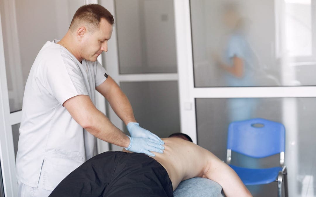
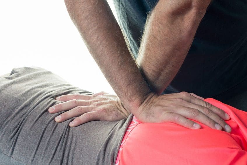
Various approaches use a form of force, hence the manipulation of the spine. The chiropractic adjustment technique that is the most common is spinal manipulation. It can also be called the diversified technique or HVLA – high-velocity, low-amplitude thrust.
However, chiropractic techniques are continually evolving. This comes from creating variations on existing techniques, a combination of techniques, or the chiropractor needs to adjust/tweak their own specific technique/s because they begin to suffer overuse injury/s from the constant adjusting, pushing, thrusting motions. Most techniques are named after the chiropractor that developed the method. These are the most common spinal manipulation techniques currently in use.
Contents
Manipulation Techniques
Chiropractic adapts to the condition/s and specific needs of each individual. Treatment plans can involve a forceful approach and a gentler force technique. This could happen during the same visit or the treatment plan could be half forceful adjustments, that could range from 6 to 10 visits, with the final visits using the gentle approach.
Spinal Manipulation
This is the High-Velocity Low-Amplitude Thrust technique. The most frequently used chiropractic technique. This is the manipulation that most are familiar with because of the audible pop that results. This is from the chiropractor’s hands applying a controlled quick forceful thrust to the spine while the body is positioned in a specific way.
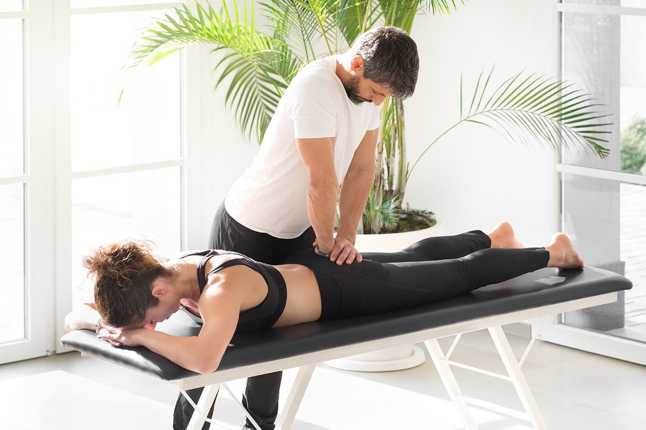
Spinal Mobilization
This is the Low-Force/Gentle Chiropractic Technique. These techniques are for individuals that require a gentler approach. The technique is known as spinal mobilization. This approach could be utilized due to:
Underlying conditions like Osteoporosis for example
Some chiropractors prefer and/or specialize in mild spinal mobilization techniques. These are techniques that do not involve twisting the body or using forceful thrusts. Along with spinal mobilization, chiropractors often employ complementary therapy, as part of an overall treatment plan. This could be:
Ice
Heat
Physical therapy
Electric stimulation
Ultrasound
Individuals need to discuss symptoms and preferences with the chiropractor. It is their role to perform a thorough examination to determine the most optimal treatment plan satisfactory to the patient. Chiropractors are not the only health care providers who utilize spinal manipulation for back pain. Osteopathic physicians can also provide types of spinal adjustments. Physical and massage therapists often work with chiropractors with continued treatment. They are fully trained in providing spinal therapy as well.
Pregnancy Lower Back Pain Chiropractic Treatment
Dr. Alex Jimenez�s Blog Post Disclaimer
The scope of our information is limited to chiropractic, musculoskeletal, physical medicines, wellness, and sensitive health issues and/or functional medicine articles, topics, and discussions. We use functional health & wellness protocols to treat and support care for injuries or disorders of the musculoskeletal system. Our posts, topics, subjects, and insights cover clinical matters, issues, and topics that relate and support directly or indirectly our clinical scope of practice.*
Our office has made a reasonable attempt to provide supportive citations and has identified the relevant research study or studies supporting our posts. We also make copies of supporting research studies available to the board and or the public upon request. We understand that we cover matters that require an additional explanation as to how it may assist in a particular care plan or treatment protocol; therefore, to further discuss the subject matter above, please feel free to ask Dr. Alex Jimenez or contact us at 915-850-0900. The provider(s) Licensed in Texas& New Mexico*
Belly dancing has been found to be an effective way to help individuals managing low back pain. It could be utilized as a part of a chiropractic treatment plan. The dancing is beneficial for improving posture and allows an individual to improve their fitness with a light form of aerobic exercise.
Regular physical activity/exercise and a healthy lifestyle go hand in hand. For individuals with spinal issues, the right stretches and exercises can make a difference in their quality of life. It increases:
Strength
Flexibility
Helps with pain management
Improves posture
Maintains spinal alignment
Belly dancing can help with injury recovery, as well as overall health. For most the trouble with exercising regularly is that it becomes routine and boring. Individuals want to live healthily, but it can be a challenge to maintain interest and motivation. An alternative form of physical activity that qualifies as exercise could be the answer.
Dancing has grown in popularity because of its fitness, flexibility, and spinal benefits. This form of belly dancing exercise does not require any special outfit or plenty of space. This utilizes the movements as a form of stretching and keeping the body moving in an aerobic fashion. They can be done at home with video instruction or an online class. Although the majority are women, men can and do belly dance.
Contents
Belly Dance
Information on the history of belly dancing. The dance has gone through various transformations since its inception. It was once considered burlesque entertainment, is now recognized as an important cultural expression, and today has been found to be a respected form of dance exercise.
Isometric exercises are contract specific muscles or groups of muscles. These types of exercise help with strength and stability enhancement. Both are vital for individuals recovering from back injuries or back pain management.
Posture
Dance posture is different than normal standing or sitting posture. Dance posture refers to the way an individual prepares/maintains their body to perform specific movements so that the motions are fluid, graceful, and with no presentation of pain. Belly dance posture maintains proper spinal alignment, which encourages reduced stress/pressure on the joints. This is beneficial for individuals managing back problems. The keys to spinal success are:
The knees should be slightly bent and not stiff so as not to pull/strain the lower back muscles
When the abdominal and back muscles maintain/support a straight spine, this alleviates stress on the low back. Lower back issues have shown a positive response to a belly dance exercise therapy program. A study looked at the effects of belly dancing on pain and function in women with chronic lower back pain. The study found that belly dancing made movements of the trunk and pelvis that are known to influence low back pain much easier.
A belly dance program in conjunction with a chiropractic or physical therapy treatment plan can help alleviate pain and improve function. A 45-minute belly dance routine/session promotes aerobic benefits, improves flexibility and core strength.
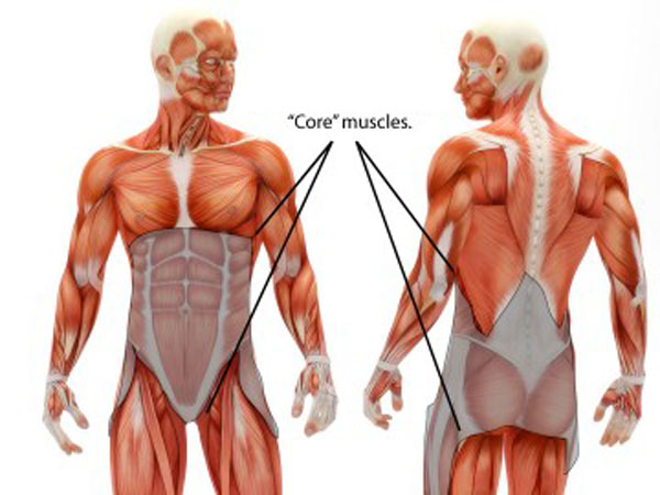
Core Strength
These are movements that train the muscles in the:
Pelvis
Abdomen
Hips
Low back
They help build strength, generate stability, protect against back pain, poor posture, and muscle injuries. Having core strength is crucial for individuals with back issues, as it increases the stabilization of the spine. Core strengthening is highly recommended and often prescribed for individuals recovering from lumbar issues.
Depression/Anxiety Improvement
Individuals with back pain also tend to experience psychological issues like depression and anxiety. Back pain can affect:
Mood
Tiredness
Sleep problems
Self-esteem problems
Belly dancing as part of a treatment/therapy program can help an individual experience benefits that improve mental health and well-being. These include:
Regaining mobility
Having a positive body image
Social interaction is enhanced
For All Ages
Belly dancing is a fantastic creative outlet and a great way to exercise. Anyone that is able can participate. Children, seniors, and everyone in between can get into belly dancing. It enhances health and strengthens the body and mind. When the body is in the proper position/posture there are no joint issues or pain. In-person classes, at home with online instruction, DVDs, or video meeting apps can benefit the body and especially the spine.
Life-Changing Orthotics
Dr. Alex Jimenez�s Blog Post Disclaimer
The scope of our information is limited to chiropractic, musculoskeletal, physical medicines, wellness, and sensitive health issues and/or functional medicine articles, topics, and discussions. We use functional health & wellness protocols to treat and support care for injuries or disorders of the musculoskeletal system. Our posts, topics, subjects, and insights cover clinical matters, issues, and topics that relate and support directly or indirectly our clinical scope of practice.*
Our office has made a reasonable attempt to provide supportive citations and has identified the relevant research study or studies supporting our posts. We also make copies of supporting research studies available to the board and or the public upon request. We understand that we cover matters that require an additional explanation as to how it may assist in a particular care plan or treatment protocol; therefore, to further discuss the subject matter above, please feel free to ask Dr. Alex Jimenez or contact us at 915-850-0900. The provider(s) Licensed in Texas& New Mexico*
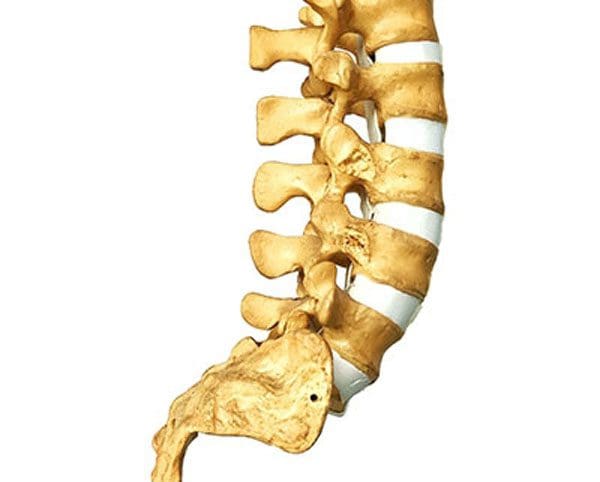
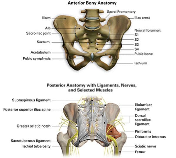
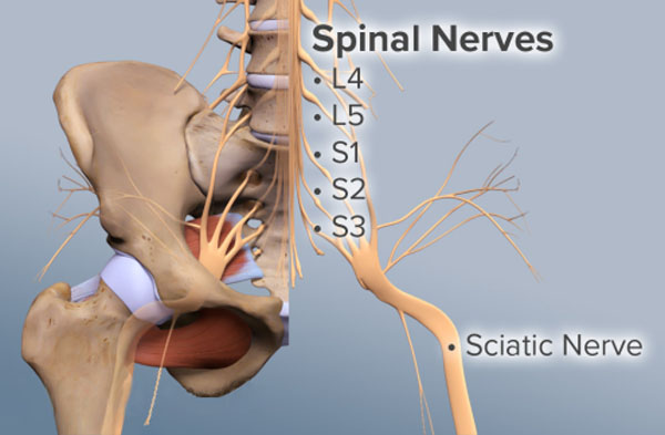
Thelumbosacral joint is the first place chiropractors start their investigation with individuals presenting with low back pain and possible sciatica. Because of the importance of the sciatic nerve, almost any lumbar condition has the potential to disturb the nerve that can lead to chronic nerve pain. For many low back conditions, the best way to start is from the bottom and work up.
Starting at the lumbosacral joint L5-S1, the chiropractor will palpate and massage the area. This is because the lumbosacral joint is a central nerve center with all kinds of possible sciatic nerve interference because of the proximity to the various nerve bundles and vertebral discs.
When sciatic nerve issues begin to develop, often the problem will be in this region of the spine. Beginning at the lumbosacral joint can generate vast insight into the root cause of radiating pain in the lower back and legs.
Contents
The Lumbosacral Joint
This pain typically presents when the nerve is inflamed, compressed, or irritated. Numbness or chronic weakness can also happen in the lower extremities and can cause unbearable discomfort. Some of the reasons that make the joint a prime suspect for sciatic pain include:
The L5 vertebrae are vulnerable to slipping forward over the connecting S1 vertebrae. The sciatic nerve goes through this area, leaving it open to compression.
A disc herniation and/or inflammation can also stress the sciatic nerves.
Deterioration of the lumbosacral facet joints is common with older individuals. This can lead to nerve compression and sciatic nerve irritation.
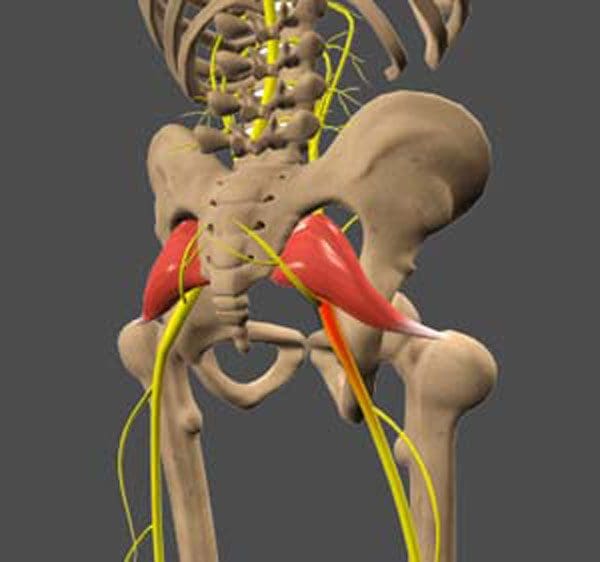
Piriformis syndrome can affect the area around the lumbosacral joint, causing nerve compression and inflammation.
The lumbosacral joint is frequently used making it a consistently stressed joint. Overuse, poor posture, and improper body mechanics affect this region of the lumbar spine. And, because of the closeness to the sciatic nerve, it is commonly affected.
Other Spinal Conditions
The lumbosacral joint also experiences problems that stem from chronic conditions, which can involve some form of sciatic pain as a symptom. They include:
Degenerative disc disease
Lumbar stenosis
Sacroiliac joint dysfunction
Spondylolisthesis
Sciatica is a condition that represents a series of symptoms. But it is often a symptom of other condition/s that affect the sciatic nerve. If spinal conditions progress, it can bring undue stress and strain to the lumbosacral joint and the sciatic nerve.
Knowing Where To Begin
The key to a proper and successful treatment plan is an accurate diagnosis. Knowing and understanding the symptoms, spinal conditions, and having an idea of the origin of these types of pain promotes a rapid diagnosis. Our chiropractic and physical therapy team thoroughly investigate the pain source using imaging, palpation, observation, and other diagnostic tools to help get individuals back on track and healthy.
Facet Syndrome Chiropractic Treatment
Dr. Alex Jimenez�s Blog Post Disclaimer
The scope of our information is limited to chiropractic, musculoskeletal, physical medicines, wellness, and sensitive health issues and/or functional medicine articles, topics, and discussions. We use functional health & wellness protocols to treat and support care for injuries or disorders of the musculoskeletal system. Our posts, topics, subjects, and insights cover clinical matters, issues, and topics that relate and support directly or indirectly our clinical scope of practice.*
Our office has made a reasonable attempt to provide supportive citations and has identified the relevant research study or studies supporting our posts. We also make copies of supporting research studies available to the board and or the public upon request. We understand that we cover matters that require an additional explanation as to how it may assist in a particular care plan or treatment protocol; therefore, to further discuss the subject matter above, please feel free to ask Dr. Alex Jimenez or contact us at 915-850-0900. The provider(s) Licensed in Texas& New Mexico*
References
Grgi?, Vjekoslav. �Lumbosakralni fasetni sindrom: funkcijski i organski poreme?aji lumbosakralnih fasetnih zglobova� [Lumbosacral facet syndrome: functional and organic disorders of lumbosacral facet joints].�Lijecnicki vjesnik�vol. 133,9-10 (2011): 330-6.
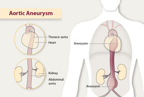
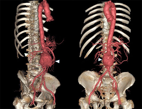
Sciatica or Aneurysm? Knowing how a missed diagnosis could be potentially fatal if not diagnosed accurately could be a deadly mistake! Doctors must not fall for a sciatica diagnosis when a possibly fatal iliac artery aneurysm lies looming and progressing.
Contents
Sciatica or Aneurysm
An example is a patient who visited an emergency clinic after a few weeks for a non-painful pulsing mass on the buttock. There was no:
Trauma
Injury
Back pain
Leg pain
Prior presentations of pain or sciatica issues
A physical examination found a small pulsing mass on the right buttock. Palpation around the site found no issues with the sensory and motor nerves.
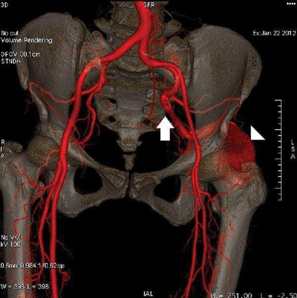
An ultrasound scan of the affected area revealed a developing aneurysm. This was followed by a CT scan of the abdomen along with the pelvis using a contrast dye found the aneurysm developing from the left internal iliac artery. If the mass was not present a doctor could easily diagnose sciatica or persistent sciatic artery. If the iliac artery presents with pulsating lesions is a tip-off that a vascular issue could be impinging on the sciatic nerve. Vascular surgery was discussed with the patient. Surgery was necessary, and the patient underwent sciatic aneurysm repair. The patient was discharged without any complications.
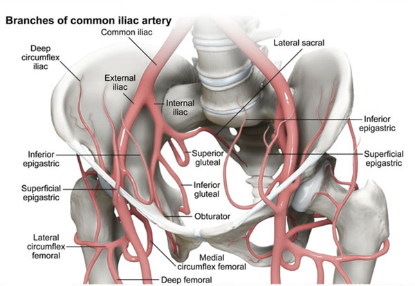
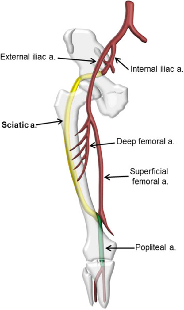
Persistent Sciatic Artery
This is a very rare congenital vascular condition. The sciatic artery runs along the sciatic nerve and functions as the major blood supply to the lower extremities. During human embryo development, the femoral artery begins to form while the sciatic arteries start to return to a less developed state. The process continues until the femoral artery takes over as the major blood supply, with only bits of the sciatic artery left.
Persistent sciatic artery can happen either from the sciatic artery not returning to its original size or during normal development the femoral artery developing properly. Most cases of persistent sciatic artery go unknown and are usually detected from another examination for another ailment. Aneurysms often develop based on the arteries/vessel’s tendency for minor trauma/injury when sitting or some form of pressure is applied on the site. Complications include:
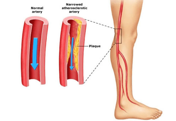
Vascular Conditions In The Leg/s That Can Present As Sciatica
The legs’ blood vessels can get infected, bulged, ruptured, or blocked. This can cause sciatica symptoms, like leg pain, weakness, tingling, and numbness. Severe cases could require medical emergency surgery to save the affected limb.
Acute Limb Ischemia
This condition occurs from a decrease or loss of blood supply to the legs. If there is leg pain, it could feel similar to sciatica pain. However, symptoms can progress rapidly and become severe. That’s when it is not sciatica. Acute limb ischemia present one or more of the following symptoms:
Pain and/or numbness in the leg while walking and when resting
Severe pain at night
Sleep problems
Pain relief when sitting on a chair with the feet hanging down
Feet and ankles become swollen
A pale color and lowered skin temperature over the toes and feet when compared to the legs
Acute limb ischemia can develop from an aneurysm, blood clot, or from the thickening of the vessel walls. Treatment should be prompt in order to preserve leg function. Differentiation diagnosis between vascular and other causes like spinal problems that can cause leg pain. A doctor may perform an Ankle/Brachial Index which is a comparison of blood flow in the arms versus the legs. This can be critical in determining if there is vascular insufficiency.
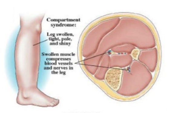
Acute Compartment Syndrome
This places increased pressure in the muscle tissues of the leg. It can lead to loss of blood supply in and around the affected area. The sciatic nerve can also get compressed from the increased pressure in the buttock, thigh, or leg. The condition can cause pain, numbness, and weakness in the buttock, thigh, and leg. Individuals have also reported an unusual/altered sensation in the web of the great toe. This is similar to sciatica, as well as one or both legs can be affected. Differentiating symptoms include:
Leg becomes swollen
Pain and tenderness present when touching the leg
A pale color and lowered skin temperature over the leg
Acute compartment syndrome is a serious condition that is considered a medical emergency. It is possible for the condition to cause complete dysfunction of the limb if not addressed in time. There are risk factors that increase the chances of developing limb ischemia or compartment syndrome. These are:
Diabetes
Heart conditions
High cholesterol
Smoking
History of having the condition can also cause a recurrence. This can be from an injury or poor health.
Kidney stones, renal failure, or cysts in the kidney can also cause back and leg pain. Other symptoms can include blood in the urine or difficulty urinating. Any sign of distressing symptoms that present with sciatica can indicate the need for medical attention. This is to check for the possibility of a serious underlying condition or medical emergency. Medical emergencies that are treated in time can help preserve the tissue/s, restore function, and save an individual�s life.
It is essential for a chiropractor or physical therapist to be familiar with diagnosing in a way that will help identify sciatica or aneurysm in individuals presenting with musculoskeletal issues/problems. Knowledge of these risk factors, understanding how to screen for non-musculoskeletal symptoms, basic competence in palpation, and how to interpret findings will help discover sciatica or aneurysm if it is there and begin timely treatment. And if it is not there then a sciatica treatment plan can be developed before it worsens.
Sciatic Nerve Pain
Dr. Alex Jimenez�s Blog Post Disclaimer
The scope of our information is limited to chiropractic, musculoskeletal, physical medicines, wellness, and sensitive health issues and/or functional medicine articles, topics, and discussions. We use functional health & wellness protocols to treat and support care for injuries or disorders of the musculoskeletal system. Our posts, topics, subjects, and insights cover clinical matters, issues, and topics that relate and support directly or indirectly our clinical scope of practice.*
Our office has made a reasonable attempt to provide supportive citations and has identified the relevant research study or studies supporting our posts. We also make copies of supporting research studies available to the board and or the public upon request. We understand that we cover matters that require an additional explanation as to how it may assist in a particular care plan or treatment protocol; therefore, to further discuss the subject matter above, please feel free to ask Dr. Alex Jimenez or contact us at 915-850-0900. The provider(s) Licensed in Texas& New Mexico*
References
Javdanfar A, Celentano C. Sciatic artery aneurysm. West J Emerg Med. 2010;11(5):516-517.
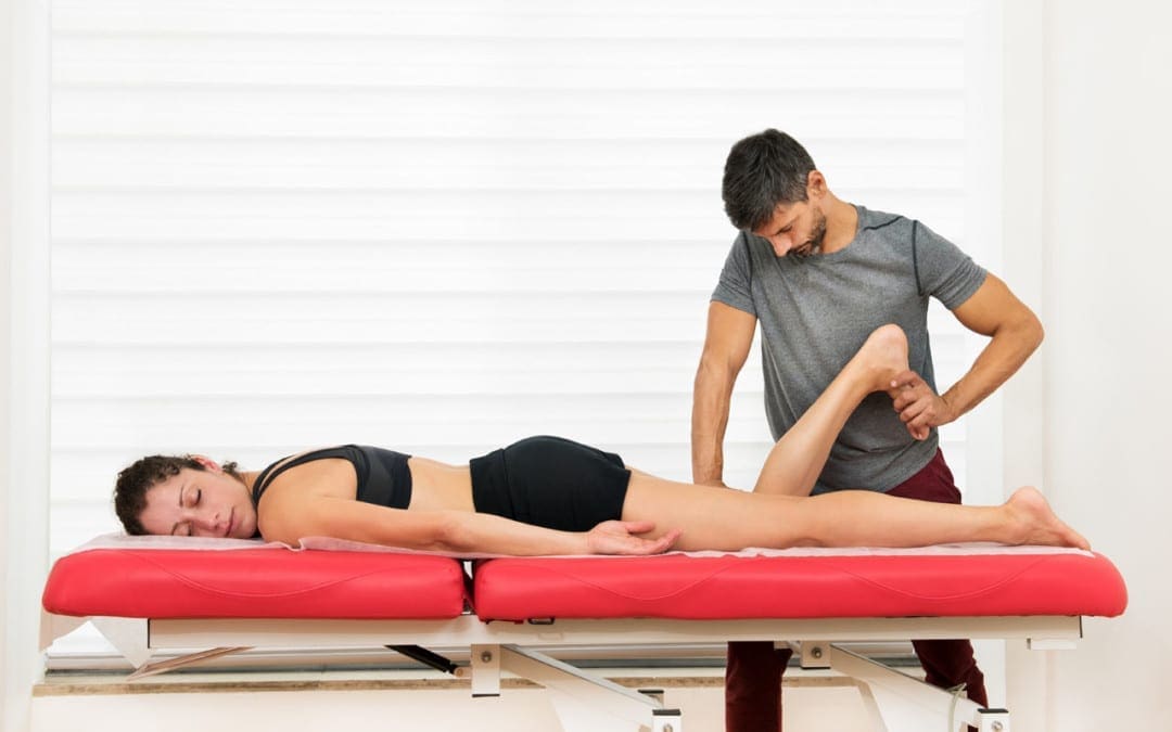
Tight and sore hamstrings commonly occur during workout and exercise, but can just as easily result from a fall or other accident. Individuals usually experience pain located at the back of the thigh with associated weakness along with the feeling of the muscles becoming tighter and a consistent soreness. Consistent tightness in the back of the legs is not only uncomfortable but can also make movement difficult.
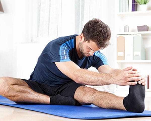
Many individuals stretch every day, do yoga, etc trying to relieve hamstring tension, with short-term relief only to have the tightness return. This is frustrating but more importantly, indicates that the problem might not have to do with the muscle�s length. There could be an underlying issue that needs to be identified and addressed. A chiropractic approach will diagnose and treat the root of the pain, not just the symptoms.
Contents
Hamstring/s Tightness
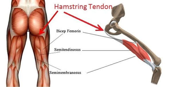
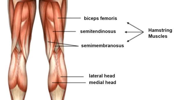
The hamstring is not a single string. It is a set of three muscles that run along the back of the thigh. The muscles allow for the bending of the leg at the knee. With a hamstring strain, one or more of these muscles can become overloaded and can start to tear. Strains often happen during activities that involve running, jumping, and/or sudden stopping and starting. This is where stretching doesn’t help, however, a chiropractic adjustment can help.
Weak Muscle Compensation
One possible cause for the tightness has to do with various related muscles and not the hamstrings themselves. Muscles that typically stabilize and facilitate movement could be too weak or are not functioning properly. What usually happens is that the muscles that are active have to work harder to compensate for the others that are not working/functioning properly.
The tension in the hamstrings can be alleviated through chiropractic exercises/stretches that activate the stabilizing muscles and get the circulation moving to promote strength and take the pressure off the hamstrings.
These muscle weaknesses can be caused by spinal misalignments that pull the body in an awkward fashion, throwing the body’s balance off. Each condition feeds the other as the hamstrings have to work harder, the body leans to the side that doesn’t cause pain, causing the spine to shift out of alignment and so begins the awkward body shifting to avoid the pain cycle. A chiropractic adjustment will restore balance and stability to the entire body.
A Pelvic Tilt
Pelvic tilt could be a contributing factor for tightness and soreness. This comes from:
Poor posture
Lack of physical activity
Weight gain
These factors can cause the pelvis to shift forward ever so slightly. But just that slight tilting could be pulling on the hamstrings. In order to rectify the tilt, exercise is recommended, and chiropractic manipulation to realign any spinal shifting.
Sciatic Irritation
Another issue is sciatic nerve irritation that could mimic tightness in the muscles. The sciatic nerve runs down the back of the leg and the irritation could make the hamstring appear to be tight. This is where stretching the hamstrings can worsen the condition by irritating the sciatic nerve, causing inflammation.
The sciatic nerve could also be experiencing irritation from spinal misalignment. A vertebral disc could be bulging or herniated. This could inflame the nerve root. A bone spur or inflamed joint could be impinging/compressing on the nerve root exits.
A chiropractic treatment plan will relieve the tension being placed on the nerve and allow for proper blood circulation and transmission of signals without interruptions. Talk to a chiropractor about an examination to identify the exact root causing the discomfort.
Advanced Chiropractic Treatment
Dr. Alex Jimenez�s Blog Post Disclaimer
The scope of our information is limited to chiropractic, musculoskeletal, physical medicines, wellness, and sensitive health issues and/or functional medicine articles, topics, and discussions. We use functional health & wellness protocols to treat and support care for injuries or disorders of the musculoskeletal system. Our posts, topics, subjects, and insights cover clinical matters, issues, and topics that relate and support directly or indirectly our clinical scope of practice.*
Our office has made a reasonable attempt to provide supportive citations and has identified the relevant research study or studies supporting our posts. We also make copies of supporting research studies available to the board and or the public upon request. We understand that we cover matters that require an additional explanation as to how it may assist in a particular care plan or treatment protocol; therefore, to further discuss the subject matter above, please feel free to ask Dr. Alex Jimenez or contact us at 915-850-0900. The provider(s) Licensed in Texas& New Mexico*
References
Hoskins, Wayne, and Henry Pollard. �Hamstring injury management–Part 2: Treatment.��Manual therapy�vol. 10,3 (2005): 180-90. doi:10.1016/j.math.2005.05.001
Finding the right sciatica chiropractic specialist to diagnose the cause especially, when it is an abdominal aortic aneurysm can be a challenge. There can cause diagnostic confusion with the root cause never being discovered or identified. Fortunately, Dr. Jimenez is a sciatica specialist with over 30 years of experience in differential sciatica diagnosis, and treatment.
Contents
Sciatica Chiropractic Specialist Diagnosis
Diagnostic Tools
Abdominal aneurysms are usually discovered for another ailment like a hernia or for routine tests like an ultrasound of the heart or stomach.Diagnosis of an abdominal aneurysm depends on the condition, medical and family history, and the physical examination. If a doctor or sciatica chiropractic specialist suspects an aortic aneurysm, then specialized tests will help with a confirmation.
Ultrasonography
The simplest and most used diagnostic test is ultrasonography. It utilizes sound waves for diagnostic purposes that send the recorded images to a monitor. It gives an accurate assessment of the size and location of the aneurysm. The patient will lie on a table while a technician moves a wand around the abdomen.
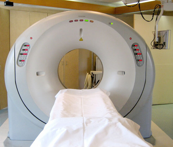
Computed tomography CT scan
This test is often used in conjunction with ultrasonography if more data/info is needed. Usually, this is to determine the exact location of the aneurysm in relation to the visceral or renal arteries. It provides cross-sectional detail with clear images of the aorta and can detect the size and shape. The patient lies on a table inside a machine. A contrast dye could be injected into the blood vessels to make the arteries more visible on the images known as CT angiography.
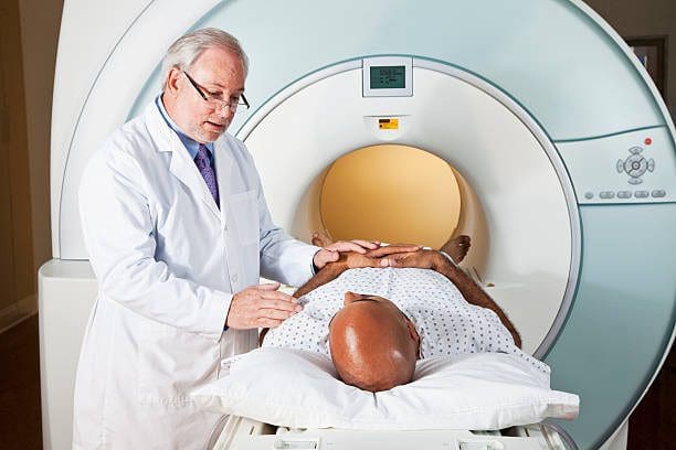
Magnetic Resonance Imaging
Magnetic resonance imaging or MRI uses a magnetic field and radio wave energy pulses to record images of the body. The patient lies on a table that slides into the imaging compartment. Contrast dye can also be injected into the blood vessels to make the images more visible known as magnetic resonance angiography.
Emergency Symptoms
Certain symptoms can indicate an emergency. The conditions are rare, but it is very important to seek medical attention should any of these symptoms present with back pain:
Severe abdominal pain
Fever out of nowhere
Bowel and/or bladder incontinence
Loss of or an unusual sensation in the groin, as well as the legs and possibly into the foot
If back pain presents after an injury medical care is recommended to check for damage/injury to the spine.
Abdominal Aneurysm Symptoms
Abdominal aneurysms often don�t present any symptoms, which is why individuals go through their days unaware, and when back pain does present a doctor may only focus on the back pain symptoms and not the cause, leaving the aneurysm to continue to develop and worsen. Aneurysms do occur in women but are more common in men and those ages 65 and older. The main cause is atherosclerosis which is a hardening of the arteries. But injury and infection can also cause an aneurysm. Those with symptoms can include:
Throbbing pain around the back or side
Deep pain in the back or side
Pain in the buttocks, groin, or legs
Sciatica symptoms
The Sciatic Connection
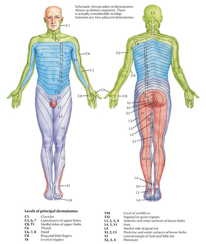
A diagnosis of the root cause of the sciatica is crucial for developing an effective treatment plan to alleviate the sciatic pain. If an aneurysm is present then referring the individual to the proper aortic aneurysm repair specialist is a top priority. If sciatica is suspected, a doctor or chiropractor will review medical history and perform a physical examination. Medical imaging tests and diagnostic nerve blocks could be used if necessary. Sciatica pain usually follows the dermatome or areas of the skin that is supplied by the sciatic nerve. The pain can also include deeper tissues called dynatomes.
Physical examination
During a physical examination, the sciatica chiropractic specialist will look for various responses when:
Straightening the leg with movements that elongate the nerve
Gently pressing the toes or calf area
Seeing if there is any type of pain associated with these movements in the low back, buttock, thigh, leg, and foot
Sciatica Clinical Tests
Two examples of clinical tests for sciatica include:
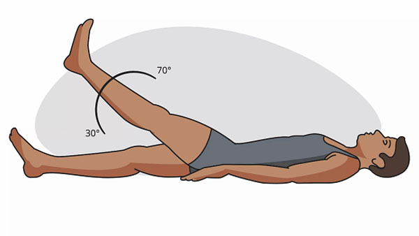
Straight leg raise – SLR
The patient lies on their back and the chiropractor lifts one leg at a time with the other leg remaining flat or bent at the knee. If pain presents while lifting the affected leg this is usually an indication of sciatica.
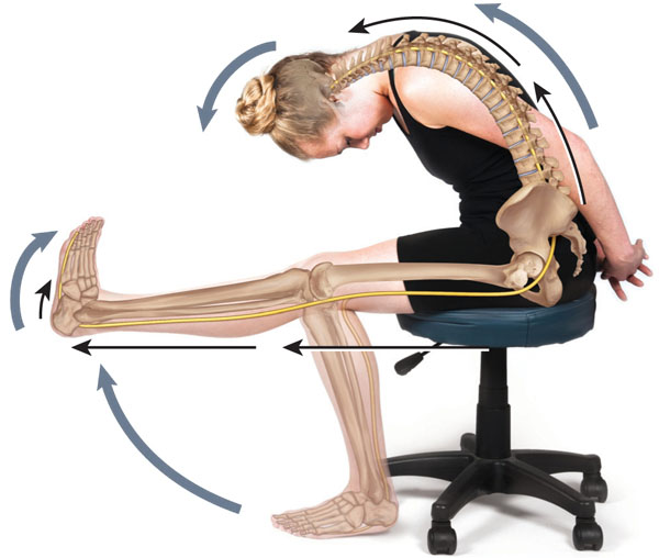
Slump
The patient sits upright with their hands behind their back. The patient then bends/slumps forward at the hips. The neck bends down with the chin touching the chest and one knee is extended as far as possible. If pain occurs in this position, sciatica could be present.
These tests could possibly be positive only when the nerve is mechanically compressed. Other causes like inflammation or chemical irritation of the nerve might not cause pain when performing these tests. This test could also help reveal a possible abdominal aneurysm as abdominal pain could present.
Chiropractic Sciatica Treatment
Manual manipulation improves the alignment of the spine. This technique helps address the underlying condition/s that can cause sciatic nerve pain, like herniated discs or spinal stenosis. Manual manipulation also creates an optimal healing environment. An aortic aneurysm specialist could work with a sciatica chiropractic specialist to help with spinal realignment if the aneurysm caused any kind of shifting or slipping of the discs along with releasing the sciatic nerve if it is compressed.
Massage Therapy
Massage therapy like deep tissue massage can also have benefits. Massage:
Improves blood circulation, which also creates an optimal healing response in the body
Releases toxins in the low back muscles that spasmed or knotted up
Relaxes tight muscles that could be contributing to the pain
Releases endorphins or the hormones that function as the body’s natural pain relievers
Sciatica Pain Chiropractor
Dr. Alex Jimenez�s Blog Post Disclaimer
The scope of our information is limited to chiropractic, musculoskeletal, physical medicines, wellness, and sensitive health issues and/or functional medicine articles, topics, and discussions. We use functional health & wellness protocols to treat and support care for injuries or disorders of the musculoskeletal system. Our posts, topics, subjects, and insights cover clinical matters, issues, and topics that relate and support directly or indirectly our clinical scope of practice.*
Our office has made a reasonable attempt to provide supportive citations and has identified the relevant research study or studies supporting our posts. We also make copies of supporting research studies available to the board and or the public upon request. We understand that we cover matters that require an additional explanation as to how it may assist in a particular care plan or treatment protocol; therefore, to further discuss the subject matter above, please feel free to ask Dr. Alex Jimenez or contact us at 915-850-0900. The provider(s) Licensed in Texas& New Mexico*
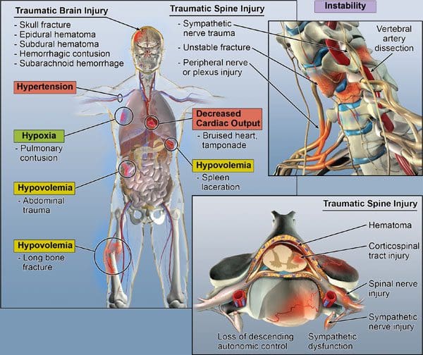
With an intense automobile, work, sporting/fitness accidents, and natural disasters, individuals can experience multiple traumas, also known as polytrauma. Recovery and rehabilitation can be challenging journeys. Multiple traumas are serious medical emergencies only given to individuals going through difficult situations. Polytrauma references multiple severe traumas that occurred at the same time. An individual can experience a:
Physical therapy and chiropractic rehabilitation could be part of a treatment program. This is to restore the body’s mobility, function and promote the body’s natural healing abilities. The importance of creating the proper rehabilitation program can make all the difference, and restoring the integrity of the spine is critical.
Contents
Spinal Stabilization
Any trauma, despite its severity, can have the potential to interfere/disrupt spinal functions.
Suffering a concussive hit to the head could cause damage to the cervical spine.
Falling off a ladder could shift the vertebral discs.
Hitting the knee on an object can cause a spinal misalignment from shifting the weight to the other leg for a day or two.
Spinal misalignments and/or dysfunction are common in multiple trauma cases. Dealing with broken bones and substantial wounds often means the spine could need to be realigned even if there are no localized injuries to the back or neck. Spinal misalignment, translation/rotation, herniated discs, or other conditions can hinder the overall recovery process and present new health issues.
Nerve Damage
Often, a lasting issue from multiple traumas is nerve damage. The severity can destroy nerve endings, making it impossible to regain feeling or movement in certain areas of the body. The nerve damage can be regenerated if the impediment is rooted in the spine and is addressed in time.
Correcting spinal discs that have shifted along with subluxations that can compress the nerves and interfere with signal communication can help restore the areas directly affected. Example: Broken arms or legs can lose sensations that can be regained by spinal realignment. Some arteries follow the spinal pathway. Proper circulation/blood flow is achieved when the spinal integrity is restored, which is essential in natural recovery.
Trauma Beyond the Injury
Developing the correct rehabilitation treatment plan could mean the difference in a speedy and healthy recovery. Chiropractic can be an integral part of the recovery process, especially for a body that has sustained severe musculoskeletal injuries. The spine is an extension of the central nervous system. This means that bringing relief to the spine can affect positive healing in the injured areas. The critical role of the spine in an individual’s rehabilitation and whole well-being is why no matter the extent of the trauma, we deliver relief.
Auto Accident Injury Treatment
References
Kroupa, J. K definici polytraumatu a polytraumatismu [Definition of “polytrauma” and “polytraumatism”].Acta chirurgiae orthopaedicae et traumatologiae Cechoslovaca vol. 57,4 (1990): 347-60.
Underlying causes for an abdominal aortic aneurysm can be challenging to diagnose and identify. Combined with sciatica symptoms, doctors could misdiagnose the ailment and prescribe the wrong treatment protocol. Then an individual has to deal with two conditions that were not properly diagnosed, continue to develop, and worsen. This is why finding the right sciatica specialist that can also identify an abdominal aneurysm is so crucial to developing the right treatment plan. There can be a variety of factors that can lead to the development of an abdominal aneurysm. They include:
Health conditions associated with an increased risk for an abdominal aneurysm include:
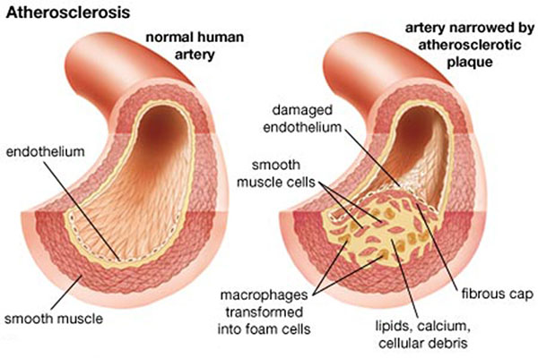
Atherosclerosis
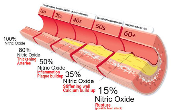
This condition occurs when there is a buildup of fats, cholesterol, and other substances that create plaque buildup in the bloodstream. This causes vessels to harden and narrow. Atherosclerosis can develop during the young adult stage and becomes an issue later in life.
High Cholesterol
Cholesterol is a waxy, fat-type substance that is found in all the cells in the body. The body needs some cholesterol for the production of hormones, vitamin D, and substances to help digest foods. The body makes all the cholesterol it needs. Too much can build up in the blood vessels, which narrows the bloodstream and hardens the arterial walls.
High Blood Pressure
High blood pressure or hypertension refers to a sustained increased force of blood moving through the aorta that can weaken artery walls. It is a common condition that is widespread among individuals that are older, those that smoke, and those that are overweight. There is an estimated 60-70% of individuals over 60 that are diagnosed with high blood pressure.
Inflamed Arteries
When the arteries become inflamed, it can cause blood flow constriction and cause the arterial walls to weaken. This increases the risk of an aneurysm. Arteries can get inflamed through:
There are hereditary conditions that can weaken the body�s connective tissues. This can lead to degeneration of the aortic walls and raise an individual�s risk for an aneurysm. Two of the most common connective tissue disorders are Ehlers-Danlos syndrome, which affects collagen production, and Marfan Syndrome. This condition increases the production of fibrillin, which is a protein that helps to build the elastic fibers in connective tissue.
Other Risk Factors
Additional health factors can strain the cardiovascular system. This increases the risk of weakening or damaging blood vessels. This significantly raises the chances of developing an abdominal aortic aneurysm. Risk factors include:
Smoking and Tobacco
All types of tobacco use can contribute to diminished cardiovascular health. Individuals that smoke or use some tobacco product pose a significantly higher risk of developing an abdominal aneurysm.
Age
Aneurysms occur most often in older adults. This is because they are more likely to have cardiovascular issues and are more likely to have higher levels of plaque buildup.
Genetics and Family History
Immediate relatives of an individual with an abdominal aneurysm often have a 12-19% chance of developing the condition.
Lack of Physical Activity
Not getting adequate physical activity puts an individual at a higher risk for heart and cardiovascular disease. Aerobic activity done on a regular basis increases the heart rate and blood flow through the body. This keeps the tissues and blood vessels strong and flowing properly.
Gender
Both men and women can develop an abdominal aortic aneurysm. However, the majority of those that do develop the condition are men. This is because men are more likely to go through the heart and cardiovascular issues.
Diagnosis
Underlying conditions that can cause sciatic pain can vary or be a combination of several conditions. The most important action to take is to consult a doctor or chiropractic sciatica specialist for a clinical diagnosis. While rare, sciatica-type pain could be caused by medical conditions like:
Spinal tumor
Spinal infection
Cauda equina syndrome
These factors can contribute to an increased chance of developing an abdominal aortic aneurysm. However, individuals can have unknown risk factors and still develop the condition. Treatments may range from regular monitoring, lifestyle changes, and physical therapy/chiropractic to urgent or emergency surgery. If you feel symptoms of pain in the buttocks, leg, numbness, tingling, or other neurological symptoms in the back and/or leg, it is very important to see a doctor or chiropractor for clinical diagnosis that identifies the cause of the symptoms.
Sciatic Nerve Treatment
Dr. Alex Jimenez�s Blog Post Disclaimer
The scope of our information is limited to chiropractic, musculoskeletal, physical medicines, wellness, and sensitive health issues and/or functional medicine articles, topics, and discussions. We use functional health & wellness protocols to treat and support care for injuries or disorders of the musculoskeletal system. Our posts, topics, subjects, and insights cover clinical matters, issues, and topics that relate and support directly or indirectly our clinical scope of practice.*
Our office has made a reasonable attempt to provide supportive citations and has identified the relevant research study or studies supporting our posts. We also make copies of supporting research studies available to the board and or the public upon request. We understand that we cover matters that require an additional explanation as to how it may assist in a particular care plan or treatment protocol; therefore, to further discuss the subject matter above, please feel free to ask Dr. Alex Jimenez or contact us at 915-850-0900. The provider(s) Licensed in Texas& New Mexico*
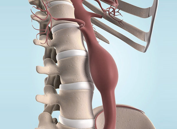
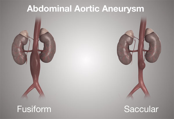
An abdominal aortic aneurysm refers to an enlargement of the abdominal aorta. If the blood vessel is enlarged and starts to leak blood or rupture, it will cause severe abdominal and lower back pain. This is a serious medical emergency that necessitates emergency surgery. Unfortunately, there is no way to reverse the damage. A prominent symptom from a rupture is severe, persistent low back pain, and pain in and around the abdomen. Treatment for an abdominal aortic aneurysmdepends on the possible complications that could develop. Approaches for treatment:
Nonsurgical treatments like anti-biotics calcium channel blockers and exercise along with monitoring are used for individuals that have a low risk of rupture.
If an aneurysm is not found until it becomes an emergency, then surgery to repair the ruptured artery is absolutely necessary. If ruptured or there is a high risk of rupturing is considered an emergency.
If a rupturing aneurysm has been diagnosed, some treatment/management will be implemented to prevent severe/fatal bleeding.
Contents
Cardiac
For low-risk cases, lifestyle changes and possible medication/s may be recommended to slow the development. Small aneurysms are monitored using ultrasound. This can be every 6 to 12 months depending on the size and growth rate of the artery.Medications for lowering blood pressure and cholesterol could be prescribed. This is to limit the amount of plaque buildup in the aorta and reduce any pressure on the arterial walls. Quitting smoking and removing tobacco altogether whether dip, chew, vape is a significant action an individual can do to minimize the risk of aortic rupture. Other lifestyle changes involve maintaining a healthy diet and regular exercise will help lower blood pressure and cholesterol levels decreasing the chance of rupture.
Surgery
Surgical treatment when necessary is to stop a rupture if leaking blood or to prevent a rupture. Surgery requires replacing the damaged portion of the aorta with a stent-graft. This is an artificial artery made from a high-tech mesh/fabric. There are two standard surgical treatments:
Open Repair
Open repair is the most common surgical treatment. It takes the enlarged portion of the aorta removes it and replaces it with a stent-graft. Open surgery repair consists of the following:
The incision is made in the abdomen at the site of the aneurysm.
The aorta gets clamped with the blood temporarily blocked from flowing through the damaged portion.
The damaged part is removed.
A tube graft is placed where the damaged portion was.
If the damage was not severe and does not require the removal and complete replacement, then less invasive options will be offered.
Endovascular Aortic Aneurysm Repair
EVAR endovascular aneurysm repair surgery is a minimally invasive procedure. There is no need for a large abdominal incision or removal of the damaged portion of the artery. This procedure does not require blood flow stoppage, which places less stress on the heart. Endovascular surgery involves:
A fluoroscopy or live X-ray is used. This is so the surgeon can look at the repair, and guide the stent into place.
2 small incisions are made in the groin.
A catheter is inserted into the femoral artery in the groin and guided to the abdominal aorta.
Through the catheter, the stent is guided to the aneurysm.
Once it reaches the aneurysm, it is compressed and closed.
The stent is placed in position, and the wireframe is expanded to fit the artery.
The stent is sewn/secured into place at both ends.
Once in place, the blood gets redirected from the enlarged area and flows only through the stent-graft. This takes the pressure off the artery’s walls and allows for size reduction over time, and decreases the risk of rupture.
The procedure is not an option for individuals with an aorta that cannot be accessed safely through the femoral arteries. Or if the artery is severely damaged that the aneurysm portion needs to be replaced. And if the aneurysm is too big or complex where an open repair is a more favorable option.
Follow Up
Follow-up monitoring is necessary after any aortic aneurysm surgical procedure. This is to ensure the stent works and the aorta is functioning without a high risk of rupture. Individuals will be advised to maintain a healthier heart and cardiovascular system. A surgeon/doctor will suggest:
Diet adjustments
Regular exercise
Quitting smoking/tobacco intake
Taking cholesterol and blood pressure medication
Chiropractic/Physical therapy for any spinal misalignment, herniation, sciatic nerve compression back pain relief.
Lower Back Pain
Dr. Alex Jimenez�s Blog Post Disclaimer
The scope of our information is limited to chiropractic, musculoskeletal, physical medicines, wellness, and sensitive health issues and/or functional medicine articles, topics, and discussions. We use functional health & wellness protocols to treat and support care for injuries or disorders of the musculoskeletal system. Our posts, topics, subjects, and insights cover clinical matters, issues, and topics that relate and support directly or indirectly our clinical scope of practice.*
Our office has made a reasonable attempt to provide supportive citations and has identified the relevant research study or studies supporting our posts. We also make copies of supporting research studies available to the board and or the public upon request. We understand that we cover matters that require an additional explanation as to how it may assist in a particular care plan or treatment protocol; therefore, to further discuss the subject matter above, please feel free to ask Dr. Alex Jimenez or contact us at 915-850-0900. The provider(s) Licensed in Texas& New Mexico*
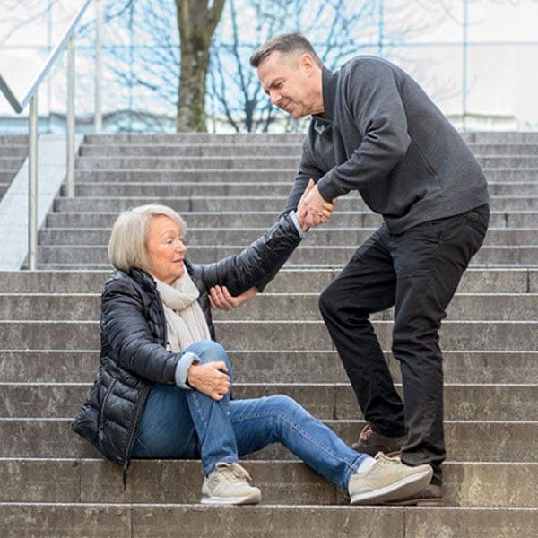
As individuals advance in age, the risk of falling starts to become a regular concern. An average of one in four adults over 65 suffers from a fall every year. Around twenty percent of falling accidents result in serious injury. However, experiencing one fall increases the risk of future falls significantly. The most common injuries sustained include:
Hip fracture
Head injury
These injuries usually necessitate hospitalization. Therefore minimizing the risks is important for increasing/enhancing an individual’s quality of life and help minimize the financial burdens that can come with these types of accidents.
Contents
Risk Factors for Falling
Various factors can determine an increased risk of falling. If there are two or more at the same time, the risk goes up significantly. These factors include:
A physical therapist or chiropractor can assess the home for safety and possible tripping hazards
Regular vision check-ups at least every two years
Spinal Health
Restoring balance to the body will make the biggest difference in increasing overall health. However, pain, stiffness, and poor health can make getting started a difficult challenge. With the right chiropractic and physical therapy team, getting started does not have to be as challenging, with the transition being made as easily and as smooth as possible.
An essential building block for overall optimal health begins with spinal alignment. When the spine is misaligned it can lead to poor nerve circulation/energy. This slows down and interferes with the body’s functions. This is often manifested with poor balance, weakness, and pain. Chiropractic spinal alignment addresses whole-body health. A chiropractic practitioner is specialized in non-invasively and systematically restoring not only the alignment of the spine but the entire body.
Chiropractic Health
Find a chiropractor and talk to them about any health concerns, including falls. A customized chiropractic adjustment treatment plan will be developed along with a fitness and stretching regimen, and dietary adjustments will significantly optimize health and help prevent falls.
Sports Injuries Chiropractic Treatment
Dr. Alex Jimenez�s Blog Post Disclaimer
The scope of our information is limited to chiropractic, musculoskeletal, physical medicines, wellness, and sensitive health issues and/or functional medicine articles, topics, and discussions. We use functional health & wellness protocols to treat and support care for injuries or disorders of the musculoskeletal system. Our posts, topics, subjects, and insights cover clinical matters, issues, and topics that relate and support directly or indirectly our clinical scope of practice.*
Our office has made a reasonable attempt to provide supportive citations and has identified the relevant research study or studies supporting our posts. We also make copies of supporting research studies available to the board and or the public upon request. We understand that we cover matters that require an additional explanation as to how it may assist in a particular care plan or treatment protocol; therefore, to further discuss the subject matter above, please feel free to ask Dr. Alex Jimenez or contact us at 915-850-0900. The provider(s) Licensed in Texas& New Mexico*
References
Hawk, Cheryl et al. �Pilot study of the effect of a limited and extended course of chiropractic care on balance, chronic pain, and dizziness in older adults.��Journal of manipulative and physiological therapeutics�vol. 32,6 (2009): 438-47. doi:10.1016/j.jmpt.2009.06.008
IFM's Find A Practitioner tool is the largest referral network in Functional Medicine, created to help patients locate Functional Medicine practitioners anywhere in the world. IFM Certified Practitioners are listed first in the search results, given their extensive education in Functional Medicine