Imaging diagnostics are an essential element in the evaluation of spine trauma. Over the last few decades, the rapid evolution of imaging technology has tremendously changed the assessment and treatment of spinal injuries. Imaging diagnostics utilizing CT and MRI, among others, are helpful in the acute and the chronic settings. Spinal cord and soft-tissue injuries are best evaluated by magnetic resonance imaging, or MRI, whereas computed tomography scanning, or CT scans, best evaluate spinal trauma or spine fracture. The purpose of the article below is to demonstrate the significance of imaging diagnostics in spine trauma.
Cervical Spine Fracture Evaluation
Practice Essentials
Approximately 5-10% of unconscious patients who present to the ED as the result of a motor vehicle accident or fall have a major injury to the cervical spine. Most cervical spine fractures occur predominantly at two levels: one-third of injuries occur at the level of C2, and one-half of injuries occur at the level of C6 or C7. Most fatal cervical spine injuries occur in upper cervical levels, either at craniocervical junction C1 or C2. [1, 2, 3, 4, 5, 6, 7, 8]
Anatomy
The normal anatomy of the cervical spine consists of 7 cervical vertebrae separated by intervertebral disks and joined by a complex network of ligaments. These ligaments keep individual bony elements behaving as a single unit. [7]
View the cervical spine as three distinct columns: anterior, middle, and posterior. The anterior column is composed of the anterior longitudinal ligament and the anterior two-thirds of the vertebral bodies, the annulus fibrosus and the intervertebral disks. The middle column is composed of the posterior longitudinal ligament and the posterior one-third of the vertebral bodies, the annulus, and intervertebral discs. The posterior column contains all of the bony elements formed by the pedicles, transverse processes, articulating facets, laminae, and spinous processes.
The anterior and posterior longitudinal ligaments maintain the structural integrity of the anterior and middle columns. The posterior column is held in alignment by a complex ligamentous system, including the nuchal ligament complex, capsular ligaments, and the ligamenta flava.
If one column is disrupted, other columns may provide sufficient stability to prevent spinal cord injury. If two columns are disrupted, the spine may move as two separate units, increasing the likelihood of spinal cord injury.
The atlas (C1) and the axis (C2) differ markedly from other cervical vertebrae. The atlas has no vertebral body; however, it is composed of a thick anterior arch with two prominent lateral masses and a thin posterior arch. The axis contains the odontoid process that represents fused remnants of the atlas body. The odontoid process is held in tight approximation to the posterior aspect of the anterior arch of C1 by the transverse ligament, which stabilizes the atlantoaxial joint. [9, 7]
Apical, alar and transverse ligaments provide further stabilization by allowing spinal column rotation; this prevents posterior displacement of the dens in relation to the atlas.
In pediatric patients, the spine is more flexible, and therefore, neural damage occurs much earlier than musculoskeletal injury in young patients. Because of this high flexibility, fatal consequences can occur with sometimes even minimal structural damage. Compared to adults, children have a different fulcrum because of a relatively large head, the vertebrae are not completely ossified, and the ligaments are firmly attached to articular bone surfaces that are more horizontal, making the pathophysiology of injury in children different from that in adults. [6, 10]
�
The neck consists of seven bones, or the cervical vertebrae, which support the head and connect it the body. A cervical fracture is commonly referred to as a broken neck. Cervical spine fractures often occur due to trauma or injury, such as from automobile accidents or slip-and-fall accidents. Imaging diagnostics have advanced to be able to help healthcare professionals diagnose cervical spine health issues.
Dr. Alex Jimenez D.C., C.C.S.T.
�
Evaluation of injury
When a cervical spine injury is suspected, neck movement should be minimized during transport to the treating facility. Ideally, the patients should be transported on a backboard with a semirigid collar, with the neck stabilized on the sides of the head with sandbags or foam blocks taped from side to side (of the board), across the forehead.
If spinal malalignment is identified, place the patient in skeletal traction with tongs as soon as possible (with very few exceptions), even if no evidence of neurologic deficit exists. The specific injury involved and capabilities of the consulting staff guide further management.
Place tongs one finger width above the earlobes in alignment with the external auditory canal. The consultant applies the tongs for traction under close neurologic and radiograph surveillance. Care must be taken while managing the airway in patients with potential cervical spine injuries. Video-assisted intubation should be considered to limit cervical spine motion during the process of securing the airway. [11, 12, 13, 1]
Cervical spine injuries are best classified according to several mechanisms of injury. These include flexion, flexion-rotation, extension, extension-rotation, vertical compression, lateral flexion, and imprecisely understood mechanisms that may result in odontoid fractures and atlanto-occipital dislocation. [1, 14, 4, 5, 15, 7, 16]
Radiographic evaluation is indicated in the following: [2, 2, 17, 18, 15, 19, 20]
Patients who exhibit neurologic deficits consistent with a cord lesion
Patients with an altered sensorium from head injury or intoxication
Patients who complain about neck pain or tenderness
Patients who do not complain about neck pain or tenderness but have significant distracting injuries
A standard trauma series is composed of 5 views: cross-table lateral, swimmer’s, oblique, odontoid, and anteroposterior. Approximately 85-90% of cervical spine injuries are evident in the lateral view, making it the most useful view from a clinical standpoint.
The advent of readily available multidetector computed tomography has supplanted the use of plain radiography at many centers. Recent literature supports CT as more sensitive with lower rates of missed primary and secondary injury. [14]
Thoracic Spinal Trauma Imaging
Computed Tomography
Findings
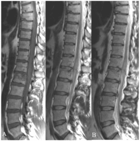
Thin-section axial CT performed by using a bone algorithm is the single most sensitive means by which to diagnose fractures of the thoracic spine. Routine helical CT scans of the thoracic spine are valuable because multisection CT scanners can generate high-resolution spinal images, even during a primary multisystemic trauma evaluation. [21, 22, 28, 29]
The CT images below display various thoracic spinal traumatic injuries.
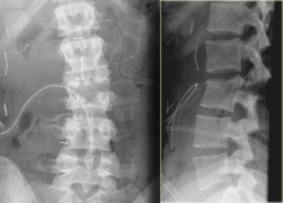
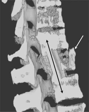
Figure 1: Lateral 3-dimensional maximum intensity projection CT scan of multiple upper thoracic and lower cervical spinous process fractures. The force necessary to fracture the spinous processes of the upper thoracic spine may also involve the lower cervical spine.
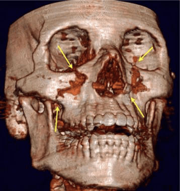
Figure 2:�Three-dimensional CT scan of complex mid-face fractures including a Le Fort I injury in a patient who had fractures of the upper thoracic and lower cervical spinous processes. Sudden deceleration of the face and skull resulted in severe stress forces on the spinous processes.
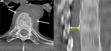
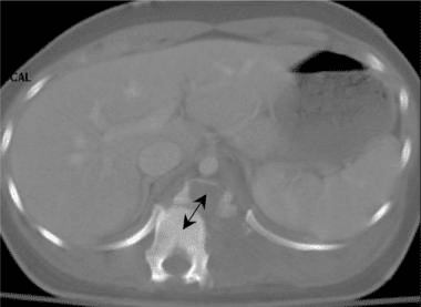
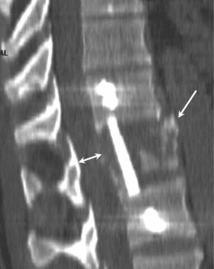
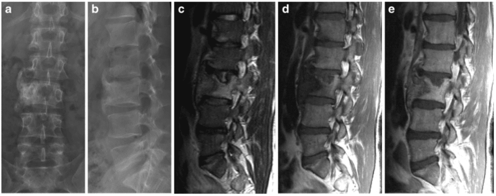
Figure 3:�Axial CT scan of a T12 compression fracture demonstrates a fracture line through the anterior body of the T12 (white arrow), posterior displacement of the T12 vertebral endplate (black arrow) into the spinal canal, and a fracture of the left transverse spinous process.
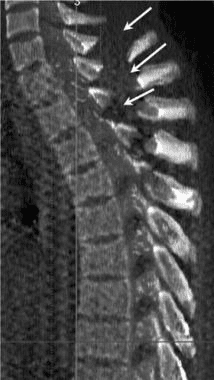
Figure 4:�Axial and sagittal CT images of an acute lower thoracic spine compression fracture. Note the paraspinal hematoma (white arrows) and the slight narrowing of the spinal canal at the level of the compression fracture (double yellow arrows).
Figure 5:�Three-dimensional CT scan of the thoracic spine demonstrates a compression fracture.
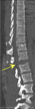
Figure 6:�Sagittal CT scan of the thoracic and lumbar spine demonstrates a complete distraction fracture at the L1-2 interspace (arrow).
Figure 7:�Axial CT image of an unstable fracture of the thoracic spine. Note the association of compression of the vertebral body with laminar and pedicle fractures. Injury to the anterior, middle and posterior columns results in an unstable fracture.
Figure 8:�Coronal multiplanar reformatted CT images of an unstable thoracic spinal fracture. The association of both anterior compression and lateral subluxation (arrows) indicates instability.
Figure 9:�Volume maximum intensity projection CT image of the entire thoracic spine demonstrates spinous process fractures of the C7 through T7 vertebra. Although spinous process fractures of the T1 may occur in a manner similar to a clay shoveler’s fracture of the C6 or C7, middle and lower thoracic spinous process fractures most likely occur due to a combination of forward flexion and axial rotation. Note the lack of findings of compression vertebral body fractures.
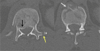
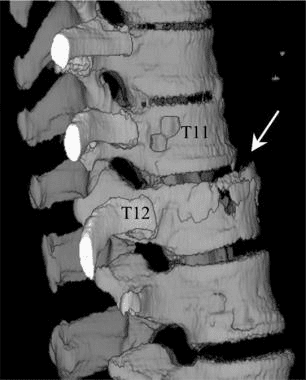
Figure 10:�Three-dimensional surface CT image of the cervical spine. Note the spinous process fractures of the C6, C7, and T1. CT examination of both the cervical and the thoracic spine was obtained as a single study using a multisection CT scanner. All images were obtained by using a 3-mm reconstruction with 1.5-mm collimation. Scanning times were 0.5 seconds per rotation. These 3-dimensional images were reconstructed by using an independent imaging workstation. In complex cases, reconstructed images are very useful in consultation with treating physicians.
Figure 11:�Scout view image from a spiral CT scan shows a complete subluxation fracture (curved blue lines) of the lower thoracic spine. Such an injury combines lateral displacement with rotational injury (arrow).
Figure 12: Fracture dislocation of the lower thoracic spine. Axial CT image demonstrates the large distance that the lower thoracic spine has been displaced.
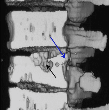
Figure 13:�Axial CT myelogram in a patient with a gunshot wound to the thoracic spine. While a fracture is obvious, the injury also resulted in a dural tear with a freely leaking cerebrospinal fluid space (white arrow). The midline fracture of the vertebral body is noted in the lower image (black arrow).
Figure 14:�Axial CT image demonstrates a complex fracture of the T12 with rotation subluxation. Air was introduced into the epidural space during the injury.
Figure 15:�Sagittal multi-planar CT image of a burst fracture following fixation. The image has been cut in the sagittal plane. Surgical repair of unstable thoracic spine fractures, such as this burst fracture, usually involves placement of an interposition graft (double black arrow) together with a lateral plate held in position by screws placed into the vertebral body above and below the injury. A residual fragment of the burst fracture is seen anteriorly (white arrow). The double white arrow illustrates the restored spinal canal.
Figure 16:�Shaded-surface 3-dimensional CT image of a burst fracture following fixation. The image has been cut in the sagittal plane. Surgical repair of unstable thoracic spine fractures, such as this burst fracture, usually involves placement of an interposition graft (double black arrow) together with a lateral plate held in position by screws placed into the vertebral body above and below the injury. A residual fragment of the burst fracture is seen anteriorly (white arrow).
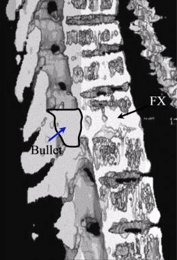
Figure 17: Shaded-surface 3-dimensional CT image of a gunshot wound to the thoracic spine. Although the bullet passed into the interspace, causing a fracture of the vertebral body, the bullet stopped within the spinal canal. Note the outline drawn around the bullet (arrow).
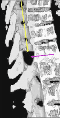
Figure 18:�Shaded-surface 3-dimensional CT scan of a gunshot wound to the thoracic spine. In other cases, the bullet may enter the spinal canal superior to the final position in the canal. The passage of the bullet within the spinal canal (yellow arrow) destroys the spinal cord and also may result in a fracture of the vertebral body. Note that the bullet has been darkened (blue arrow).
Figure 19:�Axial CT image in a man with known pulmonary tuberculosis and back pain. Note the left-sided paraspinal abscess (arrow).
Figure 20:�Sagittal shaded-surface 3-dimensional reconstruction CT scan of the lower thoracic spine. The spinal image has been cut in the midsagittal plane to demonstrate posterior displacement of the thoracic spinal vertebral body (arrow) and downward displacement of the superior endplate. Note the general wedge shape of the vertebral body.
Because of its superior contrast definition and the absence of superimposed structures, good-quality CT imaging depicts more thoracic spinal injuries than do conventional radiographic studies. However, the percentage of clinically important fractures that are seen on CT scans but not on radiographs is lower with thoracic than with cervical spinal fractures. Most of the fractures missed on radiographs were spinous process fractures, transverse processes fractures, and fractures in large patients. Because axial CT is performed with patients in a neutral position, bony distraction of the fracture fragments and subluxations of the spinal articulations may not be as significant on CT images as on they are on acute trauma-series radiographs. [22, 25, 28, 29, 30, 31, 32]
The level of a burst fracture and the percentage of spinal canal stenosis have been correlated with associated neurologic deficits. A significant correlation exists between neurologic deficit and the percentage of spinal canal stenosis. The higher the level of injury, the greater the probability of neurologic deficit. This association may be related to the smaller canal diameter in the upper thoracic spine. The severity of neurologic deficit cannot be predicted.
In patients with Chance-type fractures, CT scans often show a burst-type fracture with posterior cortex buckling or retropulsion, and serial transaxial CT images often show a gradual loss of definition of the pedicles. [23]
The thoracic spine, located between the cervical and lumbar vertebrae, consists of 12 vertebrae levels. Thoracic spinal trauma, including spinal cord injuries along the middle of the spine, can generally be severe, however, with early treatment, long-term prognosis is good. Therefore, imaging diagnostics for thoracic spinal trauma are essential. Many healthcare professionals can provide patients with these services.
Dr. Alex Jimenez D.C., C.C.S.T.
�
Degree of Confidence
The confidence level for the diagnosis of a thoracic spinal fracture with 2-mm axial sections (possible with a multisection CT unit) is greater than 98% and reportedly 99%.
Because axial CT is performed with the patient in a neutral position, a bony distraction of the fracture fragments and subluxations of the spinal articulations may not be as significant on CT images as on acute trauma-series radiographs.
False Positives/Negatives
False-positive results may occur in patients with a Schmorl node, which is a chronic internal herniation of the vertebral disk into the thoracic vertebral body endplate and failure of the fusion of the anterior vertebral endplate epiphysis, resulting in a limbus vertebra. False-negative CT studies may occur in chronic stress injuries and severe generalized osteoporotic endplate fractures.
It has been reported that among trauma patients who had a chest and/or abdominal CT, fractures of the thoracic spine are frequently underreported. Sagittal reformats of the spine obtained from thin sections, and morphometric analysis using electronic calipers help to identify fractures that might otherwise not be identified. [25]
In conclusion, imaging diagnostics of�spinal trauma or spine fracture are essential towards the assessment and treatment of patients. Magnetic resonance imaging, or MRI, is helpful in the evaluation of spinal cord and soft-tissue injuries whereas computed tomography scanning, or CT scans, is helpful in the evaluation of spinal trauma or spine fracture. The understanding of imaging technology has tremendously enhanced advances in treatment.� The scope of our information is limited to chiropractic, spinal injuries, and conditions. To discuss the subject matter, please feel free to ask Dr. Jimenez or contact us at�915-850-0900�.
Curated by Dr. Alex Jimenez
Additional Topics: Acute Back Pain
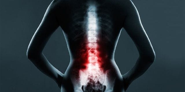
Back pain�is one of the most prevalent causes of disability and missed days at work worldwide. Back pain attributes to the second most common reason for doctor office visits, outnumbered only by upper-respiratory infections. Approximately 80 percent of the population will experience back pain at least once throughout their life. The spine is a complex structure made up of bones, joints, ligaments, and muscles, among other soft tissues. Because of this, injuries and/or aggravated conditions, such as�herniated discs, can eventually lead to symptoms of back pain. Sports injuries or automobile accident injuries are often the most frequent cause of back pain, however, sometimes the simplest of movements can have painful results. Fortunately, alternative treatment options, such as chiropractic care, can help ease back pain through the use of spinal adjustments and manual manipulations, ultimately improving pain relief.
Imaging diagnostics of the spine consist from radiographies to computed tomography scanning, or CT scans, in which CT is utilized in conjunction with myelography and most recently with magnetic resonance imaging, or MRI. These imaging diagnostics are being used to determine the presence of abnormalities of the spine, scoliosis, spondylolysis and spondylolisthesis. The following article describes various imaging modalities and their application in the evaluation of common spinal disorders described.
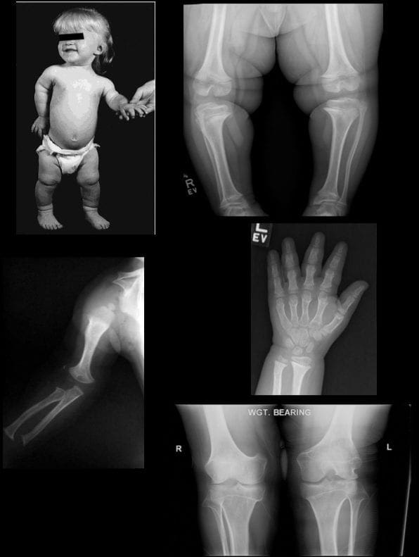
Achondroplasia
Achondroplasia is the most common cause of rhizomelic (root/proximal) short-limb dwarfism. Patients are of normal intelligence.�
It shows multiple distinct radiographic abnormalities affecting long bones, pelvis, skull, and hands.
Vertebral column changes may present with significant clinical and neurological abnormalities.�
Achondroplasia is an autosomal dominant disorder with about 80% of cases from a random new mutation. Advanced paternal age is often linked. Achondroplasia results from a mutation in the fibroblast growth factor gene (FGFR3) which causes abnormal cartilage formation.
All bones formed by endochondral ossification are affected.
Bones that form by intra-membranous ossification are not normal.
Thus, skull vault, iliac wings develop normally vs. the base of the skull, some facial bones, vertebral column, and most tubular bones are abnormal.
�
Dx: is usually made at birth with many features becoming apparent during the first few years of life.
Radiography plays an important part of clinical diagnosis.
Typical features include: shortening and widening of tubular bones, metaphyseal flaring, Trident hand with short, broad metacarpals and proximal and middle phalanges. Longer Fibular, Tibial bowing, markedly short humeri often with dislocated Radial head and elbow flexion deformity.
Spine: characteristic narrowing of L1-L5 interpedicular distance on AP views. Lateral view shows shortening of pedicles and vertebral bodies, �bullet shaped vertebrae� can be a characteristic feature. Early degenerative changes and canal narrowing occur. The horizontal sacral inclination is an important feature.
Pelvis is broad and short with characteristic �champagne glass� pelvis appearance.
Femoral heads are hypoplastic, but hip arthrosis is normally not observed even in older patients likely due to reduced leverage and lightweight (50kg) of patients.
Management of Achondroplasia
Recombinant human growth hormone (GH)�is currently being used to augment the height of patients with achondroplasia.
Most complications of Achondroplasia are related to the spine: vertebral canal stenosis, thoracolumbar kyphosis, narrowed foramen magnum and others.
Laminectomy extending to pedicles/lateral recess with foraminotomies and discectomies can be performed.
Cervical manipulations are contraindicated.
Imaging diagnostics play a fundamental role in the diagnosis the of scoliosis, an abnormality of the spine which is believed to occur due to an underlying health issue, although most cases of scoliosis are idiopathic. More over, radiographies, CT scans, and MRI, among others, can help monitor the changes of the deformity of the spine associated with this spinal manifestation. Chiropractors can provide imaging diagnostics to patients with scoliosis before proceeding with treatment.�
Dr. Alex Jimenez D.C., C.C.S.T.
�
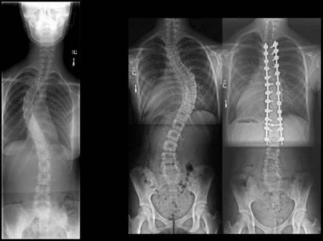
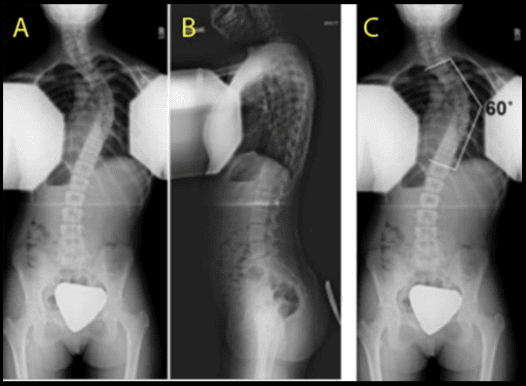
Scoliosis
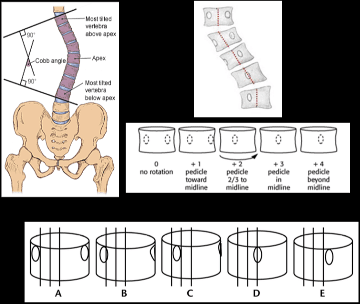
Scoliosis is defined as the abnormal lateral curvature of the spine >10-degree when examined by Cobb�s method of mensuration.
Scoliosis can be described as postural and structural.
Postural scoliosis is not fixed and can be improved by lateral flexion to the side of the convexity.
Structural scoliosis has multiple causes ranging from: ? Idiopathic (>80%) ? Congenital (wedge or hemivertebra, blocked vertebra, Marfan syndrome, skeletal dysplasias) ? Neuropathic (neurofibromatosis, neurological conditions like tethered cord, spinal dysraphism, etc.) ? Scoliosis d/t Spinal neoplasms ? Post-traumatic etc.
Idiopathic scoliosis is the most common type (>80%).
Idiopathic scoliosis can be of 3-types ( infantile, juvenile, adolescent).
Idiopathic adolescent scoliosis if patients >10y.o.
Infantile scoliosis if <3 y.o. M>F.
Juvenile scoliosis if >3 but <10-y.o.
Idiopathic Adolescent scoliosis is the most common with F:M 7:1 (adolescent girls are at particular risk).
Etiology: unknown thought to be the result of some disturbance of proprioceptive control of the spine and spinal musculature, other hypotheses exist.
Most seen in the thoracic region and most commonly convex to the right.
Dx: full spine radiography with gonadal and breast shielding (preferably PA views to protect breast tissue).
� Curves that are 50-degrees or greater and rapidly progressing will require operative intervention to prevent severe deformity of the thorax & ribs leading to cardiopulmonary abnormalities. � �? If curvature is < 20-degree, no treatment is required (observation). � �? For curves that are >20-40-degrees bracing may be used (orthosis).
Milwaukee (metal) brace (left).
Boston brace polypropylene lined with polyethylene (right) often preferred because it can be worn under clothing.
Bracing wearing is required for 24-hours for the duration of the treatment.
Note Cobb�s method of mensuration to record spinal curvature. It has some limitations: 2D imaging, not able to estimate rotation, etc.
Cobb�s method is still a standard evaluation performed in Scoliosis studies.
Nash-Moe method: determines pedicle rotation in scoliosis.
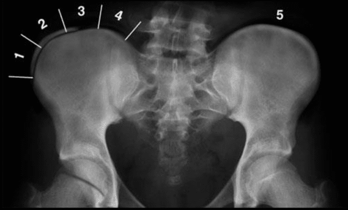
Risser index is used to estimate spinal skeletal maturity.
Iliac growth apophysis appears at ASIS (F- 14, M-16) and progresses medially and expected to be closed in 2-3-years (Risser 5).
Scoliosis progression ends at Risser 4 in females & Risser 5 in males.
During radiographic evaluation of scoliosis, it is crucial to report if Risser growth apophysis remains open or closed.
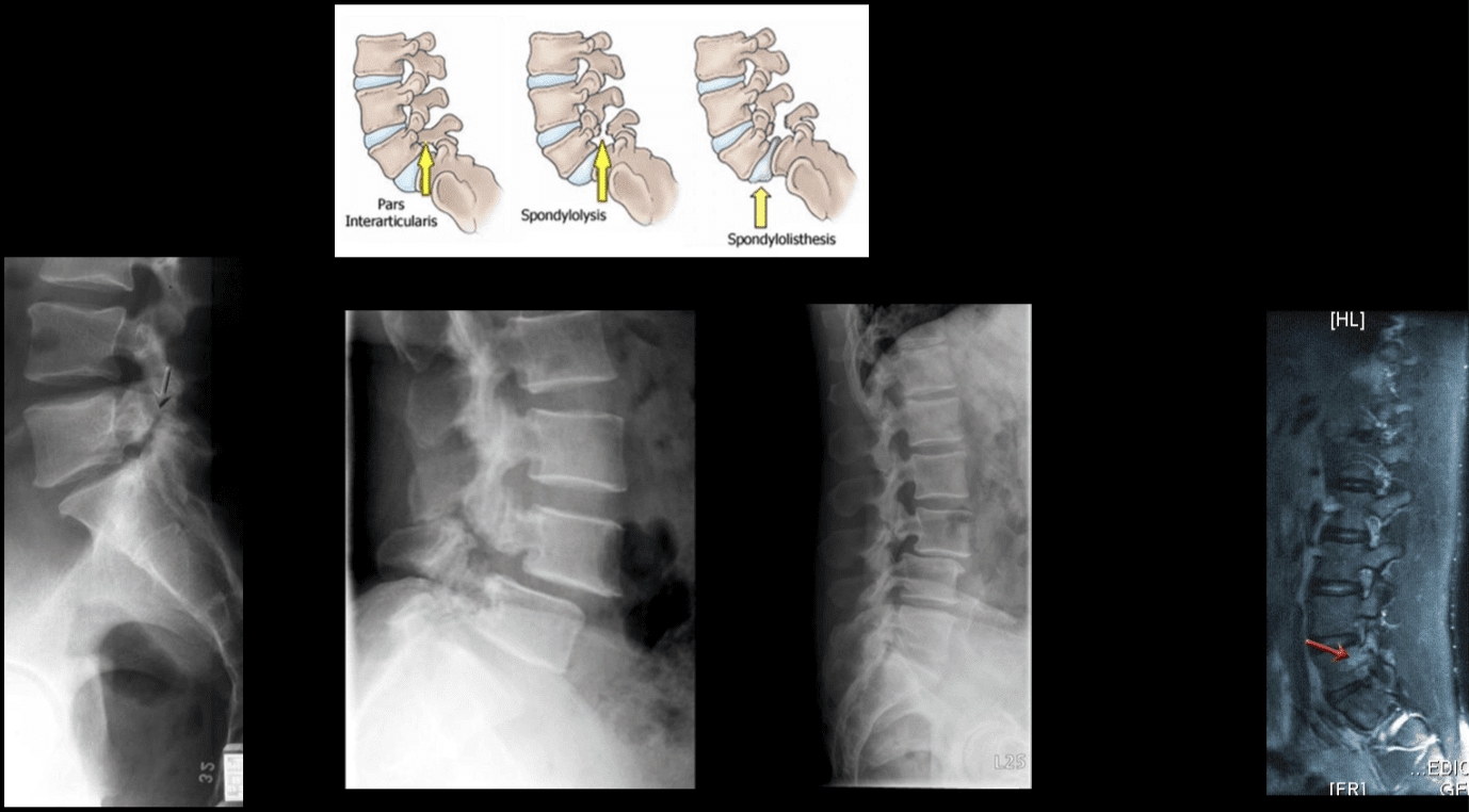
Spondylolysis and spondylolisthesis are health issues which can result in back pain. Spondylolysis is believed to be caused by repeated microtrauma leading to stress fractures in the pars interarticularis. Patients with bilateral pars defects can develop spondylolisthesis, where the degree of slippage of the adjacent vertebrae can progress gradually over time. Patients with suspected spondylolysis and spondylolisthesis may initially be evaluated with pain radiography. Chiropractic care can also help provide imaging diagnostics for these health issues.
Dr. Alex Jimenez D.C., C.C.S.T.
�
Spondylolysis & Spondylolisthesis
Spondylolysis defect in pars interarticularis or osseous bridge between superior and inferior articular processes.
Pathology stress fracture of the pars, believed to be after repeated microtrauma on extensions Men > Women, affects 5% of the general population especially in athletic adolescents.
Clinically postulated that adolescent back pain cases may be related to this process.
Typically spondylolysis remains asymptomatic.
Spondylolysis can be present with or w/o spondylolisthesis.
Spondylolysis is found in 90% at L5 with the remaining 10% in L4.
Can be uni or bilateral.
In 65%�of�cases, spondylolysis is associated with spondylolisthesis.
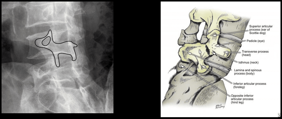
Radiographic Features: break in the Scotty dog collar around the neck on oblique lumbar views.
Radiography has low sensitivity compared to SPECT. SPECT is associated with ionizing radiation, and MRI is currently a preferred method of imaging diagnosis.
MRI can help to show reactive marrow edema next to pars defect or w/o defect so-called pending or potential to develop spondylolysis.
Types of Spondylolisthesis
Type 1 – Dysplastic, rare and found in congenital dysplastic malformation of the sacrum allowing anterior displacement of L5 on S1. Often no pars defect.
Type 2 – Isthmic, most common, often the result of a stress fracture.
Type 3 – Degenerative from the remodeling of articular processes.
Type 4 – Traumatic in an acute posterior arch fracture.
Type 5 – Pathologic due to bone disease locally or generalized.
Grading of spondylolisthesis is based on the Myereding Classification. This classification refers to the overhanging part of the superior body in relation to anterior-posterior part of the inferior body.
Grade 1 – 0-25% anterior slip
Grade 2 – 26-50%
Grade 3 – 51%-75%
Grade 4 – 76-100%
Grade 5 – >100% spondyloptosis
Note degenerative spondylolisthesis at L4 and retrolisthesis at L2, L3.
This abnormality develops due to degeneration of facets and disc with decreased local stability.
Rarely progresses beyond Grade 2.
Must be recognized in the imaging report.
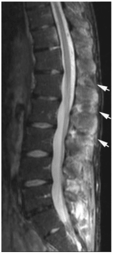
Contributes to vertebral canal stenosis.
Canal stenosis is better delineated by cross-sectional imaging.
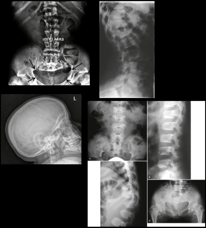
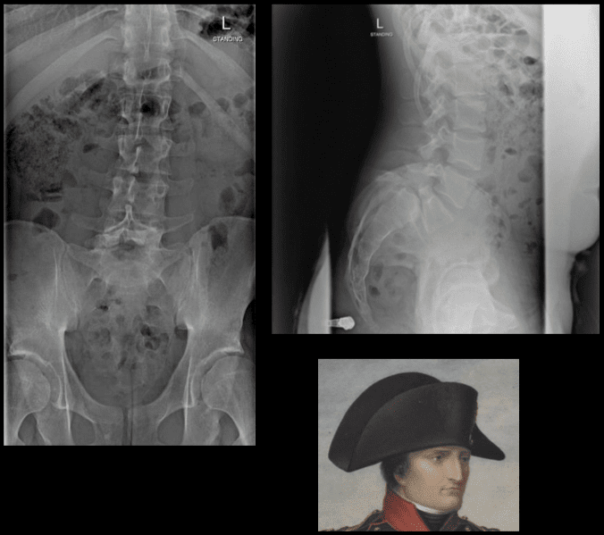
The inverted Napoleon hat sign -�seen on the frontal lumbar/pelvic radiographs at L5-S1.
Represents bilateral spondylolysis with marked anterolisthesis of L5 on S1 often with spondyloptosis and marked exaggeration of the normal lordosis.
Spondylolysis resulting in this degree of spondylolisthesis is more often congenital and/or traumatic in origin and less often degenerative.
The “brim” of the hat is formed by the downward rotation of the transverse processes, and the “dome” of the hat is formed by the body of L5.
In conclusion,�imaging diagnostics for the spine are recommended for patients with specific abnormalities of the spine, however, their increased use can help determine�their best treatment option. Understanding the abnormalities of the spine described above can help healthcare professionals and patients create a treatment program to improve their symptoms. The scope of our information is limited to chiropractic as well as to spinal injuries and conditions. To discuss the subject matter, please feel free to ask Dr. Jimenez or contact us at�915-850-0900�.
Curated by Dr. Alex Jimenez
Additional Topics: Acute Back Pain
Back pain�is one of the most prevalent causes of disability and missed days at work worldwide. Back pain attributes to the second most common reason for doctor office visits, outnumbered only by upper-respiratory infections. Approximately 80 percent of the population will experience back pain at least once throughout their life. The spine is a complex structure made up of bones, joints, ligaments, and muscles, among other soft tissues. Because of this, injuries and/or aggravated conditions, such as�herniated discs, can eventually lead to symptoms of back pain. Sports injuries or automobile accident injuries are often the most frequent cause of back pain, however, sometimes the simplest of movements can have painful results. Fortunately, alternative treatment options, such as chiropractic care, can help ease back pain through the use of spinal adjustments and manual manipulations, ultimately improving pain relief.
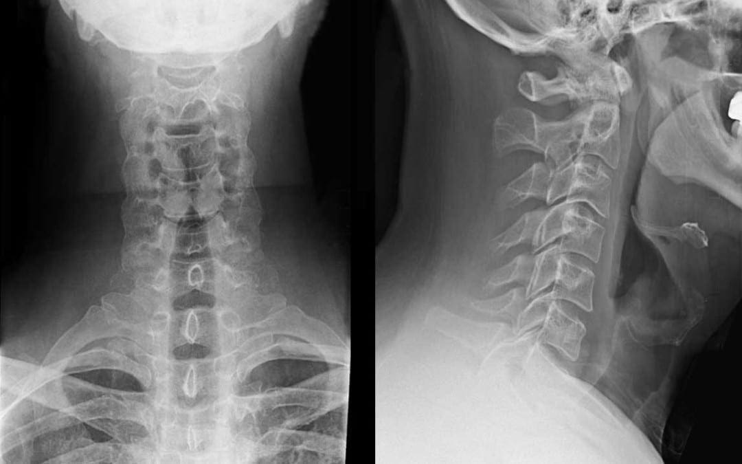
While computed tomography scanning, or CT scans, of the cervical spine are frequently utilized to help diagnose neck injuries, simple radiographs are still commonly performed for patients who have experienced minor cervical spine injuries with moderate neck pain, such as those who have suffered a slip-and-fall accident. Imaging diagnostic assessments may reveal underlying injuries and/or aggravated conditions to be more severe than the nature of the trauma. The purpose of the article is to demonstrate the significance of cervical spine radiographs in the trauma patient.�
Abstract
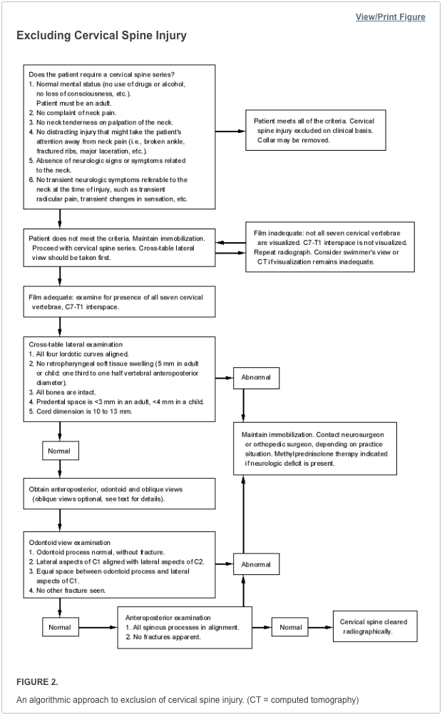
Significant cervical spine injury is very unlikely in a case of trauma if the patient has normal mental status (including no drug or alcohol use) and no neck pain, no tenderness on neck palpation, no neurologic signs or symptoms referable to the neck (such as numbness or weakness in the extremities), no other distracting injury and no history of loss of consciousness. Views required to radiographically exclude a cervical spine fracture include a posteroanterior view, a lateral view and an odontoid view. The lateral view must include all seven cervical vertebrae as well as the C7-T1 interspace, allowing visualization of the alignment of C7 and T1. The most common reason for a missed cervical spine injury is a cervical spine radiographic series that is technically inadequate. The �SCIWORA� syndrome (spinal cord injury without radiographic abnormality) is common in children. Once an injury to the spinal cord is diagnosed, methylprednisolone should be administered as soon as possible in an attempt to limit neurologic injury.
Radiographs continue to be used as a first-line imaging diagnostic assessment modality in the evaluation of patients with suspected cervical spine injuries. The aim of cervical spine radiographs is to confirm the presence of a health issue in the complex structures of the neck and define its extent, particularly with respect to instability. Multiple views may generally be necessary to provide optimal visualization.
Dr. Alex Jimenez D.C., C.C.S.T.
Introduction
Although cervical spine radiographs are almost routine in many emergency departments, not all trauma patients with a significant injury must have radiographs, even if they arrive at the emergency department on a backboard and wearing a cervical collar. This article reviews the proper use of cervical spine radiographs in the trauma patient.
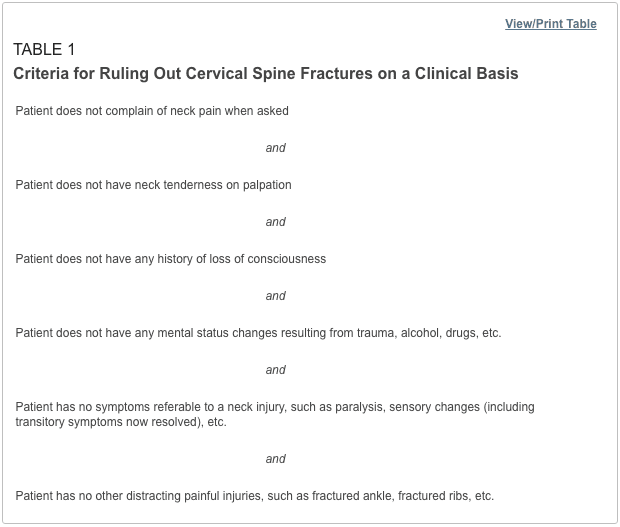
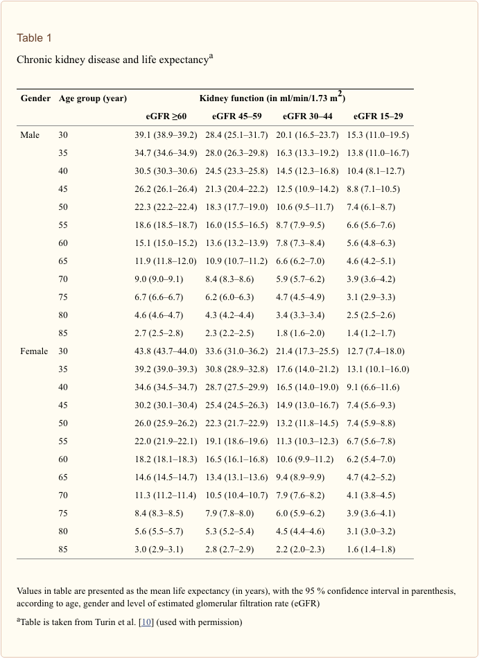
Low-risk criteria have been defined that can be used to exclude cervical spine fractures, based on the patient’s history and physical examination.1�6 Patients who meet these criteria (Table 1) do not require radiographs to rule out cervical fractures. However, the criteria apply only to adults and to patients without mental status changes, including drug or alcohol intoxication. Although studies suggest that these criteria may also be used in the management of verbal children,7�9 caution is in order, since the study series are small, and the ability of children to complain about pain or sensory changes is variable. An 18-year-old patient can give a more reliable history than a five-year-old child.
Some concern has been expressed about case reports suggesting that �occult� cervical spine fractures will be missed if asymptomatic trauma patients do not undergo radiography of the cervical spine.10 On review, however, most of the reported cases did not meet the low-risk criteria in Table 1. Attention to these criteria can substantially reduce the use of cervical spine radiographs.
Cervical Spine Series and Computed Tomography
Once the decision is made to proceed with a radiographic evaluation, the proper views must be obtained. The single portable cross-table lateral radiograph, which is sometimes obtained in the trauma room, should be abandoned. This view is insufficient to exclude a cervical spine fracture and frequently must be repeated in the radiographic department.11,12 The patient’s neck should remain immobilized until a full cervical spine series can be obtained in the radiographic department. Initial films may be taken through the cervical collar, which is generally radiolucent. An adequate cervical spine series includes three views: a true lateral view, which must include all seven cervical vertebrae as well as the C7-T1 junction, an anteroposterior view and an open-mouth odontoid view.13
If no arm injury is present, traction on the arms may facilitate visualization of all seven cervical vertebrae on the lateral film. If all seven vertebrae and the C7-T1 junction are not visible, a swimmer’s view, taken with one arm extended over the head, may allow adequate visualization of the cervical spine. Any film series that does not include these three views and that does not visualize all seven cervical vertebrae and the junction of C7-T1 is inadequate. The patient should be maintained in cervical immobilization, and plain films should be repeated or computed tomographic (CT) scans obtained until all vertebrae are clearly visible. The importance of obtaining all of these views and visualizing all of the vertebrae cannot be overemphasized. While some missed cervical fractures, subluxations and dislocations are the result of film misinterpretation, the most frequent cause of overlooked injury is an inadequate film series.14,15
In addition to the views listed above, some authors suggest adding two lateral oblique views.16,17 Others would obtain these views only if there is a question of a fracture on the other three films or if the films are inadequate because the cervicothoracic junction is not visualized.18 The decision to take oblique views is best made by the clinician and the radiologist who will be reviewing the films.
Besides identifying fractures, plain radiographs can also be useful in identifying ligamentous injuries. These injuries frequently present as a malalignment of the cervical vertebrae on lateral views. Unfortunately, not all ligamentous injuries are obvious. If there is a question of ligamentous injury (focal neck pain and minimal malalignment of the lateral cervical x-ray [meeting the criteria in Table 2]) and the cervical films show no evidence of instability or fracture, flexion-extension views should be obtained.17,19 These radiographs should only be obtained in conscious patients who are able to cooperate. Only active motion should be allowed, with the patient limiting the motion of the neck based on the occurrence of pain. Under no circumstance should cervical spine flexion and extension be forced, since force may result in cord injury.
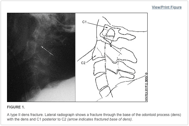
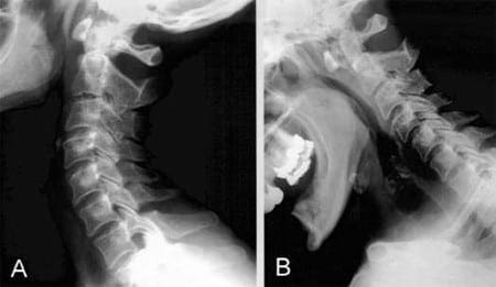
Although they may be considered adequate to rule out a fracture, cervical spine radiographs have limitations. Up to 20 percent11,20,21 of fractures are missed on plain radiographs. If there is any question of an abnormality on the plain radiograph or if the patient has neck pain that seems to be disproportionate to the findings on plain films, a CT scan of the area in question should be obtained. The CT is excellent for identifying fractures, but its ability to show ligamentous injuries is limited.22 Occasionally, plain film tomography may be in order if there is a concern about a type II dens fracture (Figure 1).
While some studies have used magnetic resonance imaging (MRI) as an adjunct to plain films and CT scanning,23,24 the lack of wide availability and the relatively prolonged time required for MRI scanning limits its usefulness in the acute setting. Another constraint is that resuscitation equipment with metal parts may not be able to function properly within the magnetic field generated by the MRI.
Cervical Spine Radiography
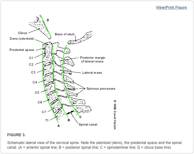
Figure 2 summarizes the approach to reading cervical spine radiographs.
Lateral View
Alignment of the vertebrae on the lateral film is the first aspect to note (Figure 3). The anterior margin of the vertebral bodies, the posterior margin of the vertebral bodies, the spinolaminar line and the tips of the spinous processes (C2-C7) should all be aligned. Any malalignment (Figures 4 and 5) should be considered evidence of ligamentous injury or occult fracture, and cervical spine immobilization should be maintained until a definitive diagnosis is made.
Confusion can sometimes result from pseudosubluxation, a physiologic misalignment that is due to ligamentous laxity, which can occur at the C2-C3 level and, less commonly, at the C3-C4 level. While pseudosubluxation usually occurs in children, it also may occur in adults. If the degree of subluxation is within the normal limits listed in Table 2 and the neck is not tender at that level, flexion-extension views may clarify the situation. Pseudosubluxation should disappear with an extension view. However, flexion-extension views should not be obtained until the entire cervical spine is otherwise cleared radiographically.
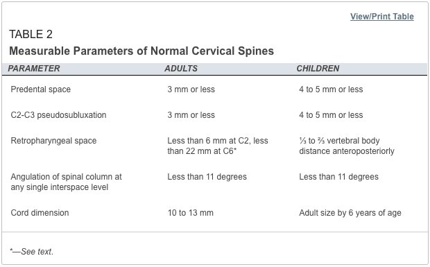
After ensuring that the alignment is correct, the spinous processes are examined to be sure that there is no widening of the space between them. If widening is present, a ligamentous injury or fracture should be considered. In addition, if angulation is more than 11 degrees at any level of the cervical spine, a ligamentous injury or fracture should be assumed. The spinal canal (Figure 2) should be more than 13 mm wide on the lateral view. Anything less than this suggests that spinal cord compromise may be impending.
Next, the predental space�the space between the odontoid process and the anterior portion of the ring of C1 (Figure 2)�is examined. This space should be less than 3 mm in adults and less than 4 mm in children (Table 2). An increase in this space is presumptive evidence of a fracture of C1 or of the odontoid process, although it may also represent ligamentous injury at this level. If a fracture is not found on plain radiographs, a CT scan should be obtained for further investigation. The bony structures of the neck should be examined, with particular attention to the vertebral bodies and spinous processes.
The retropharyngeal space (Figure 2) is now examined. The classic advice is that an enlarged retropharyngeal space (Table 2) indicates a spinous fracture. However, the normal and abnormal ranges overlap significantly.25 Retropharyngeal soft tissue swelling (more than 6 mm at C2, more than 22 mm at C6) is highly specific for a fracture but is not very sensitive.26 Soft tissue swelling in symptomatic patients should be considered an indication for further radiographic evaluation. Finally, the craniocervical relationship is checked.
Odontoid View
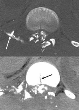
The dens is next examined for fractures. Artifacts may give the appearance of a fracture (either longitudinal or horizontal) through the dens. These artifacts are often radiographic lines caused by the teeth overlying the dens. However, fractures of the dens are unlikely to be longitudinally oriented. If there is any question of a fracture, the view should be repeated to try to get the teeth out of the field. If it is not possible to exclude a fracture of the dens, thin-section CT scans or plain film tomography is indicated.
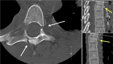
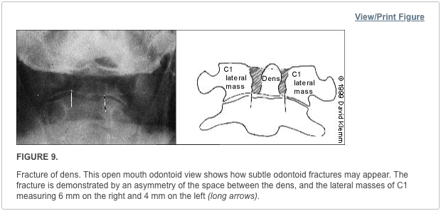
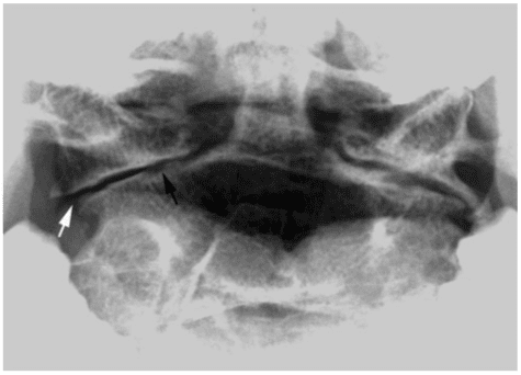
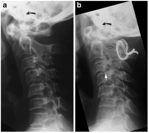
Next, the lateral aspects of C1 are examined. These aspects should be symmetric, with an equal amount of space on each side of the dens. Any asymmetry is suggestive of a fracture. Finally, the lateral aspects of C1 should line up with the lateral aspects of C2. If they do not line up, there may be a fracture of C1. Figure 6 demonstrates asymmetry in the space between the dens and C1, as well as displacement of the lateral aspects of C1 laterally.
Anteroposterior View
The height of the cervical spines should be approximately equal on the anteroposterior view. The spinous processes should be in midline and in good alignment. If one of the spinous processes is off to one side, a facet dislocation may be present.
Common Cervical Abnormalities
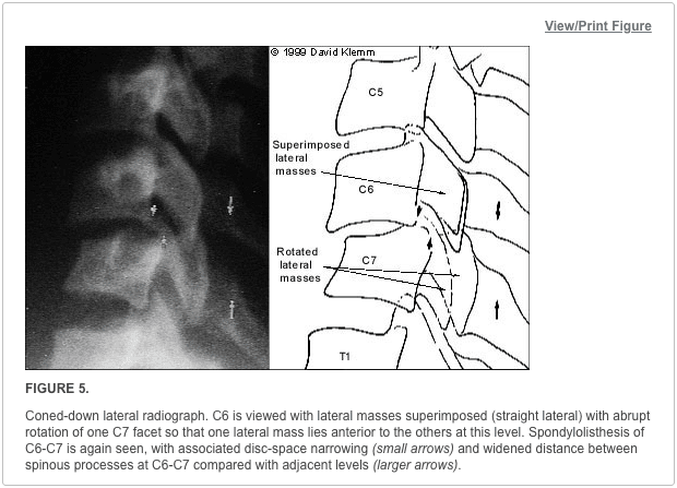
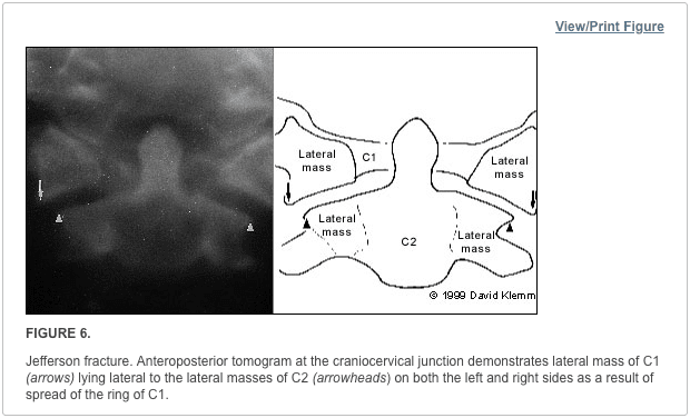
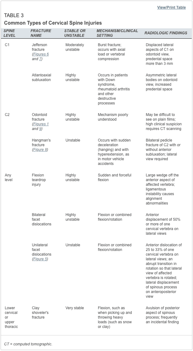
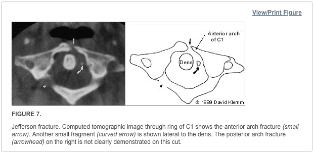
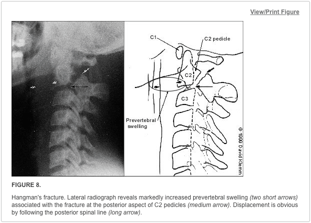
The most common types of cervical abnormalities and their radiographic findings are listed in Table 3. Except for the clay shoveler’s fracture, they should be assumed to be unstable and warrant continued immobilization until definitive therapy can be arranged. Any patient found to have one spinal fracture should have an entire spine series, including views of the cervical spine, the thoracic spine and the lumbosacral spine. The incidence of noncontiguous spine fractures ranges up to 17 percent.27,28 Figures 7 through 9 demonstrate aspects of common cervical spine fractures.
Initial Treatment of Cervical Spine and Cord
If a cervical fracture or dislocation is found, orthopedic or neurosurgical consultation should be obtained immediately. Any patient with a spinal cord injury should begin therapy with methylprednisolone within the first eight hours after the injury, with continued administration for up to 24 hours. Patients should receive methylprednisolone in a dosage of 30 mg per kg given intravenously over one hour. Over the next 23 hours, intravenous methylprednisolone in a dosage of 5.4 mg per kg per hour should be administered. This therapy has been shown to improve outcomes and minimize cord injury,29 although it is not without its problems. The incidence of pneumonia is increased in patients treated with high dosages of methylprednisolone.30
�Sciwora� Syndrome: Unique in Children
A special situation involving children deserves mention. In children, it is not uncommon for a spinal cord injury to show no radiographic abnormalities. This situation has been named �SCIWORA� (spinal cord injury without radiographic abnormality) syndrome. SCIWORA syndrome occurs when the elastic ligaments of a child’s neck stretch during trauma. As a result, the spinal cord also undergoes stretching, leading to neuronal injury or, in some cases, complete severing of the cord.31 This situation may account for up to 70 percent of spinal cord injuries in children and is most common in children younger than eight years. Paralysis may be present on the patient’s arrival in the emergency department. However, up to 30 percent of patients have a delayed onset of neurologic abnormalities, which may not occur until up to four or five days after the injury. In patients with delayed symptoms, many have neurologic symptoms at the time of the injury, such as paresthesias or weakness, that have subsequently resolved.32
It is important to inform the parents of young patients with neck trauma about this possibility so that they will be alert for any developing symptoms or signs. Fortunately, most children with SCIWORA syndrome have a complete recovery, especially if the onset is delayed.33 It is possible to evaluate these injuries with MRI, which will show the abnormality and help determine the prognosis: a patient with complete cord transection is unlikely to recover.3
The treatment of SCIWORA syndrome has not been well studied. However, the general consensus is that steroid therapy should be used.34 In addition, any child who has sustained a significant degree of trauma but has recovered completely should be restricted from physical activities for several weeks.34
Cervical spine radiographs include three standard views, such as the coned odontoid peg view, the anteroposterior view of the entire cervical spine, and the lateral view of the entire cervical spine. Most qualified and experienced healthcare professionals, including chiropractors, offer additional views to visualize the cervicothoracic junction as well as to evaluate the proper alignment of the spine in all patients.�
Dr. Alex Jimenez D.C., C.C.S.T.
About the Authors
MARK A. GRABER, M.D., is associate professor of clinical family medicine and surgery (emergency medicine) at the University of Iowa Hospitals and Clinics, Iowa City. He received his medical degree from Eastern Virginia Medical School, Norfolk, and served a residency in family medicine at the University of Iowa College of Medicine, Iowa City.
MARY KATHOL, M.D., is associate professor of radiology at the University of Iowa Hospitals and Clinics. She is also head of the musculoskeletal radiology section. She received her medical degree from the University of Kansas School of Medicine, Kansas City, Kan., and served a residency in radiology at the University of Iowa College of Medicine.
Address correspondence to Mark A. Graber, M.D., Department of Family Medicine, Steindler Bldg., University of Iowa Hospitals and Clinics, Iowa City, Iowa 52242. Reprints are not available from the authors.
In conclusion,�it is essential to evaluate all views of the cervical spine through imaging diagnostic assessments. While cervical spine radiographs can reveal injuries and conditions, not all neck injuries are detected through radiography. Computed tomography, or CT, scans of the cervical spine are highly accurate in the diagnosis of neck fractures which can help with treatment. The scope of our information is limited to chiropractic as well as to spinal injuries and conditions. To discuss the subject matter, please feel free to ask Dr. Jimenez or contact us at�915-850-0900�.
Curated by Dr. Alex Jimenez
Additional Topics: Acute Back Pain
Back pain�is one of the most prevalent causes of disability and missed days at work worldwide. Back pain attributes to the second most common reason for doctor office visits, outnumbered only by upper-respiratory infections. Approximately 80 percent of the population will experience back pain at least once throughout their life. The spine is a complex structure made up of bones, joints, ligaments, and muscles, among other soft tissues. Because of this, injuries and/or aggravated conditions, such as�herniated discs, can eventually lead to symptoms of back pain. Sports injuries or automobile accident injuries are often the most frequent cause of back pain, however, sometimes the simplest of movements can have painful results. Fortunately, alternative treatment options, such as chiropractic care, can help ease back pain through the use of spinal adjustments and manual manipulations, ultimately improving pain relief.
1.�Kreipke DL, Gillespie KR, McCarthy MC, Mail JT, Lappas JC, Broadie TA. Reliability of indications for cervical spine films in trauma patients.�J Trauma. 1989;29:1438�9.
2.�Ringenberg BJ, Fisher AK, Urdaneta LF, Midthun MA. Rational ordering of cervical spine radiographs following trauma.�Ann Emerg Med. 1988;17:792�6.
3.�Bachulis BL, Long WB, Hynes GD, Johnson MC. Clinical indications for cervical spine radiographs in the traumatized patient.�Am J Surg. 1987;153:473�8.
4.�Hoffman JR, Schriger DL, Mower W, Luo JS, Zucker M. Low-risk criteria for cervical-spine radiography in blunt trauma: a prospective study.�Ann Emerg Med. 1992;21:1454�60.
5.�Saddison D, Vanek VW, Racanelli JL. Clinical indications for cervical spine radiographs in alert trauma patients.�Am Surg. 1991;57:366�9.
6.�Kathol MH, El-Khoury GY. Diagnostic imaging of cervical spine injuries.�Seminars in Spine Surgery. 1996;8(1):2�18.
7.�Lally KP, Senac M, Hardin WD Jr, Haftel A, Kaehler M, Mahour GH. Utility of the cervical spine radiograph in pediatric trauma.�Am J Surg. 1989;158:540�1.
8.�Rachesky I, Boyce WT, Duncan B, Bjelland J, Sibley B. Clinical prediction of cervical spine injuries in children. Radiographic abnormalities.�Am J Dis Child. 1987;141:199�201.
9.�Laham JL, Cotcamp DH, Gibbons PA, Kahana MD, Crone KR. Isolated head injuries versus multiple trauma in pediatric patients: do the same indications for cervical spine evaluation apply?�Pediatr Neurosurg. 1994;21:221�6.
10.�McKee TR, Tinkoff G, Rhodes M. Asymptomatic occult cervical spine fracture: case report and review of the literature.�J Trauma. 1990;30:623�6.
11.�Woodring JH, Lee C. Limitations of cervical radiography in the evaluation of acute cervical trauma.�J Trauma. 1993;34:32�9.
12.�Spain DA, Trooskin SZ, Flancbaum L, Boyarsky AH, Nosher JL. The adequacy and cost effectiveness of routine resuscitation-area cervical-spine radiographs.�Ann Emerg Med. 1990;19:276�8.
13.�Tintinalli JE, Ruiz E, Krome RL, ed. Emergency medicine: a comprehensive study guide. 4th ed. New York: McGraw-Hill, 1996.
15.�Davis JW, Phreaner DL, Hoyt DB, Mackersie RC. The etiology of missed cervical spine injuries.�J Trauma. 1993;34:342�6.
16.�Apple JS, Kirks DR, Merten DF, Martinez S. Cervical spine fractures and dislocations in children.�Pediatr Radiol. 1987;17:45�9.
17.�Turetsky DB, Vines FS, Clayman DA, Northup HM. Technique and use of supine oblique views in acute cervical spine trauma.�Ann Emerg Med. 1993;22:685�9.
18.�Freemyer B, Knopp R, Piche J, Wales L, Williams J. Comparison of five-view and three-view cervical spine series in the evaluation of patients with cervical trauma.�Ann Emerg Med. 1989;18:818�21.
19.�Lewis LM, Docherty M, Ruoff BE, Fortney JP, Keltner RA Jr, Britton P. Flexion-extension views in the evaluation of cervical-spine injuries.�Ann Emerg Med. 1991;20:117�21.
20.�Mace SE. Emergency evaluation of cervical spine injuries: CT versus plain radiographs.�Ann Emerg Med. 1985;14:973�5.
21.�Kirshenbaum KJ, Nadimpalli SR, Fantus R, Cavallino RP. Unsuspected upper cervical spine fractures associated with significant head trauma: role of CT.�J Emerg Med. 1990;8:183�98.
22.�Woodring JH, Lee C. The role and limitations of computed tomographic scanning in the evaluation of cervical trauma.�J Trauma. 1992;33:698�708.
23.�Schaefer DM, Flanders A, Northrup BE, Doan HT, Osterholm JL. Magnetic resonance imaging of acute cervical spine trauma. Correlation with severity of neurologic injury.�Spine. 1989;14:1090�5.
24.�Levitt MA, Flanders AE. Diagnostic capabilities of magnetic resonance imaging and computed tomography in acute cervical spinal column injury.�Am J Emerg Med. 1991;9:131�5.
25.�Templeton PA, Young JW, Mirvis SE, Buddemeyer EU. The value of retropharyngeal soft tissue measurements in trauma of the adult cervical spine. Cervical spine soft tissue measurements.�Skeletal Radiol. 1987;16:98�104.
26.�DeBehnke DJ, Havel CJ. Utility of prevertebral soft tissue measurements in identifying patients with cervical spine fractures.�Ann Emerg Med. 1994;24:1119�24.
29.�Bracken MB, Shepard MJ, Collins WF Jr, Holford TR, Baskin DS, Eisenberg HM, et al. Methylprednisolone or naloxone treatment after acute spinal cord injury: 1-year follow-up data. Results of the second National Acute Spinal Cord Injury Study.�J Neurosurg. 1992;76:23�31.
30.�Galandiuk S, Raque G, Appel S, Polk HC Jr. The two-edged sword of large-dose steroids for spinal cord trauma.�Ann Surg. 1993;218:419�25.
31.�Grabb PA, Pang D. Magnetic resonance imaging in the evaluation of spinal cord injury without radiographic abnormality in children.�Neurosurgery. 1994;35:406�14.
32.�Pang D, Pollack IF. Spinal cord injury without radiographic abnormality in children�the SCIWORA syndrome.�J Trauma. 1989;29:654�64.
33.�Hadley MN, Zabramski JM, Browner CM, Rekate H, Sonntag VK. Pediatric spinal trauma. Review of 122 cases of spinal cord and vertebral column injuries.�J Neurosurg. 1988;68:18�24.
34.�Kriss VM, Kriss TC. SCIWORA (spinal cord injury without radiographic abnormality) in infants and children.�Clin Pediatr. 1996;35:119�24.
The editors of AFP welcome the submission of manuscripts for the Radiologic Decision-Making series. Send submissions to Jay Siwek, M.D., following the guidelines provided in �Information for Authors.�
Coordinators of this series are Thomas J. Barloon, M.D., associate professor of radiology and George R. Bergus, M.D., assistant professor of family practice, both at the University of Iowa College of Medicine, Iowa City.
Many types of arthritis can affect the structure and function of the muscles, bones and/or joints, causing symptoms such as, pain, stiffness and swelling. While arthritis can commonly affect the hands, wrists, elbows, hips, knees and feet, it can also affect the facet joints found along the length of the spine. One of the most well-known types of arthritis, known as rheumatoid arthritis or RA, is a chronic inflammatory disease of the joints which occurs when the human body’s own immune system attacks the synovium, the thin membrane that lines the joints. According to the article below, imaging the spine in arthritis is fundamental towards its proper treatment.
Abstract
Spinal involvement is frequent in rheumatoid arthritis (RA) and seronegative spondyloarthritides (SpA), and its diagnosis is important. Thus, MRI and CT are increasingly used, although radiography is the recommended initial examination. The purpose of this review is to present the typical radiographic features of spinal changes in RA and SpA in addition to the advantages of MRI and CT, respectively. RA changes are usually located in the cervical spine and can result in serious joint instability. Subluxation is diagnosed by radiography, but supplementary MRI and/or CT is always indicated to visualize the spinal cord and canal in patients with vertical subluxation, neck pain and/or neurological symptoms. SpA may involve all parts of the spine. Ankylosing spondylitis is the most frequent form of SpA and has rather characteristic radiographic features. In early stages, it is characterized by vertebral squaring and condensation of vertebral corners, in later stages by slim ossifications between vertebral bodies, vertebral fusion, arthritis/ankylosis of apophyseal joints and ligamentous ossification causing spinal stiffness. The imaging features of the other forms of SpA can vary, but voluminous paravertebral ossifications often occur in psoriatic SpA. MRI can detect signs of active inflammation as well as chronic structural changes; CT is valuable for detecting a�fracture.
The spine can be involved in most inflammatory disorders encompassing rheumatoid arthritis (RA), seronegative spondyloarthritides (SpA), juvenile arthritides and less frequent disorders such as, arthro-osteitis and SAPHO (synovitis, acne, pustulosis, hyperostosis, osteitis) syndrome.
During the last decade, the diagnostic use of magnetic resonance imaging (MRI) and computed tomography (CT) has increased considerably, although radiography is still the recommended initial examination. It is therefore important to know the characteristic radiographic findings in arthritides in addition to the advantages of supplementary MRI and CT. This review will focus on the different imaging features and be concentrated on the most frequent inflammatory spinal changes seen in RA and SpA, respectively. These two entities display somewhat different imaging features, which are important to recognize.
Rheumatoid arthritis is an autoimmune disease which causes the human body’s own immune system to attack and often destroy the lining of the joints. Although it commonly affects the small joints of the hands and feet, rheumatoid arthritis, or RA, can affect any joint in the human body. The neck, or cervical spine, can be affected more often than the lower back if rheumatoid arthritis affects the joints in the spine.�
Dr. Alex Jimenez D.C., C.C.S.T.
�
Rheumatoid Arthritis
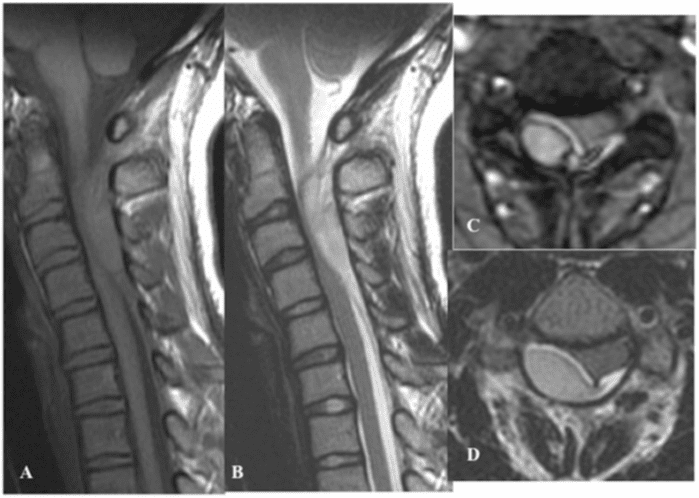
Involvement in RA is usually located in the cervical spine where erosive changes are predominantly seen in the atlanto-axial region. Inflamed and thickened synovium (pannus) can occur around the odontoid process (dens) and cause bone erosion and destruction of surrounding ligaments, most seriously if the posterior transverse ligament is involved. Laxity or rupture of the transverse ligament causes instability with a potential risk of spinal cord injury. Cervical RA involvement is a progressive, serious condition with reduced lifetime expectancy [1], and its diagnosis is therefore important [2, 3].
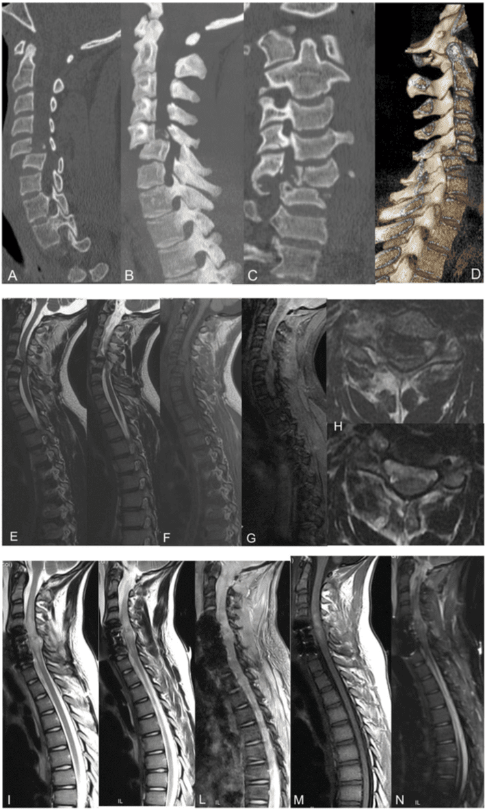
Fig. 1 Standard radiography of the cervical spine in rheumatoid arthritis (RA). (a) Lateral radiographs in neutral position and (b) during flexion in addition to (c) lateral and (d) anterior-posterior (AP) open-mouth view of the atlanto-axial region (45-year-old woman). The flexion view (b) shows abnormal distance (>3 mm) between the posterior aspect of the anterior arc of the atlas and the anterior aspect of the dens (black line). Note that the spino-laminar line of the atlas�(arrow) does not align with that of the other vertebrae, confirming the presence of anterior subluxation, but there is no stenosis of the atlanto- axial canal; the posterior atlanto-dental interval (white line) is >14 mm. The open-mouth view (d) shows erosion at the base of the dens (arrow). (a) and (b) show concomitant disc degenerative changes at the C4�C6 level.
Fig. 2 Lateral and rotatory atlanto-axial subluxation. AP open- mouth view in a 53-year-old man with RA. There is narrowing of the atlanto-axial joints with superficial erosions (black arrow) and lateral displacement of the axis with respect to the lateral masses of the atlas (white arrow); in addition signs indicating rotatory displacement with asymmetry of the distance between the dens and the lateral masses of the atlas.
Radiography of the cervical spine is mandatory in RA patients with neck pain [3]. It should always include a�lateral view in a flexed position compared with a neutral position in addition to special views of the dens area to detect any lesions and/or instability (Fig. 1). A supplementary lateral view during extension can be useful to assess reducibility of atlanto-axial subluxation possibly limited by pannus tissue between the anterior arc of the atlas and dens.
Anterior atlanto-axial subluxation is the most frequent form of RA instability in the occipito-atlanto-axial region, but lateral, rotatory and vertical subluxation can also occur. The definition of the different forms of instability by radiography is as follows [3].
Anterior atlanto-axial subluxation. Distance between the posterior aspect of the anterior arc of the atlas and the anterior aspect of the dens exceeding 3 mm in a neutral position and/or during flexion (Fig. 1). It may cause stenosis of the atlanto-axial canal presenting as a posterior atlanto-dental interval<14 mm (Fig. 1).
Lateral and rotatory atlanto-axial subluxation.�Displacement of the lateral masses of the atlas more than 2 mm in relation to that of the axis and asymmetry of the lateral masses relative to the dens, respectively (Fig. 2). Rotatory�and lateral subluxation is diagnosed on open-mouth anterior-posterior (AP) radiographs. Anterior subluxation often coexists because of the close anatomical relation between the atlas and the axis.
Posterior atlanto-axial subluxation. The anterior arc of the atlas moves over the odontoid process. This is rarely seen, but may coexist with fracture of the dens.
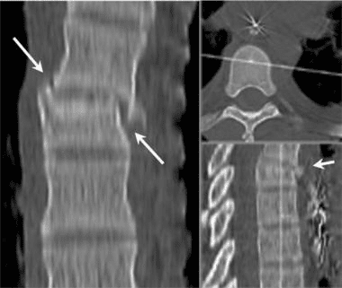
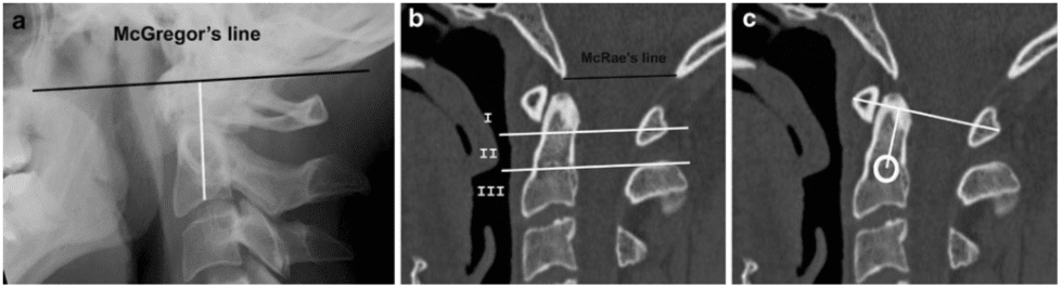
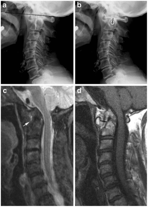
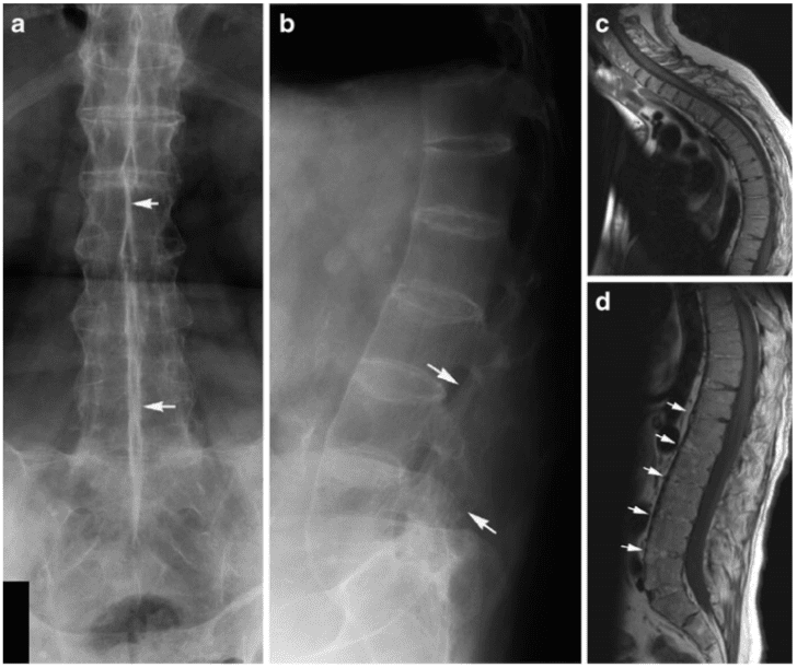
Vertical atlanto-axial subluxation is also referred to as atlanto-axial impaction, basilar invagination or cranial�setting, and is defined as migration of the odontoid tip proximal to McRae�s line corresponding to the occipital foramen. This line can be difficult to define on radiographs, and vertical subluxation has therefore also been defined by several other methods. Migration of the tip of the odontoid process >4.5 mm above McGregor�s line (between the postero-superior aspect of the hard palate and the most caudal point of the occipital curve) indicates vertical subluxation (Fig. 3).
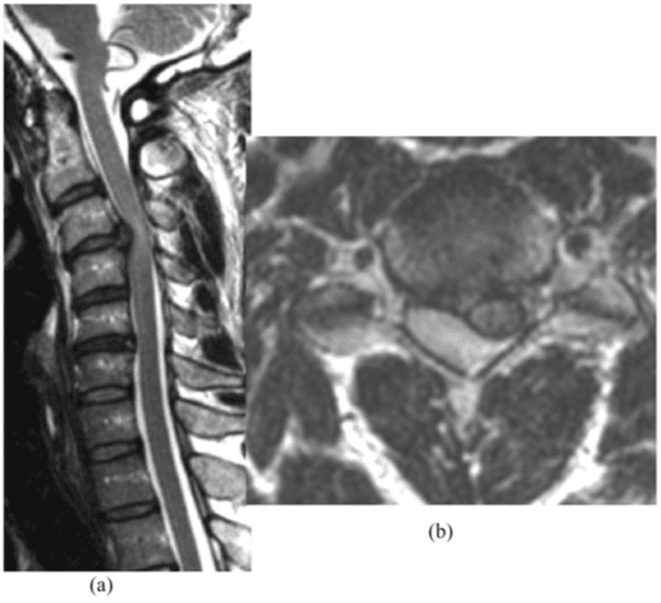
Fig. 3 Vertical atlanto-axial subluxation, measurement methods. (a) Lateral normal radiograph in neutral position showing the location of McGregor�s line (black) between the postero-superior aspect of the hard palate and the most caudal point of the occipital curve. Migration of the tip of the dens >4.5 mm above McGregor�s line indicates vertical subluxation. The distance indicated by the white line between McGregor�s line and the midpoint of the inferior margin of the body of axis is used to evaluate vertical subluxation according to Redlund-Johnell and Pettersson�s method. A distance less than 34 mm in men and 29 mm in women indicates vertical subluxation. (b) Sagittal CT�reconstruction of a normal cervical spine showing the location of McRae�s line corresponding to the occipital foramen and the division of the axis into three equal portions used by Clark�s method for diagnosing vertical subluxation. If the anterior arc of the atlas is in level with the middle or caudal third of the axis there is slight and pronounced vertical subluxation, respectively. (c) Ranawat�s method includes determination of the distance between the centre of the second cervical pedicle and the transverse axis of the atlas. A distance less than 15 mm in males and 13 mm in females indicates vertical subluxation [4].
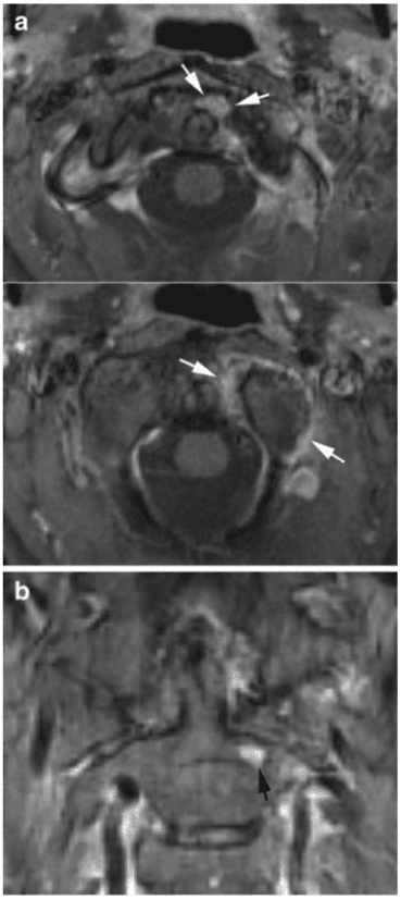
Fig. 4 Vertical subluxation. (a) Lateral radiograph with McGregor�s line (black line; 61-year-old man with RA). The tip of the dens is difficult to define, but measurement according to Redlund-Johnell�s method (white line) results in a distance of 27 mm, which is below the normal limit. In accordance with this, the anterior arc of the atlas is level with the middle third of the axis. (b) Ranawat�s method, the distance between the centre of the second cervical pedicle and the transverse axis of the atlas is below the normal limit (9 mm). Thus, all measurements indicate vertical subluxation. Supplementary MRI, (c) sagittal STIR and (d) T1-weighted images show erosion of the dens and protrusion of the tip into the occipital foramen causing narrowing of the spinal canal to 9 mm, but persistence of cerebrospinal fluid around the cord. There is a 9-mm-thick mass of pannus tissue between the dens and anterior arc (black line) exhibiting small areas with high signal intensity on the STIR image (arrow) compatible with slight activity, but signal void fibrous pannus tissue predominates.
The occurrence of dens erosion can, however, make this measurement difficult to obtain. The Redlund-Johnell method is therefore based on the minimum distance between McGregor�s line and the midpoint of the inferior margin of the body of the axis on a lateral radiograph in a neutral position (Fig. 3) [4]. Visualisation of the palate may not always be obtained. Methods without dens and/or the palate as landmarks have therefore been introduced [4]. The method described by Clark et al. (described in [4]) includes assessment of the location of the atlas by dividing the axis into three equal portions on a lateral radiograph. Location of the anterior arc of the atlas in level with the middle or caudal third of the axis indicates vertical subluxation (Fig. 3). Ranawat et al. have proposed using the distance between the centre of the second cervical pedicle and the�transverse axis of the atlas at the odontoid process (Fig. 3) [4]. To obtain the diagnosis of vertical subluxation a combination of the Redlund-Johnell, Clark and Ranawat methods has been recommended (described in [4]). If any of these methods suggests vertical subluxation MRI should be performed to visualize the spinal cord (Fig. 4). Using this combination of methods vertical subluxation will be missed in only 6% of patients [4]. It is mandatory to diagnose vertical subluxation; this can be fatal because of the proximity of the dens to the medulla oblongata and the proximal portion of the spinal cord. Risk of cord compression/injury occurs, especially in patients with flexion instability accompanied by erosive changes in the atlanto- axial and/or atlanto-occipital joints, causing the vertical subluxation with protrusion of the dens into the occipital foramen (Figs. 4, 5).
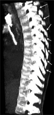
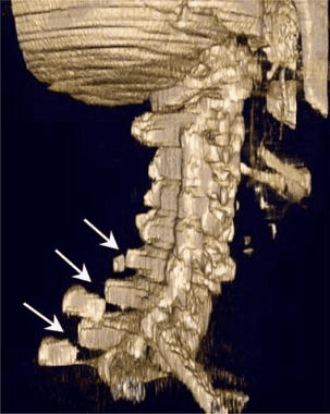
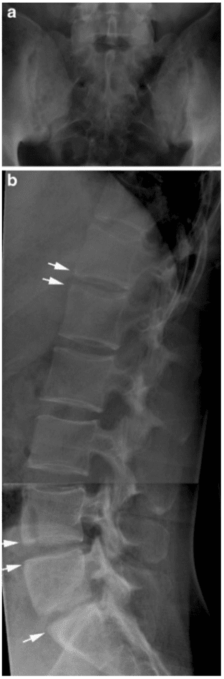
Subaxial RA changes also occur in the form of arthritis of the apophyseal and/or uncovertebral joints, appearing as narrowing and superficial erosions by radiography. It can cause instability in the C2-Th1 region, which is mainly seen in patients with severe chronic peripheral arthritis. Anterior subluxation is far more frequent than posterior subluxation. It is defined as at least 3 mm forward slippage of a vertebra�relative to the underlying vertebra by radiography including a flexion view (Fig. 6). Changes are particularly characteristic at the C3�4 and C4�5 level, but multiple levels may be involved, producing a typical �stepladder� appearance on lateral radiographs. The condition is serious if the subaxial sagittal spinal canal diameter is <14 mm, implying a possibility of spinal cord compression [2]. The instability may progress over time, especially if the C1�C2 region is stabilized surgically (Fig. 6) [5].
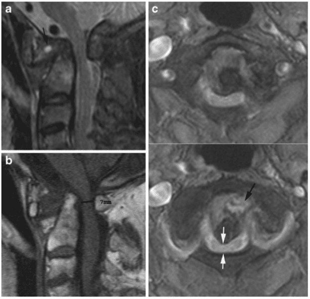
Fig. 5 Vertical subluxation with spinal cord compression. MRI of the cervical spine in a 69- year-old woman with advanced peripheral RA, neck pain and clinical signs of myelopathy. (a) Sagittal STIR, (b) sagittal T1 and (c) axial T2 fat-saturated (FS) images show erosion of the dens and protrusion of the tip into the occipital foramen causing compression of the spinal cord, which exhibits irregular signal intensity (white arrows). The osseous spinal canal has a width of approximately 7 mm (black line). There is heterogeneous signal intensity pannus surrounding the dens compatible with a mixture of fibrotic and oedematous pannus tissue (black arrows) in the widened space between the dens and the anterior arc of the atlas.
Discitis-like changes and spinous process erosion may also be detected by radiography in RA, but are relatively rare, whereas concomitant degenerative changes occur occasionally (Fig. 1).
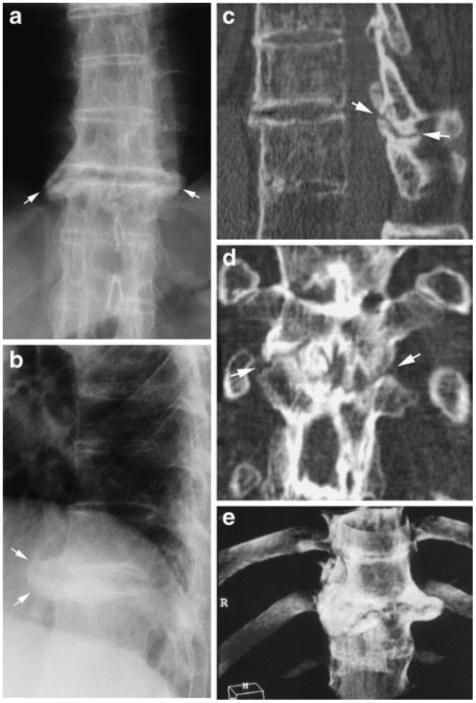
Cross-sectional imaging in the form of CT and MRI eliminates overprojecting structures and can improve the detection of RA changes. Osseous changes (erosions, etc.) can be clearly delineated by CT [6]. Additionally, MRI visualizes soft tissue structures (pannus; spinal cord, etc.), signs of disease activity and sequelae of inflammation in the form of fibrous pannus. These advantages of CT and MRI in patients with atlanto-axial involvement are illustrated in Figs. 7 and 8, including the possibility of detecting signs of arthritis by MRI before the occurrence of erosive changes (Fig. 8) [3].
Fig. 6 Subaxial instability. (a) Flexion view in a 64-year-old woman with advanced peripheral RA showing anterior atlanto-axial instability as well as subaxial instability at multiple levels. (b) Flexion view 2 years later after surgical stabilization of the atlanto-axial region demonstrates progression of the subaxial instability, especially between C3 and C4 (white arrow). There is a characteristic �stepladder� appearance, which also occurred on the initial radio- graphs (a), but is less pronounced.
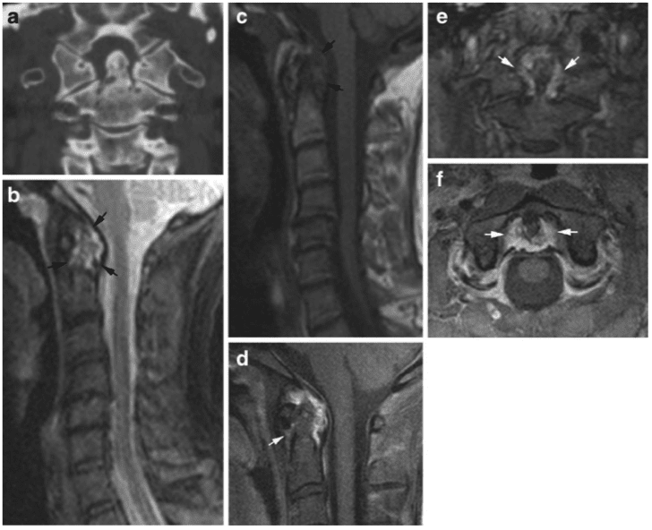
Fig. 7 Advantages of CT and MRI. (a) Supplementary CT and (b-f) MRI of the patient shown in Fig. 1. CT demonstrates erosion not only at the base of the dens, but also at the tip and at the atlanto-axial and atlanto-occipital joints, which are difficult to visualize by radiography. MRI, (b) sagittal STIR and (c) sagittal T1 of the entire cervical spine and post-contrast T1FS images of the atlanto-axial region, (d) sagittal, (e) coronal and (f) axial. Oedematous voluminous pannus surrounding the dens is seen on the STIR and T1 images (black arrows) in addition to C4/5 and C5/6 disc degeneration with posterior protrusion of the disc at C4/5. The post-contrast T1FS images confirm the presence of vascularized enhancing pannus around the dens (white arrows) and demonstrate improved anatomical delineation compared with the STIR image. There is no sign of spinal cord compression.
Fig. 8 Non-radiographic MR findings. MRI in a 41-year-old woman with peripheral erosive RA and neck pain, but normal cervical radiography. (a) Post-contrast axial and (b) coronal TIFS images show signs of active arthritis with synovial contrast enhancement at the left atlanto-axial joint in addition to enhancing pannus tissue at the left side of the dens (white arrows). There is also a subchondral enhancing area in the axis (black arrow) compatible with a pre-erosive lesion.
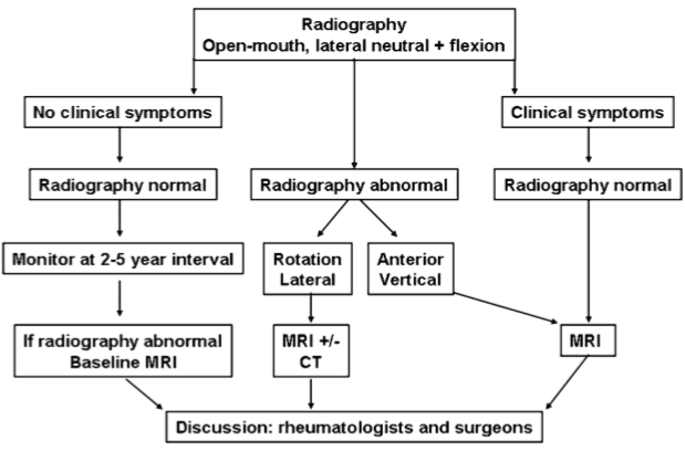
A diagnostic strategy according to Younes et al. [3] is recommended (Fig. 9). This includes an indication for radiography in all RA patients with disease duration >2 years as cervical involvement may occur in over 70% of patients and has been reported to be asymptomatic in 17% of RA patients. It is recommended to monitor patients with manifest peripheral erosions accompanied by RF (rheumatoid factor) and antiCCP (antibodies to cyclic citrullinated peptide) positivity every second year and�patients with few peripheral erosions and RF negativity at 5-year intervals. MRI is indicated in patients with neurological deficit, radiographic instability, vertical subluxation and subaxial stenosis [2, 3]. Visualisation of the spinal cord is especially important to detect cord injury or risk of injury. MRI should therefore always be performed in RA patients with neck pain and/or neurological symptoms [3, 7].
�
Seronegative Spondyloarthritides
According to European classification criteria [8, 9], SpA is divided into: (1) ankylosing spondylitis (AS), (2) psoriatic arthritis, (3) reactive arthritis, (4) arthritis associated with inflammatory bowel disorders (enteropathic arthritis) and (5) undifferentiated SpA. Inflammatory changes at the sacroiliac joints always occur in AS and are part of most other forms of SpA. Spinal changes are also a feature of SpA, especially in the late stages of AS.
�
Ankylosing Spondylitis
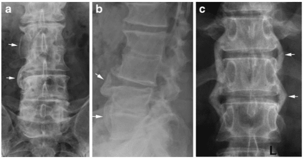
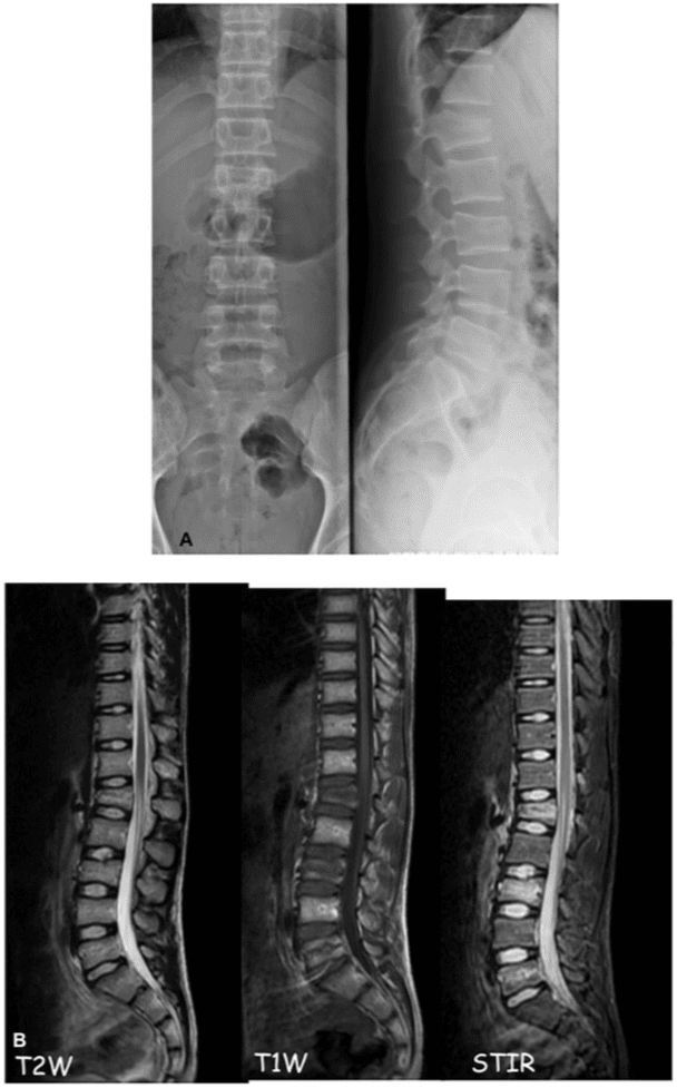
Ankylosing spondylitis is the most frequent and usually the most disabling form of SpA. It has a genetic predisposition in the form of a frequent association with the human leukocyte antigen (HLA) B27 [10]. AS often starts in early adulthood and has a chronic progressive course. It is therefore important to diagnose this disorder. According to the modified New York Criteria [11], the diagnosis of definite AS requires the following: manifest sacroiliitis by radiography (grade ?2 bilateral or unilateral grade 3�4 sacroiliitis; Fig. 10) and at least one of the following clinical criteria: (1) low back pain and stiffness for more than 3 months improving with activity, (2) limited movement of the lumbar spine and (3) reduced chest expansion. These criteria are still used in the diagnosis of AS despite the increasing use of MRI to detect the disease early. It is therefore important to know both the characteristic radiographic features and the MR features of AS.
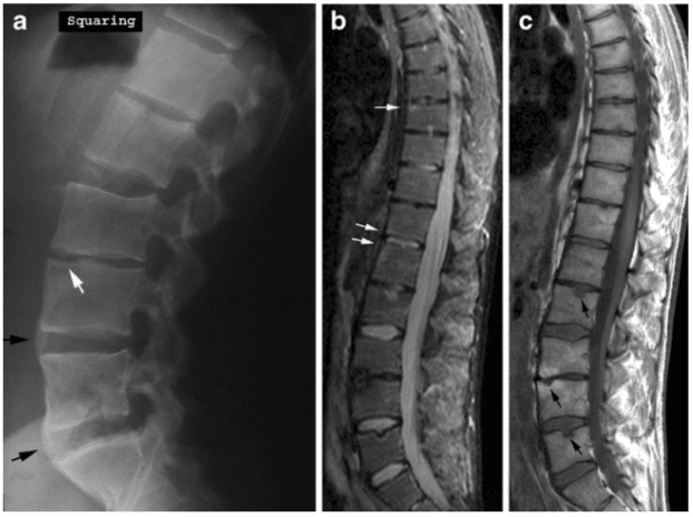
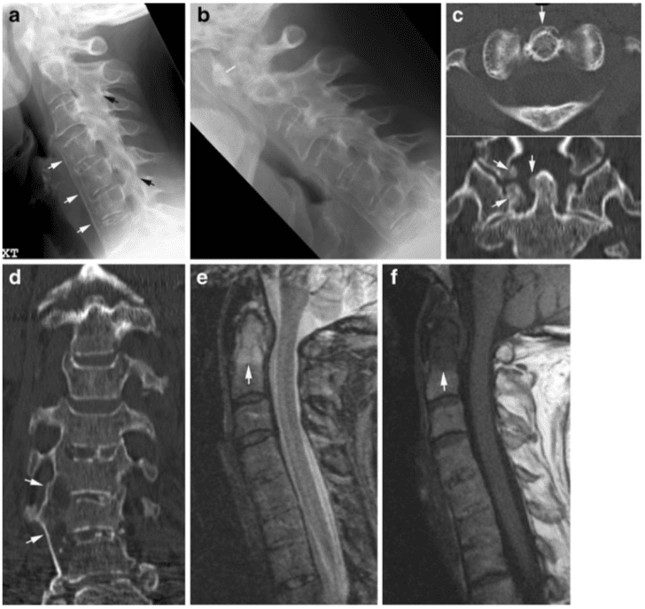
Early radiographic spinal changes encompass erosion of vertebral corners (Romanus lesions) causing vertebral squaring and eliciting reactive sclerosis appearing as condensation of vertebral corners (shiny corners; Fig. 10). These changes are caused by inflammation at the insertion of the annulus fibrosus (enthesitis) at vertebral corners provoking reactive bone formation [12]. Later on slim ossifications appear in the annulus fibrosus (syndesmo- phytes) (Fig. 11) [13]. With disease progression the spine gradually fuses because of syndesmophytes crossing the intervertebral spaces in addition to fusion of apophyseal joints, resulting in complete spinal fusion (bamboo spine;�Fig. 12). In advanced disease the supra- and interspinous ligaments may ossify and be visible on frontal radiographs as a slim ossified streak (Fig. 12). The occurrence of a single central radiodense streak has, the �dagger sign�. When the ligamentous ossification occurs together with ossification of apophyseal joint capsules, there are three vertical radiodense lines on frontal radiography (trolley-track sign).
Fig. 9 Diagnostic strategy. According to Younes et al. [3] radiography of the cervical spine is indicated in all RA patients with disease duration >2 years. It should at least include open-mouth and lateral views in neutral and flexed positions. Because of the occurrence of asymptomatic cervical involvement in 17% of RA patients, it is recommended to monitor patients with intervals of 2�5 years depending on positivity for the rheumatoid factor. MRI is indicated in patients with neurological deficit, radiographic instability, atlanto-axial impaction and subaxial stenosis. CT may add information in rotatory and lateral subluxation because of the possibility of secondary reconstruction in arbitrary planes and a clear visualisation of the atlanto-occipital joints [6].
Erosive changes within intervertebral spaces (Andersson lesions) have been detected by radiography in approximately 5% of patients with AS [14], but more frequently by MRI (Fig. 11) [15].
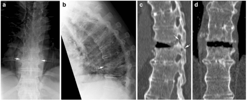
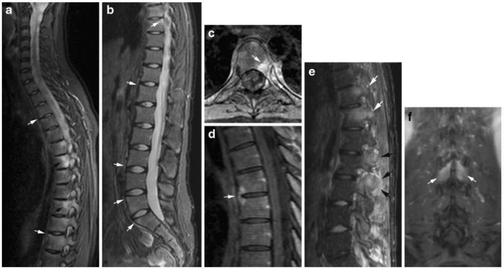
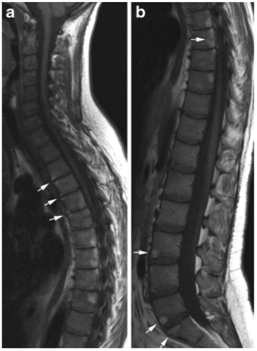
Persistent movement at single intervertebral spaces may occur in an otherwise ankylosed spine, sometimes caused by non-diagnosed fractures. This can result in pseudo- arthrosis-like changes with the formation of surrounding reactive osteophytes due to excessive mechanical load at single movable intervertebral spaces [14]. The diagnosis of such changes may require a CT examination to obtain adequate visualization (Fig. 13).
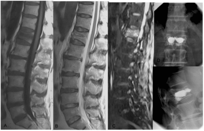
One of the life-threatening complications of AS is spinal fracture. Non-fatal fractures have been reported to occur in up to 6% of AS patients, especially in patients with long disease duration [16]. Fractures may occur after minor trauma because of the spinal stiffness and frequently accompanying osteoporosis. Fractures often occur at intervertebral spaces, but usually involve the ankylosed posterior structures and are thereby unstable (Fig. 14). Obvious fractures can visualize by radiography, but fractures may be obscured. It is therefore mandatory to supplement a negative radiography with CT if fracture is suspected (in the case of trauma history or a change in spinal symptoms). The occurrence of cervico-thoracic fractures may cause spinal cord injury and be lethal even following minor trauma [17].
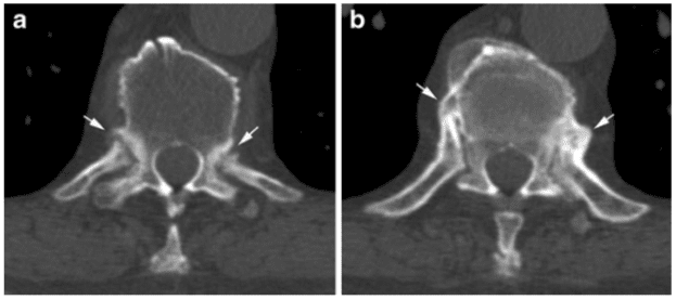
Cross-sectional CT or MR imaging can be advantageous in the diagnosis of AS changes. CT providing a clear delineation of osseous structures is the preferred technique for visualizing pseudo-arthrosis and detecting fractures (Figs. 13, 14). CT is superior to MRI in detecting minor osseous lesions such as erosion and ankylosis of the apophyseal, costo-vertebral and costo-transversal joints (Fig. 15). MRI can visualize signs of active inflammation in the form of bone marrow and soft tissue oedema and/or contrast enhancement. It has therefore gained a central role in the evaluation of disease activity [15]. MRI can, however, also detect sequelae of inflammation consisting of fatty deposition in the bone marrow and chronic structural changes such as erosion and fusion of vertebral bodies [15].
Characteristic MR findings early in the disease are activity changes mainly consisting of oedema at vertebral corners and/or costo-vertebral joints (Fig. 16) [13]. The inflammatory changes at vertebral corners are characteristic of AS. Based on the occurrence of severe or multiple (?3) lesions in young patients, AS changes can be distinguished from degenerative changes with a high reliability [18].
Fig. 10 Relatively early changes in ankylosing spondylitis (AS). (a) AP radiograph of the sacroiliac joints in a 28-year-old man presenting with typical definite bilateral AS sacroiliitis (grade 3) in the form of bilateral joint erosion accompanied by subchondral sclerosis. (b) Initial spinal changes consisting of erosion of vertebral corners (Romanus lesion) with vertebral squaring corresponding to Th11, Th12, L4 and L5 accompanied by condensation of the vertebral corners�shiny corners (arrows).
During the disease course signs of activity can also occur at syndesmophytes, apophyseal joints and interspinous ligaments (Fig. 16). Detection of inflammation at apophyseal joints by MRI, however, demands pronounced involvement�histopathologically [19]. The inflammation at vertebral corners is the most valid feature and has been observed related to the development of syndesmophytes by radiography [12], establishing a link between signs of disease activity and chronic structural changes.
Chronic AS changes detectable by MRI mainly consist of fatty marrow deposition at vertebral corners (Fig. 17), erosion (Fig. 11) and vertebral fusion in advanced disease (Fig. 12). Fatty marrow deposition seems to be an a sign of chronicity being significantly correlated with radiographic changes, in particular, vertebral squaring [15]. Erosions are more frequently detected by MRI than by radiography (Fig. 11) [15] and can present with signs of active inflammation and/or surrounding fatty marrow deposition compatible with sequels of osseous inflammation. Syndesmophytes, however, may not always be visible by MRI because they may be difficult to distinguish from fibrous tissue unless there is concomitant active inflammation or fatty deposition (Figs. 11, 16) [15, 20].
The possibility of visualizing disease activity by MRI has increased its use to monitor AS, especially during anti-TNF (anti-tumour necrosis factor) therapy [21, 22]. Several studies have shown that MR changes are frequent in the thoracic spine (Fig. 16) [15, 23]. It is therefore important to examine the entire spine using sagittal STIR or T2 fat-saturated (FS) and T1-weighted sequences. Supplementary axial slices can be necessary for visualising involvement of apophyseal, costo-vertebral and costo-transversal joints (Fig. 16) [24, 25]. Post-contrast T1FS sequences can sometimes be advantageous as they provide better anatomical delineation [26]. Additionally, dynamic contrast-enhanced MRI may be superior to static MRI in monitoring disease activity during anti-TNF therapy [27]. Whole-body MRI gives the possibility of detecting involvement in other areas without losing important information about spinal and sacroiliac joint involvement [28, 29].
Other Forms of SpA
Radiographic changes in reactive and psoriatic arthritis are often characterized by voluminous non-marginal syndesmophytes (parasyndesmophytes) or coalescing ossification of the paravertebral ligaments in addition to asymmetrical sacroiliitis (Fig. 18) [30].
Reactive arthritis is self-limiting in most patients. However, in patients with chronic reactive arthritis and HLA B27 the axial changes may progress to changes somewhat similar to those seen in AS and can then be regarded as AS elicited by infection [10].
Fig. 11 Syndesmophytes and erosions in AS. (a) Lateral radiograph in a 29-year-old man with the characteristic slim ossification (syndesmophytes) at the periphery of the annulus fibrosus (black arrows) in addition to erosion of the endplates at the intervertebral (iv) space between L3 and L4 (white arrow). Supplementary MRI, (b) sagittal STIR and (c) T1-weighted images show small oedematous areas in the�erosion at iv L3/4 on the STIR image and surrounding fatty marrow deposition on T1 as a sign of previous osseous inflammation. There are additional erosive changes (black arrows, c) not clearly delineated by radiography and slight oedema at the vertebral corners (white arrows, b). Note that the syndesmophytes demonstrated by radiogra- phy are not visible on MRI.
Fig. 12 Advanced AS. (a) AP and (b) lateral radiograph in a 55-year-old man showing vertebral fusion due to syndesmophytes crossing the intervertebral spaces in addition to fusion of the apophyseal joints (bamboo spine). The interspinous ligaments are ossified, presenting as a slim ossified streak on the frontal radiograph (dagger sign; arrows). MRI, sagittal T1- weighted images of (c) the cervico-thoracic and (d) lumbar region, respectively, shows a general narrowing of the intervertebral discs with partial osseous fusion of the vertebral bodies, especially in the lumbar region (arrows). In addition a characteristic AS deformity with reduced lumbar lordosis and thoracic kyphosis.
Fig. 13 Pseudo-arthrosis-like changes in AS. (a) AP and (b) lateral radiograph showing vertebral fusion except at iv Th10/11. There is surrounding osteophyte formation at this iv space (arrows). Supplementary CT, (c) sagittal and (d) coronal 2D reconstruction, demonstrates lack of fusion of the vertebral bodies and apophyseal joints at this level (arrows). (e) 3D reconstruction clearly demonstrates the exuberant surrounding reactive osteophytes.
Fig. 14�Spinal fracture in AS. (a) AP and (b) lateral radiograph of the thoracic spine in a 64-year-old man with advanced AS and increasing back pain over 4 weeks. The lateral view demonstrates a slight malalignment at the anterior aspects of the vertebral bodies of Th9 and Th10, and the iv is irregularly narrowed on the AP view, all�suggesting fracture (arrows). CT, (c) sagittal and (d) coronal reconstruction, shows fracture through the iv space and the posterior structures (arrows). There is widening of the intervertebral space anteriorly in the supine position used for CT compared with the upright position used during radiography.
Axial psoriatic arthritis (PsA) occurs in approximately 50% of patients with peripheral PsA [31]. It differs radiographically from AS by the voluminous paravertebral ossifications and the occurrence of spinal changes without concomitant sacroiliitis in 10% of patients [32]. Axial PsA may be clinically silent [33], and involvement of the cervical spine is frequent (atlanto-axial or apophyseal joint changes). The cervical recognize may include atlanto-axial instability as seen in RA (Fig. 19), but the pathogenesis and thereby imaging findings are different. In PsA radiography and CT usually visualize new bone formation in the region of the dens. This is elicited by osseous inflammation (osteitis) and/or inflammation at ligament/ tendon attachments (enthesitis) detectable by MRI (Fig. 19). Osteitis is often a feature of spinal PsA and can occur together with paravertebral ossification/para- syndesmophytes and erosion of vertebral plates (Fig. 20). , and illustrated MR findings in PsA are based on personal observations and seem to reflect the radiographic changes encompassing a mixture of osteitis, enthesitis and erosion. Unfortunately, there is a lack of�systematic description of spinal changes in PsA by MRI. Some of the patients described under the term SAPHO (synovitis, acne, pustulosis, hyperostosis, osteitis) syndrome may have PsA. SAPHO is a collective term often used for inflammatory disorders primarily presenting with osseous hyperostosis and sclerosis, and they are frequently associated with skin disorders. The most commonly affected site in SAPHO is the anterior chest followed by the spine [34]. The PsA changes shown in Fig. 20 are characterized by hyperostosis and sclerosis, both main features of SAPHO. However, this patient did not have anterior chest involvement.
Fig. 15 CT detection of costo-vertebral changes in AS. Axial CT slices showing erosive changes (a) and ankylosis of costo-vertebral joints (b), respectively (arrows).
Fig. 16 Activity changes in AS by MRI. Sagittal STIR of (a) the cervico-thoracic and (b) the lumbar spine of the patients shown in Fig. 10 obtained 3 years before the radiography. There are multiple high signal intensity areas corresponding to vertebral corners (white arrows). Additionally, osseous oedema of the costo-vertebral joints (a, black arrows) seen on the lateral sagittal slice of the thoracic spine. (c) Axial post-contrast T1FS of an inflamed costo-vertebral joint confirmed the presence of joint inflammation in the form of osseous enhancement in both the vertebra and the rib (arrows) in addition to joint erosion. (d) Midline sagittal post-contrast T1FS shows an�enhancing syndesmophyte. (e) Inflammatory changes at the apophy- seal joint in a 27-year-old man; sagittal STIR image of the lumbar region showing subchondral osseous oedema in the lower thoracic region (white arrows), and both osseous and soft tissue oedema corresponding to the lumbar apophyseal joints (black arrows). Note that the osseous oedema in the pedicle of Th12 extends to the region of the costo-vertebral joint. (f) Coronal post-contrast T1FS of the lumbar spine shows additional enhancement corresponding to the interspinous ligament between L2 and L3 (arrows).
Fig. 17 Chronic changes in AS by MRI. Sagittal T1 (a) the cervico-thoracic and (b) the lumbar spine of the patients shown in Fig. 10. There are multiple fatty marrow depositions at vertebral corners and also posteriorly in thoracic vertebral bodies (b, arrows). This was observed to have developed since the MRI performed 3 years previously (shown in Fig. 16 a-d) and corresponds to areas of previous inflammation.
In patients with enteropathic arthritis associated with Crohn�s disease or ulcerative colitis, the spine is often osteoporotic with various accompanying SpA features by radiography, mostly AS-like changes. However, by MRI there may be more pronounced inflammation in the posterior ligaments than seen in the other forms of SpA (Fig. 21).
Fig. 18 Psoriatic arthritis (PsA), paravertebral ossifications. (a) AP and (b) lateral radiograph of the lumbar spine in a 48-year-old man with PsA showing voluminous paravertebral new bone forma- tion (arrows) in addition to fusion of the second and third vertebral bodies. There was no concomitant sacroiliitis. (c) AP radiograph of the thoraco- lumbar junction in a female patient with axial PsA demon- strating coalescing paravertebral ossifications (arrows).
Fig. 19 Cervical PsA. (a) Lateral radiographs in the neutral position and (b) during flexion in a 61-year-old woman show atlanto-axial instability with a 4-mm distance between the anterior arc and the dens (white line). Additionally, ankylosis of the apophyseal joints (black arrows) and new bone formation anterior to the C4-7 vertebral bodies (white arrows). CT, (c) axial slice and coronal reconstruction of the dens area, demonstrates new bone formation in the atlanto-axial region (arrows); (d) coronal reconstruction of the lower cervical region shows voluminous new bone formation on the right side of the vertebral bodies (arrows). MRI, (e) sagittal STIR and (f) T1-weighted images, shows homogeneous osseous inflammation corresponding to the dens (arrows) with surrounding irregular oedema compatible with a mixture of osteitis and enthesitis. Note that the anterior new bone formation visualised by radiography is difficult to detect on MRI.
Fig. 20 Lumbar PsA. (a) AP and (b) lateral radiograph in a 50-year- old man show voluminous paravertebral ossifications anteriorly and at the right side of the third lumbar vertebra and adjacent iv spaces. MRI, (c) sagittal STIR, (d) T1 and (e) post-contrast T1-weighted images, demonstrates manifest osseous inflammation (osteitis) in the form of oedema and enhancement of the vertebral body, slight enhancement in the paravertebral new bone formation and erosion of the upper vertebral plate compatible with a mixture of osteitis, enthesitis and erosive changes.
Fig. 21 Enteropathic SpA. Sagittal STIR image of the lumbar spine in a 27-year-old man with ulcerative colitis demonstrates oedema corresponding to the interspinous ligaments (arrows) and spinous processes as signs of inflammation. There are only minimal activity changes corresponding to the vertebral bodies, located to the anterior vertebral corners.
Rheumatoid arthritis of the spine can cause neck pain, back pain, and/or radiating pain in the upper and lower extremities. In severe cases, RA can also lead to the degeneration of the spine, resulting in the compression or impingement of the spinal cord and/or the spinal nerve roots. As a chiropractor, we offer diagnostic imaging to help determine a patient’s health issue, in order to develop the best treatment program.
Dr. Alex Jimenez D.C., C.C.S.T.
�
Conclusion
Radiography is still valuable in the diagnosis of spinal inflammatory disorders. It is necessary for visualizing instability and is superior to MRI for detecting syndesmophytes. However, MRI and CT can detect signs of spinal involvement before they can be visualized by radiography. MRI adds information about potential involvement of the spinal cord and nervous roots in addition to signs of disease activity and chronic changes such as fibrous pannus in RA and fatty marrow deposition, erosion and vertebral fusion in SpA. MRI is�therefore widely used to monitor inflammatory spinal diseases, especially during anti-TNF therapy.
Computed tomography is particularly valuable in the detection of fracture and minor osseous lesions as well as in the evaluation of pseudo-arthrosis. In conclusion, rheumatoid arthritis most commonly affects the structure and function of your hands, wrists, elbows, hips, knees, ankles and feet, however, people with this chronic inflammatory disease can experience back pain. Imaging the spine�in arthritis is fundamental to determine treatment. The scope of our information is limited to chiropractic as well as to spinal injuries and conditions. To discuss the subject matter, please feel free to ask Dr. Jimenez or contact us at�915-850-0900�.
Curated by Dr. Alex Jimenez
Additional Topics: Acute Back Pain
Back pain�is one of the most prevalent causes of disability and missed days at work worldwide. Back pain attributes to the second most common reason for doctor office visits, outnumbered only by upper-respiratory infections. Approximately 80 percent of the population will experience back pain at least once throughout their life. The spine is a complex structure made up of bones, joints, ligaments, and muscles, among other soft tissues. Because of this, injuries and/or aggravated conditions, such as�herniated discs, can eventually lead to symptoms of back pain. Sports injuries or automobile accident injuries are often the most frequent cause of back pain, however, sometimes the simplest of movements can have painful results. Fortunately, alternative treatment options, such as chiropractic care, can help ease back pain through the use of spinal adjustments and manual manipulations, ultimately improving pain relief.
1. Paus AC, Steen H, Roislien J, Mowinckel P, Teigland J (2008) High mortality rate in rheumatoid arthritis with subluxation of the cervical spine: a cohort study of operated and nonoperated patients. Spine 33(21):2278�2283 2. Kim DH, Hilibrand AS (2005) Rheumatoid arthritis in the cervical spine. J Am Acad Orthop Surg 13(7):463�474 3. Younes M, Belghali S, Kriaa S, Zrour S, Bejia I, Touzi M et al (2009) Compared imaging of the rheumatoid cervical spine: prevalence study and associated factors. Joint Bone Spine 76 (4):361�368 4. Riew KD, Hilibrand AS, Palumbo MA, Sethi N, Bohlman HH (2001) Diagnosing basilar invagination in the rheumatoid patient. The reliability of radiographic criteria. J Bone Joint Surg Am 83 (2):194�200 5. Ishii K, Matsumoto M, Takahashi Y, Okada E, Watanabe K, Tsuji T et al (2010) Risk factors for development of subaxial subluxations following atlantoaxial arthrodesis for atlantoaxial subluxations in rheumatoid arthritis. Spine 35(16):1551�1555 6. Iizuka H, Sorimachi Y, Ara T, Nishinome M, Nakajima T, Iizuka Y et al (2008) Relationship between the morphology of the atlanto-occipital joint and the radiographic results in patients with atlanto-axial subluxation due to rheumatoid arthritis. Eur Spine J 17(6):826�830 7. Narvaez JA, Narvaez J, Serrallonga M, De Lama E, de AM, Mast R et al (2008) Cervical spine involvement in rheumatoid arthritis: correlation between neurological manifestations and magnetic resonance imaging findings. Rheumatology (Oxford) 47 (12):1814�1819 8. Dougados M, van der Linden S, Juhlin R, Huitfeldt B, Amor B, Calin A et al (1991) The European Spondylarthropathy Study Group preliminary criteria for the classification of spondylarthr- opathy. Arthritis Rheum 34:1218�1227 9. Rudwaleit M, van der Heijde D, Landewe R, Listing J, Akkoc N, Brandt J et al (2009) The Development of Assessment of SpondyloArthritis international Society (ASAS) classification criteria for axial spondyloarthritis (Part II): validation and final selection. Ann Rheum Dis 68(6):777�783 10. Sieper J, Rudwaleit M, Khan MA, Braun J (2006) Concepts and epidemiology of spondyloarthritis. Best Pract Res Clin Rheumatol 20(3):401�417 11. van der Linden S, Valkenburg HA, Cats A (1984) Evaluation of diagnostic criteria for ankylosing spondylitis. A proposal for modification of the New York criteria. Arthritis Rheum 27:361� 268 12. Maksymowych WP, Chiowchanwisawakit P, Clare T, Pedersen SJ, Ostergaard M, Lambert RG (2009) Inflammatory lesions of the spine on magnetic resonance imaging predict the development of new syndesmophytes in ankylosing spondylitis: evidence of a relationship between inflammation and new bone formation. Arthritis Rheum 60(1):93�102 13. Sieper J, Rudwaleit M, Baraliakos X, Brandt J, Braun J, Burgos- Vargas R et al (2009) The Assessment of SpondyloArthritis international Society (ASAS) handbook: a guide to assess spondyloarthritis. Ann Rheum Dis 68(Suppl 2:ii):1�44 14. Park WM, Spencer DG, McCall IW, Ward J, Buchanan WW, Stephens WH (1981) The detection of spinal pseudarthrosis in ankylosing spondylitis. Br J Radiol 54(642):467�472 15. Madsen KB, Jurik AG (2009) MRI grading method for active and chronic spinal changes in spondyloarthritis. Clin Radiol 65:6�14 16. Feldtkeller E, Vosse D, Geusens P, van der Linden S (2006) Prevalence and annual incidence of vertebral fractures in patients with ankylosing spondylitis. Rheumatol Int 26(3):234�239 17. Thomsen AH, Uhreholt L, Jurik AG, Vesterby A (2010) Traumatic death in ankylosing spondylitis�a case report. J Forensic Sci 55(4):1126�1129 18. Bennett AN, Rehman A, Hensor EM, Marzo-Ortega H, Emery P, McGonagle D (2009) Evaluation of the diagnostic utility of spinal magnetic resonance imaging in axial spondylarthritis. Arthritis Rheum 60(5):1331�1341 19. Appel H, Loddenkemper C, Grozdanovic Z, Ebhardt H, Dreimann M, Hempfing A et al (2006) Correlation of histopathological findings and magnetic resonance imaging in the spine of patients with ankylosing spondylitis. Arthritis Res Ther 8(5):R143 20. Braun J, Baraliakos X, Golder W, Hermann KG, Listing J, Brandt J et al (2004) Analysing chronic spinal changes in ankylosing spondylitis: a systematic comparison of conventional x rays with magnetic resonance imaging using established and new scoring systems. Ann Rheum Dis 63(9):1046�1055 21. Baraliakos X, Listing J, Brandt J, Haibel H, Rudwaleit M, Sieper J et al (2007) Radiographic progression in patients with ankylosing spondylitis after 4 years of treatment with the anti- TNF-alpha antibody infliximab. Rheumatology (Oxford) 46 (9):1450�1453 22. Lambert RG, Salonen D, Rahman P, Inman RD, Wong RL, Einstein SG et al (2007) Adalimumab significantly reduces both spinal and sacroiliac joint inflammation in patients with ankylos- ing spondylitis: a multicenter, randomized, double-blind, placebo- controlled study. Arthritis Rheum 56(12):4005�4014 23. Baraliakos X, Landewe R, Hermann KG, Listing J, Golder W, Brandt J et al (2005) Inflammation in ankylosing spondylitis: a systematic description of the extent and frequency of acute spinal�changes using magnetic resonance imaging. Ann Rheum Dis 64 (5):730�734 24. Khanna M, Keightley A (2005) MRI of the axial skeleton manifestations of ankylosing spondylitis. Clin Radiol 60(1):135�136 25. Levine DS, Forbat SM, Saifuddin A (2004) MRI of the axial skeletal manifestations of ankylosing spondylitis. Clin Radiol 59 (5):400�413 26. Baraliakos X, Hermann KG, Landewe R, Listing J, Golder W, Brandt J et al (2005) Assessment of acute spinal inflammation in patients with ankylosing spondylitis by magnetic resonance imaging: a comparison between contrast enhanced T1 and short tau inversion recovery (STIR) sequences. Ann Rheum Dis 64 (8):1141�1144 27. Gaspersic N, Sersa I, Jevtic V, Tomsic M, Praprotnik S (2008) Monitoring ankylosing spondylitis therapy by dynamic contrast- enhanced and diffusion-weighted magnetic resonance imaging. Skeletal Radiol 37(2):123�131 28. Weber U, Maksymowych WP, Jurik AG, Pfirrmann CW, Rufibach K, Kissling RO et al (2009) Validation of whole-body against conventional magnetic resonance imaging for scoring acute inflammatory lesions in the sacroiliac joints of patients with spondylarthritis. Arthritis Rheum 61(7):893�899 29. Weber U, Hodler J, Jurik AG, Pfirrmann CW, Rufibach K, Kissling RO et al (2010) Assessment of active spinal inflamma- tory changes in patients with axial spondyloarthritis: validation of whole body MRI against conventional MRI. Ann Rheum Dis 69 (4):648�653 30. Helliwell PS, Hickling P, Wright V (1998) Do the radiological changes of classic ankylosing spondylitis differ from the changes found in the spondylitis associated with inflammatory bowel disease, psoriasis, and reactive arthritis? Ann Rheum Dis 57(3):135�140 31. Chandran V, Barrett J, Schentag CT, Farewell VT, Gladman DD (2009) Axial psoriatic arthritis: update on a longterm prospective study. J Rheumatol 36(12):2744�2750 32. Lubrano E, Marchesoni A, Olivieri I, D’Angelo S, Spadaro A, Parsons WJ et al (2009) Psoriatic arthritis spondylitis radiology index: a modified index for radiologic assessment of axial involvement in psoriatic arthritis. J Rheumatol 36(5):1006�1011 33. Hanly JG, Russell ML, Gladman DD (1988) Psoriatic spondy- loarthropathy: a long term prospective study. Ann Rheum Dis 47 (5):386�393 34. Takigawa T, Tanaka M, Nakanishi K, Misawa H, Sugimoto Y, Takahata T et al (2008) SAPHO syndrome associated spondylitis. Eur Spine J 17(10):1391�1397
Spinal trauma consists of spine fractures, or spinal fractures, and spinal cord injuries. Approximately 12,000 spinal trauma cases are reported in the United States every year. While the most prevalent causes of spinal cord injuries and spine fractures are automobile accidents and falls, spinal trauma can also be attributed to assault, sports injuries, and work-related accidents. Diagnosis of spinal trauma includes imaging and assessment of nerve function, such as reflex, motor, and sensation. The following article discusses the role of emergency radiology in spinal trauma. Chiropractic care can help provide diagnostic evaluations for spinal trauma.
Abstract
Spinal trauma is very frequent injury with different severity and prognosis varying from asymptomatic condition to temporary neurological dysfunction, focal deficit or fatal event. The major causes of spinal trauma are high- and low- energy fall, traffic accident, sport and blunt impact. The radiologist has a role of great responsibility to establish the presence or absence of lesions, to define the characteristics, to assess the prognostic influence and therefore treatment. Imaging has an important role in the management of spinal trauma. The aim of this paper was to describe: incidence and type of vertebral fracture; imaging indication and guidelines for cervical trauma; imaging indication and guidelines for thoracolumbar trauma; multidetector CT indication for trauma spine; MRI indication and protocol for trauma spine.
Introduction
The trauma of the spine weighs heavily on the budget of social and economic development of our society. In the USA, 15�40 cases per million populations with 12,000 cases of paraplegia every year, 4000 deaths before admission and 1000 deaths during hospitalization are estimated. The young adult population is the most frequently involved in road accidents, followed by those at home and at work, with a prevalence of falls from high and sports injuries.1
Imaging has an important role in the management of spinal trauma. Quick and proper management of the patients with trauma, from diagnosis to therapy, can mean reduction of the neurological damage of vital importance for the future of the patient. Radiologists have a role of great responsibility to establish the presence or absence of lesions, defining the characteristics, assessing the prognostic influence and therefore treatment.
The aim of this paper was to describe:
incidence and type of vertebral fracture
imaging indication and guidelines for cervical trauma
imaging indication and guidelines for thoracolumbar trauma
multidetector CT (MDCT) pattern for trauma spine
MRI pattern for trauma spine.
Spinal trauma, including spine fractures and spinal cord injuries, represent about 3 percent to 6 percent of all skeletal injuries. Diagnostic assessments are fundamental towards the complex diagnosis of spinal trauma. While plain radiography is the initial diagnostic modality used for spine fractures and/or spinal cord injuries, CT scans and MRI can also help with diagnosis. As a chiropractic care office, we can offer diagnostic assessments, such as X-rays, to help determine the best treatment.
Dr. Alex Jimenez D.C., C.C.S.T.
Vertebral Fracture Management and Imaging Indication and Evaluation
The rationale of imaging in spinal trauma is:
To diagnose the traumatic abnormality and characterize the type of injury.
To estimate the severity, potential spinal instability or damaged stability with or without neurological lesion associated, in order to avoid neurological worsening with medical legal issue.
To evaluate the state of the spinal cord and surrounding structures (MR is the gold standard technique).
Clinical evaluation involving different specialities�emergency medicine, trauma surgery, orthopaedics, neurosurgery and radiology or neuroradiology�and trauma information is the most important key point in order to decide when and which type of imaging technique is indicated.2
A common question in patients with spine trauma is: is there still a role for plain-film X-ray compared with CT?
In order to clarify when and what is more appropriate for spinal trauma, different guidelines were published distinguishing cervical and thoracolumbar level.
Cervical Spinal Trauma: Standard X-Ray and Multidetector CT Indication
For cervical level, controversy persists regarding the most efficient and effective method between cervical standard X-ray with three film projections (anteroposterior and lateral view plus open-mouth odontoid view) and MDCT.
X-ray is generally reserved for evaluating patients suspected of cervical spine injury and those with injuries of the thoracic and lumbar areas where suspicion of injury is low. Despite the absence of a randomized controlled trial and thanks to the high quality and performance of�MDCT and its post-processing (multiplanar reconstruction and three-dimensional volume rendering), the superiority of cervical CT (CCT) compared with cervical standard X-ray for the detection of clinically significant cervical spine injury is well demonstrated.
Figure 1. (a�l). A 20-year-old male involved in a motorbike accident. The multidetector CT with multiplanar reformatted and three- dimensional volume-rendering reconstructions (a�d) showed traumatic fracture of C6 with traumatic posterior spondylolisthesis grade III with spinal cord compression. The MRI (e�h) confirmed the traumatic fracture of C6 with traumatic posterior spondylolisthesis grade III with severe spinal cord compression. The post-surgical treatment MRI control (i�l) showed the sagittal alignment of cervical level and severe hyperintensity signal alteration of the spinal cord from C3 to T1.
In order to reduce the patient radiation exposure, it is important to determine and to select patients who need imaging and those who do not, through the clinical evaluation and probability of cervical spine injury, using only MDCT for the appropriate patient as is more cost-effective screening.3
First of all, it is necessary to distinguish the type of trauma:
minor trauma (stable patient, mentally alert, not under the influence of alcohol or other drugs and who has no history or physical findings suggesting a neck injury)
major and severe trauma (multitrauma, unstable patient with a simple temporary neurological dysfunction, with focal neurological deficit or with a history or mechanism of injury sufficient to have exceeded the physiologic range of motion).
Second, it is important to establish if trauma risk factors are presents, such as:
violence of trauma: high-energy fall (high risk) or low-energy fall (low risk)
age of the patient: <5years old, >65 years old�
associated lesions: head, chest, abdomen (multitrauma) etc.
clinical signs: Glasgow Coma Scale (GCS), neurological deficit, vertebral deformation.
Combining these elements, patients can be divided into �low risk� and �high risk� for cervical injury.
The first group consists of patients who are awake (GCS 15), alert, cooperative and non-intoxicated without any distract- ing injury.
The second group consists of unconscious, sedated, intoxicated or non-cooperative patients or those with a distracting injury or an altered mental state (GCS ,15) with a 5% chance of cervical spine injuries.3,4
CCT has a wider indication than X-ray for patients at very high risk of cervical spine injury (major trauma or multitrauma). No evidence suggests CCT instead of X-ray for a patient who is at low risk for cervical spine injury.5
Figure 2. (a�g). A 30-year-old male involved in a motorbike accident. The multidetector CT with multiplanar reformatted and three-dimensional volume-rendering reconstructions (a�d) showed traumatic burst fracture of L1 (A2-type Magerl class) with posterior bone fragment dislocation into spinal canal. The MRI (e�g) confirmed the burst fracture of L1 with moderate spinal cord compression.
Figure 3. (a�d) A 50-year-old male involved in a motorbike accident with acute spinal cord compression symptoms on anticoagulation treatment. The MRI showed an acute haemorrhagic lesion at the C2�C4 posterior epidural space, hypointense on sagittal T1 weighted (a) and hyperintense on T2 weighted (b) with spinal cord compression and dislocation on axial T2* (c) and T2 weighted (d).
In 2000, the National Emergency X-Radiography Utilization (NEXUS) study, analysing 34,069 patients, established low-risk criteria to identify patients with a low probability of cervical spine injury, who consequently needed no cervical spine�imaging. To meet the NEXUS criteria, a patient must have the following conditions:
no tenderness at the posterior midline of the cervical spine
no focal neurologic deficit
normal level of alertness
no evidence of intoxication
no clinically apparent painful injury that might distract the patient from the pain of a cervical spine injury.6
If all of these roles are present, the patient does not need to undergo X-ray because he has a low possibility of having a cervical spine injury with a sensitivity of 99% and a specificity of 12.9%.7
In 2001, the Canadian C-spine rule (CCSR) study developed a second decision rule using the risk factor of the trauma: three high-risk criteria (age $ 65 years, dangerous mechanism and paraesthesias in extremities), five low-risk criteria (simple rear-end motor vehicle collision, sitting position in emergency department, ambulatory at any time, delayed onset of neck pain and absence of midline cervical spine tenderness) and the ability of the patient to actively rotate his or her neck to determine the need for cervical spine radiography. In practice, if one of these risk factors is present, the patient needs to undergo imaging evaluation. On the other hand, if the risk factors are not present, the use of the NEXUS criteria plus a functional evaluation of the cervical spine is needed (left and right cervical spine rotation .45�); if this functional evaluation is possible, imaging is unnecessary. If an incomplete cervical movement is present, then the patient needs to be checked with imaging. The results showed the criteria to have a sensitivity of up to 100% and a specificity of up to 42.5%.8
Applying these criteria, before cervical spine imaging, the authors report a decrease of about 23.9% in the number of negative CCT, and applying a more liberal NEXUS criteria including the presence or absence of pain, limited range of motion or posterolateral cervical spine tenderness, they report a decrease of up to 20.2% in the number of negative studies.2
If these clinical criteria cannot be applied, CCT must be performed.
Major and severe traumas request a direct CCT screening, especially because there could be associated lesions, according to the high-risk criteria developed by Blackmore and Hanson to identify patients with trauma at high risk of c-spine injury who would benefit from CT scanning as the primary radiological investigation9 Figure 1.
Thoracolumbar Spinal Trauma: Standard X-Ray and Multidetector CT Indication
For thoracolumbar level, MDCT is a better examination for depicting spine fractures than conventional radiography. It has wider indication in the diagnosis of patients with thoracolumbar trauma for bone evaluation. It is faster than X-ray, more sensitive, thanks to multiplanar reformatted or volume-rendering reconstruction detecting small cortical fracture, and the sagittal alignment can be evaluated with a wide segment evaluation.10
It can replace conventional radiography and can be performed alone in patients who have sustained severe trauma.10
In fact, thoracolumbar spinal injuries can be detected during visceral organ-targeted CT protocol for blunt traumatic injury.
Figure 4. A 55-year-old female involved in a car accident with acute left cervical brachialgia. The sagittal T2 weighted (a) and axial T2 weighted (b) MRI showed a post-traumatic posterolateral herniated disc with spinal cord compression and soft hyper signal alteration on the C3�C4 spinal cord.
Thanks to multidetector technology, images reconstructed using a soft algorithm and wide-display field of view that covers the entire abdomen using a visceral organ-targeted protocol with 1.5-mm collimation are sufficient for the evaluation of spine fractures in patients with trauma, given that multiplanar reformatted images are provided without performing new CT study and without increasing radiation dose11 Figure 2.
With MDCT there is no information about spinal cord status or ligament lesion or acute epidural haematoma; it can only evaluate bone status. Spinal cord injury is suspected only by clinical data.
CCT is strictly recommended in patients affected by blunt cerebrovascular injuries. Both lesions can be strictly correlated and generally; contrast medium administration to exclude hemorrhagic brain lesion and cervical fracture is not needed.10
Magnetic resonance imaging, or MRI, is a medical diagnostic assessment technique utilized in radiology to create pictures of the anatomy and the physiological processes of the human body. Alongside radiography and CT scans, MRI can be helpful in the diagnosis of spinal trauma, including spine fractures and spinal cord injuries. Magnetic resonance imaging may not be necessary for all cases of spinal trauma. However, it could provide detailed information on the other soft tissues of the spine.�
Dr. Alex Jimenez D.C., C.C.S.T.
Spinal Trauma and MRI
Even if MDCT is the first imaging modality in a patient with trauma, MRI is essential for the soft assessment of the ligament, muscle or spinal cord injury, spinal cord, disc, ligaments and neural elements, especially using T2 weighted sequences with fat suppression or T2 short tau inversion recovery (STIR) sequence.12 MRI is also used to classify burst fracture, obtaining information about the status of the posterior ligamentous complex, a critical determinant of surgical indication even if the diagnosis of ligament injuries remains complex, and its grade is also underestimated using high-field MRI.13
Figure 5. A 65-year-old female involved in domestic trauma with spinal cord symptoms. The sagittal T1 weighted (a) and T2 weighted (b) MRI showed a traumatic T12�L1 spinal cord contusion hypointense on T1 weighted and hyperintense on T2 weighted.
In the management of patients with polytrauma, MDCT total-body scan is necessary in an emergency condition, and�MRI whole-spine indication is secondary to the clinical status of the patient: spinal cord compression syndrome Figure 3�5�MRI protocols recommended for patients affected by spinal injury and trauma are the following:13,14
Sagittal T1 weighted, T2 weighted and STIR sequence for the�bone marrow and spinal cord injury or spinal cord compression evaluation owing to epidural haematoma or traumatic herniated disc
Sagittal gradient echo T2* sequence for haemorrhage evaluation of the spinal cord or into the epidural�subdural space
Sagittal diffusion-weighted imaging helpful when evaluating spinal cord injury, differentiating cytotoxic from vasogenic�oedema, assisting in detecting intramedullary haemorrhage. It can help to evaluate the degree of compressed spinal cord.
Axial T1 weighted and T2 weighted sequence for the right localization of the injury. Recently, for patients affected by acute blunt trauma and cervical spinal cord injury, the axial T2 weighted sequence has been shown to be important for trauma-predicting outcomes. On axial T2 weighted imaging, five patterns of intramedullary spinal cord signal alteration can be distinguished at the injury�s epicentre. Ordinal values ranging from 0 to 4 can be assigned to these patterns as Brain�and Spinal Injury Center scores, which encompassed the spectrum of spinal cord injury severity correlating with neurological symptoms and MRI axial T2 weighted imaging. This score improves on current MRI-based prognostic descriptions for spinal cord injury by reflecting functionally and anatomically significant patterns of intramedullary T2 signal abnormality in the axial plane.15
Figure 6. A 20-year-old female involved in domestic trauma with back pain resistance to medical therapy. The standard antero- posterior�laterolateral X-ray (a) showed no vertebral fractures. The MRI showed a bone marrow alteration at lumbar vertebral body hyperintense on T2 weighted (T2W) (a), hypointense on T1 weighted (T1W) (b) and short tau inversion recovery (STIR) (c).
MRI has also an important role in case of discordance between clinical status and CT imaging. In the absence of vertebral fracture, patients can suffer from back pain resistant to medical therapy owing to bone marrow traumatic oedema that can be detected only using STIR sequence on MRI Figure 6.
In spinal cord injury without radiologic abnormalities (SCI- WORA), MRI is the only imaging modality that can detect intramedullary or extramedullary pathologies or show the absence of neuroimaging abnormalities.16 SCIWORA refers to spinal injuries, typically located in the cervical region, in the absence of identifiable bony or ligamentous injury on complete, technically adequate, plain radiographs or CT. SCIWORA should be suspected in patients subjected to blunt trauma who report early or transient symptoms of neurologic deficit or who have existing findings upon initial assessment.17
Vertebral Fracture Type and Classification
The rationale of imaging is to distinguish the vertebral fracture type into two groups:
� vertebral compression fracture as vertebral body fracture compressing the anterior cortex, sparing the middle posterior columns associated or not with kyphosis � burst fracture as comminuted fracture of the vertebral body extending through both superior and inferior endplates with kyphosis or posterior displacement of the bone into the canal. and to distinguish which type of treatment the patient needs; by imaging, it is possible to classify fractures into stable or�unstable fracture, giving indication to conservative or surgical therapy.
Figure 7. (a�f) A 77-year-old female involved in domestic trauma with back pain resistance to medical therapy. The multidetector CT (a) showed no vertebral fractures. The MRI showed a Magerl A1 fracture with bone marrow oedema at T12�L1 vertebral body hypointense on T1 weighted (b), hyperintense on T2 weighted (c) and short tau inversion recovery (d) treated by vertebroplasty (e�f).
Figure 8. (a�d) A 47-year-old male involved in a motorbike accident with back pain resistance to medical therapy. The MRI showed a Magerl A1 fracture with bone marrow oedema at T12 vertebral body hypointense on T1 weighted (a) hyperintense on T2 weighted (b) and short tau inversion recovery (c) treated by assisted-technique vertebroplasty�vertebral body stenting technique (d).
Using MDCT and MRI, thanks to morphology and injury distribution, various classification systems have been used for identifying those injuries that require surgical intervention, distinguishing among stable and unstable fractures and surgical and non-surgical fractures.1
Denis proposed the �three-column concept�, dividing the spinal segment into three parts: anterior, middle and posterior columns. The anterior column comprises the anterior longitudinal ligament and anterior half of the vertebral body; the middle column comprises the posterior half of the vertebral body and posterior longitudinal ligament; and the posterior column comprises the pedicles, facet joints and supraspinous ligaments. Each column has different contributions to stability, and their damages may affect stability differently. Generally, if two or more of these columns are damaged, the spine becomes unstable.18
Magerl divided the vertebral compression fracture (VCF) into three main categories according to trauma force: (a) compression injury, (b) distraction injury and (c) rotation injury. Type A has conservative or non-surgical mini-invasive treatment indication.19
The thoracolumbar injury classification and severity score (TLICS) system assigns numerical values to each injury based on the categories of morphology of injury, integrity of the posterior ligament and neurological involvement. Stable injury patterns (TLICS,4) may be treated non-operatively with�brace immobilization. Unstable injury patterns (TLICS.4) may be treated operatively with the principles of deformity correction, neurological decompression if necessary and spinal stabilization.20
The Aebi classification is based on three major groups: A = isolated anterior column injuries by axial compression, B = disruption of the posterior ligament complex by distraction posteriorly and C = corresponding to group B but with rotation. There is an increasing severity from A to C, and within each group, the severity usually increases within the subgroups from 1 to 3. All these pathomorphologies are supported by the mechanism of injury, which is responsible for the extent of the injury. The type of injury with its groups and subgroups is able to suggest the treatment modality.21
Thoracolumbar Fracture and Mini-Invasive Vertebral Augmentation Procedure: Imaging Target
Recently, different mini-invasive procedures called assisted- technique vertebroplasty (balloon kyphoplasty KP or kyphoplasty-like techniques) have been developed in order to obtain pain relief and kyphosis correction as alternative treatment for non-surgical but symptomatic vertebral fracture.
The rationale of these techniques is to combine the analgesic and vertebral consolidation effect of vertebroplasty with the restoration of the physiological height of the collapsed vertebral body, reducing the kyphotic deformity of the vertebral body, delivering cement into the fractured vertebral body with a vertebral stabilization effect compared with conservative therapy (bed rest and medical therapy).22
From interventional point of view, imaging has an important role for treatment indication together with clinical evaluation. Both MDCT and MRI are recommended Figure 7 and 8.
In fact, MDCT has the advantage of diagnosing VCF with kyphosis deformity easily, while MRI with STIR sequence is useful to evaluate bone marrow oedema, an important sign of back pain.
Patients affected by vertebral fracture without bone marrow oedema on STIR sequence are not indicated for interventional procedure.
According to imaging, Magerl A1 classification fractures are the main indication of treatment.
However, the treatment must be performed within 2�3 weeks from trauma in order to avoid sclerotic bone response: the younger the fractures, the better the results and easier the treatment and vertebral augmentation effect. To exclude sclerotic bone reaction, CT is recommended.
Conclusion
The management of spinal trauma remains complex. MDCT has a wide indication for bone evaluation in patients affected by severe trauma or patients with high risk of spine injury. MRI has a major indication in the case of spinal cord injury and the absence of bone lesion. Diagnostic assessment of spinal trauma, including radiography, CT scans, and MRI are fundamental towards the diagnosis of spine fractures and spinal cord injury for treatment. The scope of our information is limited to chiropractic as well as to spinal injuries and conditions. To discuss the subject matter, please feel free to ask Dr. Jimenez or contact us at�915-850-0900�.
Curated by Dr. Alex Jimenez
Additional Topics: Acute Back Pain
Back pain�is one of the most prevalent causes of disability and missed days at work worldwide. Back pain attributes to the second most common reason for doctor office visits, outnumbered only by upper-respiratory infections. Approximately 80 percent of the population will experience back pain at least once throughout their life. The spine is a complex structure made up of bones, joints, ligaments, and muscles, among other soft tissues. Because of this, injuries and/or aggravated conditions, such as�herniated discs, can eventually lead to symptoms of back pain. Sports injuries or automobile accident injuries are often the most frequent cause of back pain, however, sometimes the simplest of movements can have painful results. Fortunately, alternative treatment options, such as chiropractic care, can help ease back pain through the use of spinal adjustments and manual manipulations, ultimately improving pain relief.
Pneumaticos SG, Triantafyllopoulos GK, Gian- noudis PV. Advances made in the treatment of thoracolumbar fractures: current trends and future directions. Injury 2013; 44: 703�12. doi: 10.1016/j.injury.2012.12.005
Griffith B, Bolton C, Goyal N, Brown ML, Jain R. Screening cervical spine CT in a level I trauma center: overutilization? AJR Am J Roentgenol 2011; 197: 463�7.doi: 10.2214/ AJR.10.5731
Hanson JA, Blackmore CC, Mann FA, Wilson AJ. Cervical spine injury: a clinical decision rule to identify high-risk patients for helical CTscreening. AJR Am J Roentgenol 2000; 174: 713�17.
Saltzherr TP, Fung Kon Jin PH, Beenen LF, Vandertop WP, Goslings JC. Diagnostic imaging of cervical spine injuries following blunt trauma: a review of the literature and practical guideline. Injury 2009; 40: 795�800. doi: 10.1016/j.injury.2009.01.015
Holmes JF, Akkinepalli R. Computed to- mography versus plain radiography to screen for cervical spine injury: a meta-analysis. J Trauma 2005; 58: 902�5. doi: 10.1097/01. TA.0000162138.36519.2A
Hoffman JR, Wolfson AB, Todd K, Mower WR. Selective cervical spine radiography in blunt trauma: methodology of the National Emergency X-Radiography Utilization Study (NEXUS). Ann Emerg Med 1998; 32: 461�9. doi: 10.1016/S0196-0644(98)70176-3
Dickinson G, Stiell IG, Schull M, Brison R, Clement CM, Vandemheen KL, et al. Retro- spective application of the NEXUS low-risk criteria for cervical spine radiography in Canadian emergency departments. Ann Emerg Med 2004; 43: 507�14. doi: 10.1016/j. annemergmed.2003.10.036
Stiell IG, Wells GA, Vandemheen KL, Clem- ent CM, Lesiuk H, De Maio VJ, et al. The Canadian C-spine rule for radiography in
alert and stable trauma patients. JAMA 2001;
286: 1841�8. doi: 10.1001/jama.286.15.1841 9. Berne JD, Velmahos GC, El-Tawil Q, Deme- triades D, Asensio JA, Murray JA, et al. Value
of complete cervical helical computed to- mographic scanning in identifying cervical spine injury in the unevaluable blunt trauma patient with multiple injuries: a prospective study. J Trauma 1999; 47: 896�902. doi: 10.1097/00005373-199911000-00014
10. Wintermark M, Mouhsine E, Theumann N, Mordasini P, van Melle G, Leyvraz PF, et al. Thoracolumbar spine fractures in patients who have sustained severe trauma: depiction with multi-detector row CT. Radiology 2003; 227: 681�9. doi: 10.1148/radiol.2273020592
11. Kim S, Yoon CS, Ryu JA, Lee S, Park YS, Kim SS, et al. A comparison of the diagnostic performances of visceral organ-targeted ver- sus spine-targeted protocols for the evalua- tion of spinal fractures using sixteen-channel multidetector row computed tomography: is additional spine-targeted computed tomog- raphy necessary to evaluate thoracolumbar spinal fractures in blunt trauma victims? J Trauma 2010; 69: 437�46. doi: 10.1097/ TA.0b013e3181e491d8
12. Pizones J, Castillo E. Assessment of acute thoracolumbar fractures: challenges in mul- tidetector computed tomography and added value of emergency MRI. Semin Musculoskelet Radiol 2013; 17: 389�95. doi: 10.1055/s- 0033-1356468
13. Emery SE, Pathria MN, Wilber RG, Masaryk T, Bohlman HH. Magnetic resonance imag- ing of posttraumatic spinal ligament injury. J Spinal Disord 1989; 2: 229�33. doi: 10.1097/ 00002517-198912000-00003
14. Zhang JS, Huan Y. Multishot diffusion- weighted MR imaging features in acute trauma of spinal cord. Eur Radiol 2014; 24: 685�92. doi: 10.1007/s00330-013-3051-3
15. Talbott JF, Whetstone WD, Readdy WJ, Ferguson AR, Bresnahan JC, Saigal R, et al. The Brain and Spinal Injury Center score: a novel, simple, and reproducible method for assessing the severity of acute cervical spinal cord injury with axial T2-weighted MRI findings. J Neurosurg Spine 2015; 23: 495�504. doi: 10.3171/2015.1.SPINE141033
16. Boese CK, Oppermann J, Siewe J, Eysel P, Scheyerer MJ, Lechler PJ. Spinal cord injury without radiologic abnormality in children: a systematic review and meta-analysis. Trauma Acute Care Surg 2015; 78: 874�82. doi: 10.1097/TA.0000000000000579
17. Brown RL, Brunn MA, Garcia VF. Cervical spine injuries in children: a review of 103 patients treated consecutively at a level 1 pediatric trauma center. J Pediatr Surg 2001; 36: 1107�14. doi: 10.1053/jpsu.2001.25665
18. Denis F. The three column spine and its significance in the classification of acute thoracolumbar spinal injuries. Spine (Phila Pa 1976) 1983; 8: 817�31. doi: 10.1097/ 00007632-198311000-00003
19. Magerl F, Aebi M, Gertzbein SD, Harms J, Nazarian S. A comprehensive classification of thoracic and lumbar injuries. Eur Spine J 1994; 3: 184�201.
20. Patel AA, Dailey A, Brodke DS, Daubs M, Harrop J, Whang PG, et al; Spine Trauma Study Group. Thoracolumbar spine trauma classification: the Thoracolumbar Injury Classification and Severity Score system and case examples. J Neurosurg Spine 2009; 10: 201�6. doi: 10.3171/2008.12.SPINE08388
21. Aebi M. Classification of thoracolumbar fractures and dislocations. Eur Spine J 2010; 19(Suppl. 1): S2�7. doi: 10.1007/s00586-009-1114-6
22. Muto M, Marcia S, Guarnieri G, Pereira V. Assisted techniques for vertebral cementoplasty: why should we do it? Eur J Radiol 2015; 84: 783�8. doi: 10.1016/j.ejrad.2014.04.002
My treatment with Dr. Alex Jimenez has tremendously helped me. It gives me just, a sense of relief knowing that I can come and see him and all of his employees and great masseuses and everyone, can just release all the tension. It just brings me back to life.�
April Hermosillo
Have you ever experienced back pain which triggers or radiates shooting pain into the buttocks or legs? Millions of people in the United States suffer from this common health issue known as sciatica. Sciatica is a general diagnostic term used to describe radiating pain, tingling sensations, numbness or weakness which runs across the length of the sciatic nerve.
Sciatica originates along the lower back and then travels from the buttocks into one or both legs. Instead of a dull achy pain, this type of pain is characterized as a sharp, shooting pain that worsens through prolonged periods of sitting. The sciatic nerve is the largest nerve in the human body comprised of many nerve roots which come together once they exit the spine. When the sciatic nerve becomes compressed, due to a variety of possible causes, symptoms will manifest and radiate down the leg.
What is Sciatica?
Sciatica is a collection of symptoms, including pain, tingling and burning sensations at the lower back and/or legs, weakness, and numbness, caused by a combination of: pressure on the sciatic nerve, inflammation to the sciatic nerve or the region directly surrounding the nerve, an irritation to the sciatic nerve, and/or a pinching of the sciatic nerves. As the largest nerve in the human body, the sciatic nerve can be easily affected.
Sciatic nerve pain is a common health issue that affects many people on a regular basis. Its consequences can range from a mild nuisance to a debilitating pain which interferes with an individual’s physical activities.� Sciatica symptoms manifest in many different ways, including:
Lower back pain
Leg Pain
Buttock pain
A sensation of tingling or �pins & needles� running down the�leg, and even into the toes
Numbness in the legs or feet
Muscle weakness in the legs
Aching or burning feeling in the legs
Pain or numbness in the�big toe, or any of the toes
Any combination of the list above
As you may see, though some individuals could experience pain from the lower back all the way down to their feet, it can be isolated to a segment of this area. Fortunately, a variety of treatment approaches are available to help treat sciatica. Below, we will discuss some of the most common causes of sciatica as well as demonstrate the best treatment approach.�Chiropractic care is one of the most common alternative treatment options utilized to provide sciatic nerve pain relief without prescriptions or surgery.
Causes of Sciatica
Sciatic nerve pain may not always be felt immediately following an injury or condition due�to an accident. Some people experience sciatica that seems to come and go, while for others, it might take years before their symptoms manifest at all. This is partly because pain is processed by only 10 percent of the�nervous system. A patient may also feel relief from their pain, but because the underlying cause of their sciatica hasn’t been fixed, the pain will come back. Below is a list of some of the causes of sciatica.
Subluxation or misalignment of the vertebrae
Disc degeneration, herniation, bulge, protrusion or other damage
A tumor pressing on the sciatic nerve
Injury to muscles
Pregnancy
Slipping, falling, or other impacts
Internal bleeding
Bad posture, either from sitting, standing or sleeping
Osteoarthritis
Spinal stenosis, a narrowing of the spinal canal the sciatic nerves pass through,
Playing sports
Poor lifting techniques
Other normal daily activities
While there are many possible causes for sciatica, the most frequent is a subluxation, or misalignment of the vertebrae in the lumbar spine,�or low back. A subluxation that is left untreated will result in the wear-and-tear of the spine, which in turn may lead�to disc protrusions, disc degeneration, disc herniations, and at some stages, even osteoarthritis. Chiropractic care will gently fix subluxations in the spine, allowing the nervous system to perform at an optimal level so that true recovery can occur.
Another common cause�of sciatica is a disc herniation. Spinal stenosis, which is a consequence of severe degeneration or alignment problems can also cause sciatic nerve pain. A condition called piriformis syndrome occurs when a tight piriformis muscle compresses the sciatic nerve. Determining the reason for sciatica is essential in understanding how to care for the health issue. Untreated sciatica can lead to problems such as:
Constipation
Difficulty getting pregnant
Digestion Problems
Edema or leg swelling
Erectile dysfunction (ED)
Incontinence
Irritable bowel syndrome (IBS)
Menstrual problems
Urinary problems
And more
Leaving sciatica untreated for any length of time can be damaging to your health, and the problem may worsen over time. The use of drugs and/or medications to help cope with your sciatic pain may only offer temporary relief from the symptoms as the real underlying source of your sciatica may not have been treated accordingly.�Diagnosis typically includes a comprehensive examination using a range of motion testing, neurological testing, imaging with MRI or X-Rays, and occasionally, further testing using nerve conduction velocity and electromyography evaluations.
Treatment for sciatica may vary depending on the cause of the symptoms. A chiropractor may use a series of spinal adjustments and manual manipulations�to help take pressure from the sciatic nerve. Other chiropractic care techniques and methods include the flexion distraction diversified technique, traction, and lumbar decompression. Passive and active exercises can also help treat sciatica. Exercises can also be recommended to restore strength, mobility, and flexibility. In severe cases of sciatica, the healthcare professional may recommend surgery.�
Sciatica occurs when an injury or condition results in the compression or impingement of the sciatic nerve, the largest nerve in the human body. When this happens, a collection of symptoms, including pain, tingling and burning sensations, as well as numbness, can develop. Chiropractic care is a well-known, alternative treatment option which can help carefully release the tension in the spine, reducing sciatic nerve pain.
Dr. Alex Jimenez D.C., C.C.S.T.
�
Chiropractic Care and Sciatica
The European Spine Journal printed the findings from a clinical trial demonstrating that chiropractic care led to a 72 percent success rate in treating sciatica and its associated symptoms compared to only a 20 percent success rate from physical therapy, and a 50 percent success rate from corticosteroid injections when treating sciatic nerve pain.
Sciatica is a widespread problem that many patients experience. Chiropractic care can find the source of the health issue in order to begin treatment and deliver pain relief accurately.�Sciatica can be painful and hinder you from living life to the fullest. Contact a chiropractor now to determine whether this is the ideal solution for you.
If you or somebody you know is suffering from sciatica and any of its associated symptoms, please recommend this article to them.�The scope of our information is limited to chiropractic as well as to spinal injuries and conditions. To discuss the subject matter, please feel free to ask Dr. Jimenez or contact us at�915-850-0900�.
Curated by Dr. Alex Jimenez
Additional Topics: Acute Back Pain
Back pain�is one of the most prevalent causes of disability and missed days at work worldwide. Back pain attributes to the second most common reason for doctor office visits, outnumbered only by upper-respiratory infections. Approximately 80 percent of the population will experience back pain at least once throughout their life. The spine is a complex structure made up of bones, joints, ligaments, and muscles, among other soft tissues. Because of this, injuries and/or aggravated conditions, such as�herniated discs, can eventually lead to symptoms of back pain. Sports injuries or automobile accident injuries are often the most frequent cause of back pain, however, sometimes the simplest of movements can have painful results. Fortunately, alternative treatment options, such as chiropractic care, can help ease back pain through the use of spinal adjustments and manual manipulations, ultimately improving pain relief.
Approximately 30 million adults in the United States have been diagnosed with chronic kidney disease, or CKD. The conditions categorized under CKD can damage the kidneys, decreasing their ability to function accordingly. Patients with this health issue can develop high blood pressure, anemia, weak bones, nerve damage and overall poor health. Chronic kidney disease may also increase a patient’s risk of developing heart and blood vessel disease, although these complications may occur slowly over time.
Chronic kidney disease may be caused by diabetes, high blood pressure and a variety of other disorders. Early detection and treatment is important to prevent CKD from getting worse. Chronic kidney disease may lead to kidney failure which may require additional care to maintain the patient’s quality of life. The purpose of the article below is to demonstrate the accurate prognosis and life expectancy of patients with chronic kidney disease. The evidence on the prediction of how long patients with CKD are expected to live provides important new data which may be useful for treatment.
Abstract
Can renal prognosis and life expectancy be accurately predicted? Increasingly, the answer is yes. The natural history of different forms of renal disease is becoming clearer; the degree of reduction in glomerular filtration rate (GFR) and the magnitude of proteinuria are strong predictors of renal outcome. Actuarial data on life expectancy from the start of renal replacement therapy are available from renal registries such as the U.S. Renal Data System (USRDS), and the UK Renal Registry. Recently, similar data have become available for patients with chronic kidney disease. Data collected from a large population-based registry in Alberta, Canada and stratified for different levels of estimated GFR (eGFR) have shown that the reduction in life expectancy with kidney failure is not a uremic event associated with starting dialysis but a continuous process that is evident from an eGFR of ?60 ml/min. Nevertheless, despite the poor prognosis of the last stages of renal failure, progress in the treatment and management of these patients and, in particular, of their cardiovascular risk factors continues to improve long-term outcome.
How much do we know about renal prognosis and life expectancy in adolescents with chronic kidney disease (CKD)? If one sees a new patient, a 19-year-old youth with a serum creatinine level of 200 ?mol/l, can one predict his likely renal prognosis and his life expectancy? The answer is yes, and this is frequently done when the question is posed in a medico-legal context; however, is the answer accurate?
We know that life expectancy is much reduced with end-stage renal failure�but what about the different degrees or stages of renal failure? For this review I have searched the adult and paediatric literature for papers cited in PubMed and Google Scholar that might contain data on life expectancy with CKD, or for series that have followed patients with CKD from childhood to end-stage kidney disease (ESKD) and through to renal replacement therapy (RRT). I summarise the evidence on the prediction of renal prognosis, describe important new data from Canada that for the first time looks at life expectancy with different stages of CKD and cite the U.S. Renal Data System (USRDS) and UK renal registries that report annual data regarding life expectancy with RRT.
Predicting Renal Outcome
To predict renal outcome I first make a number of assumptions. On the balance of probabilities (medico-legal language for a >50 % chance), at this age (19 years) the patient will have some form of renal dysplasia that would fall under the general heading of congenital anomalies of the kidney and urinary tract (CAKUT)�or some other congenital disease that might be tubular. If my history and examination make both of these possibilities unlikely, then further investigation is required which might include a biopsy.
If the patient has no proteinuria (protein creatinine ratio <50 mg/mmol), then the renal function should be currently stable. Renal deterioration will not occur until there is increasing proteinuria [1�5]. The exception to this would be a pure tubular disease, and I am assuming that this disease will have been picked up during the history, examination and other basic investigations.
Patients with inexorably progressive renal failure tend to deteriorate at a rate proportional to their proteinuria [6], but generally speaking the more proteinuria, the more the rate of progression can be slowed by angiotensin converting enzyme inhibitors (ACEIs) and good control of blood pressure [2, 7�9].
Patients with small asymmetric kidneys (renal hypodysplasia�often described in the UK as reflux nephropathy) tend to deteriorate at the slowest rates, and this is rarely greater than an estimated glomerular filtration ration (eGFR) of 3�4 ml/min/1.73 m2/year [3, 7]. Studies by of our own group have shown that controlling blood pressure and reducing proteinuria with an ACEI should reduce the rate of loss down to around 1.5 ml/min/1.73 m2/year [2, 7].
Assuming that the 19-year-old patient with a serum creatinine level of 200 ?mol/l has an eGFR of 35 ml/min/1.73 m2 and that he will need dialysis when his eGFR is around 10 ml/min/1.73 m2, then he should reach ESRD in approximately 17 years [(35 ? 10) divided by 1.5 years]. If he were to lose function at the faster rate of 3 ml/min/year, this would be 8.3 years.
Chronic kidney disease (CKD) is characterized by the gradual loss of kidney function over time. If kidney disease becomes worse, it may lead to kidney failure, requiring dialysis or a kidney transplant to maintain life. The following article demonstrates that life expectancy in patients with chronic kidney disease can be predicted. While it’s known that life expectancy in patients with end-stage renal failure is reduced, life expectancy in patients with different degrees or stages of renal failure shouldn’t necessarily be affected. Kidney function outcome predictions are not a patient’s destiny but an option for how long they are expected to live.
Dr. Alex Jimenez D.C., C.C.S.T.
Life Expectancy with CKD
Life expectancy tables for people with CKD have been created from a large population-based registry in Alberta, Canada and stratified for different levels of eGFR [10]. Data are calculated for men and women from 30 years of age to age 85 years by their levels of kidney function as defined by eGFRs of ?60, 45�59, 30�44 and 15�29 ml/min/1.73 m2 (see Table 1) [10]. These data show that life expectancy is progressively reduced with each age band of worse renal function.
Assuming our 19-year-old patient will be alive in 11 years, when he reaches 30 (the starting age of the Canadian data), what can be expected? Looking at men age 30�34 years (see Table 1), the life expectancy for those with an eGFR of ?60 ml/min/1.73 m2 is 39.1 years. This is lower than expected and certainly much less than in the UK database. For instance, data from the UK predict that a normal, healthy white male aged 30 years in 2015 has a remaining expected lifetime of 50.7 years [11]. The equivalent figure for the USA suggests that for a 30- to 34-year-old male the expected life expectancy is 45.7 years [12] (see Table 2). The authors of this latter study explain that this difference is attributed to the selective nature of their study cohort, which was limited to individuals who had outpatient serum creatinine measurements as part of routine care. They write that those with an eGFR of >60 ml/min/1.73 m2 cannot be considered as a �normal population� as patients having their creatinine measured are likely to be less well than the general population (who would not have a creatinine measure) and therefore have a lower life expectancy.
From Table 1 it can be seen that for the first three age groups (30�34, 35�39, 40�44 years), life expectancy falls by approximately 20 % with an eGFR of 45�59 ml/min/1.73 m2, by approximately 50 % with an eGFR of 30�44 ml/min/1.73 m2 and by approximately 65 % with an eGFR of 15�29 ml/min/1.73 m2, when compared with those with an eGFR of ?60 ml/min/1.73 m2 (note: these figures are calculated from the first three age groups, i.e. 30, 35 and 40 years, respectively). Thus, the GFR of our patient now age 30 would be approximately 19 ml/min/1.73 m2 (eGFR decline of 1.5 ml/min/1.73 m2) and that at this level of function his life expectancy is reduced by 70 % from 50.6 to 15 years.
The excess mortality associated with renal failure is due principally to the increased risk of cardiovascular disease. An investigation of the causes of death associated with CKD in Alberta revealed that the major cause of death was cardiovascular (including an increase in heart failure and valvular disease). The unadjusted proportion of patients who died from cardiovascular disease increased with decreasing eGFR [21, 37, 41, and 44 % of patients with an eGFR of ?60 (with proteinuria), 45�59.9, 30�44.9, and 15�29.9 ml/min/1.73 m2, respectively]. The proportion of deaths from infection also increased but not those from cancer [13].
In a separate review using meta-analysis to examine the influence of both reduced eGFR and albuminuria on cardiovascular mortality the authors found that both lower eGFR (<60 ml/min/1.73 m2) and higher albumin/creatinine ratio (ACR ?10 mg/g) were independent predictors of mortality risk in the general population [14]. Adjusted hazard ratios (HRs) for all-cause mortality at eGFRs of 60, 45 and 15 ml/min/1.73 m2 (vs. 95 ml/min/1.73 m2) were 1.18 [95 % confidence interval (CI) 1.05�1.32], 1.57 (95 % CI 1.39�1.78) and 3.14 (95 % CI 2.39�4.13), respectively. The ACR was associated with mortality risk linearly on the log-log scale without threshold effects. Adjusted HRs for all-cause mortality at ACRs of 10, 30, and 300 mg/g (vs. 5 mg/g) were 1.20 (1.15�1.26), 1.63 (1.50�1.77) and 2.22 (1.97�2.51), respectively. These data are derived from populations a higher mean age, but age was not an independent variable.
Thus, our patient, aged 19�36, even with an eGFR of approximately 45 ml/min/1.73 m2, has an increased risk of dying of around 57 % [risk ratio (RR) 1.57] compared with an eGFR of 95 ml/min/1.73 m2; similarly, with a ACR of 30 mg/g, our patient has an increased risk of dying of around 63 % (RR 1.63) compared with ACR of 5 mg/g [14]. These figures correlate with life expectancy tables [10] in which a 30-year male with an eGFR of 30�44 ml/min/1.73 m2 has a life expectancy reduced by approximately 50 % compared with a similar patient with an eGFR of ?60 ml/min/1.73 m2.
To this equation we should also consider modification of life expectancy by such factors as race, gender and socio-economic status [15, 16], as well as control of blood pressure and hyperlipidemia [17]. All of these factors are being studied in the ongoing Chronic Kidney Disease in Children (CKiD) Study.
Predicting Life Expectancy at End-Stage
If our patient is well looked after for the next 17 years, I will assume that he will not die before he reaches ESRD at the age of 36 (age 19 + 17 years at a GFR decline rate of 1.5 ml/min/1.73 m2/year). However, we now know that this assumption cannot be made. As we have seen from the Canadian data, even at age 19 years with a GFR of 35 ml/min/1.73 m2, we can extrapolate that his life expectancy is reduced by around 50 %. For a UK male aged 19 years, a life expectancy of 61.4 years [11] is reduced to 30 years (age 49 years) [10].
Assuming that our patient would be around 36 years of age when end-stage renal failure is reached, then one can use two sources of actuarial information regarding future life expectancy:-
The USRDS Annual Report�s chapter on mortality and survival has actuarial tables which show data in 5-year age bands [12] (Table 2). Thus, at 36 years of age, our patient falls into the age band 35�39 years. This shows us that a normal U.S. male of this age group can expect to live a further 41 years. The same age group will live a further 12.5 years on dialysis and 30.8 years after a successful transplant. Of course, in reality, RRT life will tend to be a mixture of the two modes.
The UK Renal Registry annual report chapter on survival also has actuarial data in 5-year age bands [18]. However, these show that the median life expectancy for patients starting RRT at the 90-day time point and for this age group (35�39 years) is a further 13.5 years (dialysis and transplant combined).
In comparison, the Canadian data show that at age 35 years with an eGFR of 15�29 ml/min/1.73 m2, the remaining life expectancy is +13.8 years [10].
Trends in Life Expectancy
A review of annual reports from the USRDS in the period 1996�2013 reveals that the life expectancy for a 36-year-old man on haemodialysis has improved steadily and linearly from 7.2 years in 1996 to 11.5 years in 2013 (see Fig. 1). Thus, one can anticipate that our current projections of life expectancy probably err on the pessimistic side of reality. This is supported by a detailed analysis of paediatric outcome over the period 1990�2010 [19].
Summary and Conclusions
We can now predict renal outcome and life expectancy with some accuracy, but data sources on life expectancy are few. The new information from Canada on life expectancy with CKD is very important but will need verifying from other parts of the world. We must not forget that collected data are often a decade old before they are analysed and published. While several long-term studies like CKiD [15�17] are running, it is still too early for them to have generated new information on life expectancy. However, trends in outcome continue to improve, suggesting that we can be more optimistic than current data suggest.
Summary Points
Life expectancy is reduced for all levels of renal function below an eGFR of 60 ml/min/1.73 m2.
Actuarial data are now available on life expectancy both for patients with chronic kidney disease and end-stage kidney disease.
The increased risk of premature death is principally related to the increase in cardiovascular morbidity.
Questions (Answers Provided Below)
Proteinuria predicts progressive renal failure if greater than:
a. 50 mg/mmol creatinine (0.5 g/d)
b. 100 mg/mmol creatinine (1.0 g/d)
c. 150 mg/mmol creatinine
d. 200 mg/mmol creatinine
Life expectancy is reduced when eGFR falls below:
a. 60 ml/min
b. 50 ml/min
c. 50 ml/min
d. 30 ml/min
Life expectancy on dialysis in USA has stopped increasing
a. Since 2000
b. Since 2005
c. Since 2010
d. Is still increasing
The increased relative risk of dying in young patients with CKD is:
a. Cardiovascular
b. Cancer
c. Infection
d. None of these
Acknowledgements
Particular thanks to Retha Steenkamp and UK Renal Registry for their generous help and advice.
Compliance with ethical standards
Conflict of Interest
The author declares no conflict of interest
Footnotes
Answers:
a
a
d
a
In conclusion, the prognosis and life expectancy predictions for patients with CKD don’t guarantee how long a patient with CKD is expected to live. Instead, these statistics may be useful towards determining an alternative treatment option which may help change these outcomes in patients with CKD. Information referenced from the National Center for Biotechnology Information (NCBI). The scope of our information is limited to chiropractic as well as to spinal injuries and conditions. To discuss the subject matter, please feel free to ask Dr. Jimenez or contact us at�915-850-0900�.
Curated by Dr. Alex Jimenez
Additional Topics: Acute Back Pain
Back pain�is one of the most prevalent causes of disability and missed days at work worldwide. Back pain attributes to the second most common reason for doctor office visits, outnumbered only by upper-respiratory infections. Approximately 80 percent of the population will experience back pain at least once throughout their life. The spine is a complex structure made up of bones, joints, ligaments, and muscles, among other soft tissues. Because of this, injuries and/or aggravated conditions, such as�herniated discs, can eventually lead to symptoms of back pain. Sports injuries or automobile accident injuries are often the most frequent cause of back pain, however, sometimes the simplest of movements can have painful results. Fortunately, alternative treatment options, such as chiropractic care, can help ease back pain through the use of spinal adjustments and manual manipulations, ultimately improving pain relief.
1.�Ardissino G, Testa S, Dacco V, Vigano S, Taioli E, Claris-Appiani A, Procaccio M, Avolio L, Ciofani A, Dello SL, Montini G. Proteinuria as a predictor of disease progression in children with hypodysplastic nephropathy. Data from the Ital Kid Project.�Pediatr Nephrol.�2004;19:172�177. doi: 10.1007/s00467-003-1268-0.�[PubMed]�[Cross Ref]
2.�Neild GH, Thomson G, Nitsch D, Woolfson RG, Connolly JO, Woodhouse CR. Renal outcome in adults with renal insufficiency and irregular asymmetric kidneys.�BMC Nephrol.�2004;5:12. doi: 10.1186/1471-2369-5-12.�[PMC free article]�[PubMed]�[Cross Ref]
3.�Gonzalez CC, Bitsori M, Tullus K. Progression of chronic renal failure in children with dysplastic kidneys.�Pediatr Nephrol.�2007;22:1014�1020. doi: 10.1007/s00467-007-0459-5.�[PubMed]�[Cross Ref]
4.�Wingen AM, Fabian-Bach C, Schaefer F, Mehls O. Randomised multicentre study of a low-protein diet on the progression of chronic renal failure in children.�Lancet.�1997;349:1117�1123. doi: 10.1016/S0140-6736(96)09260-4.�[PubMed]�[Cross Ref]
5.�Fathallah-Shaykh SA, Flynn JT, Pierce CB, Abraham AG, Blydt-Hansen TD, Massengill SF, Moxey-Mims MM, Warady BA, Furth SL, Wong CS. Progression of pediatric CKD of nonglomerular origin in the CKiD cohort.�Clin J Am Soc Nephrol.�2015;10:571�577. doi: 10.2215/CJN.07480714.�[PMC free article][PubMed]�[Cross Ref]
6.�Ruggenenti P, Perna A, Mosconi L, Pisoni R, Remuzzi G. Urinary protein excretion rate is the best independent predictor of ESRF in non-diabetic proteinuric chronic nephropathies. �Gruppo Italiano di Studi Epidemiologici in Nefrologia� (GISEN)�Kidney Int.�1998;53:1209�1216. doi: 10.1046/j.1523-1755.1998.00874.x.�[PubMed]�[Cross Ref]
7.�Neild GH. What do we know about chronic renal failure in young adults? II. Adult outcome of pediatric renal disease.�Pediatr Nephrol.�2009;24:1921�1928. doi: 10.1007/s00467-008-1107-4.�[PubMed][Cross Ref]
8.�The GISEN Group Randomised placebo-controlled trial of effect of ramipril on decline in glomerular filtration rate and risk of terminal renal failure in proteinuric, non-diabetic nephropathy.�Lancet.�1997;349:1857�1863. doi: 10.1016/S0140-6736(96)11445-8.�[PubMed]�[Cross Ref]
9.�Wuhl E, Trivelli A, Picca S, Litwin M, Peco-Antic A, Zurowska A, Testa S, Jankauskiene A, Emre S, Caldas-Afonso A, Anarat A, Niaudet P, Mir S, Bakkaloglu A, Enke B, Montini G, Wingen AM, Sallay P, Jeck N, Berg U, Caliskan S, Wygoda S, Hohbach-Hohenfellner K, Dusek J, Urasinski T, Arbeiter K, Neuhaus T, Gellermann J, Drozdz D, Fischbach M, Moller K, Wigger M, Peruzzi L, Mehls O, Schaefer F. Strict blood-pressure control and progression of renal failure in children.�N Engl J Med.�2009;361:1639�1650. doi: 10.1056/NEJMoa0902066.�[PubMed]�[Cross Ref]
10.�Turin TC, Tonelli M, Manns BJ, Ravani P, Ahmed SB, Hemmelgarn BR. Chronic kidney disease and life expectancy.�Nephrol Dial Transplant.�2012;27:3182�3186. doi: 10.1093/ndt/gfs052.�[PubMed][Cross Ref]
12.�United States Renal Data System (2015) Mortality. In: USRDS annual data report: epidemiology of kidney disease in the United States. National Institutes of Health, National Institute of Diabetes and Digestive and Kidney Diseases, Bethesda, chapter 6, vol 2, Table 6.4. Available at:�www.usrds.org/2015/download/vol2_06_Mortality_15.pdf
13.�Thompson S, James M, Wiebe N, Hemmelgarn B, Manns B, Klarenbach S, Tonelli M. Cause of death in patients with reduced kidney function.�J Am Soc Nephrol.�2015;10:2504�2511. doi: 10.1681/ASN.2014070714.�[PMC free article]�[PubMed]�[Cross Ref]
14.�Matsushita K, van der Velde ABC, Woodward M, Levey AS, de Jong PE, Coresh J, Gansevoort RT. Association of estimated glomerular filtration rate and albuminuria with all-cause and cardiovascular mortality in general population cohorts: a collaborative meta-analysis.�Lancet.�2010;375:2073�2081. doi: 10.1016/S0140-6736(10)60674-5.�[PMC free article]�[PubMed]�[Cross Ref]
15.�Wong CJ, Moxey-Mims M, Jerry-Fluker J, Warady BA, Furth SL. CKiD (CKD in children) prospective cohort study: a review of current findings.�Am J Kidney Dis.�2012;60:1002�1011. doi: 10.1053/j.ajkd.2012.07.018.�[PMC free article]�[PubMed]�[Cross Ref]
16.�Hidalgo G, Ng DK, Moxey-Mims M, Minnick ML, Blydt-Hansen T, Warady BA, Furth SL. Association of income level with kidney disease severity and progression among children and adolescents with CKD: a report from the Chronic Kidney Disease in Children (CKiD) Study.�Am J Kidney Dis.�2013;62:1087�1094. doi: 10.1053/j.ajkd.2013.06.013.�[PMC free article]�[PubMed]�[Cross Ref]
17.�Warady BA, Abraham AG, Schwartz GJ, Wong CS, Munoz A, Betoko A, Mitsnefes M, Kaskel F, Greenbaum LA, Mak RH, Flynn J, Moxey-Mims MM, Furth S. Predictors of rapid progression of glomerular and nonglomerular kidney disease in children and adolescents: the chronic kidney disease in children (CKiD) Cohort.�Am J Kidney Dis.�2015;65:878�888. doi: 10.1053/j.ajkd.2015.01.008.[PMC free article]�[PubMed]�[Cross Ref]
18.�Pruthi R, Steenkamp R, Feest T (2014) UK Renal Registry 16th annual report: chapter 8 survival and cause of death of UK adult patients on renal replacement therapy in 2012. Available at:�www.renalreg.org/wp-content/uploads/2014/09/08-Chap-08.pdf�[PubMed]
19.�Mitsnefes MM, Laskin BL, Dahhou M, Zhang X, Foster BJ. Mortality risk among children initially treated with dialysis for end-stage kidney disease, 1990�2010.�JAMA.�2013;309:1921�1929. doi: 10.1001/jama.2013.4208.�[PMC free article]�[PubMed]�[Cross Ref]
IFM's Find A Practitioner tool is the largest referral network in Functional Medicine, created to help patients locate Functional Medicine practitioners anywhere in the world. IFM Certified Practitioners are listed first in the search results, given their extensive education in Functional Medicine