Anterior Hip and Leg Muscles: What They Are, What They Do, and Why They Hurt
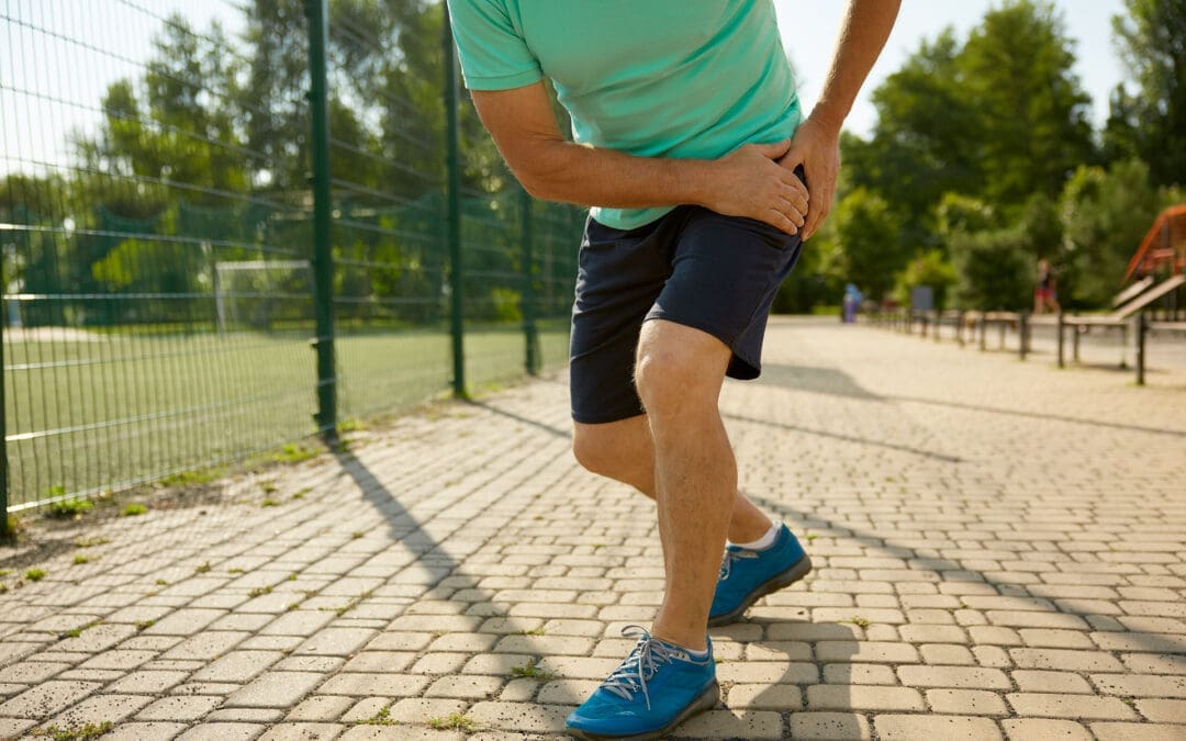
A woman holds her aching anterior hip.
Pain in the front of the hip (often felt in the hip crease or groin area) and the front of the thigh is very common. It can show up when you stand up from a chair, climb stairs, run, kick, or even after sitting for a long time. The tricky part is this: front-hip pain is not always “just a tight hip flexor.” Sometimes it’s a muscle or tendon problem, but it can also be related to the hip joint, the pelvis, or the lower back.
This guide is written for everyday people in El Paso who want clear answers, plus a practical explanation of how an integrative chiropractic approach can help reduce pain and prevent flare-ups.
At El Paso Back Clinic, Dr. Alexander Jimenez and the team often observe a pattern: tight, overworked hip flexors, underactive glutes, and poor pelvic control—especially in people who sit a lot, train hard, or are recovering after an accident.
What “anterior hip and leg muscles” means
“Anterior” means the front side. The anterior hip and leg muscles are basically your “go-forward” and “stand-tall” muscles. They help you:
Lift your knee (hip flexion)
Step forward when walking or running
Stabilize your pelvis so your lower back doesn’t overwork
Straighten your knee (knee extension)
Control your leg when you climb stairs or squat
When these muscles get overloaded, they can feel tight, sore, weak, or sharp—depending on the cause.
The main anterior hip muscles (your hip flexors)
Hip flexors are not one muscle. They’re a group that works together.
Key hip flexor muscles
Iliopsoas (iliacus + psoas): the classic “deep hip flexor”
Rectus femoris: part of the quadriceps, crosses the hip and the knee
Sartorius: a long, strap-like muscle across the front of the thigh
Tensor fasciae latae (TFL): supports hip flexion and pelvic control
Pectineus (often grouped with hip flexors in clinical discussions)
Why iliopsoas matters so much
The iliopsoas helps:
Lift the thigh toward the trunk
Support the hip joint and pelvis
Add stability near the lumbar spine/pelvis connection
At El Paso Back Clinic, iliopsoas overuse is commonly discussed among athletes and active individuals who engage in sprinting, jumping, kicking, or repeated hip flexion.
The anterior thigh muscles (front of the thigh)
The main anterior thigh group is the quadriceps. They’re designed to extend the knee and help control motion during walking, stairs, squats, and landing.
Quadriceps muscles
Rectus femoris
Vastus medialis
Vastus lateralis
Vastus intermedius
The anterior thigh compartment is also supplied and controlled by key anatomical structures, such as the femoral nerve (often described as the L2–L4 roots) and the femoral artery system. That’s one reason pain patterns can sometimes feel confusing—muscles, nerves, and joints all influence the sensation you feel.
Why the anterior hip and leg muscles sometimes hurt sometimes
There are a few “big buckets” that explain most front-hip and front-thigh pain.
You’re asking the muscles to do too much, too often (overuse)
Overuse happens when the workload increases faster than your tissues can adapt. Common triggers include:
Sudden jump in running miles
More hills or speed work than usual
Lots of kicking (soccer, martial arts)
Heavy squats/lunges with poor control
Repetitive direction changes (basketball, football)
Overuse can irritate:
The muscle belly (soreness, tightness)
The tendon (tendinopathy-like pain)
The hip flexor attachment area near the front of the hip
Prolonged sitting keeps hip flexors in a “shortened” position
Sitting puts the hips into flexion. Over time, many people notice:
Hip tightness when standing up after sitting
A “pinchy” feeling in the front of the hip
Low back stiffness that shows up with hip tightness
Dr. Jimenez has emphasized in his recent writing that prolonged sitting can contribute to tight hip flexors and poor movement patterns, and that short movement breaks, along with targeted mobility work, can help many people feel better.
The hip flexors can be tight because other muscles are not doing their job
This is one of the most common “root causes” in stubborn cases:
Weak or underactive glutes
Weak deep core stabilizers
Limited hip mobility (the hip joint doesn’t move well)
Pelvic control issues (pelvis tips forward, rotates, or drops during gait)
El Paso Back Clinic explains that when the glutes weaken from inactivity and prolonged sitting, the hips and pelvis can become less stable and shift out of alignment, thereby increasing stress on surrounding tissues.
Sometimes the pain is not in the hip flexor at all
A major clinical point from family medicine guidelines is that hip pain often groups into:
Anterior (front)
Lateral (side)
Posterior (back)
…and the cause changes based on that pattern. Anterior hip pain may result from hip flexor injury, but it can also result from intra-articular hip joint problems (such as femoroacetabular impingement or labral pathology) or from referred pain.
A helpful “body map” concept is presented in educational videos that discuss what different hip pain locations can indicate, but a hands-on evaluation remains important when symptoms persist.
What the pain feels like: common patterns that guide the next step
These are not perfect rules, but they help you decide whether you’re dealing with a likely muscle/tendon issue or something deeper.
More likely muscle/tendon irritation (common hip flexor pattern)
Pain in the front hip crease
Worse with lifting the knee (stairs, marching)
Worse with running sprints, kicking, or hills
Tenderness in the front hip region
Feels tight after sitting
More likely hip joint involvement
Deep groin pain with hip rotation
Catching, clicking, locking, or “pinching”
Pain that persists despite basic stretching/rest
Range of motion feels blocked (especially flexion + rotation)
More likely low back/nerve referral
Front thigh pain plus low back symptoms
Numbness, tingling, and burning sensations
Symptoms that change with spine position
Why “stretching only” often fails
Stretching can feel good short-term, but it may not solve the real driver if the problem is:
Weak glutes and weak core control
A stiff hip joint or pelvic restriction
Poor movement strategy (how you squat, run, or stand)
A training load problem (too much too soon)
In other words, the hip flexors may be tight because they’re protecting you or compensating for something else.
How El Paso Back Clinic approaches anterior hip and leg pain
El Paso Back Clinic describes an integrative model that blends chiropractic care, rehabilitation concepts, and movement-based strategies, with a focus on mobility, flexibility, and the restoration of balanced function.
Here’s how that “integrative” approach commonly helps front-hip and front-thigh problems.
Identify the true driver (not just the sore spot)
A good evaluation typically includes:
History (training, sitting, injury, accident history)
Differentiation between hip joint vs. lumbar referral patterns
Dr. Jimenez has written about the importance of a structured hip evaluation to sort out the likely source of pain and match care to the pattern.
Restore joint motion and reduce protective “guarding”
When the pelvis/hip/lumbar spine isn’t moving well, the body often shifts load to the hip flexors and quads. Chiropractic-style care may focus on restoring smoother motion so the muscles stop overworking.
El Paso Back Clinic also discusses how muscle imbalance and chronic guarding can make it harder for muscles to “relax on their own,” especially after injuries.
Use soft tissue + targeted techniques to normalize muscle function
A common strategy is pairing hands-on care with neuromuscular techniques. El Paso Back Clinic specifically discusses assessing hip flexors with MET therapy (muscle energy technique) as part of reducing tightness and improving hip mobility.
Rebuild strength where it matters (glutes + core + hip control)
To prevent recurrence, the plan usually includes strengthening and control, especially:
Glute bridges and progressions
Hip abduction strength (side-lying or banded work)
Gradual reloading of hip flexors (instead of only stretching)
El Paso Back Clinic’s content repeatedly emphasizes that restoring balanced muscle function around the pelvis and hips supports daily movement and performance.
Practical tips you can start today (safe, simple, and realistic)
If your symptoms are mild and you’re not dealing with red flags, these are common first steps.
For desk workers and drivers (very common in El Paso)
Take 1–2 minute movement breaks every 30–60 minutes
Do a gentle hip flexor stretch (no sharp pinching)
Add a glute activation move (bridges or mini-band walks)
Keep your daily steps consistent (don’t go from 2,000 to 12,000 overnight)
For runners and athletes
Reduce aggravating volume for 1–2 weeks (not “stop forever,” just calm it down)
Avoid sprinting/kicking if it spikes sharp pain
Strengthen glutes and hip stabilizers 2–3x/week
Return to speed and hills gradually, not all at once
Quick self-check idea (mobility clue)
The Thomas Test is commonly used to screen for hip flexor tightness and may help distinguish whether the “tight feeling” is more iliopsoas- or quadriceps-based (rectus femoris). It’s not a diagnosis, but it can be a clue.
When you should get evaluated sooner rather than later
Don’t try to “stretch through it” if you have:
Severe pain after a fall or accident
Inability to bear weight
Fever or feeling unwell with hip pain
Worsening numbness/tingling or leg weakness
Persistent catching/locking and deep groin pain
A structured clinical examination is particularly important when hip pain may involve the hip joint or referral patterns.
The main takeaway
Your anterior hip and leg muscles—especially the hip flexors and quadriceps—are essential for walking, running, stairs, and posture. They often hurt because of:
Too much repeated load (overuse)
Too much sitting (hip flexors stay shortened)
Muscle imbalance (weak glutes/core causing hip flexors to overwork)
Hip joint or low back referral (pain “shows up” in the front)
An integrative chiropractic model—such as the one described in El Paso Back Clinic’s educational resources—focuses on identifying the underlying cause, restoring motion, improving muscle balance, and developing a plan to reduce the likelihood of recurrence.
Is It Safe to Wear a Backpack? Expert Tips on Spinal Health and Back Pain Prevention in the US and El Paso, TX
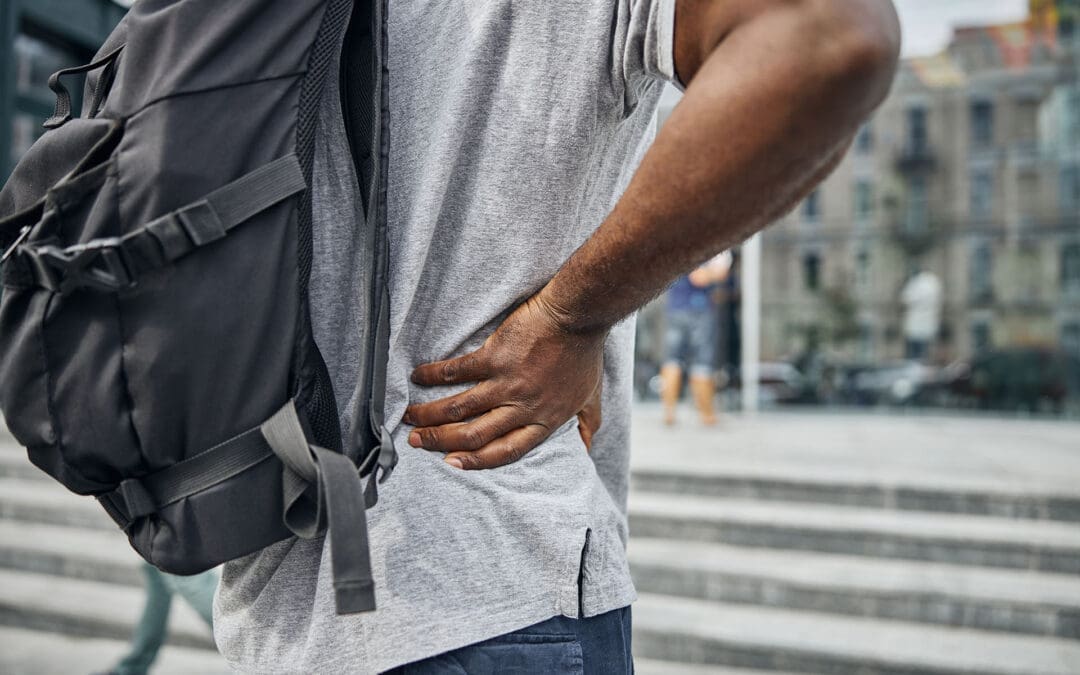
A woman walking, wearing a backpack with the recommended weight, and maintaining correct posture to prevent back pain and problems.
Back pain is a big issue for many people in the United States
Up to 80% of adults face low back pain at some point in their lives. This is one of the top reasons for doctor visits and missed workdays. The cost is huge too, with over $100 billion spent on spine problems each year. In El Paso, Texas, where people often have active jobs like industrial work or lots of driving, back pain questions focus on things like sciatica, herniated discs, and spinal stenosis. A common concern across the country, including in places like El Paso, is whether wearing a backpack is safe for the spine. The good news is that it can be safe if you follow some simple rules. This article focuses on backpack safety and then addresses other key questions about managing back pain, treatment options, and daily habits to keep your spine healthy.
Understanding Backpack Safety and Spinal Health
Wearing a backpack is common for carrying things, but if it’s too heavy or worn incorrectly, it can hurt your back. Heavy backpacks can strain muscles and joints in your back, neck, and shoulders. This might lead to pain or bad posture over time. However, backpacks do not cause scoliosis, a spinal curvature that affects about 2% to 3% of people. Scoliosis often starts in teens and is more common in girls, but it’s not linked to backpacks.
Is it safe? Yes, as long as you distribute the weight right and follow the tips to avoid strain. Improper use can cause muscle fatigue, poor posture (such as slouching), and even chronic pain if left unaddressed. In El Paso, where people might carry tools or bags for work, this is especially important to prevent issues such as sciatica, where pain radiates down the leg due to nerve pressure.
Here are some key tips for safe backpack use:
Choose the right backpack: Pick one with wide, padded straps and a padded back. It should fit your body size and have a waist strap for heavy loads. Lightweight materials help too.
Limit the weight: Keep the backpack under 10-15% of your body weight. For example, if you weigh 150 pounds, aim for no more than 15-22.5 pounds.
Distribute weight evenly: Put heavier items at the bottom and close to your back. Use compartments to balance things and stop shifting.
Wear it correctly: Always use both straps. Adjust them so the pack sits in the middle of your back, not sagging low. Bend your knees to lift it.
Make smart choices: Remove extra items often. Use lockers or storage if possible. For very heavy loads, try a rolling backpack or crossbody bag.
These steps help distribute the load across your strong back muscles and keep your spine aligned. If you feel pain, stop and adjust. In places like El Paso, with busy lifestyles, following these can help prevent accidents from becoming long-term back issues.
Common Causes of Back Pain in the US
Back pain affects millions. In the US, about 26% of adults have it at any time, and it’s more common after age 45. Among adults aged 50 and older, up to 45.6% experience it. Causes include muscle strains, ligament injuries, herniated discs (where the disc’s soft center protrudes), arthritis, and spinal stenosis (where the spinal canal narrows). Stress can make it worse by causing muscle spasms. Even factors such as obesity or infections can play a role.
Chronic back pain lasts more than 3 months and affects 8% of adults. It often comes from wear and tear on discs or joints. Poor sleep makes it worse because pain disrupts rest, and lack of sleep raises inflammation. In the US, this results in high costs, such as lost work and medical bills.
Symptoms vary. You might feel an ache in your lower back or sharp pain if it’s sciatica. Numbness, tingling, or weakness in the legs are red flags. Scoliosis, which affects 7 million Americans, can cause symptoms such as uneven shoulders or back pain; most cases are mild.
Muscle or ligament strain: From lifting incorrectly or sudden moves.
Disc problems: Bulges or herniations press on nerves.
Arthritis: Joint wear is common in older people.
Stenosis: Narrowing squeezes nerves, causing leg pain.
Stress and lifestyle: Tension builds up, leading to spasms.
Knowing these helps prevent pain. For example, strengthening your core muscles supports your spine and reduces strain from daily activities like wearing a backpack.
Managing Chronic Back Pain
Chronic back pain needs long-term plans. First, see if it’s new or ongoing. Most cases improve with rest and simple fixes, but if it lasts, get checked. Avoid bed rest; gentle movement helps recovery faster.
Daily habits matter. Exercise like walking or swimming builds strength. Maintain a healthy weight to reduce spinal load. Quit smoking, as it negatively affects spinal tissues and raises surgery risk by up to 50%. Good posture and ergonomic setups at work prevent strain.
In El Paso, with industrial jobs and driving, pain from accidents is common. Recovery focuses on building habits to avoid re-injury.
Stay active: Low-impact exercises like yoga or Pilates.
Watch your diet: Healthy foods reduce inflammation.
Manage stress: Deep breathing or mindfulness helps.
Sleep well: Use pillows to maintain spinal alignment.
Stretch daily: Loosen tight muscles, such as the hamstrings.
These steps reduce pain and improve quality of life.
Treatment Options: Surgery vs. Conservative Care
When pain doesn’t go away, choices include conservative care or surgery. Conservative means non-surgical options such as physical therapy, medications, injections, chiropractic care, or massage. These are tried first for 8-12 weeks. Surgery is indicated for severe cases, such as nerve damage or instability.
Ask your doctor: What causes my pain? What tests do I need? What are the risks and benefits? For surgery, ask about the surgeon’s experience, recovery time, and whether you’ll need help at home. Alternatives like spinal decompression stretch the spine to ease disc pressure.
Chiropractic vs. orthopedic: Chiropractors focus on spinal adjustments to realign the spine and relieve pain without medication. Orthopedists may recommend surgery for significant issues. Both can help, but chiropractic care is well-suited to conservative care.
In El Paso, many choose chiropractic for herniated discs or sciatica. It’s safe and effective for back pain, reducing symptoms by fixing alignment and boosting blood flow.
Spinal Health in El Paso, TX
El Paso has unique needs. Active lives, work injuries, and car accidents lead to questions about sciatica, where nerve pain goes down the leg, or spinal stenosis with leg weakness. Herniated discs are common from lifting or falls.
Lumbar stenosis FAQs: It causes leg pain or numbness when walking. Avoid high-impact exercises like running; try swimming instead. Treatments include therapy or decompression.
Local care often combines chiropractic and orthopedic care. Dr. Alexander Jimenez, a chiropractor in El Paso with over 30 years of experience, notes that integrative care is most effective. He uses adjustments, nutrition, and therapy for root causes. For example, a worker’s back pain improved by 50% within weeks with his plan. He stresses non-surgical options for sciatica and injuries, helping people stay active in El Paso’s environment.
Sciatica: From disc pressure; chiropractic eases it.
Chiropractic: Aligns the spine, safe for all ages.
Dr. Jimenez’s work shows personalized plans reduce pain without surgery.
Daily Habits to Prevent Spinal Injury
Preventing pain starts with habits. Lift by bending knees, not back. Stand every 15 minutes if sitting for long. For driving in El Paso, take breaks to stretch.
Core strength is key. Exercises like planks support your spine. Avoid smoking for better healing. Ergonomics: Screen at eye level, chair with back support.
For backpacks, combine with these: Even weight helps posture.
Lift right: Knees bent, close to body.
Posture: Stand tall, no slouch.
Exercise: Core and back focus.
Weight control: Less strain on the spine.
Breaks: Move often.
These reduce the risk of injury and tie into backpack safety.
Conclusion
Wearing a backpack is safe when done properly, with proper weight distribution and habits. This fits into broader questions about spinal health in the US and El Paso. Manage chronic pain with conservative care first, like chiropractic, and build daily routines to prevent issues. Experts like Dr. Jimenez show that integrative approaches work. Stay active, ask questions, and protect your spine for a better life.
Neuropathy Pain: “What’s the Best Medication?” And How El Paso Back Clinic Uses a Team Approach
Neuropathy is a common reason people contact El Paso Back Clinic®. The most common question sounds simple: “What’s the best medication for this pain?” But neuropathy is not one single problem. It is a symptom pattern (burning, tingling, numbness, electric shocks, sensitivity) that can result from various causes, such as diabetes, vitamin deficiencies, nerve compression, medication side effects, or past injuries. Getting the “best” treatment usually means combining the right medical plan with the right hands-on and movement-based care, plus lifestyle steps that protect nerves over time.
At El Paso Back Clinic, the care model described in their neuropathy education includes integrative chiropractic care coordinated with nurse practitioner (NP) oversight, aiming to improve function and quality of life while also looking for root causes.
What Peripheral Neuropathy Really Means
Peripheral neuropathy means the nerves outside the brain and spinal cord are irritated or damaged. These nerves help with:
Feeling (touch, pain, temperature)
Movement (muscle control)
Automatic body functions (sweating, digestion, blood pressure)
When signals get disrupted, symptoms can include burning pain, numbness, tingling, cramps, and weakness—often starting in the feet or hands.
Why cause matters: Treatment works best when you address both the pain and the underlying cause of the nerve’s discomfort. Primary care guidance emphasizes a careful history, exam, and targeted lab testing to look for common causes (diabetes, alcohol use, nutritional issues, toxins, nerve compression, and more).
The “Best Medication” for Neuropathy Pain: What Most Guidelines Start With
There isn’t a single perfect medication for everyone. Most major guidance starts with a few first-line options because they can reduce abnormal nerve pain signaling:
Common first-line medication groups
Gabapentinoids:gabapentin or pregabalin
SNRIs (a type of antidepressant used for nerve pain):duloxetine
TCAs (older antidepressants used for nerve pain):amitriptyline (used more often at night due to sedation)
This is consistent across multiple evidence summaries and public clinical guidance.
What patients usually want to know (in plain language)
These medicines do not “fix” the nerve overnight.
They aim to reduce the volume of nerve pain messages reaching the brain.
Many people need dose adjustments or a different medication to get the best balance of relief and side effects.
Side Effects to Expect (And Why NPs Help So Much Here)
A big reason people stop neuropathy meds is side effects—especially in the first 1–3 weeks. The NHS lists these as commonly used neuropathic pain medicines, and side effects are a key part of safe prescribing decisions.
Typical side effects patients report
Gabapentin/pregabalin: sleepiness, dizziness, “brain fog,” swelling, weight gain (for some)
Amitriptyline: dry mouth, constipation, grogginess, dizziness (often taken at night)
How an NP helps (practical, real-world):
Reviews your full medication list to avoid risky combos
Adjusts timing (for example, shifting sedating doses toward evening)
Watches for issues like fall risk, daytime sleepiness, and mood changes
Checks labs or contributing problems (blood sugar, B12, thyroid, kidney function when relevant)
Plans step-by-step changes instead of guessing
NPs are also well-positioned to manage chronic pain patterns and medication decision-making over time, because neuropathy often requires follow-up and fine-tuning.
“Are There Non-Drug Treatments?” Yes—And They Matter
Most people with neuropathy want conservative options first, or at least options that let them use less medication. The El Paso Back Clinic neuropathy education highlights several non-surgical strategies commonly used in integrative care.
Integrative chiropractic care focused on movement, joint mechanics, and nerve irritation patterns
Footwear, balance support, and fall prevention
Sleep and stress strategies (very underrated for nerve pain)
Patient-facing education materials often encourage asking about topical options, TENS, and PT because neuropathy increases fall risk and balance issues.
A safety point that matters in real life
When numbness is present, people may not notice small injuries—especially on the feet. Major cancer center patient education emphasizes routine skin checks (hands/feet) and lifestyle habits that support nerve health and safety.
How Integrative Chiropractic Care Can Help Neuropathy Symptoms
Not all neuropathy pain is the same. Some nerve pain is driven by systemic issues (like diabetes). Other nerve pain can be worsened by biomechanics—for example, irritation at the spine, pelvis, or along nerve pathways that changes movement and increases sensitivity.
The El Paso Back Clinic neuropathy resource outlines an approach focused on non-invasive, whole-person strategies and coordination with NP oversight.
What integrative chiropractic care may focus on
Finding patterns of nerve compression/irritation linked to posture or movement
Improving joint motion to reduce “mechanical stress” on sensitive areas
Corrective exercises to support better balance and gait
Soft tissue work and mobility strategies to reduce protective tension
Coordinating with medical care when neuropathy is linked to diabetes, medication effects, or other systemic causes
Important note: Chiropractic and integrative therapies should be framed as part of a broader plan—not a stand-alone “cure.” A careful diagnostic workup is still key, especially if symptoms are new, worsening, one-sided, or include weakness.
“Why Is My Neuropathy Worse at Night?”
This is one of the most common questions. Nighttime can amplify nerve pain for several reasons:
Less distraction: your brain has fewer competing signals
Stress/emotions: the day catches up, and pain feels louder
Temperature changes: some people notice symptoms more when cooler
Cleveland Clinic’s patient education explains several of these factors and also notes that approaches like PT, mindfulness, and medication adjustments may help when pain spikes at night.
Nighttime tips that are often helpful
Keep a steady sleep schedule (even on weekends)
Avoid alcohol excess (it can worsen neuropathy for some people)
Review medication timing with your NP
Use foot/hand warmth if cold triggers symptoms (not hot enough to burn)
This is where a stepwise plan matters. Many people either give up too early or keep escalating one med until side effects take over.
Evidence-based reviews emphasize recognizing when treatment is not effective and switching earlier, and they also note that combination therapy can help some patients (using moderate doses instead of maxing out on a single drug).
Common next steps an NP may consider
Confirm the diagnosis (is it neuropathy, radiculopathy, vascular, or something else?)
Adjust dose timing or switch to a different first-line option
Consider combination therapy when appropriate and safe
Severe pain with fever, unexplained weight loss, or a cancer history
Primary care guidance recommends referral for electrodiagnostic studies when symptoms are concerning (e.g., rapid progression, asymmetry, motor/autonomic issues) or when the initial workup is normal but symptoms persist.
The “Two Lanes” of Neuropathy Care at El Paso Back Clinic: Medical + Mechanical
A practical way to think about neuropathy treatment is two lanes running together:
Support nerve health with lifestyle and risk-factor control
Coordinate referrals for testing if needed
Lane 2: Integrative chiropractic + rehab
Address movement patterns that keep pain “turned up”
Improve mobility, balance, and function
Reduce mechanical stress and improve daily tolerance
Build a home plan you can actually follow
This is the kind of “integrative” model described in El Paso Back Clinic’s neuropathy content—conservative, coordinated, and focused on quality of life.
Smart Questions to Ask at Your Neuropathy Visit
Patients often feel more confident when they come in with clear questions. These are consistent with neuropathy question guides and clinical evaluation principles:
Medication questions
“What is the first medicine you recommend, and what side effects should I expect?”
“If that doesn’t work, what’s next?”
“Are topical lidocaine patches or creams right for me?”
Diagnosis and cause questions
“What type of neuropathy do I have?”
“What do you think is the most likely cause for me?”
“Will we check for diabetes/prediabetes, vitamin levels, or thyroid issues?”
“Do my symptoms suggest inherited, toxic, inflammatory, or metabolic patterns?”
Function and safety questions
“What can I do to improve balance and prevent falls?”
“What should I do for foot care if I can’t feel injuries well?”
“Which exercises are safe for me right now?”
Bottom Line
The “best medication” for neuropathy pain is the one that reduces pain enough to help you function without side effects that wreck your day. For many people, that means starting with gabapentin, pregabalin, duloxetine, or amitriptyline, and then adjusting based on response and tolerability.
At El Paso Back Clinic, the integrative approach outlined in their neuropathy resources emphasizes coordinated care—NP oversight of medical management and integrative chiropractic strategies to support mobility, comfort, and daily life.
Sugar Hangover: Why You Feel “Off” After Too Much Sugar (El Paso Back Clinic Guide)
If you’ve ever eaten a lot of sweets and then woken up (or hit a wall a few hours later) feeling tired, foggy, cranky, or headachy, you’re not imagining it. Many people call this a “sugar hangover.” It’s not an official medical diagnosis, but the experience is real for many people—and there are clear reasons it can happen.
At El Paso Back Clinic®, we see something important: when your body is stressed—by poor sleep, dehydration, inflammation, neck tension, headaches, and irregular meals—you can feel worse after a sugar-heavy day. That’s why our clinic approach is often integrative, combining chiropractic care, functional rehabilitation, and nurse practitioner support when appropriate.
Let’s break down what a sugar hangover is, what it feels like, why it happens, and what to do—without hype and without scary claims.
What Is a “Sugar Hangover”?
A sugar hangover is a short-term slump that can happen after eating a lot of added sugar or refined carbs (like candy, pastries, sweet coffee drinks, soda, or a big plate of white pasta). People often feel symptoms like:
Fatigue
Headache
Brain fog
Irritability
Cravings
Thirst or dry mouth
Low motivation
Upset stomach (sometimes)
Houston Methodist explains the basic idea: simple carbs can be digested quickly, causing a blood sugar spike, and if that spike is big enough, it can lead to unpleasant side effects.
Levels (a metabolic health education site) also describes the sugar hangover pattern as feeling “crummy” after a sugar splurge, often tied to glucose swings.
Why It Happens: The Spike → Crash Cycle
Your body runs on glucose (blood sugar). After you eat, glucose rises. Then your body releases insulin, which helps move glucose into cells for energy.
When you eat a lot of sugar (especially on an empty stomach), the swing can be bigger:
Sugar absorbs fast
Sugary and refined foods often have little fiber, so they hit your bloodstream quickly.
Insulin response can be strong
A bigger spike can trigger a bigger insulin response.
Blood sugar can drop quickly afterward
That drop is what many people call the “crash.”
Some people experience a true pattern called reactive hypoglycemia—blood sugar that drops after eating. Mayo Clinic notes that reactive hypoglycemia can improve with food choices like high-fiber meals, avoiding sugary foods on an empty stomach, and eating smaller meals spaced throughout the day.
Stress hormones can kick in
When your body senses a drop in blood sugar, it may release hormones (like adrenaline) to bring levels back up. This can feel like:
jitters
anxiety
sweating
irritability
Levels describes these hormone shifts as part of why people can feel shaky, wired, or off during a crash.
Dehydration can cause headaches and fatigue
Some people get thirstier after a sugar-heavy day, and dehydration can worsen headaches and brain fog.
What a Sugar Hangover Feels Like (And Why Headaches Are Common)
A sugar hangover can feel like your brain is “slow.” That’s partly because your brain is sensitive to energy changes.
Common complaints include:
Headache + neck tightness
Brain fog
Heavy fatigue
Mood swings
Sugar cravings
Levels connects sugar hangover symptoms to glucose swings and the body’s stress response.
At El Paso Back Clinic®, we also notice something practical: headaches often come with muscle tension, especially in the neck, upper back, and jaw—and tension can feel worse when you’re dehydrated and underslept. (This doesn’t mean sugar “causes” all headaches. It means sugar swings can be one more stressor on a tense system.)
Who Is More Likely to Get Sugar Hangovers?
Anyone can feel it, but it’s more common if you have:
Irregular meals (skipping breakfast, long gaps)
Poor sleep
High stress
A mostly refined-carb diet
A lot of sugary drinks
Prediabetes or diabetes risk factors
If you have diabetes (or take glucose-lowering meds), you should treat big swings seriously and follow your care plan.
Business Insider also notes that sugar can contribute to feeling sick a few hours after eating sweets, even separate from alcohol hangovers.
Is a Sugar Hangover Dangerous?
Usually, it’s temporary and improves within hours.
But you should get medical help if you have:
Fainting or near-fainting
Confusion that doesn’t clear
Severe weakness
Chest pain
Repeated vomiting
Symptoms plus known diabetes/insulin use
Mayo Clinic provides clear guidance that post-meal low blood sugar patterns should be managed with dietary structure and, when needed, medical evaluation.
What To Do: A Simple “Next-Day Reset” Plan
You don’t need a cleanse. You need stability.
Step 1: Hydrate first
Start the day with water.
Helpful options:
Water
Unsweetened electrolyte drink (if you’re very thirsty)
Herbal tea
Try to avoid:
Sugary coffee drinks
Soda or sweet tea (as they can restart the spike)
Levels emphasizes hydration and avoiding more sugar when you’re trying to stabilize.
Step 2: Eat a steady breakfast (protein + fiber)
Pick something that slows digestion:
Eggs + veggies
Greek yogurt + berries + nuts
Oatmeal + chia + peanut butter
Beans + avocado + salsa (easy and filling)
Mayo Clinic recommends high-fiber foods and avoiding sugary/refined carbs on an empty stomach—especially for people prone to post-meal drops.
Step 3: Walk for 10–20 minutes
A short walk after eating helps many people feel clearer and less sluggish.
Step 4: Calm the “tension loop” (neck, jaw, shoulders)
If your sugar hangover comes with headaches, try:
Gentle neck range-of-motion
Shoulder rolls
Slow nasal breathing (2–3 minutes)
Light stretching
At El Paso Back Clinic®, we focus on restoring function after neck and back strain, and many patients notice that reducing mechanical stress can help them feel better overall—especially when headaches are linked to tension patterns.
Step 5: Don’t “punish” yourself with extreme restriction
A common mistake is skipping food all day. That can create more cravings and more swings.
Better:
normal meals
protein + fiber each time
water
early bedtime
How to Prevent Sugar Hangovers (Without Giving Up All Treats)
Prevention is mostly about how you eat sugar, not whether you ever eat it.
Use the “anchor meal” rule
If you want dessert, have it after a real meal that includes:
protein
fiber
healthy fat
This slows the glucose rise.
Avoid “liquid sugar” most days
Sugary drinks are one of the easiest ways to overshoot your daily sugar without feeling full.
Keep added sugar within reasonable limits
The American Heart Association recommends:
Women: no more than 25 g (about 6 teaspoons) added sugar/day
Men: no more than 36 g (about 9 teaspoons) added sugar/day
Watch for hidden sugar
Johns Hopkins points out that added sugar hides in many “normal” foods and can add up fast.
Common hidden sources:
flavored yogurt
granola bars
cereals
sauces and dressings
“coffee drinks”
sports/energy drinks
Where Chiropractic + Nurse Practitioner Support Fits (El Paso Back Clinic Approach)
A sugar hangover is usually a metabolic + lifestyle issue first. Chiropractic is not a “blood sugar cure.” But integrative care can help because real life is not a one-system-only world.
At El Paso Back Clinic®, our clinical model is built around restoring function and supporting whole-body recovery with a multidisciplinary team.
How a Nurse Practitioner (NP) can help
An NP can:
review symptoms and timing (what you ate + when you crashed)
screen for risk factors (prediabetes, diabetes, anemia, thyroid issues)
recommend lab work when appropriate
build a realistic food plan (not extreme)
help with sleep and stress strategies
Dr. Alexander Jimenez, DC, APRN, FNP-BC often frames this as building repeatable daily habits that support recovery—rather than chasing “quick fixes.”
How chiropractic care can support the “stress and tension side”
Sugar hangovers often come with:
headaches
neck tension
poor sleep
low activity the next day
Chiropractic care may help by:
assessing neck/back mechanics that contribute to tension headaches
improving mobility so you can move and walk more comfortably
El Paso Back Clinic focuses on restoring function after neck, back, and soft-tissue issues through integrated approaches.
Why a combined approach can be stronger
Because a “sugar hangover” often sits on top of other real-world problems:
bad sleep
dehydration
stress overload
chronic tightness
irregular meals
low protein/fiber patterns
Integrative chiropractic + NP care can address both:
the chemical side (glucose swings, nutritional structure)
the structural side (tension, headaches, movement limits)
That’s the practical “whole-person” logic behind multidisciplinary care at El Paso Back Clinic®.
A Quick Word on Nutrition Scope and Safety
Nutrition rules can differ by state and profession. The American Nutrition Association explains that nutrition regulations vary and that the scope of practice can differ across states and providers.
If your symptoms are frequent, intense, or confusing, the safest move is a clinical evaluation—especially if you might have reactive hypoglycemia or diabetes risk.
When to Get Checked (Don’t Ignore These Patterns)
Make an appointment if:
you crash after meals often (2–5 hours later)
headaches + fatigue are frequent
cravings feel out of control
you have a family history of diabetes
you feel shaky, sweaty, or confused after eating
Mayo Clinic recommends a dietary structure for reactive hypoglycemia patterns and supports evaluation when symptoms persist.
Key Takeaways
A “sugar hangover” is a real experience for many people, often driven by glucose spikes and crashes.
Symptoms can include fatigue, headache, brain fog, irritability, and cravings.
The best fix is stable meals, hydration, and light movement, not extreme restriction.
Long-term prevention includes limiting added sugar and watching hidden sugars.
At El Paso Back Clinic®, integrative care can support both the metabolic plan (NP) and the tension/movement side (chiropractic + rehab).
Sciatica Numbness in the Hamstring and Foot (Without Low Back Pain): An El Paso Back Clinic Guide to What It Means and What to Do
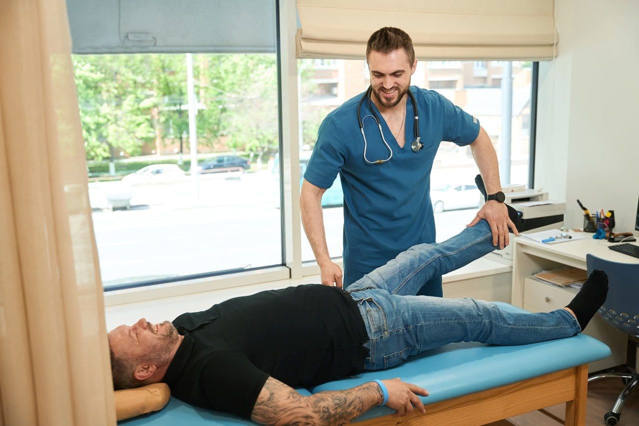
Patient with sciatica symptoms but no back pain, only leg and foot numbness and pain, lies supine on the examination table while the chiropractor/nurse practitioner lifts his extended leg with resistance.
If your hamstring feels numb or your foot feels tingly or “asleep,” it’s easy to think you pulled a muscle. But many people in El Paso are surprised to learn that sciatica can show up as leg numbness without much (or any) low back pain. That pattern is common—and it’s one reason sciatica can get missed at first. (Yale Medicine, n.d.; Penn Medicine, n.d.; AMA, 2024)
At El Paso Back Clinic, we often see this exact concern:
“My lower back doesn’t hurt… so how can this be sciatica?”
“Why is there numbness in my hamstring and foot?”
“Is this a hamstring strain or a nerve issue?”
“When should I worry and get checked?”
This article explains the “why,” helps you distinguish between muscle and nerve pain, and shows how an integrative chiropractic approach may reduce sciatica-related numbness by addressing the spine, hips, soft tissues, and movement habits that keep the nerve irritated. (HSS, 2024; Fletcher Family Chiropractic, 2025; Auburn Hills Chiropractic, n.d.)
Important: Numbness can have several causes. A careful evaluation matters—especially if symptoms persist or worsen.
What Sciatica Really Is (And Why It Can Feel Like a Hamstring/Foot Problem)
Sciatica is a set of symptoms caused by irritation or compression of nerve roots in the lower back or of the sciatic nerve pathway itself. The sciatic nerve is the largest nerve in the body. It starts in the lower back and travels through the buttocks, down the back of the thigh, and into the lower leg and foot. (Yale Medicine, n.d.; Penn Medicine, n.d.; HSS, 2024)
That pathway explains a big point:
You can feel the problem far away from where it starts. So even if your low back feels “fine,” the nerve signals going into your hamstring, calf, or foot can still be affected. (Yale Medicine, n.d.; Mayo Clinic, 2025)
Common sciatica symptoms include:
Pain that travels down the leg
Tingling (“pins and needles”)
Numbness in the thigh, leg, or foot
Burning or electric-like feelings
Weakness in the leg or foot (Mayo Clinic, 2025; Penn Medicine, n.d.)
Why Sciatica Can Cause Hamstring and Foot Numbness Without Back Pain
The nerve is irritated “upstream,” but you feel it “downstream”
A nerve can be irritated near the spine, but the symptoms often show up where the nerve travels—like the hamstring or foot. This is one reason people feel confused: the pain isn’t always in the back. (Yale Medicine, n.d.; Penn Medicine, n.d.)
Some sciatica patterns are leg-dominant
Some people mainly feel sciatica below the knee (calf/foot) with little low back pain. That’s still consistent with nerve involvement. (AMA, 2024; Mayo Clinic, 2025)
The irritation may be outside the spine (hip/buttock region)
Not every case is a disc issue. Sometimes the sciatic nerve becomes irritated where it passes through the buttocks. Tight, overworked muscles can compress or irritate the nerve, leading to numbness down the leg. (Total Ortho Sports Med, 2025; HSS, 2024)
Common Causes of Sciatica-Like Numbness (Even When the Low Back Doesn’t Hurt)
Think of these as the “usual suspects.” A proper exam helps pinpoint which one fits your pattern.
A) Lumbar nerve root irritation (radiculopathy)
A disc bulge/herniation, arthritic changes, or narrowing of the spaces in the spine can irritate nerve roots. You may feel numbness in the legs even if the back pain is mild. (Mayo Clinic, 2025; Penn Medicine, n.d.)
Clues that this may be happening:
Symptoms travel below the knee
Sitting makes it worse (especially long drives)
Coughing/sneezing increases symptoms
You notice weakness or heaviness in the foot (Mayo Clinic, 2025; Goodman Campbell, 2025)
B) Piriformis syndrome / deep buttock compression
When the buttock area is the main source of compression, you may feel:
Buttock tightness or a deep ache
Symptoms worsen with sitting
Numbness/tingling down the leg with minimal back pain (Total Ortho Sports Med, 2025)
C) Mobility and movement problems that keep the nerve irritated
Even when the “main” cause is a disc or nerve root, symptoms can stick around if:
The hips don’t move well
The pelvis is rotating during walking
The core and glutes aren’t supporting the spine
Work and driving keep you in nerve-irritating positions (HSS, 2022; Mayo Clinic, 2025)
In clinical settings like El Paso Back Clinic, we often see a pattern where spine mechanics + hip tension + repeated sitting/positioning team up to keep the nerve cranky. (Jimenez, n.d.)
D) Non-sciatica causes that mimic sciatica
Some issues look like sciatica but are different, such as:
Peripheral neuropathy
Other nerve entrapments lower in the leg
Vascular problems (circulation)
Rare but serious spinal conditions (AMA, 2024; Mayo Clinic, 2025)
That’s why ongoing numbness deserves a focused exam.
Sciatica vs. Hamstring Strain: How to Tell the Difference
This is one of the biggest “either/or” questions.
Hamstring strain is usually a muscle problem
Hamstring strains often occur during sprinting, sudden acceleration, or deep stretching. (Ducker Physio, 2025)
Typical hamstring strain signs:
Local pain in the back of the thigh
Tenderness to touch in the muscle
Pain with resisted knee bending or stretching the hamstrings
Usually no tingling or numbness in the foot (Ducker Physio, 2025)
Sciatica is a nerve problem
Sciatica symptoms often behave differently.
Typical sciatica signs:
Tingling, numbness, burning, or electric sensations
Symptoms can travel below the knee into the foot
Sitting, bending, or twisting can trigger it
The sensation may come and go with certain positions (Mayo Clinic, 2025; Yale Medicine, n.d.)
Quick comparison (simple and practical)
Hamstring strain: muscle pain, tender spot, worse with stretch/strength work, no foot numbness (Ducker Physio, 2025)
Sciatica: numbness/tingling, traveling symptoms, position-sensitive, may include weakness (Mayo Clinic, 2025)
Why You Can Have Foot Numbness and Not Much Pain
People often say, “It doesn’t hurt that badly, it’s just numb.” That can still be significant.
Numbness can happen when nerve signals are disrupted. Instead of sharp pain, your body gives you:
Reduced sensation
Tingling
A “sock-like” strange feeling
A foot that feels off when you walk (Mayo Clinic, 2025)
If numbness persists, spreads, or is accompanied by weakness, it’s a strong reason to get evaluated. (AMA, 2024; Mayo Clinic, 2025)
When to Get Help: Red Flags You Shouldn’t Ignore
Get urgent care if you have:
New or worsening leg weakness
Trouble lifting the foot (or frequent tripping)
Loss of bowel or bladder control
Numbness in the groin/saddle area
Severe symptoms after trauma (AMA, 2024; Mayo Clinic, 2025)
Schedule an evaluation soon if:
Numbness lasts more than 1–2 weeks
Symptoms keep returning
Numbness is moving farther down the leg
Pain/numbness is affecting sleep or walking
Home care isn’t working (Mayo Clinic, 2025; Goodman Campbell, 2025)
How El Paso Back Clinic Approaches Sciatica-Related Numbness (Integrative Chiropractic Perspective)
In Dr. Alexander Jimenez’s clinical observations, leg-dominant sciatica symptoms often improve best when care focuses on more than one area:
Spine mechanics (how the lumbar joints and discs are loading)
Hip and pelvis motion (how the leg is moving under the trunk)
Soft tissue tension (especially deep gluteal and posterior chain tightness)
Movement habits (sitting, driving posture, bending technique, sports training patterns) (Jimenez, n.d.)
This integrative approach aims to answer a simple question:
“Where is the nerve being stressed, and why is it staying stressed?” (Jimenez, n.d.)
Orthopedic tests (to reproduce or reduce symptoms)
Movement checks (hip hinge, gait, pelvic control)
Posture and work/drive habit review If findings suggest serious compression or a non-spine cause, referral or imaging may be appropriate. (Mayo Clinic, 2025; Penn Medicine, n.d.)
How Integrative Chiropractic Therapy May Help Reduce Hamstring and Foot Numbness
Sciatica-related numbness can improve when you reduce mechanical stress and calm irritation around the nerve.
Spinal and pelvic adjustments (when appropriate)
Chiropractic adjustments are often used to improve joint motion and reduce mechanical irritation patterns. Many chiropractic resources describe symptom improvement by addressing mobility restrictions and reducing stress on sensitive tissues. (Auburn Hills Chiropractic, n.d.; Alliance Ortho, 2024)
Soft tissue therapy for buttock/hip and posterior chain tension
Soft-tissue methods can help when muscle tension and fascial tightness contribute to irritation—especially in the deep gluteal region. (AFCadence, n.d.; Collective Chiro, 2024)
Common tools include:
Myofascial release
Trigger point work
Targeted stretching (symptom-guided)
Gentle mobilization
Rehab exercises that “retrain” movement, not just stretch
When numbness is linked to nerve irritation, the goal is often:
Better hip mobility without nerve flare-ups
Stronger glute support and core stability
Improved walking mechanics and posture
Gradual return to bending and lifting patterns (HSS, 2022; Mayo Clinic, 2025)
Technique options like flexion-distraction (case-by-case)
Some clinics use flexion-distraction approaches for certain disc-related patterns to reduce irritation and improve movement tolerance. (Fletcher Family Chiropractic, 2025; Spinal Recovery Center, n.d.)
The best plan depends on the pattern. If numbness is your main symptom, a clinician should check for weakness, reflex changes, and other signs that require faster escalation of care. (AMA, 2024; Mayo Clinic, 2025)
Practical Self-Care Tips for Sciatica Numbness (Simple, Safe, and Nerve-Friendly)
These are general strategies commonly recommended in conservative sciatica care.
Helpful basics
Take walking breaks if walking helps
Avoid long sitting without standing up
Use heat or ice based on what feels better
Don’t force stretches that shoot symptoms into the foot (Mayo Clinic, 2025; HSS, 2022)
If symptoms are not improving—or if weakness is appearing—get reassessed.
Key Takeaways
Sciatica can cause hamstring and foot numbness without back pain, because nerve irritation is often felt along the nerve’s path. (Yale Medicine, n.d.; Penn Medicine, n.d.)
It’s important to tell nerve symptoms apart from a hamstring strain, since numbness/tingling usually points to nerve involvement. (Ducker Physio, 2025)
An integrative chiropractic plan often combines mobility care, soft tissue work, and rehab exercises to reduce irritation and restore movement. (HSS, 2022; Alliance Ortho, 2024; Jimenez, n.d.)
Red flags like weakness or bowel/bladder changes require urgent evaluation. (AMA, 2024; Mayo Clinic, 2025)
If you’re dealing with hamstring or foot numbness—especially if it’s lingering—getting a focused evaluation can help you figure out whether it’s sciatica or something else and build a plan that fits your life in El Paso.
How Integrative Chiropractic Care Prevents Future Injuries in Athletes Using Functional Movement Assessments
Sports: an athlete is in action on the field, ready to hit the ball during the game.
Athletes often push their bodies hard during training and competition. Small problems can build up over time and turn into painful injuries that force time off from sports. To catch these issues early, many athletes now ask for functional movement assessments as part of integrative chiropractic care. This method spots hidden imbalances like muscle tightness, weak spots, or stiff joints before pain starts. By addressing these problems with adjustments, soft-tissue work, and targeted exercises, practitioners help athletes stay healthy, move better, and avoid overuse injuries.
Functional movement assessments check how the body moves during everyday and sport-specific actions. These tests look at mobility, stability, balance, and coordination. Common movements include squats, lunges, reaching overhead, or stepping in different directions. The goal is to find areas where the body does not move smoothly or evenly. Even if nothing hurts yet, these assessments reveal subclinical imbalances—small issues that do not cause pain right away but can lead to bigger problems later.
Early detection of poor posture or uneven weight distribution
Spotting a limited range of motion in the hips, shoulders, or ankles
Identifying weak core or glute muscles that affect overall stability
Noting tight muscles that pull joints out of proper alignment
Integrative chiropractic care
Integrative chiropractic care combines spinal adjustments, soft-tissue therapies, and corrective exercises to effectively address these findings. Gentle adjustments move joints back into better positions, improving nerve signals and reducing pressure on surrounding tissues. Soft tissue work, such as massage or instrument-assisted techniques, loosens tight muscles and breaks up scar tissue. Corrective exercises then build strength and teach proper movement patterns. Together, these steps enhance nervous system function, optimize biomechanics, and stop the body from developing harmful compensation patterns.
The nervous system controls every muscle movement. When the spine or joints are misaligned, nerve messages can get disrupted. This leads to weaker muscle coordination or slower reaction times. Chiropractic adjustments help restore clear nerve pathways, so muscles fire at the right time and with the right force. Better biomechanics means joints move through their full, natural range without extra stress. This reduces wear and tear on knees, hips, shoulders, and the lower back.
Compensation patterns occur when one part of the body works harder to compensate for a weakness elsewhere. For example, tight hip flexors or a tilted pelvis in runners can cause the knees to track incorrectly, leading to pain or stress fractures over time. Faulty shoulder mechanics in swimmers or weightlifters can overload the rotator cuff. Integrative care addresses these root causes rather than just treating symptoms later.
Common subclinical imbalances identified through functional movement assessments include:
Muscle tension in the lower back or hamstrings that limits forward bending
Weak glute muscles that fail to stabilize the pelvis during running or jumping
Joint restrictions in the ankles that change walking or landing mechanics
Uneven shoulder mobility that affects throwing or overhead lifting
Poor core stability causes excessive arching in the lower back during lifts
By addressing these early, athletes lower their injury risk and maintain consistent training. Regular care also speeds recovery if minor issues arise, resulting in less downtime overall.
Practitioners often start with a thorough history and physical exam. They watch the athlete perform key movements and note any asymmetries or compensations. Based on the results, they create a personalized plan. Spinal adjustments realign the vertebrae to take pressure off nerves. Soft tissue therapies release tight fascia and muscles. Then, corrective exercises strengthen weak areas and retrain proper form. Over time, these steps improve balance, coordination, flexibility, and power output.
Key benefits of combining functional movement assessments with integrative chiropractic care:
Reduced chance of sprains, strains, tendonitis, and stress fractures
Improved joint mobility and muscle flexibility for better performance
Faster reaction times and coordination through better nerve function
Less inflammation and quicker recovery between workouts
Longer sports careers by preventing chronic overuse problems
Runners frequently show pelvic imbalances that tilt the hips and strain the iliotibial band or shins. Chiropractic adjustments and exercises that strengthen the glutes and core help keep the pelvis level, improving stride efficiency and cutting injury risk. Weightlifters with restricted shoulder mobility may compensate by excessively arching their backs, which can lead to low-back strain. Targeted soft tissue work and mobility drills correct this pattern before pain develops.
Football players and other contact-sport athletes benefit from regular checks of spinal alignment to better handle impacts. Swimmers gain from improved shoulder mechanics that prevent rotator cuff irritation. Weekend warriors who lift weights or cycle also see gains in endurance and reduced soreness. The approach works for athletes of all levels because it focuses on the root causes rather than waiting for symptoms.
Dr. Alexander Jimenez, DC, APRN, FNP-BC, brings valuable clinical observations to this field. As a chiropractor and board-certified family nurse practitioner with certifications in functional medicine, he emphasizes non-invasive, root-cause approaches. His work highlights how chiropractic adjustments, combined with functional assessments of mobility and biomechanics, help treat sports injuries, sciatica, and musculoskeletal imbalances. Dr. Jimenez observes that addressing nerve compression, inflammation, and movement dysfunction early—through adjustments, nutrition support, and tailored rehabilitation—enhances recovery and prevents recurrence in athletes and active individuals. His integrative practice in El Paso integrates chiropractic care with functional medicine to optimize performance, reduce chronic pain, and support long-term wellness.
This holistic view aligns with broader chiropractic principles that view the body as interconnected. When one area is restricted, it affects the whole kinetic chain. Integrative care breaks that cycle by restoring proper alignment and teaching sustainable movement habits.
Additional advantages athletes notice include:
Better posture during daily activities and sports
Enhanced proprioception (body awareness) for safer landings and cuts
Decreased muscle fatigue during long training sessions
Greater overall strength and power from efficient mechanics
Support for mental focus through reduced nagging discomfort
Preventing injuries this way also saves time and money by avoiding expensive treatments or missed competitions later. Many athletes report feeling stronger, more balanced, and more confident in their movements after consistent care.
To maintain results, athletes typically schedule regular visits. Frequency depends on training intensity, sport demands, and individual findings. Some come weekly during heavy training periods, while others maintain monthly check-ins. Between visits, they perform prescribed exercises at home or in the gym to reinforce new patterns.
Education plays a big role, too. Chiropractors teach proper warm-up routines, cool-down stretches, and body mechanics for specific sports. Nutritional guidance can sometimes complement care to support tissue repair and reduce inflammation. Collaboration with coaches, physical therapists, or trainers creates a complete support team.
In summary, functional movement assessments allow integrative chiropractic care to identify subclinical imbalances long before pain appears. Adjustments restore joint function, soft tissue therapies release restrictions, and corrective exercises build resilience. This combination enhances nervous system communication, optimizes biomechanics, and prevents compensation patterns that cause overuse injuries. Athletes—from runners dealing with pelvic tilts to lifters correcting shoulder mechanics—benefit by training more consistently, performing at higher levels, and enjoying longer, healthier careers. By addressing small issues proactively, this approach helps athletes stay in the game without painful interruptions.
IFM's Find A Practitioner tool is the largest referral network in Functional Medicine, created to help patients locate Functional Medicine practitioners anywhere in the world. IFM Certified Practitioners are listed first in the search results, given their extensive education in Functional Medicine