Do you often feel low brain endurance for focus and concentration? Do you often crave sugar and sweets in the afternoon? Or do you feel energized after meals? Glucose, or sugar, is the main source of energy in the human body. And, because the human brain has so many nerve cells or neurons, it is one of the most energy-demanding organs, which utilizes about one-half of all the energy from glucose in the human body. Sugar is important but too much of it can also have its downsides. �
Brain functions, such as memory, thinking, and learning, are relatively associated with glucose levels and how efficiently the brain utilizes this essential energy fuel source. If there isn�t enough glucose, or sugar, in the brain, by way of instance, neurotransmitters, or the human brain�s chemical messengers, don’t develop properly and the communications between neurons can ultimately break down. Additionally, dysglycemia, a common health issue caused by abnormal blood glucose levels, can cause loss of energy for brain function and has also been associated with poor attention and cognitive function. �
�The human brain is dependent on sugar or glucose as its main energy fuel source,� stated Vera Novak, MD, Ph.D., an HMS associate professor of medicine at Beth Israel Deaconess Medical Center. �It just simply cannot be without it.� �
What is Dysglycemia?
As previously mentioned, brain structure and function, such as cognition, can be affected by dysglycemia, or blood glucose abnormalities, in older adults. Researchers conducted a cross-sectional and longitudinal cohort research study, analyzing the association of dysglycemia with brain health. The researchers found that dysglycemia is associated with an increased number of brain infarcts, white matter hyperintensities volume, and decreased total white matter, gray matter, and hippocampus volume cross-sectionally. According to the research study, there was also a decrease in gray matter volume longitudinally. Dysglycemia was ultimately associated with reduced language performance, speed, and visuospatial function. �
�Our results suggest that dysglycemia affects brain health in elderly survivors, evidenced by higher cerebrovascular disease, lower white, and gray matter volume as well as language, visuospatial function, and cognitive speed,� stated the authors. �
Dysglycemia can cause changes in blood glucose levels which may cause a variety of health issues. Dysglycemia is also not necessarily defined by specific blood sugar levels. Instead, having an abnormally low, high, or unstable blood glucose levels suggests an underlying health issue that requires further investigation. Moreover, while type 1 and type 2 diabetes are the most common causes of dysglycemia, other examples of blood sugar level abnormalities can include gestational diabetes and pre-diabetic conditions as well as drug-related and genetically related abnormalities of the blood sugar levels. �
Furthermore, dysglycemia can be a result of hereditary or environmental factors, or it can even be a combination of both. Genes can predispose a person to ultimately develop dysglycemia over time, just as much as several lifestyle habits can, too. A poor diet high in unhealthy fats, sugars, and processed foods can commonly cause a person to develop dysglycemia. Lacking certain vitamins and minerals that enhance the human body�s sensitivity to insulin can also cause dysglycemia. �
Dysglycemia and Brain Health
Although the brain needs glucose or sugar, too much of this energy fuel source can also have several side-effects. A 2012 research study on animals conducted by researchers at the University of California at Los Angeles demonstrated a positive relationship between the consumption of fructose, another form of sugar, and the aging of cells. A 2009 research study, also utilizing animal models and conducted by a team of scientists at the University of Montreal and Boston College, connected excess glucose consumption to memory and cognitive deficiencies. Further research studies are still required. �
The effects of glucose and other forms of sugar on the human brain may be the most profound in diabetes, a group of health issues in which high blood glucose levels persist over a prolonged period of time. Type 1 diabetes is a health issue in which the immune system destroys the cells in the pancreas that produce insulin, a hormone utilized by the human body to maintain and regulate blood glucose levels. Type 2 diabetes, caused by dietary and other environmental factors, is a health issue in which cells become overwhelmed by insulin and fail to properly respond and they ultimately become insulin resistant. �
Long-term diabetes, either type 1 or type 2, can have many consequences for the brain cells, or neurons, as well as the brain. High blood glucose levels can affect the brain�s functional connectivity which connects brain regions that share functional properties and brain matter. It can also cause the brain to atrophy or shrink and it can lead to small-vessel disease, which restricts blood flow in the brain, causing cognitive difficulties and it can cause the development of vascular dementia. �
In her laboratory, Novak evaluated several ways to prevent these effects in people with type 2 diabetes. One of these ways involves a nasal spray known as intranasal insulin (INI). When used, INI enters the brain and binds to receptors in its memory networks, including the hippocampus, hypothalamus, and insular cortex. As signaling within these memory networks become more efficient, cognitive functions in these areas, such as learning and visual perceptions of spatial relationships, improve. �
�Type 2 diabetes accelerates brain aging,� says Novak, �which, in turn, accelerates the progression of functional decline. With intranasal insulin, we�re hoping to find a new avenue for treatment to slow down these effects or prevent them altogether.� �
In a pilot research study, Novak and her colleagues found that a single dose of INI had a positive effect on memory, verbal learning, and spatial orientation. She is now planning the first clinical trial of INI in older adults with type 2 diabetes. The results of the clinical trial are especially relevant because of the high prevalence of dementia and significant cognitive decline among older adults with diabetes. Sugar, or glucose, is fundamental, however, it must be controlled for overall brain health. �
Glucose, or sugar, is an important source of energy fuel for every cell in the human body, especially the brain. However, excess amounts of blood glucose, or sugar, levels can be more harmful than beneficial and it can ultimately cause a variety of brain health issues, including neurological diseases like dementia and Alzheimer’s disease. Dysglycemia, or abnormal blood glucose, or sugar, levels, is a common condition in diabetes. Managing and regulating glucose, or sugar, in patients with diabetes is essential to promote overall brain health and wellness, according to research studies. – Dr. Alex Jimenez D.C., C.C.S.T. Insight
Neurotransmitter Assessment Form
The following Neurotransmitter Assessment Form can be filled out and presented to Dr. Alex Jimenez. Symptoms listed on this form are not intended to be utilized as a diagnosis of any type of disease, condition, or any other type of health issue. �
Do you often feel low brain endurance for focus and concentration? Do you often crave sugar and sweets in the afternoon? Or do you feel energized after meals? Glucose, or sugar, is the main source of energy in the human body. And, because the human brain has so many nerve cells or neurons, it is one of the most energy-demanding organs, which utilizes about one-half of all the energy from glucose in the human body. Sugar is important but too much of it can also have its downsides. �
Brain functions, such as memory, thinking, and learning, are relatively associated with glucose levels and how efficiently the brain utilizes this essential energy fuel source. If there isn�t enough glucose, or sugar, in the brain, by way of instance, neurotransmitters, or the human brain�s chemical messengers, don’t develop properly and the communications between neurons can ultimately break down. Additionally, dysglycemia, a common health issue caused by abnormal blood glucose levels, can cause loss of energy for brain function and has also been associated with poor attention and cognitive function. �
The scope of our information is limited to chiropractic, musculoskeletal, and nervous health issues or functional medicine articles, topics, and discussions. We use functional health protocols to treat injuries or disorders of the musculoskeletal system. Our office has made a reasonable attempt to provide supportive citations and has identified the relevant research study or studies supporting our posts. We also make copies of supporting research studies available to the board and or the public upon request. To further discuss the subject matter above, please feel free to ask Dr. Alex Jimenez or contact us at 915-850-0900.�
Curated by Dr. Alex Jimenez �
References:
Marchione, Victor. �Cognition and Brain Structure Affected by Dysglycemia in Older Adults: Study.� Bel Marra Health – Breaking Health News and Health Information, Bel Marra Health, 10 Jan. 2017, www.belmarrahealth.com/cognition-brain-structure-affected-dysglycemia-older-adults-study/.
Edwards, Scott. �Sugar and the Brain.� Sugar and the Brain | Department of Neurobiology, neuro.hms.harvard.edu/harvard-mahoney-neuroscience-institute/brain-newsletter/and-brain-series/sugar-and-brain.
Additional Topic Discussion: Chronic Pain
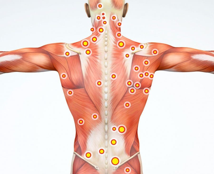
Sudden pain is a natural response of the nervous system which helps to demonstrate possible injury. By way of instance, pain signals travel from an injured region through the nerves and spinal cord to the brain. Pain is generally less severe as the injury heals, however, chronic pain is different than the average type of pain. With chronic pain, the human body will continue sending pain signals to the brain, regardless if the injury has healed. Chronic pain can last for several weeks to even several years. Chronic pain can tremendously affect a patient’s mobility and it can reduce flexibility, strength, and endurance.
Neural Zoomer Plus for Neurological Disease
Dr. Alex Jimenez utilizes a series of tests to help evaluate neurological diseases. The Neural ZoomerTM Plus is an array of neurological autoantibodies which offers specific antibody-to-antigen recognition. The Vibrant Neural ZoomerTM Plus is designed to assess an individual�s reactivity to 48 neurological antigens with connections to a variety of neurologically related diseases. The Vibrant Neural ZoomerTM Plus aims to reduce neurological conditions by empowering patients and physicians with a vital resource for early risk detection and an enhanced focus on personalized primary prevention. �
Food Sensitivity for the IgG & IgA Immune Response
Dr. Alex Jimenez utilizes a series of tests to help evaluate health issues associated with food sensitivities. The Food Sensitivity ZoomerTM is an array of 180 commonly consumed food antigens that offers very specific antibody-to-antigen recognition. This panel measures an individual�s IgG and IgA sensitivity to food antigens. Being able to test IgA antibodies provides additional information to foods that may be causing mucosal damage. Additionally, this test is ideal for patients who might be suffering from delayed reactions to certain foods. Utilizing an antibody-based food sensitivity test can help prioritize the necessary foods to eliminate and create a customized diet plan around the patient�s specific needs. �
Formulas for Methylation Support
XYMOGEN�s Exclusive Professional Formulas are available through select licensed health care professionals. The internet sale and discounting of XYMOGEN formulas are strictly prohibited.
Proudly,�Dr. Alexander Jimenez makes XYMOGEN formulas available only to patients under our care.
Please call our office in order for us to assign a doctor consultation for immediate access.
If you are a patient of Injury Medical & Chiropractic�Clinic, you may inquire about XYMOGEN by calling 915-850-0900.
�
For your convenience and review of the XYMOGEN products please review the following link. *XYMOGEN-Catalog-Download �
* All of the above XYMOGEN policies remain strictly in force.
Health coaches are becoming more and more crucial as modern medicine continues to improve. Now more than ever, the health care field is progressing at high speeds and professionals do not always have the available time some patients desire. Here is where health coaches become involved. Basically, the position of a health coach was produced to fill the emptiness in several doctor offices. Many physicians contribute but do not have the time or resources to assist each individual and aid in constructing healthy habits on a day to day basis. However, health coaches are available to be a supportive mentor that assists and guides patients in making healthy lifestyle changes. Many patients who seek help to change their lifestyle are those suffering from some kind of chronic pain, headaches, or joint inflammation.
In the previous weeks, we have defined and explained what a health coach is and what they really do, as well as the first two steps a health coach might take with a patient. Throughout this article, the third and fourth steps will be broken down and analyzed.
Need a refresher? No problem!
Health Coaching in El Paso: Part 1 can be found by clicking here.�
Health Coaching in El Paso: Part 2 can be found by clicking here.�
Step 3: Building A Plan For Action
Once the health coach understands the values and goals of the patient, a plan for change can get mapped out. One thing that is unique about building a plan, is that the health coach encourages the patient to have a say in it and contribute to building the plan. The ways of medicine have changed, and this aspect is one of them. Before, many patients would sit silently as doctors instructed them on their new protocol. However, it has been shown that patients who build a plan of action with the practitioner, are more likely to comply and complete a program.
In addition to this, the perspective of the patient can help maintain expectations and keep the plan of action at a realistic timeline. The health coach will offer their suggestions during this process as well as their perspective. Often times, this will help the patient break down their overall goal, into smaller more specific goals or tasks.
As soon as the overall goals are broken down into specific tasks, the health coach will then map out the process to complete these tasks. It can be simple to overlook small steps when thinking of a bigger picture, so the health coach will provide tools to better help the patient understand.
An example of this would be for a patient who wants to lose weight. Mapping out these tasks will have an end result that looks similar to these:
� I will try a new fruit and vegetable every day this week and identify what I enjoy
� I will think of different, creative ways to work movement into my day, such as finding a walking trail in my neighborhood
� I will always keep a water bottle with me and refill it every two hours
� I will cook dinner two nights this week
� I will go for a walk after dinner every day this week
By providing the patients with these smaller tangible tasks, the patient now has “homework” in a sense to complete these throughout the week. The health coach will set a deadline with these tasks and check-in with the patient regularly to ensure they are on track.
Step 4: Tracking Progress And Results
Before progress can be tracked, the health coach will take into consideration the patient’s goal and determine how often the patient will need to come in for follow-ups. For many patients, a combination of follow up techniques are used. Health coaches understand that in-person is not always the most convenient and does not always fit into the patient’s schedule. If this is the scenario, health coaches work around that to create follow-ups by using some in-person visits, some phone conversations, or other virtual check-in meetings that are HIPAA compliant.
Often times, during a lifestyle change patients may become confused or discouraged. It is important to remember that this is normal and progress is not always a straight line up, but rather includes bumps along the way. In order to better help the patient, the health coach will provide them with a helpful “where to turn” guide.
As humans, at different times we require different types of support. The where to turn guide will be a supporting reminder of things to do to counteract these feelings when they arise. Items included in this guide will be ideas such as:
� Pursuing a hobby, like dancing or playing an instrument
� Getting out in nature
� Starting a mindfulness practice
� Making art, like drawing or writing
� Joining a community, religious, or spiritual group
In addition to these activities, the health coach will determine with the patient what kind of support (internal or external) is appropriate depending on the situation.
Lastly,� progress does not always look like a dip in the number on the scale. Progress can come in many different forms. In order to help the patient appreciate and realize all the progress they are making, a health coach will ask questions like:
1. How can you appreciate your progress?
2. How would you describe the benefits of your experience?
3. What�s been good about this experience?
4. How have you grown?
As mentioned earlier, a health coach is important to have as they help one realize all the steps it truly takes to be successful and reach their health goals. There are many critical steps that are easily overlooked when the big picture is on their minds. The final two steps that a health coach will work on with a patient is to help them visualize their best self and to create a plan for resiliency. These two topics will be discussed in the next article.
�Using a health coach to complete a lifestyle change is similar to the work of going to therapy. One must be willing to accept the tools and resources they are givien, and actually do the work provided or it will not produce results. If a patient is truly serious about completing a lifestyle change, using a health coach is an extremly beneifical resource! As one can see, they work with the patients to hammer down tasks and ideas that a patient might not have orignally thought of. By utilizing a health coach, the patient has a higher chance of reaching their goals. – Kenna Vaughn, Senior Health Coach
All information and resources for this post came from an Integrative Practioner article titled, “A Six-Step Approach To Health And Wellness Coaching: A Toolkit for Practice Implementation” and can be found by clicking here; as well as listed below in the proper bibliography.
*The scope of our information is limited to chiropractic, musculoskeletal, and nervous health issues or functional medicine articles, topics, and discussions. We use functional health protocols to treat injuries or disorders of the musculoskeletal system. Our office has made a reasonable attempt to provide supportive citations and has identified the relevant research study or studies supporting our posts. We also make copies of supporting research studies available to the board and or the public upon request. To further discuss the subject matter above, please feel free to ask Dr. Alex Jimenez or contact us at 915-850-0900.
Bibliography:
American Psychological Association (2019). The Road to Resilience. Retrieved from: https://www.apa.org/helpcenter/road-resilience
Jonas, W. (2019). Empowering patients with chronic diseases to live healthier through health coaching: Integrative primary care case study. Samueli Integrative Health Programs.�Retrieved from: https://www.health.harvard.edu/staying-healthy/give-yourself-a-health-self-assessment
Miller, W. and Rose, G. (1991). Motivational Interviewing: Preparing People to Change Addictive Behavior. Guilford Publications.
Pecoraro, Wendy. �A Six-Step Approach to Health and Wellness Coaching: A Toolkit for Practice Implementation.� Official Media Integrative Practitioner, 17 Oct. 2019, www.integrativepractitioner.com/resources/e-books/a-six-step-approach-to-health-and-wellness-coaching-a-toolkit-for-practice-implementation.
Trzeciak, S. and Mazzarelli, A. (2019). Compassionomics. Studer Group. Virginia Polytechnic Institute and State University. The Stages of Change.Retrieved from: http://www.cpe.vt.edu/gttc/presentations/8eStagesofChange.pdf
Your Coach (2009). SMART goals.Retrieved from: https://www.yourcoach.be/en/coaching-tools/
Physical therapists (PTs) are healthcare professionals that treat patients of all ages with various ailments/conditions. A spine surgeon, physiatrist, orthopedist, primary care physician, neurosurgeon, and a chiropractor may refer patients to a physical therapist as part of a non-operative treatment plan.
An organized physical therapy plan may be an integral part of after-care following surgery. Therapists practice in a variety of settings, like hospitals, outpatient clinics, rehabilitation centers, and nursing homes. Physical therapists provide:
Treatments
Exercises
Mechanics
The primary goals�of physical therapy include:
Maintain practical skills
Improvement
Building endurance and strength
Increasing flexibility
Reducing pain
Preventing impairment
Physical therapists also instruct patients on the best way to exercise to enhance overall physical fitness, move about safely (biomechanics and ergonomics), and injury prevention. Physical therapists also help patients with long-term physical incapacity (eg, spinal cord injury).
Aquatic Therapy
Patients with osteoarthritis have found�waterexercise to be beneficial. With a gravity-free environment,�patients can perform simple exercises without stressing the tender joints. Movement increases circulation to the affected joints and can relieve stiffness. Swimming is also a great exercise for anyone for loosening up stiff joints and strengthening muscles.
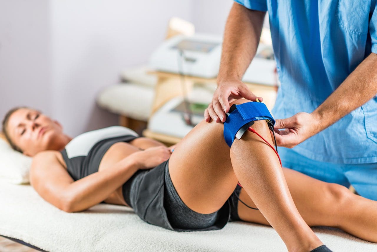
Electrical Stimulation
This type of therapy forces a muscle or muscle group to contract and relax. Therapists place surface patches containing electrodes on the skin over the area to be treated. The therapist programs the equipment to deliver the correct amount of stimulation for a set time.
The electrical current flows through nerve and muscle cells. The treatment is not painful. The patient feels gentle pulsating or an on/off sensation. This treatment stimulates circulation and supplies the area with oxygen and nourishment for healing. Electrical stimulation enhances healing and alleviates swelling and pain.
Electrical stimulation in physical therapy. Therapist positioning electrodes onto a patient’s knee
Heat and Ice
Heat increases circulation, decreases stiffness, pain and muscle spasms.
Patients with early arthritis symptoms find relief by taking a warm bath or hot shower.
It is best when done in the morning to help loosen up and alleviate stiffness.
Physical therapists use moist hot packs wrapped in a towel that is laid or wrapped around the affected area.
A moist hot pack transfers moist heat that penetrates deeply into soft tissues and stimulates local circulation more than heat alone.
Ice decreases pain by slowing the nerve impulses.
Inflammation subsides with forms of cold therapy:
Cold packs
Ice massage
Iced towels
They are usually the first aid following trauma.
When treating an overworked body part�ice�treatment should be supervised by a physical therapist.
Hydrotherapy
This is like a whirlpool bath. The water temperature and agitation loosen up joints, stimulate muscles and are controlled for maximum benefit.
Myofascial Release
This therapy improves circulation, decreases muscular tension and increases range of motion.
It is a type of massage that stimulates the muscles. The muscle tissue is manipulated by hand to stretch the tissue. Tight tissues become loosened using a cross friction motion with this therapy.
Movement & Conditioning
A physical therapist teaches patients how to move properly while being able to work through the pain. Therapists want patients to work as pain-free as possible. This does not mean that the exercises will be easy. More than likely they are going to be tough, but that is what you want to get back into top physical form.
Physical therapist assisting woman on an exercise ball at the clinic
Warming-Up can be accomplished by riding a stationary bike and some light stretching. The type of warm-up that goes with the therapy is determined by the individual treatment plan.
There will be muscle soreness for 24 to 48 hours following exercise therapy. This is completely normal and should be expected. As the exercises become a normal part of the day the discomfort will gradually go away. Stretching will increase flexibility. And as the treatment goes on resistive and strength exercises could be added.
Couple warming up
Home Exercise
With any treatment plan, there is usually a custom home exercise program. Exercises pretty much follow clinic exercises with variations and added stretches to keep the body from stiffening staying limber. Changes can be discussed with a physician.
Body Mechanics & Posture
Proper body mechanics helps to prevent further injury/s from occurring. Patients willing to maintain
Physical fitness
Reduce stress
Apply proper body mechanics
This reduces the risk of injury.
Proper posture is defined as keeping the natural curve of the spine. Proper posture minimizes stress to the spine. This is the first lesson a physical therapist teaches a patient.
Poor posture and poor body mechanics are leading contributors to neck and back pain.
Proper Work Habits
Do not lean over a desk for long periods.
Try not to sit without back support.
Adjust chair height so the knees are bent at a 90-degrees.
Bend the elbows at a 90-degree angle or they can rest on the work surface.
Don’t cradle the phone against the ear and shoulder as this can cause neck and shoulder issues.
Lifting and Carrying Objects
First, look at the object to be moved. If it looks too heavy find help.
Remove obstacles from the pathway where the object will be going through.
Visualize maintaining proper posture.
Get as close to the object as you can.
Place feet slightly apart and flat on the floor.
Bend at the knees to provide stable support.
Tighten the stomach muscles.
Breathe deeply.
Smoothly lift the object using arms and legs and not the back.
Hold the object at the sides and bottom.
Keep the object close to the body.
Keep back straight and carry the object with elbows slightly bent.
With shopping bags or luggage split the load in two, and try to carry the same amount of weight in each hand.
Pushing versus Pulling
Pushing is the more efficient and safer method of moving objects. Keep the back straight and use the knees to push. Keep close to the object and reposition the body every so often.
Reaching for Objects
Check the size, weight, and location of the object.
Use a proper stool or step-ladder to get the object. Stand with both feet flat.
One hand can be used for additional support.
Try not to look over too much as this can cause neck strain.
Think about storing regularly used items within easy reach.
Physical therapists may work directly for or with a physician, therapist, chiropractor and other healthcare providers to organize aspects of physical treatment plans. For example, a doctor may send physical therapist information of graphs, medications, analysis, and imaging results.
Massage Rehabilitation El Paso, Texas
NCBI Resources
During the first consultation, a physical therapist will talk about symptoms, analysis, and medical history.� Severity the location, type, and variables that decrease or increase pain are significant, and the PT will ask many questions regarding pain.
Physical therapists are healthcare professionals and members of your medical team. While physical therapy may be challenging or demanding at first, there are many benefits. It�s an opportunity to take charge of back or neck pain while building a stronger more resilient body.
Stomach pains, burning, or aching 1-4 hours after eating?
Excessive belching, burping, or bloating?
Inflammation in your stomach?
Is gas immediately following a meal?
If you are experiencing any of these situations, then try these eating mushrooms for your immune system.
Mushrooms
Medicinal mushrooms have been traditionally used for centuries by protecting anyone against infectious diseases, and various cancers. The positive biological effects of mushrooms are due in part to the indirect action of stimulating the immune cells. These mushrooms have a long history of usages by supporting health, especially in early Chinese, Egyptian, Greek, Mexican, and Roman cultures. In fact in 1991, a 5,300-year-old mummy was discovered carrying polypore fungus, which exerts a purgative effect. It may have been used to treat the mummies’ intestinal parasites.
What Are the Benefits of Mushrooms?
Modern research has shown that medicinal mushrooms can provide a rich source of nutrients and bioactive compounds that are associated with a few health effects that primarily support the immune system. Mushrooms act as an anti-bacterial, immune system enhancer and cholesterol-lowering agents. Additionally, they are an essential source of bioactive compounds, and some mushroom extracts are used to promote human health as well as being found as dietary supplements.
Since medicinal mushrooms are edible macroscopic fungi that are visible to the naked eye and are used for their beneficial health properties. Fungi, which includes yeasts molds, and mushrooms, live on the dead matter that is found in soil, plants, animals, and other fungi. It is estimated that there are 14000 to 22000 known species of mushrooms worldwide and approximately 20 to 30 mushrooms that are cultivated edible species. Even though there approximately 15 species that are wild foraged for consumption, they can be part of functional foods or dietary supplements.
Mushrooms are a source of many nutrients, including fiber, protein, selenium, potassium, and vitamins, B1, B2, B12, C, D, and E. They also possess several bioactive components like alkaloids, flavonoids, terpenes, phenolic compounds, polyunsaturated fatty acids, and polysaccharides. Mushrooms have been studied for not only its immune-stimulating and prebiotic properties, but they notably contain ?- glucan, which is a polysaccharide that is commonly present in mushrooms.
Research has been examining the health effects of mushrooms and has identified approximately 130 possible therapeutic properties, including:
Antibacterial
Antidiabetic
Antifungal
Anti-inflammatory
Antioxidants
Antiparasitic
Antitumor
Antiviral
Hepatoprotective
Immunomodulating
The research on medicinal mushrooms is based on animal or in-vitro trails that are up to date. Some earlier clinical trials suggested that individuals who consume mushrooms can have the benefits of reducing cancer and it�s many symptoms in the body. There are several mechanisms that have been proposed to explain the beneficial effects of mushrooms for immune health. Certain mushrooms can positively influence the gut microbiota by protecting it from harmful pathogens. There are even several mushrooms that have been shown to support immune health by enhancing the innate and adaptive responses in the body and exerting anti-allergic effects. Here are eight mushrooms that have immune supportive properties.
The Eight Mushrooms
Chaga
The Chaga mushroom is also referred to as birch mushroom or Chaga conk. It is a dark brown and black fungus that grows on birch trees. Several beneficial compounds are found in this mushroom and contains anti-oxidant polyphenols, betulin, and betulinic acid that are associated with anti-cancer effects for the body.
Studies show that Chaga mushrooms are used in traditional medicine and can be used in different remedies. This includes using Chaga as an anthelminthic, curing digestive disorders, and to help prevent chronic illness that affects the heart and liver.
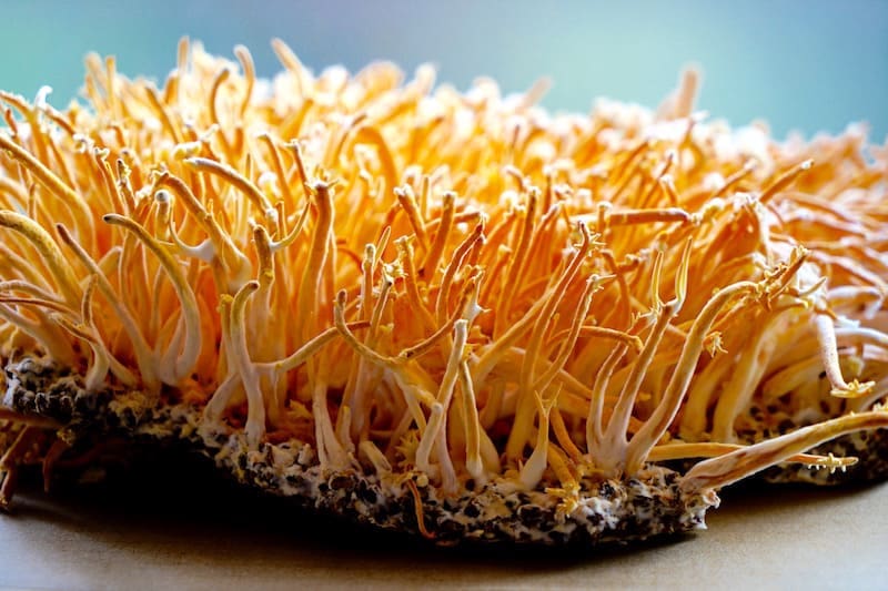
Cordyceps
Even though it is not technically a mushroom, this rare caterpillar fungus grows only in high-altitude regions in northeast India. Studies found that the bioactive components in cordyceps include polysaccharides, cordycepin, and cordycepin acid. Cordyceps was described in old Chinese medical books that traditional healers used on patients to improve their energy, stamina, and their sleeping patterns.
In a study, healthy Koreans individuals took supplements that contain cordyceps extract for eight weeks, and the results were that the extract increased the activity of NK-cells (natural killer) immune cells and improving the immune system in the body.
Lion’s Mane
Also known as Hericium Erinaceus, this mushroom has a white, fur-like appearance that resembles a lion�s mane. This mushroom can be beneficial for a healthy gut microbe and is associated with reducing colon tissue damage from inflammatory bowel disease.
Researchers suggested that lion�s mane may help individuals regulate their immune system and can improve the health of those who have IBD, but there is still more research being done to confirm this finding for the future.
Maitake
Maitake is both a culinary and medicinal mushroom that has proven to have anticancer activity for a variety of cancers that can affect the body. Maitake has a component called proteoglycan, and it has been associated with the immune-simulating effects.
Studies have been shown that proteoglycan can decrease mammary tumor cell behavior in animals and more research shows that maitake can exert anti-viral activity against hepatitis B and HIV from the body.
Oyster
Oyster mushrooms are a genus-group of fungi that has serval species like Pleurotus ostreatus and Pleurotus florida. Research has found that polysaccharides that are present in P. ostreatus mushrooms can activate N.K. cells against cancer cells. While another research shows that the extract of P. florida contains several active components containing anti-inflammatory properties in animal models.
Reishi
Known as the �king of mushrooms�, reishi has been shown to prevent various diseases and can modulate inflammation that is associated with a high cholesterol diet on people.
The health effects of this mushroom may be a result of its ability to regulate the body�s microbiota composition.� The beneficial effect that is found in reishi can help increase the beneficial bacteria that are in a person�s body.
Shiitake
Shiitake mushrooms have been traditionally used to treat common ailments that a person may encounter. Studies have shown that people who consume shiitake mushroom saw that there were changes in their body as their gut immunity and the anti-inflammatory components were improving over time.
As with many mushrooms, shiitake mushrooms have anticancer effects and lentinan that is being currently used as a complementary treatment for tumors.
Turkey Tail
The turkey tail mushroom gets its name from the tan and brown rings on its surface, resembling the tail feathers of a turkey. Research has shown that in traditional medicine, healers use the turkey tail mushroom to treat fungal infections, cancer, and AIDS on patients.
A 2007 study that was conducted by the Kyoto University Graduate School of Medicine in Japan found that over 8,000 cancer patients that took turkey tail and combined it with chemotherapy have an increased chance of survival.
Conclusion
From coming back to the body, mushrooms are used to stop diseases and cancers. Using its many health advantages of supporting the entire body can be helpful for anyone who wants to incorporate them into their diet. Mushrooms are edible while some are poisonous from the wild consuming these eight mushrooms are safe for individuals. Combining these mushrooms and some products are beneficial in supporting the immune system and are designed for more excellent stability, bioavailability, and digestive comfort.
The scope of our information is limited to chiropractic, musculoskeletal, and nervous health issues or functional medicine articles, topics, and discussions. We use functional health protocols to treat injuries or disorders of the musculoskeletal system. Our office has made a reasonable attempt to provide supportive citations and has identified the relevant research study or studies supporting our posts. We also make copies of supporting research studies available to the board and or the public upon request. To further discuss the subject matter above, please feel free to ask Dr. Alex Jimenez or contact us at 915-850-0900.
References:
El-Deeb, Nehal M, et al. �Modulation of NKG2D, KIR2DL and Cytokine Production by Pleurotus Ostreatus Glucan Enhances Natural Killer Cell Cytotoxicity Toward Cancer Cells.� Frontiers in Cell and Developmental Biology, Frontiers Media S.A., 13 Aug. 2019, www.ncbi.nlm.nih.gov/pmc/articles/PMC6700253/.
Feeney, Mary Jo, et al. �Mushrooms and Health Summit Proceedings.� OUP Academic, Oxford University Press, 8 May 2014, academic.oup.com/jn/article/144/7/1128S/4569770.
Ganeshpurkar, Aditya, and Gopal Rai. �Experimental Evaluation of Analgesic and Anti-Inflammatory Potential of Oyster Mushroom Pleurotus Florida.� Indian Journal of Pharmacology, Medknow Publications & Media Pvt Ltd, 2013, www.ncbi.nlm.nih.gov/pmc/articles/PMC3608298/.
G�ry, Antoine, et al. �Chaga ( Inonotus Obliquus), a Future Potential Medicinal Fungus in Oncology? A Chemical Study and a Comparison of the Cytotoxicity Against Human Lung Adenocarcinoma Cells (A549) and Human Bronchial Epithelial Cells (BEAS-2B).� Integrative Cancer Therapies, SAGE Publications, Sept. 2018, www.ncbi.nlm.nih.gov/pmc/articles/PMC6142110/.
He, Yanli, et al. �Grifola Frondosa Polysaccharide: A Review of Antitumor and Other Biological Activity Studies in China.� Discovery Medicine, 23 Apr. 2018, www.discoverymedicine.com/Yanli-He/2018/04/grifola-frondosa-polysaccharide-antitumor-and-other-biological-activity-studies-in-china/.
Integrative, PDQ, and Alternative and Complementary Therapies Editorial Board. �Medicinal Mushrooms (PDQ�).� PDQ Cancer Information Summaries [Internet]., U.S. National Library of Medicine, 30 Nov. 2016, www.ncbi.nlm.nih.gov/books/NBK401261/.
Jayachandran, Muthukumaran, et al. �A Critical Review on Health Promoting Benefits of Edible Mushrooms through Gut Microbiota.� International Journal of Molecular Sciences, MDPI, 8 Sept. 2017, www.ncbi.nlm.nih.gov/pmc/articles/PMC5618583/.
Jung, Su-Jin, et al. �Immunomodulatory Effects of a Mycelium Extract of Cordyceps (Paecilomyces Hepiali; CBG-CS-2): a Randomized and Double-Blind Clinical Trial.� BMC Complementary and Alternative Medicine, BioMed Central, 29 Mar. 2019, www.ncbi.nlm.nih.gov/pmc/articles/PMC6441223/.
Lindequist, Ulrike, et al. �Medicinal Mushrooms.� Evidence-Based Complementary and Alternative Medicine: ECAM, Hindawi Publishing Corporation, 2014, www.ncbi.nlm.nih.gov/pmc/articles/PMC4095656/.
Lindequist, Ulrike, et al. �The Pharmacological Potential of Mushrooms.� Evidence-Based Complementary and Alternative Medicine: ECAM, Oxford University Press, Sept. 2005, www.ncbi.nlm.nih.gov/pmc/articles/PMC1193547/.
Oba, Koji, et al. �Efficacy of Adjuvant Immunochemotherapy with Polysaccharide K for Patients with Curative Resections of Gastric Cancer.� Cancer Immunology, Immunotherapy: CII, Centre for Reviews and Dissemination (U.K.), June 2007, www.ncbi.nlm.nih.gov/pubmed/17106715.
Panda, Ashok Kumar, and Kailash Chandra Swain. �Traditional Uses and Medicinal Potential of Cordyceps Sinensis of Sikkim.� Journal of Ayurveda and Integrative Medicine, Medknow Publications Pvt Ltd, Jan. 2011, www.ncbi.nlm.nih.gov/pmc/articles/PMC3121254/.
Valverde, Mar�a Elena, et al. �Edible Mushrooms: Improving Human Health and Promoting Quality Life.� International Journal of Microbiology, Hindawi Publishing Corporation, 2015, www.ncbi.nlm.nih.gov/pmc/articles/PMC4320875/.
Wasser, Solomon P. �Medicinal Mushroom Science: Current Perspectives, Advances, Evidences, and Challenges.� Biomedical Journal, U.S. National Library of Medicine, 2014, www.ncbi.nlm.nih.gov/pubmed/25179726.
Do you feel:
Aches, pains, and swelling throughout the body?
Stomach pains, burning, or aching 1-4 hours after eating?
Excessive belching, burping, or bloating?
Inflammation in your stomach?
Is gas immediately following a meal?
If you are experiencing any of these situations, then try these eight edible mushrooms for your immune system.
Mushrooms
Medicinal mushrooms have been traditionally used centuries for protecting anyone against infectious diseases, and various cancers. The positive biological effects of mushrooms are due in part to the indirect action of stimulating the immune cells. These mushrooms have a long history of usages by supporting health, especially in early Chinese, Egyptian, Greek, Mexican, and Roman cultures. In fact, in 1991, a 5,300-year-old mummy was discovered carrying polypore fungus, which exerts a purgative effect.� It may have been used to treat the mummies’ intestinal parasites.
Mushroom Benefits
Modern research has shown that medicinal mushrooms can provide a rich source of nutrients and bioactive compounds that are associated with a few health effects, primarily supporting the immune system. Mushrooms act as an anti-bacterial, immune system enhancer and cholesterol-lowering agents. Additionally, they are an essential source of bioactive compounds, and some mushroom extracts are used to promote human health as well as being found as dietary supplements.
Medicinal mushrooms are edible macroscopic fungi that are visible to the naked eye and are used for their beneficial health properties. Fungi, which includes yeasts molds, and mushrooms, live on the dead matter that is found in soil, plants, animals, and other fungi. It is estimated that there are 14000 to 22000 known species of mushrooms worldwide, and approximately 20 to 30 mushrooms that are cultivated edible species, while approximately 15 species are wild foraged for consumption and can be part as functional foods or dietary supplements.
Mushrooms are a source of many nutrients, including fiber, protein, selenium, potassium, and vitamins, B1, B2, B12, C, D, and E. They also possess several bioactive components like alkaloids, flavonoids, terpenes, phenolic compounds, polyunsaturated fatty acids, and polysaccharides. Mushrooms have been studied for not only its immune-stimulating and prebiotic properties, but they notably contain ?- glucan, which is a polysaccharide that is commonly present in mushrooms.
Research has been examining the health effects of mushrooms and has identified approximately 130 possible therapeutic properties, including:
The research on medicinal mushrooms is based on animal or in-vitro trails that are up to date. Some earlier clinical trials suggested that individuals who consume mushrooms can be beneficial for reducing the risk of breast cancer and can help improve cancer-related symptoms like insomnia and sweating.� Several mechanisms have been proposed to explain the beneficial effects of mushrooms for immune health. Certain mushrooms can positively influence the gut microbiota by improving the protection against pathogens. There are even several mushrooms that have been shown to support immune health by enhancing the innate and adaptive immune responses as well as suppressing the immune response, thereby exerting anti-allergic effects.
The Top 8 Mushrooms
Here are the top 8 mushrooms that have immune supportive properties.
Chaga (Inonotus obliquus)
The Chaga mushroom is also referred to as birch mushroom and Chaga conk. It is a dark brown and black fungus that often grows on birch trees. Several compounds are found in Chaga, with its beneficial effects that contain anti-oxidant polyphenols, betulin, and betulinic acid that are associated with anticancer effects.
Studies show that the Chaga mushrooms are used in traditional medicine for different therapeutic indications, such as using it as an anthelminthic, as an antitubercular, to cure digestive disorders (gastritis, ulcers, etc.), or even to prevent cardiac or hepatic illnesses.
Cordyceps (Ophiocordyceps Sinensis)
Even though cordyceps is not technically a mushroom, this rare caterpillar fungus grows only in high-altitude regions of Sikkim, a state in northeast India. Studies found that the bioactive components in cordyceps include polysaccharides, cordycepin, and cordycepic acid. Cordyceps was described in old Chinese medical books in ancient times and used by traditional healers to improve energy, appetite, stamina, libido, endurance, and sleeping patterns.
In an eight week study, healthy Koreans individuals took supplements that contain cordyceps extract, and the results were that with the cordyceps extract, it increased the activity of NK-cells (natural killer immune cells). This change was accompanied by improving the immune regulation in the body.
Lion’s Mane (Hericium Erinaceus)
Also known as Hericium Erinaceus, the lion’s mane mushroom has a white, fur-like appearance and may promote beneficial gut microbiota growth and be associated with reducing colon tissue damage from inflammatory bowel disease.
Researchers suggested that lion�s mane may help individuals regulate their immune system and can improve the health of those who have IBD, but there is still more research being done to confirm this finding.
Maitake (Grifola frondosa)
Maitake is both a culinary and medicinal mushroom that has proven to have anticancer activity on breast cancer, melanoma, and hepatoma cells. Maitake has a component called proteoglycan, and it has been associated with the immune-simulating effects.
Studies have been shown that proteoglycan can decrease mammary tumor cell behavior in mice, and research shows that maitake can exert anti-viral activity against hepatitis B and HIV (human immunodeficiency virus.)
Oyster (Pleurotus)
Oyster mushrooms are a genus of fungi that has serval species like Pleurotus ostreatus and Pleurotus florida.� Research has found that polysaccharides that are present in P. ostreatus mushrooms can activate N.K. cells against lung and breast cancer cells. Another research shows that an extract of P. florida contains several active components like phenolics, flavonoids, and polysaccharides having anti-inflammatory analgesic effects in animal models.
Reishi (Ganoderma lingzhi)
Known as the �king of mushrooms� or the “mushrooms of immortality,” reishi has been shown to prevent or treat various diseases and modulate inflammation that is associated with a high cholesterol diet on people.
The health effects of this mushroom may be a result of its ability to regulate microbiota composition in the body, as the polysaccharides that are found in reishi demonstrates prebiotic effects and may increase the beneficial bacteria in a person’s body.
Shiitake (Lentinula edodes)
Shiitake mushrooms have been traditionally used to treat reasonable conditions like the common cold. Studies have shown that people who consume shiitake were associated with favorable changes in secretion patterns of various immune compounds and that the changes caused by consuming shiitake mushrooms can improve the gut immunity and anti-inflammatory response.
As with many mushrooms, shiitake mushrooms have anticancer effects and contains a glucan called lentinan that is being currently used as a complementary treatment for tumors, especially in China and Japan.
Turkey Tail (Coriolus Versicolor)
The turkey tail mushroom gets its name from the tan and brown rings on its surface, and its appearance is similar to the tail feathers of a turkey. Research has shown that in traditional medicine, the turkey tail mushroom has been used to therapeutically to treat fungal infections, cancer, and AIDS (acquired immunodeficiency syndrome.) Turkey tail mushrooms have PSK (polysaccharide-K)� and have been used as a complementary cancer treatment
A 2007 study that was conducted by the Kyoto University Graduate School of Medicine in Japan found that over 8,000 patients that took turkey tail and combined it with chemotherapy have increased the survival rate of patients following gastric cancer resection.
Conclusion
Mushrooms have been used for a long time to prevent infectious diseases and various cancers from coming into the body. With its many health benefits for immune support, it can be beneficial to provide anti-inflammatory properties. Certain mushrooms are edible while others are poisonous in the wild, so consuming these eight mushrooms are safe for people. Combining these mushrooms and some products are beneficial in supporting the immune system and are designed for more excellent stability, bioavailability, and digestive comfort.
The scope of our information is limited to chiropractic, musculoskeletal, and nervous health issues or functional medicine articles, topics, and discussions. We use functional health protocols to treat injuries or disorders of the musculoskeletal system. Our office has made a reasonable attempt to provide supportive citations and has identified the relevant research study or studies supporting our posts. We also make copies of supporting research studies available to the board and or the public upon request. To further discuss the subject matter above, please feel free to ask Dr. Alex Jimenez or contact us at 915-850-0900.
References:
El-Deeb, Nehal M, et al. �Modulation of NKG2D, KIR2DL and Cytokine Production by Pleurotus Ostreatus Glucan Enhances Natural Killer Cell Cytotoxicity Toward Cancer Cells.� Frontiers in Cell and Developmental Biology, Frontiers Media S.A., 13 Aug. 2019, www.ncbi.nlm.nih.gov/pmc/articles/PMC6700253/.
Feeney, Mary Jo, et al. �Mushrooms and Health Summit Proceedings.� OUP Academic, Oxford University Press, 8 May 2014, academic.oup.com/jn/article/144/7/1128S/4569770.
Ganeshpurkar, Aditya, and Gopal Rai. �Experimental Evaluation of Analgesic and Anti-Inflammatory Potential of Oyster Mushroom Pleurotus Florida.� Indian Journal of Pharmacology, Medknow Publications & Media Pvt Ltd, 2013, www.ncbi.nlm.nih.gov/pmc/articles/PMC3608298/.
G�ry, Antoine, et al. �Chaga ( Inonotus Obliquus), a Future Potential Medicinal Fungus in Oncology? A Chemical Study and a Comparison of the Cytotoxicity Against Human Lung Adenocarcinoma Cells (A549) and Human Bronchial Epithelial Cells (BEAS-2B).� Integrative Cancer Therapies, SAGE Publications, Sept. 2018, www.ncbi.nlm.nih.gov/pmc/articles/PMC6142110/.
He, Yanli, et al. �Grifola Frondosa Polysaccharide: A Review of Antitumor and Other Biological Activity Studies in China.� Discovery Medicine, 23 Apr. 2018, www.discoverymedicine.com/Yanli-He/2018/04/grifola-frondosa-polysaccharide-antitumor-and-other-biological-activity-studies-in-china/.
Integrative, PDQ, and Alternative and Complementary Therapies Editorial Board. �Medicinal Mushrooms (PDQ�).� PDQ Cancer Information Summaries [Internet]., U.S. National Library of Medicine, 30 Nov. 2016, www.ncbi.nlm.nih.gov/books/NBK401261/.
Jayachandran, Muthukumaran, et al. �A Critical Review on Health Promoting Benefits of Edible Mushrooms through Gut Microbiota.� International Journal of Molecular Sciences, MDPI, 8 Sept. 2017, www.ncbi.nlm.nih.gov/pmc/articles/PMC5618583/.
Jung, Su-Jin, et al. �Immunomodulatory Effects of a Mycelium Extract of Cordyceps (Paecilomyces Hepiali; CBG-CS-2): a Randomized and Double-Blind Clinical Trial.� BMC Complementary and Alternative Medicine, BioMed Central, 29 Mar. 2019, www.ncbi.nlm.nih.gov/pmc/articles/PMC6441223/.
Lindequist, Ulrike, et al. �Medicinal Mushrooms.� Evidence-Based Complementary and Alternative Medicine: ECAM, Hindawi Publishing Corporation, 2014, www.ncbi.nlm.nih.gov/pmc/articles/PMC4095656/.
Lindequist, Ulrike, et al. �The Pharmacological Potential of Mushrooms.� Evidence-Based Complementary and Alternative Medicine: ECAM, Oxford University Press, Sept. 2005, www.ncbi.nlm.nih.gov/pmc/articles/PMC1193547/.
Oba, Koji, et al. �Efficacy of Adjuvant Immunochemotherapy with Polysaccharide K for Patients with Curative Resections of Gastric Cancer.� Cancer Immunology, Immunotherapy: CII, Centre for Reviews and Dissemination (U.K.), June 2007, www.ncbi.nlm.nih.gov/pubmed/17106715.
Panda, Ashok Kumar, and Kailash Chandra Swain. �Traditional Uses and Medicinal Potential of Cordyceps Sinensis of Sikkim.� Journal of Ayurveda and Integrative Medicine, Medknow Publications Pvt Ltd, Jan. 2011, www.ncbi.nlm.nih.gov/pmc/articles/PMC3121254/.
Valverde, Mar�a Elena, et al. �Edible Mushrooms: Improving Human Health and Promoting Quality Life.� International Journal of Microbiology, Hindawi Publishing Corporation, 2015, www.ncbi.nlm.nih.gov/pmc/articles/PMC4320875/.
Wasser, Solomon P. �Medicinal Mushroom Science: Current Perspectives, Advances, Evidences, and Challenges.� Biomedical Journal, U.S. National Library of Medicine, 2014, www.ncbi.nlm.nih.gov/pubmed/25179726.
Do you often feel energy level drops in the afternoon? Do you often crave sugar and sweets in the afternoon? Do you often have difficulty concentrating before eating? Various medical conditions can affect the overall health of our body and mind. However, research studies have found that anemia caused by iron deficiency can tremendously affect our brain health. �
Iron deficiency is considered to be one of the most prevalent nutritional health issues, affecting approximately 2.5 billion people worldwide. In developing countries, about 40 percent of children and 50 percent of pregnant women have an iron deficiency. Iron is an essential mineral found in approximately 5 percent of the earth�s crust, however, inefficiency in absorption, low iron levels in staple grain foods, and a variety of medical conditions can make iron deficiency a common problem among humans. In first world countries, iron deficiency is still one of the most common nutrient deficiencies. �
What Causes Iron Deficiency and Anemia?
Poor iron intake and increased iron loss, generally through bleeding or breastfeeding, are several of the main causes of iron deficiency. Pregnant women, breastfeeding women, women with heavy periods, children or picky eaters, vegetarians and vegans, as well as people with digestion health issues which cause decreased iron absorption like celiac disease or post gastric bypass, and people with increased bleeding, such as cancer, ulcers, gastritis, or parasites, are generally at higher risk for iron deficiency. High calcium intake, by way of instance, children who drink a lot of milk, can also affect iron absorption, together with drugs and/or medications, such as antacids and proton-pump inhibitors for gastroesophageal reflux disease. �
Although low iron levels are well-known for causing anemia because red blood cells need iron as a part of hemoglobin, iron is also needed for the brain and nerves. Severe iron deficiency in younger children can ultimately cause irreversible damage to cognition and result in lower IQ and developmental delays, especially during the most fundamental stages of human development and up to 16 months of age. Even in adults. the most common symptoms associated with iron deficiency are generally neurological symptoms, including fatigue, brain fog, and restless legs that cause insomnia, among other symptoms. �
Pica, the abnormal behavioral compulsion to eat non-nutritional foods like dirt or clay, is tremendously prevalent in regions of the world where iron deficiency is common. In the developed world, pica is a rare health issue, however, it still frequently occurs in children, pregnant women, and among other groups of people that are at higher risk for iron deficiency, including people who have had gastric bypass. Non-neurological symptoms associated with iron deficiency ultimately include pallor, generalized weakness, and higher than usual heart rate along with shortness of breath, especially with exertion. �
What are the Symptoms of Iron Deficiency?
As previously mentioned above, iron deficiency can cause problems associated with cognition and neurological health issues, such as restless legs and insomnia. The exact mechanisms of why this happens are unknown, however, without enough iron in the brain and nerves, there are problems with neurotransmitter signaling, the development of nerve insulation known as myelin, and brain energy metabolism. Reduced central neuron processing is considered to be one of the most critical problems associated with iron deficiency, which can be a cause of psychiatric symptoms and ongoing psychiatric problems. �
Occasionally, iron deficiency may also cause anxiety, depression, irritability, and even poor concentration and restlessness. By way of instance, iron deficiency has a much higher prevalence in children with ADHD but the symptoms can improve with iron supplements. People with iron deficiency have higher risks of developing psychiatric disorders, especially ADHD, and developmental disorders. Evidence has demonstrated that iron deficiency can cause a variety of other health issues. �
Iron enters the brain through the blood-brain barrier via transferrin receptors. Iron uptake into the brain is highly regulated but it also does highly depend on the iron status of the human body. Therefore, people with low iron levels will have much less iron going into the brain and people with high iron levels will have much more iron going into the brain. Several regions of the brain also appear to gather iron and have higher levels than others. Moreover, neurological symptoms can manifest before developing iron deficiency anemia. Thus, healthcare professionals can’t rule out iron deficiency anemia from the most commonly utilized basic screening test, a complete blood count. A better general screen involves ferritin levels, where less than 15 ng/ml presents the diagnosis for iron deficiency but less than 40 ng/ml presents with fatigue, brain fog, restless legs, and other neurological symptoms. Ferritin on its own can be misleading in populations of people with chronic inflammation, including people on dialysis, where ferritin can be high even if the person is diagnosed with iron deficiency. Furthermore, a full iron workup includes hemoglobin, MCV, ferritin, total iron-binding capacity, serum iron, and transferrin saturation. �
What is the Treatment for Iron Deficiency Anemia?
Treating iron deficiency is considerably simple through the utilization of iron supplements or in mild or moderate cases by encouraging the consumption of foods that are high in iron. Occasionally, people with severe absorption health issues will need iron transfusions intravenously. Meat and seafood are the best sources of easily absorbable heme iron, however, non-heme iron is naturally found in leafy greens, beans, and nuts. Make sure to talk to your doctor if you have iron deficiency. �
It is ultimately essential to make sure if you have iron deficiency before treating it with increased amounts of iron supplements. With the exception of blood loss, the only way to reduce excess iron is through the process of skin cells flaking off. Therefore, adult men who take a lot of iron supplements and people with a genetic tendency to absorb more iron from foods are at a higher risk of developing a medical condition, known as hemochromatosis or severe iron overload. �
Excess iron is stored in the liver and can lead to scarring of the liver, known as cirrhosis. Iron overload can also lead to joint and hormonal problems and it can also cause a bronze-ish skin color. Symptoms of hemochromatosis include joint pain, fatigue, and low sex drive as well as a higher risk of developing diabetes. High serum iron is associated with health issues like high blood pressure. People who aren�t iron deficient can consider regular blood donations to prevent accidental iron overload. Iron is one of those types of minerals that should neither be too high or too low but rather, just right. More research studies, especially clinical trials analyzing common medical conditions, such as restless legs, insomnia, and ADHD, are fundamental to help healthcare professionals understand the relationship between iron deficiency anemia and brain health. �
Recent research studies have demonstrated that iron deficiency anemia may be associated with brain health issues. Because the brain and nerves need iron for many functions, iron deficiency can cause a variety of symptoms and medical conditions, including brain fog, fatigue, restless legs with insomnia, anxiety, depression, and cognitive problems, besides anemia or lack of healthy red blood cells. Treatment for iron deficiency anemia may utilize iron supplements, however, it’s important to make sure to talk to a qualified healthcare professional in order to avoid risks and side effects through iron supplementation.� – Dr. Alex Jimenez D.C., C.C.S.T. Insight
Neurotransmitter Assessment Form
The following Neurotransmitter Assessment Form can be filled out and presented to Dr. Alex Jimenez. Symptoms listed on this form are not intended to be utilized as a diagnosis of any type of disease, condition, or any other type of health issue. �
Do you often feel energy level drops in the afternoon? Do you often crave sugar and sweets in the afternoon? Do you often have difficulty concentrating before eating? Various medical conditions can affect the overall health of our body and mind. However, research studies have found that anemia caused by iron deficiency can tremendously affect our brain health. � Iron deficiency is considered to be one of the most prevalent nutritional health issues, affecting approximately 2.5 billion people worldwide. In developing countries, about 40 percent of children and 50 percent of pregnant women have an iron deficiency. Iron is an essential mineral found in approximately 5 percent of the earth�s crust, however, inefficiency in absorption, low iron levels in staple grain foods, and a variety of medical conditions can make iron deficiency a common problem among humans. In first world countries, iron is still considered to be the most common nutrient deficiency. �
The scope of our information is limited to chiropractic, musculoskeletal, and nervous health issues or functional medicine articles, topics, and discussions. We use functional health protocols to treat injuries or disorders of the musculoskeletal system. Our office has made a reasonable attempt to provide supportive citations and has identified the relevant research study or studies supporting our posts. We also make copies of supporting research studies available to the board and or the public upon request. To further discuss the subject matter above, please feel free to ask Dr. Alex Jimenez or contact us at 915-850-0900.�
Curated by Dr. Alex Jimenez �
References:
Deans, Emily. �Heavy Metal: Iron and the Brain.� Psychology Today, Sussex Publishers, 29 Nov. 2015, www.psychologytoday.com/us/blog/evolutionary-psychiatry/201511/heavy-metal-iron-and-the-brain.
Additional Topic Discussion: Chronic Pain
Sudden pain is a natural response of the nervous system which helps to demonstrate possible injury. By way of instance, pain signals travel from an injured region through the nerves and spinal cord to the brain. Pain is generally less severe as the injury heals, however, chronic pain is different than the average type of pain. With chronic pain, the human body will continue sending pain signals to the brain, regardless if the injury has healed. Chronic pain can last for several weeks to even several years. Chronic pain can tremendously affect a patient’s mobility and it can reduce flexibility, strength, and endurance.
Neural Zoomer Plus for Neurological Disease
Dr. Alex Jimenez utilizes a series of tests to help evaluate neurological diseases. The Neural ZoomerTM Plus is an array of neurological autoantibodies which offers specific antibody-to-antigen recognition. The Vibrant Neural ZoomerTM Plus is designed to assess an individual�s reactivity to 48 neurological antigens with connections to a variety of neurologically related diseases. The Vibrant Neural ZoomerTM Plus aims to reduce neurological conditions by empowering patients and physicians with a vital resource for early risk detection and an enhanced focus on personalized primary prevention. �
Food Sensitivity for the IgG & IgA Immune Response
Dr. Alex Jimenez utilizes a series of tests to help evaluate health issues associated with food sensitivities. The Food Sensitivity ZoomerTM is an array of 180 commonly consumed food antigens that offers very specific antibody-to-antigen recognition. This panel measures an individual�s IgG and IgA sensitivity to food antigens. Being able to test IgA antibodies provides additional information to foods that may be causing mucosal damage. Additionally, this test is ideal for patients who might be suffering from delayed reactions to certain foods. Utilizing an antibody-based food sensitivity test can help prioritize the necessary foods to eliminate and create a customized diet plan around the patient�s specific needs. �
Formulas for Methylation Support
XYMOGEN�s Exclusive Professional Formulas are available through select licensed health care professionals. The internet sale and discounting of XYMOGEN formulas are strictly prohibited.
Proudly,�Dr. Alexander Jimenez makes XYMOGEN formulas available only to patients under our care.
Please call our office in order for us to assign a doctor consultation for immediate access.
If you are a patient of Injury Medical & Chiropractic�Clinic, you may inquire about XYMOGEN by calling 915-850-0900.
�
For your convenience and review of the XYMOGEN products please review the following link. *XYMOGEN-Catalog-Download �
* All of the above XYMOGEN policies remain strictly in force.
Biomarkers are molecules that can help diagnose a health issue. These have become important for verifying investigations, choosing the best remedies, and monitoring disease progression. One exception, however, includes biomarkers for neurological diseases. Neurological biomarkers are found in the cerebral spinal fluid (CSF) or, in undetectable amounts, in the blood vessels. The human brain is closely guarded by the blood-brain barrier which protects it from damaging compounds circulating throughout the blood vessels. The blood-brain barrier has made it inaccessible to use these biomarkers. �
Biomarkers may be analyzed using the CSF but this also needs an invasive lumbar puncture process. Biomarker signatures, or recent improvements in discovery, in addition to the ability of clusters of biomarkers, are currently helping to make neurological disorders more treatable and more reachable. Treating and preventing neurological disorders, such as chronic traumatic encephalopathy (CTE), Alzheimer’s disease, Parkinson’s disease, autism, and major depressive disorder, is very likely to become less difficult to diagnose with the recent arrival of neurological biomarkers found in the blood. �
Biomarker for Brain Health Issues
Biomarker signatures, found with panels of high-quality antibodies, are yet another safe and effective tool for evaluating neurological disorders and diseases. Assistant professor of neurology and immunobiology at the University of Arizona College of Medicine at Tucson, Kristian Doyle, utilizes biomarkers to examine how the immune system deals with dead brain tissue. The system eliminates brain tissue with a procedure called liquefactive necrosis following a stroke but the pathophysiology of the procedure is unknown. This information is essential because liquefactive necrosis may be neurotoxic. �
“We utilize multiplex immunoassays to describe inflammation within chronic stroke infarcts in the point of liquefactive necrosis, and to describe changes by ordinary stroke comorbidities,” says Doyle. Because over 10 million individuals survive a stroke every year, Doyle expects that biomarkers may help them monitor the development of liquefactive necrosis and start to tailor remedies that mitigate the secondary harm due to this procedure,” he states. Another connection between inflammation and neurotoxicity is analyzed by Alysson Muotri, professor of molecular and cellular medicine and director of the Stem Cell Program at the University of California, San Diego School of Medicine. The Muotri laboratory uses induced pluripotent stem cells (iPSCs) from individuals with schizophrenia and autism to search for biomarkers of those ailments. �
Muotri’s laboratory started analyzing the cytokine interleukin-6 (IL-6) as a biomarker because evidence indicates that chronic exposure to elevated cytokines might be neurotoxic together with elevated levels associated with depression, autism, and schizophrenia. “The gap of one of the many brain disorders could function as cytokines act on particular types or subtypes of nerves, or within a particular brain area,” states Muotri. His laboratory differentiates iPSCs to cells, which they suspect might be releasing cytokines from patients. Also, because IL-6 can also be involved in immune-inflammatory pathways, Muotri supposes a connection between autism and in utero exposure to infection, such as the Zika virus. �
“Our forecast is that the inflammation caused by Zika vulnerability is sufficient to make a neurotoxic environment which could rewire how the human brain is shaped,” he states. “We see that in mice, therefore, we believe some Zika-exposed children are going to develop autism or have intellectual disabilities” Larger biomarker signatures are available with technologies from CDI Laboratories, which provides microarrays of practical human proteins, including over 20,000 to a single variety, to check the antibodies within human liquid biopsy samples, including blood, serum, plasma, CSF, or tissue lysates. The consequent “autoantibody profile” is a helpful tool for study and for diagnoses or prognoses of individuals. �
“We have worked in the area of biomarker discovery for various neurodegenerative diseases like multiple sclerosis, neuropsychiatric lupus, Alzheimer’s disease, and Parkinson’s disease,” states George Dorfman, director of business development in CDI Laboratories, a spin-off firm located in Baltimore, Maryland, and Mayaguez, Puerto Rico which was created from research in the High Throughput Biology Center at Johns Hopkins University. CDI’s stage is particularly beneficial in building panels for biomarker discovery because researchers can start by utilizing patient samples or banked trials to evaluate resistant profiles of cohorts that reveal specific symptoms or no indications in the event of control trials. �
“This provides us with an inherent candidate biomarker panel that offers advice on the following clinical outcome or curative efficacy, which is confirmed to yield the last panel, then interpreted into the state an ELISA-based kit or any other immunodiagnostic format at the clinical setting,” says Dorfman. “In the event of multiple sclerosis, as a patient grows through measures of this disease, their entire body creates novel antibodies or greater present antibody titers against specific proteins, such as myelination proteins. Our panels may discover these, to provide a notion about exactly what patients’ disease development might seem like, and supply a signature which may be interpreted into another evaluation or an FDA-approved diagnostic” CDI’s technologies have also been utilized to create an autoantibody profile for neuropsychiatric lupus, a beneficial diagnostic tool to ultimately help diagnose a neurological disorder that typically lacks obvious clinical signs. �
Understanding Biomarkers for Brain Health
The amount and types of biomarkers, as well as the quantity of information which researchers have to arrange, can help provide better remedies and prevention methods and techniques. “It ought to be no surprise that researchers spend around 80 percent of the time handling and not assessing statistics,” states Scott Marshall, managing director of translational informatics and diagnostic sciences in Precision for Medicine in Frederick, Maryland. The biomarker data management system, PATH, was made to incorporate any sort of biomarker information for further neurological disease diagnosis. �
“The true power of biomarkers comes if you connect this data to clinical information,” states Marshall. Their biomarker information management system supports translational research and biomarker-guided medicine development and puts no limitation on the number of biomarkers that may be tracked. “It can manage multiple biomarker technologies concurrently, such as complicated flow cytometry, next-generation sequencing, immuno-sequencing, epigenetic profiling, and other varieties of assays measuring biological variant too,” states Marshall. Their kind of “translational informatics” instrument is much more efficient than generating reams of information” with no strategy to acquire actionable insights out of these.” �
Research teams utilize Precision to Medicine’s platform for neuro-related programs that vary from illness pathogenesis to creating complicated signatures that are predictive of treatment response. By way of instance, the system was utilized in a research study including the evaluation of transcriptomic and genomic data in the treatment of major depressive disorder. The outcome is a genomically defined subset of individuals utilizing a probability of improvement. “This type of signature can now be evaluated by means of an assay, which may subsequently be developed to accompany diagnostic or free diagnostic to successfully target the correct individual group,” states Marshall. Biomarker data management systems become more fundamental as distinct kinds of biomarkers are examined collectively, ultimately including proteins and miRNAs. �
Combining kinds of biomarkers will very likely boost their usefulness. “Diagnostics is becoming increasingly more important as we know that the interplay between microRNAs, proteins, DNA, and messenger RNA is necessary,” states Pregibon. Clinical decision-making may profit particularly in which the human brain has been blocked by the blood-brain barrier until lately. “The chance to leverage biomarker-driven targeted treatments means that the sufferers that are more inclined to react to treatments are getting them quicker,” states Marshall. “For researchers, that’s the energy of biomarkers.” �
�
The recent ability to be able to detect neurological biomarkers in the blood, despite the blood-brain barries, is largely due in part to new technological advances in diagnosis and detection. Several of these technologies can ultimately increase sensitivity, however, increased sensitivity can help improve earlier detection or diagnosis of biomarkers for neurological diseases and disorders. Researchers and healthcare professionals currently believe that the presence of these biomarkers may be present earlier than we currently understand, which can help improve health issue diagnosis and treatment. – Dr. Alex Jimenez D.C., C.C.S.T. Insight
Neurotransmitter Assessment Form
The following Neurotransmitter Assessment Form can be filled out and presented to Dr. Alex Jimenez. Symptoms listed on this form are not intended to be utilized as a diagnosis of any type of disease, condition, or any other type of health issue.
Biomarkers are molecules that can help diagnose a health issue. These have become important for verifying investigations, choosing the best remedies, and monitoring disease progression. One exception, however, includes biomarkers for neurological diseases. Neurological biomarkers are found in the cerebral spinal fluid (CSF) or, in undetectable amounts, in the blood vessels. The human brain is closely guarded by the blood-brain barrier which protects it from damaging compounds circulating throughout the blood vessels. The blood-brain barrier has made it inaccessible to use these biomarkers. �
Biomarkers may be analyzed using the CSF but this also needs an invasive lumbar puncture process. Biomarker signatures, or recent improvements in discovery, in addition to the ability of clusters of biomarkers, are currently helping to make neurological disorders more treatable and more reachable. Treating and preventing neurological disorders, such as chronic traumatic encephalopathy (CTE), Alzheimer’s disease, Parkinson’s disease, autism, and major depressive disorder, is very likely to become less difficult to diagnose with the recent arrival of neurological biomarkers found in the blood. �
The scope of our information is limited to chiropractic, musculoskeletal, and nervous health issues or functional medicine articles, topics, and discussions. We use functional health protocols to treat injuries or disorders of the musculoskeletal system. Our office has made a reasonable attempt to provide supportive citations and has identified the relevant research study or studies supporting our posts. We also make copies of supporting research studies available to the board and or the public upon request. To further discuss the subject matter above, please feel free to ask Dr. Alex Jimenez or contact us at 915-850-0900.�
Curated by Dr. Alex Jimenez �
References:
Smith, Caitlin. �Biomarkers on the Brain: Putting Biomarkers Together for a Better Understanding of the Nervous System.� Science, 15 Mar. 2018, www.sciencemag.org/features/2017/12/biomarkers-brain-putting-biomarkers-together-better-understanding-nervous-system.
Additional Topic Discussion: Chronic Pain
Sudden pain is a natural response of the nervous system which helps to demonstrate possible injury. By way of instance, pain signals travel from an injured region through the nerves and spinal cord to the brain. Pain is generally less severe as the injury heals, however, chronic pain is different than the average type of pain. With chronic pain, the human body will continue sending pain signals to the brain, regardless if the injury has healed. Chronic pain can last for several weeks to even several years. Chronic pain can tremendously affect a patient’s mobility and it can reduce flexibility, strength, and endurance.
Neural Zoomer Plus for Neurological Disease
�
Dr. Alex Jimenez utilizes a series of tests to help evaluate neurological diseases. The Neural ZoomerTM Plus is an array of neurological autoantibodies which offers specific antibody-to-antigen recognition. The Vibrant Neural ZoomerTM Plus is designed to assess an individual�s reactivity to 48 neurological antigens with connections to a variety of neurologically related diseases. The Vibrant Neural ZoomerTM Plus aims to reduce neurological conditions by empowering patients and physicians with a vital resource for early risk detection and an enhanced focus on personalized primary prevention. �
Food Sensitivity for the IgG & IgA Immune Response
�
Dr. Alex Jimenez utilizes a series of tests to help evaluate health issues associated with food sensitivities. The Food Sensitivity ZoomerTM is an array of 180 commonly consumed food antigens that offers very specific antibody-to-antigen recognition. This panel measures an individual�s IgG and IgA sensitivity to food antigens. Being able to test IgA antibodies provides additional information to foods that may be causing mucosal damage. Additionally, this test is ideal for patients who might be suffering from delayed reactions to certain foods. Utilizing an antibody-based food sensitivity test can help prioritize the necessary foods to eliminate and create a customized diet plan around the patient�s specific needs. �
Formulas for Methylation Support
XYMOGEN�s Exclusive Professional Formulas are available through select licensed health care professionals. The internet sale and discounting of XYMOGEN formulas are strictly prohibited.
Proudly,�Dr. Alexander Jimenez makes XYMOGEN formulas available only to patients under our care.
Please call our office in order for us to assign a doctor consultation for immediate access.
If you are a patient of Injury Medical & Chiropractic�Clinic, you may inquire about XYMOGEN by calling 915-850-0900.
�
For your convenience and review of the XYMOGEN products please review the following link. *XYMOGEN-Catalog-Download �
* All of the above XYMOGEN policies remain strictly in force.
Lower back pain is a condition that pretty much affects most of us at some point in our lives. Some find:
Sitting
Standing
Walking
can be painfully difficult or helpful, depending on the position or the activity they’re involved in.
Low back pain varies from person to person, yet there remains no consensus on the optimal way to diagnose and treat patients.
Lower back pain can be caused by a multitude of factors and can be difficult to diagnose the exact cause. However, there is an advanced method of diagnosing and treating individuals with musculoskeletal conditions/injuries.
TENS, Transcutaneous Electrical Nerve Stimulation in Physical Therapy. Therapist Positioning Electrodes onto Patient’s Lower Back
Successful treatment
Dividing patients with lower back pain into more similar groups based on the same factors that aggravate and alleviate the pain means that a more custom based treatment plan can be created.
The one size fits all method just doesn’t cut it. A more focused approach for every individual leads to better results.
Patients find that placing their bodies in certain positions and certain physical activities can:
Activate
Aggravate
Deactivate their back pain.
Patients also find the pain being either better or worse.
Understanding why sitting, standing, and walking can change the severity of low back pain can be helpful in diagnosis.
These are important cues that help to diagnose and treat low back pain.
People sit, stand, and walk all day. This is why so much research has been conducted on how these specific positions and activities contribute to low back pain.
Spine Anatomy
The alignment of the spinal column, from the skull to the pelvis, is S-shaped.
The cervical and lumbar spinal segments curve towards the front of the body, and so are lordotic,�while the thoracic spine curves towards the back of the body and is kyphotic.
The amount of curvature does not stay in one place and changes based on body position.
Compared to standing, sitting decreases lumbar lordosis by about 50%.
Changes in lumbar lordosis can relieve pain from certain forms of back pain but can aggravate others.
Picture The Spine Bending
Imagine you are holding a garden hose. Holding it vertically in front of you. The hollow part of the tube represents the spinal canal, the part of the hose facing you is the posterior spinal column, and the part of the hose facing away from you is the anterior spinal column.
Bend the hose in half. The hose on the outward-facing side of the curve will stretch out, while the hose on the inward-facing side of the tube will be compressed.
This exercise helps visualize that an object being bent will experience two forces, either:
Compression
Tension
Increasing lumbar curvature will compress the posterior column�and stretch out the anterior column vertebrae and discs.
Decreasing lumbar curvature will�stretch out�the posterior column and�compress�the anterior column. This is the basis of why certain body positions can relieve back pain symptoms for some people and make it worse for others.
Prolonged sitting, and especially if poor posture is involved, can cause overstretching of spinal muscles.
Arthritis can affect the joints.
The facet joints are posterior based structures and can become compressed with increased curvature. When the facet joints are the primary cause of back pain, patients usually find that sitting improves the pain, while standing and walking make it worse.
When the discs are the pain generator, sitting seems to aggravate the pain as there is more compression in the tissue.
With certain types of disc herniations, sitting can compress the disc to the point that the herniated tissue starts to press against a nerve root causing radicular pain or pain that radiates out to another area of the body like the legs. In this case, walking seems to alleviate the pain.
Spinal Conditions
Various spinal conditions definitely contribute to back pain and have the same presentations and alleviations.
Spinal Stenosis which means the narrowing of the spinal canal. Patients with this type of spine pain improve with leaning forward sitting. This is because the posture increases/opens the size of the canal�and decreases the nerve root compression.
Spondylolisthesis is when one vertebra slips forward over the vertebra right underneath. This is more commonly known as a slipped disc. There are various forms of this condition. But the pain occurs immediately when sitting, bending or walking and is usually relieved when standing still or in a neutral position for short periods.
Sacroiliac Joint Dysfunction comes from the sacroiliac joint becoming inflamed. Pain is usually aggravated when sitting or about to sit. especially when there is added weight on the affected side. This can be difficult to diagnose because of trying to figure out if it’s the hip or low back causing the pain.
Locating the Pain Generator
Sitting, standing, walking low back pain causes are different for everyone. But finding the root cause helps to prescribe the best treatment possible. A chiropractor uses many different techniques to relieve pain. These include:
Applied pressure
Massage
Hands-on manipulation (adjustments)
X-rays
MRI’s
Lab work
Health coaching
Chiropractors don�t prescribe medications, but they do recommend therapeutic, rehabilitative exercises and nutritional lifestyle counseling to help the body heal itself.
Low Back Pain Treatment El Paso, Texas
NCBI Resources
A chiropractor sets up treatment approaches based on the individual. This begins with the natural, non-invasive treatments before moving on to more aggressive techniques. And as an added bonus patient�s who have received chiropractic treatment have experienced improvements to their digestive health and have reported better and deeper sleep after a chiropractic visit.
IFM's Find A Practitioner tool is the largest referral network in Functional Medicine, created to help patients locate Functional Medicine practitioners anywhere in the world. IFM Certified Practitioners are listed first in the search results, given their extensive education in Functional Medicine