Podcast: Athletic Strength Training vs Military Strength Training
[embedyt] https://www.youtube.com/watch?v=s75Q7sypEwQ[/embedyt]
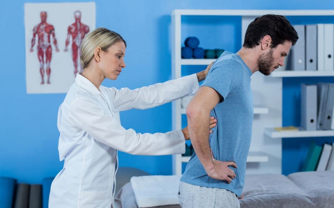
PODCAST: In today’s podcast, Dr. Alex Jimenez, chiropractor, and Kenna Vaughn, health coach, introduce Jeremy McGowan and Taylor Lyle, both experts in nutrition and strength training, as they discuss the differences between what is athletic strength training vs what is military strength training. By sharing a wide variety of nutrition and fitness recommendations, Jeremy McGowan and Taylor Lyle bring their knowledge and experience to El Paso, Tx where they offer performance improvements and injury recovery wherever they are needed. Dr. Alex Jimenez, Kenna Vaughn, Jeremy McGowan, and Taylor Lyle share what it is they do in their careers to provide overall health.� – Podcast Insight
[00:00:18] All right, guys, we’re here today. We’re excited. It’s a real special day for me here in El Paso because as you guys know, my job is to advance the science of wellness and fitness and to bring people that we have in El Paso to the forefront and to, you know, kind of show the individuals that are out there and the options we have. A lot of people don’t know. I’ve been out here for 30 years and I’ve seen El Paso kind of develop over the last three decades. And I’ve been nothing but proud to see the young kids and young men that are heading to the fitness programs all around the city, along with the insights that they’re bringing from where they come from people coming from all over the world. We have Olympians. We have specialists. We have top trainers, power trainers, fitness trainers, Crossfit trainers all around the world. These individuals bring a huge amount of talent and they all do the same thing. They get old. And as we get older, they were once the best in the world. And they come back and they share with. If you’re an Olympian and you know what? For the youth. So we bring certain individuals that have sciences and technologies. And some people are in the middle of their flight, in the beginnings and in the new starts of their lives where they actually bring us some great sciences. Today we have Jeremy McGowan and Taylor Lyle. These were two individuals that we brought in last time and we’re gonna hopefully have them come back and share with us their technologies. Jeremy brings a background. He works in the military, really smart guy. All these kids are really smarter than I am. It’s a beginning of time where we can see that the knowledge that the military has brought out has brought really great talent from around the world. [00:02:11][113.8]
[00:02:12] Jeremy is from Panama City, correct? Yes, sir. Yes. And Taylor, where are you from? Dallas. Fort Worth. [00:02:17][5.5]
[00:02:18] Dallas, Fort Worth. And one of the great things that I love about this, this whole story is that they’re here in El Paso and a lot of people don’t know this. And their expertise and knowledge are not only for us to to to benefit from, but they’re benefitting and they’re teaching the people that are here, the military, through their sciences and their techniques and their specialties and their licenses. So we really, really do have a moment in time where now the world is advancing in El Paso. So what I’d like to do is I’d like to say, introduce, you know, Kenna Vaughn. She’s over there on the side so you can see her. She’s there, she’s out. She’s anchoring on the side, making sure that my cameras work well and that I don’t stutter too much. Then we have Taylor Lyle and we have Jeremy. Jeremy McGowan. Correct. Yes. OK. And what we want to talk about a little bit about fitness training and the ideas about specifically about strength training, nutrition and as it pertains to collegiate sports and as well as power training for the military. So these kinds of sciences are very important for people to correlate. Now, do they cross lines? They cross lines for athletes in high school. So these sciences and these techniques are going to be good. But I like to know a little bit about Jeremy today. Jeremy, welcome to the show. And the people are here watching and they’re interested in understanding what it is that you do. So tell us a little bit about what you do and what you’ve done and where you came from. And we’ll leave it from there. Go ahead. [00:03:42][84.7]
[00:03:43] So, like you said, first, I’m from close to Panama City, Florida, a little tiny, small town. I went to Troy University on a baseball scholarship. It’s a D1 University in Alabama, close to Montgomery. So southeast Alabama, played there for five years. Soon as I got done playing slid right into coaching, I coached there for a little bit over three years. I ran baseball and softball, mostly assisted with other sports, the football, volleyball, soccer. A lot of others. Got offered a job out here to slot into the military side of things to coach. Getting really turn it down. Really, really enjoy what I’m doing here, running the physical training programs or the PT programs for a battalion at Fort Bliss. So I’ve worked with two separate battalions. We run the PT program, reconditioning program and then we work hand-in-hand. Kind of help write their PT programs so that when we’re not with them, they have a better idea of what to do. [00:04:36][53.1]
[00:04:37] I got a question for you. And you said that you’re an athlete going back to that. What position did you play? I pitched. You pitched. Oh, so you’re the dude. You’re the man. [00:04:44][7.0]
[00:04:45] You know, I was a closer started a little bit, but mostly closed. I really just tried to throw as hard as I could. That’s about it. Were you good? I like to think so. But you know some people might tell you differently. [00:04:57][11.8]
[00:04:58] Well, don’t be modest. Don’t be modest. You gotta say that you’re good at what you do. You know where I came from when I was a little boy. We got to see this out here in Mexico. We got this guy named Fernando Valenzuela. Well, remember that guy. Yeah. [00:05:11][13.4]
[00:05:11] Man was he was a Dodgers. Oh, man. I remember this big chunky looking dude that just could rip the ball. Definitely it didn’t look like he was a pitcher. But really, you know. But here’s the thing. Here’s the thing that I know now that I didn’t know then that people who got strong cores can really, really propel their force. Right. And this dude had a thick core, he busted up some gloves. Oh, hey. So let me ask you, what did the military see in you that they wanted to provide for this local community? [00:05:44][32.8]
[00:05:45] So the way that it kind of worked this whole program started as a very small pilot. There were five strength conditioning coaches and that was the first people on the ground. So that was it. And then it expanded. Now there are 60 coaches across a few bases in the U.S. So basically they needed qualified coaches that had an experience. So they wanted guys that had been a strength coach, you know, three-plus years, whatever, to lead the way. The assistants could have a little bit less experience but had to be certified, had to have a little bit of experience in the field so that they could get some, you know, guys in the run PT programs that were used to running large groups. So they wanted guys mainly from the collegiate side because we’re a little more used to running large groups, the private sector as well, depending on where they were at. Obviously, if they’re, you know, only working there for a very small amount of time, it might not be as much, but… They really wanted to focus on, you know, the qualifications, so having that Master’s degree and having that call and having that certification was the main thing. [00:06:44][59.0]
[00:06:45] I’ll tell you what, I saw you’re, clearly that you’re a really, really smart that both of you guys have resumes that are just amazing. And I got to tell you, the talent that the military brings this town is amazing. So don’t feel shy. Go ahead and tell people you got the big ‘ol Masters because that’s huge because you’re only one step away from a Ph.D. Let me ask you this because that’s very curious. The military has different departments, different battalions. What is it? [00:07:07][22.2]
[00:07:07] How many people in the battalion, the one that we were in originally there was around 410, 450. This one’s larger. So there are five companies. Each company is comprised of around 100 people. So there’s upwards of 550 in the battalion right now that we work with. [00:07:22][14.3]
[00:07:22] I’ll tell you what, we’re used to running a little bit of a Crossfit center. We’re actually coming from the Push Fitness Center. And 20, 30 kids at one time is a lot. How can you manage the largeness or the immensity of those groups together? [00:07:34][12.2]
[00:07:35] We kind of set up circuit style training for the most part. So we try to run stations with them. Luckily, I do have another strength coach, it’s not just me. So that helps a lot. We split the group up into two. Normally we’ll run a lifting type station and then a running type station and one of us will run each and we get about the halfway point. We’ll switch. So he’ll come over if I’m running the training. The strength training station to begin with. We’ll just flip flop. So he’ll bring his group over the straight training. I’ll take my group of the running and we’ll do that for the last half. So we usually have around outside tops around 80 people in a group. It would be the most that we would have and we would have 40 and 40 apiece. [00:08:09][34.0]
[00:08:10] Jeremy so you can pretty much see all these guys in different, I guess, techniques whether this is a running area. This is a strength area. You can see them all line sight kind of in the distance. [00:08:19][8.8]
[00:08:19] Yeah. That’s the goal. So with the strength training sessions, we set up kind of a semicircle on those stations so that I can just walk around the semicircle and then be able to see everyone. [00:08:29][9.6]
[00:08:30] And then as far as the running goes, it’s normally we do more anaerobic style training, some more sprint type work so that we can be right there telling them, you know, running the rest times, telling them, you know what, Tom, we’re trying to be on the run, whatever might be so that we can actually manage it a little more. [00:08:44][13.8]
[00:08:44] Wow. Taylor, we’re gonna get with you in a second there, so go ahead and drink some water. We’re going to get to you in a minute. But I got to ask a question for you. When you look at as a strength coach, do you have a deep-seated philosophy and the way things are done? And I assume and I don’t know, it seems like you’re beginning’s were with baseball. Correct. How do you apply that? That science and the level of mechanical sciences to the different kinds of levels and different types of specialties in the military. Let’s say you got some. Right, you know. I don’t know what kind of things they do. Let’s say the mechanics versus the heavy-duty artillery gunners. How do you change that up for them? [00:09:20][35.9]
[00:09:21] So one thing that’s really, really changed over for me with baseball to this is obviously with baseball I worked, with a lot of overhead throwing athletes. So a lot of shoulder problems, a lot of shoulder stability, things like that, that I was really trying to work with something that I’ve noticed in this military sector because of the way they’ve trained for so long. There they have a lot of shoulder injuries. There’s a lot of shoulder problems, a lot of instabilities as far as their, you know, way overcompensating. Their shoulders are starting to round from doing push-ups for so many years and not getting the proper training along with that. So having that expertise on that side of things, it’s helped me a lot as far as training, you know, different types of people. So I work in a BSB right now, so I haven’t really worked a whole lot with infantry, BSB Brigade Support Battalion. [00:10:06][45.2]
[00:10:06] Okay, got you. [00:10:07][0.4]
[00:10:07] We have a lot of mechanics, medics, communications people. It’s not a whole lot of high-speed guys. So we’re not really working with a lot of infantry type people. [00:10:17][9.7]
[00:10:17] We’re not working with a lot of guys that are really, really out there and really, really active. So a lot of the times, the people that we work with, the main things that we’re working on is landing mechanics, proper lifting technique, because we do have guys that have to lift some heavier stuff with transport and stuff like that. And in landing mechanics, guys jump out at trucks all the time. They’re in big, tall trucks, whatever might be. So those two things are something that we really try to work on so that in their day to day jobs, they don’t get hurt. [00:10:46][28.6]
[00:10:46] You know, when you say landing mechanics, whether it’s volleyball or anything, you know, that’s got to be the almost second nature. Oh, yeah. You know, I’ve seen that in the last couple of the last decade or two. I see the philosophy changing in the military, specifically in their ideas and their fitness goals. Recently, they’ve done some changes in their new programs where they actually if you don’t pass these certain things, you don’t even get the vacation time or even have even time to or migrate up in the ranks. But based on this performance, I’ve heard a lot about this ruck thing. What is this ruck thing? Yeah. Yeah, I heard. How much weight is it? Because they don’t care if you’re a 180-pound person or a ninety-five-pound lady, they’re still going to carry the same weight. [00:11:27][40.8]
[00:11:27] So there are different size rucksacks. It can be depending on really what your unit wants for that day or what type of thing you’re doing. So. Sure, you might have heard of the Baton Death March that happens here once a year. Yes, it did. So there are two separate standards for that. There’s a military light and the military heavy on the rucksacks are different. I don’t remember the exact way, but I want to say it’s 40 and 80 pounds. OK. If I remember right. Could be wrong on that. But it’s somewhere around that. And so that’s the light standard and the heavy standard as far as what they do in a normal setting for a rucksack. They kind of set it up for themselves. So basically, if a unit’s going on a ruck, they might tell you, hey, load it with as much as you want. Here’s how long we’re going, be able to do that in this fast. So they get to kind of pick their rucksack weight, depending on what they can handle. [00:12:13][45.6]
[00:12:13] Is it 40 through 80 or 40 and 80? [00:12:15][1.8]
[00:12:16] So in the baton it’s 40 and 80. But if they set it up themselves, they could do 40 through 80 as just depending on what they want to run with. [00:12:23][6.6]
[00:12:23] Yeah. You know, what do you look for in terms of an individual in order for them to say, oh, this dude’s going to just kind of wreck his back or he’s going to mess his shoulders up? What do you how do you tweak it so that you can kind of help them not get injured? [00:12:35][12.3]
[00:12:36] It’s, so posture’s a big part of it. Again, a lot of guys have rounded shoulders, so that translates over into the ruck as well. Well, they’ve got a heavy rucksack on their back. They start to hunch over round their back, their shoulders are already rounded. So you’re putting a lot of stress on the back, which I know you’re kind of the guy for that. [00:12:54][18.0]
[00:12:55] Oh, my God. I live with that every day, you know. Oh, you mean how we treat them? [00:13:05][10.2]
[00:13:05] You know what that can do to a back. And, you know, so there’s an issue that we try to fix. We do a lot of pulling, a lot of rows, a lot of rear delt work to try to get those shoulders back right. And stop the hunching. Stop the rolled shoulders. So that’s one thing that we try to do. And then again, as far as the lower body goes, proper gait is something that we try to work on, on the PTs work on that a little bit more than we do. But proper running mechanics, proper gait can obviously help with a lot of hip ankle knee issues. That a lot of guys have when they’re out there because they’re on uneven terrain. A lot of times rucking. They’re wearing their boots. You know, they’re not necessarily in the best running gear. So we try to do as much as we can to combat the problems that that can cause. [00:13:49][43.5]
[00:13:49] I find this to be so amazing that the both of you guys are here. Taylor, I know that you guys work together and I know we were introduced with you and the vast amount of expertise that you have and shared with us last time. But how do you guys interact? How does the diet world and the physical training world work together with Jeremy’s dynamics? [00:14:09][20.3]
[00:14:11] Yes. So we work hand in hand. I mean, you really can’t train without nutrition. So I’m out there a lot of times at the P.T. sessions, whether I’m trying to participate myself or just help the soldiers. So, you know, just making sure that they eat something in the morning, that’s a big issue that we see, is that they don’t have enough energy. And they wonder why they can’t finish their workout sometimes. So, you know, that is something that we both preach and then making sure that they eat something afterward, whether it’s going straight to breakfast or they’re getting some type of post-workout recovery modality. So we work with that. And then, you know, I do quite a bit of one on one counseling. And so a lot of times when I’m meeting with a variety of soldiers, you know, strength and conditioning come up in my conversation and we do a referral system. So I’ll refer them to Jeremy and, you know, follow up with him. And then, you know, a lot of times they’ll meet with them individually, give them a training program. And so we’re constantly urging communication with the best practices and, you know, how do we work towards the common goal. [00:15:25][74.0]
[00:15:26] So, you know, Jeremy, in terms of when you look at someone and you see them, they’re just they need help. You know, this kid is. He means well. But you can see him falling apart because you get that instinct like this kid’s going to blow out at something. He’s just not there. He looks ashy. He’s not eating well. How do you bring in Taylor in this dynamics, in that situation? [00:15:47][20.4]
[00:15:48] So a lot of the times I can really see it closer to the end of a workout as she said. Their energy levels are just low. You know, they can’t even, during the break period, they’re sitting down, they’re lying down. They’re trying to drink something and they can hardly drink as their stomach’s upset, you know. So I can tell pretty quickly if somebody has not eaten or is struggling with the nutrition side of things. And if that’s the case, then I’ll tell them, hey, you know, we’ve got a dietitian. We’ve got somebody that can help you. I can help a little bit in terms of telling you you need to eat something before you come out here. But she can help you, you know, in a better way than I can. [00:16:22][34.2]
[00:16:23] You guys coordinate a little bit. Kind of like this one is going to be a rough one. OK. We need to know where they’re going. They’re gonna be on the floor today. [00:16:29][6.4]
[00:16:30] There are some times that, you know, we can tell, you know, I can tell when I set up the circuit, like, okay. These guys are gonna get broke off a little bit, you know, and especially the ones that I’ve looked at and I know that she’s talked to, I’d make sure with them before those days. Hey, did you eat anything? And if not, then, you know, I’ll try to help out as much as I can, like, take breaks, you know, make sure you eat something next time, though, because this is how the sessions are going to continue to be for right now. [00:16:58][28.1]
[00:16:58] Guys, can you feel what I’m seeing, guys? And I’ve got to tell you when I started here in 1991, literally the military treated from my vantage point, again, I’m civilian and I don’t have to follow the rules, but like, they are set up there. But I could sense that the world was like Full Metal Jacket. It was really intense. It was a really harsh environment. And as you can tell, these two individuals are the forefront of the military to this day. So one of the things is I have to ask you both one question. Do you guys care about your guys? Oh, yeah, yeah, yes. You know what I got to tell you? You know what? I see this from the captains. Now, the world in the military is totally pro. There are people in a way that I have never seen go back two decades ago, three decades to 1991. I could not even get my hands on a military patient. They just would not let anyone outside the military take care of the people today. You guys are. Are you in the military? Both of you. No. No. [00:17:55][56.2]
[00:17:55] Contractors. See they’re bringing in the outside world. They’re also letting the inside go out. It’s awesome to see that because from my point of view, the caring that’s involved had to move from the top down and to have you guys from around the world, there’s got to be some amazing crew of people recruiting you guys. And I got to tell you, it makes me very proud because from the senators, you know, that actually made the Fort Bliss to become as big as it is now. And as it’s moved up, you see a lot of kind, caring sergeants, colonels, commanders that really care about their people. [00:18:33][38.1]
[00:18:34] And I got to tell you, it makes me feel really cool for an individual out there because I’ve got a kid who is your age. Right. So, you know, you guys got, you know, your guys taking care of him. So it’s a great thing. [00:18:43][9.8]
[00:18:44] Let me ask you, in terms of focusing on the dynamics of, let’s say over the shoulder, you had mentioned that shoulder thing going into that particular area is now for my vantage point, I’m a real lover of the shoulder girdle and the way the word and how it works together when you put something on the shoulder back in the day, there was one thing that really destroyed everyone’s shoulder. People didn’t realize this was this military. It was like a football jacket that had weights on it and they’d load it up in the front in the back, and you could put on, you know, some weights on it. These people had shoulder problems because of the pressure of the on the chromium, on the clavicle. And this happened. How is it that you kind of prevent a shoulder injury in terms of what you’ve seen when they wear things that are compressing them like a rucksack? [00:19:32][48.3]
[00:19:33] So part of that is the way they wear their rucksack. Our PTs do a really good job of demonstrating to them the proper technique of how to wear a rucksack, how to tie it down the right way so that it’s not putting a lot of pressure on their shoulders. That’s not something that necessarily I do, but that’s one way of combating it. As far as my role in it, I’m really just trying to strengthen the whole shoulder girdle and that whole area of the upper back, upper traps, whatever it might be done to try to take some load off so that they have a little bit of a shelf or something to sit it on. So we do a lot of like I said, we’re dealt work. We do a lot of rotator cuff work and a lot of trap work as well, so that they do get a little bit of that shelf. [00:20:15][41.7]
[00:20:15] All right. Well, that gives me a good understanding. I want to know the difference between an NCAA Division One athlete and the military athlete. How do you go about training and start like what are the similarities? And we’re going to try to look at the differences to contrast that specifically in that science. Go ahead and tell me a little bit about what you do for with your philosophies. [00:20:37][21.8]
[00:20:38] So similarities wise I would say the main things is their want to. A lot of times the military guys, the ones that are a little more high speed, they really want to get after it on PT. Right. So they’re one harder sessions. They want to sweat. They want to feel like they got something done. The NCAA guys are the same way. You know, they don’t want to come in and do one exercise and be done. They want to lift heavy. They want to get big. They want to get strong. And it’s the same way here. The only issue is here the training age is so much lower as compared to an NCAA Division One athlete. So when I would get a guy at college, you know, 18 years old. But he came straight out of high school. That was a 6A, 7A, 5A high school, you know, some bigger school played football for four years. He’s been working out since he was in eighth grade. These guys come here and, you know, I’ve got a lot of people that are 30 years old that didn’t play sports in high school, that have been in the military since they were 18. And they’ve been training wrong for 12 years since they got in the military. So their true training age is really nothing. [00:21:43][64.8]
[00:21:44] You know, they don’t really have good movement pattern. They don’t have an idea of really how to lift. They don’t have an idea of, you know, the right way to warm up, the right way to cool down anything like that. So it’s a lot more teaching here as compared to I could really get up and running at a Division One school like I was in about three or four weeks. I was up and running, had guys going full speed almost. So and here it’s a lot of teaching. [00:22:08][23.6]
[00:22:08] Jeremy, do you work with the reserves also? I do not. So we’re just with the active duty. Active duty. [00:22:14][5.6]
[00:22:15] So you mentioned 30 years old. OK. How does that work? And what’s your approach for a 30-year-old versus an 18-year-old? That’s got to do the same procedure. [00:22:22][7.6]
[00:22:23] The 18-year-olds are a little bit easier to teach. Their movement patterns are a little bit easier to pick up on because they haven’t been doing it wrong for so many years. Right. So if an 18-year-old and this is true across any population, whether it’s military or whatever, these guys, it kind of sticks a little faster. Right. So you teach them something two or three times they might have it, whereas this 30, 35-year-old guy that’s been doing this movement, but he’s been doing it wrong for 12 years. You know, when you try to teach him the correct way to do it, it might take eight, 10, 12, 15 sessions for him to finally get it down. And the issue with that is because of how many people are in the battalion, we might only get one or two sessions with him a week. So it might take four months for him to finally get this movement pattern down. And that slows down a lot of people in the process. [00:23:08][44.9]
[00:23:09] Do you separate them to kind of keep them on a different sack of or direction? [00:23:12][3.3]
[00:23:13] So we try to the issue with that is there. You know, if you’ve got one guy in Bravo Company and one guy, an Alpha company that is in the same boat, they don’t really do PT together. So it’s hard to separate within the same company, those people, because you might get that company once or twice a week. So if I’m really trying to separate the guys that are picking up on the guys that aren’t the groups, you’re going to be one of the really small. Or they’re just going to stop coming because they’re not getting enough out of it. [00:23:38][24.8]
[00:23:38] Taylor, in answering that same question, when you see those young kids that and versus the older or how do you approach the diet changes as well as just the approach of nutrition for them going through the same process in terms of the program? [00:23:55][16.3]
[00:23:56] Yes. Just what Jeremy said, you know, the 18-year-old scenario, they typically you know, they want to get better. They want to do what it takes to make it to the next level, which would be professional. And so I feel like they strive to want to get better. They’re a little bit more intuitive to that and receptive. And the, you know, 30-year-old, it’s not that they aren’t receptive. But, you know, a lot of them will have a family, whether that’s a spouse and children. And, you know, you have to take, you know, other factors that may be out of their control to have this success. So really, just in both scenarios, education component, there is so much room to grow, you know, unless someone maybe you went through like Ranger school a little bit more elite on the tactical side, you know, they might be a little bit more attuned to the nutrition and already know what to do around training and recovery. So they might not need as much education and guidance. But definitely there’s a lot of room to grow and both collegiate and military setting for nutrition. [00:25:09][73.5]
[00:25:10] All right. We’re gonna throw it to another gear here. Now, we’re dealing with in my thought process, as you take these young men to the next level, you’re going to deal with some elite guys. And that’s where a lot of my, you know, kids here, the Division one athletes, they correlate. And I got to tell you, from what I’ve seen, because I treat quite a few of the strange cats that go off to the journeys and they go into their, you know, the jungles, these are different kinds of characters. They have different mindsets. And there are at the highest level. Some of these guys are literally in their early, late 30s. And they’re just like that, you can see. In their eyes, they’re just ready to go climb trees, get in the jungle. [00:25:47][36.6]
[00:25:48] These individuals, these elite, these tactical guys, these ones that are that have percolated up to the highest level. How do you work with those individuals and what do you do in terms of trying to maintain them at their sharpest level? [00:26:01][13.5]
[00:26:03] So those guys are a little bit more obviously, like you said, they’re high speed. So they are more like working with a Division One athlete. Honestly, there’s been strength conditioning coaches in the special ops side of things for years and years. There are a lot more in tune with that side of things, with knowing the proper technique. Knowing how things are really supposed to work and knowing how they’re supposed to feel. So, you know, if they have a problem, they’re a lot more likely to either know if it’s actually pain or an actual injury. They can actually handle the two of them whereas guys that are not used to working out to them, you know, having pain and being sore the day after a workout, they’re hurt. You know, these guys are a little more in tune with their body and they’re a lot more likely to be able to push themselves through your workouts so you can go a lot heavier with them. You can do more of a, you know, true tier-based or strength-based or whatever it might be program that you want to do to get them better and better. [00:27:05][61.7]
[00:27:05] You know, when I was going to college, there were these programs that came out, strength training programs, where you could actually calculate how strong an individual was if they followed this tier, you know, go through these many deadlifts, do it this way, do it these reps. And over time, you were gonna go, you know, in a linear progression upwards. It was amazing that you could actually do it that way. Do you feel that if you push these athletes, you watch them improve, especially the top tier one that you can actually push them to, you know, an amazing level of accomplishment with tough training? [00:27:41][36.0]
[00:27:42] …�[00:32:55][48.8]
[00:32:56] Jeremy, how do you look at that stuff? And do you are you privy to that information and do you apply it to the flight that you’re doing? [00:33:02][6.0]
[00:33:02] So I don’t really get the actual numbers. Taylor is the one that gets those numbers and she would just share with me, hey, you know, this guy might need a little extra help. You know, as far as losing some weight goes, this guy is in the standards. He wants to gain a little bit of weight and he can, you know, that kind of thing, whatever it might be. So I don’t get the actual numbers, but I do get some information from her that I can help the guys with. [00:33:23][20.1]
[00:33:23] You know, one of the things that we realize in health care is the unification of data as well as integration of other sciences. You two guys met at a… Obviously, I’d like to know a little bit about how you guys introduce yourselves and how did you guys interact and how did you. Because, Taylor, you kind of talk to me about Jeremy. And I got to tell you, Jeremy seems to be an amazing guy. That’s got a lot of knowledge. And we and I really appreciate that. But how did you guys get to interact together? How did that process go in terms of for the purpose of the military? [00:33:56][33.1]
[00:33:58] Yes, so, yes, Jeremy is an excellent strength coach and it’s been a pleasure working with him. We actually work for two different contract companies, so we just were put together by chance, to be honest. And I mean, we just really clicked since day one, our personalities match really well. So that’s really where it began. And Jeremy has been here for almost two years and I’ve been here for almost a year. So he’s been here a lot longer than me. But so we met when I started. [00:34:28][29.3]
[00:34:28] Gotcha. In terms of your overall goals for the military and the dynamics for the athletes, let’s go back into the world of a little bit of the athletic division one. And now let’s also consider the fact that the sciences you have can also be applied to even the general public and even to kids at that level. And I know a lot of my patients have parents out there that want their kids to benefit from the best ideas and philosophies. And one of the things is that you realize that it’s not so much about knowledge. It’s about philosophy. It’s about your point of view. It’s the way you stand in what you think about how can we take what the military does in its sciences and its progression sciences to get these athletes and these individuals ready for battle. To our kids, how can we apply that if you can kind of reach into I don’t know if you’ve got kids, but if you do deal with kids, how would you apply those sciences to even the young, young high school, younger people population? [00:35:32][63.3]
[00:35:33] So I actually one of my papers or whatever for my masters was about strength training in kids because it was something that really, really interested me, because all my life I heard kids shouldn’t lift weights. Kids shouldn’t do this. It stunts their growth. It does. You know, it’s bad for them, whatever. [00:35:49][15.8]
[00:35:50] And honestly, everything that you read research-wise says otherwise. That’s just been a myth that’s been out there for so long that people started to believe it. So for me, as far as translating my side over to the general population. Younger kids all the way up to high school, it honestly starts with GPP, which is just general physical preparedness. So being able to handle their body weight, being able to learn movement patterns. So obviously push-ups, pull-ups, things like that for body weight, but then movement patterned on the squat, the landing mechanics like we talked about, things like that, and then just the general agility and movement stuff. So playing tag, doing things that are actually active outdoors. [00:36:29][38.9]
[00:37:12] So, you know, have him, you know, just practice squatting and making sure the knees are pointing out over the toes. He’s not getting valgus knee is not caving and he’s not you know, when he’s walking his gait pattern is good. When he’s running his gait pattern is good when he’s planting his foot. You know, stop and go playing tag with his friends. He’s, you know, actually planting sinking into that hip and driving off. You know, there’s little tiny things that you can look at that can help with those movement patterns as they get older and hopefully combat the chances of injuries as they get older. And then once as they get older and those movement patterns are more ingrained, then you can start adding some weight to stuff you can start doing. You know, even just goblet squats is where I would start. So a kettlebell or dumbbell holding a single thing. So you’re not actually loading the spine things like that and floor press and med ball throws and different things like that where you’re adding weight once those get learned more ingrained than you just are getting into the bigger lift. You know, you get to the big three, the squat bench deadlift, the Olympic lifting type stuff, whatever it is, Taylor, he is good. [00:38:12][59.6]
[00:38:13] ... [00:42:17][48.6]
[00:42:18] Taylor, you know you’re talking, right? I mean, this is amazing stuff in terms of its dynamics and specifically for recovery. How do you guys play into kids or young men that are injured in the nutrition component? How do you help them? How do you support the dynamics of the nutrition component? I know we talked about a little bit, but can you go back into it and talk about the things that you look at at the micronutrient level, as well as the macronutrient level to get these guys to be able to sustain the loads that they’re going to be under and provide them their best option? [00:42:53][34.6]
[00:42:54] Yes. So it goes back to recovery, nutrition, and the nutrient timing and making sure I mean, you’re breaking down your muscles when you’re working out and you’re trying to build them back up, grow. And so, you know, what’s going to do that is protein and carbohydrates. So making sure you have a three to one ratio of carbohydrates to protein. You know, that’s going to help them replenish their stores, their energy stores and also build muscle. And then from an injury standpoint, it’s just again, you know, making sure that depending on the injury will depend on the prescription for nutrition. But overall, you want to make sure that they have enough energy needs first and foremost, and they’re going to be less active typically. So, you know, you might not need as high of calorie needs that they would when they would be training. And the same with carbohydrates. It is your primary energy source, but you’re not going to be training as hard. So typically that is going to be lower. Now, your protein needs are going to be almost twice as high as they normally would be to really make sure that you’re, you know, getting the growth and nutrients you need for the protein and for the muscles to just recover from the injury, and then fat also plays a huge role as well. So and then micronutrients, you’re going to look at your B vitamins, zinc, vitamin C, vitamin A. You know, magnesium, those are all going to help in the wound healing injury recovery aspect as well. And then also immune support, which is really important. [00:44:36][101.9]
[00:44:37] Jeremy, thank you. Jeremy, they’re leaving now the day, they’re all exhausted. They’re all whooped on. Right. What are the words of advice that you give them about what they’re gonna eat tonight? You know, and let’s say you got an individual that’s just they just look bad. And what do you tell them? How do you tell them to rehab? Recover? I guess is a good word. [00:44:55][18.4]
[00:44:56] So for me, I’d try to preach high carb, high protein once after. So obviously, like she said, protein plays a big part in the recovery side of things. And they just depleted a lot of their carb sources during the workout. So that’s really what I try to preach, our sessions are in the morning. So a lot of times they’ve barely eaten anything as we’ve mentioned before. And if they have, it’s a lot of times not enough. So I try to preach. Get some carb sources, get some protein, get some eggs, get an omelet. They make you omelets in there. I know they do because I’ve been in there, eat one, you know, get something that can actually help you recover from this workout. [00:45:35][38.3]
[00:45:35] You mentioned, you know, they would sometimes show up without eating properly. You know, that’s a problem with a lot of the athletes. So they’re, you know, especially younger ones. They want to look good for some of the ones so they can volleyball. But some wrestlers, they got to, you know, have the basics, too. And for different types of athletes, different things for the population that you’re dealing with in order to get them better. What is the baseline good level of carbohydrates and what type of drinks or what kind of foods do you offer or recommend them at least get in that much so that they don’t end up totally running and being depleted by the end of the program. [00:46:12][36.3]
[00:46:13] So they’ll need 30 to 60 grams of carbohydrate, 30 minutes to an hour right before working now. And like Jeremy said, a lot of times, the workout is at six-thirty in the morning. So you’re not going to have the ideal scenario where people are eating three hours meal before they, you know, train. So. [00:46:30][17.7]
[00:46:31] So when you just wait. I’m sorry. When you said that 30 to 60 grams. So. So thirty-one twenty to 240 calories. Just a start up the engine. Right. Is that right? Is that a good fare. [00:46:40][9.0]
[00:46:41] Yeah. So that is fair. So what that looks like is 30 grams could be a banana or it could be a couple of slices of toast. You typically want something that is going to digest very well. So that’s going to be low in protein, low in fat and low in fiber. So that is going to be a carbohydrate source. You’re going to want to isolate that carbohydrate to avoid any digestion issues. So, you know, for people that can’t handle solid foods as well, I always recommend liquids. It’s already converted. So something as simple as a 20 ounce Gatorade. You know, if they can take something a little in between solid and liquid applesauce pouch, you know, there are so many varieties at the grocery store now for kids or adults. And, you know, just taking one of those apple sauces will also help meet that need. [00:47:33][51.8]
[00:47:33] You know, as you start your training program in the morning, what kind of things do you do? How do you ramp up the training program? Jeremy, I’d like to know a little bit about that, like take me through a day in your world. [00:47:45][11.7]
[00:47:46] So with ramping it up goes. As I said, we might get guys once or twice a week. So it’s a very, very slow process. It’s also dependent upon their battle rhythm. So, you know, we might get guys say twice a week. So we do have to get a group twice a week, which is what we were that whole battalion. We get every company twice a week. We might get them for six weeks and then they’re gone for three weeks doing a field training exercise and they completely detrain. Right. They’re doing nothing but sitting there for a lot of the time and, you know, practice in military type stuff, they’re not getting any physical training in. It’s not mandatory out there. It’s not necessary. And nobody does it and they can’t really shower. So nobody really wants to get sweaty and stuff. Right. So those three weeks when they come back, we kind of have to reset. There’s not really that much of a ramp-up. It’s a lot of general physical preparedness stuff. We do a lot of bodyweight stuff. And then a lot of the big three, we try to progress those as much as we can. So like right now, because we just kind of restarted with this battalion, with the whole COVID thing going on. We’re doing a lot of goblet squats. We’re doing trap bar. Deadlifts are extremely important. That’s going to be in their new PT test. [00:48:58][71.9]
[00:48:59] What was that? Trap bar deadlift. A different name for it, but we do that. And then right now, we’re doing floor press and we’re planning to progress the goblet squad into a front squat, front squat to back squat. Right. So that’ll be the progression there. The floor press will progress into the bench press. [00:49:16][17.6]
[00:49:17] Are those the three that you’re talking about? The three? [00:49:18][1.3]
[00:49:19] Yeah. So those are kind of the big three is your deadlift, your squat, and your bench. And so that’s your main three strength lift, right. That’s what everybody wants to be good at. So that’s the three that we kind of focus on. But we’ll set up circuits around that. So if we’re doing so, you have floor press, right? We’ll try to do some kind of a pull with that, whether it’s rear delt or an actual row. So it might be a kettlebell row, dumbbell row. Some like that. And then we’ll do a lower body exercise with that. So we try to go full body every workout session. So we’re getting upper, lower-end core. We try to do the main lift is for strength. So if it’s floor press, squats, or deadlifts, it’s more of your strength-based stuff. So it’s more that max effort. So it might be sets of four sets of five. Some like that with a heavier weight. We try to work up to a heavy load. Then everything else is more hypertrophy based. So it’s more work capacity. We’re trying to do, you know, a little bit of a lighter weight, but it’s still going to be heavy not to where it makes them work for those eight to 12 to 15 reps, whatever we might do. [00:50:21][61.6]
[00:50:21] Do you mix it? Like, do you have some hypertrophy versus agility and versus body mechanics stuff or do you have like certain days. Today’s Body Mechanic Day today is power today. This today is Hypertrophy Day. [00:50:30][9.3]
[00:50:31] So right now, because we don’t really know what group we’re going to get every day with stuff going on so they’re, kind of work in shifts. They’re not there every day. So we might have one group one day. It might be the same exact people, you know, for a full week. It might be. They come every other day. So the plan right now basically is we go up there, we set up three lifts. Monday was a Friday or lift days, Tuesday a more run day, though. And like I said, the running is more anaerobic stuff. So sprint stuff. But on those sprint days, we do more. We do lift more. But it’s more bodyweight work capacity stuff. So we’ll do a lot of push-ups, pull-ups, sit-ups, squats, lunges. But it’s all bodyweight type stuff and that’ll be all in a circuit with some running involved. And then on the lift days, it’s, you know, like I said, that one lift strength. Everything else is more hypertrophy/work capacity. So it’s all high reps and. [00:51:24][53.2]
[00:51:26] It’s kind of tough to get a lot of hypertrophy type stuff in because of the box that we’re working out of. So we have a gym that’s inside a box. You had to pull all the weight out. There’s not really enough weight to load up a lot of stuff if we want to do a lot of squats. We need weights for that. Right. So we need weights. But on the barbell, well, there’s only eight forty-fives. Eight thirty-five, eight twenty-five, and eight tenths. So if I have four stations of squats like that up, I’d need almost all of that weight to be able to handle that. So I can’t use that weight on anything else, whether it’s the sleds, the trap bars, whatever it might be. So I have to come up with stuff with bands and kettlebells is really I see an invention there. [00:52:02][36.8]
[00:52:03] I think there’s an invention in there and what I’m hearing is, is that your gym doesn’t go out to the outside that easy. So is that what I’m getting? Like you want to be able to have a piece of equipment, has all your stuff on it, so you just drag it off that thing. [00:52:14][11.5]
[00:52:14] … [00:56:58][62.0]
[00:56:59] I really believe that what he just said was a huge component? Now he has spent his whole life understanding body dynamics. And he ended up understanding and now the military gets it in a different level. The translation of force comes from the core. It is huge. When you hear med ball slams, that is a body that’s going to its fullest out and slamming at a full range of motion. When you’re seeing hip flexes, you’re pulling that hip to the furthest, deepest dungeon of movement, to the furthest extreme on the outside. So to be able to do that, to be able to translate, weight and slowed and sled movements, you’re gonna need a powerful core. The dynamics of it are the ability to move it through time and space at a certain rate of speed. How long you do it, you can do it a little bit, that’s strength. But power means you can translate it over twenty-five feet or so and hit back and forth. So we’re really pushing the body to a level that is amazing. I have sat down with certain patients of mine and they find that that theory. And I found it to be very interesting that deep tuck, the knee tuck, and the deep flexion movements. Where did that philosophy come from and you have as a physiologist and the nutrition strength coach? How did that come in? Where did that come from that they realized that those particular movements, the slam ball, as well as the deep tuck, became a crucial component in the military action? [00:58:26][86.2]
[00:58:27] So I know the people are well, Major Matthews’, that actually used to run H two F, she’s transferred over to a different side of the military now, but she used to work at the Olympic training facility in Colorado. And I know she helped develop the test. So I would guess I don’t know for sure, but I would guess she played a big role in that. Yeah, because she, you know, does know a lot more about that side of things. I know she helped create, you know, with the power throw and stuff. [00:58:57][29.5]
[00:58:57] What’s her name, shout out again? Major Matthews. Major Matthews. OK. [00:58:59][2.7]
[00:59:00] So you know we met her, she came down I think it was a little bit before Taylor got here so she came down to talk to us and explained about the test and why they were doing it and whatever. And she played a big part in developing a test because of her background. [00:59:18][18.4]
[00:59:19] Have you guys gone to Colorado Springs before to take a look at the Olympic Center? I have not. I have not either. You know what? I got to go there. But there, you know, I got to watch from the outside inward. And you got to I got to tell you that you can see top athletes from around the world. I mean, from powerlifters. But you can see that they’re not very big in the sense of muscular build. But you can see that every athlete had a trainer with them and usually it’s a physical therapist that was right with them. And they were talking mechanics and movements. And these athletes and all the sports that they have, you see this amazing. It’s almost like watching something out of an amazing superpower show where you see these athletes running from all different directions…
And these are the top athletes in the world training centers from swimmers to high bolt whatever the sport is, I can imagine, but you can see them training in the center and they really focus on the range of motion. And you can see the physical therapist showing the motion. And actually the intensity of the movement is really, really important. So that science of Deep Tuck and translation of force is huge now. And it’s amazing that now to be able to do that is at the forefront of the military’s progression. Let me ask you this. Now that you know and you’re in your science and understanding is about the youth. How do you correlate that and take me into the progression of how to get kids, let’s say, a high school kid into doing that particular component of translation of forces so that we can make them great at being a lineman or just torquing the heck out of someone in wrestling. You know, kind of that deal. [00:00:54][54.1]
…
You know, I’ve got to tell you, you know, I could sit here and talk for over an hour. This is it. We’ve been over at least 60 minutes here. People are gonna look at me and YouTube is going to shut me out. But I’ve got to tell you, this has been literally an exciting moment because, between the both of you, I feel like I’m in a show of Jumanji of knowledge. You know, it’s like I just opened up a Pandora and you guys are full of knowledge. That is great. Again, I got to tell you, El Paso has these individuals. And if you, again, I don’t yield, the information will be on there for them if you want to communicate with them. I’ve got to tell you, we have them. We have such great talents, such smart individuals out there. Birds of a feather flock together. So for both of you, I can see how you guys migrated into appreciating the levels of vast knowledge and in the direction that you have for both of you. I honestly see you guys being Ph.D.s and whatever you guys do. So it’s only one step away from being Ph.D.s I will say that strength coaches are different kinds of characters, huh? They’re just different, man. They just there’s no joke. There’s serious. This is life-threatening. And when you’re under that bar, they want to take care of you. So they’re the most compassionate people. And they’re the most serious of all people. And as you said in the gym, basically everyone seeks out, both of you guys, for the greater order. That is what you guys do. You guys have great knowledge. And I’ve been a big proponent of great order rules. So you guys have been pulled in through whatever the sources are to bring you to create great order for these young kids and young men so that they can perform the best that they can in the world that they have to go into. So I got to tell you. Thank you, guys. Thank you. I know that this information was something that correlates to children. I could open up each one of those conversations and open it up for another hour each. So, Taylor, I got to tell you. Thank you so much for bringing us some knowledge. And I look forward to talking to you guys some more in the future and bringing you in and breaking it up into a different. Because we talked about the leg. We talked about the knees. We talked about nutrition. Each one of these are directions that we can spend hours talking about. And it’s out there. And just to let you know, my goal is to bring it out so that the parents can also see what’s important. I think all we got here is good nutrition, good body mechanics, range of motion, dynamic transfer of power, and also the progression from even young that, you know, you can’t be accused of abusing your children when you put them under a weight machine. If you have the understanding is the proper mechanics and the right age and the right dynamics of it. So nutrition plays a huge role. I always knew that the core held the secret. Now, I’m not the smartest guy in the world, but when God put the baby, he put it where? He put it in the core. OK. So when you look at it, the Orientals called it the Chi, the center of the power right in kung fu. Watch the hips. Watch the hips where you can see where the guy’s going because of the center of the order rules in sports in translating. And when your life depends on it, your core, it has to be one of the most important components as to where you translate force and reaction time comes from there. As a matter of fact, it’s the basis of what the body dynamics are. The pelvis, the hips, the range of motion, and the knees. Those are the sciences that these young individuals have brought in the nutrition of it. Because when it comes down to circulation, you know, what’s in the circulation, the food, the stuff that you put in that hole in your face and the rest and the sleep and the water and hydration. What I’m very pleased about is that I’m a lot older and I appreciate the level of youth and youngness in them, so to speak, that is going to be changing the world for the future individuals and families around El Paso and in the regions that this kind of can reach. So thank you, guys. I appreciate your information. And I’m a fan of both of you guys, by the way, OK, because you guys are an amazing talent that I got to tell you, I do have a window. Before you were here in the 1990s where there was a different world, El Paso is different. And Sylvester Reyes, by the way, that’s the senator that I wanted to call out, it was his dream to make that military force out here and make it as big as it was. It’s got a long history. But in that impact of those big centers, those training centers was this dream. So I got to tell you for that, Senator, I don’t know if he anticipated you guys come in, but he did create the great order so that you guys would come and share your knowledge. So I wish you the best and thank you guys for everything you guys have offered. And I look forward to hearing from you best. And thank you, Kenna. Thank you for everything.
Thank you. Thank you.