Nutrition is integral to optimal health and can help treat and manage diseases that threaten the body. Mushrooms come in various shapes, sizes, and colors and have been used for their unique ability to add flavor and taste without sodium or fat. They are also healthy and tasty and contain various vitamins and minerals. Different mushrooms can provide distinct health benefits that can be increased brain function, help with hormonal balance, and as an antioxidant.
Mushroom
Research continues to uncover how mushrooms can improve everyday health and help mitigate the risk of developing health conditions like Alzheimer’s, heart disease, cancer, and diabetes. Mushrooms are recommended because they are:
Fat-free
Low in sodium
Low-calorie
Cholesterol-free
Packed with fiber
Nutritional benefits vary depending on the type of mushroom.
B vitamins
Mushrooms are rich in B vitamins: riboflavin, niacin, and pantothenic acid, which help maintain heart health. Riboflavin supports red blood cells. Niacin assists the digestive system and helps keep healthy skin. Pantothenic acid supports nervous system function and helps the body make necessary hormones.
Minerals
They are a great source of minerals – Selenium, Copper, Thiamin, Magnesium, and Phosphorus. Copper helps the body create red blood cells to deliver oxygen and maintain healthy bones and nerves. Potassium supports heart, muscle, and nerve function.
Antioxidants
Antioxidants help protect the body from damaging free radicals that can cause heart disease and cancer. They also protect against damage from aging and increase immune system function.
Beta-glucan
Beta-glucan is a soluble dietary fiber linked to improved cholesterol levels and supports heart health. It helps the body regulate blood sugar, which helps reduce the risk of type 2 diabetes.
Cordyceps
Cordyceps increases energy levels by utilizing oxygen more efficiently and enhancing circulation. This can be especially helpful for athletes or individuals who regularly work out and has been shown to improve exercise and athletic performance and speed up muscle recovery.
Shiitake
This mushroom has benefits that are particularly good for the heart, as they contain phytonutrients, which aid in:
Preventing plaque buildup
Maintaining blood pressure
Maintaining circulation
Lowering cholesterol
Chaga
Chaga mushrooms are full of antioxidants, making them excellent for fighting free radicals and inflammation. This mushroom combats oxidative stress, inflammation, and aging. And it can help prevent or slow cancer growth and has been found to help lower low-density lipoprotein – LDL cholesterol.
Mushroom Preparation
Mushrooms are almost always available in the produce section of any grocery or health food store. Make sure to wash them thoroughly first. Example: Cremini mushrooms can be:
Eaten raw or cooked, sliced or unsliced.
Simmered in water for 5 minutes until soft
Sauteed – cook the mushrooms in a pan with olive oil on medium heat for eight minutes, frequently stirring until they brown at the edges.
Sprinkled raw over meals to add more texture and flavor.
Ways to add mushrooms to a nutrition plan:
With eggs in the morning.
Mix into cooked beef, chicken, or turkey.
Cook mushrooms with garlic and butter for a side dish.
Add to a stir-fry with other vegetables.
Add to homemade pizza.
As an ingredient in pasta sauce.
Add to salads.
Make cream of mushroom soup.
Always talk to a doctor, nutritionist, or dietician before to confirm whether adding mushrooms is safe, especially if pregnant or using medications, as certain mushrooms can cause side effects like an upset stomach or allergies.
Food as Medicine
References
Fukushima, M et al. “Cholesterol-lowering effects of maitake (Grifola frondosa) fiber, shiitake (Lentinus edodes) fiber, and enokitake (Flammulina velutipes) fiber in rats.” Experimental biology and medicine (Maywood, N.J.) vol. 226,8 (2001): 758-65. doi:10.1177/153537020222600808
Kabir, Y et al. “Effect of shiitake (Lentinus edodes) and maitake (Grifola frondosa) mushrooms on blood pressure and plasma lipids of spontaneously hypertensive rats.” Journal of nutritional science and vitaminology vol. 33,5 (1987): 341-6. doi:10.3177/jnsv.33.341
Kolotushkina, E V et al. “The influence of Hericium erinaceus extract on myelination process in vitro.” Fiziolohichnyi zhurnal (Kiev, Ukraine : 1994) vol. 49,1 (2003): 38-45.
Ma, Gaoxing, et al. “Health benefits of edible mushroom polysaccharides and associated gut microbiota regulation.” Critical reviews in food science and nutrition vol. 62,24 (2022): 6646-6663. doi:10.1080/10408398.2021.1903385
Rop, Otakar, et al. “Beta-glucans in higher fungi and their health effects.” Nutrition reviews vol. 67,11 (2009): 624-31. doi:10.1111/j.1753-4887.2009.00230.x
Tuli, Hardeep S et al. “Pharmacological and therapeutic potential of Cordyceps with special reference to Cordycepin.” 3 Biotech vol. 4,1 (2014): 1-12. doi:10.1007/s13205-013-0121-9
Venturella, Giuseppe, et al. “Medicinal Mushrooms: Bioactive Compounds, Use, and Clinical Trials.” International journal of molecular sciences vol. 22,2 634. 10 Jan. 2021, doi:10.3390/ijms22020634
Any form of physical sports activity puts the body at risk for injury. Chiropractic care can prevent injury for all athletes, weekend warriors, and fitness enthusiasts. Regular massaging, stretching, adjusting, and decompressing enhances strength and stability, maintaining the body’s readiness for physical activity. A chiropractor assists in sports injury prevention through analysis of the body’s musculoskeletal system addressing any abnormalities from the natural frame and adjusts the body back into proper alignment. Injury Medical Chiropractic and Functional Medicine Clinic provides various sports injury prevention therapies and treatment plans personalized to the athlete’s needs and requirements.
Sports Injury Prevention
Individuals involved in sports activities push themselves through rigorous training and play sessions to new levels. Pushing the body will cause musculoskeletal wear and tear despite meticulous care and training. Chiropractic addresses potential injuries by proactively correcting the problematic areas within the musculoskeletal system to improve body functionality. It ensures that all system structures, spine, joints, muscles, tendons, and nerves are working correctly and at their healthiest, most natural state.
Performance
When muscles are restricted from moving how they are designed to, other areas over-compensate and over-stretch to make the movement possible, increasing the risk of injury as they overwork. This is how the vicious cycle starts. Regular professional chiropractic:
Regularly assesses the alignment of the body.
Keeps the muscles, tendons, and ligaments loose.
Spots any imbalances and weaknesses.
Treats and strengthens the imbalances and deficiencies.
Advises on maintaining alignment.
Treatment Schedule
Consecutive treatments are recommended to allow the musculoskeletal system to adapt to regular treatments. This allows the therapists to get used to how the body looks, feels, and is aligned. The chiropractic team gets used to the body’s strengths and weaknesses and learns the areas that need attention during each treatment. Initial treatment could be every week or two, allowing the chiropractor to spot any discrepancies in movement patterns and giving the body a chance to acclimate to the therapy. Then regular treatment every four to five weeks depending on the sport, training, games, recovery schedule, etc., helps maintain a relaxed, balanced, and symmetrically aligned body.
Pre-Workouts
References
Hemenway, David, et al. “Injury prevention and control research and training in accredited schools of public health: a CDC/ASPH assessment.” Public health reports (Washington, D.C.: 1974) vol. 121,3 (2006): 349-51. doi:10.1177/003335490612100321
Nguyen, Jie C et al. “Sports and the Growing Musculoskeletal System: Sports Imaging Series.” Radiology vol. 284,1 (2017): 25-42. doi:10.1148/radiol.2017161175
Van Mechelen, W et al. “Incidence, severity, etiology and prevention of sports injuries. A review of concepts.” Sports medicine (Auckland, N.Z.) vol. 14,2 (1992): 82-99. doi:10.2165/00007256-199214020-00002
Weerapong, Pornratshanee et al. “The mechanisms of massage and effects on performance, muscle recovery, and injury prevention.” Sports medicine (Auckland, N.Z.) vol. 35,3 (2005): 235-56. doi:10.2165/00007256-200535030-00004
Wojtys, Edward M. “Sports Injury Prevention.” Sports health vol. 9,2 (2017): 106-107. doi:10.1177/1941738117692555
Woods, Krista et al. “Warm-up and stretching in the prevention of muscular injury.” Sports medicine (Auckland, N.Z.) vol. 37,12 (2007): 1089-99. doi:10.2165/00007256-200737120-00006
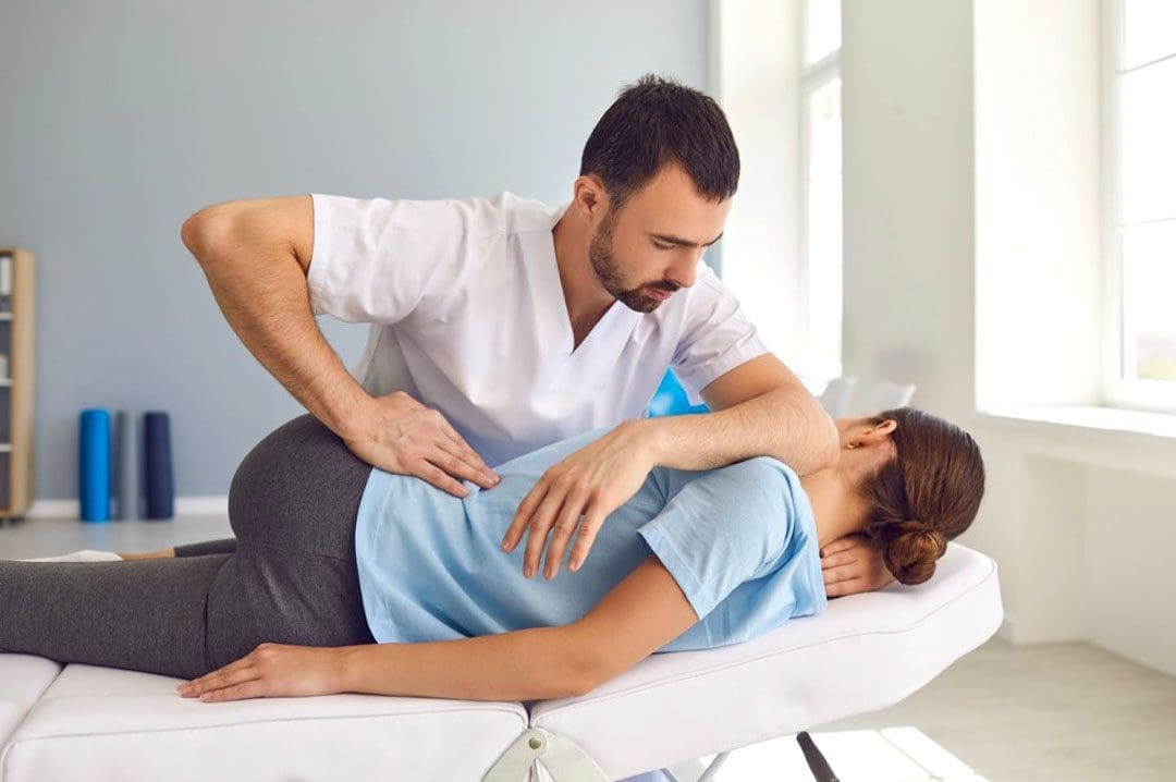
Every day, the body is in constant rest or active motion when needed, from working to exercising and getting adequate rest to repeat the cycle. However, as the body is in this dynamic/rest motion, unintentionally, many individuals will be hunched forward, causing their posture to be slouched for long periods. To that point, it can cause the surrounding neck, shoulder, and back muscles to be pulled and overly stretched, causing pain when the individual gets out of the reclined position. When a person is constantly being hunched over, the action alone could lead to poor posture, which can cause misalignment to the spine and be associated with many chronic conditions that affect their way of life. Fortunately, various treatments can help alleviate poor posture and its associated symptoms. Today’s article examines what defines good posture, the influences that can affect body posture, and how treatment techniques like MET (muscle energy technique) can help improve posture. We mention our patients to certified medical providers that provide available therapy treatments like MET (muscle energy techniques) for individuals suffering from chronic conditions associated with poor posture that can correlate with overlapping risk profiles. We encourage each patient when it is appropriate by referring them to associated medical providers based on their diagnosis or needs. We understand and accept that education is a marvelous way when asking our providers crucial questions at the patient’s request and acknowledgment. Dr. Alex Jimenez, D.C., uses this information as an educational service. Disclaimer
What Defines Good Posture?
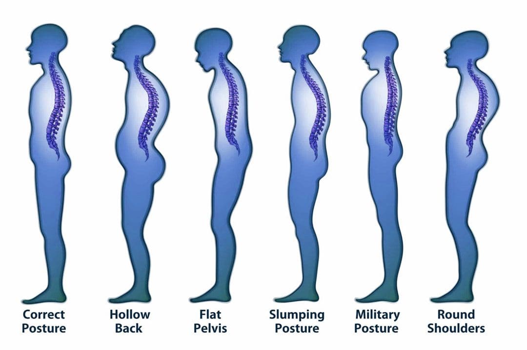
Have you been experiencing referred pain in your neck, shoulders, or lower back? Do you feel pain when stretching after being hunched over throughout the day? Or have you noticed that your neck is slanted, which causes your head to poke in front of your shoulders? Many of these issues are correlated with poor posture. Many of us have heard the saying from our parents, “Stand up straight!” And this is a reminder that having good posture correlates with good spinal health. The book, “Clinical Applications of Neuromuscular Techniques,” written by Leon Chaitow, N.D., D.O, and Judith Walker DeLany, L.M.T, mentions that posture is used to describe the static state of the spine. There are two different types of posture: static and dynamic. Static posture is when the body is in motion, while dynamic posture is when the body is resting. So good posture allows the spine to naturally curve with minimal pain affecting the cervical, thoracic, and lumbar regions.
Influences That Affect Body Posture
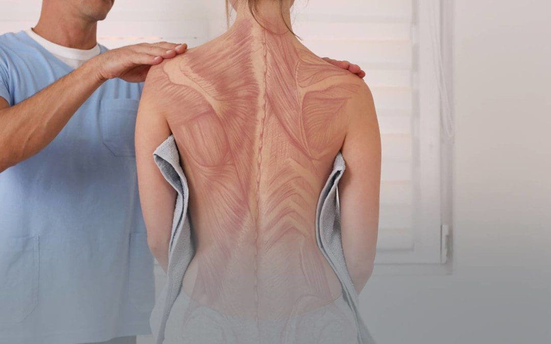
As stated early, many of us unintentionally hunch our bodies over time. This is one of the issues as we constantly look down on our phones, and as we get older, it can affect our ability to balance ourselves. Research studies reveal that improper posture can affect static and dynamic balance as we age. This means that when we are constantly hunched over as older adults, we are more prone to the risk of falling and causing long-term disability to our bodies. Additional research studies also mentioned that chronic conditions like forward head posture (which correlates to constantly looking down at the phone) could cause a persistent and abnormal contraction of the neck and shoulder muscles to become dysfunctional. To that point, it can cause pressure on the muscles, fascia, and nerves in the cervical-thoracic regions of the body. When bad posture affects the body over time, it can develop into musculoskeletal disorders if not treated immediately.
5 Way To Improve Posture- Video
Have you felt muscle strain on your neck, shoulders, and back? Have you felt relief when you stretch after being hunched over? Do you feel unstable when walking? These issues could be correlated with your posture if you have been experiencing these issues. When it comes to the body, it is important to make sure that maintaining good posture is not just to please your parents but to have a healthy spine. When we are constantly hunched over, it can cause the muscles and connective tissues to have gravitational strain and shorten the length of the muscles. However, realizing that you have poor posture early on can be treated. The video above shows the five best ways to improve your posture and how to strengthen the back, neck, and shoulder muscles from developing chronic conditions. Exercise alone can not be the only solution; combining it with chiropractic therapy allows the body to be fully restored with various techniques to reduce pain-like symptoms.
How The Met Technique Helps Improve Posture
So how would chiropractic care help with improving posture? Many chiropractors use techniques like MET (muscle energy technique) and spinal manipulation to help restore the body to realignment. Studies reveal that the combinations of MET and stretching can help lengthen the short muscles and restore range of motion to the body. Chiropractors use their hands and various tools to help realign the spine from subluxation and return the body to normal while freeing the tense muscles. Chiropractic care decreases the body’s risk of back injuries while reducing wear and tear on the muscles and joints, contributing to poor posture.
Conclusion
Overall, it is important to maintain good posture to prevent unwanted chronic issues from causing pain-like symptoms to the body. Recognizing the problems contributing to poor posture, treatment, and exercise can help stretch and strengthen the back muscles from hunching over. Maintaining good posture allows the body to be pain-free and prevents many unwanted symptoms from developing.
References
Chaitow, Leon, and Judith Walker DeLany. Clinical Applications of Neuromuscular Techniques. Churchill Livingstone, 2003.
Cohen, Rajal G, et al. “Lighten up! Postural Instructions Affect Static and Dynamic Balance in Healthy Older Adults.” Innovation in Aging, U.S. National Library of Medicine, 24 Mar. 2020, https://www.ncbi.nlm.nih.gov/pmc/articles/PMC7092748/.
Lee, Joon-Hee. “Effects of Forward Head Posture on Static and Dynamic Balance Control.” Journal of Physical Therapy Science, U.S. National Library of Medicine, Jan. 2016, https://www.ncbi.nlm.nih.gov/pmc/articles/PMC4756019/.
Phadke, Apoorva, et al. “Effect of Muscle Energy Technique and Static Stretching on Pain and Functional Disability in Patients with Mechanical Neck Pain: A Randomized Controlled Trial.” Hong Kong Physiotherapy Journal : Official Publication of the Hong Kong Physiotherapy Association Limited = Wu Li Chih Liao, U.S. National Library of Medicine, 14 Apr. 2016, https://www.ncbi.nlm.nih.gov/pmc/articles/PMC6385145/.
Postural dysfunction happens when unhealthy postures are practiced and maintained for prolonged periods. This can occur in any sitting, standing, or lying down position and is a major factor in musculoskeletal injuries. Injuries related to poor posture are normally caused by overuse that builds up over time. When the body starts to go out of alignment, the muscles must work harder to compensate, which further strains the body. This stress can lead to soft tissue injury and excess joint wear and tear. These injuries start as minor aches and pains in the short term. However, if left untreated, they can lead to chronic conditions. Injury Medical Chiropractic and Functional Medicine Clinic can rehabilitate the body to optimal function and provide postural training.
Postural Dysfunction
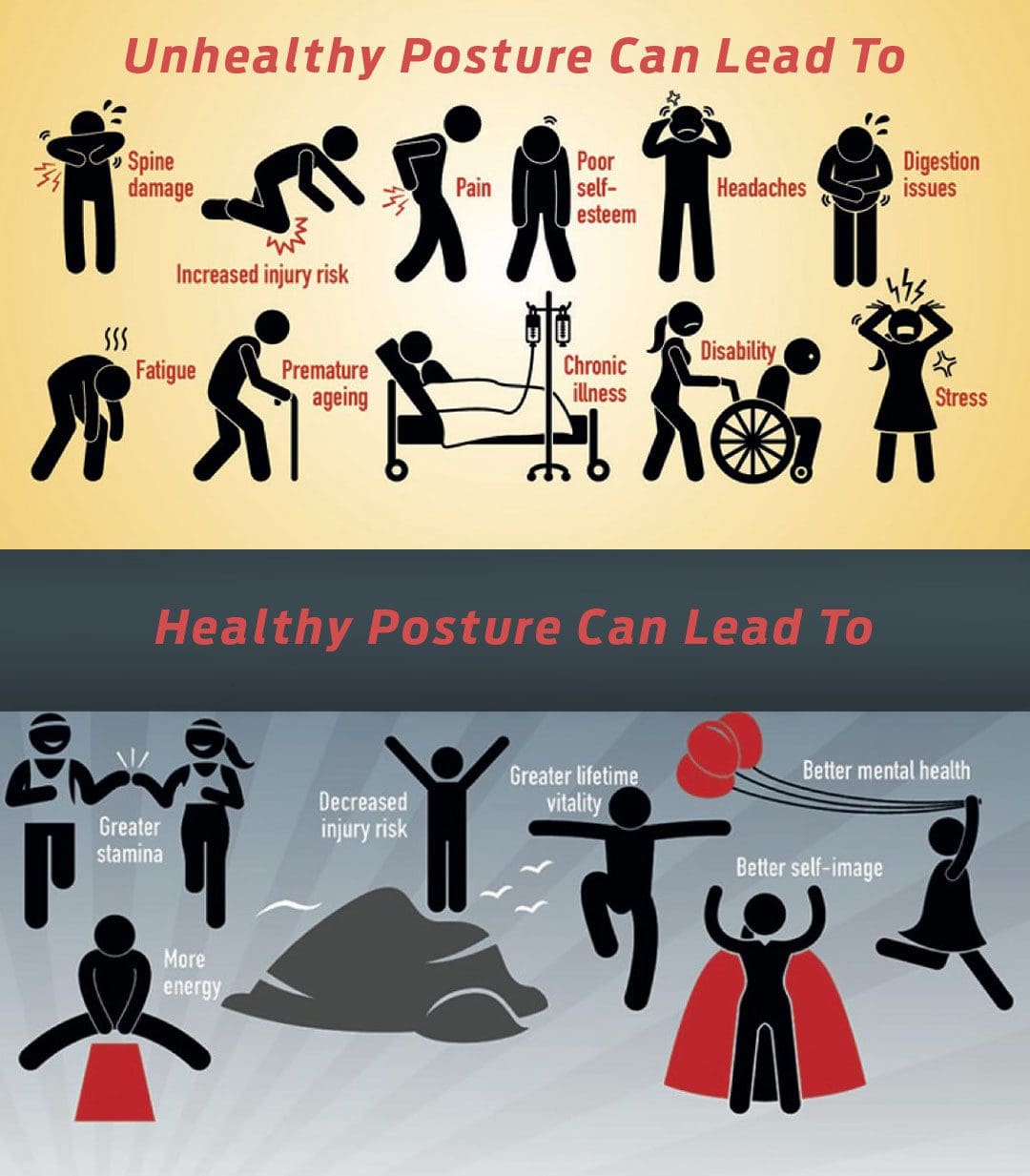
Posture is how the skeleton and muscles hold the body in a healthy position while standing or sitting, affecting breathing, muscle growth, and mobility. Practicing healthy posture means:
The bones are properly aligned.
The muscles, joints, and ligaments function correctly.
The organs, like the stomach, kidneys, and GI tract, are in the right position and can work efficiently.
The nervous system can operate at its full potential.
This allows the body to have:
More energy.
More room for the lungs to expand.
Experience less stress.
Alleviate muscle fatigue.
Achieve physical fitness.
Imbalance Causes
Unhealthy body positioning causes imbalances in muscle strength that pull the body out of alignment. This leads to muscles becoming tight/shortened and others becoming weak/lengthened, and it can also cause internal organ problems. For example, individuals that slump excessively cause the abdomen to compress, crowding the stomach and intestines, which leads to digestive issues. Postural dysfunction can be caused by the following:
Stress and strain from day-to-day activities.
Job responsibilities that involve sitting/standing for long periods and/or repetitive tasks like bending, lifting, reaching, twisting, etc.
Unhealthy driving position.
Non-supportive footwear.
Joint stiffness usually of the neck, upper and lower back, and hips.
Sedentary habits.
Lack of physical activity and exercise.
Muscle tightness.
Muscle weakness.
Weakened core stability.
Inadequate or failed post-surgical recovery.
Effects
Decreased blood circulation resulting in fatigue.
Overuse Injuries.
Breathing difficulties.
Balance issues.
Knee pain.
Joint misalignment.
Increased strain on the spine.
Compression of discs and joints.
Neck pain.
Lower back pain.
Less space for nerves to move due to compression.
Nerve problems.
Piriformis syndrome.
Shoulder impingement.
Chiropractic Rehabilitation
Chiropractic treatment for postural dysfunction provides adjustments, massage and decompression therapy, targeted stretching and exercises, retraining movement patterns, and nutritional and health coaching. Personalized treatment plans can include the following:
Targeted stretches and exercises to maintain posture correction.
Fix Posture
References
Korakakis, Vasileios, et al. “Physiotherapist perceptions of optimal sitting and standing posture.” Musculoskeletal science & practice vol. 39 (2019): 24-31. doi:10.1016/j.msksp.2018.11.004
Lee, Yongwoo, and Ki Bum Jung. “Effect of Physiotherapy to Correct Rounded Shoulder Posture in 30 Patients During the COVID-19 Pandemic in South Korea Using a Telerehabilitation Exercise Program to Improve Posture, Physical Function, and Reduced Pain, with Evaluation of Patient Satisfaction.” Medical science monitor: international medical journal of experimental and clinical research vol. 28 e938926. 27 Dec. 2022, doi:10.12659/MSM.938926
Shih, Hsu-Sheng, et al. “Effects of Kinesio taping and exercise on forward head posture.” Journal of back and musculoskeletal rehabilitation vol. 30,4 (2017): 725-733. doi:10.3233/BMR-150346
Snodgrass, Suzanne J et al. “Relationship between Posture and Non-Contact Lower Limb Injury in Young Male Amateur Football Players: A Prospective Cohort Study.” International journal of environmental research and public health vol. 18,12 6424. 14 Jun. 2021, doi:10.3390/ijerph18126424
Zhao, Mingming, et al. “Driver posture monitoring in highly automated vehicles using pressure measurement.” Traffic injury prevention vol. 22,4 (2021): 278-283. doi:10.1080/15389588.2021.1892087
Environmental factors can affect the body and lead to chronic conditions involving the musculoskeletal system. When issues like stress, physical inactivity, and traumatic events affect the muscle groups in the upper and lower extremities, it causes the various muscles to tense up and be succumbed to multiple injuries that could potentially develop trigger points. Now trigger points can cause overlapping risk profiles and pain-like issues that can affect a person’s mobility and stability. However, many ways can alleviate the pain-like symptoms associated with trigger points affecting the musculoskeletal system. Many pain specialists use techniques to stretch the tense muscle and release the trigger point nodule in the muscle fibers. Today we will look at how myofascial trigger point formation affects the body, how MET (muscle energy techniques) are used to relieve trigger point formation, and how chiropractic care uses the MET technique on trigger points. We mention our patients to certified medical providers that provide available therapy treatments like MET (muscle energy techniques) for individuals suffering from chronic conditions associated with trigger point formation on the musculoskeletal system. We encourage each patient when it is appropriate by referring them to associated medical providers based on their diagnosis or needs. We understand and accept that education is a marvelous way when asking our providers crucial questions at the patient’s request and acknowledgment. Dr. Alex Jimenez, D.C., uses this information as an educational service. Disclaimer
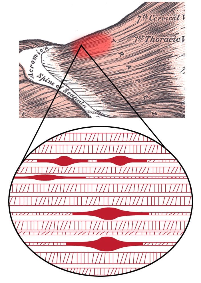
Myofascial Trigger Points Affecting The Body
Have you been dealing with pain in different locations in your body? Do you feel that your muscles feel tight or tensed constantly? Or do you feel muscle strain when lifting or carrying heavy objects? Many of these pain-like issues correlate with myofascial trigger points affecting the body. According to research studies, myofascial pain syndrome or trigger points are hard palpable nodules discrete along the taut skeletal muscle band that can be painful when active or compressed. Now trigger points can cause the affected muscles to be hypersensitive, which to that point, can spread pain when being touched, known as referred pain. A great example would be if tense shoulder muscles have a cluster of trigger points and, when touched, send pain to the neck.
Trigger points in the musculoskeletal system can be present in soft tissues that can cause dysfunction and promote pain in the affected muscle area. Trigger points are developed in any scenario, from trauma like an auto accident to repetitive motions for extended periods. Two features can cause trigger point formation that can create these nodules: active and latent trigger points. Active trigger points, according to “Clinical Applications of Neuromuscular Techniques,” written by Leon Chaitow, N.D, D.O, and Judith Walker DeLany, L.M.T, mentioned that when pressure is applied to active trigger points it can cause referred pain associated with symptoms of painful sensations to the affected muscle. While latent trigger points, when pressure is applied to them, can cause referred pain that a person experienced in the past and occurs recently. Latent trigger points can also develop into active trigger points correlating to overlapping risk profiles. The book also stated that when the fascia and connective muscle tissues have been overused or strained, it can lead to trigger point formation development.
MET Trigger Point Therapy-Video
Have you been dealing with referred pain in different areas of your body? Do you feel that your muscles are tense and aching? Or do you feel muscle strain when lifting or carrying heavy objects? If you have been dealing with these issues, they are correlated to trigger point formation in your musculoskeletal system. Why not try MET or muscle energy technique therapy? Studies reveal that muscle energy techniques were developed originally to treat soft tissue, stretch tight muscles and fascia, and mobilize joints while improving blood circulation and draining the lymphatic system. So how do trigger point formation can be treated with MET techniques? Well, since trigger points can cause tight, hypersensitive spots that can be located in various taut muscle bands, MET techniques from pain specialists can help stretch and break up the tight nodules in the muscles to achieve muscle restoration at full resting length. The video above demonstrates how MET is used as trigger point therapy.
MET Techniques On Trigger Point Formation
So how do MET techniques work on trigger point formation in the musculoskeletal system? According to research studies, MET techniques utilize soft tissue manipulation to improve the myofascial system’s and joints’ functional parameters. Many pain specialists, like chiropractors, use this technique and other tools to help restore the body’s natural range of motion in the joints while providing a pain-reducing effect to the numerous musculoskeletal disorders. Additional research studies also mentioned that MET/NET (neuro-emotional) techniques could help relieve pain sensitivity from the affected muscle area.
How Chiropractic Care Uses MET Techniques On Trigger Points
So how would chiropractic care utilize MET techniques on an individual with trigger points? Due to its effectiveness and drug-free approach, chiropractic care can help smooth out the muscle and fascia by applying pressure with their hands or special tools to relieve trigger point pain. With MET techniques, chiropractors can help release muscle stiffness, tightness, and shortness to restore the body and re-align the spine. With continued chiropractic treatment, the body can reduce the future formation of trigger points in the muscle fibers while preventing further issues from developing.
Conclusion
Trigger point formation can occur in different muscle areas in the body, leading to overlapping risk profiles associated with pain. When the body is dealing with referred pain caused by trigger points, it can cause numerous issues affecting a person’s daily activity. Luckily, pain specialists like chiropractic care can incorporate techniques like MET and spinal manipulation to re-align the body, stretch out the stiff muscles, and promote a restored range of motion back to the musculoskeletal system. By going through daily treatments, the body can begin to heal naturally and prevent future injuries.
References
Bablis, Peter, et al. “Neuro Emotional Technique for the Treatment of Trigger Point Sensitivity in Chronic Neck Pain Sufferers: A Controlled Clinical Trial.” Chiropractic & Osteopathy, U.S. National Library of Medicine, 21 May 2008, https://www.ncbi.nlm.nih.gov/pmc/articles/PMC2427032/.
Chaitow, Leon, and Judith Walker DeLany. Clinical Applications of Neuromuscular Techniques. Churchill Livingstone, 2003.
Shah, Jay P, et al. “Myofascial Trigger Points Then and Now: A Historical and Scientific Perspective.” PM & R : the Journal of Injury, Function, and Rehabilitation, U.S. National Library of Medicine, July 2015, https://www.ncbi.nlm.nih.gov/pmc/articles/PMC4508225/.
Thomas, Ewan, et al. “The Efficacy of Muscle Energy Techniques in Symptomatic and Asymptomatic Subjects: A Systematic Review.” Chiropractic & Manual Therapies, U.S. National Library of Medicine, 27 Aug. 2019, https://www.ncbi.nlm.nih.gov/pmc/articles/PMC6710873/.
Wendt, Michał, and Małgorzata Waszak. “Evaluation of the Combination of Muscle Energy Technique and Trigger Point Therapy in Asymptomatic Individuals with a Latent Trigger Point.” International Journal of Environmental Research and Public Health, U.S. National Library of Medicine, 14 Nov. 2020, https://www.ncbi.nlm.nih.gov/pmc/articles/PMC7696776/.
Prolonged standing can cause the pelvis to push backward, increasing the curve of the lower back/lumbar region. This increased pressure on the soft tissues surrounding the spine causes the lower back muscles to tighten and/or spasm, resulting in discomfort in the joints and nerves. Weakened core muscles and unhealthy posture/postural syndrome are the most common causes, but injury, aging, congenital malformations, or a disease/condition can also contribute to the symptoms. Injury Medical Chiropractic and Functional Medicine Clinic has a top team of professional therapists to evaluate the problem, diagnose the cause/s accurately, and develop a customized treatment and rehabilitation plan.
Prolonged Standing Back Discomfort
Back Structure
The lower back is one of the most used areas of the spine, moving around and bending during a normal day. When the body stands, the spine naturally curves both in and outwards.
The inward curve, called lordosis, curves towards the front of the body at the lower back and neck regions.
The outward curve, called kyphosis, curves towards the back of the body at the chest.
When bending over while standing, the five lumbar vertebrae of the lower back change position and shift from lordosis to kyphosis when bent completely.
When standing up from bending, the lumbar vertebrae change position again and return to the lordosis position.
Causes
The facet joints allow movement between each spine level. The standing spinal curvature can increase contact between the facet joints. As the body ages, the facet joints and discs begin to wear out, which can cause the discs and facet joints to become inflamed. Prolonged standing during normal daily activity combined with inflammation in these joints can aggravate the inflammation and cause symptoms. Regular routines and habits may contribute to low back discomfort during prolonged standing. These include:
Sleeping on a sinking or unsupportive mattress.
Practicing unhealthy postures that cause imbalances with proper weight distribution.
Not wearing proper footwear and/or supportive orthotics forces the lower spine into increased curvature and can compress the facet joints.
Not getting enough physical activity that strengthens the core.
Chiropractors are experts on the musculoskeletal system. They will:
Listen to the patient about symptoms, medical history, and occupation.
A physical examination of muscle tone, strength, and range of motion.
Therapeutic massage, electric muscle stimulation, and ultrasound therapy can help reduce muscle inflammation and increase circulation to injured soft tissues.
Chiropractic adjustments will reset joints, removing pressure from the surrounding muscles and nerves.
Targeted therapeutic strength training is recommended for core and leg muscles to improve hip flexibility.
Non-surgical decompression or traction, either with a machine or suspension, can reverse the pressure in spinal discs.
Standing Lower Back Relief Exercises
References
Hasegawa, Tetsuya, et al. “Association of low back load with low back pain during static standing.” PloS one vol. 13,12 e0208877. 18 Dec. 2018, doi:10.1371/journal.pone.0208877
Jo, Hoon, et al. “Negative Impacts of Prolonged Standing at Work on Musculoskeletal Symptoms and Physical Fatigue: The Fifth Korean Working Conditions Survey.” Yonsei medical journal vol. 62,6 (2021): 510-519. doi:10.3349/ymj.2021.62.6.510
Ognibene GT, Torres W, von Eyben R, Horst KC. Impact of a sit-stand workstation on chronic low back pain: randomized trial results. J Occup Environ Med. 2016;58(3):287-293. Abstract. https://www.ncbi.nlm.nih.gov/pubmed/26735316. Accessed March 2, 2017.
Parry, Sharon P et al. “Workplace interventions for increasing standing or walking for decreasing musculoskeletal symptoms in sedentary workers.” The Cochrane database of systematic reviews vol. 2019,11 CD012487. November 17, 2019, doi:10.1002/14651858.CD012487.pub2
Rodríguez-Romero, Beatriz, et al. “Thirty Minutes Identified as the Threshold for Development of Pain in Low Back and Feet Regions, and Predictors of Pain Intensity During 1-h Laboratory-Based Standing in Office Workers.” International journal of environmental research and public health vol. 19,4 2221. February 16, 2022, doi:10.3390/ijerph19042221
Smith, Michelle D et al. “The Influence of Using a Footstool during a Prolonged Standing Task on Low Back Pain in Office Workers.” International journal of environmental research and public health vol. 16,8 1405. April 18. 2019, doi:10.3390/ijerph16081405
The various muscles, tendons, and ligaments inside the body surround the skeletal joint to provide movement and multiple actions to allow the host to be mobile. The body also has various muscle groups, with soft tissues surrounding the vital organs to help support the body. Since the human body is mobile, many factors can cause issues to the body’s host and lead to chronic overlapping risk profiles that can correlate with pain in the joints and muscle tissues. When these factors are causing pain in the musculoskeletal system, various treatment techniques can help reduce the pain-like symptoms and help restore the body. MET, or muscle energy technique, is one of the different treatment techniques used by pain specialists like chiropractors, massage therapists, physical therapists, and occupational therapists on many individuals with musculoskeletal pain. Today’s article looks at the musculoskeletal system, how the issues affect the muscles, and how muscle energy technique is utilized to reduce muscle pain associated with the musculoskeletal system. We mention our patients to certified medical providers that provide available therapy treatments like MET (muscle energy techniques) for individuals suffering from chronic conditions associated with the musculoskeletal system. We encourage each patient when it is appropriate by referring them to associated medical providers based on their diagnosis or needs. We understand and accept that education is a marvelous way when asking our providers crucial questions at the patient’s request and acknowledgment. Dr. Alex Jimenez, D.C., uses this information as an educational service. Disclaimer
An Overview Of The Musculoskeletal System
The musculoskeletal system plays a huge role in the body, consisting of numerous muscle groups, tissues, ligaments, joints, and organs controlled by the central nervous system. The central nervous system provides the motor-sensory function to the musculoskeletal system, allowing the body to rest and move around. What the central nervous system does to the musculoskeletal system, according to research studies, it is revealed that these two systems have a relationship with each other as they are interconnected. Besides the various muscle groups that help surround the skeletal joints and provide mobility to the body, we will look at the connective tissue associated with the facial system and how muscle activity is affected by chronic issues.
Connective Tissue & The Fascial System
Regarding the musculoskeletal system, the connective tissue is one of the single abundant materials that allow each muscle group to be connected to its specific body region. The connective tissue comprises the body’s bones, muscles, blood vessels, and lymph nodes while embracing all the soft tissues and organs. The body’s connective tissue also works with the fascial system, giving the body the fundamental requirements. The fascial system is the structural form of the body since the fascial system is composed of connective tissues. With these two systems connecting and working together, it allows the muscles in the body to respond to various actions thrown at in different environments. The fascia web allows all muscle tissues to exist in isolation and interwoven with other structures to provide mobility.
Muscle Activity
Everything from the connective tissues to the fascia is involved in muscle activity in the musculoskeletal system. When the various muscles start to work with the body’s most movement, it is combined with one or more muscles acting as the prime mover or antagonist, allowing synergistic muscles to assist and contract simultaneously. The various muscle groups in the musculoskeletal system allow different actions, often repeated, to become stabilizing or antagonizing muscles. A great example is looking at the upper and lower extremities of the body. The upper extremities allow the arms, neck, head, and shoulders to have mobility when it comes to bending, twisting, and turning. While the lower extremities allow the hips, low back, legs, and feet to allow, stability and flexion to make the body move. However, these muscle groups can be affected by multiple factors that can affect muscle activity and lead to overlapping soft tissue pain profiles.
Issues Affect Muscle Activity
Since the body is a complex machine, different environmental factors can affect muscle groups in various ways and cause numerous pain issues. Now when it comes to environmental factors, many negative influences do play a role in affecting the musculoskeletal system in three categories:
Biomechanical: trauma, overusing the muscles, congenital, etc.
Biochemical: endocrine imbalances, inflammation, ischemia, nutritional deficiency, etc.
Psychosocial: anxiety, depression, chronic stress, etc.
These influences can cause the muscles to tense up and restrict blood flow, causing pain and trigger points to form in the muscle fibers and making a person feel miserable. Fortunately, therapeutic techniques allow the muscles to relax and release the tension that the person is feeling.
MET(Muscle Energy Technique)-Video
What Is Muscle Energy Technique?
When people feel stressed, and their muscles become tight, they can develop pain-like symptoms that correlate with chronic issues. Fortunately, a revolution has taken place that many pain specialists like chiropractors and massage therapists take place when it comes to manipulative therapy through a technique known as MET or muscle energy technique. According to research studies, MET is an osteopathic manipulative medicine designed to improve the body’s musculoskeletal function. This technique helps target soft tissues and contributes to joint mobilization. The muscle energy technique allows the tight muscles and fascia to be stretched, improving circulation and lymphatic flow since chiropractors or doctors of chiropractic care utilize spinal manipulation to realign the body and restore joint function.
Additional studies also reveal that MET combined with chiropractic care allows pain reduction in the muscles and can increase the body’s range of motion. This technique is essential for chronic and acute low back pain, trigger point pain, and other musculoskeletal dysfunctions associated with environmental factors.
The Various Stretching Techniques Of MET
The main objective of MET is to induce relaxation of hypertonic musculature, which also stretches the muscles to reduce pain-like symptoms. Now many treatments like chiropractic care can combine different techniques to reduce pain and restore mobility to the individual. With MET, various stretching techniques can allow chiropractors to stretch the tense muscles while restoring the range of motion. Some of the stretching techniques that pain specialists use include:
Facilitated stretching: Allows chiropractors and massage therapists to use strong/light isometric contractions to treat the muscles and be actively stretched. Reduces muscle cramps, tissue damage, or pain to the affected muscle group while utilizing breathing techniques and producing sufficient post-isometric relaxation.
Active-isolated stretching: Allows chiropractors and massage therapists to stretch the affected muscle actively while using precise localization to allow the affected muscle to receive a specific extension. This allows the muscles to relax through a short repetitive contraction and retraction to increase oxygenated blood flow. This stretching technique prevents the activation of the myotatic stretch reflex on the affected muscle.
Static stretching: In yoga, the individual can maintain a position for a few minutes to allow deep breathing and slowly release contracted and tensed muscle tissues to relax. This stretch also releases myofascial trigger points from the affected muscle groups.
Ballistic stretching: This stretch provides a series of rapid, bouncing movements that allow the short muscles in the body to be lengthened rapidly.
Conclusion
When the body encounters environmental factors that can cause pain-like symptoms to the host, it can develop into pain and other chronic conditions affecting a person’s life. Many techniques like MET (muscle energy technique) allow the musculoskeletal system to stretch out tense muscles and help restore mobility to the body. Pain specialists like chiropractors can incorporate various MET stretching techniques combined with spinal manipulation to restore the body to its original state.
References
Chaitow, Leon, and Judith Walker DeLany. Clinical Applications of Neuromuscular Techniques. Churchill Livingstone, 2003.
Murphy, Andrew C, et al. “Structure, Function, and Control of the Human Musculoskeletal Network.” PLoS Biology, U.S. National Library of Medicine, 18 Jan. 2018, https://www.ncbi.nlm.nih.gov/pmc/articles/PMC5773011/.
Thomas, Ewan, et al. “The Efficacy of Muscle Energy Techniques in Symptomatic and Asymptomatic Subjects: A Systematic Review.” Chiropractic & Manual Therapies, U.S. National Library of Medicine, 27 Aug. 2019, https://www.ncbi.nlm.nih.gov/pmc/articles/PMC6710873/.
Waxenbaum, Joshua A, and Myro Lu. “Physiology, Muscle Energy – StatPearls – NCBI Bookshelf.” In: StatPearls [Internet]. Treasure Island (FL), StatPearls Publishing, 25 July 2022, https://www.ncbi.nlm.nih.gov/books/NBK559029/.
IFM's Find A Practitioner tool is the largest referral network in Functional Medicine, created to help patients locate Functional Medicine practitioners anywhere in the world. IFM Certified Practitioners are listed first in the search results, given their extensive education in Functional Medicine