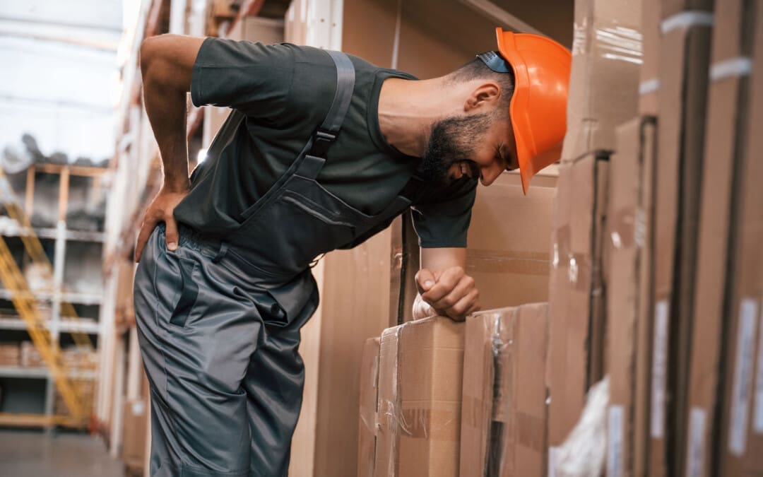
Individuals who have experienced spinal or back trauma, suffered fractures, are going through spinal degeneration, or are dealing with a spinal condition have an increased risk of anterolisthesis, where a vertebra slips forward relative to the vertebra below it. Can healthcare providers help prevent and treat the condition?
Anterolisthesis
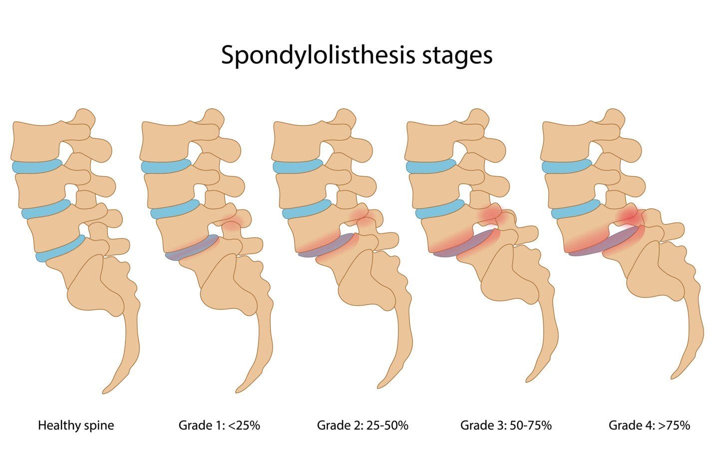
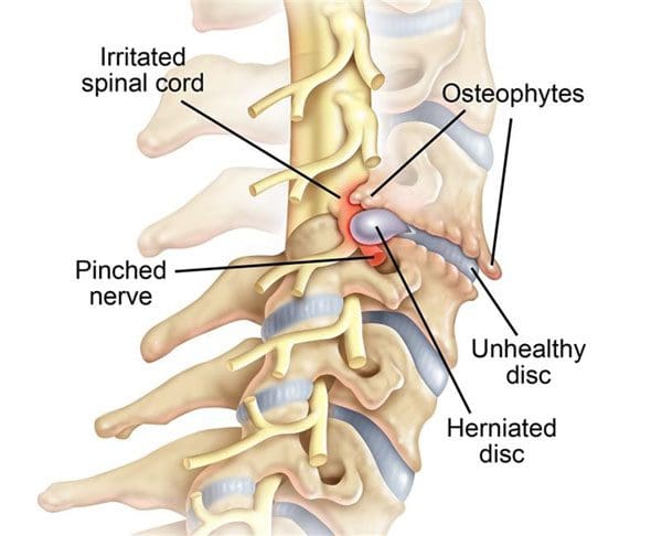
The spine consists of 33 individual bones or vertebrae stacked on one another. Anterolisthesis occurs when one vertebral segment slips forward over another. The condition can be mild, asymptomatic, or cause significant pain and neurological symptoms. Many different things, including osteoarthritis, osteoporosis, trauma, or a fracture, can cause this vertebral shifting. (Cedars Sinai, 2022) Spondylolisthesis is a general term for shifting a spinal vertebra over the one below it. It includes anterolisthesis, forward moving, and the less common retrolisthesis, or backward shifting.
Grades
Anterolisthesis is typically graded using the Meyerding scale, which assigns one of five grades according to how much slippage has occurred. These grades include:
Anterolisthesis can lead to various symptoms, depending on the severity and if the surrounding spinal nerves have been affected. The most common complaints include:
Diagnosis begins with a subjective evaluation and a physical examination. During these, the healthcare provider will assess sensation, strength, and reflexes and will order one of several diagnostic tests, including:
X-rays
Visualizes the vertebrae in the spine and their position relative to those above and below.
Also provides a clear picture of spinal arthritis or disc degeneration.
Magnetic Resonance Imaging – MRI
Allows the spinal cord, nerves, muscles, and discs to be assessed for compression or damage.
Several factors determine how the condition is treated, including:
The grade of the slippage.
The cause.
The symptoms.
The presence of instability on a diagnostic test such as an X-ray.
Stable and mildly symptomatic cases are usually treated with a combination that can involve:
Physical therapy
Activity modification
Bracing
Nonsteroidal anti-inflammatory medications/NSAIDs like ibuprofen.
Spinal injections
In more severe cases in which spinal instability or significant neurological symptoms are present, surgery may be recommended. This commonly involves a spinal decompression or fusion procedure. The technique varies based on the surgeon’s preferences and anatomy. (Koslosky E., and Gendelberg D. 2020)
Prognosis
Most individuals with this condition don’t know they have it until it is found accidentally on an X-ray or an MRI for something else. Mild cases can cause minimal symptoms and can be well-managed with conservative treatments. Cases of unstable anterolisthesis or those with neurological compression often require surgical intervention. These surgeries restore stability to the spine and alleviate any pressure on the nerves. More than 85% of individuals who need surgery have a successful outcome. (American Academy of Orthopaedic Surgeons, 2021)
Self-Care and Management
For individuals experiencing pain, numbness, or tingling from anterolisthesis, getting symptoms evaluated by a healthcare provider is an important first step. The healthcare provider may suggest one of several management strategies, which include:
Core Strengthening
To alleviate symptoms, exercises targeting the core muscles in the hips, pelvis, abdomen, and lower back are recommended.
Formal physical therapy may also be recommended.
Over-the-counter Meds
A healthcare provider may suggest pain-relieving medications like ibuprofen or naproxen to reduce soreness.
Activity Modification
Sticking to gentle, pain-free activities and avoiding excessive or repetitive extension of the spine can help prevent symptom aggravation. (American Academy of Orthopaedic Surgeons, 2021)
Injury Medical Chiropractic and Functional Medicine Clinic
At Injury Medical Chiropractic and Functional Medicine Clinic, our areas of practice include Chronic Pain, Personal Injury, Auto Accident Care, Work Injuries, Back Injury, Low Back Pain, Neck Pain, Migraine Headaches, Sports Injuries, Severe Sciatica, Scoliosis, Complex Herniated Discs, Fibromyalgia, Chronic Pain, Complex Injuries, Stress Management, Wellness & Nutrition, Functional Medicine Treatments, and in-scope care protocols. We focus on what works for you to relieve pain and restore function. If other treatment is needed, individuals will be referred to a clinic or physician best suited to their injury, condition, and/or ailment.
Koslosky, E., & Gendelberg, D. (2020). Classification in Brief: The Meyerding Classification System of Spondylolisthesis. Clinical orthopaedics and related research, 478(5), 1125–1130. https://doi.org/10.1097/CORR.0000000000001153
American Academy of Orthopaedic Surgeons. (2021). Adult spondylolisthesis in the low back. https://orthoinfo.aaos.org/en/diseases–conditions/adult-spondylolisthesis-in-the-low-back
Hospital for Special Surgery. (2023). Spondylolisthesis. https://www.hss.edu/condition-list_spondylolisthesis.asp
Can understanding the nucleus pulposus help in body positioning and prevention for individuals wanting to practice spinal hygiene and protect their discs from injury?
Nucleus Pulposus
The spinal discs are located between the spine’s vertebrae and are the body’s natural impact and shock absorbers. Within the disc is the nucleus pulposus, which plays a major role in providing the spine with shock absorption during movement. (Zhou Z. et al., 2014) The discs have a tough outer portion and a soft inner core. They are the:
It forms the tough circular exterior and comprises concentric sheets of collagen fibers or lamellae surrounding the inner core.
It has cartilaginous endplates that firmly attach to the vertebrae above and below.
Nucleus Pulposus
The nucleus pulposus is the inner core soft filling of the discs.
It contains a network of fibers suspended in a mucoprotein gel with a water base to maintain strength and pliability.
The near-liquid consistency makes it responsive to movement to handle the body’s axial load.
It helps maintain spinal suspension to prevent pressure on the bones and prevent bone-to-bone contact, reducing the potential for injuries and pain.
Shock Absorber
Each intervertebral disc is a shock-absorbing cushion, with the nucleus pulposus providing shock-absorbing properties (Zhou Z. et al., 2014). The intervertebral discs move as the body moves. For example, when arching the back, the disc moves forward slightly, and when twisting, the disc twists as well.
Spinal Action
The intervertebral disc supports spinal movements. When bending, twisting, arching, or tilting the spine, the nucleus pulposus swivels to accommodate these actions. These repeated spinal actions, which occur throughout the day and night, contribute to shifting positions while sitting, working, playing sports, carrying groceries, performing house chores, etc. An example is bending forward to pick something up. This action involves forward spinal flexion, which is bending the spine forward, flattening, or rounding. When bending using flexion, the spinal bones come closer together, pushing the nucleus pulposus toward the back.
Injuries
The disc can be pushed too far back with persistent or excessive spinal flexion. If the fibers of the annulus fibrosus become weak, they can tear, causing the nucleus pulposus to leak out and disc herniation. Generally, the nucleus pulposus will leak to the side and back; however, this corresponds to the location of the very sensitive nerve root/s with which it can come into contact, causing pain and other symptoms. The most common causes of disc herniation are degenerative wear and tear changes of the disc and trauma. Disc degeneration occurs as the body ages; it weakens the annulus fibers, allowing the nucleus pulposus to distend, bulge, or herniate.
Aging
Disc degeneration occurs with age but can also occur with injuries to the area. In young individuals, the nucleus pulposus is mostly water. For this age group, a herniation from trauma is more likely than in older individuals. (Ucar, D. et al., 2021) But as the body ages, the discs, especially the nucleus pulposus, begin to dry out. This dehydration leads to a significant loss of disc height. (UCLA Health, 2024) By age 60 or 70, the discs may be composed entirely of fiber, which can cause the shock absorption function not to work and disappear.
Chiropractic therapy is among the more conservative treatment options for a herniated disc and may be tried first before proceeding with more invasive treatments. Injury Medical Chiropractic and Functional Medicine Clinic works with primary healthcare providers and specialists to develop an optimal health and wellness solution that fully benefits the individual to get back to normal.
The Science of Functional Healing
References
Zhou, Z., Gao, M., Wei, F., Liang, J., Deng, W., Dai, X., Zhou, G., & Zou, X. (2014). Shock absorbing function study on denucleated intervertebral disc with or without hydrogel injection through static and dynamic biomechanical tests in vitro. BioMed research international, 2014, 461724. https://doi.org/10.1155/2014/461724
Nosikova, Y. S., Santerre, J. P., Grynpas, M., Gibson, G., & Kandel, R. A. (2012). Characterization of the annulus fibrosus-vertebral body interface: identification of new structural features. Journal of anatomy, 221(6), 577–589. https://doi.org/10.1111/j.1469-7580.2012.01537.x
Ucar, D., Duman, S., Bayram, Y., & Ucar, B. Y. (2021). Extruded disc herniations are experienced earlier by inactive young people in the high-tech gaming era. Journal of medicine and life, 14(3), 402–407. https://doi.org/10.25122/jml-2021-1059
For individuals looking to improve their spinal health, can understanding the anatomy of the intervertebral foramen help in injury rehabilitation and prevention?
Intervertebral Foramen
The intervertebral foramen, aka neural foramen, is the opening between the vertebrae through which spinal nerve roots connect and exit to other body areas. If the foramina narrows, it can place added pressure on the nerve roots near and around them, causing pain symptoms and sensations. This is known as neuroforaminal stenosis. (Sumihisa Orita et al., 2016)
Anatomy
The vertebrae comprise the spinal column.
They protect and support the spinal cord and most of the weight placed on the spine.
Foramen is the singular form, and foramina is the plural form.
Structure
The body is the large, round part of the bone that makes up each vertebra.
The body of each vertebra is attached to a bony ring.
Stenosis can occur in the spinal canal, known as central canal stenosis, and the foramina.
Pain brought on by neuroforaminal spinal stenosis and arthritis-related bone growth/bone spurs/osteophytes that are present in one or more foramen rub against the nerve root that passes through the space, causing radicular pain.
Pain accompanied by other sensations, like tingling or numbness, is known as radiculopathy. (Young Kook Choi, 2019)
The main symptom is pain.
Numbness and/or tingling can present depending on the injury.
Neurogenic claudication occurs as a result of ischemia or a lack of blood circulation to the nerves and typically presents with a heaviness in the legs.
It is typically associated with central stenosis rather than foraminal stenosis.
Most individuals with spinal stenosis feel better when flexing or bending forward and worse when arching their backs.
Stenosis treatment aims to relieve pain and prevent nerve symptoms from occurring or worsening. Conservative treatments are recommended and can be highly effective.
These include:
Myelopathy in the neck and/or upper or mid-back (myelopathy symptoms are spinal cord related and occur in central canal stenosis) (Cleveland Clinic. 2021)
Intense incapacitating pain
Different surgical techniques include:
Decompression laminectomy – entails removing the buildup of bone in the spinal canal.
Spinal fusion – when there is instability of the spine or severe foraminal stenosis.
Orita, S., Inage, K., Eguchi, Y., Kubota, G., Aoki, Y., Nakamura, J., Matsuura, Y., Furuya, T., Koda, M., & Ohtori, S. (2016). Lumbar foraminal stenosis, the hidden stenosis including at L5/S1. European journal of orthopaedic surgery & traumatology : orthopedie traumatologie, 26(7), 685–693. https://doi.org/10.1007/s00590-016-1806-7
American Academy of Orthopaedic Surgeons. (2020). Spine Basics (OrthoInfo, Issue. https://orthoinfo.aaos.org/en/diseases–conditions/spine-basics/
American Academy of Orthopaedic Surgeons. (2021). Lumbar spinal stenosis (OrthoInfo, Issue. https://orthoinfo.aaos.org/en/diseases–conditions/lumbar-spinal-stenosis/
Choi Y. K. (2019). Lumbar foraminal neuropathy: an update on non-surgical management. The Korean journal of pain, 32(3), 147–159. https://doi.org/10.3344/kjp.2019.32.3.147
Lee, S. Y., Kim, T. H., Oh, J. K., Lee, S. J., & Park, M. S. (2015). Lumbar Stenosis: A Recent Update by Review of Literature. Asian spine journal, 9(5), 818–828. https://doi.org/10.4184/asj.2015.9.5.818
Lurie, J., & Tomkins-Lane, C. (2016). Management of lumbar spinal stenosis. BMJ (Clinical research ed.), 352, h6234. https://doi.org/10.1136/bmj.h6234
“Various problems with the sacrum make up or contribute to a significant portion of lower back problems. Can understanding the anatomy and function help prevent and treat back injuries?”
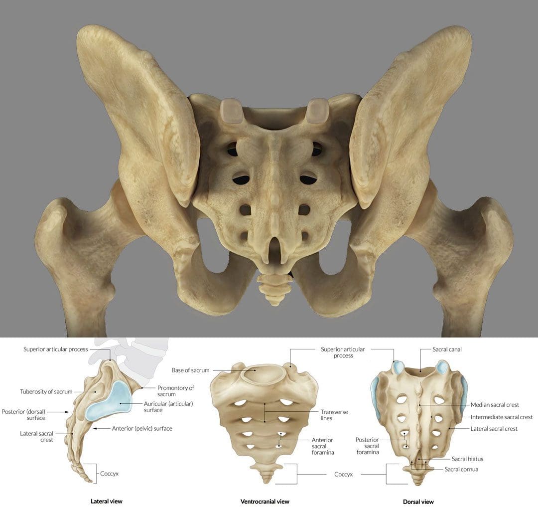
The Sacrum
The sacrum is a bone shaped like an upside-down triangle located at the base of the spine that helps support the upper body when sitting or standing and provides pelvic girdle flexibility during childbirth. It comprises five vertebrae that fuse during adulthood and connect to the pelvis. This bone takes and endures all of the body’s pressure and stress from everyday activities and movements.
Formation
Humans are born with four to six sacral vertebrae. However, fusion does not take place in all sacral vertebrae simultaneously:
Fusion starts with the S1 and S2.
As the individual gets older, the overall shape of the sacrum begins to solidify, and the vertebrae fuse into a single structure.
The process usually starts in the mid-teens and finishes in the early to mid-twenties.
It is believed to start earlier in females than males.
The sacrum in a female is wider and shorter and has a more curved top or the pelvic inlet.
The male sacrum is longer, narrower, and flatter.
Structure
The sacrum is an irregular bone that makes up the back/posterior third of the pelvic girdle. There is a ridge across the front/anterior portion of the S1 vertebra known as the sacral promontory. Small holes/foramen on both sides of the sacrum are left over after the vertebrae fuse together. Depending on the number of vertebrae, there can be three to five foramen on each side, though there are usually four. (E. Nastoulis, et al., 2019)
Each anterior foramen is typically wider than the posterior or dorsal/backside foramen.
Each sacral foramina/plural of foramen provides a channel for the sacral nerves and blood vessels.
Small ridges develop between each of the fused vertebrae, known as transverse ridges or lines.
The top of the sacrum is called the base and is connected to the largest and lowest of the lumbar vertebrae – L5.
The bottom is connected to the tailbone/coccyx, known as the apex.
The sacral canal is hollow, runs from the base to the apex, and serves as a channel at the end of the spinal cord.
The sides of the sacrum connect to the right and left hip/iliac bones. The attachment point is the auricular surface.
Right behind the auricular surface is the sacral tuberosity, which serves as an attachment area for the ligaments that hold the pelvic girdle together.
Location
The sacrum is at the level of the lower back, just above the intergluteal cleft or where the buttocks split. The cleft starts at around the level of the tailbone or coccyx. The sacrum is curved forward and ends at the coccyx, with the curvature being more pronounced in females than males. It connects to the L5 lumbar vertebra by way of the lumbosacral joint. The disc between these two vertebrae is a common source of low back pain.
On either side of the lumbosacral joint are wing-like structures known as the sacral ala, which connect to the iliac bones and form the top of the sacroiliac joint.
These wings provide stability and strength for walking and standing.
Anatomical Variations
The most common anatomical variation applies to the number of vertebrae. The most common is five, but anomalies have been documented, including individuals with four or six sacral vertebrae. (E. Nastoulis, et al., 2019)
Other variations involve the sacrum’s surface and curvature, where the curvature differs widely between individuals.
In some cases, the first and second vertebrae do not fuse and remain separately articulated.
Failure of the canal to completely close during formation is a condition known as spina bifida.
Function
Studies on the sacrum are ongoing, but some proven functions include:
It serves as an anchor point for the spinal column to attach to the pelvis.
It provides stability for the body’s core.
It acts as a platform for the spinal column to rest on when sitting.
It facilitates childbirth, providing pelvic girdle flexibility.
It supports upper body weight when sitting or standing.
It provides extra stability for walking, balance, and mobility.
Conditions
The sacrum can be a main source or focal point for lower back pain. It is estimated that 28% of men and 31.6% of women aged 18 years or older have experienced low back pain in the past three months. (Centers for Disease Control and Prevention. 2020) Conditions that can cause sacrum pain symptoms include.
Sacroiliitis
This is a common condition of sacroiliac/SI joint inflammation.
A doctor only makes the diagnosis when all other possible causes of pain have been ruled out, known as a diagnosis of exclusion.
About half of all chordomas form in the sacrum, but the tumors can also develop elsewhere in the vertebral column or at the base of the skull. (National Library of Medicine. 2015)
Spina Bifida
Individuals can be born with conditions that affect the sacrum.
Spina bifida is a congenital condition that can arise from the malformation of the sacral canal.
Unlocking the Secrets of Inflammation
References
Gruss, L. T., & Schmitt, D. (2015). The evolution of the human pelvis: changing adaptations to bipedalism, obstetrics and thermoregulation. Philosophical transactions of the Royal Society of London. Series B, Biological sciences, 370(1663), 20140063. https://doi.org/10.1098/rstb.2014.0063
Nastoulis, E., Karakasi, M. V., Pavlidis, P., Thomaidis, V., & Fiska, A. (2019). Anatomy and clinical significance of sacral variations: a systematic review. Folia morphologica, 78(4), 651–667. https://doi.org/10.5603/FM.a2019.0040
Barros, G., McGrath, L., & Gelfenbeyn, M. (2019). Sacroiliac Joint Dysfunction in Patients With Low Back Pain. Federal practitioner : for the health care professionals of the VA, DoD, and PHS, 36(8), 370–375.
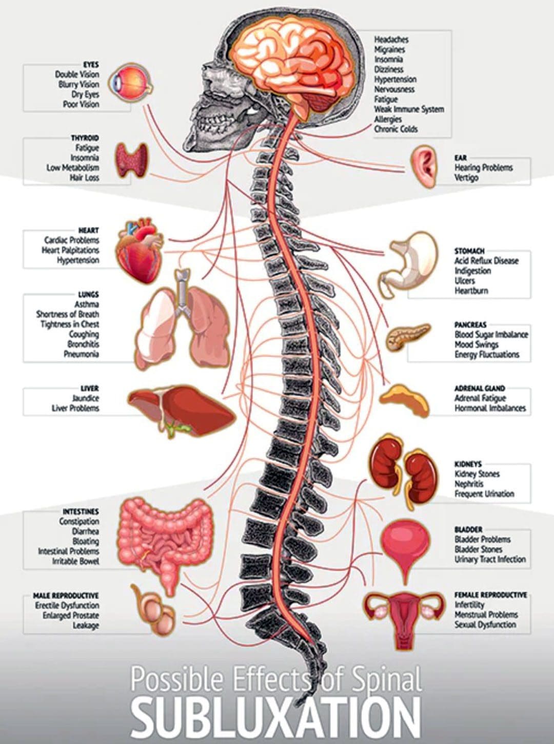
Body misalignment can cause various symptoms to be experienced, ranging from headaches, neck and back pain, sore feet, discomfort in the joints, muscles, or nerves, and digestive problems. Improperly aligned vertebrae can press against nerves, pinching or compressing them, causing the nerve signals of the digestive system, including those in the stomach and intestines, to misfire or fail to transmit at the appropriate moment. This can cause the organs to malfunction, resulting in heartburn, gas, constipation, cramping, diarrhea, and other symptoms. Chiropractic realignment adjustments are an effective treatment option for frequent stomachaches, reflux, constipation, and other gastrointestinal conditions.
Body Misalignment Digestive Problems
There are over a million nerve cells within the digestive system. A collection of nerves branch out from the lower part of the spinal cord and travels to the stomach and intestines. Nerve transmission plays an essential role in the following:
Digestion.
Movement of food through the gastrointestinal system.
Absorption of nutrients and minerals.
Removal of waste products.
Misalignments of the vertebrae are known as subluxations. Pressure on nerve roots caused by misalignment can interfere with the function of the bowel and other organs, which can lead to gastrointestinal issues. Muscle tension in the abdomen can also contribute to digestive problems, whether because of stress or sitting for long hours daily.
Misalignment Symptoms
When the body is out of alignment, symptoms of discomfort begin to appear. The most common include:
Fatigue.
Stiff neck.
Sore shoulders.
Chronic headaches.
Sore muscles.
Pain throughout the back.
Joint pain throughout the body.
Chronic aches.
Tight hips.
Difficulty walking.
Tingling, pins and needles, and numbness nerve sensations – sciatica.
Constantly getting sick.
Healthy Gut
A balanced healthy gut will have less difficulty processing food and eliminating waste, leading to reduced and eventually alleviated symptoms. The following show healthy gut function:
Regular, consistent energy levels.
Increased mental clarity.
Regular and healthy bowel movements.
No pain or discomfort symptoms.
A normal amount of gas and bloating.
Healthy stress levels.
Chiropractic
Chiropractic care will realign the body to its proper form, improving gastrointestinal issues. The chiropractic team will use various tools and techniques to guide and correct any subluxations, relax the muscles, and increase nerve and blood circulation.
Healthy Diet and Chiropractic
References
Ernst, Edzard. “Chiropractic treatment for gastrointestinal problems: a systematic review of clinical trials.” Canadian Journal of Gastroenterology = Journal canadien de Gastroenterologie vol. 25,1 (2011): 39-40. doi:10.1155/2011/910469
Hills, Ronald D Jr, et al. “Gut Microbiome: Profound Implications for Diet and Disease.” Nutrients vol. 11,7 1613. 16 Jul. 2019, doi:10.3390/nu11071613
Hornbuckle, William E., et al. “Gastrointestinal Function.” Clinical Biochemistry of Domestic Animals (2008): 413–457. doi:10.1016/B978-0-12-370491-7.00014-3
Leeming, Emily R et al. “Effect of Diet on the Gut Microbiota: Rethinking Intervention Duration.” Nutrients vol. 11,12 2862. 22 Nov. 2019, doi:10.3390/nu11122862
Li, Yuanyuan, et al. “The Role of Microbiome in Insomnia, Circadian Disturbance, and Depression.” Frontiers in psychiatry vol. 9 669. 5 Dec. 2018, doi:10.3389/fpsyt.2018.00669
Redwood, Daniel. “Chiropractic and visceral disorders.” Journal of Alternative and complementary medicine (New York, N.Y.) vol. 13,5 (2007): 479-80. doi:10.1089/acm.2007.7146
Valdes, Ana M et al. “Role of the gut microbiota in nutrition and health.” BMJ (Clinical research ed.) vol. 361 k2179. 13 Jun. 2018, doi:10.1136/bmj.k2179
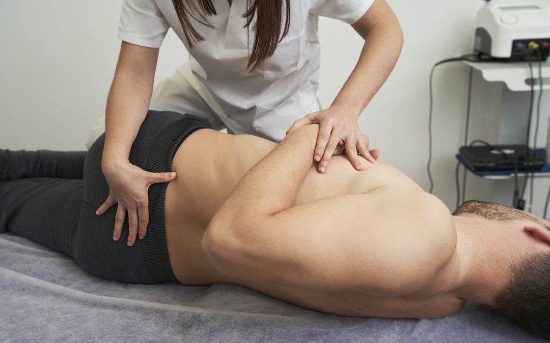
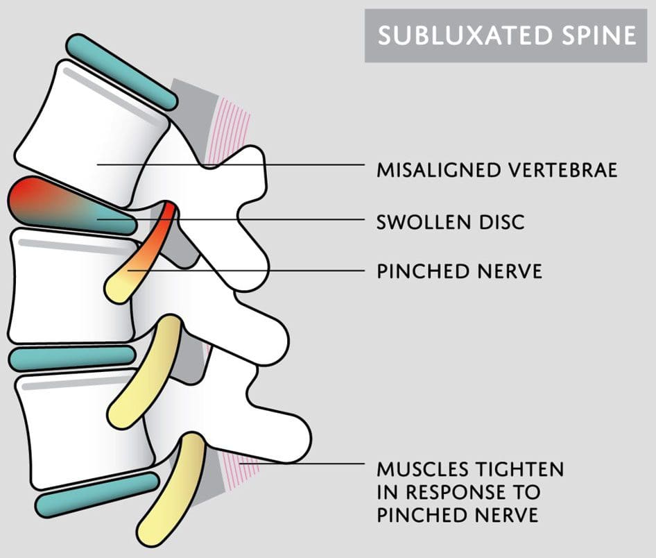
Subluxation is when a joint shifts out of alignment, which can happen to any joint in the body. Spinal subluxation indicates a misalignment of one or more portions of the spinal vertebrae. It is common in the spine from all the reaching, bending, twisting, and flexing the body goes through. Spinal subluxations, if left untreated, can cause disc degeneration, permanent nerve damage, neurological conditions, and chronic pain symptoms. A subluxation chiropractor will realign and decompress the spine combined with massage therapy to relax the muscles and restore mobility and function.
Subluxation Chiropractor
Some subluxations don’t cause any problems or pain, but that doesn’t mean they aren’t affecting the back and body. A spinal subluxation can cause long-term problems by:
Research shows that spinal subluxations can affect many facets of the body. Long-term effects may include:
Sleep problems
Low energy
Brain fog
Mood swings
Anxiety and depression
Digestive issues
Respiratory problems
Bone spurs
Spinal arthritis
Chiropractic Care
When the spine is out of alignment, it can cause issues throughout the body. Changes in one area affect the rest of the body. A subluxation chiropractor looks at the spine’s neurological and mechanical components and aims to reset everything back into its proper position. Similar to the way a massage helps the mind and body relax and de-stress, a spinal adjustment helps by:
Increasing circulation
Relieving discomfort and pain
Releasing tension
Improving mood
Reducing stress levels
Improving sleep function
Increasing energy levels
When the spine is properly aligned, the body can operate at its full potential.
Adrenal Dysfunction
References
Brian S. Budgell, Reflex effects of subluxation: the autonomic nervous system, Journal of Manipulative and Physiological Therapeutics, Volume 23, Issue 2,
2000, Pages 104-106, ISSN 0161-4754, https://doi.org/10.1016/S0161-4754(00)90076-9. (https://www.sciencedirect.com/science/article/pii/S0161475400900769)
Green, J D et al. “Anterior subluxation of the cervical spine: hyperflexion sprain.” AJNR. American journal of neuroradiology vol. 2,3 (1981): 243-50.
Meyer, S. “Thoracic spine trauma.” Seminars in roentgenology vol. 27,4 (1992): 254-61. doi:10.1016/0037-198x(92)90004-l
Neva MH, Häkkinen A, Mäkinen H, et al. High prevalence of asymptomatic cervical spine subluxation in patients with rheumatoid arthritis waiting for orthopedic surgeryAnnals of the Rheumatic Diseases 2006;65:884-888.
Nourollahi, Maryam, et al. “Awkward trunk postures and their relationship with low back pain in hospital nurses.” Work (Reading, Mass.) vol. 59,3 (2018): 317-323. doi:10.3233/WOR-182683
Vernon, Howard. “Historical overview and update on subluxation theories().” Journal of chiropractic humanities vol. 17,1 (2010): 22-32. doi:10.1016/j.echu.2010.07.001
Ankylosing spondylitis/AS is a common type of arthritis that can cause damage to spinal structures, body parts, and organs. Ankylosing spondylitis causes inflammation in the spine’s ligaments and joints which can cause affected vertebrae to fuse, but other symptoms/complications are skin disorders. Ankylosing spondylitis flare-ups can present with skin disorders like rashes and the possible development of skin diseases like psoriasis.
Ankylosing Spondylitis
The inflammation causes back stiffness and pain that causes the spine to become inflexible and rigid. The vertebrae can fuse in extreme cases.
It is typically seen in the early adult population as back pain and hip pain.
Symptoms are more common in individuals between 17 and 45.
Men are more likely to be affected than women.
Genetics can play a role in this condition.
Doctors utilize multiple approaches to relieve symptoms and manage the condition through combined exercise, chiropractic, physical therapy, diet, and stress management to help improve quality of life.
Skin Disorders
A flare-up can present as a skin rash but can also affect the skin in other ways that include:
Rashes brought on by medication treatments.
Trouble healing from incisions after surgery.
Psoriasis
Psoriasis presents as red skin patches appearing anywhere on the body.
The most common areas are the scalp, palms, elbows, and knees.
The affected skin can itch, become tender, and can also sting and burn.
Some psoriasis outbreaks result in lesions or blisters.
Ankylosing Spondylitis vs. Psoriatic Arthritis
Ankylosing spondylitis and psoriatic arthritis are related and come under spondyloarthritis/SpA rheumatic disease.
Ankylosing spondylitis is typically localized to the spine, whereas psoriatic arthritis can affect almost any joint in the body and presents with tendinopathy.
Some individuals with AS can begin to develop psoriasis.
Medications can also help but can produce side effects.
Ankylosing spondylitis skin disorders present ongoing challenges. However, increasing treatment options are helping to minimize the condition’s impact on a better quality of life.
AS Causes, Symptoms, Diagnosis, Treatment
References
Meier, Katharina, et al. “Skin manifestations in spondyloarthritis.” Therapeutic advances in musculoskeletal disease vol. 12 1759720X20975915. 8 Dec. 2020, doi:10.1177/1759720X20975915
Myers, Elisha et al. “An Update on Narrowband Ultraviolet B Therapy for the Treatment of Skin Diseases.” Cureus vol. 13,11 e19182. 1 Nov. 2021, doi:10.7759/cureus.19182
National Institutes of Health. (n.d.) “Ankylosing spondylitis.” https://www.niams.nih.gov/health-topics/ankylosing-spondylitis
Ye, Chao, and Wenyuan Li. “Cutaneous vasculitis in a patient with ankylosing spondylitis: A case report.” Medicine vol. 98,3 (2019): e14121. doi:10.1097/MD.0000000000014121
IFM's Find A Practitioner tool is the largest referral network in Functional Medicine, created to help patients locate Functional Medicine practitioners anywhere in the world. IFM Certified Practitioners are listed first in the search results, given their extensive education in Functional Medicine