Physicians, neurologists, and other healthcare professionals may often run a cranial nerve examination as part of a neurological evaluation to analyze the operation of the cranial nerves. This involves a highly formalized series of tests that evaluate the status of each cranial nerve. A cranial nerve test begins with observation of the patient partly due to the fact that cranial nerve lesions may ultimately affect the symmetry of the face or eyes, among other signs and symptoms.
The visual fields for neural lesions or nystagmus�are tested via an evaluation of particular eye movements. The sensation of the face is tested by asking patients to execute different facial movements, like puffing out their cheeks. Hearing is tested through voice and tuning forks. The position of the individual’s uvula is also examined because asymmetry in its placement could indicate a lesion of the glossopharyngeal nerve. After the capability of the individual to use their shoulder to test the accessory nerve (XI), the patient’s tongue operation is generally assessed by detecting various tongue movements.
Damage or Injury of the Cranial Nerves
Compression
Cranial nerves may be compressed due to increased intracranial pressure, a profound effect of an intracerebral haemorrhage, or tumour which presses against the cranial nerves and interferes with the communication of impulses along the length of a nerve. In some instances, a loss of functionality of one cranial nerve may on occasion be the first symptom of an intracranial or skull base cancer.
An increase in intracranial pressure can lead to dysfunction of the optic nerves (II) because of the compression of the surrounding veins and capillaries, resulting in swelling of the eyeball, known as papilloedema. A cancer, such as an optic glioma, can also affect the optic nerve (II). A pituitary tumour can compress the optic tracts or the optic chiasm of the optic nerve (II), causing visual field loss. A pituitary tumour may also extend into the cavernous sinus, compressing the oculuomotor nerve (III), the trochlear nerve (IV) and the abducens nerve (VI), often leading to double-vision and strabismus. These cranial nerves may also be impacted by herniation of the temporal lobes of the brain via the falx cerebri.
The cause of trigeminal neuralgia, where one side of the face experiences painful signs and symptoms, is believed to be due to the compression of a cranial nerve by an artery as the nerve exits from the brain stem. An acoustic neuroma, especially at the junction between the pons and medulla, may compress the facial nerve (VII) and the vestibulocochlear nerve (VIII), resulting in hearing and sensory loss on the affected side.
Stroke
Occlusion of blood vessels which supply the cranial nerves or their nuclei, or an ischemic stroke, might cause specific signs and symptoms which could localize where the occlusion happened. A clot in a blood vessel draining the cavernous sinus, also known as the cavernous sinus thrombosis, may affect the oculomotor (III), the trochlear (IV), and the opthalamic branch of the trigeminal nerve (V1) and the abducens nerve (VI).
Inflammation
Inflammation caused by an infection may impair the operation of any of the cranial nerves. Infection of the facial nerve (VII), for instance, can result in Bell’s palsy. Multiple sclerosis, an inflammatory process which can produce a loss of the myelin sheathes that encircle the cranial nerves, may cause a variety of shifting signs and symptoms which can ultimately affect multiple cranial nerves.
Other
Trauma to the skull, bone disease like Paget’s disease, and damage or injury to the cranial nerves through neurosurgery, by way of instance, through tumor removal, are other potential causes of cranial nerve health issues.
Dr. Alex Jimenez’s Insight
There are 12 pairs of cranial nerves which exit the brain, one in each side. These cranial nerves are named and numbered (I-XII) according to their location in the brain as well as their specific function in the body. Common conditions, such as multiple sclerosis, may affect one or more of the cranial nerves, resulting in dysfunction of the specific regions innervated by them. Signs and symptoms associated with health issues affecting specific cranial nerves can help healthcare professionals determine the source of the problem. Testing the cranial nerves involves a number of steps in order to be certain which function of the human body has been ultimately affected.
Clinical Significance of the Cranial Nerves
Most commonly, humans are believed to have twelve pairs of cranial nerves which have been assigned Roman numerals I-XII for identification. The numbering of the cranial nerves is based on the order in which they emerge from the brain, or from the front to the back of the brainstem. These include: the olfactory nerve (I), the optic nerve (II), the oculomotor nerve (III), the trochlear nerve (IV), the trigeminal nerve (V), the abducens nerve (VI), the facial nerve (VII), the vestibulocochlear nerve (VIII), the glossopharyngeal nerve (IX), the vagus nerve (X), the accessory nerve (XI), and the hypoglossal nerve (XII). Below we will narrow down the clinical significance of the cranial nerves.
Olfactory Nerve (I)
The olfactory nerve (I) communicates the sensation of smell to the brain. Lesions resulting in anosmia, or loss of the sense of smell, have been previously described to occur through trauma, damage or injury to the head, especially in the instance that a patient hits the back of their head. In addition, frontal lobe masses, tumors, and SOL have also been associated with the loss of the sense of smell. Healthcare professionals have previously identified that the loss of the sense of smell is one of the first symptoms seen in Alzheimer’s and early dementia patients.
Healthcare professionals may test the function of the olfactory nerve (I) by having the patient close their eyes and cover one nostril at a time in order to have them breathe out through their nose while placing a scent under the nostril and having them breathe in. The doctor will ask the patient, “do you smell anything?”, and record the findings. This tests whether the nerve is operating appropriately. If the patient says yes, the doctor will then ask the patient to identify the scent. This tests whether the processing pathway, known as the temporal lobe, is functioning accordingly.
Optic Nerve (II)
The optic nerve (I) communicates visual information to the retina. Lesions to this cranial nerve can be the result of CNS disease, such as MS, or CNS tumors and SOL. Most health issues associated with the visual system emerge from direct trauma, metabolic or vascular diseases. FOV lost in the periphery can also indicate that SOL may be affecting the optic chiasm, including a pituitary tumor.
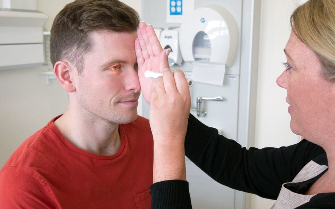
A healthcare professional will often test the function of the optic nerve (II) by asking whether the patient can see. If the patient describes having vision in each eye, the optic nerve is functional. Doctors may also perform visual acuity testing using the Snellen chart, first one eye at a time, then the two eyes together, or they may perform distance vision testing. Near vision testing will often involve the Rosenbaum chart, first one eye at a time, then the two eyes together. Additional associated testing for the visual system can include, the ophthalmoscopic or funduscopic exam, which assess the A/V ratio and vein/artery health as well as assess cup to disc ratio of the visual system. Other testing methods include field of vision testing, intraoccular pressure testing and the iris shadow test.
Oculomotor Nerve (III), Trochlear Nerve (IV), and Abducens Nerve (VI)
The oculomotor nerve (III), the trochlear nerve (IV), the abducens nerve (VI) and the ophthalmic division of the trigeminal nerve (V1) travel through the cavernous sinus to the superior orbital fissure, passing out of the skull into the orbit. These cranial nerves control the tiny muscles that move the eye and also offer sensory innervation to the eye and orbit.
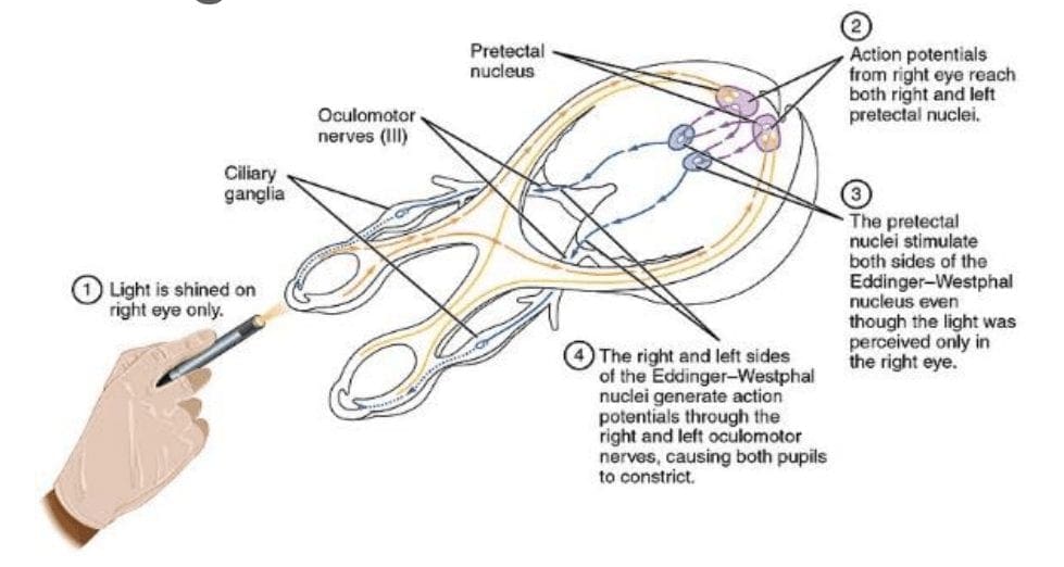
The clinical significance of the oculomotor nerve (III) includes diplopia, lateral strabismus (unopposed lateral rectus m.), head rotation away from the side of the lesion, a dilated pupil (unopposed dilator pupillae m.), and ptosis of the eyelid (loss of function of the levator palpebrae superioris m.). Lesions to the oculomotor nerve (III) can occur due to inflammatory diseases, such as syphilitic and tuberculous meningitis, aneurysms of the posterior cerebral or superior cebellar aa., and SOL in the cavernous sinus or displacing the cerebral peduncle to the opposite side. Testing this cranial nerve is performed by moving a light in front of the patient’s pupil from the lateral side and hold for 6 seconds. The doctor should watch for direct (ispilateral eye) and consensual (contralateral eye) pupillary constriction in order to distinguish dysfunction of the oculomotor nerve (III).
The clinical significance of the trochlear nerve (IV) is characterized where the patient presents diplopia and difficulty while maintaining a downward gaze, often complaining of having difficulties when walking down stairs, resulting in more frequent tripping and/or falling, followed by extortion of the affected eye (unopposed inferior oblique m.) and a head tilt to the unaffected side. Lesions to the trochlear nerve (IV) can commonly be the result of inflammatory diseases, aneurysms of the posterior cerebral or superior cerebellar aa., SOL in the cavernous sinus or superior orbital fissure and surgical damage during mesencephalon procedures. Head tilts in superior oblique palsy (CN IV failure) may also be identified.
The clinical significance of the abducens nerve (VI) includes diplopia, medial strabismus (unopposed medial rectus m.), and head rotation towards the side of the lesion. Lesions to this cranial nerve can be the result of aneurysms of the posterior inferior cerebellar or basilar aa., SOL in the cavernous sinus or 4th ventricle, such as a cerebellar tumor, fractures of the posterior cranial fossa, and increased intracranial pressure. Testing this cranial nerve is performed through the H-Pattern testing, where the healthcare professional will have the patient follow an object no bigger than 2 inches. It’s essential for the doctor to follow these specific guidelines as patient’s can have difficulties focusing on items that are too large, and it’s also important for the doctor not to hold the object too close to the patient. Convergence and accommodation testing is performed by bringing the object close to the bridge of the patient’s nose and back out at least 2 times. The physician must look for pupillary constriction response as well as convergence of the eyes.
Trigeminal Nerve (V)
The trigeminal nerve (V) is made up of three different parts: The . When put together, these nerves provide sensation to the skin of the face and also controls the muscles of mastication, or chewing. Cranial nerve dysfunction along any of the separate sections of the trigeminal nerve (V) can manifest as decreased bite strength on the ipsilateral side of the lesion, loss of sensation along the distribution of V1, V2, and V3, and loss of corneal reflex. Lesions to the trigeminal nerve (V) can be the result of aneurysms or SOL affecting the pons, particularly tumors at the cerebellopontine angle, skull fractures on the facial bones or damage to the foramen ovale, and Tic doloureux, most frequently referred to as trigeminal neuralgia, characterized by sharp pain along the distributions of the different parts of the trigeminal nerve (V). Physicians may utilize analgesic, anti-inflammatory or contralateral stimulation to control the signs and symptoms.
Testing the trigeminal nerve (V) includes pain & light touch testing along the ophthalmic (V1), the maxillary (V2), as well as the Mandibular (V3) nerves of the cranial nerve.�Testing is best done toward the more medial or proximal areas of
the face, where the V1, the V2 and the V3 are better delineated. A healthcare professional may also assess dysfunction along this cranial nerve using the blink/corneal reflex testing, performed by puffing air or doing a small tissue tap from the lateral side of the eye on the cornea. If normal, the patient blinks. The CN V provides the sensory (afferent) arc of this reflex. Bite strength may also be tested by having the patient bite down on a tongue depressor while the doctor tries to remove it. The jaw jerk/Masseter reflex may also be performed with the patient�s mouth slightly open, by placing the thumb on a patient�s chin and tapping the own thumb with a reflex hammer. Strong closure of the mouth indicates UMN lesion. CN V provides both the motor and sensory of this reflex.
Facial Nerve (VII) and Vestibulocochlear Nerve (VIII)
The facial nerve (VII) and the vestibulocochlear nerve (VIII) both input the inner auditory canal in the temporal bone. The facial nerve subsequently extends to the side of the face then distributes to control and reach all of the muscles in charge of facial expressions. The vestibulocochlear nerve reaches the organs which control equilibrium and hearing in the temporal bone.
As with all cranial nerves, signs and symptoms along the facial nerve (VII) describe the location of the lesion. Lesion in the lingual nerve will manifest as loss of taste, general sensation in the tongue and salivary secretion. Lesion proximal to the branching of the chorda tympani, such as in the facial canal, will result in the same signs and symptoms, without the loss of general sensation of the tongue, partly due because the V3 has not yet joined the facial nerve (VII). Corticobulbar innervation is asymmetric to the upper and lower parts of the facial motor nucleus. In the instance of an UMN lesion, or a lesion to the corticobulbar fibers, the patient will experience paralysis of the muscles in charge of facial expression in the contralateral lower quadrant. If there is an LMN lesion, or a lesion to the facial nerve itself, the patient will experience paralysis of the muscles of facial expression in the ipsilateral half of the face, otherwise known as Bell’s palsy.
A healthcare professional will test the facial nerve (VII) initially by asking the patient to mimic or follow specific instructions to make certain facial expressions. The doctor should make sure to evaluate all four quadrants of the face by asking the patient to raise their eyebrows, puff their cheeks, smile and then close their eyes tightly. Subsequently, the doctor will test the facial nerve (VII) by checking the strength of the buccinator muscle against resistance. The healthcare professional will achieve this by asking the patient to hold air in their cheeks as they press gently from the outside. The patient should be able to hold air in against the resistance.
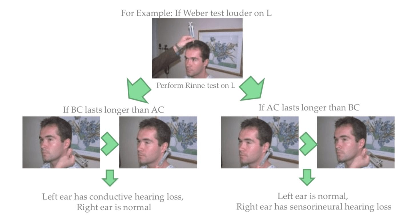
Signs and symptoms of dysfunction in the vestibulocochlear nerve (VIII) often involve changes in hearing alone, most commonly as a result of infections in the otitis media and/or as a result of skull fractures. The most common lesion to this nerve is caused by an acoustic neuroma which affects the CN VII and the CN VIII, particularly the cochlear and vestibular divisions, as a result of proximity in the internal auditory meatus. Signs and symptoms of the health issue include nausea, vomiting, dizziness, hearing loss, tinnitus, and Bell’s palsy, etc.
Testing the vestibulocochlear nerve (VIII) for dysfunction commonly involves an otoscopic exam, the scratch test, which determines whether a patient can hear equally on both sides, the Weber test, tests for lateralization, a 256 Hz tuning fork placed on top of the patient�s head in the center, which can help point out whether a patient hears it louder on one side than the other, and finally the Rinne test, which compares air conduction to bone conduction. Normally, air conduction should last twice as long as bone conduction.
Glossopharyngeal Nerve (IX), Vagus Nerve (X) and Accessory Nerve (XI)
The glossopharyngeal (IX), the vagus nerve (X) and the accessory nerve (XI) all emerge from the skull to enter the neck. The glossopharyngeal nerve (IX) provides innervation to the upper throat and the back of the tongue, the vagus nerve (X) offers innervation to the muscles at the voicebox, and proceeds down to provide parasympathetic innervation to the chest and abdomen. The accessory nerve (XI) controls the trapezius and sternocleidomastoid muscles at the neck and shoulder.
The glossopharyngeal nerve (IX) is rarely damaged alone, due to it�s proximity to the CN X and XI. A healthcare professional should perform a test to look for signs of CN X & XI damage as well if CN IX involvement is suspected.
Patients with clinical signs and symptoms caused by vagus nerve (X) dysfunction may experience dysarthria, or difficulty speaking clearly, as well as dysphagia, or difficulty swallowing. These may present as food or liquid coming out of their nose or frequent chocking or coughing when eating and/or drinking. Further clinical presentations include hyperactivity of a visceral motor component, leading to the hypersecretion of gastric acid and resulting in ulcers. Hyper-stimulation of the general sensory component can cause coughing, fainting, vomiting and reflex visceral motor activity. The visceral sensory component of this nerve only provides general feelings of un-wellness but visceral pain may transfer on to the sympathetic nerves.
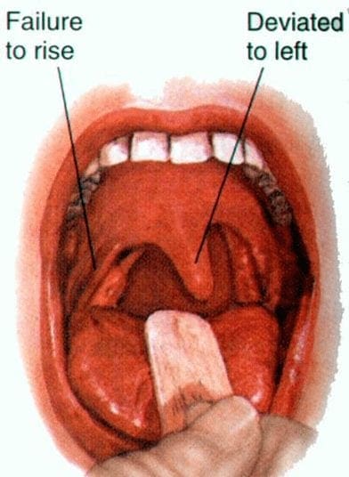
Testing for the glossopharyngeal nerve (IX) and the vagus nerve (X) can include the gag reflex, where the�CN IX provides the afferent (sensory) arc and the�CN X provides the efferent (motor) arc. Approximately 20 percent�of patients have a minimal or absent gag reflex. Other tests may include wwallowing, gargling, etc., as it requires CN X function. Healthcare professionals may also test palatal elevation because it requires CN X function. Furthermore, the doctor will see whether the palate elevates and uvula deviates
contralateral to damaged side. Finally, the healthcare professional will test the auscultation of the heart, since the R CN X innervates SA node (more rate regulation) and the L CN X the AV node (more rhythm regulation).
Lesions in the accessory nerve (XI)�may occur due to radical surgeries in the neck area, such as the removal of the laryngeal carcinomas. Testing for the accessory nerve (XI) may include the strength test SCM m. Patients with clinical signs and symptoms due to lesions in the accessory nerve (XI) will experience difficulties turning their head against the resistance of a healthcare professional, particularly toward the side opposite of the lesion. Testing for the accessory nerve (XI) may also include the strength test trapezius m. Patients with clinical signs and symptoms due to lesions in the accessory nerve (XI) will experience difficulties with shoulder elevation on the side of the lesion.
Hypoglossal Nerve (XII)
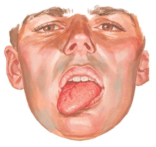
The hypoglossal nerve (XII) originates from the skull to reach the tongue in order to control essentially all of the muscles involved in the movements of the tongue. The clinical significance of health issues associated to the hypoglossal nerve (XII) can manifest as a deviating tongue towards the side of an inactive genioglossus m. upon tongue protrusion. This may often be contralateral to a corticobulbar, or UMN, lesion or from an ipsilateral to a hypoglossal n., or LMN, lesion.
Testing for the hypoglossal nerve (XII) involves the healthcare professional asking a patient to stick out their tongue. The doctor will look for any deviation which may signal a health issue along the length of the hypoglossal nerve (XII). Another test the doctor may perform as a part of the evaluation may include the physician asking the patient to place their tongue inside their cheek and apply light resistance, one side at a time. The patient should be able to resist moving their tongue with pressure.
The clinical significance of the signs and symptoms which manifest as a result of cranial nerve dysfunction are essential in order for the healthcare professional to properly diagnose the patient’s specific health issue. The clinical findings described above are often unique to the affected cranial nerve and the tests and evaluations for each can help confirm a diagnosis. Proper diagnosis is fundamental in order for the doctor to continue with the patient’s appropriate treatment. The scope of our information is limited to chiropractic as well as to spinal injuries and conditions. To discuss the subject matter, please feel free to ask Dr. Jimenez or contact us at 915-850-0900 .
Curated by Dr. Alex Jimenez
Additional Topics: Sciatica
Sciatica is medically referred to as a collection of symptoms, rather than a single injury and/or condition. Symptoms of sciatic nerve pain, or sciatica, can vary in frequency and intensity, however, it is most commonly described as a sudden, sharp (knife-like) or electrical pain that radiates from the low back down the buttocks, hips, thighs and legs into the foot. Other symptoms of sciatica may include, tingling or burning sensations, numbness and weakness along the length of the sciatic nerve. Sciatica most frequently affects individuals between the ages of 30 and 50 years. It may often develop as a result of the degeneration of the spine due to age, however, the compression and irritation of the sciatic nerve caused by a bulging or herniated disc, among other spinal health issues, may also cause sciatic nerve pain.
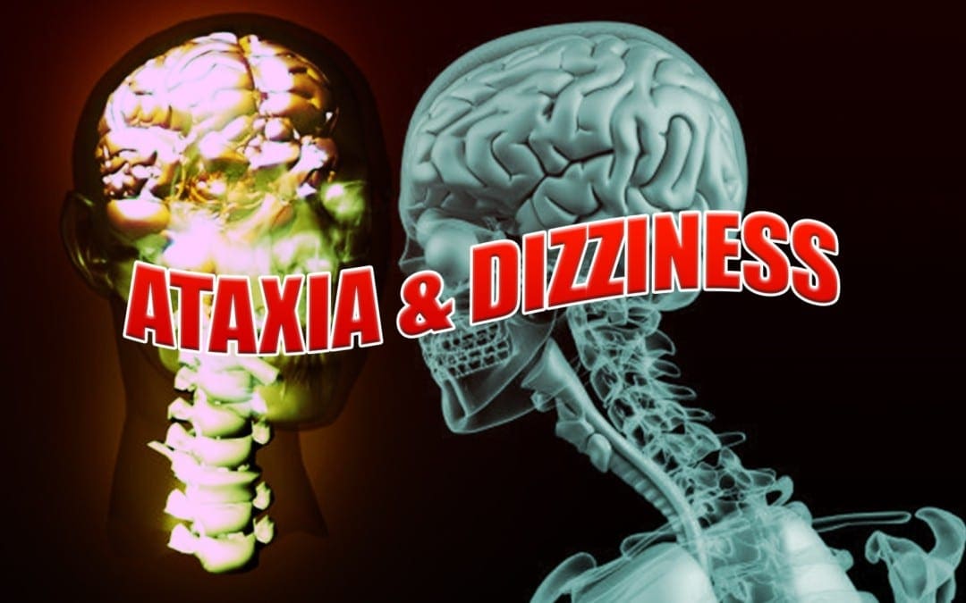
Ataxia is a degenerative disease of the nervous system. Symptoms can mimic those of being inebriated/intoxicated, with� slurred speech, stumbling, falling, and unable to maintain coordination. This comes from degeneration of the cerebellum, which is the part of the brain responsible for coordinating movement. It is a disease that affects people of all ages. However, age of symptom onset can vary, from childhood to late adulthood. Complications from the disease can be serious, even debilitating and life shortening.
Symptoms can vary from person to person, as well as, the type of Ataxia. Symptom onset and progression can vary as well. Symptoms can worsen slowly, over decades or quickly, over a few months. The common symptoms are lack of coordination, slurred speech, trouble eating, swallowing, eye movement abnormalities, motor skill deterioration, difficulty walking, gait abnormalities, tremors, and heart problems. People with Ataxia usually require wheelchairs, walkers, and/or scooters to aid in mobility.
Ataxia
The Loss Of Full Control Of Bodily Movements, Especially Gait
History Of Ataxia
How long has it been present?
Slow onset ? Degenerative disease?
Acute onset ? Stroke?
When does it occur?
If worsened by walking on uneven surfaces, or with limited vision ? Sensory ataxia?
Are there any coexisting symptoms?
Vertigo, weakness, stiffness, cognitive changes, etc.
Have others noticed this gait disturbance?
If no, consider psychogenic cause
Is the gait change explainable by physical problems such as pain or weakness?
Antalgic gait, limp, etc.
Weakness
Proximal muscle weakness�? Myopathy?
Distal muscle weakness ? Neuropathy?
UMN signs?
LMN signs?
Has the patient fallen? Or at risk for fall?
Is ataxia limiting ADLs?
Balance
Utilizes
Vestibular system
Cerebellar system
Conscious proprioceptive information (joint position sense)
Visual information
Motor strength and coordination
Vestibular System
Generally, if the problem lies in the vestibular system the patient will experience dizziness, possibly having vertigo or nystagmus
Does the dizziness feel similar to when you stand up too fast?
Pre-Syncope
�Light-headedness�
CardiacOrigin
Output disorders
Arrhythmias
Holter monitor testing
Postural/Orthostatic hypotension
May be secondary to other problems (diabetic neuropathy, adrenal hypofunction, Parkinsons, certain medications, etc.)
Vasovagal episodes
Slow heart rate with low blood pressure
Often brought on by stress, anxiety or hyperventilation
Migraine
Due to cerebrovascular instability
Blood sugar dysregulation
Disequilibrium Hx Questions
Does the dizziness only occur when you�re on your feet?
Does it get better if you touch/hold onto something?
Disequilibrium
Common in the elderly
Due to sensory deficits
Gradual onset
Worsened by reduced vision
Dark
Eyes closed
Visual acuity losses
Improved by touching a stationary object
Subjective of dizziness often improves with a gait assistive device (cane, walker, etc.)
Other Causes
Psychological stress
Often patient will describe dizziness as �floating�
Rule out hyperventilation and other types of dizziness
Sources
Blumenfeld, Hal. Neuroanatomy through Clinical Cases. Sinauer, 2002.
Alexander G. Reeves, A. & Swenson, R. Disorders of the Nervous System. Dartmouth, 2004.
You have been diagnosed with Benign Paroxysmal Positional Vertigo. This brochure is designed to help increase your understanding of this disorder and its potential treatments.
Benign Paroxysmal Positional Vertigo
What Is BPPV?
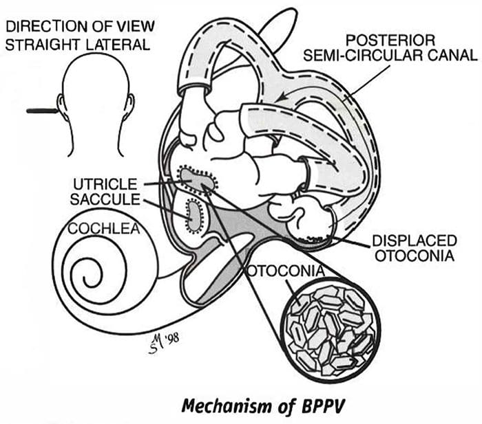
Benign paroxysmal positional vertigo (BPPV) is a disorder of the inner ear. People with BPPV typically experi�ence brief episodes of vertigo (dizziness) when they change the position of their head with respect to gravity. Approximately 20 percent of all vertigo is due to BPPV.
What Causes BPPV?
Benign Paroxysmal Positional Vertigo is thought to be due to tiny crystals, called otoconia, that have collected within a sensitive part of the inner ear. Otoconia are crystals of calcium carbonate that are normally located in a structure of the ear called the utricle.
Dizziness occurs when the crystals are displaced from the utricle into the semicircular canals of the inner ear.
Otoconia may become displaced when the utricle is injured, if there is an infection or other disorder of the inner ear, or simply due to advanced age. When you change the position of your head, the otoconia move within the semicircular canals and this causes the dizziness. The dizziness subsides when the otoconia stop moving.
The most common cause of BPPV in people under age 50 is head injury. In older people, the most common cause is degeneration of the vestibular system of the inner ear. BPPV becomes much more common with advancing age. Other causes include minor strokes, Meniere’s disease, and viruses such as those causing vestibular neuritis. In approximately half of all BPPV cases, no cause can be determined.
What Are The Symptoms?
The symptoms of BPPV include dizziness or vertigo, lightheadedness, imbalance, and nausea. Activities that
bring on symptoms vary among individuals, but symptoms are usually associated with a change in the position of the head with respect to gravity. Getting out of bed, rolling over in bed, and tipping the head back to look up are common “problem” motions. The use of shampoo bowls in hair salons may bring on symptoms. An intermittent pattern is common. BPPV may be present for a few weeks, then stop, and then come back again.
How Is Benign Paroxysmal Positional Vertigo (BPPV) Diagnosed?
BPPV is diagnosed with the Dix-Hallpike test. This test involves observing the eyes with the head and body positioned in specific ways. It can be performed either by the clinician, or as part of a laboratory test called an electronystagmography, or ENG. If the Dix-Hallpike test is abnormal and the findings are “dassic” for BPPV, then additional testing is not necessary. If the results are normal or not “classic” then the diagnosis of BPPV is less certain and other tests may be suggested.
What Are The Treatments For BPPV?
There are four approaches to treating BPPV.
1. Do Nothing And Wait For It To Go Away By Itself
BPPV symptoms sometimes go away within six months of onset, therefore you might want to wait and see if your symptoms subside on their own. During this waiting period, medications to prevent motion sickness or nausea are sometimes helpful in controlling the nausea associated with BPPV.
2. Physical Maneuvers Performed In The Clinic
(The Epley and Semont Maneuvers)
The Epley and Semont maneuvers, named for their inventors, are treat�ments that are performed in the clinic. These treatments are specifi�cally intended to move the otoconia from the semicircular canals to a less sensitive location within the inner ear. Your clinician will select the treatment that is most appropriate for you.
Each of these treatments takes about 15 minutes and alleviates symptoms in about 80 percent of patients. In the remaining 20 percent, a second treatment may be necessary, or you may be instructed to perform the Brandt-Daroff exercises (see “Home Treatment”).
The Epley maneuver, also called the canalith reposi�tioning procedure (CRP) and particle repositioning, is a procedure in which the clinician moves your head into five positions, maintaining each position for ap�proximately 30 seconds. The Semont maneuver (also called the liberatory maneuver) is a procedure in which the clinician rapidly moves you from lying on one side to lying on the other side. These maneuvers may not be appropriate for patients with neck or back problems. Pa�tients who experience nausea or anxiety may wish to take medication prior to the treatment.
INSTRUCTIONS FOR PATIENTS AFTER CLINIC TREATMENTS
Follow these instructions after the Epley or Semont maneuver. B.Y doing so you will minimize the opportunity for otoconia to return to the semicircular canals of the inner ear and reduce the potential that your dizziness will recur.
Wait at least 10 minutes after the maneuver before going home.
This is to avoid “quick spins” or brief bursts of vertigo as the otoconia reposition themselves immedi�ately after the maneuver. If possible, arrange to have someone drive you home.
The following two days:
Sleep semi-recumbent for the next two nights. This means sleeping with your head halfway between flat and upright, at a 45-degree angle. This is most easily done by sleeping in a recliner chair or by sleeping with pillows appropriately arranged on a couch.
During the day, try to keep your head vertical. A soft neck brace may be helpful.
Do not go to the barber, hairdresser or dentist.
When shaving, keep your head vertical by bending forward at your hips with your neck extended.
If you need to administer eye drops, try to keep your head as vertical as possible.
Sham�poo only under the shower.
During the following week, avoid provoking head positions that might bring on BPPV.
Use two pillows when you sleep.
Avoid sleeping on the affected side.
Don’t turn your head far up or far down.
Avoid tilting your head back especially when lying on your back with your head turned toward the affected side.
If possible, postpone elective surgery and going to the beauty parlor or the dentist’s office.
Avoid far head-forward positions and exercises where the head is not kept upright, for example toe touches.
The effectiveness of the clinic treatment cannot be determined for one week.
Wait one week after treatment to test the effectiveness of treatment. Place yourself in the position that usually makes you dizzy. Be sure to position yourself cautiously and under conditions in which you can’t fall or hurt yourself.
3. Home Treatment Of Benign Paroxysmal Positional Vertigo (Brandt-Daroff Exercises)
When the clinic treatment (Epley or Semont) fails, when the involved side is not determined, or when a case is mild, the Brandt-Daroff exercises may be recommended. These exercises succeed in 95 percent of cases but take longer to work than the clinic treatments. You should perform these exercises only if instructed to do so by your clinician. If your clini�cian performed the Epley or Semont maneuver, you must wait one week after that treatment before you begin the Brandt-Daroff exercises.
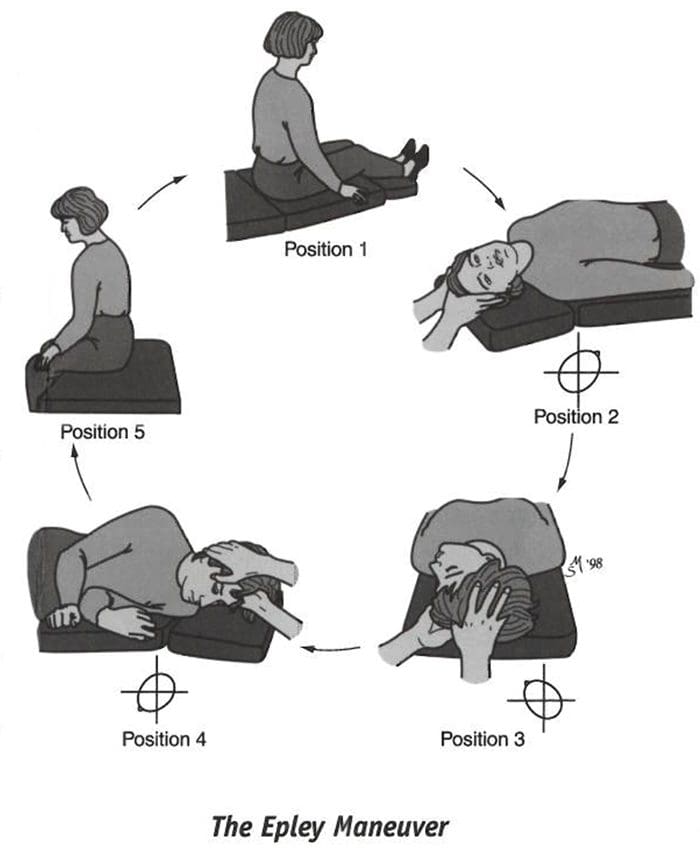
These exercises should be performed on a flat surface, without a pillow.
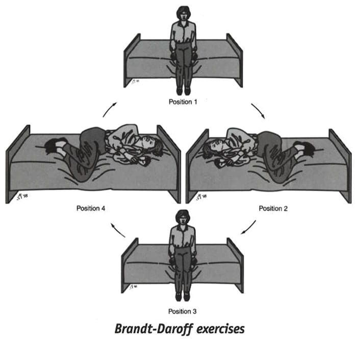
Start sitting upright on the edge of the bed or on the floor.
(Position 1)�Turn your head 45 degrees to the left and lie down on your right side.
(Position 2)�When in the right side-lying position, your head should be at a 45-degree angle turned halfway between the flat surface and the ceiling. Stay in the side-lying position for at least 30 seconds. If you are still dizzy, stay until the dizziness subsides or one minute, whichever is less.
Then sit up (Position 3} and stay in the sitting posi�tion for 30 seconds. Turn your head 45 degrees to the right and lie down on your left side.
(Position 4)�Again keeping your head turned halfway toward the ceiling for 30 seconds or until the dizziness subsides. Return to Position 1 (sit upright) for 30 seconds. This is one repetition.
One set (five repetitions) takes about 10 minutes to complete and should be performed each morning, mid-day and evening.
The Brandt-Daroff exercises should be performed for two weeks, three sets each day, or for three weeks, two sets each day (52 sets total). In most individuals, complete relief from symptoms is obtained after 30 sets, or about 10 days. In approximately 30 percent of patients, BPPV will recur within one year. If BPPV recurs you may wish to add one 10-minute exercise (one set) to your daily routine.
If the maneuvers or exercises do not control symptoms that have persisted for a year or longer and the diagnosis is very clear, surgery may be recommended. The most common surgical procedure, called posterior canal plugging, blocks most of the posterior canal’s function without affecting the functions of the other canals or parts of the ear. There is, however, a small risk of hearing loss. This surgery is effective in about 90 percent of individuals who have not responded to other treatments and when symptoms are severe and long-standing.
�?2000 Northwestern University. Authors: Timothy C. Hain, MD, Janet Odiry Helminski, PhD, PT.
This information is for educational purposes and is not intended as a substitute for examination, diagnosis, or medical care provided by a licensed and qualified health professional. This work was supported by the Center for Sensory and Communicotion Disorders at Northwestern University, a national research and training center funded by the National Institute on Deafness and Other Communication Disorders.
Back Pain Specialist: Mike Melgoza is a very active person who is always engaging in physical activity, as a result, he occasionally suffers from debilitating back pain symptoms. Mike Melgoza was struggling to sleep properly due to his symptoms of back pain before receiving chiropractic care with Dr. Alex Jimenez. Mike Melgoza has already started experiencing tremendous relief from his back pain and he highly recommends Dr. Alex Jimenez as the non-surgical choice for back pain.
Back Pain Specialist
Back pain is one of the most common reasons people visit the doctor or miss work and it is also a leading cause of disability globally. The majority of people have back pain at least once throughout their lifetimes. Luckily, you can take steps to prevent or relieve back pain. If prevention fails, easy treatment and appropriate body mechanics frequently will heal your back in a few weeks and keep it operational for the long haul. Surgery is rarely required to treat back pain.
We are blessed to present to you�El Paso�s Premier Wellness & Injury Care Clinic.
As El Paso�s Chiropractic Rehabilitation Clinic & Integrated Medicine Center,�we passionately are focused treating patients after frustrating injuries and chronic pain syndromes. We focus on improving your ability through flexibility, mobility and agility programs tailored for all age groups and disabilities.
If you have enjoyed this video and/or we have helped you in any way please feel free to subscribe and share us.
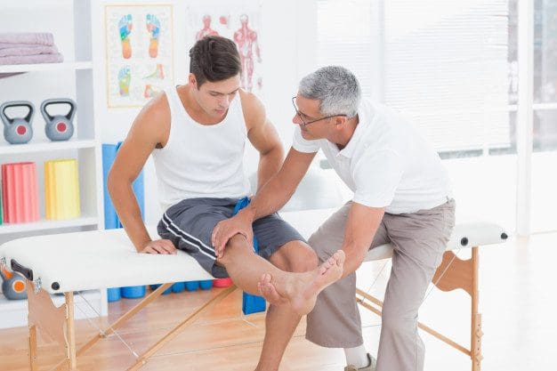
Patellofemoral Syndrome: As the weather warms and spring is in full swing, more and more runners are heading outside, hitting the pavement to train for upcoming races or to just step up their game after a long winter. While there are some die hard runners who don�t let even the most brutal winter stop them, most tend to retreat indoors, waiting for warmer days and a more pleasant environment. Unfortunately, increased activity can also lead to an increased risk of injury, particularly patellofemoral pain syndrome (PFPS), also known as runner�s knee.
What Is Patellofemoral Pain Syndrome?
Runner�s knee is often used to describe PFPS, but runner�s knee is actually a broader term describing several different knee injuries or ailments. PFPS is a painful condition that is caused when the tissue that is between the femur (thigh bone) and the patella (kneecap) becomes inflamed or irritated.
Most people will notice pain in the front portion or anterior part of the knee, but pain can be experienced in other parts of the knee and even back pain may occur. Running increases the discomfort, as does sitting for long periods and going up or down stairs.
The causes of PFPS can also vary widely. Overuse is often the first thing that people think, but a problem with the way the knee is aligned is actually the most common reason.
If the patella is not properly aligned, when it moves through the groove that is at the end of the femur, it causes irritation to the surrounding tissues. This usually happens because the muscles and joints are out of balance.
For instance, if the quad muscle on one side is weaker than the other side it throws the entire system out of balance, causing the knee to become misaligned. This leads to knee pain and discomfort.
Treatment For Patellofemoral Syndrome – Runner�s Knee
When treating PFPS, rest is usually first on the list, followed by icing the area to reduce inflammation. Once the pain is under control, the next step is to determine what is causing the problem. It is important to rule out more serious conditions or injuries first in order to determine the best course of treatment.
If it is indeed PFPS, strengthening the muscles in and around the knee is generally the first step in treatment. It is important that the muscle strength is balanced so that the knee can be properly aligned. Getting a good pair of running shoes is also recommended so that future injury can be prevented.
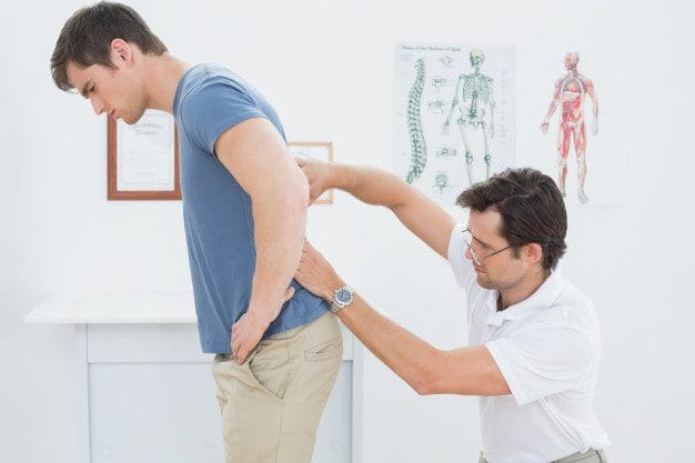
Chiropractic For Runner�s Knee
Runner�s knee, or PFPS, responds very well to chiropractic treatment. The chiropractor is able to do a complete exam and find the cause of the problem, then tailor the treatment accordingly. This is typically done on a case by case basis with treatment that is based on the individual�s unique needs. The chiropractor may do various chiropractic alignments and manipulations on the spine, hip, ankle, and knee in order to bring the body back into proper balance.
The chiropractor may also recommend other complimentary treatments including special supplements, dietary adjustments, and an exercise plan. The chiropractor may also recommend certain stretching exercises to aid in healing. Kinesio taping is another common treatment that may be used in conjunction with chiropractic care. It is particularly beneficial when there is a muscular strength imbalance. The tape can help support the weaker muscle group.
Chiropractic care is a very effective treatment for patellofemoral syndrome and the associated knee pain. It works to correct the problem by bringing the body back into proper alignment, allowing it to function as it should.
Chiropractic Benefits: If you have ever had a migraine before then you know that it is much more than a simple headache. The symptoms of a migraine can be debilitating, lasting hours and even days. According to the Migraine Research Foundation, it is the eighth most disabling disease in the world. It is estimated that 38 million people in the United States alone suffer from migraine headaches. That�s around one in every ten people.
According to the Migraine Research Foundation, migraine headaches are extremely difficult to treat and even more difficult to control. This is mainly due to the fact that doctors still don�t know exactly what causes it. This leaves it undiagnosed in many patients and often terribly under treated in those with a diagnosis.
The best many doctors seem to be able to do is prescribe pain medication that has undesirable side effects in an effort to manage the symptoms. However, chiropractic has been shown in several studies to not only effectively manage the pain of migraines, it also helps stop and prevent them.
Anatomy Of A Migraine Headache
There are two types of migraines, those with an aura and those without an aura. An aura can appear up to an hour before the onset of a migraine. It is a warning sign that usually presents as a disturbance that is either visual or olfactory. The person may see flashes of light or smell particular odors before the headache begins. About one in six migraines are preceded by an aura.
Once the migraine itself begins, the pain is typically on one side of the head, although this is not always the case. Other symptoms may include nausea, vomiting, sensitivity to noise, sensitivity to light, and sensitivity to smell. Some patients experience an inability to concentrate, hot or cold flashes, stiffness in neck or shoulders, slurred speech, loss of coordination, and in rare cases, loss of consciousness.
The migraine can last several minutes, hours, or even days. Afterwards the patient may feel fatigued or washed out. They may be unable to concentrate and either lethargic or extremely energetic.
Studies Show: Chiropractic As A Migraine Treatment
There have been several clinical studies on chiropractic as a treatment for migraine headaches. The results of one study reported that 22 percent of patients who received chiropractic treatment for their migraines reported that their attacks were reduced by more than 90 percent. Additionally, 49 percent reported that the intensity of their migraines was significantly reduced.
Another study randomly assigned people with migraine headaches several different treatments. One group was given Elavil, a daily medication, another group was given chiropractic treatment and a third group received a combination of the two treatments. The results showed that chiropractic was as effective in reducing migraines as the medication and it had fewer side effects. Other studies have also found that chiropractic is as effective as medication for the treatment and prevention of migraine or tension headaches.
Chiropractic Benefits For Migraines Headaches
Spinal adjustments are very effective as a treatment for migraines. The whole body approach of chiropractic also utilizes dietary recommendations, including foods to avoid, as well as lifestyle changes.
The patient may be counseled on managing stress, advised to engage in exercise, and given supplements. The treatments may be used to reduce the pain and severity of a migraine once it begins or it can be used to prevent migraines and reduce their frequency.
Chiropractic benefits everyone and is a safer treatment with fewer side effects than�prescription medications. Chiropractic is quickly becoming the treatment of choice for many migraine sufferers. As the studies show, it works! So if you or a loved one suffer from migraines, give us a call. Our Doctor of Chiropractic is here to help!
Ataxia is a medical term used to describe a lack of muscle control or coordination of voluntary movements, including everyday physical activities like walking or picking up objects. Often referred to as a symptoms of an underlying health issue, ataxia can affect various movements, causing difficulties with speech patterns and language, eye movement and even swallowing.
Persistent ataxia generally results from damage to the part of the brain which controls muscle coordination, known as the cerebellum. Many causes and conditions can lead to ataxia, such as alcohol abuse, certain drugs and/or medications, stroke, tumors, cerebral palsy, brain degeneration and multiple sclerosis. Inherited faulty genes have also been associated to lead to ataxia.
Diagnosis and treatment for ataxia depends largely on the cause and/or condition. Adaptive devices, including walkers or canes, can help patients with ataxia maintain their independence. Chiropractic care, physical therapy, occupational therapy, speech therapy and regular aerobic stretches and exercises can also help improve the symptoms associated with this health issue.
Symptoms of Ataxia
Ataxia is a health issue which can develop gradually over time or it can come on unexpectedly. As a symptom of a number of neurological disorders, ataxia may ultimately lead to:
Poor coordination
Unsteady walk along with a tendency to stumble
Difficulty with fine motor tasks, such as eating, writing or buttoning a shirt
Changes in speech
Involuntary back-and-forth eye movements, known as nystagmus
Difficulty swallowing
When to Visit a Doctor
In the instance that a patient is not aware of whether they may have an underlying health issue that causes ataxia, such as multiple sclerosis, it’s essential to visit a doctor immediately if the patient:
Loses equilibrium
Loses muscle coordination at a hand, leg or arm
Has difficulty walking
Slurs their speech
Has trouble swallowing
Causes of Ataxia
Damage, degeneration or loss of neural cells in the section of the brain which controls muscle coordination, or the cerebellum, often results in ataxia. The cerebellum is made up of two pingpong-ball-sized parts of folded tissue located at the base of the brain close to the brainstem. The right side of the cerebellum controls coordination over the right side of the body; the left side of the cerebellum controls coordination on the left side of the body. Diseases that damage the spinal cord and peripheral nerves which connect the cerebellum to the muscles can also lead to ataxia. Ataxia causes include:
Head trauma. Damage to the brain or spinal cord due to a blow to the head, such as in the case of an automobile accident, can cause acute cerebellar ataxia, which comes on unexpectedly.
Stroke. After the blood supply to part of the brain is interrupted or severely reduced, depriving brain tissue of nutrients and oxygen, brain cells die.
Cerebral palsy. This can be a general term for a group of disorders brought on by damage to a child’s brain during early development, before, during or shortly after birth, which affects the child’s ability to coordinate body movements.
Autoimmune diseases. Multiple sclerosis, sarcoidosis, celiac disease and other autoimmune conditions can cause ataxia.
Infections. Ataxia may be an uncommon complication of chickenpox and other viral ailments. It may manifest in the healing phases of the infection and can last for days or weeks. Generally, the ataxia resolves over time.
Paraneoplastic syndromes. These are rare, degenerative health issues triggered by the body’s own immune system’s reaction to a cancerous tumor, referred to as neoplasm, most frequently from lung, ovarian, breast or lymphatic cancer. Ataxia can appear months or years before the cancer is even diagnosed.
Tumors. A growth on the brain, cancerous, or malignant, or noncancerous, or benign, can also harm the cerebellum, leading to ataxia.
Toxic reaction. Ataxia is a possible side effect of certain drugs and/or medications, particularly barbiturates, like phenobarbital; sedatives, like benzodiazepines; as well as some kinds of chemotherapy. These are important to diagnose because the effects are usually reversible. Also, some drugs and/or medications can cause problems with age, which means a person may need to reduce their dose or discontinue its use. Alcohol and drug intoxication; heavy metal poisoning, such as from mercury or lead; and solvent poisoning, like from paint thinner, can also cause ataxia.
Vitamin E, vitamin B-12 or thiamine deficiency. Not getting enough of these nutrients, due to the inability to absorb them enough, alcohol misuse or other reasons, may also ultimately lead to ataxia.
For a number of adults that develop sporadic ataxia, no particular cause is found. Sporadic ataxia can take lots of forms, including multiple system atrophy, a progressive and degenerative disease.
Dr. Alex Jimenez’s Insights
The cerebellum is the region of the brain which is in charge of controlling movement in the body. Electrical signals are transmitted from the brain through the spinal cord and into the peripheral nerves to stimulate a muscle to contract and initiate movement. Sensory nerves also gather data from the environment regarding position and proprioception. When one or more of these pathway components experiences a problem, it can subsequently lead to ataxia. Ataxia is a medical term utilized to describe the lack of muscle coordination when a voluntary movement is attempted. It can make any motion which requires muscles to function a challenge, from walking to picking up an object, even swallowing. Diagnosis and treatment can help manage and improve the symptoms associated with ataxia.
Diagnosis of Ataxia
If an individual has developed symptoms of ataxia, a healthcare professional may perform a diagnosis in order to look for a treatable cause. Besides running a physical examination and a neurological examination, including assessing a patient’s memory and concentration, vision, hearing, balance, coordination, and reflexes, your doctor might request lab tests, including:
Imaging studies. A CT scan or MRI of a patient’s brain might help determine possible causes of ataxia. An MRI can sometimes reveal shrinkage of the cerebellum and other brain structures in people with ataxia. It might also demonstrate other findings that are treatable, such as a blood clot or benign tumor, which may be pressing on the cerebellum.
Lumbar puncture (spinal tap). A needle is inserted into the lower spine, or the lumbar spine, between two lumbar bones, or vertebrae, to remove a sample of cerebrospinal fluid. The fluid, which surrounds and protects the brain and spinal cord, is transported to a laboratory for testing.
Genetic testing. A healthcare professional might recommend genetic testing to determine whether a child has the gene mutation which causes hereditary ataxia. Gene tests are available for many but not all of the hereditary ataxias.
Furthermore, diagnosing ataxia may depend on which system is affected. For instance,�if the health issue lies in the vestibular system, the patient will experience dizziness, possibly having vertigo or nystagmus. They may also be unable to walk in a straight line and when walking, they will tend to veer to one side. If the health issue lies in the cerebellar system, cerebellar gaits present with a wide-base and generally involves staggering and titubation. Patient will also have difficulty doing the Rhomberg�s test with their eyes open or closed, because they cannot stand with their feet together, as described below.
Testing the Vestibular System
Testing the vestibular system to determine the diagnosis of ataxia can include the Fakuda Stepping Test and the Rhomberg Test. The�Fakuda Stepping Test is performed by having the patient march in place with their eyes closed and their arms raised to 90 degrees in front of them. If they rotate more than 30 degrees, the test is considered to be positive. It’s important to note that the patient will rotate toward the side of the vestibular dysfunction. The Rhomberg Test will confirm a diagnosis of ataxia if the patient sways a different direction every time their eyes are closed, as this may indicate vestibular dysfunction.
Testing the Cerebellar System
Testing the cerebellar system to determine the diagnosis of ataxia can include the piano-playing test and the hand-patting test as well as the finger-to-nose test. The piano-playing test and hand-patting test both assess for dysdiadochokinesia. Also in both tests, the patient will have more difficulty moving the limb on the side of cerebellar dysfunction. With the finger-to-nose test, the patient may be hyper/hypo metric in movement and intention tremor may be reveled.
Joint Position Sense
In patients with changes to their joint position sense, conscious proprioception may be diminished, especially in elderly patients and patients with neuropathy. Patients with joint position sense losses often rely on visual information to help compensate. When visual input is removed or diminished, these patient�s have exaggerated ataxia.
Motor Strength and Coordination
If the patient has reduced frontal lobe control, they may end up with an apraxia of gait, where they have difficult with the volitional control of movement. Extrapyramidal disorders, such as Parkinson disease, result in the inability to control motor coordination. Pelvic girdle muscle weakness due to a myopathy in this instance will produce an abnormal gait pattern.
There’s no specific treatment for ataxia. In some cases, treating the underlying health issue often resolves the ataxia, such as quitting the use of drugs and/or medications that cause it. In other cases, such as ataxia that results from chickenpox or other viral infection, it’s likely to resolve on its own. A healthcare professional might recommend treatment to manage symptoms, such as pain, fatigue or nausea, or they may recommend the use of adaptive devices or therapies to help with ataxia. Chiropractic care is a safe and effective, alternative treatment option which focuses on the treatment of a variety of injuries and/or conditions associated with the musculoskeletal and nervous system. A chiropractor commonly uses spinal adjustments and manual manipulations to correct any spinal misalignment, or subluxation, which may be causing a patient’s symptoms. In addition, a doctor of chiropractic, or chiropractor, may also recommend a series of appropriate lifestyle modifications, including nutritional advice and exercise plans, in order to restore a patient’s strength, mobility and flexibility. Chiropractic care together with the proper fitness routine can help speed up the patient’s recovery process.
Adaptive Devices
Ataxia brought on by conditions like multiple sclerosis or cerebral palsy might not be curable. In that circumstance, a healthcare professional might have the ability to recommend adaptive devices. These can include:
Hiking sticks or walkers for walking
Modified utensils for eating
Communication aids for speaking
Other therapies
A patient with ataxia might benefit from particular therapies, including: physical therapy to help improve coordination and enhance mobility; occupational treatment to help with daily living activities, such as eating on their own; and speech therapy to improve speech as well as aid with swallowing.
Coping and Support
The challenges a person face when living with ataxia or with a child with the condition might make the patient feel lonely or it may contribute to depression and anxiety. Talking to a counselor or therapist may help. Or perhaps the patient may find encouragement and understanding in a support group, possibly for ataxia or for their specific underlying condition, such as cancer or multiple sclerosis.
Although support groups aren’t for everyone, they may be good sources of advice. Group members often know about the newest treatments and tend to share their own experiences. If you’re interested, your healthcare professional may be able to recommend a group in your area. The scope of our information is limited to chiropractic as well as to spinal injuries and conditions. To discuss the subject matter, please feel free to ask Dr. Jimenez or contact us at 915-850-0900 .
Curated by Dr. Alex Jimenez
Additional Topics: Sciatica
Sciatica is medically referred to as a collection of symptoms, rather than a single injury and/or condition. Symptoms of sciatic nerve pain, or sciatica, can vary in frequency and intensity, however, it is most commonly described as a sudden, sharp (knife-like) or electrical pain that radiates from the low back down the buttocks, hips, thighs and legs into the foot. Other symptoms of sciatica may include, tingling or burning sensations, numbness and weakness along the length of the sciatic nerve. Sciatica most frequently affects individuals between the ages of 30 and 50 years. It may often develop as a result of the degeneration of the spine due to age, however, the compression and irritation of the sciatic nerve caused by a bulging or herniated disc, among other spinal health issues, may also cause sciatic nerve pain.
IFM's Find A Practitioner tool is the largest referral network in Functional Medicine, created to help patients locate Functional Medicine practitioners anywhere in the world. IFM Certified Practitioners are listed first in the search results, given their extensive education in Functional Medicine


 May develop spontaneously, especially in the elderly
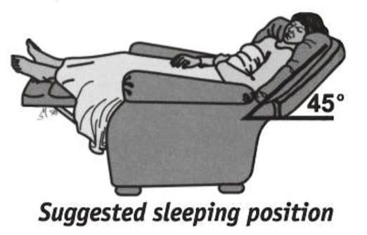 Sleep semi-recumbent for the next two nights. This means sleeping with your head halfway between flat and upright, at a 45-degree angle. This is most easily done by sleeping in a recliner chair or by sleeping with pillows appropriately arranged on a couch.