Relieve: A small muscle located deep in the buttocks, the piriformis muscle performs the essential function of rotating the leg outwards. Piriformis Syndrome is a painful condition that occurs when the piriformis muscle is tight and intrudes upon the sciatic nerve in the buttocks. Causing pain and tenderness and sometimes numbness in the buttocks, piriformis syndrome pain may also radiate down the sufferer’s leg, and in some cases, even into the calf.
There are two commonly identifiable potential causes for piriformis syndrome. One is sitting for prolonged periods of time, which can cause tightening of the muscle. The second cause is an injury to the buttocks, either by a fall, an accident, or a sports injury. Trauma causes the piriformis muscle to swell and irritate the sciatic nerve.
Spasms can also cause piriformis syndrome, however, the underlying cause of the spasms frequently remains unknown.
Unfortunately, once an individual has suffered from piriformis syndrome, the condition can recur periodically, usually brought on by too much exercise or sitting for a long time without stretching.
Whatever the initial cause, piriformis syndrome treatment options are vital in relieving the painful symptoms and healing the condition.
Relieve:
These Four Treatment Options Are Frequently Used To Treat Piriformis Syndrome.
Medication. Over-the-counter or prescribed pain medicines, anti-inflammatory drugs, or muscle relaxers frequently serve to reduce the pain from piriformis syndrome. A doctor may also inject medicine directly into the piriformis muscle to improve the condition.
Heat. A common way to relax tight muscles is to apply heat. Piriformis syndrome sufferers may find relief from painful symptoms by periodically applying heat directly to the tender area.
Heat therapy may relieve the tightness of the muscle and promote healing of the entire area. However, it’s important to avoid treating the muscle with heat if there is a chance the muscle may be torn.
Exercise. The overall cause of the condition is a tight piriformis muscle, so it stands to reason a proper exercise regimen will loosen the muscle and alleviate the symptoms associated with piriformis syndrome. A doctor can prescribe the correct exercises to stretch and subsequently strengthen the muscles and the body’s other muscles. A strong body will reduce the chances of the issue recurring down the road.
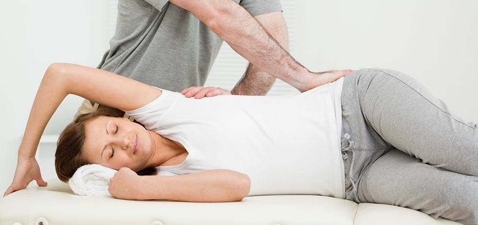
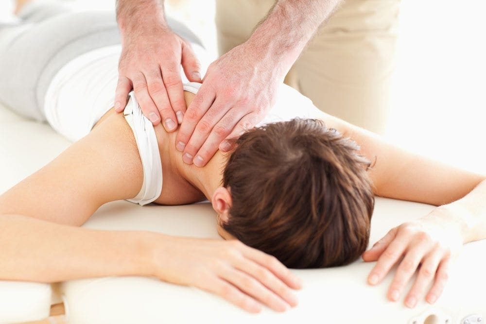
Hands on therapy. Used with other types of treatment or on their own, these types of therapies are popular because of their effectiveness, as well as the fact they are drug-free ways to gain relief from the pain. Massage is a commonly used therapy for piriformis syndrome, as it helps increase blood flow to the area. The massage therapist can manipulate the area to relieve the tightness of the muscle.
Another hands on therapy that produces positive results is chiropractic care.
Chiropractors view the body in its entirety, and will often treat other parts of the body, such as a foot or leg, in order to improve the condition of the piriformis muscle. They may also utilize a regimen of pelvic and spinal adjustments along with joint manipulation and stretching to loosen up the muscle and help heal the afflicted area.
As stated earlier, once the condition has been controlled and the area has healed, it’s vital to take precautions to avoid re-aggravating the area. Proper stretching before exercise, periodic breaks when sitting, and maintaining spinal and pelvic alignment will increase an individual’s chances of living free of the pain of piriformis syndrome in the future.
Treating Sciatica
If you have a question about how chiropractic care can help with the symptoms of piriformis syndrome, or other health conditions, contact us today.
This article is copyrighted by Blogging Chiros LLC for its Doctor of Chiropractic members and may not be copied or duplicated in any manner including printed or electronic media, regardless of whether for a fee or gratis without the prior written permission of Blogging Chiros, LLC.
The importance of Magnetic Resonance Imaging to evaluate the integrity of the lumbar posterior ligament complex post trauma.
Abstract: Posterior ligamentous complex(PLC), consisting of the supraspinous ligament, interspinous ligament, ligamentum flavum, and the facet joint capsules is thought to contribute significantly to the stability of the lumbar spine. There has been much debate on whether Magnetic Resonance Imaging(MRI) is specific and sensitive in diagnosing pathology to the PLC. The objective is to determine the necessity of MRI imaging for evaluating the integrity of the lumbar posterior ligament complex post trauma.
Key Words: Magnetic Resonance Imaging(MRI), interspinous ligament, posterior ligament complex, low back pain, ligament laxity, electromyography, impairment rating
A 41-year-old male, presented to my office for an examination with complaints of low back pain with numbness, tingling and weakness into the left lower extremity after he was the restraint driver in a motor vehicle collision approximately three and a half months� post trauma.�He�rated the pain as a�3/10 on a visual analog scale with 10/10 being the worst and the pain and noted the pain as being�present most of the time.� He stated that he was on pain killers daily and this helped manage his daily activities. Without pain killers his pain levels are rated 8/10 being present most of the time. The pain killers stated by the patient are Oxycodone and Naproxen. He�reported that the pain would be aggravated by activities which required excessive standing, repetitive bending, and lifting. He further noted that in the morning the pain was increased and his left leg would be numb and weak for about the first hour.
The patient stated that his care to date had been managed by a pain management clinic and that he had minimal improvement with treatment which has included physical therapy and massage therapy. He reported the pain clinic next recommended steroid injections which he refused. He states there has been was no imaging ordered and that an Electromyography(EMG) had been performed. He was told the test was negative for pathology.
Prior History: No significant medical history was reported. Clinical Findings:�The patient is 6�0� and weighs 210 lbs.
Physical Exam Findings:
Cervical Spine:
Cervical spine range of motion is full and unrestricted. Maximum cervical compression is negative. Motor and other regional sensory exam are unremarkable at this time.
Thoracic Spine:
Palpation of the thoracic spine region reveals taught and tender fibers in the area of the bilateral upper and mid thoracic musculature. Thoracic spine range of motion is restricted in flexion, extension, bilateral lateral flexion, and bilateral rotation. Regional motor and sensory exam are unremarkable at this time.
Lumbar Spine:
Palpation of the lumbosacral spine region reveals taught and tender fibers in the area of the lumbar paraspinal musculature. Lumbar spine range of motion is limited in flexion, extension, bilateral lateral flexion and bilateral rotation. Extension restriction is due to pain and spasm. Straight leg raise causes pain at approximately 50 degrees when testing either side in the left low back. There is no radicular symptomatology down the leg. Kemp�s maneuver recreates pain in the L4 region on the left. No radicular symptoms are noted. The patient is able to heel and toe walk. Regional motor and sensory exam is unremarkable at this time other than L4, L5 and S1 dermatomes having decreased sensation with light touch.
Muscle testing of the upper and lower extremities was tested at a 5/5 with the exception of the left quadricep tested at a 4/5.� The patient�s deep tendon reflexes of the upper and lower extremities were tested including triceps, biceps, brachioradialis, patella, and Achilles and all were tested at 2+ bilaterally except the left patellar reflex was 1+.
RANGES OF MOTION EVALUATION
All range of motions are based on the�American Medical Association�s Guides to the Evaluation of Permanent Impairment, 5th�Edition1�and performed by a dual inclinometer for the lumbar spine.
�� Range of Motion������Normal�������� Examination�������� % Deficit
Flexion
60
48
20
Extension
25
12
52
Left Lateral Flexion
25
16
36
Right Lateral Flexion
25
18
28
An MRI was ordered to rule out gross pathology.
Imaging:
�
A lumbar MRI reveals;
1)��� Mild disc bulges at T11-T12, T12-L1, L1-L2 and L5-S1
2)��� Low disc signals indicative of disc desiccation at T11-T12, T12-L1, L1-L2, L2-L3, L3-L4 and L4-L5
3)��� Retrolisthesis of 2mm at L3-L4
4)��� Mild ligamentous hypertrophy at L1-L2, L2-L3, L3-L4, L4-L5 and L5-S1
5)��� L4-L5 has a Grade 1-2 tear of the interspinous ligament with mild inflammation
6)��� L5-S1 has a Grade 1 interspinous ligament tear with mild inflammation
After reviewing the MRI I ordered lumbar x-rays to rule out ligament laxity.
X-RAY STUDIES
Lumbar x-rays reveal the following:
1)��� Left lateral tilt
2)��� Retrolisthesis at L1 of 3mm
3)��� Retrolisthesis at L2 of 3mm
4)��� Combined excessive translation of 4mm of L1 during flexion-extension
5)��� Combined excessive translation of 4mm of L2 during flexion-extension
6)��� Excessive translation of L3 in extension posteriorly of 2.5mm
7)��� Decreased disc space at L5-S1
Chiropractic care was initiated. The patient was placed on an initial care plan of 2-3x/week for 3 months and then a recommended break in care for one month so the patient could be evaluated for permanency while he was not care dependent.
At maximum medical improvement, he had continued low back pain rated 4/10, continued numbness and tingling into his left leg and left quadricep weakness rated 4/5. He does not need pain killers for pain management anymore. He continues chiropractic care every two weeks to manage his symptoms.
Conclusion:
In this specific case, pathology to the posterior ligament complex diagnosed on MRI lead to the x-ray finding of excessive translation at L1-L2 and L2-L3. The patient was given a permanent impairment rating of 22% based on my interpretation of the American Medical Association�s Guides to the Evaluation of Permanent Impairment, 5th�Edition1. The interspinous ligament tears at the L4-L5 and L5-S1 level would not have been diagnosed without the MRI.
There has been much debate on whether MRI imaging has a role in evaluating lumbar PLC. MRI is a powerful diagnostic tool that can provide important clinical information regarding the condition of the PLC. Useful sequences for spinal MRI in trauma include sagittal and axial T1-weighted images, T2-weighted FSE, fat-saturated T2-weighted FSE, and STIR sequences to highlight bone edema.2�Ligamentous injuries are best identified on T2-weighted images with fat saturation because the ligaments are thin and bonded on either side by fat, which can appear as hyperintense on both T1 and T2 images.3�T1-weighted images are inadequate in isolation for identifying ligamentous injuries.4�
The diagnostic accuracy for MRI was reported for both supraspinous ligament and interspinous ligament injury with a sensitivity of 89.4% and 98.5%, respectively, and a specificity of 92.3% and 87.2% in 35 patients.5
For patients with persistent symptoms after trauma an MRI may be indicated to evaluate posterior ligamentous complex integrity.
Competing Interests:� There are no competing interests in the writing of this case report.
De-Identification: All of the patient�s data has been removed from this case.
References:
1. Cocchiarella L., Anderson G. Guides to the Evaluation of Permanent Impairment, 5th Edition, Chicago IL, 2001 AMA Press.
2. Cohen, W.A., Giauque, A.P., Hallam, D.K., Linnau, K.F. and Mann, F.A., 2003. Evidence-based approach to use of MR imaging in acute spinal trauma.�European journal of radiology,�48(1), pp.49-60.
3. Terk, M.R., Hume-Neal, M., Fraipont, M., Ahmadi, J. and Colletti, P.M., 1997. Injury of the posterior ligament complex in patients with acute spinal trauma: evaluation by MR imaging.�AJR. American journal of roentgenology,�168(6), pp.1481-1486.
4. Saifuddin, A., Green, R. and White, J., 2003. Magnetic resonance imaging of the cervical ligaments in the absence of trauma.�Spine,�28(15), pp.1686-1691.
5. Haba H, Taneichi H, Kotani Y, et al. Diagnostic accuracy of magnetic resonance imaging for detecting posterior ligamentous complex injury associated with thoracic and lumbar fractures.�J Neurosurg. 2003; 99(1 Suppl):20-26.
Chiropractor, Dr. Alexander Jimenez gives insight into the relevant anatomy and functional biomechanics of the piriformis muscle, highlights the role it plays in musculoskeletal dysfunction and looks at management options in cases of muscle dysfunction.
The piriformis muscle (PM) is well-known in the fraternity of sports medicine as a significant muscle in the posterior hip. It is a muscle that has a role in controlling hip joint rotation and abduction, and it is also a muscle made famous due to its �inversion of action� in rotation. Furthermore, the PM also grabs attention due to its role in the contentious �piriformis syndrome�, a condition implicated as a potential source of pain and dysfunction, not only in the general population but in athletes as well.
Relevant Anatomy
The name piriformis was first coined by Belgian Anatomist Adrian Spigelius in the early 17th century. Its name is derived from the Latin word �pirum� meaning �pear� and �forma� meaning �shape� � ie a pear shaped muscle (see Figure 1)(1).
The PM originates on the anterior surface of the sacrum and is anchored to it by three fleshy attachments between the first, second, third and fourth anterior sacral foramina(2). Occasionally its origin may be so broad that it joins the capsule of the sacroiliac joint above and with the sacrotuberous and/or sacrospinous�ligament below(3,4).
PM is a thick and bulky muscle, and as it passes out of the pelvis through the greater sciatic foramen, it divides the foramen into the suprapiriform and infra-piriform foramina(5). As it courses antero-laterally through the greater sciatic foramen, it tapers out to form a tendon that is attached to the superior-medial surface of the greater trochanter, commonly blending with the common tendon of the obturator internus and gemelli muscles(6).
The nerves and blood vessels in the suprapiriform foramen are the superior gluteal nerve and vessels, and in the infra- piriforma fossa are the inferior gluteal nerves and vessels and the sciatic nerve (SN)(5). Due to its large volume in the greater sciatic foramen, it has the potential to compress the numerous vessels and nerves that exit the pelvis.
PM is closely associated with the other short hip rotators that lie inferior such as the superior gemellus, obturator internus, inferior gemellus and obturator externus(2). The primary difference between the PM and other short rotators is the relationship to the SN. The PM passes posterior to the�nerve whereas the other otators pass anterior (see figure 2).
Variants
A few anatomical variants have been found with the PM:
1. Additional medial attachments to the first and fifth sacral vertebrae and to the coccyx(7).
2. The tendon may fuse with the gluteus medius or minimus above, or superior gemellus below(7).
3. In less than 20% of cases it is divided into two distinct portions through which part or all of the sciatic nerve may pass(7).
4. It may blend with the posterior hip joint capsule as a conjoined tendon with the obturator internus(8).
5. The distal attachment of the PM has shown to vary in dimensions and position on the supero-medial surface of the greater trochanter. It can span a distance of between 25-64% of the anterior-posterior length on the greater trochanter, with 57% attaching more anterior and 43% more posterior(9).
6. Pine et al (2011) studied the insertion point extensively and found that four types of insertion existed and these were classified based on the relationship to the obturator internus(10). The variability in position and breadth of the distal attachment of the PM muscle may influence the validity of the concept known as �inversion of action� (see below).
The other hotly debated issue is the relationship between the PM and the SN. The conclusion is that there are several anatomical variations of the PM and its SN relationship. The sub-types of this variation include(11-13):
Type 1 (A below). Typical pear shape muscle with the nerve running anteriorly and inferiorly to this (in 70%-85% of cases).
Type 2 (B below). The PM is divided into two parts with the common peroneal nerve running between the two parts and the tibial nerve running anterior and below (found in 10-20% of cases).
Type 3 (C below). The peroneal portion loops over the top of the muscle and the tibial portion is below (found in 2-3% of cases).
Type 4 (D below). Undivided nerve passing through the PM (occurs in about 1% of cases).
It is also believed that two other very uncommon variations occur (see E and F below).
Type A is the most common variation, showing the SN passing below the PM
Functional Considerations
The primary functional roles of the PM are;
1. Hip external rotation(15).
2. Abductor at 90 degrees of hip flexion(15).
3. In weight-bearing, the PM restrains the femoral internal rotation during stance phase of walking and running(2).
4. Assists the short hip rotators in compressing the hip joint and stabilising the joint(6).
5. As it can exert an oblique force on the sacrum, it may produce a strong rotary shearing force on the sacroiliac joint (SIJ). This would displace the ipsilateral base of the sacrum anteriorly (forward) and the apex of the sacrum posteriorly(16).
As the PM is the most posterior of the hip external rotators due to its attachment on the anterior surface of the sacrum, it has the greatest leverage to exert a rotation effect on the hip joint. It is often seen clinically that the PM appears to be tight and hypertonic, while the other short hip�rotators that are closer to the axis of rotation become inhibited and hypotonic.
Inversion Of Action
The most contentious issue related to the function of the PM is its �reversal-of- function role� or �inversion of action� role. Many authors have suggested that as the hip approaches angles of 60-90 degrees and greater, the tendon of the PM shifts superiorly on the greater trochanter. As a result, its line of pull renders it ineffective as a hip external rotator; however it does contribute to internal hip rotation. Therefore it reverses its rotation role at high hip flexion angles(15,17,18).
The function of the PM at varying joint angles is an important consideration for the clinician who is evaluating and treating �piriformis syndrome�. Often it has been advocated to stretch the hip into flexion, adduction and external rotation to stretch the PM over the glutes by utilising the �reversal of function� concept.
However, more recent anatomical dissection studies have shown that the attachment of the PM onto the greater trochanter can be variable and in some instances it may insert in a position whereby it is unable to reverse its function, for example in a more posteriorly placed attachment(19). Therefore, stretching the PM into external rotation when the hip is flexed beyond 90 degrees � based upon reversal of function � would be ineffective as a treatment or misleading as an examination technique(19)
MSK Dysfunction & PM Syndrome
Many decades ago, the role that the PM played in creating sciatic-like symptoms was first suggested by Yeoman (1928) when it was considered that some cases of sciatica may originate outside the spine(20). This was supported soon after when Freiberg and Vinkle (1934) successfully cured sciatica by surgically dividing the PM(21). Based on cadaver dissections Beaton and Anson (1938) gave the hypothesis that the spasm of the PM could be responsible for the irritation of the SN(12).
The term �piriformis syndrome� was first coined by Robinson in 1947(22) and was applied to sciatica thought to be caused by an abnormality in the PM (usually traumatic in origin) with emphasis on ruling out more common causes of sciatica such as nerve root impingement from a disc protrusion. It soon became an accepted clinical entity � but with no consensus about the exact clinical signs and diagnostic tests to differentiate it from other sources of sciatica(23,24).
Piriformis syndrome can be defined as a clinical entity whereby the interaction�between the PM and SN may irritate the SN and produce posterior hip pain with distal referral down the posterior thigh, imitating �true sciatica�. Isolating the dysfunction to this region usually follows exclusion of the more common causes of buttock pain and sciatica.
More specifically, complaints of buttock pain with distal referral of symptoms are not unique to the PM. Similar symptoms are prevalent with the more clinically evident lower back pain syndromes and pelvic dysfunctions. Thus, a thorough evaluation of these regions must be performed to exclude underlying pathology(4). It has been suggested that piriformis syndrome� is responsible for 5-6% of cases of sciatica(25,26). In the majority of cases, it occurs in middle-aged patients (mean age 38 yr)(27) and is more prevalent in women(28).
Pathogenesis Of Piriformis Syndrome (PS)
PS may be caused by or relate to three primary causative factors;
1. Referred pain due to myofascial trigger points (see Figure 4)(2,28-30). Examples include tight and shortened muscle fibres precipitated by muscle overuse such as squat and lunge movements in external rotation, or�direct trauma(16). This increases the girth of the PM during contraction, and this may the source of the compression/entrapment.
2. Entrapment of the nerve against the greater sciatic foramen as it passes through the infrapiriform fossa, or within a variant PM(29,31).
3. SIJ dysfunction causing PM spasm(29,32).
Janvokic (2013) has presented a number of causative factors in PS(29);
1. Gluteal trauma in the sacroiliac or gluteal areas.
2. Anatomical variations.
3. Myofascial trigger points.
4. Hypertrophy of the PM or spasm of the PM.
5. Secondary to spinal surgery such as laminectomy.
6. Space occupying lesions such as neoplasm, bursitis, abscess, myositis. 7. Intragluteal injections.
8. Femoral nailing.
Symptoms
Typical symptoms reported in piriformis syndrome include:
A tight or cramping sensation in the buttock and/or hamstring(33).
Gluteal pain (in 98% of cases)(34).
Calf pain (in 59% of cases)(34).
Aggravation through sitting and squatting(35), especially if the trunk is inclined forward or the leg is crossed over the unaffected leg(36).
Possible peripheral nerve signs such as pain and paraesthesia in the back, groin, buttocks, perineum, back of the thigh (in 82% of cases)(34).
Physical Findings & Examinations
Palpable spasm in and around the PM and obturator internus and external tenderness over the greater sciatic notch (in 59-92% of cases)(34,35). The patient is placed in the Sims position. The piriformis line overlies the superior border of the PM and extends from immediately above the greater trochanter to the cephalic border of the greater sciatic foramen at the sacrum. The line is divided into equal thirds. The fully rendered thumb presses on the point of maximum trigger-point tenderness, which is usually found just lateral to the junction of the middle and last thirds of the line.
Hip flexion with active external rotation or passive internal rotation may exacerbate the symptoms(36).
Positive SLR that is less than 15 degrees the normal side(37).
Positive Freiberg�s sign (in 32-63% of cases)(34,35). This test involves reproducing pain on passive forced internal rotation of the hip in the supine position � thought to result from passive stretching of the PM and pressure on the sciatic nerve at the sacrospinous ligament.
Pacers sign (in 30-74% of cases)(34,35). This test involves reproducing pain and weakness on resisted abduction and external rotation of the thigh in a sitting position.
Pain in a FAIR position(34). This involves the reproduction of pain when the leg is held in flexion, adduction and internal rotation.
An accentuated lumbar lordosis and hip flexor tightness predisposes one to increased compression of the sciatic nerve against the sciatic notch by a shortened piriformis(38).
Electro-diagnostic tests may prove useful (see below).
Investigations
Conventional imaging such as X-ray, CT scan and MRI tend to be ineffective in diagnosing piriformis syndrome.
However, some value may exist in electro- diagnostic testing.
It is beyond the scope of this paper to discuss in detail the process of electro- diagnostic testing; the reader is directed to references for more a more detailed description of how these tests are administered(35,36,39). However the purpose of these tests is to find conduction faults in the SN. Findings such as long-latency potentials (for example the H reflex of the tibial nerve and/or peroneal nerve) may be normal at rest but become delayed in positions where the hip external rotators are tightened(27,36,39).
It is accepted that the tibial division of the SN is usually spared, the inferior gluteal nerve that supplies the gluteus maximus may be affected and the muscle becomes atrophied(40). However testing of the peroneal nerve may provide more conclusive results as is more likely to be the�impinged portion of the SN. The H-wave may become extinct during the painful position of forced adduction-internal rotation of the affected leg(36).
The �Myth� Of Piriformis Syndrome
Stewart 2003 argues that piriformis syndrome is an often over-used term to describe any non-specific gluteal tenderness with radiating leg pain(41). He argues that only in rare cases is the PM implicated in nerve compression of the SN to truly qualify as a piriformis syndrome. He cites only limited evidence and cases where the diagnosis of piriformis syndrome can be made.
1. Compressive damage to the SN by the PM. Stewart cites studies whereby in few isolated studies, the SN was seen to be compressed by the PM in instances such as hypertrophy of the muscle,�usual anatomical anomalies such as a bifid PM, and due to compression by fibrous bands.
2. Trauma and scarring to the PM leading to SN involvement; it is possible that rare cases of true Piriformis Syndrome have been caused by direct heavy trauma to the PM due to a blunt trauma to the muscle. This is termed �post- traumatic PS�.
McCory (2001) supports this argument by stating that it is more likely that (given the anatomical relationship of the PM to the various nerves in the deep gluteal region) the buttock pain represents entrapment of the gluteal nerves, and the hamstring pain entrapment of the posterior cutaneous nerve of the thigh, rather than the SN alone(33). This would explain the clinically observed phenomenon in the absence of distal sciatic neurological signs. Whether the PM is the cause of the compression has not been clearly established. It is possible that the obturator internus/gemelli complex is an alternative cause of neural compression. He suggests using the term �deep gluteal syndrome� rather than piriformis syndrome.
Treatment
When it is believed that a piriformis syndrome exists and the clinician feels that a diagnosis has been made, the treatment will usually depend on the suspected cause. If the PM is tight and in spasm then initially conservative treatment will focus on stretching and massaging the tight muscle to remove the PM as being the source of the pain. If this fails, then the following have been suggested and may be attempted(23,36):
Local anaesthetic block � usually performed by anaesthesiologists who have expertise in pain management and in performing nerve blocks.
Steroid injections into the PM.
Botulinum toxin injections into the PM.
Surgical Neurolysis.
Here, we will focus on therapist-directed interventions such as stretching of the PM and direct trigger point massage. It has always been advocated that PM stretches are done in positions of hip flexion greater than 90 degrees, adduction and external rotation to utilize the �inversion of action� effect of the PM to isolate the stretch to this muscle independent of the other hip external rotators.
However, recent evidence from Waldner (2015) using ultrasound investigation discovered that there was no interaction between hip flexion angle and the thickness of the PM tendon in both internal and lateral hip rotation stretching � suggesting that the PM does not invert its action(19). Furthermore, Pine et al (2011)(9) and Fabrizio et al (2011)(10) in their cadaveric studies found that the PM insertion is a lot more complex and varied than first thought. It is possible that the PM may invert its action only in some subjects but not others.
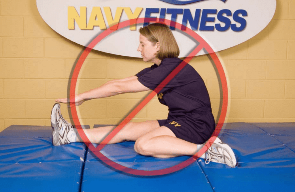
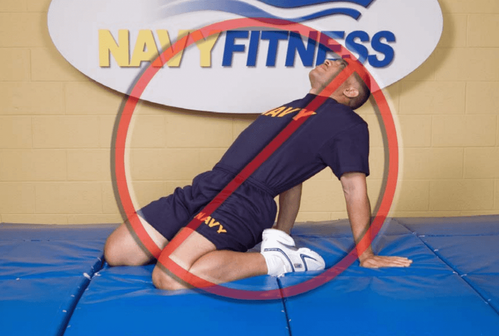
Therefore, due to the disagreements and confusions over the �inversion of action� concept, it is recommended that the clinician �covers all bases� and performs two variations of a PM stretch � stretches in flexion, adduction and external rotation and stretches in flexion, adduction and internal rotation. Examples of these stretches are given in figures 5-7 below.
Trigger Points & Massage
(see Figure 8)
The best approach to palpate the PM trigger points is in the position suggested by Travel and Simons(2) and this is shown below. In this position, the clinician can feel for the deep PM trigger points and apply a sustained pressure to alleviate the trigger�points � and also apply a flush massage to the muscle in this position.�In this position the large gluteus maximus is relaxed and it is easier to feel the deeper PM.
Summary
The PM is a deep posterior hip muscle that is closely related anatomically to both the sacroiliac joint and the sciatic nerve. It is a hip external rotator at hip flexion angles of neutral to 60 degrees of hip flexion, an abductor when in flexion and also contributes to hip extension.
It has been previously accepted that the PM will �invert its action� or �reverse its function� after 60 degrees of flexion to become a hip internal rotator. However, recent ultrasound and cadaveric studies has found conflicting evidence that this �inversion of action� may in fact not exist.
PM is a muscle that is a dominant hip rotator and stabiliser, and thus has a tendency to shorten and become hypertonic. Therefore, stretching and massage techniques are best utilised to reduce the tone through the muscle. Furthermore, it has also been implicated in compression and irritation of the sciatic nerve � often referred to as piriformis syndrome�.
References
1. Contemp Orthop 6:92-96, 1983.
2. Simons et al (1999) Travell and Simons� Myofascial Pain and Dysfunction. Volume 1 Upper Half of the Body (2nd edition). Williams and Wilkins. Baltimore.
3. Anesthesiology; 98: 1442-8, 2003.
4. Joumal of Athletic Training 27(2); 102-110, 1996.
5. Journal of Clinical and Diagnostic Research. Mar, Vol-8(3): 96-97, 2014.
6. Clemente CD: Gray�s Anatomy of the Human Body, American Ed. 30. Lea & Febiger, Philadelphia, 1985 (pp. 568-571).
7. Med J Malaysia 36:227-229, 1981.
8. J Bone Joint Surg;92-B(9):1317-1324, 2010.
9. J Ortho Sports Phys Ther. 2011;41(1):A84, 2011.
10. Clin Anat;24:70-76, 2011.
11. Med Sci Monit, 2015; 21: 3760-3768, 2015.
12. J Bone Joint Surg Am 1938, 20:686-688,1938.
13. Journal of Clinical and Diagnostic Research. 2014 Aug, Vol-8(8): 7-9, 2014.
14. Peng PH. Piriformis syndrome. In: Peng PH, editor. Ultrasound for Pain Medicine Intervention: A Practical Guide. Volume 2. Pelvic Pain. Philip Peng Educational Series. 1st ed. iBook, CA: Apple Inc.; 2013 .
15. Kapandji IA. The Physiology of Joints. 2nd ed. London: Churchill Livingstone; 1970: 68.
16. J Am Osteopath Assoc 73:799-80 7,1974.
17. J Biomechanics. 1999;32:493-50, 1999.
18. Phys Therap. 66(3):351-361, 1986.
19. Journal of Student Physical Therapy Research. 8(4), Article 2 110-122, 2015.
20. Lancet. 212: 1119-23, 1928.
21. J Bone Joint Surg Am 16:126�136, 1934.
22. Am J Surg 1947, 73:356-358, 1947.
23. J Neurol Sci; 39: 577�83, 2012.
24. Orthop Clin North Am; 35: 65-71, 2004
25. Arch Phys Med Rehabil; 83: 295-301,2002.
26. Arch Neurol. 63: 1469�72, 2006.
27. J Bone Joint Surg Am; 81: 941-9,1999.
28. Postgrad Med 58:107-113, 1975.
29. Can J Anesth/J Can Anesth;60:1003�1012, 2013.
30. Arch Phys Med Rehabil 69:784, 1988.
31. Muscle Nerve; 40: 10-8, 2009.
32. J Orthop Sports Phys Ther;40(2):103-111, 2010.
33. Br J Sports Med;35:209�211, 2001.
34. Man Ther 2006; 10: 159-69, 2006.
35. Eur Spine J. 19:2095�2109, 2010.
36. Journal of Orthopaedic Surgery and Research, 5:3, 2010.
37. Muscle & Nerve. November. 646-649, 2003.
38. Kopell H, Thomnpson W. Peripheral Entrapment Neuropathies. Huntington, NY: Krieger, 1975:66.
39. Arch Phys Med Rehabil;73:359�64, 1992.
40. J Bone and Joint Surg, 74-A:1553-1559, 1992.
41. Muscle & Nerve. November. 644-646, 2003
Corticosteroid injections are widely used to aid injury rehabilitation but we still understand very little about their mechanism. Chiropractor, Dr. Alexander Jimenez examines the current thinking and discusses how this potentially impacts treatment options…
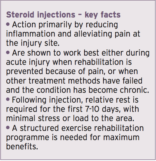
Corticosteroids are used for their anti- inflammatory and pain reducing effects. They can also reduce muscle spasms and influence local tissue metabolism for faster healing. Injection therapy is now widely available from specially trained general practitioners, physiotherapists and consultants, and can be offered for a wide range of clinical conditions. Because of this wide availability and the growing desire for injury �quick fixes�, it is important that they are used correctly and the full consequences are understood prior to injection.
The main indications for corticosteroid injection use are(1):
Acute and chronic bursitis
Acute capsulitis (tight joint capsule)
Chronic tendinopathy
Inflammatory arthritis
Chronic ligament sprains
Steroid injections of hydrocortisone are a synthetic form of a naturally produced hormone within the body called cortisol. Cortisol is important for regulating carbohydrate, protein and fat metabolism. It is also involved in metabolic responses in times of stress such as emotional problems, trauma, and infection, where levels of inflammation are elevated. Steroid injections work on the immune system by blocking the production of chemicals that activate the inflammatory reactions, therefore reducing inflammation and pain within injury locations.
Steroid injections can be directed into a joint, muscle, tendon, bursa, or a space around these structures. Figure one shows an injection aiming for the bursa within the shoulder joint. This is often a source of irritation and causes impingement when the shoulder moves. The location will depend on what tissue is causing the symptoms. When injected locally to the specific structure, the effects are primarily only produced there and widespread detrimental effects are minimal(2).
When To Use
Identifying the correct time to issue a steroid injection following injury requires careful consideration. The mechanical status of the tissue is important because this will vary depending on the stage of healing and therefore the effectiveness of the injection will also vary.
Figure 2 shows the different stages that a tendon can progress through following trauma. This is equally applicable to muscles, fascia, and other tissues too. A reactive tendinopathy (tendon degeneration/damage) will present shortly after injury/trauma/stress/ excessive loading, and will display acute swelling and inflammation. The initial care should be 2-3 weeks of rest, analgesia, ice application and gentle physiotherapy. If symptoms have not significantly improved after this period, then the introduction of a corticosteroid injection is appropriate for providing symptomatic relief by reducing inflammation and eliminating the occurrence of further damage because mechanical normality will be quickly restored(3).
If the tendon continues to be placed under excessive load, swelling and inflammation will remain or escalate, and continuous loading will eventually cause micro trauma and further tendon degeneration. If this is prolonged for long enough then the tendon will fail structurally(4).
The use of corticosteroids here is questionable because there is unlikely to be inflammation present to combat, and the injection alone will not repair this physical damage. Injection treatment at this stage may only be indicated if the athlete is in too much pain to participate in any significant rehabilitation. The symptomatic relief the injection may bring at this point could allow exercises to be performed, which can help accelerate the repair of physical damage. Ultimately, physical exercise is a key component in recovery following corticosteroid injections.
Impact On Treatment & Performance
For the best outcome, post-injection care � particularly with respect to timing � is important. Relative rest is recommended for the first two weeks post-injection. During this first two weeks the tissues are weakened and their failing strengths are reduced by up to 35%; this means the strength at which they would fail (tear) is much lower and more susceptible to rupturing(8).
By six weeks the bio-mechanical integrity is reestablished and the tissues are deemed �normal� again, with increased strength and function(8). Benefits are optimal within this 6-week period and often short-lived; therefore the athlete must comply strictly to a rehabilitation program to gradually load the tissues and ensure the correct load is applied during this period(9). Research has also shown that at twelve weeks post-injection�there is little significance in the difference between those who received a steroid injection and those who focused on exercise therapy alone, suggesting this early symptom relief should be used to enhance rehabilitation(10). If loading is accelerated in the early stages the athlete risks re-aggravation of the injury, delayed healing, further weakening and thus rupture.
If this rehabilitation protocol is followed, the athlete will likely maximise their outcome. They can return to training, and with the severity of their symptoms reduced, this can allow progression to the next stage of training. If the injury is severe enough that surgery may be considered within three months, a steroid injection should not be performed as this can affect the success of the surgery.
Evidence For Sports Injuries
Here we will consider some of the more common sports injuries and summarize what the current evidence regarding steroid injection suggests.
Shoulders
Injection therapy is indicated in subacromial impingement or bursitis (as in Figure 3 below) to allow the inflammation reduction and restoration of normal movement. It is also indicated in rotator cuff pathology where the tendons are again inflamed, but also damaged and unable to undergo exercise therapy. Shoulder injections are shown to produce early improvements in pain and function with a high level of patient satisfaction(10). Symptoms are similar to those without injection at 12 weeks however, suggesting physical therapy is also important(10). Injection is not appropriate for shoulder instability as it can make the joint more unstable. Exercise therapy alone is recommended for this condition.
Hip Pain
Two soft tissue conditions that benefit the most from injection are piriformis syndrome (muscle tightness running deep to the buttock muscles), and greater trochanter pain syndrome (affecting the bursa surrounding the hip joint, or the gluteal tendons that are all in close proximity to the lateral hip)(11). Injection success is reported to be approximately 60-100% if the diagnosis is accurate and the correct protocols are adhered to(12). Other regions such as the adductor and hamstring tendons can also be treated for tendinitis or groin pains. However, injections into these�regions are deep and painful, and require extensive rest afterwards.
Knee Pain
Knee joint injections for arthritic conditions are most commonly used, with injection to the soft tissues much less common due to the complex diagnosis, and risk of detrimental side effects. The various bursa around the knee, the iliotibial band, and quadriceps and patellar tendons have all been shown to significantly benefit in the short-term; however accurate location is essential to ensure the tendon itself is not penetrated � only the surrounding regions(13).
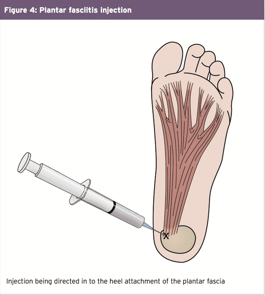
Plantar Fasciitis
This is a painful injection to receive, and pain can last for well over one week post- injection (see figure 4). There is an approximate 2-4% risk that the fascia can rupture. In addition, there�s a risk of local nerve damage and wasting of the fat pad within the heel. Studies have demonstrated that at 4 weeks post-injection pain and thickness of the injured plantar fascia are reduced and these benefits remain three months later, suggesting a good outcome if the risks are avoided(14).
Many people have back pain so often that it starts to become a daily struggle. You don�t have to put up with pain every day.
Share Free Ebook!
TIP! One interesting fact is that good chiropractic care can actually strengthen your immune system. Spine issues can cause problems with the nervous system, which is linked to the immune system.
Many doctors work with alternative therapy. This makes it important that your insurance policy and see what back care therapies are covered. This can improve your health even more effective.
Pay attention to your sleep in order to fix back-related problems.Put a pillow underneath your head and shoulders. Place rolled-up towels underneath your neck and knees to help support the body�s curves. You should also have a mattress that is comfy.
TIP! Don�t think you�re going to get all the care you need from just a single visit to a�chiropractor. While it may make you feel better right away, it will take many sessions to see great improvements.
You should not expect one treatment at the chiropractor to solve all your pain problem. You will probably see some immediate relief; however, but regular visits are required for lasting relief. Stick with whatever regiment your chiropractor�recommends. If you fail to do this, you will end up disappointed with the results.
There are lots of solid reasons as to why a person ought to visit a chiropractor. If your back or neck hurt, you should find a reputable chiropractor right away. Your body will not run correctly if your skeletal structure.
TIP! Don�t be afraid to ask your chiropractor�about discounts; they may offer one the more frequently you visit. Chiropractic treatment usually involves multiple visits.
Ask your chiropractor if there are frequency discounts in their office. Chiropractic treatment usually requires a series of office visits. You may need to visit several times weekly for months to come. It can quickly become quite costly. The doctor�s office may have some sort of discount if you visit a lot so things don�t cost you so much more affordable.
Ask you doctor to recommend a chiropractor. Even if you don�t need a referral, your doctor can suggest a good chiropractor.
TIP! Ask your regular physician if they can refer you to a quality�chiropractor. A referral may not be required, but it helps you find the most qualified, trusted professionals in your local area.
Make a wise decision when searching for a chiropractor. Most chiropractors are honest, but others cannot be trusted. There are actually some of people going to a chiropractor and feeling much worse afterwards. Make sure you�re doing research prior to choosing a chiropractor.
Check out the references of a chiropractor before scheduling an appointment with them. While lots of chiropractors are interested in their patients� health, there are some that attempt to extend treatments beyond what is actually needed. Look at reviews online and get recommendations from your regular doctor.
TIP! Before ever contacting a�chiropractor, ask for references from your doctor or physician. The majority of professional chiropractors are experts in their field who actually care about your health, but there are a few bad eggs in the bunch.
A cervical pillow or roll up a towel and position it beneath your neck when you sleep can really help. They let your head drop down while a regular pillow has your head being pushed forward.
Blood Pressure
TIP! Is high blood pressure something you have? Studies show that vertebrae manipulation is more effective than blood pressure medications. Certain manipulations of the vertebrae can help get your blood pressure regular.
Is your blood pressure something you have? Studies show that vertebrae is as good as using two hypertension medications together. When the vertebrae are manipulated, the blood pressure can be regulated.
Chiropractic care can also help your immune system as well. Your nervous system can malfunction when the bones in your spine are misaligned. Because your nervous system controls tissue, cell and organ function, if it gets impacted it can make your health go wrong. Fixing the issue can get your immune system back to optimal performance.
TIP! Chiropractic care is not just for back and necks, it boosts your immune system as well. Bones that are out of alignment in the spine often interfere with your nervous system�s functioning.
Stay away from chiropractors that want to give you dietary supplements and other products. They are likely charlatans if they offer and cannot be trusted. Nutritionists and doctors are reliable sources for such advice.
It is easy to find a qualified chiropractor in the United States. Chiropractic care makes up the second largest health care profession. It is also happens to be the fastest. On top of that number, as many as 10,000 students or more are learning the practice themselves.
TIP! Do not carry a wallet inside your back pocket. Many men�carry a wallet in their back pocket and don�t understand how it affects their back.
Meet with the chiropractor before scheduling an appointment. A chiropractor can really improve your quality of living. A poorly qualified chiropractor can make matters so much worse. Find a chiropractor you feel comfortable. Be sure you talk with a chiropractor before scheduling treatment.
Avoid slumping when you are sitting or standing in a hunched position for long periods. This strains your back and will give you lower back pain and that�s going to hurt you stand straight. If sitting or standing hunched over is unavoidable, make sure you stretch well and periodically get up from your position.
TIP! Don�t work with a�chiropractor that�s going to try to place you on supplements or homeopathic products that can help to treat disease. If they are selling these items from their offices, they are not entirely trustworthy.
If you do a lot of sit-ups and crunches to build up your core strength, it�s time to find other core exercises, since these are two that often make things worse. The Yoga plank position is a good alternative and can help your back and core.
The thoracic spinal area of your spine is responsible for communication regarding digestion and other stomach functions. You can have problems with things like acid reflux or other irritations when the thoracic area is irritated. Chiropractic care can fix any alignment issues and prevent misalignment of this area to help heal the stomach.
TIP! When you carry around a wallet, don�t put it in the pocket on the back of your pants. You may not believe it, but doing that can strain your lower back.
Don�t be afraid to ask for assistance lifting heavy item. Ask someone else to help or use proper equipment. A dolly that�s good and secure can assist you in moving something. A dolly is a great investment if you are moving heavy things often.
To keep headache pain at bay, you may want to go to your chiropractor or a therapist that can massage the pain out of your body.Tightness in the pain. The temperomandibular joint (TMJ) can suffer from teeth in your sleep. A guard might keep this case.
TIP! Now you can confidently find the�chiropractor who is perfect for you. Many people in the United States and around the world seek chiropractic care today.
It is important to take your time when searching for a good chiropractor. There are tons of chiropractors out there. Once you find the ideal one, shop around for a good price. Call each one and ask for a quote. Make sure that their quote includes all of their services and they are quoting you doesn�t neglect to mention any hidden fees.
You may believe that your use of a smartphone is making your life better. It may actually be hurting your neck though. When you look at the screen, your neck is pulled down, which puts too much weight on the muscles. Use your phone at eye level to avoid this.
TIP! You can get back strain from standing for long periods of time. If you need to stand, do this with one foot on something low every so often to relieve the strain on your lower back.
Clearly, there really is no reason to endure ongoing back pain. You can help yourself through the situation. Try some of the advice here, and you can get relief quickly.
The purpose of this article is to provide an update of a previously published evidence-based practice guideline on chiropractic management of low back pain.
Methods
This project updated and combined 3 previous guidelines. A systematic review of articles published between October 2009 through February 2014 was conducted to update the literature published since the previous Council on Chiropractic Guidelines and Practice Parameters (CCGPP) guideline was developed. Articles with new relevant information were summarized and provided to the Delphi panel as background information along with the previous CCGPP guidelines. Delphi panelists who served on previous consensus projects and represented a broad sampling of jurisdictions and practice experience related to low back pain management were invited to participate. Thirty-seven panelists participated; 33 were doctors of chiropractic (DCs). In addition, public comment was sought by posting the consensus statements on the CCGPP Web site. The RAND-UCLA methodology was used to reach formal consensus.
Results
Consensus was reached after 1 round of revisions, with an additional round conducted to reach consensus on the changes that resulted from the public comment period. Most recommendations made in the original guidelines were unchanged after going through the consensus process.
Conclusions
The evidence supports that doctors of chiropractic are well suited to diagnose, treat, co-manage, and manage the treatment of patients with low back pain disorders.
Key Indexing Terms:
Chiropractic, Low Back Pain, Manipulation, Spinal, Guidelines
Early development of the chiropractic profession in the 1900s represented the application of accumulated wisdom and traditional practices.1, 2 As was the practice of medicine, philosophy and practice of chiropractic were informed to a large extent by an apprenticeship and clinical experiential model in a time predominantly absent of clinical trials and observational research.
The traditional chiropractic approach, in which a trial of natural and less invasive methods precedes aggressive therapies, has gained credibility. However, the chiropractic profession can gain wider acceptance in the role as the first point of contact health care provider to patients with low back disorders, particularly within integrated health care delivery systems, by embracing the scientific approach integral to evidence-based health care.3, 4, 5,6, 7 It is in this context that these guidelines were developed and are updated and revised.8, 9, 10, 11, 12
By today’s standards, it is the responsibility of a health profession to use scientific methods to conduct research and critically evaluate the evidence base for clinical methods used.13, 14 This scientific approach helps to ensure that best practices are emphasized.15 With respect to low back disorders, clinical experience suggests that some patients respond to different treatments. The availability of other clinical methods for conditions that are unresponsive to more evidence-informed approaches (primary nonresponders) introduces the opportunity for patients to achieve improved outcomes by alternative and personalized approaches that may be more attuned to individual differences that cannot be informed by typical clinical trials.16, 17, 18 To a large degree, variability in the selection of treatment methods among doctors of chiropractic (DCs) continues to exist, even though the large body of research on low back pain (LBP) has focused on the most commonly used manipulative methods.17, 19, 20
Although the weight of the evidence may favor the evidence referenced in a guideline for particular clinical methods, an individual patient may be best served in subsequent trials of care by treatment that is highly personalized to their own mechanical disorder, experience of pain and disability, as well as preference for a specific treatment approach. This is consistent with the 3 components of evidence-based practice: clinician experience and judgment, patient preferences and values, and the best available scientific evidence.3, 13
Doctors of chiropractic use methods that assist patients in self-management such as exercise, diet, and lifestyle modification to improve outcomes and their stabilization to avoid dependency on health care system resources.19, 21 They also recognize that a variety of health care providers play a critical role in the treatment and recovery process of patients at various stages, and that DCs should consult, refer patients, and co-manage patients with them when in the patient’s best interest.19
To facilitate best practices specific to the chiropractic management of patients with common, primarily musculoskeletal disorders, the profession established the Council on Chiropractic Guidelines and Practice Parameters (CCGPP) in 1995.6 The organization sponsored and/or participated in the development of a number of “best practices” recommendations on various conditions.21, 22, 23, 24, 25, 26, 27, 28, 29, 30, 31, 32 With respect to chiropractic management of LBP, a CCGPP team produced a literature synthesis8 which formed the basis of the first iteration of this guideline in 2008.9 In 2010, a new guideline focused on chronic spine-related pain was published,12 with a companion publication to both the 2008 and 2010 guidelines published in 2012, providing algorithms for chiropractic management of both acute and chronic pain.10 Guidelines should be updated regularly.33, 34 Therefore, this article provides the clinical practice guideline (CPG) based on an updated systematic literature review and extensive and robust consensus process.9, 10, 11, 12
Methods
This project was a guideline update based on current evidence and consensus of a multidisciplinary panel of experts in the conservative management of LBP. It has been recommended that, although periodic updates of guidelines are necessary, “partial updating often makes more sense than updating the whole CPG because topics and recommendations differ in terms of the need for updating.”33 Logan University Institutional Review Board determined that the project was exempt. We used Appraisal of Guidelines for Research & Evaluation (AGREE) in developing the guideline methodology.
Systematic Review
Between March 2014 through July 2014, we conducted a systematic review to update the literature published since the previous CCGPP guideline was developed. The search included articles that were published between October 2009 through February 2014. Our question was, “What is the effectiveness of chiropractic care including spinal manipulation for nonspecific low back pain?” Table 1 summarizes the eligibility criteria for the search.
Table 1
Eligibility Criteria for the Literature Search
Inclusion
Exclusion
Published between October 2009-February 2014
Case reports and case series
English language
Commentaries
Human participants
Conference proceedings
Age >17 y
In-patients
Manipulation
Letters
LBP
Narrative and qualitative reviews
Duration chronic (>3 mo)
Non–peer-reviewed publications
Patient outcomes reported
Pilot studies
Non-manipulation comparison group
Pregnancy-related LBP
RCTs, cohort studies, systematic reviews, and meta-analyses
Secondary analyses and descriptive studies
LBP, low back pain; RCT, randomized controlled trial.
Search Strategy
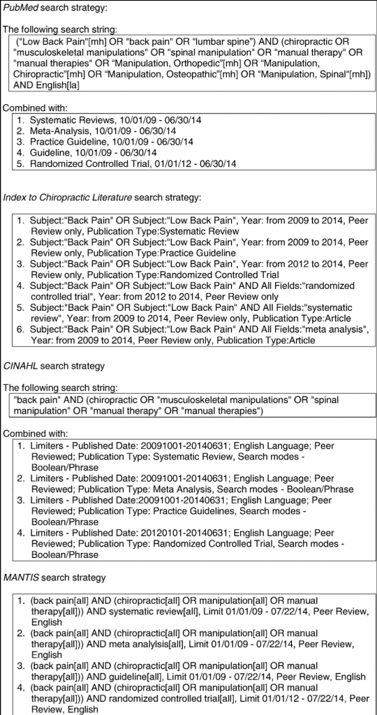
The following databases were included in the search: PubMed, Index to Chiropractic Literature, CINAHL, and MANTIS. Details of the strategy for each database are provided in Figure 1. Articles and abstracts were screened independently by 2 reviewers. Data were not further extracted.
Fig 1
Search strategies used in the literature search.
Evaluation of Articles
We evaluated articles using the Scottish Intercollegiate Guideline Network checklists (http://www.sign.ac.uk/methodology/checklists.html) for randomized controlled trials (RCTs) and systematic reviews/meta-analyses. For guidelines, the AGREE 2013 instrument35 was used. At least 2 of the 3 investigators conducting the review (CH, SW, MK) reviewed each article. If both reviewers rated the study as either high quality or acceptable, it was included for consideration; if both reviewers rated it as unacceptable, it was removed. For AGREE, we considered “unacceptable” to be a sum of <4. If there was disagreement between reviewers, a third also reviewed the article, and the majority rating was used.
Results of Literature Review
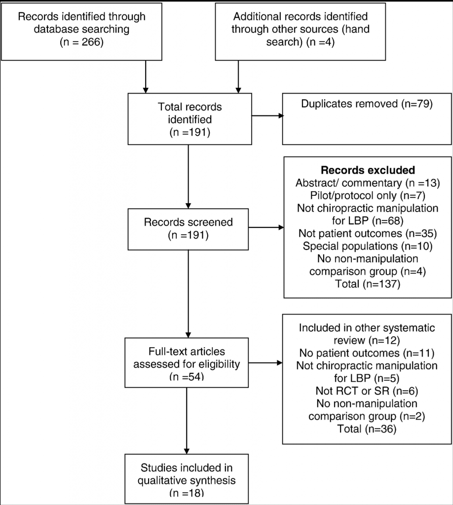
This search yielded 270 articles. Screening the articles for eligibility resulted in 18 articles included for evaluation, as detailed in Figure 2, using the Preferred Reporting Items for Systematic Reviews and Meta-Analyses flowchart.36
Fig 2
Flow diagram for literature search. LBP, low back pain; RCT, randomized controlled trial; SR, systematic reviews.
Of the 18 articles included after screening, 16 were retained as acceptable/high quality12, 17, 37, 38, 39, 40, 41, 42,43, 44, 45, 46, 47, 48, 49, 50 and 251, 52 (both systematic reviews) were excluded as being of unacceptable quality according to the Scottish Intercollegiate Guideline Network checklist. Those with new relevant information were summarized and provided to the Delphi panel as background information. Table 2 lists the articles by lead author and date, and the topic addressed, if new findings were present.
Table 2
Articles Evaluated
Lead Author
Year
Relevant New Findings
Guidelines and systematic reviews
Clar17
2014
None
Dagenais38
2010
Standards for assessment of LBP
Dagenais37
2010
Standards for assessment of LBP
Farabaugh12
2010
Basis for current update
Furlan39
2010
None
Goertz40
2012
None
Hidalgo41
2014
None
Koes42
2010
None
McIntosh43
2011
None
Posadzki44
2011
None
Rubinstein45
2013
None
Rubinstein46
2011
Excluded as unacceptable quality
Ernst51
2012
Menke52
2014
RCTs
Haas47
2013
Dosage information
Senna48
2011
Dosage information
Von Heymann49
2013
None
Walker50
2013
None
LBP, low back pain; RCT, randomized controlled trial.
Seed Documents & Seed Statements
Along with the literature summary, seed documents were comprised of the 3 previous CCGPP guidelines9, 10, 12; links were provided to full text versions. The original guidelines had been developed based on the evidence, including guidelines and research available at the time.16, 53, 54, 55, 56, 57, 58, 59, 60, 61, 62, 63 The steering committee, composed of authors on these previous guidelines, developed 51 seed statements based on the background documents, revising the previous statements if it seemed advisable based on the literature. The steering committee did not conduct a formal consensus process; however, the seed statement development was a team effort, with changes only made if all members of the steering committee were in agreement. Before conducting this project, these seed statements had gone through a local Delphi process among clinical and academic faculty at Logan University as part of their development of care pathways for their clinical faculty. This was done to assess the readability of the seed statements to a group of practicing clinicians. In the Delphi process, 7 statements were slightly modified from the original, and none of those changes were substantive, but rather for purposes of clarification. Consensus was reached for the seed document, which was then adopted by that institution for use in its teaching clinics. That document formed the seed document for the current project. For the Delphi rounds, the 51 statements were divided into 3 sections to be less onerous for the panelists to rate in a timely manner.
Delphi Panel
Panelists who served on the 3 previous consensus projects10, 11, 12 related to LBP management were invited to participate. Steering committee members made additional recommendations for experts in management of LBP who were not DCs to increase multidisciplinary input. There were 37 panelists; 33 were DCs, one of whom had dual licensure—DC and massage therapist. The 4 non-DC panelists consisted of an acupuncturist who is also a medical doctor, a medical doctor (orthopedic surgeon), a massage therapist, and a physical therapist. Thirty-three of the 37 panelists were in practice (89%); the mean number of years in practice was 27. Seventeen were also affiliated with a chiropractic institution (46%), with 2 of these associated with Logan University; 3 were affiliated with a different health care professional institution (8%); and 1 was employed with a government agency. Because this guideline focuses primarily on chiropractic practice in the United States, geographically, all panelists were from the United States, with 19 states represented. These were Arizona (1), California (4), Florida (3), Georgia (3), Hawaii (2), Iowa (2), Illinois (3), Kansas (1), Michigan (1), Minnesota (1), Missouri (3), North Carolina (1), New Jersey (2), New York (5), South Carolina (1), South Dakota (1), Texas (1), Virginia (1), and Vermont (1). Of the 33 DCs, 21 (64%) were members of the American Chiropractic Association, 2 (6%) were members of the International Chiropractors Association, and 10 (30%) did not belong to any national chiropractic professional organization.
Delphi Rounds and Rating System
The consensus process was conducted by e-mail. For purposes of analyzing the ratings and comments, panelists were identified by an ID number only. The Delphi panelists were not aware of other panelists’ identity during the duration of the study. As in our previous projects, we used the RAND-UCLA methodology for formal consensus.64
This methodology uses an ordinal scale of 1-9 (highly inappropriate to highly appropriate) to rate each seed statement. RAND/UCLA defines appropriateness to mean that expected patient health benefits are greater than expected negative effects by a large enough margin that the action is worthwhile, without considering costs.64
After scoring each Delphi round, the project coordinator provided the medians, percentages, and comments (as a Word table) to the steering committee. They reviewed all comments and revised any statements not reaching consensus as per these comments. The project coordinator circulated the revised statements, accompanied by the deidentified comments, to the Delphi panel for the next round.
We considered consensus on a statement’s appropriateness to have been reached if both the median rating was 7 or higher and at least 80% of panelists’ ratings for that statement were 7 or higher. Panelists were provided with space to make unlimited comments on each statement. If consensus could not be reached, it was planned that minority reports would be included.
Public Comments
As per recommendations for guideline development such as AGREE, we invited public comment on the draft CPG. This was accomplished by posting the consensus statement on the CCGPP Web site. Press releases and direct e-mail contacts announced a 2-week public comment period, with comments collected via an online Web survey application. Organizations and institutions who were contacted included the following: all US chiropractic colleges; members of all chiropractic state organizations; state boards of chiropractic examiners; chiropractic practice consultants; chiropractic attorneys; chiropractic media (including 1 publication sent to all US-licensed DCs); and chiropractic vendors, whose contacts also included interested laypersons. The steering committee then crafted additional or revised statements as per the comments collected through this method, and these statements were then recirculated through the Delphi panel until consensus was reached.
Data Analysis
For scoring purposes, ratings of 1-3 were collapsed as “inappropriate,” 4-6 as “uncertain,” and 7-9 as “appropriate.” If a panelist rated a statement as “inappropriate,” he or she was instructed to articulate a specific reason and provide a citation from the peer-reviewed literature to support it, if possible. The project coordinator entered ratings into a database (SPSS v. 22.0, Armonk, NY: IBM Corp, 2013).
Results
The verbatim evidence-informed consensus-based seed statements, as approved by the Delphi panel, are presented below. Consensus was reached after 1 round of revisions, with an additional round conducted to reach consensus on the changes that resulted from the public comment period. No minority reports are included because consensus was reached on all statements. There were 7 comments received, 6 from DCs and 1 from a layperson. Three did not require a response; statements were added or modified in response to the other 4 comments.
General Considerations
Most acute pain, typically the result of injury (micro- or macrotrauma), responds to a short course of conservative treatment (Table 3). If effectively treated at this stage, patients often recover with full resolution of pain and function, although recurrences are common. Delayed or inadequate early clinical management may result in increased risk of chronicity and disability. Furthermore, those responding poorly in the acute stage and those with increased risk factors for chronicity must also be identified as early as possible.
Table 3
Frequency and Duration for Trial(s) of Chiropractic Treatment
Scheduled ongoing care for management of chronic painb
1-4 visits per month
At minimum every 6 visits, or as necessary to document condition changes.
aFor acute and subacute stages; up to 12 visits per trial of care. If additional trials of care are indicated, supporting documentation should be available for review, including, but not necessarily limited to, documentation of complicating factors and/or comorbidities coupled with evidence of functional gains from earlier trial(s). Efforts toward self-care recommendations should be documented.
bFor chronic presentations, exacerbations, and scheduled ongoing care for management of chronic pain, additional care must be supported with evidence of either functional improvement or functional optimization. Such presentations may include, but are not limited to, the following: (1) substantial symptom recurrences following treatment withdrawal, (2) minimization/control of pain, (3) maintenance of function and ability to perform common ADLs, (4) minimization of dependence on therapeutic interventions with greater risk(s) of adverse events, and (5) care which maintains or improves capacity to perform work. Efforts toward self-care recommendations should be documented.
Clinicians must continually be vigilant for the appearance of clinical red flags that may arise at any point during patient care. In addition, biopsychosocial factors (also known as clinical yellow flags) should be identified and addressed as early as possible as part of a comprehensive approach to clinical management.
Chiropractic doctors are skilled in multiple approaches of functional assessment and treatment. Depending on the clinical complexity, DCs can work independently or as part of a multidisciplinary team approach to functional restoration of patients with acute and chronic LBP.
It is the ultimate goal of chiropractic care to improve patients' functional capacity and educate them to accept independently the responsibility for their own health.
Informed Consent
Informed consent is the process of proactive communication between a patient and physician that results in the patient's authorization or agreement to undergo a specific medical intervention. Informed consent should be obtained from the patient and performed within the local and/or regional standards of practice. The DC should explain the diagnosis, examination, and proposed procedures clearly and simply and answer patients’ questions to ensure that they can make an informed decision about their health care choices. He or she should explain material risks* of care along with other reasonable treatment options, including the risks of no treatment. (*Note: The legal definition of material risk may vary state by state.)
Examination Procedures
Thorough history and evidence-informed examination procedures are critical components of chiropractic clinical management. These procedures provide the clinical rationale for appropriate diagnosis and subsequent treatment planning.
Assessment should include but is not limited to the following38:
Health history (eg, pain characteristics, red flags, review of systems, risk factors for chronicity)
Specific causes of LBP (eg, aortic aneurysm, inflammatory disorders)
Diagnostic testing (indications) for red flags (eg, imaging and laboratory tests)
Routine imaging or other diagnostic tests are not recommended for patients with nonspecific LBP.55
Imaging and other diagnostic tests are indicated in the presence of severe and/or progressive neurologic deficits or if the history and physical examination cause suspicion of serious underlying pathology.55
Patients with persistent LBP accompanied by signs or symptoms of radiculopathy or spinal stenosis should be evaluated, preferably, with magnetic resonance imaging or computed tomography.55
Imaging studies should be considered when patients fail to improve following a reasonable course of conservative care or when there is suspicion of an underlying anatomical anomaly, such as spondylolisthesis, moderate to severe spondylosis, posttrauma with worsening symptomatogy (consider imaging, referral, or co-management) with evidence of persistent or increasing neurological (ie, reflex, motor, and/or sensory) compromise, or other factors which might alter the treatment approach. Lateral view flexion/extension studies may be warranted to assess for mechanical instability due to excessive intervertebral translation and/or wedging. Imaging studies should be considered only after careful review and correlation of the history and examination.65
Severity and Duration of Conditions
Conditions of illness and injury are typically classified by severity and/or duration. Common descriptions of the stages of illness and injuries are acute, subacute, chronic, and recurrent, and further subdivided into mild, moderate, and severe.
Acute—symptoms persisting for less than 6 weeks.
Subacute—symptoms persisting between 6 and 12 weeks.
Chronic—symptoms persisting for at least 12 weeks' duration.
Recurrent/flare-up—return of symptoms perceived to be similar to those of the original injury at sporadic intervals or as a result of exacerbating factors.
Treatment Frequency and Duration
Although most patients respond within anticipated time frames, frequency and duration of treatment may be influenced by individual patient factors or characteristics that present as barriers to recovery (eg, comorbidities, clinical yellow flags). Depending on these individualized factors, additional time and treatment may be required to observe a therapeutic response. The therapeutic effects of chiropractic care/treatment should be evaluated by subjective and/or objective assessments after each course of treatment (see “Outcome Measurement”).
Recommended therapeutic trial ranges are representative of typical care parameters. A typical initial therapeutic trial of chiropractic care consists of 6 to 12 visits over a 2- to 4-week period, with the doctor monitoring the patient's progress with each visit to ensure that acceptable clinical gains are realized (Table 3).
For acute conditions, fewer treatments may be necessary to observe a therapeutic effect and to obtain complete recovery. Chiropractic management is also recommended for various chronic low back conditions where repeated episodes (or acute exacerbations) are experienced by the patient, particularly when a previous course of care has demonstrated clinical effectiveness and reduced the long-term use of medications.
Initial Course of Treatments for Low Back Disorders
To be consistent with an evidence-based approach, DCs should use clinical methods that generally reflect the best available evidence, combined with clinical judgment, experience, and patient preference. For example, currently, the most robust literature regarding manual therapy for LBP is based primarily on high-velocity, low-amplitude (HVLA) techniques, and mobilization (such as flexion-distraction).17, 20, 66 Therefore, in the absence of contraindications, these methods are generally recommended. However, best practices for individualized patient care, based on clinical judgment and patient preference, may require alternative clinical strategies for which the evidence of effectiveness may be less robust.
The treatment recommendations that follow, based on clinical experience combined with the best available evidence, are posited for the “typical” patient and do not include risk stratification for complicating factors. Complicating factors are discussed elsewhere in this document.
An initial course of chiropractic treatment typically includes 1 or more “passive” (ie, nonexercise) manual therapeutic procedures (ie, spinal manipulation or mobilization) and physiotherapeutic modalities for pain reduction, in addition to patient education designed to reassure and instill optimal strategies for independent management.
Although the evidence reviewed does not generally support the use of therapeutic modalities (ie, ultrasonography, electrical stimulation, etc) in isolation,67 their use as part of a passive-to-active care multimodal approach to LBP management may be warranted based on clinician judgment and patient preferences. Because of the scarcity of definitive evidence,68 lumbar supports (bracing/taping/orthoses) are not recommended for routine use, but there may be some utility in both acute and chronic conditions based upon clinician judgment, patient presentation, and preferences. Caution should be exercised as these orthopedic devices may interfere with conditioning and return to regular activities of daily living (ADLs).
The initial visits allow the doctor to explain that the clinician and the patient must work as a proactive team and to outline the patient's responsibilities. Although passive care methods for pain or discomfort may be initially emphasized, “active” (ie, exercise) care should be increasingly integrated to increase function and return the patient to regular activities. Table 3 lists appropriate frequency and duration ranges for trials of chiropractic treatment for different stages of LBP.
Reevaluation & Reexamination
After an initial course of treatment has been concluded, a detailed or focused reevaluation should be performed. The purpose of this reevaluation is to determine whether the patient has made clinically meaningful improvement. A determination of the necessity for additional treatment should be based on the response to the initial trial of care and the likelihood that additional gains can be achieved.
As patients begin to plateau in their response to treatment, further care should be tapered or discontinued depending on the presentation. A reevaluation is recommended to confirm that the condition has reached a clinical plateau or has resolved. When a patient reaches complete or partial resolution of their condition and all reasonable treatment and diagnostic studies have been provided, then this should be considered a final plateau (maximum therapeutic benefit, MTB). The DC should perform a final examination, typically following a trial of therapeutic withdrawal, to verify that MTB has been achieved and provide any necessary patient education and instructions in effective future self-management and/or the possible need for future chiropractic care to retain the benefits achieved.
Continuing Course Of Treatment
If the criteria to support continuing chiropractic care (substantive, measurable functional gains with remaining functional deficits) have been achieved, a follow-up course of treatment may be indicated. However, one of the goals of any treatment plan should be to reduce the frequency of treatments to the point where MTB continues to be achieved while encouraging more active self-therapy, such as independent strengthening and range of motion exercises and rehabilitative exercises. Patients also need to be encouraged to return to usual activity levels as well as to avoid catastrophizing and overdependence on physicians, including DCs. The frequency of continued treatment generally depends on the severity and duration of the condition. Patients who are interested in wellness care (formerly called maintenance care11) should be given those options as well. (Wellness or maintenance care was defined by Dehen et al11 as “care to reduce the incidence or prevalence of illness, impairment, and risk factors and to promote optimal function.”)
When the patient's condition reaches a plateau or no longer shows ongoing improvement from the therapy, a decision must be made on whether the patient will need to continue treatment. Generally, progressively longer trials of therapeutic withdrawal may be useful in ascertaining whether therapeutic gains can be maintained without treatment.
In a case where a patient reaches a clinical plateau in their recovery (MTB) and has been provided reasonable trials of interdisciplinary treatments, additional chiropractic care may be indicated in cases of exacerbation/flare-up or when withdrawal of care results in substantial, measurable decline in functional or work status. Additional chiropractic care may be indicated in cases of exacerbation/flare-up in patients who have previously reached MTB if criteria to support such care (substantive, measurable prior functional gains with recurrence of functional deficits) have been established.
Outcome Measurement
For a trial of care to be considered beneficial, it must be substantive, meaning that a definite improvement in the patient's functional capacity has occurred. Examples of measurable outcomes and activities of daily living and employment include the following:
1.Pain scales such as the visual analog scale and the numeric rating scale.
2.Pain diagrams that allow the patient to demonstrate the location and character of their symptoms.
3.Validated ADL measures, such as the Revised Oswestry Back Disability Index, Roland Morris Back Disability Index, RAND 36, and Bournemouth Disability Questionnaire.
4.Increases in home and leisure activities, in addition to increases in exercise capacity.
5.Increases in work capacity or decreases in prior work restrictions.
6.Improvement in validated functional capacity testing, such as lifting capacity, strength, flexibility, and endurance.
Spinal Range Of Motion Assessment
Range of motion testing may be used as a part of the physical examination to assess for regional mobility, although evidence does not support its reliability in determining functional status.69
Benefit Vs Risk
Care rendered by DCs has been documented to be quite safe and effective compared with other common medical treatments and procedures. A 2010 systematic review concluded that serious adverse events were no more than 1 per million patient visits for lumbar spine manipulation.20 Another systematic review found that the risk of major adverse events with manual therapy is low, but many patients experience minor to moderate short-lived (<48 hours) adverse events after treatment.70
These are usually brief episodes of muscle stiffness or soreness.20 The relative risk (RR) of adverse events appears greater with drug therapy but less with usual medical care.70 Comparatively, an earlier study from 1995 related to cervical manipulation found that the RR for high-velocity manipulation causing minor/moderate adverse events was significantly less than the RR of the comparison medication (usually nonsteroidal anti-inflammatory drugs [NSAIDs]).71 The risk of death from NSAIDs for osteoarthritis was estimated to be 100-400 times the risk of death from cervical manipulation.71 Because lumbar spine manipulation is considered lower risk than cervical manipulation, it is reasonable to extrapolate that NSAIDs pose at least the same comparative risk when prescribed for the treatment of LBP. Special attention must be given to each patient’s individual history and presentation. In that context, it should be noted that for patients who are not good candidates for HVLA manipulation, DCs should modify their manual approach accordingly.
Cautions & Contraindications
Chiropractic-directed care, including patient education, and passive and active care therapy, is a safe and effective form of health care for low back disorders. As stated in the previous section, there are certain clinical situations where HVLA manipulation or other manual therapies may be contraindicated. It is incumbent upon the treating DC to evaluate the need for care and the risks associated with any treatment to be applied. Many contraindications are considered relative to the location and stage of severity of the morbidity, whether there is co-management with one or more specialists, and the therapeutic methods being used by the chiropractic physician. Figure 3 lists contraindications for high-velocity manipulation to the lumbar spine (red flags); however, these do not necessarily prohibit soft-tissue, low-velocity, low-amplitude procedures and mobilization.
Fig 3
Contraindications for high-velocity manipulation to the lumbar spine (red flags). aIn some cases, soft-tissue, low-velocity, low-amplitude mobilization procedures may still be clinically reasonable and safe.
Conditions Contraindicating Certain Chiropractic-Directed Treatments Such As Spinal Manipulation & Passive Therapy
In some complex cases where biomechanical, neurological, or vascular structure or integrity is compromised, the clinician may need to modify or omit the delivery of manipulative procedures. Chiropractic co-management may still be appropriate using a variety of treatments and therapies commonly used by DCs. It is prudent to document the steps taken to minimize the additional risk that these conditions may present. Figure 4 lists conditions which present contraindications to spinal manipulation and passive therapy, along with conditions requiring co-management and/or referral.
Fig 4
Conditions contraindicating certain chiropractic-directed treatments such as spinal manipulation and passive therapy.
During the course of ongoing chronic pain management of spine-related conditions, the provider must remain alert to the emergence of well-known and established “red flags” that could indicate the presence of serious pathology. Patients presenting with “red flag” signs and/or symptoms require prompt diagnostic workup which can include imaging, laboratory studies, and/or referral to another provider. Ignoring these “red flag” indicators increases the likelihood of patient harm. Figure 5 summarizes red flags that present contraindications to ongoing HVLA spinal manipulation.
Fig 5
Complicating factors that may document the necessity of ongoing care for chronic conditions.
Management of Chronic LBP
Definition of chronic pain patients. Note: MTB is defined as the point at which a patient's condition has plateaued and is unlikely to improve further. Chronic pain patients are those for whom ongoing supervised treatment/care has demonstrated clinically meaningful improvement with a course of management and who have reached MTB, but in whom substantial residual deficits in activity performance remain or recur upon withdrawal of treatment. The management for chronic pain patients ranges from home-directed self-care to episodic care to scheduled ongoing care. Patients who require provider-assisted ongoing care are those for whom self-care measures, although necessary, are not sufficient to sustain previously achieved therapeutic gains; these patients may be expected to progressively deteriorate as demonstrated by previous treatment withdrawals.
Chronic Care Goals
Minimize lost time on the job
Support patient's current level of function/ADL
Pain control/relief to tolerance
Minimize further disability
Minimize exacerbation frequency and severity
Maximize patient satisfaction
Reduce and/or minimize reliance on medication
Application of Chronic Pain Management
Chronic pain management occurs after the appropriate application of active and passive care including lifestyle modifications. It may be appropriate when rehabilitative and/or functional restorative and other care options, such as psychosocial issues, home-based self-care, and lifestyle modifications, have been considered and/or attempted, yet treatment fails to sustain prior therapeutic gains and withdrawal/reduction results in the exacerbation of the patient's condition and/or adversely affects their ADLs.
Ongoing care may be inappropriate when it interferes with other appropriate care or when the risk of supportive care outweighs its benefits, that is, physician dependence, somatization, illness behavior, or secondary gain. However, when the benefits outweigh the risks, ongoing care may be both medically necessary and appropriate.
Appropriate chronic pain management of spine-related conditions includes addressing the issues of physician dependence, somatization, illness behavior, and secondary gain. Those conditions that require ongoing supervised treatment after having first achieved MTB should have appropriate documentation that clearly describes them as persistent or recurrent conditions. Once documented as persistent or recurrent, these chronic presentations should not be categorized as “acute” or uncomplicated.
Factors Affecting the Necessity for Chronic Pain Management of LBP
Prognostic factors that may provide a partial basis for the necessity for chronic pain management of LBP after MTB has been achieved include the following:
Older age (pain and disability)
History of prior episodes (pain, activity limitation, disability)
Duration of current episode >1 month (activity limitation, disability)
Leg pain (for patients having LBP) (pain, activity limitation, disability)
Psychosocial factors (depression [pain]; high fear-avoidance beliefs, poor coping skills [activity limitation]; expectations of recovery)
High pain intensity (activity limitation; disability)
Occupational factors (higher job physical or psychological demands [disability])
The list above is not all-inclusive and is provided to represent prognostic factors most commonly seen in the literature. Other factors or comorbidities not listed above may adversely affect a given patient's prognosis and management. These should be documented in the clinical record and considered on a case-by-case basis.
Each of the following factors may complicate the patient's condition, extend recovery time, and result in the necessity of ongoing care:
Nature of employment/work activities or ergonomics: The nature and psychosocial aspects of a patient's employment must be considered when evaluating the need for ongoing care (eg, prolonged standing posture, high loads, and extended muscle activity)
Impairment/disability: The patient who has reached MTB but has failed to reach preinjury status has an impairment/disability even if the injured patient has not yet received a permanent impairment/disability award.
Medical history: Concurrent condition(s) and/or use of certain medications may affect outcomes.
History of prior treatment: Initial and subsequent care (type and duration), as well as patient compliance and response to care, can assist the physician in developing appropriate treatment planning. Delays in the initiation of appropriate care may complicate the patient's condition and extend recovery time.
Lifestyle habits: Lifestyle habits may impact the magnitude of treatment response, including outcomes at MTB.
Psychological factors: A history of depression, anxiety, somatoform disorder, or other psychopathology may complicate treatment and/or recovery.
Treatment Withdrawal Fails to Sustain MTB
Documented flare-ups/exacerbations (ie, increased pain and/or associated symptoms, which may or may not be related to specific incidents), superimposed on a recurrent or chronic course, may be an indication of chronicity and/or need for ongoing care.
Complicating/Risk Factors for Failure to Sustain MTB
Figure 5 lists complicating factors that may document the necessity of ongoing care for chronic spine-related conditions. Such lists of complicating/risk factors are not all-inclusive. Individual factors from this list may adequately explain the condition chronicity, complexity, and instability in some cases. However, most chronic cases that require ongoing care are characterized by multiple complicating factors. These factors should be carefully identified and documented in the patient's file to support the characterization of a condition as chronic.
Risk Factors for the Transition of Acute/Subacute Spine-Related Conditions to Chronicity (Yellow Flags)
A number of prognostic variables have been identified as increasing the risk of transition from acute/subacute to chronic nonspecific spine-related pain. However, their independent prognostic value is low. A multidimensional model, that is, a number of clinical, demographic, psychological, and social factors are considered simultaneously, has been recommended. This model emphasizes the interaction among these factors, as well as the possible overlap between variables such as pain beliefs and pain behaviors.
Chronicity may be described in terms of pain and/or activity limitation (function) and/or work disability. Risk factors for chronicity have been categorized by similar domains:
Symptoms
Psychosocial factors
Function
Occupational factors
Factors directly associated with the clinician/patient encounter may influence the transition to chronicity:
Treatment expectations: Patients with high expectations for a specific treatment may contribute to better functional outcomes if they receive that treatment.
Significant others' support: Patients’ risk of chronicity may be reduced when family members encourage their participation in social and recreational activities.
Diagnosis Of Chronic LBP
The diagnosis should never be used exclusively to determine need for care (or lack thereof). The diagnosis must be considered with the remainder of case documentation to assist the physician or reviewer in developing a comprehensive clinical picture of the condition/patient under treatment.
Clinical Reevaluation Information
Clinical information obtained during reevaluation that may be used to document the necessity of chronic pain management for persistent or recurrent spine-related conditions includes, but is not limited to, the following:
Response to date of care management for the current and previous episodes.
Response to therapeutic withdrawal (either gradual or complete withdrawal) or absence of care.
MTB has been reached and documented.
Patient-centered outcome assessment instruments.
Analgesic use patterns.
Other health care services used.
Clinical Reevaluation Information to Document Necessity for Ongoing Care of Chronic LBP
In addition to standard documentation elements (ie, date, history, physical evaluation, diagnosis, and treatment plan), the clinical information typically relied upon to document the necessity of ongoing chronic pain management includes the following:
Documentation of having achieved a clinically meaningful favorable response to initial treatment or documentation that the plan of care is to be amended.
Documentation that the patient has reached MTB.
Substantial residual deficits in activity limitations are present at MTB.
Documented attempts of transition to primary self-care.
Documented attempts and/or consideration of alternative treatment approaches.
Documentation of those factors influencing the likelihood that self-care alone will be insufficient to sustain or restore MTB.
Once the need for additional care has been documented, findings of diagnostic/assessment procedures that may influence treatment selection include the following:
Biomechanical analysis (pain, asymmetry, range of motion, tissue tone changes);
Palpation (static, motion);
Nutritional/dietary assessment with respect to factors related to pain management (such as vitamin D intake).
This list is provided for guidance only and is not all-inclusive. All items are not required to justify the need for ongoing care. Each item of clinical information should be documented in the case file to describe the patient's clinical status, present and past.
In the absence of documented flare-up/exacerbation, the ongoing treatment of persistent or recurrent spine-related disorders is not expected to result in any clinically meaningful change. In the event of a flare-up or exacerbation, a patient may require additional supervised treatment to facilitate return to MTB status. Individual circumstances including patient preferences and previous response to specific interventions guide the appropriate services to be used in each case.
Chronic Pain Management Components in Physician-Directed Case Management
Case management of patients with chronic LBP should be based upon an individualized approach to care that combines the best evidence with clinician judgment and patient preferences. In addition to spinal manipulation and/or mobilization, an active care plan for chronic pain management may include, but is not restricted to, the following:
Procedures
Massage therapy
Other manual therapeutic methods
Physical modalities
Acupunctur
Bracing/orthoses
Behavioral and exercise recommendations
Supervised rehabilitative/therapeutic exercise
General and/or specific exercise programs
Mind/body programs (eg, yoga, Tai Chi)
Multidisciplinary rehabilitation
Cognitive behavioral programs
Counseling recommendations
ADL recommendations
Co-management/coordination of care with other physicians/health care providers
Monitoring patient compliance with self-care recommendations
Chronic Pain Management Treatment Planning
A variety of functional and physiological changes may occur in chronic conditions. Therefore, a variety of treatment procedures, modalities, and recommendations may be applied to benefit the patient. The necessity for ongoing chronic pain management of spine-related conditions for individual patients is established when there is a return of pain and/or other symptoms and/or pain-related difficulty performing tasks and actions equivalent to the appropriate minimal clinically important change value for more than 24 hours, for example, change in numeric rating scale of more than 2 points for chronic LBP.
Although the visit frequency and duration of supervised treatment vary and are influenced by the rate of recovery toward MTB values and the individual's ability to self-manage the recurrence of complaints, a reasonable therapeutic trial for managing patients requiring ongoing care is up to 4 visits after a therapeutic withdrawal. If reevaluation indicates further care, this may be delivered at up to 4 visits per month. (Caution: The majority of chronic pain patients can self/home-manage, be managed in short episodic bursts of care, or require ongoing care at 1-2 visits per month, to be reevaluated at a minimum of every 12 visits. It is rare that a patient would require 4 visits per month to manage even advanced or complicated chronic pain.) Clinicians should routinely monitor a patient's change in pain/function to determine appropriateness of continued care. An appropriate reevaluation should be completed at minimum every 12 visits. Reevaluation may be indicated more frequently in the event a patient reports a substantial or unanticipated change in symptoms and/or there is a basis for determining the need for change in the treatment plan/goals.
When pain and/or ADL dysfunction exceeds the patient's ability to self-manage, the medical necessity of care should be documented and the chronic care treatment plan altered appropriately.
Patient recovery patterns vary depending on degrees of exacerbations. Mild exacerbation episodes may be manageable with 1-6 office visits within a chronic care treatment plan. There is not a linear effect between the intensity of exacerbation and time to recovery.
Moderate and severe exacerbation episodes within a chronic care treatment plan require acute care recommendations and case management.12
Algorithms
Figure 6 summarizes the pathways for the chiropractic management of LBP.
Fig 6
Algorithms for chiropractic management of LBP.
Discussion
With the chiropractic profession’s establishment of the CCGPP to facilitate the development of best practices, 3 guidelines addressing the management of low back disorders were ultimately published.9, 10, 12 This set in motion an effort to improve clinical methods by reducing variation in chiropractic treatment patterns that has long been unaddressed by any other evidence-informed and consensus-driven official guideline.16, 54, 55, 62, 63,72 The approach to the development of these recommendations has been evolutionary so as to guide the profession toward the utilization of more evidence-informed clinical methods intended to improve patient outcomes. Historically, this also explains why the initial low back guideline, published in 2008, required 2 subsequent additional guidelines to expand on acute and chronic conditions. This was practical to introduce additional guidance in a stepwise fashion.
The focus of these recommendations has been patient centered and not practitioner centered. Practices and techniques that have not demonstrated superior efficacy in published studies may be used as alternative approaches to those methods that have more robust evidence. No other guidelines have been specific to this purpose within the chiropractic profession and endorsed as broadly, making this guideline unique. It is also important to consider that guidelines specific to other professions may or may not include clinical approaches that do not best inform chiropractic management of low back disorders. Although evidence produced under the auspices of other professions is important to consider, it is also important to consider whether this evidence informs a conservative care approach. For example, from a chiropractic viewpoint, drug and surgical treatment approaches are generally regarded as more invasive and should be considered as second- and third-line approaches to the treatment of low back disorders. That is why we believe that professional guidelines specific to a profession’s scope and approach to intervening in the natural course of disease are important.
It is the responsibility of a profession to periodically update guidelines to ensure consistency with new research findings and subsequent clinical experience. As such, an updated literature review was conducted, and the previous best practice guidelines were revised. The evidence reviewed has informed several important new recommendations to this updated guideline. For example, the evidence informs us that the routine use of radiographic imaging studies is not in the best interest of most patients with nonspecific LBP.53, 55 However, there may be exceptions to this based upon history and clinical examination characteristics. Doctors of chiropractic are advised that it is frequently in the best interest of patients to select manual method approaches that do not rely on radiographs to determine the method of manipulation or adjustment.69 In addition, it is not in the patient’s best interest for the DC to use the least evidence-informed chiropractic techniques as their first-line approach over those where the evidence is more robust.
While adding important new recommendations, it is useful to note that the updated literature synthesis did not ultimately require many other changes from the original guideline recommendations. The changes reflected in this current update were as follows: (1) a brief description of key elements that should standardly be included during an informed consent discussion; (2) the recommendation that routine radiographs, other imaging, and other diagnostic tests are not recommended for patients with nonspecific LBP (along with recommendations for when these studies should be considered); (3) recommendation that the hierarchy of clinical methods used in patient care should generally correspond to the supporting level of existing evidence; (4) additional clarification about the limited use of therapeutic modalities and lumbar supports that reflects patient preferences with the intention to best facilitate the shift from passive-to-active care and not dependency on passive modalities with limited evidence of efficacy; (5) recognition that although range of motion testing may be clinically useful as a part of the physical examination to assess for regional mobility, the evidence does not support its reliability in determining functional status; and (6) inclusion of a brief summary of the evidence informing manipulation risk vs benefit assessment.
Although this revision contemplates new guidance on key practice areas, it is not expected that these new recommendations will necessarily apply to every patient seen by a DC.
Similarly, with respect to the dosage recommendations (ie, treatment frequency and duration) within this guideline, dosage should be modified to fit the individual patient’s needs. For example, the majority of chronic pain patients can self-manage, can be managed in short episodic bursts of care, or require ongoing care at 1-2 visits per month, to be reevaluated at a minimum of every 12 visits. It is rare that a patient would require 4 visits per month to manage advanced or complicated chronic pain. Thus, it is important to consider this guideline’s recommendations for visit frequency as ranges rather than specific numbers. In addition, with regard to continuing assessments to evaluate the effectiveness of treatment, after the initial round of up to 6 visits, a brief evaluation should be performed to evaluate the progress of care. Such reevaluations at a minimum should include assessment of subjective and/or objective factors. These might include using pain scales such as the visual analog scale, the numeric rating scale, pain diagrams, and/or validated ADL measures, such as the Revised Oswestry Back Disability Index, Roland Morris Back Disability Index, RAND 36, or the Bournemouth Disability Questionnaire. Additional orthopedic/neurological tests may be considered on a case-by-case basis.
Nothing in this guideline should be interpreted as saying that patients should never have imaging ordered based upon examination and clinical judgment. Similarly, the conclusion should not be that every patient should only receive treatment methods with the highest level of evidence. It is the recommendation of this guideline that imaging and clinical methods have evidence to inform their use. In addition, patients should be informed when their care appears to require a trial of an alternate, less evidence-informed strategy.
Regarding the evidence used to support these guidelines, most clinical trials are limited in duration and usually reflect a target patient population that is not necessarily representative of all patients encountered in standard practice. Patients possess characteristics that include risk factors (ie, age, history of previous episodes of LBP, etc) and other clinical characteristics that were not specifically assessed in clinical trials. Therefore, it is important to view practice guidelines in this context and that a 1-size-fits-all approach will not fit all patients. It is the collective judgment of CCGPP, the Delphi panelists, and the authors that unexplainable and unnecessary variation in treatment patterns for standard presentations of nonspecific LBP, without considering or using the best evidence, will not necessarily lead to improvements in clinical methods and improved patient outcomes.
Future Studies
The work of developing and improving guidelines is a never-ending and time-consuming task. Therefore, the authors have suggested areas of patient management that should be considered during future revisions. Three areas suggested during the manuscript review process were (1) guidance on the evidence of the value of limited rest at various phases of recovery across the range of low back disorders, (2) more detailed guidance as to what history findings would/should lead to imaging, and (3) review of the literature describing efforts to develop assessment methods and tools to characterize the predictors of outcomes and inform selection and greater standardization of clinical methods.73, 74 Two areas of focus for future updates are also strongly recommended by the coauthors as well. The first concerns attempting to achieve a more detailed understanding of the hierarchy of chiropractic techniques that should be used based upon various archetypal patient presentations across the range of low back disorders. This would require reviewing head-to-head comparative research to determine relative efficacy of clinical methods using specific chiropractic techniques.
The authors recognize that some legacy outcome measures used in clinical practice and in clinical trials were not developed specifically with patients who may be interested in prioritizing conservative care approaches first. Also, because a measure’s ability to detect change and clinically minimal important difference (CMID) is linked directly to the target population and contextual characteristics, it is unlikely that there is a monolithic CMID value for a clinical outcomes assessment tool (including patient rated outcome measures) across all contexts of use and patient cohorts. More likely, there would be a range in CMID estimates that differs across varying patient cohorts and clinical trial contexts.75 The chiropractic profession has relied upon instruments that are less sensitive to changes in the types of risks, adverse effects, symptoms, and impacts that chiropractic patients might consider most important. This includes the benefits of avoidance of risks and adverse events associated with medication use and surgical interventions. As such, a comprehensive review is recommended to determine the evidence for the use of these legacy instruments in practice as well as, most critically, clinical trials that include the evaluation of the outcomes of the treatment of low back disorders that include chiropractic subjects. This type of review should include members who have a background in outcomes measurement and the development of de novo patient-reported outcomes instruments. Finally, an ever-broadening horizon of new and ongoing areas of related research constantly needs to be scanned for updated and applicable learnings, such as improved understanding of the interplay between functional anatomy (eg, muscular and fascial) and the generation of LBP.76, 77
Limitations
This guideline did not address several important issues that future efforts should focus on, including the following: the important issues of appropriate recommendations on limited rest; guidance on how DCs should assess history findings that might require imaging; expanded review and assessment of comparative efficacy of chiropractic manipulative techniques; and a full-scale review of outcome measures used by chiropractors and chiropractic researchers to evaluate the suitability of legacy measures as well as the robustness of their reported CMID in the context of populations frequently treated by chiropractors.78, 79, 80
Our Delphi panel may not have represented the broadest spectrum of DCs in terms of philosophy and approach to practice. In addition, this guideline is most applicable to chiropractic practice in the United States. Input from other professions was present but also limited to 4 members from other professions (acupuncture, massage therapy, medicine, and physical therapy). However, the panel had geographic diversity and was clearly based upon practice expertise with 33 of 37 panelists being in practice an average 27 years.
Another limitation relates to the literature included in the systematic review, which extended through February 2014 to provide time for project implementation. It is possible that articles were inadvertently excluded. An important issue related to the literature is that issues of great practical importance, such as the determination of optimal procedures and protocols for specific patients, do not yet have enough high-quality evidence to make detailed recommendations. An example of this is the use of a wide variety of manipulative techniques by DCs,19even though most randomized trials use only HVLA manipulation, due to the requirements of the study design for uniformity of the intervention. As the evidence base for manipulative techniques grows and expands its scope, it is essential that CPGs continue to be updated in response to new evidence. Although the authors did not task themselves with the responsibility of developing a formal dissemination plan, CCGPP is currently developing one to coordinate with the timing of the publication of this guideline.
Finally, any guideline recommendations are limited by those who would use partial statements, out of context, to justify a treatment, utilization, and/or reimbursement decision. It is critical to the appropriate use of this CPG that recommendations are not misconstrued by being taken out of context by the use of partial statements. To avoid such practice, we strongly recommend that when a quote from this guideline is to be used, an entire paragraph be included to contextualize the recommendation being cited.
Conclusion
This publication is an update of the best practice recommendations for chiropractic management of LBP.9, 10, 12This guide summarizes recommendations throughout the continuum of care from acute to chronic and offers the chiropractic profession and other key stakeholders an up-to-date evidence- and clinical practice experience–informed resource outlining best practice approaches for the treatment of patients with LBP.
Funding Sources & Conflicts of Interest
All authors and panelists participated without compensation from any organization. Logan University made an in-kind contribution to the project by allowing Drs. Hawk and Kaeser and Ms. Anderson and Walters to devote a portion of their work time to this project. The University of Western States also provided in-kind support for a portion of Dr. Hawk’s time. Dr. Farabaugh currently holds the position of the National Physical Medicine Director of Advanced Medical Integration Group, LP. Dr. Morris is a post-graduate faculty member of the National University of Health Sciences and receives access to library resources. There were no conflicts of interest were reported for this study.
Contributorship Information
Concept development (provided idea for the research): C.H., G.G., C.M., W.W., G.B.
Design (planned the methods to generate the results): C.H., G.G.
Supervision (provided oversight, responsible for organization and implementation, writing of the manuscript): C.H., G.G., C.M.
Data collection/processing (responsible for experiments, patient management, organization, or reporting data): C.H.
Analysis/interpretation (responsible for statistical analysis, evaluation, and presentation of the results): C.H., G.G., C.M., G.B.
Literature search (performed the literature search): C.H., M.K., S.W., R.F., G.G., C.M.
Writing (responsible for writing a substantive part of the manuscript): C.H., R.F., G.G., C.M., W.W., G.B.
Critical review (revised manuscript for intellectual content; this does not relate to spelling and grammar checking): C.H., M.K., S.W., R.F., M.D., G.G., C.M., W.W., M.D., G.B., T.A.
Acknowledgment
The authors thank Michelle Anderson, project coordinator, who ensured that all communications were completed smoothly and in a timely manner. The experts, listed below, who served on the Delphi panel made this project possible by generously donating their expertise and clinical judgment.
Logan University panelists who developed the seed document that served as the basis for the consensus process: Robin McCauley Bozark, DC; Karen Dishauzi, DC, MEd; Krista Gerau, DC; Edward Johnnie, DC; Aimee Jokerst, DC; Jeffrey Kamper, DC; Norman Kettner, DC; Janine Ludwinski, DC; Donna Mannello, DC; Anthony Miller, DC; Patrick Montgomery, DC; Michael J. Wittmer, DC. Muriel Perillat, DC, MS, Logan Dean of Clinics, also provided an independent review of the document.
Delphi panelists for the consensus process: Charles Blum, DC; Bryan Bond, DC; Jeff Bonsell, DC; Jerrilyn Cambron, LMT, DC, MPH, PhD; Joseph Cipriano, DC; Mark Cotney, DC; Edward Cremata, DC; Don Cross, DC; Donald Dishman, DC; Gregory Doerr, DC; Paul Dougherty, DC; Joseph Ferstl, DC; Anthony Q. Hall, DC; Michael W. Hall, DC; Robert Hayden, DC, PhD; Kathryn Hoiriis, DC; Lawrence Humberstone, DC; Norman Kettner, DC; Robert Klein, DC; Kurt Kuhn, DC, PhD; William Lauretti, DC; Gene Lewis, DC, MPH; John Lockenour, DC; James McDaniel, DC; Martha Menard, PhD, LMT; Angela Nicholas, DC; Mariangela Penna, DC; Dan Spencer, DC; Albert Stabile, DC; John S. Stites, DC; Kasey Sudkamp, DPT; Leonard Suiter, DC; John Ventura, DC; Sivarama Vinjamury, MD, MAOM, MPH, LAc; Jeffrey Weber, MA, DC; Gregory Yoshida, MD.
References
Meeker, S HW. Chiropractic: a profession at the crossroads of mainstream and alternative medicine. Ann Intern Med. 2002; 136: 216–227
Coulter, I. The roles of philosophy and belief systems in complementary and alternative health care.in: Paper presented at: Conference on Philosophy of Chiropractic Education 2000; Toronto. ; 2000
LeFebvre, R, Peterson, D, and Haas, M. Evidence-based practice and chiropractic care. JEBCAM. 2013; 18: 75–79
Triano, J and Raley, B. Chiropractic in the interdisciplinary team practice. Top Clin Chiropr. 1994; 1: 58–66
Triano, JJ. Literature syntheses for the Council on Chiropractic Guidelines and Practice Parameters: methodology. J Manipulative Physiol Ther. 2008; 31: 645–650
Triano, JJ. What constitutes evidence for best practice?. J Manipulative Physiol Ther. 2008; 31: 637–643
Triano, JJ, Goertz, C, Weeks, J et al. Chiropractic in North America: toward a strategic plan for professional renewal—outcomes from the 2006 Chiropractic Strategic Planning Conference. J Manipulative Physiol Ther. 2010; 33: 395–405
Lawrence, DJ, Meeker, W, Branson, R et al. Chiropractic management of low back pain and low back-related leg complaints: a literature synthesis. J Manipulative Physiol Ther. 2008; 31: 659–674
Globe, GA, Morris, CE, Whalen, WM, Farabaugh, RJ, and Hawk, C. Chiropractic management of low back disorders: report from a consensus process. J Manipulative Physiol Ther. 2008; 31: 651–658
Baker, G, Farabaugh, RJ, Augat, TJ, and Hawk, C. Algorithms for the chiropractic management of acute and chronic spine-related pain. Top Integr Health Care. 2012; 3
Dehen, MD, Whalen, WM, Farabaugh, RJ, and Hawk, C. Consensus terminology for stages of care: acute, chronic, recurrent, and wellness. J Manipulative Physiol Ther. 2010; 33: 458–463
Farabaugh, RJ, Dehen, MD, and Hawk, C. Management of chronic spine-related conditions: consensus recommendations of a multidisciplinary panel. J Manipulative Physiol Ther. 2010; 33: 484–492
Sackett, DL, Rosenberg, WM, Gray, JA, Haynes, RB, and Richardson, WS. Evidence based medicine: what it is and what it isn't. BMJ. 1996; 312: 71–72
Sackett, DL, Straus, SE, Richardson, WS et al. Evidence-based medicine: how to practice and teach EBM. 2nd ed. Church Livingston, Edinburgh; 2000
Slaughter, AL, Frith, K, O'Keefe, L, Alexander, S, and Stoll, R. Promoting best practices for managing acute low back pain in an occupational environment. Workplace Health Saf. 2015; 63: 408–414
Haldeman, S and Dagenais, S. What have we learned about the evidence-informed management of chronic low back pain?. Spine J. 2008; 8: 266–277
Clar, C, Tsertsvadze, A, Court, R, Hundt, GL, Clarke, A, and Sutcliffe, P. Clinical effectiveness of manual therapy for the management of musculoskeletal and non-musculoskeletal conditions: systematic review and update of UK evidence report. Chiropr Man Ther. 2014; 22: 12
Council on Chiropractic Education. Accreditation standards, principles, processes & requirements for accreditation. (Scottsdale, AZ); 2013
Christensen, M, Kollasch, M, and Hyland, JK. Practice analysis of chiropractic. NBCE, Greeley, CO; 2010
Bronfort, G, Haas, M, Evans, R, Leiniger, B, and Triano, J. Effectiveness of manual therapies: the UK evidence report. Chiropr Osteopath. 2010; 18: 3
Hawk, C, Schneider, M, Evans, MW, and Redwood, D. Consensus process to develop a best-practice document on the role of chiropractic care in health promotion, disease prevention, and wellness. J Manipulative Physiol Ther. 2012; 35: 556–567
Hawk, C, Khorsan, R, Lisi, AJ, Ferrance, RJ, and Evans, MW. Chiropractic care for nonmusculoskeletal conditions: a systematic review with implications for whole systems research. J Altern Complement Med. 2007; 13: 491–512
Hawk, C, Schneider, M, Dougherty, P, Gleberzon, BJ, and Killinger, LZ. Best practices recommendations for chiropractic care for older adults: results of a consensus process. J Manipulative Physiol Ther. 2010; 33: 464–473
Hawk, C, Schneider, M, Ferrance, RJ, Hewitt, E, Van Loon, M, and Tanis, L. Best practices recommendations for chiropractic care for infants, children, and adolescents: results of a consensus process. J Manipulative Physiol Ther. 2009; 32: 639–647
Schneider, M, Vernon, H, Ko, G, Lawson, G, and Perera, J. Chiropractic management of fibromyalgia syndrome: a systematic review of the literature. J Manipulative Physiol Ther. 2009; 32: 25–40
Vernon, H and Schneider, M. Chiropractic management of myofascial trigger points and myofascial pain syndrome: a systematic review of the literature. J Manipulative Physiol Ther. 2009; 32: 14–24
Pfefer, MT, Cooper, SR, and Uhl, NL. Chiropractic management of tendinopathy: a literature synthesis. J Manipulative Physiol Ther. 2009; 32: 41–52
Brantingham, JW, Bonnefin, D, Perle, SM et al. Manipulative therapy for lower extremity conditions: update of a literature review. J Manipulative Physiol Ther. 2012; 35: 127–166
Brantingham, JW, Cassa, TK, Bonnefin, D et al. Manipulative therapy for shoulder pain and disorders: expansion of a systematic review. J Manipulative Physiol Ther. 2011; 34: 314–346
Brantingham, JW, Cassa, TK, Bonnefin, D et al. Manipulative and multimodal therapy for upper extremity and temporomandibular disorders: a systematic review. J Manipulative Physiol Ther. 2013;36: 143–201
Brantingham, JW, Globe, G, Pollard, H, Hicks, M, Korporaal, C, and Hoskins, W. Manipulative therapy for lower extremity conditions: expansion of literature review. J Manipulative Physiol Ther. 2009; 32: 53–71
Brantingham, JW, Parkin-Smith, G, Cassa, TK et al. Full kinetic chain manual and manipulative therapy plus exercise compared with targeted manual and manipulative therapy plus exercise for symptomatic osteoarthritis of the hip: a randomized controlled trial. Arch Phys Med Rehabil. 2012; 93: 259–267
Becker, M, Neugebauer, EA, and Eikermann, M. Partial updating of clinical practice guidelines often makes more sense than full updating: a systematic review on methods and the development of an updating procedure. J Clin Epidemiol. 2014; 67: 33–45
Shekelle, P, Woolf, S, Grimshaw, JM, Schunemann, HJ, and Eccles, MP. Developing clinical practice guidelines: reviewing, reporting, and publishing guidelines; updating guidelines; and the emerging issues of enhancing guideline implementability and accounting for comorbid conditions in guideline development. Implement Sci. 2012; 7: 62
The AGREE Next Steps Consortium. Appraisal of guidelines for research and evaluation II. ([Ontario, Canada]); 2013
Moher, D, Liberati, A, Tetzlaff, J, Altman, DG, and Group, P. Preferred reporting items for systematic reviews and meta-analyses: the PRISMA statement. BMJ. 2009; 339: b2535
Dagenais, S, Gay, RE, Tricco, AC, Freeman, MD, and Mayer, JM. NASS contemporary concepts in spine care: spinal manipulation therapy for acute low back pain. Spine J. 2010; 10: 918–940
Dagenais, S, Tricco, AC, and Haldeman, S. Synthesis of recommendations for the assessment and management of low back pain from recent clinical practice guidelines. Spine J. 2010; 10: 514–529
Furlan, AD, Yazdi, F, Tsertsvadze, A et al. Complementary and alternative therapies for back pain II.Evid Rep Technol Assess (Full Rep). 2010; : 1–764
Goertz, CM, Pohlman, KA, Vining, RD, Brantingham, JW, and Long, CR. Patient-centered outcomes of high-velocity, low-amplitude spinal manipulation for low back pain: a systematic review. J Electromyogr Kinesiol. 2012; 22: 670–691
Hidalgo, B, Detrembleur, C, Hall, T, Mahaudens, P, and Nielens, H. The efficacy of manual therapy and exercise for different stages of non-specific low back pain: an update of systematic reviews. J Man Manip Ther. 2014; 22: 59–74
Koes, BW, van Tulder, M, Lin, CW, Macedo, LG, McAuley, J, and Maher, C. An updated overview of clinical guidelines for the management of non-specific low back pain in primary care. Eur Spine J. 2010; 19: 2075–2094
McIntosh, G and Hall, H. Low back pain (acute). BMJ Clin Evid. 2011; 2011
Posadzki, P and Ernst, E. Spinal manipulations for cervicogenic headaches: a systematic review of randomized clinical trials. Headache. 2011; 51: 1132–1139
Rubinstein, SM, Terwee, CB, Assendelft, WJ, de Boer, MR, and van Tulder, MW. Spinal manipulative therapy for acute low back pain: an update of the cochrane review. Spine (Phila Pa 1976). 2013; 38: E158–E177
Rubinstein, SM, van Middelkoop, M, Assendelft, WJ, de Boer, MR, and van Tulder, MW. Spinal manipulative therapy for chronic low-back pain: an update of a Cochrane review. Spine (Phila Pa 1976). 2011; 36: E825–E846
Haas, M, Vavrek, D, Peterson, D, Polissar, N, and Neradilek, MB. Dose-response and efficacy of spinal manipulation for care of chronic low back pain: a randomized controlled trial. Spine J. 2014;14: 1106–1116
Senna, MK and Machaly, SA. Does maintained spinal manipulation therapy for chronic nonspecific low back pain result in better long-term outcome?. Spine (Phila Pa 1976). 2011; 36: 1427–1437
von Heymann, WJ, Schloemer, P, Timm, J, and Muehlbauer, B. Spinal high-velocity low amplitude manipulation in acute nonspecific low back pain: a double-blinded randomized controlled trial in comparison with diclofenac and placebo. Spine (Phila Pa 1976). 2013; 38: 540–548
Walker, BF, Hebert, JJ, Stomski, NJ, Losco, B, and French, SD. Short-term usual chiropractic care for spinal pain: a randomized controlled trial. Spine (Phila Pa 1976). 2013; 38: 2071–207
Ernst, E. Chiropractic spinal manipulation: what does the 'best' evidence show?. Focus Altern Complement Ther. 2012; 17: E463–E472
Menke, JM. Do manual therapies help low back pain? A comparative effectiveness meta-analysis.Spine (Phila Pa 1976). 2014; 39: E463–E472
Chou, R, Fu, R, Carrino, JA, and Deyo, RA. Imaging strategies for low-back pain: systematic review and meta-analysis. Lancet. 2009; 373: 463–472
Chou, R and Huffman, LH. Nonpharmacologic therapies for acute and chronic low back pain: a review of the evidence for an American Pain Society/American College of Physicians clinical practice guideline. Ann Intern Med. 2007; 147: 492–504
Chou, R, Qaseem, A, Snow, V et al. Diagnosis and treatment of low back pain: a joint clinical practice guideline from the American College of Physicians and the American Pain Society. Ann Intern Med. 2007; 147: 478–491
Chou, WC, Tinetti, ME, King, MB, Irwin, K, and Fortinsky, RH. Perceptions of physicians on the barriers and facilitators to integrating fall risk evaluation and management into practice. J Gen Intern Med. 2006; 21: 117–122
Guzman, J, Haldeman, S, Carroll, LJ et al. Clinical practice implications of the Bone and Joint Decade 2000-2010 Task Force on Neck Pain and Its Associated Disorders: from concepts and findings to recommendations. Spine. 2008; 33: S199–S213
Haas, M, Bronfort, G, and Evans, RL. Chiropractic clinical research: progress and recommendations. J Manipulative Physiol Ther. 2006; 29: 695–706
Haas, M, Jacobs, GE, Raphael, R, and Petzing, K. Low back pain outcome measurement assessment in chiropractic teaching clinics: responsiveness and applicability of two functional disability questionnaires. J Manipulative Physiol Ther. 1995; 18: 79–87
Haas, M, Sharma, R, and Stano, M. Cost-effectiveness of medical and chiropractic care for acute and chronic low back pain. J Manipulative Physiol Ther. 2005; 28: 555–563
Haldeman, S, Carroll, L, Cassidy, JD, Schubert, J, and Nygren, A. The Bone and Joint Decade 2000-2010 Task Force on Neck Pain and Its Associated Disorders: executive summary. Spine (Phila Pa 1976). 2008; 33: S5–S7
in: S Haldeman, D Chapman-Smith, DJ Petersen (Eds.) Guidelines for chiropractic quality assurance and practice parameters. Aspen Publishers, Gaithersburg, MD; 1993
Haldeman, S and Dagenais, S. A supermarket approach to the evidence-informed management of chronic low back pain. Spine J. 2008; 8: 1–7
Fitch, K, Bernstein, SJ, Aquilar, MS et al. The RAND UCLA Appropriateness Method user's manual.RAND Corp., Santa Monica, CA; 2003
American Medical Association. Guide to the evaluation of permanent impairment. 6th ed. American Medical Association, Chicago; 2008
Schneider, M, Haas, M, Glick, R, Stevans, J, and Landsittel, D. Comparison of spinal manipulation methods and usual medical care for acute and subacute low back pain: a randomized clinical trial.Spine (Phila Pa 1976). 2015; 40: 209–217
National Institute for Health and Care Excellence. Early management of persistent non-specific low back pain. NICE, UK; 2009
van Duijvenbode, I, Jellema, P, van Poppel, MN, and van Tulder, MW. Lumbar supports for prevention and treatment of low back pain. Cochrane Database Syst Rev. 2011; : CD001823
Triano, J, Budgell, B, Bagnulo, A et al. Review of methods used by chiropractors to determine the site for applying manipulation. Chiropr Man Ther. 2013; 21: 36
Carnes, D, Mars, TS, Mullinger, B, Froud, R, and Underwood, M. Adverse events and manual therapy: a systematic review. Man Ther. 2010; 15: 355–363
Dabbs, V and Lauretti, WJ. A risk assessment of cervical manipulation vs. NSAIDs for the treatment of neck pain. J Manipulative Physiol Ther. 1995; 18: 530–536
Boswell, MV, Trescot, AM, Datta, S et al. Interventional techniques: evidence-based practice guidelines in the management of chronic spinal pain. Pain Physician. 2007; 10: 7–111
Deyo, RA, Dworkin, SF, Amtmann, D et al. Report of the NIH task force on research standards for chronic low back pain. Spine (Phila Pa 1976). 2014; 39: 1128–1143
Russell, R. The rationale for primary spine care employing biopsychosocial, stratified and diagnosis-based care-pathways at a chiropractic college public clinic: a literature review. ([Online access only 11 p.])Chiropr Man Ther. 2013; 21
Revicki, D, Hays, RD, Cella, D, and Sloan, J. Recommended methods for determining responsiveness and minimally important differences for patient-reported outcomes. J Clin Epidemiol. 2008; 61: 102–109
Bush, HM, Pagorek, S, Kuperstein, J, Guo, J, Ballert, KN, and Crofford, LJ. The association of chronic back pain and stress urinary incontinence: a cross-sectional study. J Womens Health Phys Ther. 2013;37: 11–18
Bi, X, Zhao, J, Zhao, L et al. Pelvic floor muscle exercise for chronic low back pain. J Int Med Res. 2013; 41: 146–152
Parkin-Smith, GF, Norman, IJ, Briggs, E, Angier, E, Wood, TG, and Brantingham, JW. A structured protocol of evidence-based conservative care compared with usual care for acute nonspecific low back pain: a randomized clinical trial. Arch Phys Med Rehabil. 2012; 93: 11–20
Peterson, CK, Bolton, J, and Humphreys, BK. Predictors of outcome in neck pain patients undergoing chiropractic care: comparison of acute and chronic patients. Chiropr Man Therap. 2012;20: 27
Peterson, CK, Bolton, J, and Humphreys, BK. Predictors of improvement in patients with acute and chronic low back pain undergoing chiropractic treatment. J Manipulative Physiol Ther. 2012; 35: 525–533
Exercise Physiologist and PUSH-as-Rx � fitness facility owner Daniel Alvarado takes a look at common mistakes that people make in their workout routines.
Time is precious. That’s why making the most of every sweat session and avoiding common workout mistakes is key. I’m a huge fan of sneaking in mini-workouts whenever I could. That’??s one of the benefits of high-intensity interval training: You only need several minutes.
But there are so many other ways you can shift your own exercise routine to more efficient workouts. Tiny tweaks may make a huge different when it comes to losing body fat increasing lean muscle mass and reducing stress.
12 Common Workout Mistakes
Here are some of the most common workout mistakes you need to avoid:
1. You Only Use Machines.
There??s no doubt about it. Resistance training is medicine.
In fact, a 2012 study found just 10 weeks of resistance training can increase lean muscle mass by nearly 4 pounds and reduce 4 pounds of fat while increasing your metabolic rate by 7 percent. Translation, you’ll burn more fat when you’re out of the gym, too. But that does not so much more than strength training . It’s a key factor in the natural management of type 2 diabetes, thanks to its ability to help create normal blood sugar levels. Resistance training increases bone mineral density by 1 to 3 percent, helps shed that dangerous belly fat and also lowers your resting blood pressure.
Its pain-relieving properties can help ease fibromyalgia symptoms in women. Clearly, we all need to be make sure strength training is in our lives.
But you’re short-changing yourself if you’??re only using machines. (In fact, this is one of the classic workout mistakes.) Strength training machines lock your movement into a pre-determined plane of motion, meaning you’re working those large, primer mover muscles without a lot of assistance from stabilizing muscles. Keeping these muscles out of the mix fails to strengthen them and also largely eliminates the use of balance in each lift.
Certainly, if your only goal is increase muscle mass in one area, or if you’re focusing on one muscle group for rehab purposes, machines have their place. But a lot of us want training that is more functional so we could move around with more ease, and in much less pain, every day. Free weights strengthens total-body movements and increases coordination between muscle groups that are different. Free weights may improve performance better than a machine-only approach, too. For instance, squats are more effective at increasing vertical jumping compared to machine leg presses.
Be sure to make free weights part of your lifting routine. And don’t forget to incorporate bodyweight exercises, too. Remember, even the ancient Greeks understood the insane fitness value of calisthenics.
2. You Wait To Work Out.
Studies show you’ll be able to optimize your workouts by targeting a specific time of day, depending on your goals, although working out at night is not working out at all. (Of course, pushing your workout to nights also means more excuses can pop up through the day, derailing your PM workout efforts, too.)
But the best time to sneak in a sweat session largely depends on your own main fitness goal. This great Medical Daily article sheds some light on optimal workout times:
Walking to lose weight? Getting your steps in can transform your diet in a way that better leads to weight loss, based on an 2011 study published in the Journal of Sports Medicine and Physical Fitness. Walking later in the day prompted exercisers to eat during breakfast, an important factor to reduce late-night cravings, lower the risk of obesity and improve weight-loss success. you are able to also optimize fat-burn should you workout in a fasted state before breakfast. Doing this helps improve insulin sensitivity even in case you eat a high-fat diet, researchers say. The good news for walkers? You’ve got options.
If you’re looking to construct muscle fast and increase strength, evening workouts are optimal.
To beat work-day brain fog and increase focus and performance, aim for afternoon sweat sessions between noon and 6 p.m.
Exercise is one of the most potent stress relievers on the planet. To help prime your blood pressure for optimal levels for sleep, opt for morning exercise. A 2010 study by Appalachian State University researchers found morning resistance training helped drop blood pressure levels on average by 20 percent. That’s as good or even better than common hypertension-fighting drugs. �A 2011 study published in the Journal of Strength and Conditioning Research discovered moderate 7 a.m. morning walking resulted in a 10 percent blood pressure drop throughout the day; at night, it sank 25 percent. It helped exercises reach deeper sleep compared to working out at other times of the day. The AM exercises spent up to 75 percent more time in deep sleep compared to afternoon and evening exercisers.
3. You Forget The “?Little Muscles.”
Sure, biceps and pecs and quads usually get all of the glory, but there’s more to movement than those big prime mover showoff muscles. There’s a whole other cast of characters you need to nurture. Stabilizers are muscles that support the body while the prime movers do their thing. Synergists help assist those prime movers to create movement patterns that are functional. Should you ignore these little guys, you might be setting yourself up for posture problems that may manifest into pain and injuries down the line. Workout mistakes such as these will only snowball and lead to inflammation, pain, altered movements and eventually injury.
Using resistance band exercises and exercises that involve multiple planes of motion that mimic more real-life movements (not just the up-and-down of a bicep curl) can help target those important, albeit less famous, muscles.
Targeting the dynamic stabilizers of the rotator cuff, erector spinae (deep core muscles that keep your body upright), gluteus medius and minimum, tibialis anterior and obliques.
Men’s Health shares some ideas on how best to strengthen a few of these important muscles:
Back extensions
Bicycle crunches
Side steps using an exercise band around both feet
4. Your Recovery Is All Wrong.
In case your post-workout recovery consists of 2 minutes of stretching and a shower, it’s time to get real. Workout mistakes such as these may not seem just like a huge deal now, but as you age you’ll start feeling it. Chances are it’ll catch up with your joints and muscles as you age, making injury and pain inevitable. Here, I want to cover foam rolling exercises.
But let’s back up a sec. The organs, muscles, nerves, bones and arteries and veins of your body are all enveloped in a densely woven webbing called fascia. Much like the yard of a sweater, your fascia connects you entire inner body, highlighting the fact that trouble in one spot could impact a totally different part of the body.
When you work your muscles hard, microspasms occur, triggering the formation of knots?� or adhesions in the soft tissue. This, subsequently, starts leading to abnormal movement that can, over time, result in chronic pain and injury. Luckily, self-myofascial release, including foam rolling, can help �??break up those knots to help get your muscle length and functioning back to normal.
And here’s the big takeaway: foam rolling short, tight muscles riddled with knots in combination with proper stretching can help return your body to some more normal selection of motion. This could improve not only performance, but just the manner in which you feel in general, too. A Texas Woman’s University study found this combo can serve as one of the remedies for fast back pain relief.
And this is something I’m really excited about. Foam rolling effects your brain and stress hormones, too, not just your muscles. Emerging science suggests foam rolling impacts the nervous system and can lower cortisol levels, reducing not just physical stress, but emotional stress, too. After exercise, foam rolling can improve cortisol levels in fact better than rest. Exciting stuff!
To get a full-spectrum foam rolling and corrective exercise program, your best bet is to have other posture and movement assessments and an overhead squat by way of a qualified personal trainer with high-level certifications and also a college degree to do so. The National Academy of Sports Medicine focuses heavily on these assessments and corrective exercise programs.
Several key points:
Common muscles include the calves, peroneals, IT- band, TFL, piriformis, adductors, hamstrings, quadriceps, latissimus dorsi and thoracic spine.
It’s possible for you to foam roll holding tender spots for 30 to 90 seconds, 1 set daily.
DON’T make the classic foam rolling of quickly rolling over a muscle back and forth mistakes. So the neural, skeletal and muscular systems can work together to more effectively break up the adhesion, you need to hold tender spots.
To improve flexibility, follow foam rolling with static stretches of the same muscles, holding the static stretches for at least 30 seconds. Do this before and following a workout.
Keep proper posture as you’re rolling.
If you want more pressure, you are able to use your own body to create it. For instance, if you’re rolling your calves and need more pressure, it is possible to cross your one leg within the other that’s being rolled.
If�you have a health condition or are pregnant, talk with your doctor before foam rolling. Foam rolling is not suitable for people with cancer, congestive heart failure or alternative organ failure, skin lesions, goiters, uncontrolled high blood pressure, blood clots, bleeding disorders and certain other health issues.
5. You Force Yourself To Run.
If you’ve gone over the best running tips for beginners and still dread lacing up your sneaks for a jog, it might be time to find another form of cardio. The key is to do something so you stick with it, you like, not torture yourself.
If you do stick with running, remember, it’s not about speed. In fact tend to live longer. Fast marathon runners gain no increase in lifespan compared to people who avoid exercise.
Cycling, mountain biking, spinning (I love the Peloton bike) are just a few of the other ways to work cardio into your life. I recently did an article to try. Just make it fun and find a workout buddy. We know working out using a buddy increases your odds of sticking to an exercise routine. But did you know a virtual buddy works? That may be one huge factor in Pelot’??s success, where people from throughout the world indoor cycle as they’re connected virtually to other riders.
6. You’re Jarring Your Joints (And Maybe Your Lungs).
Love basketball? Opt for indoor instead of a game on asphalt to save your valuable knees. Runner? Get your job on along the side of the road for more give. And consider trail running to get a more natural, less jarring surface. Runner’s World’s annual sneaker guide features an annual featuring many options with better shock absorption, too, but shoes can only go so far.
And beware of other workouts that were popular linked to joint injuries. They dynamic, twisting movements of Zumba make it a fun class. In one study, nearly 30 percent of Zumba participants experienced an injury; 42 percent of those involved the knee. The people most likely to suffer injury took class nearly 4 times a week, so if your joints are feeling it, maybe dial back a bit and fill in a class or two with something more forgiving, like gentle yoga. (Maybe sure you’ve a qualified instructor.)
Flooring matters. For indoor gyms, I prefer flooring made from natural cork or real linoleum (NOT vinyl knockoffs). And this brings up an important side point: Natural floorings like solid or certified formaldehyde-free manufactured cork don’t off-gas toxic fumes common to popular gym flooring and mats. In 2014, the University of Lisbon in Portugal and the Delft University of Technology in the Netherlands released first-of-its-kind data showing the horrific state of indoor air in gyms. We know vinyl-based products off-gas carcinogenic formaldehyde, as well as plasticizing phthalate chemicals.
Popular gym flooring made from recycled tires labeled as rubber?� are often laden with compounds on the verge of being classified as hazardous waste. Tire crumb used in several gym floors contains distillate aromatic extract, oils that can make up to 30 percent of a tires mass. Unfortunately, these are among the world’??s most harmful chemicals, rich in polyaromatic hydrocarbons and other carcinogens, in accordance with the Healthy Building Network.
Urge your gym to use more healthy flooring materials and invest in a air exchange system. If you’re setting up a part of your home for working out, looking into cork or real linoleum flooring with good shock absorption qualities. Green Building Supply is a good place to look for safer building materials.
Athletic Tips
7. Your Workout Rest Periods Are Wrong.
The amount of time you rest between exercise sets matters, depending on your own fitness goals.
Here’s a nice breakdown in the National Academy of Sports Medicine:
Muscle endurance & stabilization: This is great if you’re just getting started or back on the wagon with exercise. It’ll help strength key muscles for joint stability in order to build a strong foundation and progress in a safer way as you become more fit. In this stage, you’ll focus on lifting lighter and taking a rest period anywhere from zero to 90 seconds long between sets. The short rest period keeps the heart rate elevated, optimizing weight loss and fat burn.
When hypertrophy is the goal, we’re focusing on increased muscle size. Short rest periods increases testosterone levels and human growth hormone in men. It’??s is best achieved with relatively short rests periods often ranging from 0 to 60 seconds. Longer rest periods may be appropriate depending on condition of the lifter and the amount of weight lifted.
Maximal strength & power: When you’re focusing on lifting your max weight and when you’re training for maximum power, you’re going to focus on taking much longer rest periods. Three to five minutes are necessary.
8. You Forget To Rest.
Overtraining is a problem. Not giving your body and hormones the time to adjust to exercise or not resting adequately between workouts �?? can cause injuries, mood problems, negative changes in your metabolism and burnout within a couple of months time. While too much exercise mightn’t be the sole reason for negative symptoms in some people, overtraining combined with stress from other factors like imbalanced hormones, a diet that is poor, and a lack of sleep or rest can accumulate to serious bodily damage.
And get this: Overtraining can actually cause weight gain. Excessive exercise can lead to high cortisol levels, which could switch your body into fat-storage mode.
Signs of overtraining include:
Changes in your heart rate
Trouble sleeping
Increased soreness
Joint pain
Moodiness, depression or anxiety
Chronic fatigue or exhaustion
Changes in your appetite
Feeling more thirsty
Digestion issues
Irregular periods or changes to your menstrual cycle, such as with all the severe overtraining phenomenon called female athlete triad
Running is definitely an exercise that helps you live longer, but you’ll want to mix it up to include the benefits of yoga and HIIT training, too.
9. You Arrive Undernourished.
Working out on an empty stomach does burn fat. But showing up having an empty tank doesn’t work for everybody. And in some, having the right pre-workout snack enhances fat burn in fact. In The Event you end up burning out halfway during your workout, you could possibly want to try many of these pre-workout snacks before you hit the gym.
A study published in the Journal of the International Society of Sports Nutrition found no difference in weight loss between women who ate a meal-replacement shake before exercising without eating and those who got straight into their workouts. So the moral of the story? Do what’s right for you personally, but probably get some sort of natural, pre-workout drink or snack in your routine.
10. You’re Stretching Dangerously.
This is really a biggie when it comes to workout mistakes. If you played sports growing up, chances are you did one of these dangerous stretches that place torque and unnecessary strain on your joints.
Common Exercise Mistake: You Endanger Your Knee with This Hurdler’s Stretch
The above stretch hurdler’s stretch is intended to stretch the hamstring, but it’s one of the major workout mistakes. But the problem lies for the reason that left knee. See how it’s rotated and stressing the knee? This could stretch the joint capsule, damaging cartilage and ligaments, according to a lot of studies. The U.S. Navy IDs this as a stretch to avoid.
To get a hamstring stretch with no unneeded stress, put the foot of the leg opposite the one being stretched to the inner thigh of the leg that is stretched.
Common Exercise Mistake: You Do This Quadriceps Stretch with Both Knees Flexed
Here’s another one that stresses your knees. You can get an effective quad stretch minus the damaging effects of the one above. The Navy trainers recommend then reaching around with one arm, lying on your own stomach and grasping the same-side foot. Before you feel a stretch in the very front of the leg, pull the foot toward the buttocks. To protect your neck, turn your head toward the same side that you’re reaching.
Exercise Mistake: You Do This Overhead Bicycle, Yoga Plow Stretch
This may be the most dangerous stretch on the list. And there’s no safe alternative. It places your neck into extreme positions that are forward, putting pressure on the cervical discs.
11. You’re Too Predictable.
We do anything with no variation, we can get bored and our fitness goals can plateau. The nervous system and muscle can adapt to your own routine, sometimes as early as 6 to 8 weeks. Now it’s time to shake things up!
Here are a few tricks for climbing your way from a plateau so you don�??t get stuck in this workout mistake:
If you’re a runner, try the Swedish training trick called fartlek.
Trade in a few longer, moderate cardio workouts for BurstFit ideas you can do at home.
Eliminate, or drastically cut back on, alcohol. I’ve seen so many people fall off of the fitness wagon, sometimes with as little as one or two drinks. If you’re drinking and hitting a plateau, it’s time for mocktails instead.
Eat more fat. That’s right, your body may be craving more healthy fats. Experiment by means of your calorie intake and find out if adding more avocados and other healthy fats into your diet stokes your fat burn.
12. You Forget Corrective Exercise.
We touched on foam rolling earlier, but that’s just one important component of corrective exercise. To workout without this component that is critical is making one of the workouts mistakes that are most common. To get a personalized read on what you need to work on, it’s best to consult using a certified personal trainer (I recommend ones through National Academy of Sports Medicine, the American College of Sports Medicine or the National Sports and Conditioning Association.) Physical therapists and chiropractors can help.
The idea is getting a postural and movement assessment will help show:
Muscles that need stretching and rolling
Muscles that need strengthening
Running issues like supination or pronation
Broken body mechanics, including postural distortions like forward- head posture, upper – and lower-crossed syndrome
Final Thoughts On Common Workout Mistakes
The best time of day depends on your specific fitness and health goals.
To avoid injury and pain, it’s imperative to work corrective exercise into your routine.
Foam rolling can improve both your physical and mental stress levels, but be sure to hold tender spots for at least 30 and you have to roll the proper muscles to be effective.
If you’re starting to notice joint pain, pay attention to the classes you take, your shoes and the surfaces you exercise on. Workout mistakes are the culprit, and adjustments may be necessary.
The rest period you take in between exercise sets varies depending on your fitness level and goals.
IFM's Find A Practitioner tool is the largest referral network in Functional Medicine, created to help patients locate Functional Medicine practitioners anywhere in the world. IFM Certified Practitioners are listed first in the search results, given their extensive education in Functional Medicine

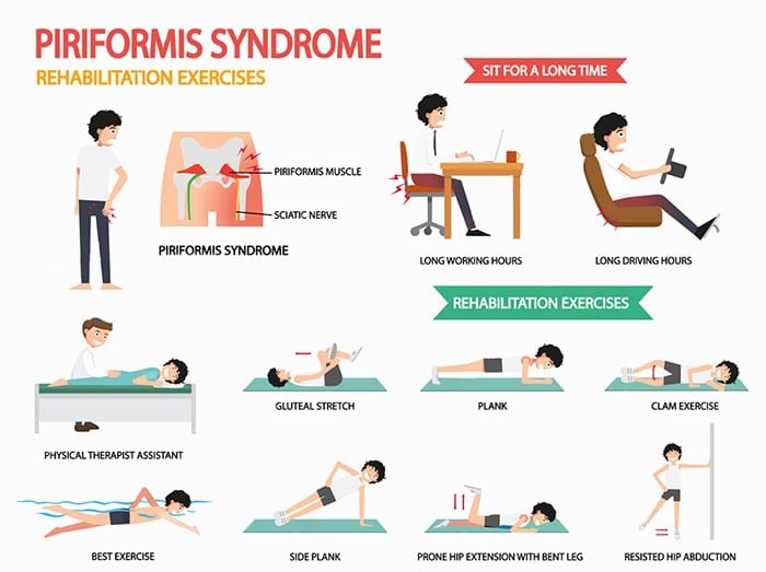


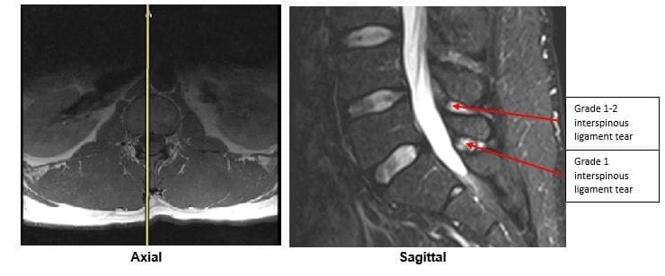


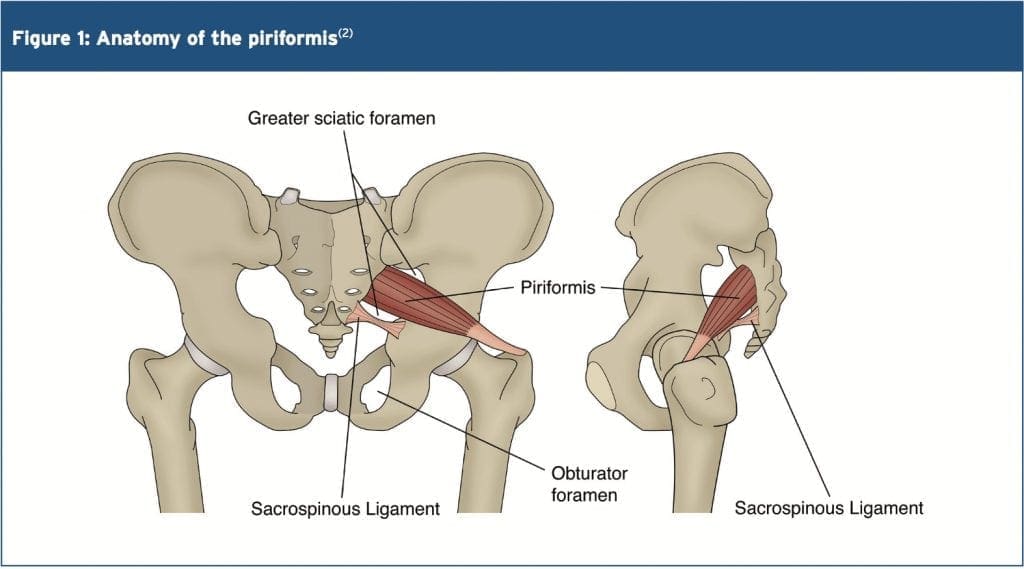
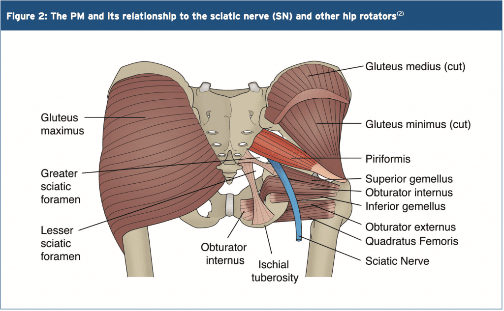 Variants
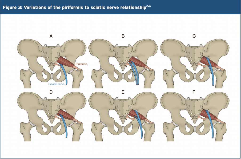
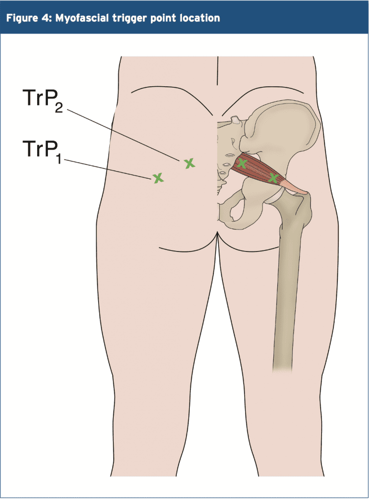
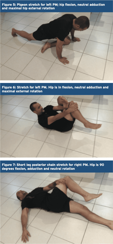 Trigger Points & Massage
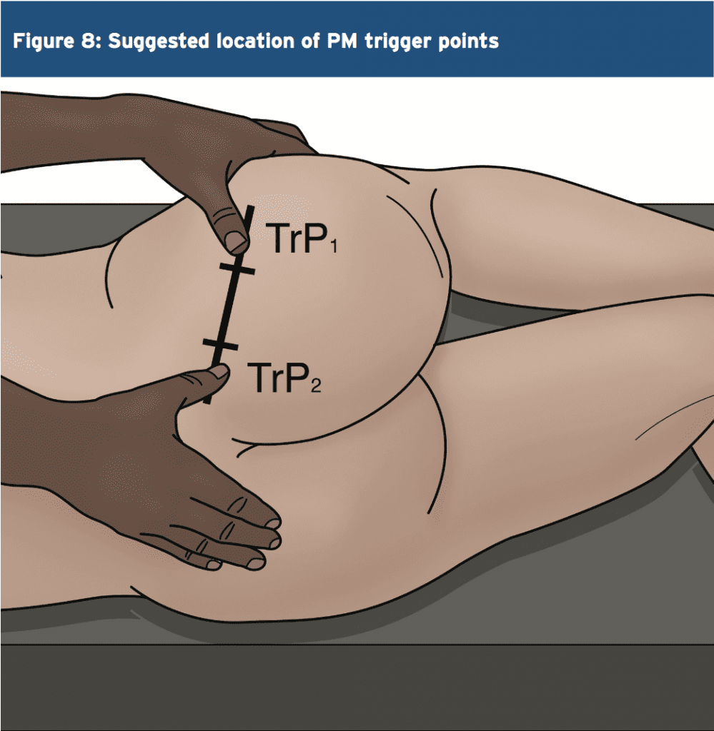

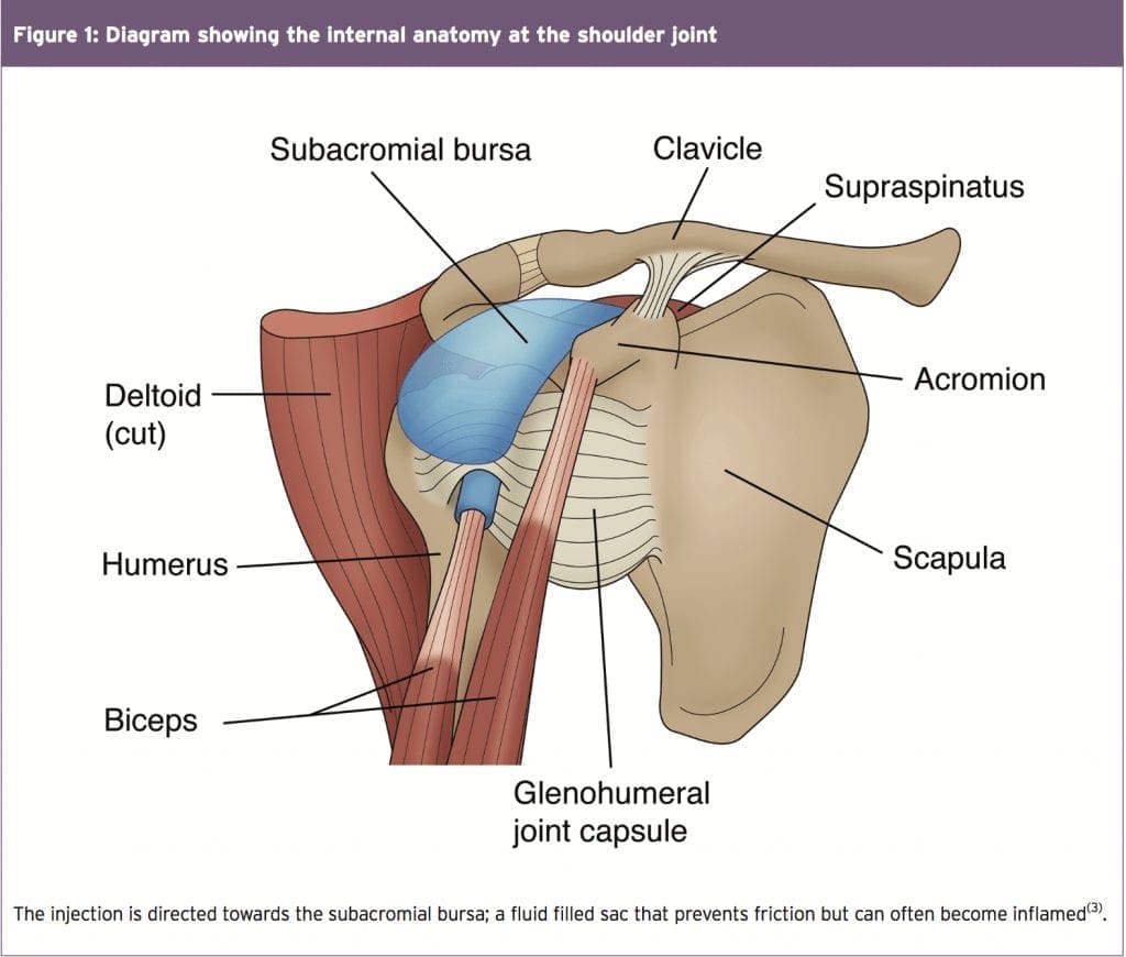
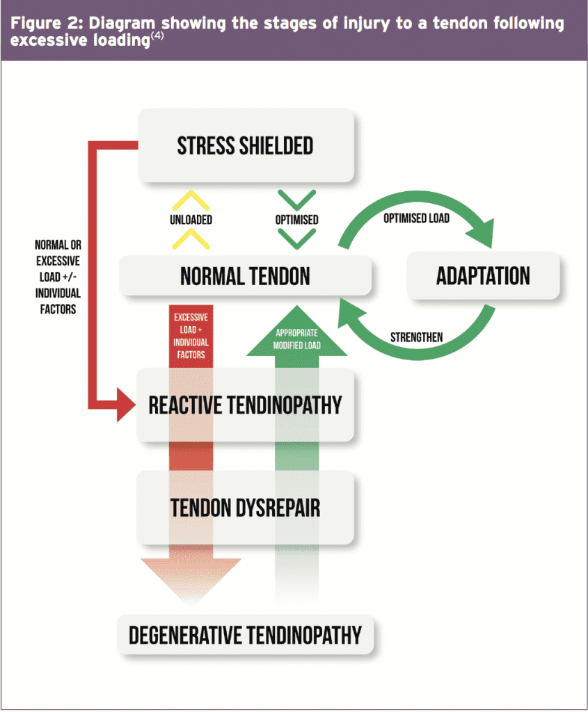
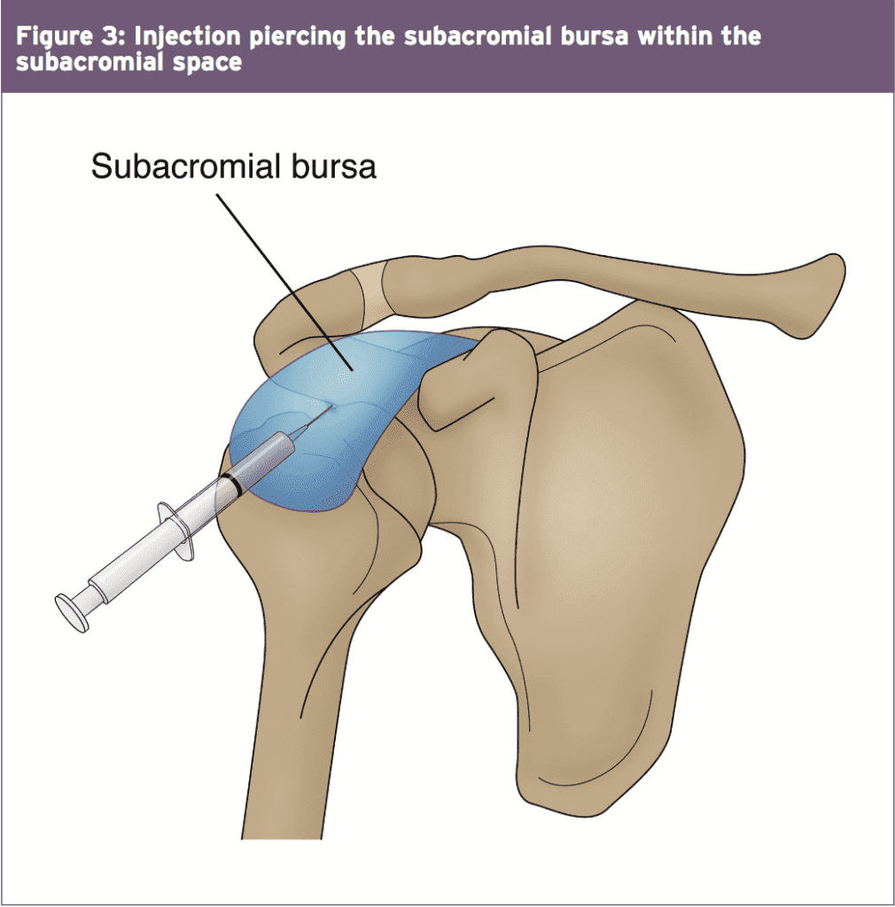 Hip Pain