Footwear can cause lower back pain and problems for some individuals. Can understanding the connection between footwear and back problems help individuals find the right shoes to maintain back health and relieve pain?
Footwear Back Pain
The back provides the strength for physical activities. Back pain affects daily life and can have various causes. Unhealthy posture, walking, twisting, turning, bending, and reaching can contribute to back problems that result in pain. According to the CDC, 39% of adults report living with back pain (Centers for Disease Control and Prevention, 2019). Improper footwear can also contribute to back pain. Selecting footwear carefully can help bring pain relief and help maintain spinal health. Individuals can enjoy less pain and manage symptoms by choosing shoes that maintain spinal alignment and protect the feet from blunt impact.
Understanding the Back Pain-Footwear Connection
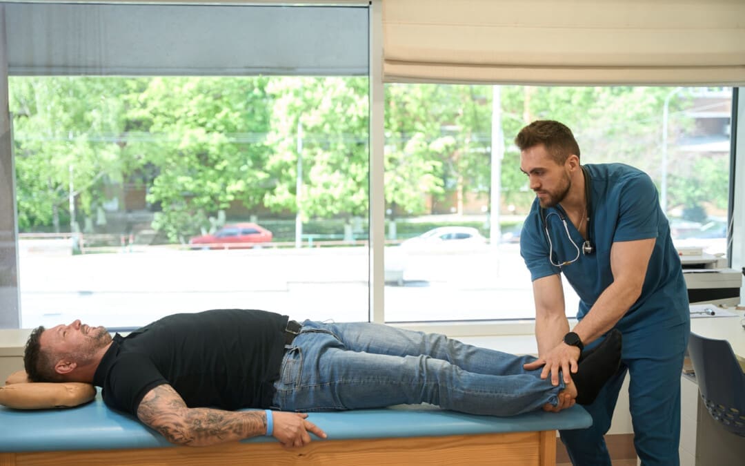
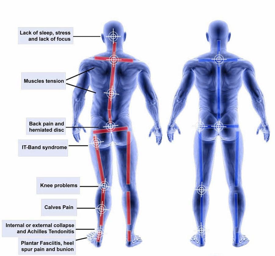
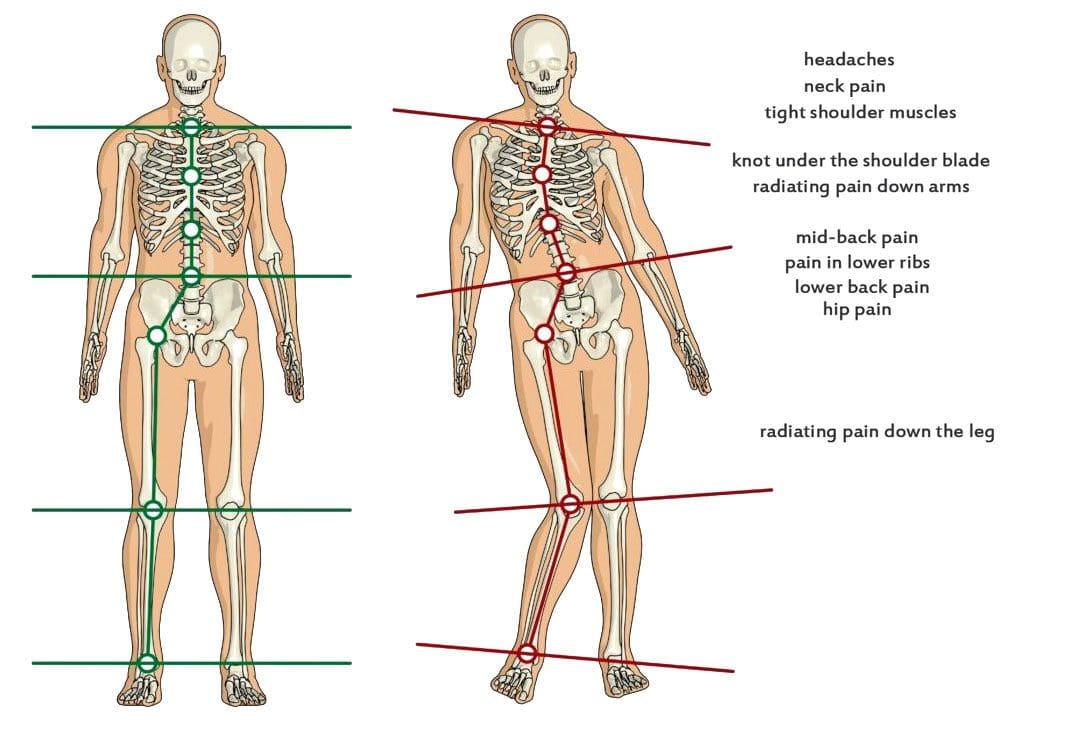
Improper footwear could be the cause of lower back pain. What impacts the bones at the bottom of the neuromusculoskeletal system radiates upward and affects the spine and back muscles. What footwear is used travels upward, impacting gait, posture, spinal alignment, and more. When back problems originate from the feet, these are biomechanical issues. Biomechanics means how the bones, joints, and muscles work together and how changes in external forces impact the body.
Movement
When the feet impact the ground, they are the first extremities to absorb shock for the rest of the body. Individuals will start to walk differently if they have a problem or change in their feet. Wearing shoes with improper support can increase the wear and tear on the muscles and joints, leading to awkward and unnatural movement. For example, consider the difference between standing on tiptoes in high heels and the natural flat-footed state. Well-cushioned shoes help absorb impact and lessen pain sensations. The pressures on each of the joints shift balance, which causes instability problems with less pressure on some and more on others. This creates an imbalance that leads to pain and joint conditions.
Posture
Maintaining a healthy posture is another factor in preventing or alleviating back pain. With the right footwear, the body can maintain a healthier stance and the right curvature throughout the spine, and it helps distribute the weight evenly. This results in decreased stress on ligaments, muscles, and joints. (Harvard Health Publishing. 2014) It’s recommended to see an orthopedist to get to the root of an individual’s condition. For some, a herniated disc, sciatica, automobile collision, fall, unhealthy ergonomics, or a combination, as well as other underlying issues, may be contributing to their back pain.
Shoe Types and Their Impact on The Back
How various shoes impact posture, potentially causing or relieving back pain.
High Heels
High heels can definitely contribute to back pain. They change body posture, causing a domino effect on the spine. The body’s weight is shifted to increase pressure on the balls of the feet, and the spine’s alignment becomes altered. High heels also affect how the ankles, knees, and hips move when walking, balance, and how the back muscles operate, all of which can worsen back pain.
Flat Shoes
Flat shoes may not be the best choice for spinal health. If they lack arch support, they can cause the foot to roll inward, known as pronation. This can contribute to misalignment, which can strain the knees, hips, and lower back. However, they can be a decent choice if they provide arch support. When wearing flat shoes with healthy support, the weight is distributed evenly on the feet and the spine. This helps maintain correct posture, which can help prevent and/or alleviate back pain.
Sneakers, Tennis, and Athletic Shoes
Sneakers, tennis, and athletic shoes can relieve back pain with thorough cushioning and support. Choosing the right ones involves determining the activity that will be done in them. There are tennis, running, basketball, pickleball, skating shoes, and more. Research what features will be needed for the sport or activity. This could include:
Heel cups
Insole cushioning
Wide base
Other features to meet individual foot needs.
It is recommended that athletic shoes be changed every 300 to 500 miles of walking or running or with any signs of unevenness when placed on a flat surface, as worn-out soles and degraded materials can increase the risk of injury and back pain. (American Academy of Podiatric Sports Medicine, 2024). If a certain pair puts the legs, hips, or ankles into an unnatural position or impedes regular movement, it may be time to replace them.
Choosing the Right Shoes
The ideal solution for choosing shoe wear is to get a gait analysis and a review of how you walk and run. Various healthcare professionals may offer this service to tailor each individual’s search for the right shoes for back pain. In gait analysis, individuals are asked to run and walk, sometimes on camera, while a professional notes physical tendencies, like when the foot hits the ground and whether it rolls inward or outward. This provides data on affected posture, movement, pain levels, how much arch support is needed, and what type to wear to help prevent back pain. Once the analysis is complete, it will guide you on what to look for, such as what level of arch support, heel height, or material is best for you.
Injury Medical Chiropractic and Functional Medicine Clinic specializes in progressive, cutting-edge therapies and functional rehabilitation procedures focused on clinical physiology, total health, practical strength training, and complete conditioning. We focus on restoring normal body functions after trauma and soft tissue injuries. We use Specialized Chiropractic Protocols, Wellness Programs, Functional and integrative Nutrition, Agility and mobility Fitness Training, and Rehabilitation Systems for all ages. Our programs are natural and use the body’s ability to achieve specific measured goals rather than introducing harmful chemicals, controversial hormone replacement, unwanted surgeries, or addictive drugs. We have teamed up with the city’s premier doctors, therapists, and trainers to provide high-quality treatments that empower our patients to maintain the healthiest way of living and live a functional life with more energy, a positive attitude, better sleep, and less pain.
Benefits of Using Custom Foot Orthotics
References
Centers for Disease Control and Prevention. (2019). Back, lower limb, and upper limb pain among U.S. adults, 2019. Retrieved from https://www.cdc.gov/nchs/products/databriefs/db415.htm
Harvard Health Publishing. (2014). Posture and back health. Harvard Health Education. https://www.health.harvard.edu/pain/posture-and-back-health
American Academy of Podiatric Sports Medicine. Ayne Furman, D. F., AAPSM. (2024). How do I know when it is time to replace my athletic shoes?
Can knowing treatment options for a dislocated hip help individuals expedite rehabilitation and recovery?
Dislocated Hip
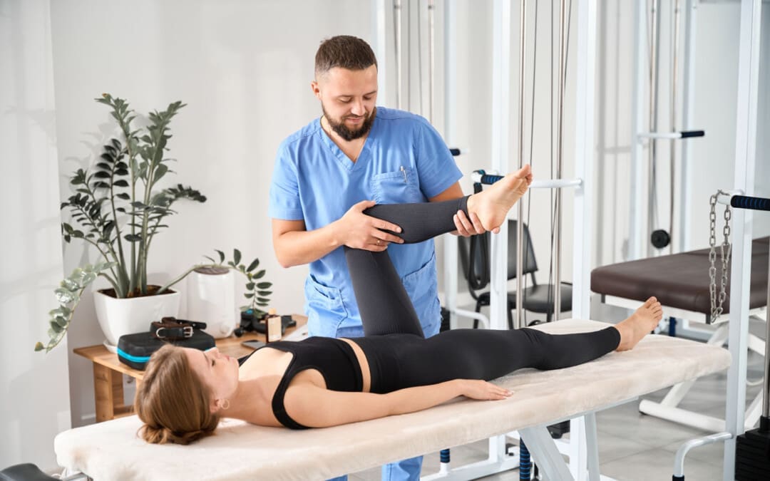
A dislocated hip is an uncommon injury but can happen due to trauma or following hip replacement surgery. It usually occurs after severe trauma, including motor vehicle collisions, falls, and sometimes sports injuries. (Caylyne Arnold et al., 2017) A dislocated hip can also occur after hip replacement surgery. Other injuries like ligament tears, cartilage damage, and bone fractures can occur alongside the dislocation. Most hip dislocations are treated with a joint reduction procedure that resets the ball into the socket. It is usually done with sedation or general anesthesia. Rehabilitation takes time and could be a few months before full recovery. Physical therapy can help restore motion and strength in the hip.
What Is It?
If the hip is only partially dislocated, it’s called a hip subluxation. When this happens, the hip joint head only partially emerges from the socket. A dislocated hip is when the head or ball of the joint shifts or pops out of the socket. Because an artificial hip differs from a normal hip joint, the risk of dislocation increases after joint replacement. A study found that around 2% of individuals who undergo total hip replacement will experience hip dislocation within a year, with the cumulative risk increasing by approximately 1% over five years. (Jens Dargel et al., 2014) However, new technological prosthetics and surgical techniques are making this less common.
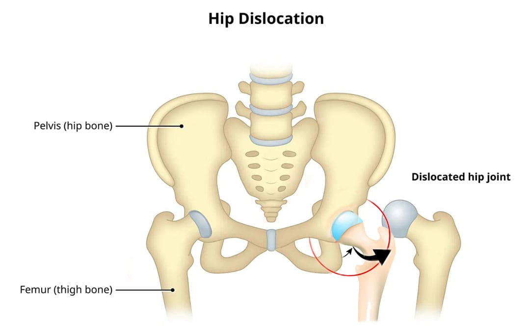
Hip Anatomy
The hip ball-and-socket joint is called the femoroacetabular joint.
The socket is called the acetabulum.
The ball is called the femoral head.
The bony anatomy and strong ligaments, muscles, and tendons help to create a stable joint. Significant force must be applied to the joint for a hip dislocation to occur. Some individuals report feeling a snapping sensation of the hip. This usually is not a hip dislocation but indicates a different disorder known as snapping hip syndrome. (Paul Walker et al., 2021)
Posterior Hip Dislocation
Around 90% of hip dislocations are posterior.
In this type, the ball is pushed backward from the socket.
A hip dislocation increases the risk of developing joint arthritis following the injury and can raise the risk of needing a hip replacement later in life. (Hsuan-Hsiao Ma et al., 2020)
Developmental Dislocation of the Hip
Some children are born with developmental dislocation of the hip or DDH.
Children with DDH have hip joints that did not form correctly during development.
This causes a loose fit in the socket.
In some cases, the hip joint is completely dislocated.
Joint reduction is the most common way to treat a dislocated hip. The procedure repositions the ball back into the socket and is usually done with sedation or under general anesthesia. Repositioning a hip requires significant force. A hip dislocation is considered an emergency, and reduction should be performed immediately after the dislocation to prevent permanent complications and invasive treatment. (Caylyne Arnold et al., 2017)
Once the ball is back in the socket, the healthcare provider will look for bone, cartilage, and ligament injuries.
Depending on what the healthcare provider finds, further treatment may be necessary.
Fractured or broken bones may need to be repaired to keep the ball within the socket.
Damaged cartilage may have to be removed.
Surgery
Surgery could be necessary to return the joint to its normal position. Hip arthroscopy can minimize the invasiveness of certain procedures. A surgeon inserts a microscopic camera into the hip joint to help the surgeon repair the injury using instruments inserted through other small incisions.
Hip replacement surgery replaces the ball and socket, a common and successful orthopedic surgical procedure. This surgery may be performed for various reasons, including trauma or arthritis, as it is common to develop early arthritis of the hip after this type of trauma. This is why many who have a dislocation ultimately need hip replacement surgery. As a major surgical procedure, it is not without risks. Possible complications include:
Infection
Aseptic loosening (the loosening of the joint without infection)
Hip dislocation
Recovery
Recovering from a hip dislocation is a long process. Individuals will need to walk with crutches or other devices early in recovery. Physical therapy will improve the range of motion and strengthen the muscles around the hip. Recovery time will depend on whether other injuries, such as fractures or tears, are present. If the hip joint was reduced and there were no other injuries, it may take six to ten weeks to recover to the point where weight can be placed on the leg. It could be between two and three months for a full recovery. Keeping weight off the leg is important until the surgeon or physical therapist gives the all-clear. Injury Medical Chiropractic and Functional Medicine Clinic will work with an individual’s primary healthcare provider and other surgeons or specialists to develop an optimal personalized treatment plan.
Chiropractic Solutions for Osteoarthritis
References
Arnold, C., Fayos, Z., Bruner, D., Arnold, D., Gupta, N., & Nusbaum, J. (2017). Managing dislocations of the hip, knee, and ankle in the emergency department [digest]. Emergency medicine practice, 19(12 Suppl Points & Pearls), 1–2.
Dargel, J., Oppermann, J., Brüggemann, G. P., & Eysel, P. (2014). Dislocation following total hip replacement. Deutsches Arzteblatt international, 111(51-52), 884–890. https://doi.org/10.3238/arztebl.2014.0884
Walker, P., Ellis, E., Scofield, J., Kongchum, T., Sherman, W. F., & Kaye, A. D. (2021). Snapping Hip Syndrome: A Comprehensive Update. Orthopedic reviews, 13(2), 25088. https://doi.org/10.52965/001c.25088
Cornwall, R., & Radomisli, T. E. (2000). Nerve injury in traumatic dislocation of the hip. Clinical orthopaedics and related research, (377), 84–91. https://doi.org/10.1097/00003086-200008000-00012
American Academy of Orthopaedic Surgeons. (2021). Hip dislocation. https://orthoinfo.aaos.org/en/diseases–conditions/hip-dislocation
Kellam, P., & Ostrum, R. F. (2016). Systematic Review and Meta-Analysis of Avascular Necrosis and Posttraumatic Arthritis After Traumatic Hip Dislocation. Journal of orthopaedic trauma, 30(1), 10–16. https://doi.org/10.1097/BOT.0000000000000419
Ma, H. H., Huang, C. C., Pai, F. Y., Chang, M. C., Chen, W. M., & Huang, T. F. (2020). Long-term results in the patients with traumatic hip fracture-dislocation: Important prognostic factors. Journal of the Chinese Medical Association : JCMA, 83(7), 686–689. https://doi.org/10.1097/JCMA.0000000000000366
American Academy of Orthopaedic Surgeons. (2022). Developmental dislocation (dysplasia) of the hip (DDH). https://orthoinfo.aaos.org/en/diseases–conditions/developmental-dislocation-dysplasia-of-the-hip-ddh/
For individuals who lift weights, are there ways to protect the wrists and prevent injuries when lifting weights?
Wrist Protection
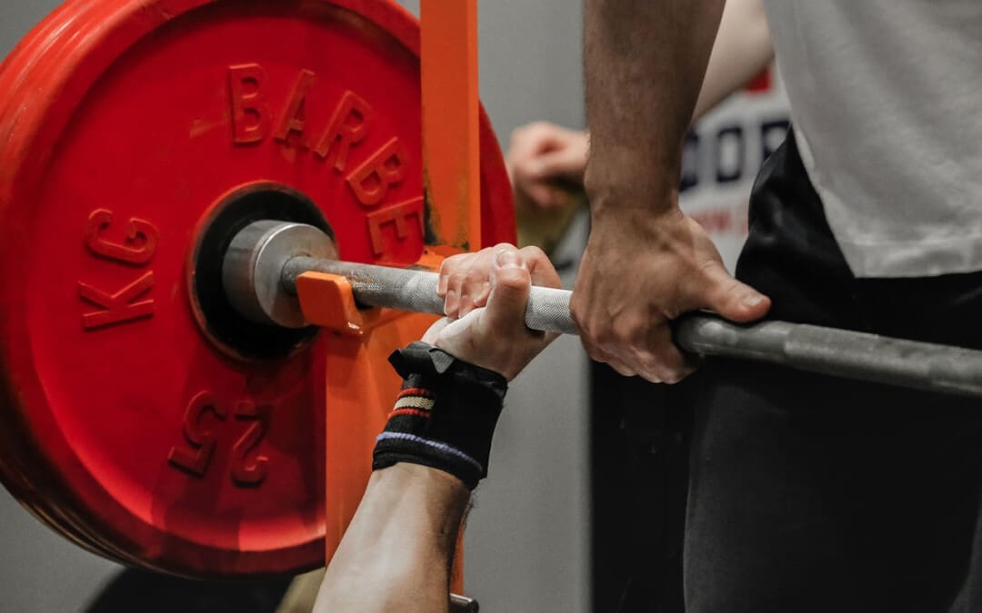
The wrists are complex joints. The wrists significantly contribute to stability and mobility when performing tasks or lifting weights. They provide mobility for movements using the hands and stability to carry and lift objects securely and safely (National Library of Medicine, 2024). Lifting weights is commonly performed to strengthen and stabilize the wrists; however, these movements can cause wrist pain and lead to injuries if not performed correctly. Wrist protection can keep wrists strong and healthy and is key to avoiding strains and injuries.
Wrist Strength
The wrist joints are set between the hand and forearm bones. Wrists are aligned in two rows of eight or nine total small bones/carpal bones and are connected to the arm and hand bones by ligaments, while tendons connect the surrounding muscles to the bones. Wrist joints are condyloid or modified ball and socket joints that assist with flexion, extension, abduction, and adduction movements. (National Library of Medicine. 2024) This means the wrists can move in all planes of motion:
Side to side
Up and down
Rotate
This provides a wide range of motion but can also cause excessive wear and tear and increase the risk of strain and injury. The muscles in the forearm and hand control finger movement necessary for gripping. These muscles and the tendons and ligaments involved run through the wrist. Strengthening the wrists will keep them mobile, help prevent injuries, and increase and maintain grip strength. In a review on weightlifters and powerlifters that examined the types of injuries they sustain, wrist injuries were common, with muscle and tendon injuries being the most common among weightlifters. (Ulrika Aasa et al., 2017)
Protecting the Wrists
Wrist protection can use a multi-approach, which includes consistently increasing strength, mobility, and flexibility to improve health and prevent injuries. Before lifting or engaging in any new exercise, individuals should consult their primary healthcare provider, physical therapist, trainer, medical specialist, or sports chiropractor to see which exercises are safe and provide benefits based on injury history and current level of health.
Increase Mobility
Mobility allows the wrists to have a full range of motion while retaining the stability necessary for strength and durability. Lack of mobility in the wrist joint can cause stiffness and pain. Flexibility is connected to mobility, but being overly flexible and lacking stability can lead to injuries. To increase wrist mobility, perform exercises at least two to three times a week to improve range of motion with control and stability. Also, taking regular breaks throughout the day to rotate and circle the wrists and gently pull back on the fingers to stretch them will help relieve tension and stiffness that can cause mobility problems.
Warm-Up
Before working out, warm up the wrists and the rest of the body before working out. Start with light cardiovascular to get the synovial fluid in the joints circulating to lubricate the joints, allowing for smoother movement. For example, individuals can make fists, rotate their wrists, perform mobility exercises, flex and extend the wrists, and use one hand to pull back the fingers gently. Around 25% of sports injuries involve the hand or wrist. These include hyperextension injury, ligament tears, front-inside or thumb-side wrist pain from overuse injuries, extensor injuries, and others. (Daniel M. Avery 3rd et al., 2016)
Strengthening Exercises
Strong wrists are more stable, and strengthening them can provide wrist protection. Exercises that improve wrist strength include pull-ups, deadlifts, loaded carries, and Zottman curls. Grip strength is vital for performing daily tasks, healthy aging, and continued success with weightlifting. (Richard W. Bohannon 2019) For example, individuals who have difficulty increasing the weight on their deadlifts because the bar slips from their hands could have insufficient wrist and grip strength.
Wraps
Wrist wraps or grip-assisting products are worth considering for those with wrist issues or concerns. They can provide added external stability while lifting, reducing grip fatigue and strain on the ligaments and tendons. However, it is recommended not to rely on wraps as a cure-all measure and to focus on improving individual strength, mobility, and stability. A study on athletes with wrist injuries revealed that the injuries still occurred despite wraps being worn 34% of the time prior to the injury. Because most injured athletes did not use wraps, this pointed to potential preventative measures, but the experts agreed more research is needed. (Amr Tawfik et al., 2021)
Preventing Overuse Injuries
When an area of the body undergoes too many repetitive motions without proper rest, it becomes worn, strained, or inflamed faster, causing overuse injury. The reasons for overuse injuries are varied but include not varying workouts enough to rest the muscles and prevent strain. A research review on the prevalence of injuries in weightlifters found that 25% were due to overuse tendon injuries. (Ulrika Aasa et al., 2017) Preventing overuse can help avoid potential wrist problems.
Proper Form
Knowing how to perform movements correctly and using proper form during each workout/training session is essential for preventing injuries. A personal trainer, sports physiotherapist, or physical therapist can teach how to adjust grip or maintain correct form.
Be sure to see your provider for clearance before lifting or starting an exercise program. Injury Medical Chiropractic and Functional Medicine Clinic can advise on training and prehabilitation or make a referral if one is needed.
Fitness Health
References
Erwin, J., & Varacallo, M. (2024). Anatomy, Shoulder and Upper Limb, Wrist Joint. In StatPearls. https://www.ncbi.nlm.nih.gov/pubmed/30521200
Aasa, U., Svartholm, I., Andersson, F., & Berglund, L. (2017). Injuries among weightlifters and powerlifters: a systematic review. British journal of sports medicine, 51(4), 211–219. https://doi.org/10.1136/bjsports-2016-096037
Avery, D. M., 3rd, Rodner, C. M., & Edgar, C. M. (2016). Sports-related wrist and hand injuries: a review. Journal of orthopaedic surgery and research, 11(1), 99. https://doi.org/10.1186/s13018-016-0432-8
Bohannon R. W. (2019). Grip Strength: An Indispensable Biomarker For Older Adults. Clinical interventions in aging, 14, 1681–1691. https://doi.org/10.2147/CIA.S194543
Tawfik, A., Katt, B. M., Sirch, F., Simon, M. E., Padua, F., Fletcher, D., Beredjiklian, P., & Nakashian, M. (2021). A Study on the Incidence of Hand or Wrist Injuries in CrossFit Athletes. Cureus, 13(3), e13818. https://doi.org/10.7759/cureus.13818
For individuals experiencing back pain from a herniated disc, can understanding the difference between surgery and chiropractic help individuals find the right treatment plan?
Surgery or Chiropractic
Living with back pain can be a nightmare, and yet many struggle without seeking care. Today, there are a vast number of surgeries and noninvasive techniques that are better at treating spine and back problems and managing symptoms. For individuals who may have a herniated disc or are curious about ways to relieve their back pain, a health care provider, physical therapist, spine specialist, and chiropractor can inform them of treatment options. Surgery and chiropractic therapy are popular treatments for a herniated, bulging, or slipped disc.
A herniated disc is when the cartilage discs that cushion the vertebrae shift out of position and leak out.
Surgery for a herniated disc involves removing or repairing the disc.
Chiropractic nonsurgically repositions the disc and realigns the spine.
Both treatments have the same goals with key differences.
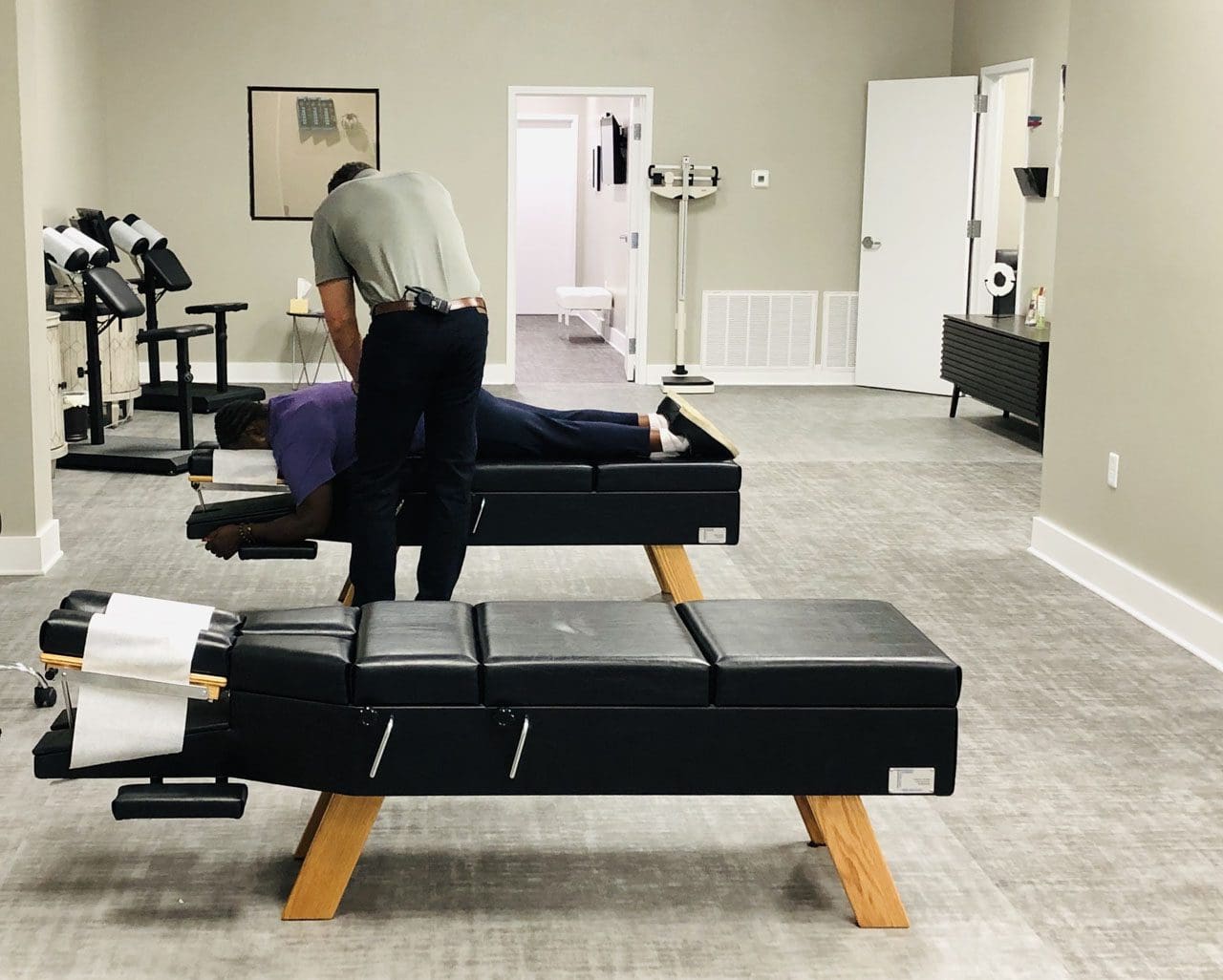
Chiropractic Care
Chiropractic is a system of therapy that focuses on adjusting and maintaining spinal alignment to help with back and posture problems. Chiropractors are trained and licensed medical professionals who take a nonsurgical approach, a proven therapy for chronic pain, flexibility, and mobility issues.
The Way It Works
Chiropractic treatment encourages and supports the body’s natural healing processes. It is considered for joint pain in the back, neck, legs, arms, feet, and hands. It typically involves sessions in which the chiropractor physically and carefully adjusts the vertebrae by hand, also known as spinal manipulation or chiropractic adjustments. (MedlinePlus. 2023). A chiropractor performs a thorough medical evaluation and runs tests to establish a diagnosis. A chiropractor will develop a treatment plan that may involve a team of massage and physical therapists, acupuncturists, health coaches, and nutritionists to treat affected areas with various techniques, recommend targeted exercises, adjust lifestyle and nutrition to support the treatment, and monitor progress. Combined with stretching and sustained pressure, the multiple methods can increase joint mobility and relieve pain symptoms. (National Center for Complimentary and Integrative Health. 2019) Added protocols to support or enhance chiropractic therapy include:
Heating and ice therapies to reduce inflammation and increase blood circulation.
Using devices to stimulate muscles and nerves electrically.
Developing relaxation and deep breathing techniques.
Incorporating exercises to promote rehabilitation.
Establishing a regular fitness routine.
Making adjustments to diet and lifestyle.
Taking certain dietary supplements.
Spinal manipulation and chiropractic adjustments have been shown to improve symptoms and restore mobility in cases of chronic back pain. One review found that individuals with chronic lumbar/low back pain reported significant improvement after six weeks of chiropractic treatment. (Ian D. Coulter et al., 2018)
Prices
The out-of-pocket expenses of chiropractic treatment depend on a variety of factors.
Insurance may or may not cover the treatment, and the amount an individual has to pay can vary based on the severity of their case, what their plan covers, and where they live. One review found the cost can range between $264 and $6,171. (Simon Dagenais et al., 2015)
Surgery
There is a range of minimally invasive surgical procedures to treat herniated discs. These work to ease nerve compression by removing or replacing damaged discs or stabilizing the vertebrae, relieving pain and inflammation.
The Way It Works
A herniated disc can happen in any part of the spine but is more common in the lower back/lumbar spine and in the neck/cervical spine. Surgery is recommended when: (American Academy of Orthopaedic Surgeons. 2022)
More conservative treatments, like medications and physical therapy, are unable to manage symptoms.
The pain and symptoms impact daily life and functioning.
Standing or walking becomes difficult or impossible.
The herniated disc causes difficulty walking, muscle weakness, and bladder or bowel control loss.
The individual is reasonably healthy, without infection, osteoporosis, or arthritis.
Specific surgical procedures used include:
Fusion Surgery
Spinal fusion is the most common procedure for a lower back herniated disc.
It involves using artificial bone material to fuse vertebrae to increase stability and release and prevent nerve irritation and compression. (American Academy of Neurological Surgeons. 2024)
Laminotomy and Laminectomy
Herniated disc symptoms appear from compression placed on the nerves.
Laminotomy involves making a small cut in the lamina, or the arch of the spinal vertebrae, to release the pressure.
Another approach involves implanting an artificial disc.
This is most often used for hernia in the lower spine; the worn or damaged disc is removed, and a specialized prosthetic replaces the removed disc. (American Academy of Orthopaedic Surgeons. 2022)
This allows for more mobility.
The success of herniated disc surgery depends on different factors. Advances in minimally invasive techniques have significantly improved long-term outcomes, with one review finding that around 80% reported good—excellent results at a six-year follow-up. (George J. Dohrmann, Nassir Mansour 2015) However, there is the possibility of recurrence. About 20% to 25% of individuals with herniated lumbar discs experience re-herniation at some point. (American Academy of Neurological Surgeons. 2024)
Prices
Surgery for a herniated disc is specialized, and the costs depend on the scope and scale of the treatment.
The individual’s specific insurance plan also determines the expenses.
When choosing between chiropractic and surgery for a herniated disc, a number of factors can determine the decision, including:
Chiropractic is the less invasive nonsurgical option.
Chiropractic adjustments cannot help certain severe cases of herniated discs.
Chiropractic adjustments prevent the herniated disc from getting worse and ease symptoms.
Surgery provides pain and symptom relief faster than chiropractic or conservative treatment but requires significant recovery time and is expensive. (Anna N A Tosteson et al., 2008)
Surgery may not be appropriate for individuals with osteoarthritis or osteoporosis.
Chiropractic therapy is among the more conservative treatment options for a herniated disc and may be tried first before proceeding with surgery. Generally, surgery is only recommended when noninvasive methods haven’t been able to stop or manage the pain and symptoms. Injury Medical Chiropractic and Functional Medicine Clinic works with primary healthcare providers and specialists to develop an optimal health and wellness solution that fully benefits the individual to get back to normal.
Quick Patient Process
References
MedlinePlus.MedlinePlus. (2023). Chiropractic. Retrieved from https://medlineplus.gov/chiropractic.html
National Center for Complimentary and Integrative Health. (2019). Chiropractic: in depth. Retrieved from https://www.nccih.nih.gov/health/chiropractic-in-depth
Coulter, I. D., Crawford, C., Hurwitz, E. L., Vernon, H., Khorsan, R., Suttorp Booth, M., & Herman, P. M. (2018). Manipulation and mobilization for treating chronic low back pain: a systematic review and meta-analysis. The spine journal : official journal of the North American Spine Society, 18(5), 866–879. https://doi.org/10.1016/j.spinee.2018.01.013
Dagenais, S., Brady, O., Haldeman, S., & Manga, P. (2015). A systematic review comparing the costs of chiropractic care to other interventions for spine pain in the United States. BMC health services research, 15, 474. https://doi.org/10.1186/s12913-015-1140-5
American Academy of Orthopaedic Surgeons. (2022). Herniated disk in the lower back. https://orthoinfo.aaos.org/en/diseases–conditions/herniated-disk-in-the-lower-back/
American Academy of Neurological Surgeons. Surgeons, A. A. o. N. (2024). Herniated disc. https://www.aans.org/en/Patients/Neurosurgical-Conditions-and-Treatments/Herniated-Disc
Dohrmann, G. J., & Mansour, N. (2015). Long-Term Results of Various Operations for Lumbar Disc Herniation: Analysis of over 39,000 Patients. Medical principles and practice : international journal of the Kuwait University, Health Science Centre, 24(3), 285–290. https://doi.org/10.1159/000375499
Tosteson, A. N., Skinner, J. S., Tosteson, T. D., Lurie, J. D., Andersson, G. B., Berven, S., Grove, M. R., Hanscom, B., Blood, E. A., & Weinstein, J. N. (2008). The cost effectiveness of surgical versus nonoperative treatment for lumbar disc herniation over two years: evidence from the Spine Patient Outcomes Research Trial (SPORT). Spine, 33(19), 2108–2115. https://doi.org/10.1097/brs.0b013e318182e390
For individuals experiencing or managing low back pain and/or sciatica, can lumbar traction therapy help provide consistent relief?
Lumbar Traction
Lumbar traction therapy for lower back pain and sciatica could be a treatment option to help restore mobility and flexibility and safely support an individual’s return to an optimal level of activity. It is often combined with targeted therapeutic exercise. (Yu-Hsuan Cheng, et al., 2020) The technique stretches the space between the vertebrae in the lower spine, relieving lower back pain.
Lumbar or low back traction helps to separate the spaces between the vertebrae.
Separating the bones restores circulation and helps relieve the pressure on pinched nerves like the sciatic nerve, decreasing pain and improving mobility.
Research
Researchers say lumbar traction with exercise did not improve individual outcomes compared to physical therapy exercises on their own (Anne Thackeray et al., 2016). The study examined 120 participants with back pain and nerve root impingement who were randomly selected to undergo lumbar traction with exercises or simple exercises for pain. Extension-based exercises focused on bending the spine backward. This movement is considered effective for individuals with back pain and pinched nerves. The results indicated that adding lumbar traction to physical therapy exercises did not offer significant benefits over extension-based exercise alone for back pain. (Anne Thackeray et al., 2016)
A 2022 study found that lumbar traction is helpful for individuals with lower back pain. The study investigated two different lumbar traction techniques and found that variable-force lumbar traction and high-force lumbar traction helped to relieve lower back pain. High-force lumbar traction was also found to reduce functional disability. (Zahra Masood et al., 2022) Another study found lumbar traction improves the range of motion in the straight leg raise test. The study examined different forces of traction on herniated discs. All the levels improved the individuals’ range of motion, but the one-half body-weight traction setting was associated with the most significant pain relief. (Anita Kumari et al., 2021)
Treatment
For individuals with only low back pain, exercise, and postural correction may be all that is needed to provide relief. Research confirms physical therapy exercises can help decrease pain and improve mobility (Anita Slomski 2020). Another study revealed the importance of centralizing sciatic symptoms during repetitive movements. Centralization is moving the pain back to the spine, which is a positive sign that the nerves and discs are healing and occurs during therapeutic exercise. (Hanne B. Albert et al., 2012) A chiropractor and physical therapy team can educate patients on preventing back pain episodes. Chiropractors and physical therapists are body movement experts who can show which exercises are best for your condition. Starting an exercise program that centralizes symptoms can help individuals return to their normal lifestyle quickly and safely. Consult a healthcare provider before starting any exercise program for back pain.
Movement Medicine: Chiropractic
References
Cheng, Y. H., Hsu, C. Y., & Lin, Y. N. (2020). The effect of mechanical traction on low back pain in patients with herniated intervertebral disks: a systemic review and meta-analysis. Clinical rehabilitation, 34(1), 13–22. https://doi.org/10.1177/0269215519872528
Thackeray, A., Fritz, J. M., Childs, J. D., & Brennan, G. P. (2016). The Effectiveness of Mechanical Traction Among Subgroups of Patients With Low Back Pain and Leg Pain: A Randomized Trial. The Journal of orthopaedic and sports physical therapy, 46(3), 144–154. https://doi.org/10.2519/jospt.2016.6238
Masood, Z., Khan, A. A., Ayyub, A., & Shakeel, R. (2022). Effect of lumbar traction on discogenic low back pain using variable forces. JPMA. The Journal of the Pakistan Medical Association, 72(3), 483–486. https://doi.org/10.47391/JPMA.453
Kumari, A., Quddus, N., Meena, P. R., Alghadir, A. H., & Khan, M. (2021). Effects of One-Fifth, One-Third, and One-Half of the Bodyweight Lumbar Traction on the Straight Leg Raise Test and Pain in Prolapsed Intervertebral Disc Patients: A Randomized Controlled Trial. BioMed research international, 2021, 2561502. https://doi.org/10.1155/2021/2561502
Slomski A. (2020). Early Physical Therapy Relieves Sciatica Disability and Pain. JAMA, 324(24), 2476. https://doi.org/10.1001/jama.2020.24673
Albert, H. B., Hauge, E., & Manniche, C. (2012). Centralization in patients with sciatica: are pain responses to repeated movement and positioning associated with outcome or types of disc lesions?. European spine journal : official publication of the European Spine Society, the European Spinal Deformity Society, and the European Section of the Cervical Spine Research Society, 21(4), 630–636. https://doi.org/10.1007/s00586-011-2018-9
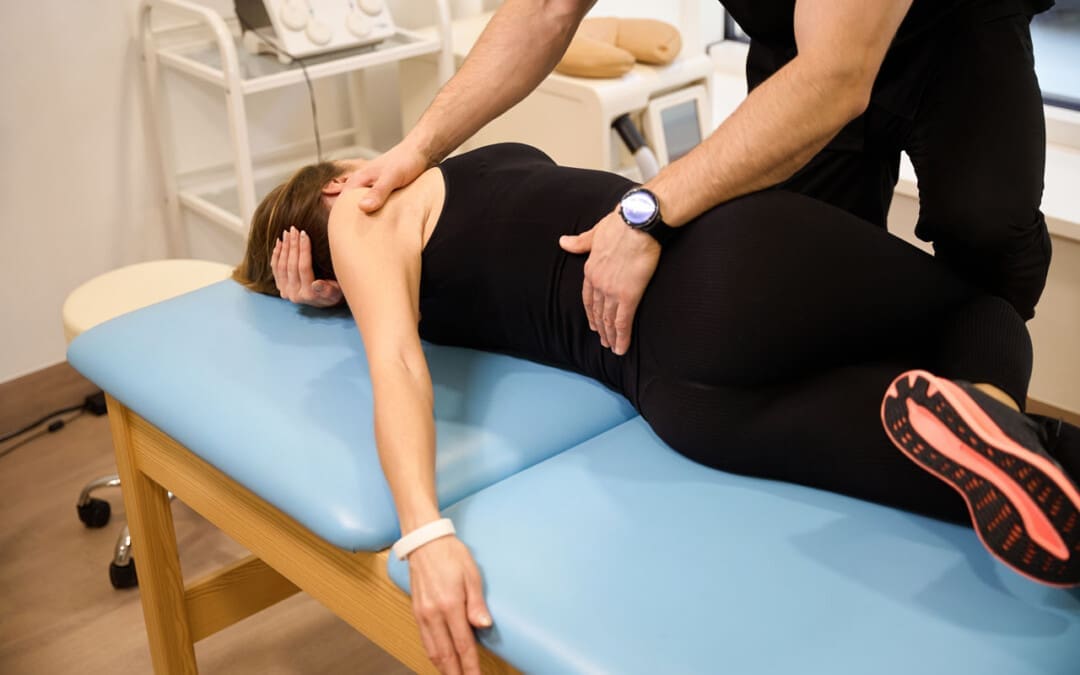
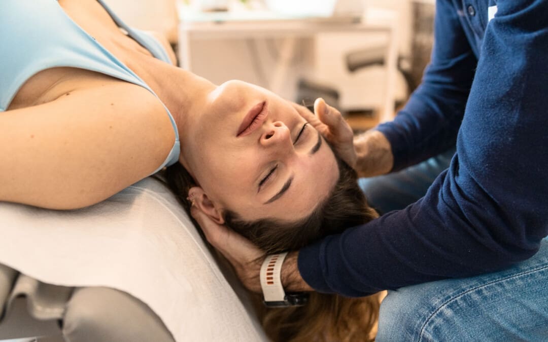
Those experiencing neck pain, stiffness, headache, shoulder and back pain may suffer from a whiplash injury. Can knowing whiplash signs and symptoms help individuals recognize the injury and help healthcare providers develop an effective treatment plan?
Whiplash Signs and Symptoms
Whiplash is a neck injury that typically occurs after a motor vehicle collision or accident but can happen with any injury that rapidly whips the neck forward and backward. It is a mild to moderate injury of the neck muscles. Common whiplash signs and symptoms include:
Some individuals can develop chronic pain and headaches.
The symptoms and treatment depend on the severity of the injury. Treatment can include over-the-counter pain medicines, ice and heat therapy, chiropractic, physical therapy, and stretching exercises.
Frequent Signs and Symptoms
The sudden whipping movement of the head can affect several structures within the neck. These structures include:
Muscles
Bones
Joints
Tendons
Ligaments
Intervertebral discs
Blood vessels
Nerves.
Any or all of these can be affected by a whiplash injury. (MedlinePlus, 2017)
Statistics
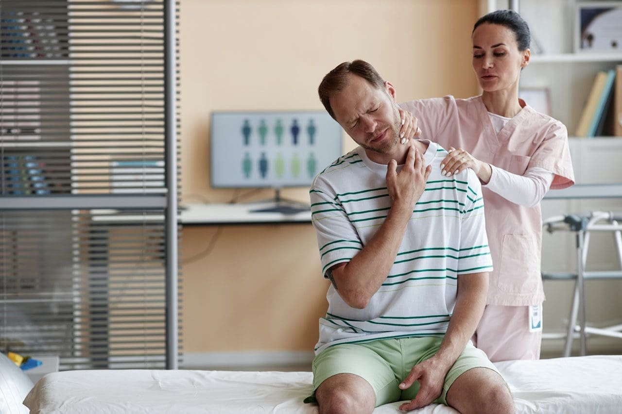
Whiplash is a neck sprain that occurs from a fast neck-jerking motion. Whiplash injuries account for more than half of vehicle traffic collision injuries. (Michele Sterling, 2014) Even with a minor injury, the most frequent symptoms include: (Nobuhiro Tanaka et al., 2018)
Neck pain
Next stiffness
Neck tenderness
Limited range of motion of the neck
Individuals can develop neck discomfort and pain shortly after an injury; however, the more intense pain and stiffness typically do not occur right after the injury. Symptoms tend to worsen the next day or 24 hours later. (Nobuhiro Tanaka et al., 2018)
Beginning Symptoms
Researchers have found that approximately more than half of individuals with whiplash develop symptoms within six hours of the injury. Around 90% develop symptoms within 24 hours, and 100% develop symptoms within 72 hours. (Nobuhiro Tanaka et al., 2018)
Whiplash vs. Traumatic Cervical Spine Injury
Whiplash describes a mild to moderate neck injury without significant skeletal or neurological symptoms. Significant neck injuries can lead to fractures and dislocations of the spine that can affect the nerves and spinal cord. Once an individual develops neurological problems associated with a neck injury, the diagnosis changes from whiplash to traumatic cervical spine injury. These differences can be confusing as they are on the same spectrum. To better understand the severity of a neck sprain, the Quebec classification system divides neck injury into the following grades (Nobuhiro Tanaka et al., 2018)
Grade 0
This means there are no neck symptoms or physical examination signs.
Grade 1
There is neck pain and stiffness.
Very few findings from the physical examination.
Grade 2
Indicates neck pain and stiffness
Neck tenderness
Decreased mobility or neck range of motion on physical examination.
Grade 3
Involves muscle pain and stiffness.
Neurologic symptoms include:
Numbness
Tingling
Weakness in the arms
Decreased reflexes
Grade 4
Involves a fracture or dislocation of the bones of the spinal column.
Other Symptoms
Other whiplash signs and symptoms that can be associated with the injury but are less common or only occur with a severe injury include (Nobuhiro Tanaka et al., 2018)
Tension headache
Jaw pain
Sleep problems
Migraine headache
Difficulty concentrating
Reading difficulties
Blurred vision
Dizziness
Driving difficulties
Rare Symptoms
Individuals with severe injuries can develop rare symptoms that often indicate traumatic cervical spine injury and include: (Nobuhiro Tanaka et al., 2018)
Amnesia
Tremor
Voice changes
Torticollis – painful muscle spasms that keep the head turned to one side.
Bleeding in the brain
Complications
Most individual generally recover from their symptoms within a few weeks to a few months. (Michele Sterling, 2014) However, whiplash complications can occur, especially with severe grade 3 or grade 4 injuries. The most common complications of a whiplash injury include chronic/long-term pain and headaches. (Michele Sterling, 2014) Traumatic cervical spine injury can affect the spinal cord and be associated with chronic neurological problems, including numbness, weakness, and difficulty walking. (Luc van Den Hauwe et al., 2020)
Treatment
The pain is typically more severe the next day than after the injury. Whiplash musculoskeletal injury treatment depends on whether it is an acute injury or the individual has developed chronic neck pain and stiffness.
Acute pain can be treated with over-the-counter medicines like Tylenol and Advil, which effectively treat the pain.
Advil is a nonsteroidal anti-inflammatory that can be taken with the pain reliever Tylenol, which works in different ways.
The mainstay of treatment is encouraging regular activity with stretching and exercise. (Michele Sterling, 2014)
Physical therapy uses various range of motion exercises to strengthen the neck muscles and relieve the pain.
Chiropractic adjustments and non-surgical decompression can help realign and nourish the spine.
Acupuncture can cause the body to release natural hormones that provide pain relief, help relax the soft tissues, increase circulation, and reduce inflammation. The cervical spine can return to alignment when the soft tissues are no longer inflamed and spasming. (Tae-Woong Moon et al., 2014)
Neck Injuries
References
Medicine, J. H. (2024). Whiplash Injury. https://www.hopkinsmedicine.org/health/conditions-and-diseases/whiplash-injury
MedlinePlus. (2017). Neck Injuries and Disorders. Retrieved from https://medlineplus.gov/neckinjuriesanddisorders.html#cat_95
Sterling M. (2014). Physiotherapy management of whiplash-associated disorders (WAD). Journal of physiotherapy, 60(1), 5–12. https://doi.org/10.1016/j.jphys.2013.12.004
Tanaka, N., Atesok, K., Nakanishi, K., Kamei, N., Nakamae, T., Kotaka, S., & Adachi, N. (2018). Pathology and Treatment of Traumatic Cervical Spine Syndrome: Whiplash Injury. Advances in orthopedics, 2018, 4765050. https://doi.org/10.1155/2018/4765050
van Den Hauwe L, Sundgren PC, Flanders AE. (2020). Spinal Trauma and Spinal Cord Injury (SCI). In: Hodler J, Kubik-Huch RA, von Schulthess GK, editors. Diseases of the Brain, Head and Neck, Spine 2020–2023: Diagnostic Imaging [Internet]. Cham (CH): Springer; 2020. Chapter 19. Available from: https://www.ncbi.nlm.nih.gov/books/NBK554330/ doi: 10.1007/978-3-030-38490-6_19
Moon, T. W., Posadzki, P., Choi, T. Y., Park, T. Y., Kim, H. J., Lee, M. S., & Ernst, E. (2014). Acupuncture for treating whiplash associated disorder: a systematic review of randomised clinical trials. Evidence-based complementary and alternative medicine : eCAM, 2014, 870271. https://doi.org/10.1155/2014/870271
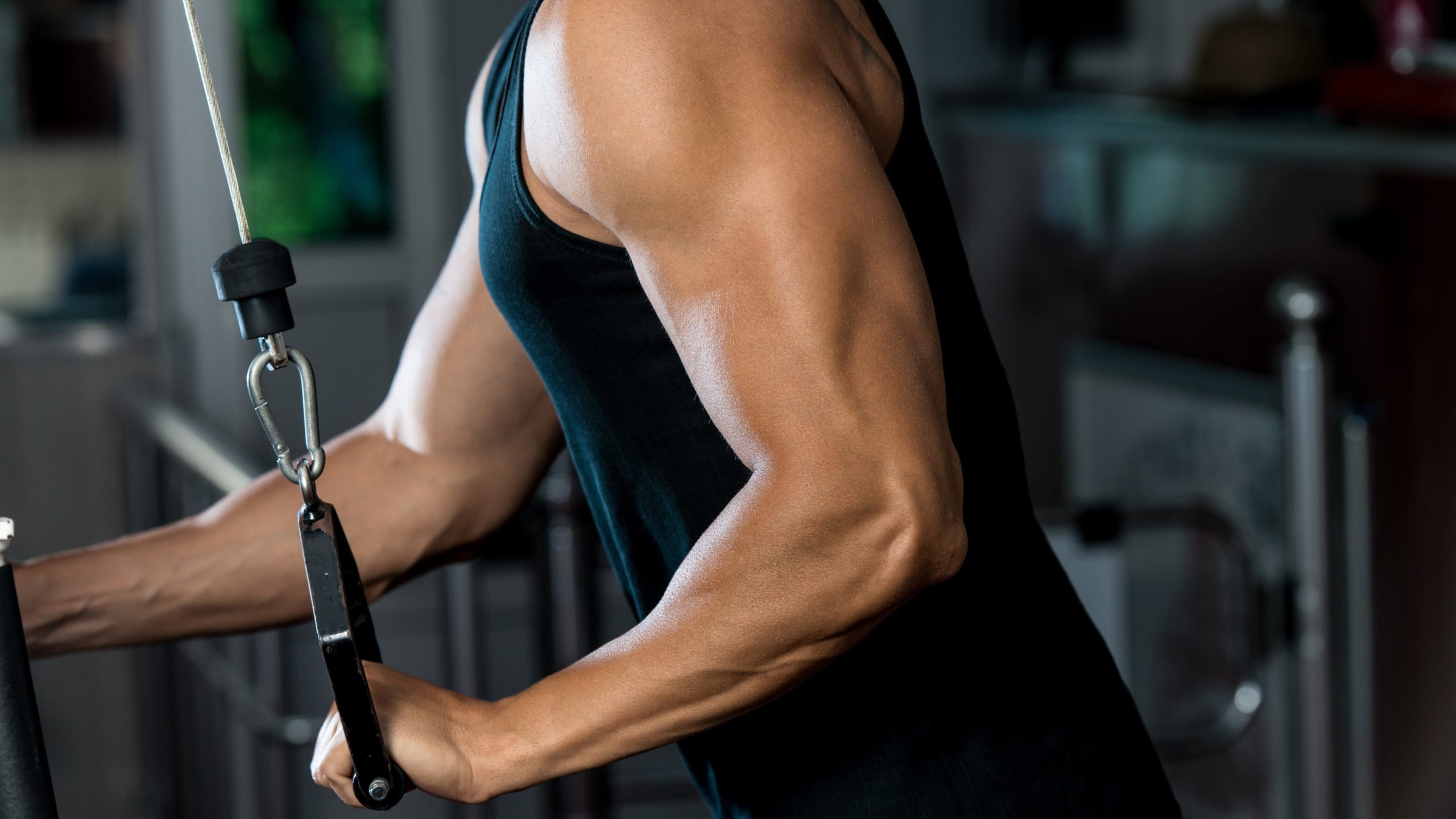
For athletes and sports enthusiasts, a torn triceps can be a serious injury. Can knowing their symptoms, causes, risk factors, and potential complications help healthcare providers develop an effective treatment plan?
Torn Triceps Injury
The triceps is the muscle on the back of the upper arm that allows the elbow to straighten. Fortunately, triceps tears are uncommon, but they can be serious. The injury affects men more often than women and usually occurs from trauma, sports, and/or exercise activities. Depending on the extent and severity of the injury, a torn triceps injury can require splinting, physical therapy, and possibly surgery to regain movement and strength. Recovery after a triceps tear typically lasts around six months. (The Ohio State University Wexner Medical Center. 2021)
Anatomy
The triceps brachii muscle, or triceps, runs along the back of the upper arm. It is named tri- because it has three heads – the long, medial, and lateral head. (Sendic G. 2023) The triceps originates at the shoulder and attaches to the shoulder blade/scapula and upper arm bone/humerus. At the bottom, it attaches to the point of the elbow. This is the bone on the pinky side of the forearm, known as the ulna. The triceps cause movement at the shoulder and the elbow joint. At the shoulder, it performs extension or backward movement of the arm and adduction or moving the arm toward the body. The main function of this muscle is at the elbow, where it performs extension or straightening of the elbow. The triceps work the opposite of the biceps muscle on the front of the upper arm, which conducts flexion or bending of the elbow.
Triceps Tear
Tears can occur anywhere along the length of a muscle or tendon, which is the structure that attaches the muscle to the bones. Triceps tears commonly occur in the tendon connecting the triceps to the back of the elbow. Muscle and tendon tears are graded from 1 to 3 based on severity. (Alberto Grassi et al., 2016)
Grade 1 Mild
These small tears cause pain that worsens with movement.
There is some swelling, bruising, and minimal loss of function.
Grade 2 Moderate
These tears are larger and have moderate swelling and bruising.
The fibers are partially torn and stretched.
Up to 50% loss of function.
Grade 3 Severe
This is the worst type of tear, where the muscle or tendon is completely torn.
These injuries cause severe pain and disability.
Symptoms
Triceps tears cause immediate pain in the back of the elbow and upper arm that worsens when trying to move the elbow. Individuals might also feel and/or hear a popping or tearing sensation. There will be swelling, and the skin will likely be red and/or bruised. With a partial tear, the arm will feel weak. If there is a complete tear, there will be significant weakness when straightening the elbow. Individuals may also notice a lump on the back of their arm where the muscles have contracted and knotted together.
Causes
Triceps tears usually occur during trauma, when the muscle is contracted and an external force pushes the elbow into a bent position. (Kyle Casadei et al., 2020) One of the most common causes is by falling on an outstretched arm. Triceps tears also occur during sports activities like:
Throwing a baseball
Blocking in a football game
Gymnastics
Boxing
When a player falls and lands on their arm.
Tears can also happen when using heavy weights during triceps-targeted exercises, such as the bench press.
Tears can also occur from direct trauma to the muscle, like a motor vehicle accident, but are less common.
Long-Term
Triceps tears can develop over time as a result of tendonitis. This condition usually occurs from repetitive use of the triceps muscle during activities like manual labor or exercise. Triceps tendonitis is sometimes referred to as weightlifter’s elbow. (Orthopedic & Spine Center. N.D.) The strain on tendons causes tiny tears that the body typically heals. However, if more strain is placed on the tendon than it can keep up with, the tiny tears can begin to grow.
Risk Factors
Risk factors can increase the risk of a triceps tear. Underlying medical conditions can weaken tendons, increasing the risk of injury, and can include: (Tony Mangano et al., 2015)
Diabetes
Rheumatoid arthritis
Hyperparathyroidism
Lupus
Xanthoma – fatty deposits of cholesterol under the skin.
Hemangioendothelioma – cancerous or noncancerous tumors caused by abnormal growth of blood vessel cells.
Chronic kidney failure
Chronic tendonitis or bursitis in the elbow.
Individuals who have had cortisone shots in the tendon.
Individuals using anabolic steroids.
Triceps tears tend to occur more commonly in males between 30 and 50. (Ortho Bullets. 2022) This comes from participating in activities like football, weightlifting, bodybuilding, and manual labor, which also increases the risk of injury.
Treatment
Treatment depends on which part of the triceps is affected and the extent of the damage. It may only need resting for a few weeks, physical therapy, or require surgery.
Nonsurgical
Partial tears in the triceps that involve less than 50% of the tendon can often be treated without surgery. (Mehmet Demirhan, Ali Ersen 2016) Initial treatment includes:
Splinting the elbow with a slight bend for four to six weeks allows the injured tissue to heal. (Ortho Bullets. 2022)
During this time, ice can be applied to the area for 15 to 20 minutes several times daily to help decrease pain and swelling.
Non-steroidal anti-inflammatory medications/NSAIDs – Aleve, Advil, and Bayer can help reduce inflammation.
Other over-the-counter medications like Tylenol can help decrease the pain.
Once the splint is removed, physical therapy will help restore movement and strength in the elbow.
Full movement is expected to return within 12 weeks, but full strength will not return until six to nine months after the injury. (Mehmet Demirhan, Ali Ersen 2016)
Surgery
Triceps tendon tears that involve more than 50% of the tendon require surgery. In some cases, however, surgery may still be recommended for tears smaller than 50% if the individual has a physically demanding job or plans to resume playing sports at a high level. Tears in the muscle belly or area where the muscle and tendon join are typically sewn back together. If the tendon is no longer attached to the bone, it is screwed back on. Recovery and physical therapy after surgery depend on the specific surgeon’s protocols. In general, individuals will spend a couple of weeks in a brace. Around four weeks after surgery, individuals will be able to start moving the elbow again. However, they won’t be able to start doing heavy lifting for four to six months. (Ortho Bullets. 2022) (Mehmet Demirhan, Ali Ersen 2016)
Complications
Complications can occur after triceps repair, whether there was surgery or not. For example, individuals may have problems regaining full elbow extension or straightening. They are also at a higher risk of re-rupture if they try to use the arm before it’s fully healed. (Mehmet Demirhan, Ali Ersen 2016)
Chiropractic Care for Healing After Trauma
References
The Ohio State University Wexner Medical Center. (2021). Distal triceps repair: clinical care guideline. (Medicine, Issue. https://medicine.osu.edu/-/media/files/medicine/departments/sports-medicine/medical-professionals/shoulder-and-elbow/distaltricepsrepair.pdf?
Sendic G. Kenhub. (2023). Triceps brachii muscle Kenhub. https://www.kenhub.com/en/library/anatomy/triceps-brachii-muscle
Grassi, A., Quaglia, A., Canata, G. L., & Zaffagnini, S. (2016). An update on the grading of muscle injuries: a narrative review from clinical to comprehensive systems. Joints, 4(1), 39–46. https://doi.org/10.11138/jts/2016.4.1.039
Casadei, K., Kiel, J., & Freidl, M. (2020). Triceps Tendon Injuries. Current sports medicine reports, 19(9), 367–372. https://doi.org/10.1249/JSR.0000000000000749
Mangano, T., Cerruti, P., Repetto, I., Trentini, R., Giovale, M., & Franchin, F. (2015). Chronic Tendonopathy as a Unique Cause of Non Traumatic Triceps Tendon Rupture in a (Risk Factors Free) Bodybuilder: A Case Report. Journal of orthopaedic case reports, 5(1), 58–61. https://doi.org/10.13107/jocr.2250-0685.257
IFM's Find A Practitioner tool is the largest referral network in Functional Medicine, created to help patients locate Functional Medicine practitioners anywhere in the world. IFM Certified Practitioners are listed first in the search results, given their extensive education in Functional Medicine
 Shoe Types and Their Impact on The Back