Yips are involuntary wrist muscle spasms that affect athletes. They are often associated with golf, baseball, and sports that involve swinging and throwing motions, such as bowling, darts, cricket, and others. Can understanding the information and causes help diagnose and find the right therapy or training?
Yips
Yips are involuntary wrist spasms that athletes experience. The term is also used to refer to performance anxiety without physical spasms. Researchers believe they are caused by muscle overuse that leads to dystonia (a condition that causes muscles to contract involuntarily), and combined with psychological factors like performance anxiety and overthinking, can make them worse. (Beacon Health Systems, 2024)
The most common symptom is muscle spasms, often in the hands and wrists. That’s why it is the most common among athletes who play sports that require precision hand and wrist movements. Yips affect fine motor skills. (Aoyama, T. et al., 2021) In addition to muscle spasms, symptoms can also include: (Beacon Health Systems, 2024)
Twitching
Tremors
Freezing up
Psychological distress
Causes
Healthcare providers, trainers, coaches, and researchers know that psychological and physical factors cause yips. Underlying physical causes include overusing wrist muscles, which leads to dystonia or involuntary muscle movements. Also known as task-specific dystonia, it can also affect individuals who engage in repetitive muscle movements, like factory and assembly line workers, store check-out clerks, musicians, etc. (Clarke P., Sheffield D., and Akehurst S. 2020). Performance anxiety and psychological stress can worsen dystonia. (Aoyama, T. et al., 2021) Athletes can become so focused on their movements that they overthink their actions and perform worse. Individuals who have anxiety, self-consciousness, or stress about a game or performance often find that their involuntary wrist spasms are worse. (Clarke P., Sheffield D. and Akehurst S. 2020)
Increased Risk
Yips are most common in athletes who use their hands and wrists for their sport and are likely to impact more experienced, competing, and older athletes. (Beacon Health Systems, 2024) They are more common in athletes focused on smaller movements or shorter distances. For example, golfers commonly experience involuntary wrist spasms when putting, and baseball players are likelier to experience them when throwing less than 20 meters. (Clarke P., Sheffield D. and Akehurst S. 2020)
Diagnosis
There is no official diagnosis for yips. However, a coach, athletic trainer, sports doctors, and others can observe the pattern of symptoms and behavior and provide an informed diagnosis.
Once trigger/s are identified, they can be addressed. Treatments that can help include: (Beacon Health Systems, 2024)
Alternate Hand Positions
This can provide relief from dystonia and overthinking.
Using Different Equipment or Stabilizers
This allows the immobilization of certain muscles and the activation of different muscles.
Mindfulness
Reducing anxiety and distress can help relax the body.
Practicing mindfulness before games or tournaments can help reduce psychological triggers.
Botox Injections
Botox injections can treat certain types of dystonia.
Sports Psychology
A sports psychologist is a healthcare provider who studies individual athletes’ sports performance and how it affects their minds and skills.
A sports psychologist can help individuals create a program that reduces stress or anxiety around games and performance.
Injury Medical Chiropractic and Functional Medicine Clinic
Yips are common among athletes. It is important to address the physical and psychological components to treat the condition. Talking with a coach or a sports psychologist, other athletes, and supporting staff like trainers can help you find a solution. Injury Medical Chiropractic and Functional Medicine Clinic works with primary healthcare providers and specialists to develop an optimal health and wellness solution. We focus on what works for you to relieve pain, restore function, and prevent injury. Regarding musculoskeletal pain, specialists like chiropractors, acupuncturists, and massage therapists can help mitigate the pain through spinal adjustments that help the body realign itself. They can also work with other medical professionals to integrate a treatment plan to resolve musculoskeletal issues.
Sports Injuries
References
Beacon Health Systems. (2024). Yips. https://www.beaconhealthsystem.org/library/diseases-and-conditions/yips/
Clarke, P., Sheffield, D., & Akehurst, S. (2020). Personality Predictors of Yips and Choking Susceptibility. Frontiers in psychology, 10, 2784. https://doi.org/10.3389/fpsyg.2019.02784
Aoyama, T., Ae, K., Souma, H., Miyata, K., Kajita, K., Kawamura, T., & Iwai, K. (2021). Difference in Personality Traits and Symptom Intensity According to the Trigger-Based Classification of Throwing Yips in Baseball Players. Frontiers in sports and active living, 3, 652792. https://doi.org/10.3389/fspor.2021.652792
For individuals dealing with newly formed or chronic lower back pain, can making daily walks a part of a weekly routine help relieve pain and discomfort symptoms and prevent strains and injuries?
Walking For Low Back Pain Relief
Walking is recommended to treat and prevent chronic or recurrent lower back pain. A study found that a personalized and progressive weekly walking program that builds up to 130 minutes of moderate intensity can significantly relieve severe lower back pain and prevent future flare-ups. (Pocovi N. C. et al., 2024) Walking is a cost-effective and easily accessible way to relieve lower back pain and prevent recurring or future injuries. It strengthens the back muscles, improves posture, and stabilizes the spine. (Suh JH, et al., 2019) Other benefits include improved overall physical health, posture, and circulation.
How Walking Helps
Walking for low back pain relief and general movement is better than not engaging in physical activities for individuals with recurrent lower back pain. Being sedentary can worsen back pain symptoms. (National Library of Medicine. 2019) Walking is second nature and is easy to incorporate into a weekly routine to help relieve back pain and improve overall health (Macquarie University, 2024)
Increases Spinal Flexibility
Walking and gentle movements increase the lower back’s functional range of motion, improve spinal flexibility, and reduce stiffness. (Smith J. A. et al., 2022)
Stabilizes Lumbar/Low Back Muscles
Walking builds muscle endurance and strength in the paraspinal muscles, increasing lumbar spinal stabilization. (Suh JH, et al., 2019)
Strengthens Core Muscles
Walking increases the body load and strengthens core muscles like the transversus abdominis, which lowers the risk of chronic lower back pain. (Lee J. S. and Kang S. J. 2016)
Improves Posture
Movement of the legs during walking enhances bodily awareness and helps correct posture.(Henry M. and Baudry S. 2019)
Increases Blood Circulation
Walking increases blood circulation to the muscles, supplying essential nutrients to spinal discs. It also reduces the frequency and severity of lower back muscle spasms. (Sitthipornvorakul E. et al., 2018)
Lubricates Spinal joints
Low-impact walking improves synovial fluid production and circulation, lubricating the lumbar spine’s facet joints and other joints that tend to get achy, such as the knees. (Zhang S. L. et al., 2013)
Relieves Inflammation
Walking helps reduce the presence of pro-inflammatory cytokines, like (IL-8 and TNF-alpha) associated with chronic lower back pain. (Slouma M. et al., 2023)
Promotes Weight Loss
Walking and a healthy diet can help individuals lose excess fat, which puts added strain on the lower back and correlates with lumbar intervertebral disc degeneration. (Wang M. et al., 2024)
Stress Relief
Regular walking can reduce mental stress associated with chronic lower back pain. (Choi S. et al., 2021)
Releases Endorphins
Moderate to vigorous physical activity, like walking at about 3 miles per hour for a half-hour daily, stimulates the release of endorphins, the body’s natural pain relievers. (Bruehl S. et al., 2020)
Walking Correctly
To get all the benefits of walking for low back pain relief, it is recommended to practice the following (Macquarie University, 2024)
Start slowly.
Gradually build intensity.
Stay consistent with the walking program.
Track progress to maintain motivation.
Healthcare Provider Consultation
Walking is a low-risk, low-impact activity well-tolerated by most individuals with nonspecific low back pain. (Pocovi N. C. et al., 2022) Because it doesn’t involve twisting or vigorous movements, it is considered a safe exercise for individuals with back pain symptoms (Gordon R. and Bloxham S. 2016). However, individuals experiencing severe lower back pain due to a traumatic injury or medical condition should consult a healthcare provider before starting a regular walking program.
Limit High Impact Activities
High-impact activities like running on hard surfaces or playing sports can exacerbate chronic lower back pain. If there is chronic lower back pain, it is recommended to limit activities that involve: (Al-Otaibi S. T. 2015)
Heavy lifting
Repetitive bending
Twisting motions
Injury Medical Chiropractic and Functional Medicine Clinic
Walking for low back pain relief. Injury Medical Chiropractic and Functional Medicine Clinic works with primary healthcare providers and specialists to develop an optimal health and wellness solution. We focus on what works for you to relieve pain, restore function, and prevent injury. Regarding musculoskeletal pain, specialists like chiropractors, acupuncturists, and massage therapists can help mitigate the pain through spinal adjustments that help the body realign itself. They can also work with other medical professionals to integrate a treatment plan to resolve musculoskeletal issues.
Movement as Medicine
References
Pocovi, N. C., Lin, C. C., French, S. D., Graham, P. L., van Dongen, J. M., Latimer, J., Merom, D., Tiedemann, A., Maher, C. G., Clavisi, O., Tong, S. Y. K., & Hancock, M. J. (2024). Effectiveness and cost-effectiveness of an individualised, progressive walking and education intervention for the prevention of low back pain recurrence in Australia (WalkBack): a randomised controlled trial. Lancet (London, England), 404(10448), 134–144. https://doi.org/10.1016/S0140-6736(24)00755-4
Suh, J. H., Kim, H., Jung, G. P., Ko, J. Y., & Ryu, J. S. (2019). The effect of lumbar stabilization and walking exercises on chronic low back pain: A randomized controlled trial. Medicine, 98(26), e16173. https://doi.org/10.1097/MD.0000000000016173
National Library of Medicine., & InformedHealth.org [Internet]. Cologne, G. I. f. Q. a. E. i. H. C. I. (2022). Low back pain: Learn More – Why movement is so important for back pain. https://www.ncbi.nlm.nih.gov/books/NBK284944/
Macquarie University. (2024). Macquarie University. Walking to combat back pain: world-first study shows dramatic improvement. https://lighthouse.mq.edu.au/article/june-2024/walking-away-from-pain-world-first-study-shows-dramatic-improvement-in-lower-back-trouble
Smith, J. A., Stabbert, H., Bagwell, J. J., Teng, H. L., Wade, V., & Lee, S. P. (2022). Do people with low back pain walk differently? A systematic review and meta-analysis. Journal of sport and health science, 11(4), 450–465. https://doi.org/10.1016/j.jshs.2022.02.001
Suh, J. H., Kim, H., Jung, G. P., Ko, J. Y., & Ryu, J. S. (2019). The effect of lumbar stabilization and walking exercises on chronic low back pain: A randomized controlled trial. Medicine, 98(26), e16173. https://doi.org/10.1097/MD.0000000000016173
Lee, J. S., & Kang, S. J. (2016). The effects of strength exercise and walking on lumbar function, pain level, and body composition in chronic back pain patients. Journal of exercise rehabilitation, 12(5), 463–470. https://doi.org/10.12965/jer.1632650.325
Henry, M., & Baudry, S. (2019). Age-related changes in leg proprioception: implications for postural control. Journal of neurophysiology, 122(2), 525–538. https://doi.org/10.1152/jn.00067.2019
Sitthipornvorakul, E., Klinsophon, T., Sihawong, R., & Janwantanakul, P. (2018). The effects of walking intervention in patients with chronic low back pain: A meta-analysis of randomized controlled trials. Musculoskeletal science & practice, 34, 38–46. https://doi.org/10.1016/j.msksp.2017.12.003
Zhang, S. L., Liu, H. Q., Xu, X. Z., Zhi, J., Geng, J. J., & Chen, J. (2013). Effects of exercise therapy on knee joint function and synovial fluid cytokine levels in patients with knee osteoarthritis. Molecular medicine reports, 7(1), 183–186. https://doi.org/10.3892/mmr.2012.1168
Slouma, M., Kharrat, L., Tezegdenti, A., Metoui, L., Ghazouani, E., Dhahri, R., Gharsallah, I., & Louzir, B. (2023). Pro-inflammatory cytokines in patients with low back pain: A comparative study. Reumatologia clinica, 19(5), 244–248. https://doi.org/10.1016/j.reumae.2022.07.002
Wang, M., Yuan, H., Lei, F., Zhang, S., Jiang, L., Yan, J., & Feng, D. (2024). Abdominal Fat is a Reliable Indicator of Lumbar Intervertebral Disc Degeneration than Body Mass Index. World neurosurgery, 182, e171–e177. https://doi.org/10.1016/j.wneu.2023.11.066
Choi, S., Nah, S., Jang, H. D., Moon, J. E., & Han, S. (2021). Association between chronic low back pain and degree of stress: a nationwide cross-sectional study. Scientific reports, 11(1), 14549. https://doi.org/10.1038/s41598-021-94001-1
Bruehl, S., Burns, J. W., Koltyn, K., Gupta, R., Buvanendran, A., Edwards, D., Chont, M., Wu, Y. H., Qu’d, D., & Stone, A. (2020). Are endogenous opioid mechanisms involved in the effects of aerobic exercise training on chronic low back pain? A randomized controlled trial. Pain, 161(12), 2887–2897. https://doi.org/10.1097/j.pain.0000000000001969
Pocovi, N. C., de Campos, T. F., Christine Lin, C. W., Merom, D., Tiedemann, A., & Hancock, M. J. (2022). Walking, Cycling, and Swimming for Nonspecific Low Back Pain: A Systematic Review With Meta-analysis. The Journal of orthopaedic and sports physical therapy, 52(2), 85–99. https://doi.org/10.2519/jospt.2022.10612
Gordon, R., & Bloxham, S. (2016). A Systematic Review of the Effects of Exercise and Physical Activity on Non-Specific Chronic Low Back Pain. Healthcare (Basel, Switzerland), 4(2), 22. https://doi.org/10.3390/healthcare4020022
Al-Otaibi S. T. (2015). Prevention of occupational Back Pain. Journal of family & community medicine, 22(2), 73–77. https://doi.org/10.4103/2230-8229.155370
The iliacus muscle is a triangle-shaped muscle in the pelvic bone that flexes and rotates the thigh bone. It works with the other muscles in the hip and thigh to help bend, run, walk, sit, and maintain correct posture. Injuries and common medical conditions can affect its function, causing pain and stiffness. Can physical therapy help?
The Iliacus Muscle
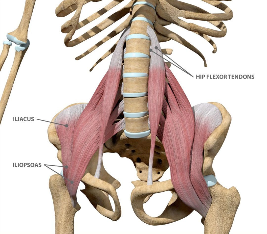
The iliacus is one of the body’s most important hip flexor muscles. The iliacus and surrounding muscles work together to produce the stability and range of motion required for bending, dancing, sitting, and walking.
Anatomy
The iliacus muscle is part of a complex muscle system in the hip and pelvis. Two iliacus muscles on each side of the pelvic bone enable the thigh to flex and rotate. They are innervated by the femoral nerve, which provides movement and sensation to the lower limbs. (Bordoni B. and Varacallo M. 2023) The iliacus muscle sits on the wing-shaped ilium and fits into the curved surface of the ilium, called the iliac fossa. The top of the muscle is attached to the upper wings of the ilium or iliac crest. It extends past the hip joint, which connects to the upper thigh bone/femur at the lesser trochanter protrusion. The iliacus is part of a major trio of muscles called the iliopsoas, including the major psoas and minor psoas muscles. These muscles are also attached to the upper femur but extend upward, connecting to the lumbar/lower spine at several attachment points. The iliopsoas also interact with the quadratus lumborum muscle, the deepest muscle of the lower back that starts at the iliac crest and attaches to the lumbar spine at several points. The quadratus lumborum enables flexion and elevation of the spine, while the iliopsoas enable the flexion and rotation of the hip and thigh.
Functions
The iliacus muscle has many functions that include: (Physiopedia, 2024)
Flexing and rotating the femur.
Helps maintain proper body posture while standing and sitting.
Produces hip movement that enables walking, running, and climbing stairs.
Provides hip flexion – bringing the knee to the chest.
Enables the forward tilt of the pelvis and side-bending.
Conditions
Several conditions can affect the iliacus muscle, specifically from under and/or overuse injuries. These conditions, collectively known as Iliopsoas syndrome, are typically the result of overuse/repetitive strain or injuries. These include:
Iliopsoas tendinopathy – which affects tendons.
Iliopsoas bursitis – which affects cushioning sacs known as bursae.
Iliopsoas syndrome can affect anyone but is common in:
Individuals and athletes who repeatedly use movements that flex the hips.
Track-and-field athletes
Gymnasts
Dancers
Iliopsoas Bursitis
This is the inflammation of the cushioning sac or bursa under the iliacus muscle, which helps the muscle slide over the pelvic bone. Symptoms can range from mild discomfort to pain that radiates through parts of the leg and hips. Runners, skiers, and swimmers are commonly affected, and individuals who regularly have tight hips and individuals with different forms of arthritis can also be affected. Early treatment can prevent the symptoms from worsening. Mild cases can be treated with self-care and stretching to help relieve tightness, rest, ice application, and over-the-counter nonsteroidal anti-inflammatory drugs. In severe cases, treatment options that may be recommended include: (Physiopedia, 2024)
Physical therapy
Assistant walking devices to relieve pressure – for example, a cane.
Corticosteroid steroid injections
Prescription anti-inflammatory medications
Iliopsoas Tendinopathy
Another condition affecting the iliacus muscles is iliopsoas tendinopathy, sometimes called snapping hip syndrome, because individuals can hear an audible snapping sound (Davenport KL. 2019). The condition is often experienced by dancers who repeatedly flex and hyperextend their hips and can result in hip and groin pain that gets worse with kicking or rotation. Treatment of iliopsoas tendinopathy can include:
Retraining muscle imbalances with strengthening and stretching exercises.
If these fail to provide relief, corticosteroid injections may be used. A saline hydro dissection can relieve stress around the tendon by injecting fluids that cushion and release trapped tissues.
Tendon release surgery may be recommended when all other options have failed. The surgical release involves severing the tendon to reduce pain and improve the range of motion.
Rehabilitation
Core muscle strengthening is essential to the rehabilitation of iliacus muscle injuries. The iliopsoas is an integral component of the core group and can benefit from stretching and strengthening exercises (Yogateket, 2019)
Lunge stretches
Straight leg raises
Knee-to-chest stretches
Standing hip flexion with resistance bands
Certain yoga poses can also help and include variations on the bridge pose that encourage hip flexion. (Yoga International, 2024)
Injury Medical Chiropractic and Functional Medicine Clinic
Iliopsoas pain is often felt at the front of the hips, thigh, mid-back, and lower back. Chiropractic care can help with iliacus muscle injuries through:
Evaluation
A chiropractor can evaluate the condition and determine if the iliacus muscle is causing pain.
Treatment plan
A chiropractor can create a personalized treatment plan that may include exercise instructions, manipulation, and other therapies.
Rehabilitation
A chiropractor can create a rehabilitation program to expedite healing.
Injury Medical Chiropractic and Functional Medicine Clinic works with primary healthcare providers and specialists to develop an optimal health and wellness solution. We focus on what works for you to relieve pain, restore function, and prevent injury. Regarding musculoskeletal pain, specialists like chiropractors, acupuncturists, and massage therapists can help mitigate the pain through spinal adjustments that help the body realign itself. They can also work with other medical professionals to integrate a treatment plan to resolve musculoskeletal issues.
Hip Labral Tear and Chiropractic Care
References
Bordoni, B., & Varacallo, M. (2024). Anatomy, Bony Pelvis, and Lower Limb, Iliopsoas Muscle. In StatPearls. https://www.ncbi.nlm.nih.gov/pubmed/30285403
Davenport KL. (2019). The professional dancer’s hip. Performing Arts Medicine, 77-87. https://doi.org/https://doi.org/10.1016/B978-0-323-58182-0.00009-2
Yogateket. Lizette Pompa. (2019). Essential yoga body parts. Hip flexor/psoas and yoga. Yogateket. https://www.yogateket.com/blog/hip-flexor-psoas-and-yoga
Can correcting body misalignments and the elements of unhealthy posture help achieve a healthy posture?
Healthy Posture
Maintaining a healthy posture is more important than ever, as individuals from all walks of life realize how practicing awkward and unhealthy postures can wreak havoc on their bodies and quality of life. Unhealthy postures include rounding the upper and lower back, slouching, and forward head posture. Over time, these postures make daily activities more difficult or painful. Mobility, stability, and strengthening exercises can address unhealthy posture problems and issues, along with practicing correct sitting, standing, and resting postural habits to reinforce proper alignment daily. A chiropractic and physical therapy team can treat and train individuals to restore correct and healthy posture.
Body Alignment
Ideal posture involves correct body alignment or how the structural parts, such as the head, trunk, hips, knees, etc., relate to an individual’s form. Whether standing, sitting, lying down, or moving, body parts need to be balanced in relationship to each other to avoid unnecessary stress on the spine and musculoskeletal system. (Bone Health & Osteoporosis Foundation, 2024)
Posture Types
Posture is considered static when sitting or standing still and dynamic when moving. Both are categorized as active postures as they require the activation of stabilizing muscles to deal with gravity and maintain alignment. Lying down and remaining still is considered an inactive posture, as muscle involvement is minimal. However, both have the potential to be healthy or unhealthy.
Proper Body Alignment
An easy way to check proper alignment while standing is to stand against a wall with the base of the head, shoulder blades, and buttocks flush against the wall, with enough space for a hand wide enough to slide in between the wall and the small of the back. This exercise correctly lines up the head, shoulders, and hips to reduce or eliminate undue stress on the spine. Body balance is the foundation for active and inactive postures concerning workstations, industrial ergonomics, daily activities, and sports. (Mayo Clinic, 2023) Healthy alignment is a standard position in which all body joints are centered and balanced and the most mechanically efficient position for static or dynamic activities. Biomechanical efficiency (the ability to use the body’s muscles and joints to perform movements while minimizing energy use and maximizing output) enables the muscles surrounding the joints to work in balance and efficiently activate, which, in turn, helps reduce strain, tension, and injury. Balanced muscles also conserve energy, leading to better daily stamina.
Development of Imbalances
Individuals develop position and movement habits in joint positioning that have led to imbalanced muscles. When this is the case, some muscles can be chronically stretched, and others become chronically tight, all to hold the body up or move around, which can lead to postural conditions like upper crossed (Physiopedia, 2024) or lower crossed syndrome (Physiopedia, 2024) which often leads to pain and/or mobility issues.
Posture Assessment
The recommended way to determine if one’s posture is healthy or poor is by conducting a posture assessment. The examination looks at joint positions and gathers visual information about the planes into which parts of each joint move and the axes around which those movements occur (Singla D. and Veqar Z., 2014) (Debra Coglianese et al., 2006). In a posture assessment, body alignment is compared with the ideal standard, a plumb line, usually a string with a small weight attached to the bottom to help maintain straightness. The other end of the string is affixed to the ceiling to be used as an accurate reference for correct alignment. (Singla D. and Veqar Z. 2014) During a posture assessment, the patient stands next to the plumb line while the doctor or therapist compares the relative positions of the following areas:
Ears
Shoulder joint
Spine
Hip joint
Knee joint
Ankle joint
Feet
Any areas that don’t match the reference can indicate misalignments in one or several regions.
Making Corrections
Chiropractic care can help correct unhealthy posture by realigning the spine and strengthening the musculoskeletal system:
Spinal Adjustments
Chiropractors use their hands or instruments to apply controlled force to the spine to realign the vertebrae. This can help relieve pressure on muscles, ligaments, and nerves, which can improve posture.
Corrective Exercises
Chiropractors can create custom exercises to strengthen postural muscles and maintain proper alignment.
Massage Therapy
Chiropractors can use massage therapy to work on strained ligaments and soft tissue.
Guidance and Training
Chiropractors can teach patients how to move to encourage a neutral spine and provide strategies for maintaining healthy posture.
A posture corrector or brace can also help teach and engage the correct muscles to achieve proper alignment. However, it should not be relied on long-term because promoting and activating one’s stabilizing muscles is important rather than relying on a brace for prolonged periods.
Injury Medical Chiropractic and Functional Medicine Clinic
Achieving and maintaining proper posture requires consistent work and development. Retraining the body and maintaining its optimal health requires daily efforts through exercise, conscious position corrections, and ergonomics. Injury Medical Chiropractic and Functional Medicine Clinic works with primary healthcare providers and specialists to develop an optimal health and wellness solution. We focus on what works for you to relieve pain, restore function, and prevent injury. Regarding musculoskeletal pain, specialists like chiropractors, acupuncturists, and massage therapists can help mitigate the pain through spinal adjustments that help the body realign itself. They can also work with other medical professionals to integrate a treatment plan to resolve musculoskeletal issues.
Upper Cross Syndrome
References
Bone Health & Osteoporosis Foundation. (2024). Proper body alignment. https://www.bonehealthandosteoporosis.org/patients/treatment/exercisesafe-movement/proper-body-alignment/
Mayo Clinic. (2023). Mayo Clinic Q and A: Proper posture and body alignment. https://newsnetwork.mayoclinic.org/discussion/mayo-clinic-q-and-a-proper-posture-and-body-alignment/
Singla, D., & Veqar, Z. (2014). Methods of postural assessment used for sports persons. Journal of clinical and diagnostic research: JCDR, 8(4), LE01–LE4. https://doi.org/10.7860/JCDR/2014/6836.4266
Coglianese, D. (2006). Muscles: Testing and Function With Posture and Pain, ed 5 (with Primal Anatomy CD-ROM). Physical Therapy, 86(2), 304-305. https://doi.org/https://doi.org/10.1093/ptj/86.2.304
An axillary nerve injury can cause pain, weakness, and shoulder mobility loss. Can physical therapy help restore and maintain shoulder joint flexibility?
Axillary Nerve
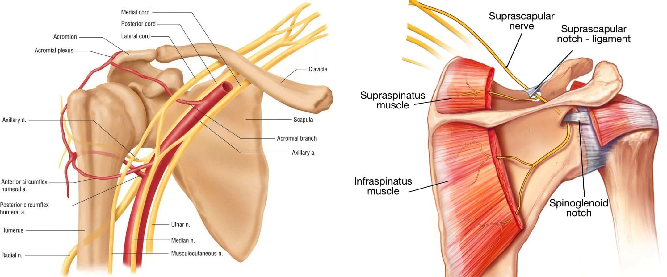
The axillary nerve, or the circumflex nerve, is a peripheral nerve that runs through the shoulder and supports movement and sensation in the upper limbs. It originates in the neck at the brachial plexus, a network of nerves that extends from the neck and upper torso to the shoulders and arms. Its primary purpose is to supply nerve function to the shoulder joint and three muscles in the arm and also innervates some skin in the region.
Anatomy
Except for the cranial nerves, all the body’s nerves branch off from the spinal cord, emerge from between vertebrae and continue to branch off as they travel to various muscles and other structures. The axillary nerve is named after the axilla, the medical name for the armpit. Individuals have two, one on each side. After leaving the spinal column, the axillary nerve runs behind the axillary artery and continues to the shoulder blade’s lower edge of the subscapularis muscle. It winds back and travels down the arm along the posterior humeral circumflex artery, which then passes through the quadrangular space (a small area of the shoulder blade just above the armpit where there is a gap in the muscles that allows nerves and blood vessels to pass through to the arm before it divides into terminal branches, which are:
Anterior Division
Supplies motor innervation to the deltoid’s anterior and middle heads, allowing the arm to abduct or move away from the body.
It winds around the neck of the humerus/funny bone, goes beneath the deltoid muscle, and then connects to the forward edge of the deltoid.
A few small cutaneous branches serve the skin in that area.
Posterior Division
Innervates the teres minor muscles and the lower part of the deltoid.
It enters the deep fascia and becomes the superior lateral cutaneous nerve.
It then wraps around the lower edge of the deltoid, connects to the skin over the lower two-thirds of the muscle, and covers the long head of the triceps brachii.
Articular Branch
Comes from the trunk of the axillary nerve and enters the glenohumeral joint, which is in the shoulder, below the subscapularis muscle.
Anatomical Variations
In a case report, healthcare providers noted an incidence of the nerve branching directly off the upper trunk of the brachial plexus rather than the posterior cord. (Subasinghe S. K. and Goonewardene S. 2016) In this case, it innervated the subscapularis muscle, latissimus dorsi, and the deltoid and teres minor muscles and also had a communicating branch to the posterior cord. Another case documented multiple abnormalities in the course of the axillary nerve in an individual with pain and severely limited shoulder mobility. (Pizzo R. A. et al., 2019) During reverse shoulder arthroplasty, the surgeon discovered that the axillary nerve ran beside the coracoid process instead of underneath and stayed close to the subscapularis muscle instead of traveling through the quadrangular space. The case noted earlier reports of axillary nerves not running through the quadrangular space. In those cases, the nerve pierced the subscapularis muscle or split into branches before reaching the quadrangular space.
Function
The axillary nerve functions as a motor nerve that controls movement and a sensory nerve that controls sensations like touch or temperature.
Motor
As a motor nerve, the axillary nerve innervates three muscles in the arm and includes:
Deltoid
Allows flexing of the shoulder joint and rotating the shoulder inward.
Long Head of the Triceps
It runs down the back of the outer arm, allowing straightening, pulling the upper arm toward the body, or extending it backward.
The radial nerve can also innervate this muscle.
Teres Minor
One of the rotator cuff muscles starts outside the shoulder and runs diagonally along the bottom edge of the shoulder blade.
It works with other muscles to allow for the external rotation of the shoulder joint.
Sensory
In its sensory role, the nerve carries information to the brain from the following:
Glenohumeral joint or the ball-and-socket joint in the shoulder.
The skin on the lower two-thirds of the deltoid muscle through the superior lateral cutaneous branch.
Injuries and Conditions
Problems with the axillary nerve can be caused by injuries anywhere along the arm and shoulder and by disease. Common injuries include:
Dislocations
Of the shoulder joint, which can cause axillary nerve palsy.
Fracture
Of the surgical neck of the humerus.
Compression
This stems from walking with crutches, also known as crutch palsy.
Direct Trauma
This can be from an impact sports, work, automobile accident, collision, or laceration.
Added Pressure
This can be from wearing a cast or splint.
Surgical Accidental Injury
An injury or damage can come from shoulder surgery, especially arthroscopic surgery on the inferior glenoid and capsule.
Quadrangular Space Syndrome
This is where the axillary nerve is compressed where it passes through that space, which is most common in athletes who perform frequent overhead motions)
Nerve Root Damage
Between the fifth and sixth cervical vertebrae, where the nerve emerges from the spinal cord, which can be caused by traction, compression, spinal disc prolapse, or a bulging disc.
Systemic Neurological Disorders
Example – multiple sclerosis
Erb’s Palsy
A condition often is the result of a birth injury called shoulder dystocia, in which a baby’s shoulder/s becomes stuck during childbirth.
Axillary Nerve Palsy
Damage can result in a type of peripheral neuropathy that can cause weakness in the deltoid and teres minor muscles.
This can result in losing the ability to lift the arm away from the body and weakness in various shoulder movements.
If the damage is severe enough, it can cause paralysis of the deltoid and other minor muscles, resulting in flat shoulder deformity, in which individuals cannot lay their shoulders flat when lying down.
Axillary nerve damage also can lead to a change, reduction, or loss of sensation in a small part of the arm just below the shoulder.
Nerve Injury Statistics
Three times more common in men than women.
It may be present in as many as 65% of shoulder injuries.
The risk of injury due to dislocation is significantly increased after age 50.
Tests
If a healthcare provider suspects a problem with axillary nerve function, they’ll test the shoulder’s range of motion and skin sensitivity. A difference in the range of motion between the shoulders can indicate a nerve injury. Individuals may be sent for electromyography and a nerve conduction study to verify nerve palsy. In some cases, an MRI and/or X-rays may be ordered, especially if the cause of possible nerve damage is unknown.
Rehabilitation
Depending on the severity and cause of the injury, non-surgical treatments may be recommended, with surgery as a last resort. Non-surgical treatment can include some combination of immobilization, rest, ice, physical therapy, and anti-inflammatory meds. Physical treatment typically lasts about six weeks and focuses on strengthening and stimulating the muscles to prevent joint stiffness, which can impair long-term function.
Surgery
If conservative treatments don’t work, surgery may be recommended, especially if several months have passed without improvement. Surgical outcomes are generally better if surgery is performed within six months of the injury, and regardless of the time frame, the prognosis is considered positive in about 90% of cases. Surgical procedures performed for axillary nerve dysfunction or injury include:
Neurolysis
This procedure involves targeted damage/degeneration of nerve fibers, interrupts the nerve signals, and eliminates pain while the damaged area heals.
Neurorrhaphy
This procedure stitches a severed nerve back together.
Nerve Grafting
Grafting involves transplanting a portion of another nerve, usually the sural nerve, to reconnect severed nerves.
This helps, especially when the damaged portion is too large to be repaired by neurorrhaphy.
It allows a pathway for signals and encourages the regrowth of nerve axons.
Neurotization or Nerve Transfer
Similar to grafting but used when the nerve is too damaged to heal.
This procedure involves transplanting a healthy but less important nerve, or a portion of a nerve, to replace the damaged one and restore function.
Injury Medical Chiropractic and Functional Medicine Clinic
Injury Medical Chiropractic and Functional Medicine Clinic works with primary healthcare providers and specialists to develop an optimal health and wellness solution. We focus on what works for you to relieve pain, restore function, and prevent injury. Regarding musculoskeletal pain, specialists like chiropractors, acupuncturists, and massage therapists can help mitigate the pain through spinal adjustments that help the body realign itself. They can also work with other medical professionals to integrate a treatment plan to resolve musculoskeletal issues.
Shoulder Pain Chiropractic Treatment
References
Subasinghe, S. K., & Goonewardene, S. (2016). A Rare Variation of the Axillary Nerve Formed as Direct Branch of the Upper Trunk. Journal of clinical and diagnostic research : JCDR, 10(8), ND01–ND2. https://doi.org/10.7860/JCDR/2016/20048.8255
Pizzo, R. A., Lynch, J., Adams, D. M., Yoon, R. S., & Liporace, F. A. (2019). Unusual anatomic variant of the axillary nerve challenging the deltopectoral approach to the shoulder: a case report. Patient safety in surgery, 13, 9. https://doi.org/10.1186/s13037-019-0189-1
Can individuals maintain proper hydration levels in their bodies to prevent spinal pain and other musculoskeletal conditions?
Introduction
When making small changes to better their health and wellness, many understand that eating nutritional foods, exercising, and finding ways to reduce the pain and stress that environmental factors have affected the body. However, there is also another crucial component to a person’s health and wellness, and that is staying hydrated. Many individuals sometimes don’t realize that the human body contains about 60% of the water, which helps every vital organ, muscle, tissue, and vertebra in the body to function. However, many individuals who do not consume enough water due to a stressful event, exercising, or just wanting to drink sugary drinks will feel pain and discomfort in their bodies. This can range from mild headaches to back pain. Today’s article focuses on what spinal pain correlates with the back, why staying hydrated can reduce spinal pain and ways to stay hydrated. We talk with certified associated medical providers who inform our patients to maintain proper hydration to reduce the chances of spinal pain affecting their bodies. While asking their associated medical provider intricate questions, we advise patients to incorporate daily hydration levels as part of their routine to reduce the chances of spinal pain from returning. Dr. Alex Jimenez, D.C., includes this information as an academic service. Disclaimer.
Spinal Pain Is Correlated With The Back
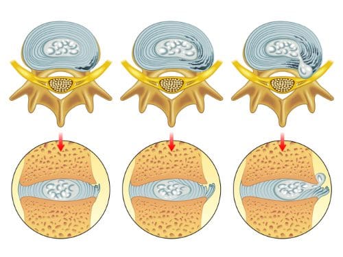
Do you feel constant aches or pains after a stressful day? Do you feel like chugging gallons of water but still feel thirsty? Or have you been noticing that you have been dealing with limited mobility that has been causing you pain and discomfort in your lower back? These pain-like scenarios are multifactorial and can correlate with spinal pain affecting the back. Now, many people wonder why spinal pain is associated with back pain. The spine is crucial in allowing the individual to be upright and move around during daily activities. The spinal vertebrae discs are between the bones, which would enable shock absorption when a person twists, bends, or flexes so the bones don’t rub together. However, when the body ages, the spine does, too, which causes the intervertebral disc to gradually fall and change the dynamics of the affected spine segments. (Kos et al., 2019) To that point, when the intervertebral disc becomes compressed due to degeneration, it can lead to the development of back pain.
Since low back pain is a multifactorial musculoskeletal condition, many individuals often develop low back pain from numerous environmental risk factors like physical labor, excessive sitting or standing, poor dieting, andphysical inactivity, which can cause individuals to have a loss of sensation, reduced reflexes, limited range of movements and tenderness can affect a person’s gait and posture. (Kabeer et al., 2023) When the spinal discs are being compressed, the water inside the disc becomes dehydrated and is linked to lower back pain. (Cheung et al., 2023) When the spinal discs are dehydrated in the spine due to improper hydration, it can lead to immobility, cause the disc to degenerate, and cause spinal structural defects that cause the surrounding muscles and tissues to have biomechanical instability and inflammation. (Hauser et al., 2022) When the body starts dehydrating, it retrieves water from the spine and other parts to sustain life and keep the vital organs functioning.
Eating Right To Feel Better- Video
Why Staying Hydrated Can Reduce Spinal Pain
Proper hydration is key to reducing the chances of spinal pain. By staying hydrated with plain, pure water, individuals can stay focused, remove toxins from the body, aid in weight reduction, and reduce muscle and low back pain. (Nsiah-Asamoah & Buxton, 2021) Drinking at least 64 ounces of water a day can rehydrate the body and prevent future issues that can cause pain.Proper hydration allows the body to maintain cell activity and regulate body temperature, reducing the chances of pain affecting the body.
Ways to Stay Hydrated
There are ways to stay hydrated and allow the body to have proper hydration levels. Incorporating hydrating fruits and vegetables can help aid in proper hydration as these fruits and vegetables contain about 90% water and the essential nutrients. Additionally, when individuals try to get their hydration levels back up, they are encouraged to drink more fluids as they prefer, especially if they feel unwell. This is to ensure that the body is hydrated enough so that individuals can receive subcutaneous or intravenous fluid supplements combined with regular fluid intake. (Li et al., 2023) Another way many individuals can stay hydrated is to combine non-surgical treatments like chiropractic care and spinal decompression to help realign and lengthen the spine to reduce unwanted pressure on the spinal discs and rehydrate them. The combination of non-surgical treatments and drinking plenty of hydrating fluids can help many individuals dealing with back pain associated with the spine as they start to make small changes to their routine and can live a life pain-free.
References
Cheung, S. T. Y., Cheung, P. W. H., & Cheung, J. P. Y. (2023). Why Are Some Intervertebral Discs More Prone to Degeneration?: Insights Into Isolated Thoracic “Dysgeneration”. Spine (Phila Pa 1976), 48(12), E177-E187. https://doi.org/10.1097/BRS.0000000000004632
Hauser, R. A., Matias, D., Woznica, D., Rawlings, B., & Woldin, B. A. (2022). Lumbar instability as an etiology of low back pain and its treatment by prolotherapy: A review. J Back Musculoskelet Rehabil, 35(4), 701-712. https://doi.org/10.3233/BMR-210097
Kabeer, A. S., Osmani, H. T., Patel, J., Robinson, P., & Ahmed, N. (2023). The adult with low back pain: causes, diagnosis, imaging features and management. Br J Hosp Med (Lond), 84(10), 1-9. https://doi.org/10.12968/hmed.2023.0063
Li, S., Xiao, X., & Zhang, X. (2023). Hydration Status in Older Adults: Current Knowledge and Future Challenges. Nutrients, 15(11). https://doi.org/10.3390/nu15112609
Nsiah-Asamoah, C. N. A., & Buxton, D. N. B. (2021). Hydration and water intake practices of commercial long-distance drivers in Ghana: what do they know and why does it matter? Heliyon, 7(3), e06512. https://doi.org/10.1016/j.heliyon.2021.e06512
For individuals with chronic pain conditions, what are the risks of developing peptic ulcers?
NSAIDs and Peptic Ulcers
A peptic ulcer is a sore in the lining of the stomach or duodenum, the first area of the small intestine. The most common symptom is a burning stomach pain that may come and go for several days or weeks. Other symptoms include:
Feeling full
Feeling bloated
Belching
Heartburn
Nausea
Stress and spicy foods can worsen ulcers but do not cause them.
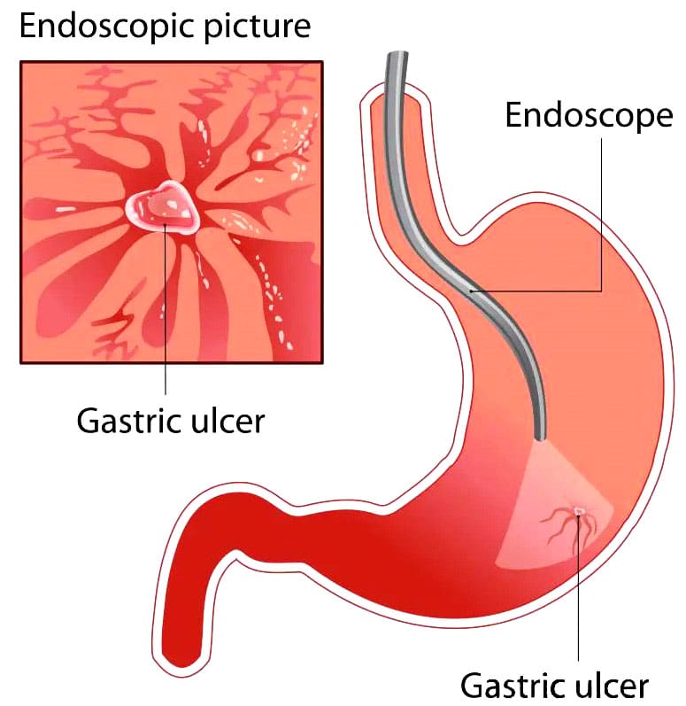
A peptic ulcer is a sore that occurs in the mucosal lining of the stomach, small intestine, or esophagus. It is caused by stomach acids or other digestive juices damaging the stomach or duodenum lining. When the ulcer is in the stomach, it might also be called a gastric ulcer. The acid can create a painful open sore that may bleed. The most common cause of a peptic ulcer is a type of bacteria called Helicobacter pylori (H. pylori). A second, less common cause of peptic ulcers is the use of non-steroidal anti-inflammatory medications or NSAIDs like aspirin, ibuprofen, and naproxen. (Fashner J. & Gitu A. C. 2015) Using over-the-counter NSAIDs for the occasional headache or achy back won’t cause a peptic ulcer. Rather, peptic ulcer disease is something that can occur with longer-term use, especially at high doses, such as for chronic pain associated with arthritis or other inflammatory conditions. (National Institute of Diabetes and Digestive and Kidney Diseases, 2022)
NSAIDs and Ulcer Development
NSAIDs can cause ulcers because they can interfere with the stomach’s ability to protect itself from gastric acids as they slow the production of protective mucus in the stomach and change its structure. (National Institute of Diabetes and Digestive and Kidney Diseases, 2022) While these acids are vital to the digestive process, they can compromise the stomach’s protective barriers. The stomach has three protections against gastric acid:
Foveolar cells that line the stomach produce a protective mucus.
Bicarbonate is produced by the foveolar cells, which help neutralize stomach acid.
Blood circulation aids in repairing and renewing cells in the stomach’s mucosal layer.
Specific lipids called prostaglandins, which the body makes, affect pain receptors. NSAIDs work to reduce pain by blocking enzymes involved in the production of certain prostaglandins. Prostaglandins also protect the stomach’s mucosal layer, which can be broken when depleted. Suppressing the body’s natural defenses against gastric acids can lead to inflammation in the stomach lining. Over time, this can cause a capillary blood vessel to rupture, causing bleeding and the development of an open, ulcerative sore. (Bjarnason I. et al., 2018)
Symptoms
A peptic ulcer may cause symptoms in the digestive tract, but some individuals may have no symptoms. The most common symptom is upper abdominal pain, which can feel dull or burning. The pain can range in severity, with some experiencing mild discomfort and others having severe pain. Most of the time, the pain will occur after a meal, but it might also happen at night for some. It could go on from a few minutes to a few hours. (National Institute of Diabetes and Digestive and Kidney Diseases, 2022) Other symptoms are less common but can include:
Bloating
Burping
Gas
Nausea
Vomiting
Loss of appetite
Weight loss
Feeling sick in the stomach.
Feeling full after even a small meal.
In rare cases, individuals with peptic ulcers may see blood in their stool or have black stools because blood is present. Blood coming from one or more peptic ulcers could also be visible in vomit. Call a healthcare provider right away if there is blood in stool or vomit, as this can be a sign of excessive bleeding or other serious problems. (National Institute of Diabetes and Digestive and Kidney Diseases, 2022)
Diagnosis
When peptic ulcer symptoms occur, a healthcare provider may order several tests to determine the cause. For individuals who are receiving NSAIDs for chronic pain, a healthcare provider may already have a high suspicion that the medication is the cause or is contributing to peptic ulcer disease. Because infection with H. pylori is the most common cause, it is normally ruled out through a breath, blood, or stool test. (National Institute of Diabetes and Digestive and Kidney Diseases, 2022) Tests to look for ulcers inside the upper digestive tract can include:
Upper GI Series
Patients drink a barium substance to help the internal organs appear on imaging.
A series of X-rays are taken.
Upper Endoscopy
A flexible tube with a camera is used to look inside the esophagus, the stomach, and the duodenum.
All NSAIDs have the potential to cause indigestion, gastric bleeding, and ulcers. However, some individuals are more susceptible to developing peptic ulcer disease than others. Peptic ulcers caused by NSAIDs are more likely to occur in individuals who: (National Institute of Diabetes and Digestive and Kidney Diseases, 2022)
Are 70 or older
Have a history of ulcers
Take corticosteroids
Take high-dose NSAIDs
Take NSAIDs regularly for a long time
Take more than two types of NSAIDs
Use aspirin daily, including low-dose aspirin, for cardioprotective purposes.
Take blood thinners
Drink alcohol regularly
Smoke
Studies suggest that 25% of those who use NSAIDs long-term will develop an ulcer, but only a small percentage will go on to develop serious complications. (Lanza F. L. et al., 2009)
Treatment
NSAID-induced ulcers usually heal once the NSAID is stopped. Certain treatments may be recommended to expedite the healing process depending on the severity of the peptic ulcer. In severe cases, surgery may be recommended to repair the damage.
Medications
A healthcare provider may recommend taking one or more medications. Over-the-counter options may include:
Antacids help neutralize stomach acid.
A proton pump inhibitor or PPI, such as omeprazole, lowers acid levels in the stomach. (Begg M. et al., 2023)
The bigger problem for individuals is how to manage pain when the medications are discontinued. For individuals with chronic pain, this may require the help of a physical therapy team, including a pain management healthcare provider. Certain medications called COX inhibitors could be used to control pain for some. COX inhibitors have been shown to work for pain relief and are associated with fewer digestive side effects than other types of NSAIDs. These meds have also been shown to have cardiovascular side effects, however, so it’s usually recommended they be used at the lowest dosage. (Scarpignato C. et al., 2015)
Lifestyle Adjustments
Lifestyle adjustments may be recommended to heal peptic ulcers, including:
Avoiding foods that worsen symptoms.
Avoiding caffeine
Avoiding alcohol
Quitting smoking
Surgery
In some cases, surgery may be recommended, but this is more often the case when there are complications as a result of the ulcer, such as:
Avoiding long-term and high-dose use of NSAIDs or not using these medications can help protect and prevent peptic ulcers. For individuals who have to take an NSAID due to a condition they are trying to manage, a healthcare provider may prescribe one of the meds used to treat peptic ulcers to prevent them from occurring. Some believe that spicy food and stress cause ulcers, but that has been discredited. (Cleveland Clinic, 2022) Most individuals who take NSAIDs will not experience peptic ulcer disease. (Drini M. 2017) However, those who have chronic pain and who are receiving high doses should be aware of the potential.
Injury Medical Chiropractic and Functional Medicine Clinic
Individuals who have any concerns about the use of NSAIDs and how the digestive system will be affected should ask a healthcare provider if there are ways to prevent ulcers and if those measures should be implemented while receiving high doses of NSAIDs. Left untreated, ulcers can lead to complications, which is why it is important to get a diagnosis and receive treatment. Injury Medical Chiropractic and Functional Medicine Clinic works with primary healthcare providers and specialists to develop an optimal health and wellness solution. We focus on what works for you to relieve pain, restore function, and prevent injury. Regarding musculoskeletal pain, specialists like chiropractors, acupuncturists, and massage therapists can help mitigate the pain through spinal adjustments that help the body realign naturally.
Root Causes of Gut Dysfunction
References
Fashner, J., & Gitu, A. C. (2015). Diagnosis and Treatment of Peptic Ulcer Disease and H. pylori Infection. American family physician, 91(4), 236–242.
National Institute of Diabetes and Digestive and Kidney Diseases. (2022). Peptic ulcers (stomach ulcers). Retrieved from https://www.niddk.nih.gov/health-information/digestive-diseases/peptic-ulcers-stomach-ulcers
Bjarnason, I., Scarpignato, C., Holmgren, E., Olszewski, M., Rainsford, K. D., & Lanas, A. (2018). Mechanisms of Damage to the Gastrointestinal Tract From Nonsteroidal Anti-Inflammatory Drugs. Gastroenterology, 154(3), 500–514. https://doi.org/10.1053/j.gastro.2017.10.049
Lanza, F. L., Chan, F. K., Quigley, E. M., & Practice Parameters Committee of the American College of Gastroenterology (2009). Guidelines for prevention of NSAID-related ulcer complications. The American journal of gastroenterology, 104(3), 728–738. https://doi.org/10.1038/ajg.2009.115
Begg, M., Tarhuni, M., N Fotso, M., Gonzalez, N. A., Sanivarapu, R. R., Osman, U., Latha Kumar, A., Sadagopan, A., Mahmoud, A., & Khan, S. (2023). Comparing the Safety and Efficacy of Proton Pump Inhibitors and Histamine-2 Receptor Antagonists in the Management of Patients With Peptic Ulcer Disease: A Systematic Review. Cureus, 15(8), e44341. https://doi.org/10.7759/cureus.44341
Scarpignato, C., Lanas, A., Blandizzi, C., Lems, W. F., Hermann, M., Hunt, R. H., & International NSAID Consensus Group (2015). Safe prescribing of non-steroidal anti-inflammatory drugs in patients with osteoarthritis–an expert consensus addressing benefits as well as gastrointestinal and cardiovascular risks. BMC medicine, 13, 55. https://doi.org/10.1186/s12916-015-0285-8
Lee, C. W., & Sarosi, G. A., Jr (2011). Emergency ulcer surgery. The Surgical clinics of North America, 91(5), 1001–1013. https://doi.org/10.1016/j.suc.2011.06.008
Cleveland Clinic. (2022). Can stress give you an ulcer? https://health.clevelandclinic.org/can-stress-give-you-an-ulcer
Drini M. (2017). Peptic ulcer disease and non-steroidal anti-inflammatory drugs. Australian prescriber, 40(3), 91–93. https://doi.org/10.18773/austprescr.2017.037
IFM's Find A Practitioner tool is the largest referral network in Functional Medicine, created to help patients locate Functional Medicine practitioners anywhere in the world. IFM Certified Practitioners are listed first in the search results, given their extensive education in Functional Medicine