I’m definitely able to do day-to-day stuff a lot easier. It’s just like a much happier life with less pain. Just doing anything like working out or any type of activity that a person would take for granted if you don’t have pain, it’s different when you have pain, and so to get pain relief is amazing.
Gale Grijalva
Head and neck injuries are health issues commonly caused by�automobile accidents. Due to the force of the impact, a�moderate fender bender can sometimes even jerk a victim enough to make them hit their head inside the vehicle. The brain�can be very susceptible to suffering damage�after an auto accident, leading to neurological issues which can have lasting effects.
Nerve damage is a prevalent consequence after a car crash, and it can�cause debilitating symptoms, such as pain, headaches, and mental health issues, among others, ultimately making it difficult for anyone to go about their everyday activities.�When it comes to nerve damage, the most common types of automobile accident injuries include:
Whiplash, an intense jerking motion of the head and neck which can cause the nerves to stretch or be pinched;
Blunt-force trauma, hitting your head, arms, or legs on a hard surface inside or outside the vehicle, compressing the nerves; and
Lacerations, deep cuts into the skin sustained during an automobile accident that can sever the nerves in the affected region.
Several signs and symptoms can help indicate when nerves are damaged. These include�pain; partial or full paralysis of limbs and appendages like fingers and/or toes; muscular fatigue; twitching or uncontrolled movements of muscles; a prickling sensation; tingling or numbness on the skin or in limbs; or increased sensitivity to cold and hot temperatures on the surface. Below, we will discuss the effects of nerve damage after an auto accident.
Neuropathy After Auto Injuries
Neuropathy, or nerve damage, may be brought on by sports injuries, work-related injuries, automobile accident injuries, or repetitive motion injuries. These scenarios may cause the nerves to be completely or partially compressed, stretched or even severed. Dislocated or broken, fractured, bones may also place an unnecessary quantity of pressure on the nerves, where slipped intervertebral discs can compress the nerve fibers.
Neuropathy,�a term used to describe nerve damage, usually involves�the peripheral nerves instead of the central nervous system, or the brain and spinal cord. This health issue may not only develop due to the causes�explained above,�but nerve damage can also occur for many other reasons. The most prevalent nerves to be affected by neuropathy include the motor nerves, the autonomic nerves, and the sensory nerves.
The motor nerves enable movement and power;
The autonomic nerves control the systems of the body; and
The sensory nerves control feeling.
Diagnosing neuropathy to determine the best treatment options can help a victim regain a healthy lifestyle. The healthcare professional will begin their evaluation by reviewing the patient’s medical history, including general health, signs and symptoms, any other�type of neuropathy in the family, current or recent prescriptions used, any exposure to poisons or toxins, alcohol consumption, and sexual history.
They will then diagnose the cause of the neuropathy by checking the skin, taking their pulse in different places, examining for feeling, such as analyzing vibration sensations with a tuning fork and evaluating tendon reflexes. The healthcare professional may determine your precise treatment options once the source of the neuropathy is narrowed down. The proper treatment approach can help manage the symptoms.
Radiculopathy After Auto Injuries
Radiculopathy is the medical term used to describe compression or irritation of a nerve in the spine. It is not a specific condition, but instead, a description of a general health issue in which or more nerves are affected, causing symptoms. Radiculopathy may cause pain, tingling sensations, numbness, or fatigue. This condition can occur in any portion of the spine, although it may be more common in some areas than others.
It is most common in the lower back (lumbar radiculopathy);
And in the neck (cervical radiculopathy);
It is�less common in the middle portion of the spine (thoracic radiculopathy), but it’s still tremendously debilitating.
Cervical radiculopathy is pain and other symptoms resulting from any condition which affects the nerves in the cervical, thoracic, or lumbar spine. Degeneration of the cervical region of the spine may lead to a myriad of conditions that might result in problems. These are usually divided between problems that come from health issues originating from pinched or irritated nerves as well as other underlying problems in the neck.
Lumbar radiculopathy causes pain which occurs in the lower back. Damage or injuries to the lumbar spine and compression or impingement of the nerve roots can cause pain, tingling sensations, and numbness. Automobile accident injuries can result in very significant pathologies including damage to the intervertebral discs, muscles, tendons, and ligaments as well as to the nerves traveling down the length of the spine.
Like neuropathy, a diagnosis for radiculopathy begins with a review of a patient’s medical history and a physical evaluation by the healthcare professional. The doctor might be able to determine the source of the symptoms by evaluating the patient’s muscle strength, sensation, and reflexes. These tests often comprise of a CT scan, an MRI or X-rays. The exam may also include an electromyogram or a nerve conduction study which analyzes the current threshold of sensibility in patients.
Millions of people are involved in automobile accidents every year, many of which result in long-term injuries and disability. Chiropractic care is one of the most frequently considered forms of treatment after an auto accident. Through the use of spinal adjustments and manual manipulations, a doctor of chiropractic can help restore normal function to the nervous system in order to allow the body to naturally heal itself.
Dr. Alex Jimenez D.C., C.C.S.T.
Treatment After Auto Injuries
The force that’s often placed on the�neck and the spine during an auto accident can cause nerve damage.�If you experience any signs and symptoms after being involved in a car crash, it’s essential to seek immediate medical attention from a healthcare professional, such as a chiropractor, to receive the proper diagnosis and treatment. Chiropractic care is a popular treatment for automobile accident injuries.
Chiropractic care is an alternative treatment approach which focuses on the diagnosis, treatment, and prevention of a variety of injuries and/or conditions associated with the musculoskeletal and nervous system. Through the use of spinal adjustments and manual manipulations, a chiropractor can carefully correct any spinal misalignments�which may be placing unnecessary amounts of stress on the nerves.�
By naturally restoring the original integrity of the spine, chiropractic care has become one of the most common treatments for a variety of injuries and conditions, including nerve damage associated with automobile accident injuries. The scope of our information is limited to chiropractic as well as to spinal injuries and conditions. To discuss the subject matter, please feel free to ask Dr. Jimenez or contact us at�915-850-0900�.
Curated by Dr. Alex Jimenez
Additional Topics: Central Sensitization After Auto Injuries
Central sensitization is a health issue affecting the nervous system which is commonly associated with the development of chronic pain. With central sensitization, the nervous system experiences a “wind-up” process that causes it to become regulated in a constant state of high reactivity. This constant, or persistent, state of high reactivity lowers the threshold for what should be causing pain in the human body, ultimately maintaining pain even after the initial injury has healed. Central sensitization is identified by two main characteristics, both of which involve a heightened sensitivity to pain and the sensation of touch, known as allodynia and hyperalgesia.
After a neurological exam, physical exam, patient history, x-rays and any previous screening tests, a doctor may order one or more of the following diagnostic tests to determine the root of a possible/suspected neurological disorder or injury. These diagnostics generally involve neuroradiology, which uses small amounts of radioactive material to study organ function and structure and ordiagnostic imaging, which use magnets and electrical charges to study organ function.
Neurological Studies
Neuroradiology
MRI
MRA
MRS
fMRI
CT scans
Myelograms
PET scans
Many others
Magnetic Resonance Imaging (MRI)
Shows organs or soft tissue well
No ionizing radiation
Variations on MRI
Magnetic resonance angiography (MRA)
Evaluate blood flow through arteries
Detect intracranial aneurysms and vascular malformations
Magnetic resonance spectroscopy (MRS)
Assess chemical abnormalities in HIV, stroke, head injury, coma, Alzheimer’s disease, tumors, and multiple sclerosis
Functional magnetic resonance imaging (fMRI)
Determine the specific location of the brain where activity occurs
Computed Tomography (CT or CAT Scan)
Uses a combination of X-rays and computer technology to produce horizontal, or axial, images
Shows bones especially well
Used when assessment of the brain needed quickly such as in suspected bleeds and fractures
Myelogram
Contrast dye combined with CT or Xray
Most useful in assessing spinal cord
Stenosis
Tumors
Nerve root injury
Positron Emission Tomography (PET Scan)
Radiotracer is used to evaluate the metabolism of tissue to detect biochemical changes earlier than other study types
Used to assess
Alzheimer’s disease
Parkinson’s disease
Huntington’s disease
Epilepsy
Cerebrovascular accident
Electrodiagnostic Studies
Electromyography (EMG)
Nerve Conduction Velocity (NCV) Studies
Evoked Potential Studies
Electromyography (EMG)
Detection of signals arising from the depolarization of skeletal muscle
May be measured via:
Skin surface electrodes
Not used for diagnostic purposes, more for rehab and biofeedback
Needles placed directly within the muscle
Common for clinical/diagnostic EMG
Diagnostic Needle EMG
Recorded depolarizations may be:
Spontaneous
Insertional activity
Result of voluntary muscle contraction
Muscles should be electrically silent at rest, except at the motor end-plate
Practitioner must avoid insertion in motor end-plate
At least 10 different points in the muscle are measured for proper interpretation
Procedure
Needle is inserted into the muscle
Insertional activity recorded
Electrical silence recorded
Voluntary muscle contraction recorded
Electrical silence recorded
Maximal contraction effort recorded
Samples Collected
Muscles
Innervated by the same nerve but different nerve roots
Innervated by the same nerve root but different nerves
Different locations along the course of the nerves
Helps to distinguish the level of the lesion
Motor Unit Potential (MUP)
Amplitude
Density of the muscle fibers attached to that one motor neuron
Proximity of the MUP
Recruitment pattern can also be assessed
Delayed recruitment can indicated loss of motor units within the muscle
Early recruitment is seen in myopathy, where the MUPs tend to be of low amplitude short duration
Polyphasic MUPS
Increased amplitude and duration can be the result of reinnervation after chronic denervation
Complete Potential Blocks
Demyelination of multiple segments in a row can result in a complete block of nerve conduction and therefore no resulting MUP reading, however generally changes in MUPs are only seen with damage to the axons, not the myelin
Damage to the central nervous system above the level of the motor neuron (such as by cervical spinal cord trauma or stroke) can result in complete paralysis little abnormality on needle EMG
Denervated Muscle Fibers
Detected as abnormal electrical signals
Increased insertional activity will be read in the first couple of weeks, as it becomes more mechanically irritable
As muscle fibers become more chemically sensitive they will begin to produce spontaneous depolarization activity
Fibrillation potentials
Fibrillation Potentials
DO NOT occur in normal muscle fibers
Fibrillations cannot be seen with the naked eye but are detectable on EMG
Often caused by nerve disease, but can be produced by severe muscle diseases if there is damage to the motor axons
Positive Sharp Waves
DO NOT occur in normally functioning fibers
Spontaneous depolarization due to increased resting membrane potential
Abnormal Findings
Findings of fibrillations and positive sharp waves are the most reliable indicator of damage to motor axons to the muscle after one week up to 12 months after the damage
Often termed �acute� in reports, despite possibly being visible months after onset
Will disappear if there is complete degeneration or denervation of nerve fibers
Nerve Conduction Velocity (NCV) Studies
Motor
Measures compound muscle action potentials (CMAP)
Sensory
Measures sensory nerve action potentials (SNAP)
Nerve Conduction Studies
Velocity (Speed)
Terminal latency
Amplitude
Tables of normal, adjusted for age, height and other factors are available for practitioners to make comparison
Terminal Latency
Time between stimulus and the appearance of a response
Useful in assessing demyelinative peripheral neuropathies
Sources
Alexander G. Reeves, A. & Swenson, R. Disorders of the Nervous System. Dartmouth, 2004.
Day, Jo Ann. �Neuroradiology | Johns Hopkins Radiology.� Johns Hopkins Medicine Health Library, 13 Oct. 2016, www.hopkinsmedicine.org/radiology/specialties/ne uroradiology/index.html.
IFM's Find A Practitioner tool is the largest referral network in Functional Medicine, created to help patients locate Functional Medicine practitioners anywhere in the world. IFM Certified Practitioners are listed first in the search results, given their extensive education in Functional Medicine


 Diagnostic Needle EMG
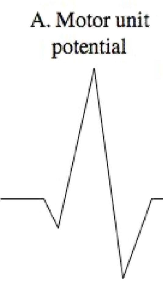 Polyphasic MUPS
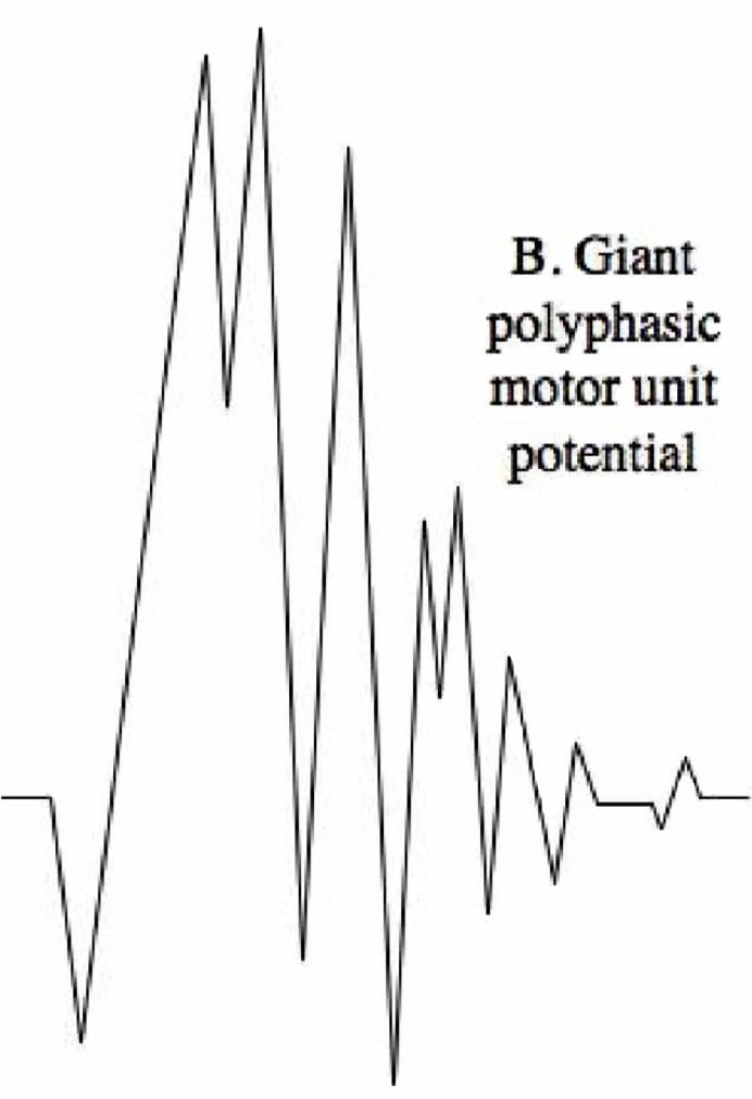 Complete Potential Blocks
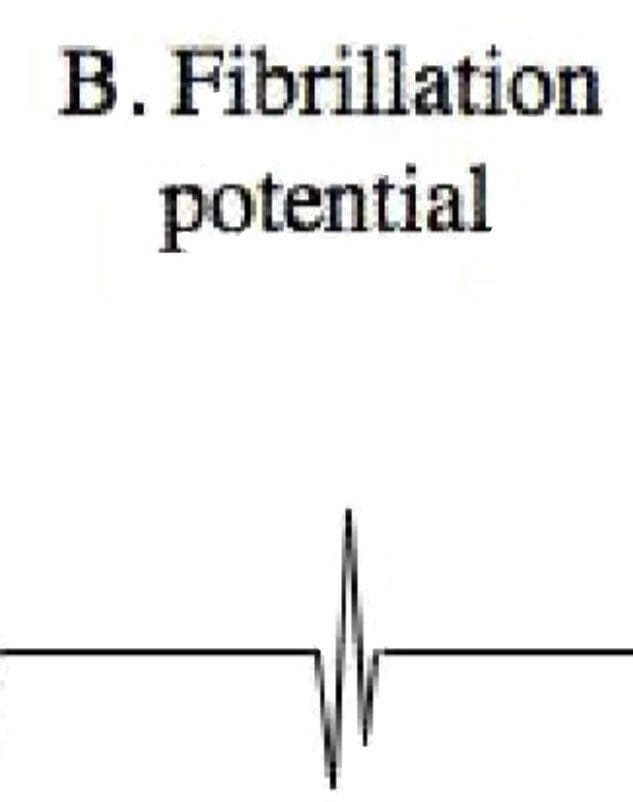 Positive Sharp Waves
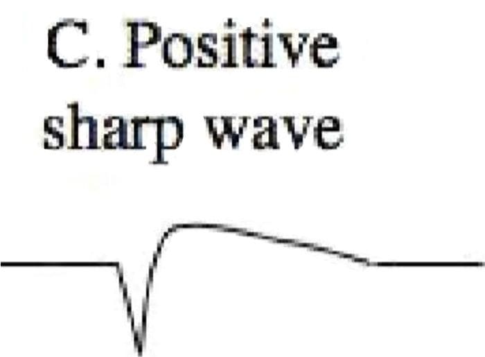 Abnormal Findings