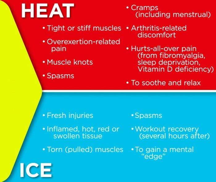
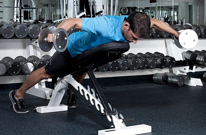
Most if not all of us have probably used heat and/or ice on a sprain, strain, or sore area of the body. Having a pinched nerve, however, has a different feeling than a sprain or strain. Chiropractic treatment for a pinched nerve is recommended, but if the pain isn’t too bad, then home care can work. Which is better for a pinched nerve, heat or ice? Both. Using heat and ice helps reduce swelling, increases blood flow to the area, and relaxes the muscles around the pinched nerve. The objective is to know when to use ice and/or heat.
Applying Heat on a Pinched Nerve
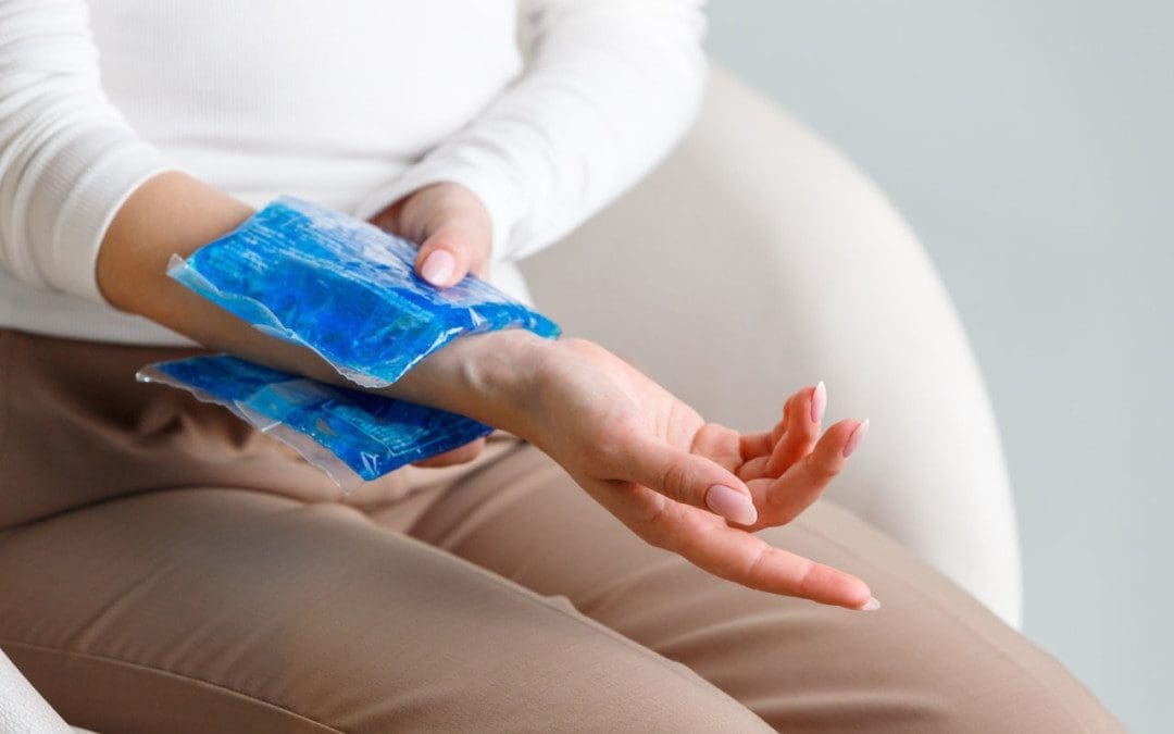
Applying heat on a pinched nerve is fine. The general guideline for a pinched nerve is to use heat only after the pain has subsided/reduced.
When pain presents or flares up, use ice before using heat.
Do not apply heat to the area directly after applying ice.
Wait 30 minutes to an hour.
Keep the heat on the affected area for 10 to 20 minutes at a time.
Take a minimum 30-minute break between sessions.
If the heat helps, make the heat moderate and use it on the area for an hour or more.
Extended heat therapy is beneficial for severe pain from a pinched nerve.
The equivalent is like soaking in a hot bath.
Heat for a Pinched Nerve Benefits
Heat soothes and relaxes both the muscles and the mind.
Heat increases healing abilities by circulating new blood to the injured/affected area, helping to flush toxins away.
Decreases tension and spasms in the muscles.
Increases the range of motion in the joints.
When Not To Use Heat Therapy
It can be dangerous for individuals with pre-existing conditions. Conditions include:
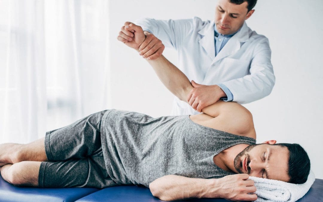
If home remedies don’t work, know when to consult a chiropractor.
Doctors of chiropractic specialize in pinched/compressed nerves. A chiropractor is trained in different techniques to relieve the pressure and release the nerve back to its proper position.
Body Composition
Peripheral Artery Disease
Peripheral artery disease or PAD is the narrowing of the arteries that carry blood away from the heart to the other areas of the body. What to know about PAD:
Peripheral artery disease risks increase with age.
Over half of affected individuals do not present with symptoms.
Around one-fourth of individuals with peripheral artery disease have diabetes mellitus.
Smokers have an increased risk of developing PAD.
This is why it’s important to monitor blood pressure.
References
Chandler, Anne, et al. “Using heat therapy for pain management. (clinical practice).” Nursing Standard, vol. 17, no. 9, 13 Nov. 2002, pp. 40+. Accessed 15 Sept. 2021.
Edzard Ernst, Veronika Fialka, Ice freezes pain? A review of the clinical effectiveness of analgesic cold therapy, Journal of Pain and Symptom Management, Volume 9, Issue 1, 1994, Pages 56-59, ISSN 0885-3924, https://doi.org/10.1016/0885-3924(94)90150-3.
Shu, Jun, and Gaetano Santulli. “Update on peripheral artery disease: Epidemiology and evidence-based facts.” Atherosclerosis vol. 275 (2018): 379-381. doi:10.1016/j.atherosclerosis.2018.05.033
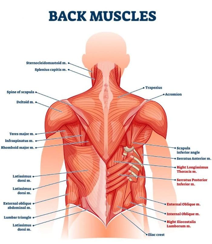
Relaxing the low back muscles. The low back supports the weight of the upper body and provides mobility. The lumbar spine/low back is a complex structure of interconnecting:
Bones
Joints
Nerves
Ligaments
Muscles
All work together to provide support, strength, and flexibility.
However, this complex structure is highly susceptible to injury and pain. The muscles in the low back support the spinal column and are responsible for flexing, rotating the hips when walking. The nerves in the low back supply sensation and power the muscles in the pelvis, legs, and feet. The most common acute low back pain cases are brought on from injury to the:
Muscles
Ligaments
Joints
Discs
The body also reacts to injury by triggering an inflammatory response. Inflammation can cause severe pain. The key is relaxing the muscles and keeping them loose.
Lower back pain causes
The most common cause is muscle spasms, which can be triggered by:
Quick awkward movements like twisting, reaching, and/or bending at the wrong angle can set off pain symptoms.
If the same back muscles are getting strained and/or pulled continuously, this could indicate an underlying problem with a misaligned vertebra.
Correct improper postures
Improper posture and body positioning eventually present with discomfort and pain. Individuals are constantly hunched over and slumped in front of computers and devices, along with sitting with crossed legs, sleeping on a non-supportive mattress, and there is a perfect recipe for low back pain. Adjusting sitting, desk, and computer ergonomics and using smart devices with posture awareness will go a long way to correct improper posture.
Stretch and loosen tight muscles
Stretch out throughout the day and before going to bed. Sitting all day at work, then going home and sitting all evening is not healthy for the body’s muscles. The muscles shorten, as do the ligaments and tendons. Stretching keeps the body loose and limber. Try out different forms of stretching, as well as foam rolling for the back.
Get the body moving
Cramped or pulled muscles need time to recover. Too much rest or being completely inactive makes sense but is not recommended. Light movement keeps the blood flowing, especially in and around the injured/sore area. This increases healing and shortens recovery time. Just some light walking is recommended. Getting up and moving while relaxing around the house will get the muscles back in shape.
Body Composition
Benefits of collagen
Gastrointestinal Health
Collagen generates a protective barrier effect on the intestinal mucosa that lines the digestive tract. One study found that collagen supplementation protects against the breakdown of the intestinal wall after a burn injury. A lack of collagen from aging or other factors could impair the structure of the intestinal mucosa. This could lead to digestive problems like leaky gut syndrome and irritable bowel syndrome.
Joint Health and Mobility
Collagen provides structure and cushion in the joints. As the body ages, the cushion wears down, and joint mobility decreases. This increases the risk of injury. Taking collagen supplements can help improve symptoms of osteoarthritis, rheumatoid arthritis, and other conditions.
References
Finta, Regina et al. “The effect of diaphragm training on lumbar stabilizer muscles: a new concept for improving segmental stability in the case of low back pain.” Journal of pain research vol. 11 3031-3045. 28 Nov. 2018, doi:10.2147/JPR.S181610
Lugo, James P et al. “Efficacy and tolerability of an undenatured type II collagen supplement in modulating knee osteoarthritis symptoms: a multicenter randomized, double-blind, placebo-controlled study.” Nutrition journal vol. 15 14. 29 Jan. 2016, doi:10.1186/s12937-016-0130-8
National Institute of Neurological Disorders and Stroke. Pain: Hope Through Research. https://www.ninds.nih.gov/Disorders/Patient-Caregiver-Education/Hope-Through-Research/Pain-Hope-Through-Research. June 9, 2017.
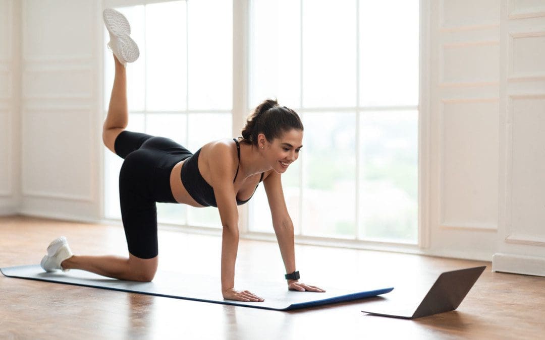
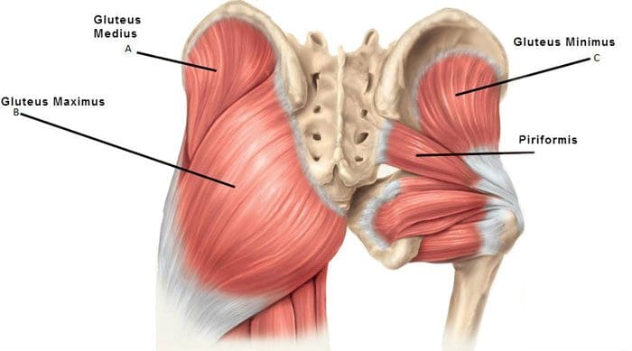
The Gluteus Maximus Muscle is the largest and the outermost of the gluteal muscles. It stretches from the sacrum and coccyx, parts of the spinal column, down to the femur. The other gluteal muscles include the gluteus minimus and medius. They each have a role in the normal function of the hips and legs. They are prone to tightness resulting in soreness, aches, and pain around the hips and in and around the buttocks.
The Gluteal/Rear End Muscles
There are three rear-end muscles:
The Gluteus Maximus extends the hip and rotates the thigh outwards, straightens the legs when moving, and provides strength.
Gluteus Minimus
Gluteus Medius
The minimus and medius are underneath the Maximus and stabilize the hip when:
Walking
Jumping
Other physical activities
There is another group of diagonal muscles under the gluteus minimus that attaches to the femur. The uppermost is the piriformis that is attached to the sacrum. The sciatic nerve and major arteries run below it.
Tightness and Irritation
The primary function of the muscles is to open the hips and push the legs out. Sitting for long periods shortens the gluteal muscles causing them to become tight, limiting normal hip function. The gluteal muscles are susceptible to tightness brought on from overuse and lack of development/strength. This can develop into tender/tight muscle bands that interfere with the normal function of the muscles. To get an example of what is happening imagine flexing and contracting one of the bicep muscles 6-10 hours a day. It would be extremely sore, tight, and tender.
Sports and Physically Active
Athletes and individuals that are physically active can also have tight gluteal muscles. This can cause post-game/exercise muscle soreness. Intense activities force the gluteal muscles to work overtime to support the back and knees. Sports that require a lot of leg muscle activation include:
Running
Soccer
Football
Crossfit
Dancing
Weight training
Awkward Walking Gait
Individuals that move with an unusual gait are vulnerable to straining the muscles. What happens is the muscles become stiff from the awkward positions/postures. This places additional strain on the back and hip muscles and worsens their overall posture. The hip muscles are also attached to the pelvis, and when the muscles begin to tighten they can pull on the gluteal muscles. Irritation of the sacroiliac joint can also place pressure on the piriformis, causing spasms that affect the gluteal muscles. Piriformis muscle spasms can also place pressure on the sciatic nerve, causing sciatica.
Diagnosis and Treatment
An examination will be necessary to diagnose whether the soreness or pain is due to muscle inflammation or other cause. Sciatica symptoms and problems at the hip level that involves the gluteus minimus and medius can be felt in the leg. The examination includes seeing and feeling muscle reactions, responses, contractions through a series of motion exercises and movements that involve different muscles. Common treatment includes:
Range of motion exercises
Strengthening exercises like bridging and resistance bands
Deep tissue massages
Heat and cold packs
Physical therapy
Electric muscle stimulation
Tightness in the glutes can be managed with chiropractic treatment. This includes:
Soft tissue work
Spinal joint manipulation
Lifestyle adjustments
Stretching
Diet
Health coaching
Simple exercises can help engage and strengthen the muscles. These include:
Individuals that sit for long periods of time, don’t get enough physical activity, and have an unhealthy diet can experience insulin resistance. This happens when insulin is not able to transport excess blood sugar out of the blood and into the muscles. A study found that women who sat for eight hours a day had an increased chance of developing diabetes. Diabetics can be inclined to have more fat within the body, specifically visceral fat. This further encourages insulin resistance. Diabetics also experience rapid loss of muscle mass as they age, intensifying symptoms and further affecting body composition.
References
Cochrane, Darryl J et al. “Does short-term gluteal activation enhance muscle performance?.” Research in sports medicine (Print) vol. 25,2 (2017): 156-165. doi:10.1080/15438627.2017.1282358
Coratella, Giuseppe et al. “The Activation of Gluteal, Thigh, and Lower Back Muscles in Different Squat Variations Performed by Competitive Bodybuilders: Implications for Resistance Training.” International journal of environmental research and public health vol. 18,2 772. 18 Jan. 2021, doi:10.3390/ijerph18020772
Distefano, Lindsay J et al. “Gluteal muscle activation during common therapeutic exercises.” The Journal of orthopedic and sports physical therapy vol. 39,7 (2009): 532-40. doi:10.2519/jospt.2009.2796
Kalyani, Rita Rastogi et al. “Age-related and disease-related muscle loss: the effect of diabetes, obesity, and other diseases.” The lancet. Diabetes & endocrinology vol. 2,10 (2014): 819-29. doi:10.1016/S2213-8587(14)70034-8
Selkowitz, David M et al. “Which exercises target the gluteal muscles while minimizing activation of the tensor fascia lata? Electromyographic assessment using fine-wire electrodes.” The Journal of orthopedic and sports physical therapy vol. 43,2 (2013): 54-64. doi:10.2519/jospt.2013.4116
The latissimus dorsi or lats are the large flat muscles on each side covering the width of the middle and lower back. They connect the bone of the upper arm to the spine and the hip. When pain presents in these muscles, it is typically caused by:
Repetitive overuse in a job or doing a task/chore that requires constant
Bending
Pulling
Pushing
Reaching
Twisting
Kneeling
A result of poor technique in sports or similar physical activities.
Chiropractic treatment, along with exercises, can help prevent and relieve this pain.
Symptoms of lat pain
The objective is to diagnose whether the pain is located in the latissimus dorsi or other muscles in the shoulders or back. If the latissimus dorsi is injured, an individual might feel pain in several areas, these include:
Lower, middle, and upper back
Back of the shoulders
The base of the shoulder blade
Lower arms
Inside of the arms, extending down to the fingers
In certain cases, the pain will present without warning and can be felt in the surrounding muscles. This type of pain often gets worse when the individual:
Extends their hands forward and out in front
Raises their hands above their head
Tosses or throws an object
Damage or injury to the latissimus dorsi
Tissue damage or injury can cause other symptoms to present. These include:
If the source of the back pain cannot be identified, or if it is accompanied by:
Fever
Breathing problems
Abdominal pain
Consult a doctor as these could be symptoms of a more serious condition.
Uses and Causes
The lat muscles are used in everyday activities. These include:
Picking up objects like grocery bags
Opening heavy doors
Chest expansion for breathing
Pushing against the armrests of a chair to stand up
Using handrails to climb stairs
For sports or working out, the lats are used in:
Weightlifting exercises using the upper body
Bench-presses
Rowing
Swimming
Throwing
Common causes of pain include:
Overusing the muscles
Using poor techniques
Exercising without warming up
Risk of injury
Individuals that are at risk of developing this injury include those that:
Are continually reaching overhead
Regularly chop wood
Perform regular shoveling
Move furniture or other heavy objects
Regularly practice poor posture
Tearing the latissimus dorsi is possible, especially for athletes. Some athletes with increased risk include:
Golfers
Baseball pitchers
Gymnasts
Swimmers
Tennis players
Exercises that can help bring relief
Certain exercises can alleviate the aches, pain, and strengthen the lat muscles to prevent and/or worsen the injury. It is recommended to consult a doctor, sports chiropractor, or personal trainer before beginning a therapeutic exercise regimen. This is to ensure that the exercises are right for the individual and their condition and that they use the correct form. Here are two exercises that can help reduce the pain. The doctor, chiropractor, or trainer will recommend the frequency the individual should perform the exercises.
Back bow
This pose is known as the superman pose. To perform:
Lay facedown on the floor
Extend the legs so they are straight
Stretch arms away from the body, so they are in front of the head
Bend the knees like for a sit-up with the heels close to the buttocks
Keeping the hands and feet in place
Lift the pelvis upward
Slowly lower back to the floor
Prevention
Individuals can prevent lat pain with lifestyle adjustments. These include:
Using proper technique and posture during work, sports, and exercise
Staying aware to not overuse the muscles
Staying hydrated
Warming up and cooling down thoroughly before and after a workout, sports, physical activities
Regular stretching
Applying ice and heat before and after work, sports, and physical activities
Chiropractic care
Physical therapy massage
Body Composition
Nutrition and Recovery Advantage
Two important steps to achieve optimal health include:
Nutrition
Having a proper protein intake is important for muscle adaptability or the way muscles adapt to stress during exercise and/or strength training. This is also important to stimulate muscle protein synthesisafter exercising and/or strength training. To ensure the body is getting the strength and hypertrophy improvement from exercise and strength training, it is recommended to eat around 25g of high-quality protein after workout sessions.
Recovery
For those doing aerobic and strength training, maximize recovery time between workout sessions. This is because strength and aerobic fitness health gains are low when the two only have a separation of 6 hours or less. Twenty-four hours between sessions is recommended especially if the priority is endurance performance.
References
Anderson, S. E., Hertel, R., Johnston, J. O., Stauffer, E., Leinweber, E., & Steinbach, L. S. (2005, November). Latissimus dorsi tendinosis and tear: imaging features of a pseudotumor of the upper limb in five patients. American Journal of Roentgenology, 185(5), 1145–1151
https://www.ajronline.org/doi/abs/10.2214/AJR.04.1247
Donohue, Benjamin F et al. “Sports Injuries to the Latissimus Dorsi and Teres Major.” The American journal of sports medicine vol. 45,10 (2017): 2428-2435. doi:10.1177/0363546516676062http://journals.sagepub.com/doi/abs/10.1177/0363546516676062?journalCode=ajsb
Henseler, J. F., Nagels, J., Nelissen, R. G. H. H., & de Groot, J. H. (2014, April). Does the latissimus dorsi tendon transfer for massive rotator cuff tears remain active postoperatively and restore active external rotation? Journal of Shoulder and Elbow Surgery, 23(4), 553–560
http://www.jshoulderelbow.org/article/S1058-2746(13)00399-6/fulltext%20
George, Michael S, and Michael Khazzam. “Latissimus Dorsi Tendon Rupture.” The Journal of the American Academy of Orthopaedic Surgeons vol. 27,4 (2019): 113-118. doi:10.5435/JAAOS-D-17-00581
Lehman, Gregory J et al. “Variations in muscle activation levels during traditional latissimus dorsi weight training exercises: An experimental study.” Dynamic medicine: DM vol. 3,1 4. 30 Jun. 2004, doi:10.1186/1476-5918-3-4
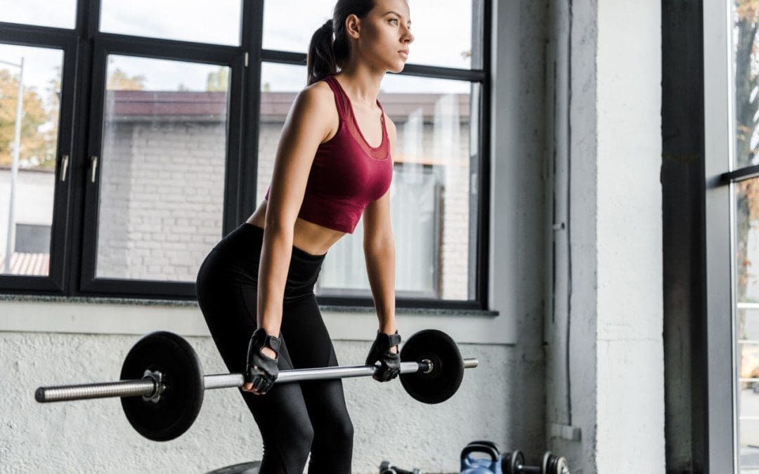
When back pain presents for a prolonged period, the back muscles reduce in mass but increase fat content, resulting in more stiffness. This leads to chronic muscle fatigue and results in chronic pain symptoms. Adding resistance to a workout routine in weight machines, free weights, and/or resistance bands helps reduce back pain. Studies have shown that specific therapeutic back muscle weight training is safe and can help relieve pain. A sports chiropractic specialist can recommend appropriate exercises for individuals and their specific condition/s to safely participate in strength training.
Back muscles development
With time, back pain and increased fatigue can lead to a fear of moving the body and engaging in physical activity. This results in spinal deconditioning and instability. Weight training works on incrementally/progressively increasing the load that the back muscles can tolerate. This technique gradually improves the body’s ability and strength to perform regular daily activities without strain and in optimal fashion. Weight training improves whole-body health because:
Back muscles and core muscles increase in function and performance.
Muscles are strengthened.
Lean muscle mass increases.
The range of motion of the spine increases.
Body fat decreases.
Guidelines while using weights
When weight training, it is important to understand safety guidelines to help relieve back pain and not worsen or cause further injury. Weighted treatment exercises are for individuals that have been cleared by their physician or chiropractor and are specific to their injury and /or condition. Depending on the underlying pain source, weight training may not be suitable for individuals that have:
Severe pain.
Back pain that originates from:
Previous spinal surgery
Tumor
Nerve root compression
Neurological symptoms
Sciatica
Spinal fracture/s
Spinal infection/s
Medical professionals and chiropractors can accurately diagnose and determine if weight training is safe and which specific exercises to perform. Guidance from a trained therapist or therapeutic trainer is recommended for optimal results.
Weight training techniques to alleviate back pain are different from regular weight or resistance exercises.
Trained physical/occupational therapists and sports chiropractors can educate an individual on:
Correct techniques
Frequency
Type of training that will help an individual’s condition.
Therapeutic training can significantly reduce the risk of further injury and damage to the spine.
After initial training, individuals are encouraged to exercise to maintain back muscles and total body health.
Smaller weights build strength progressively
Effective ways to strengthen the spine.
Begin with small/light weights and exercise slowly.
Fast rapid movements or incorrect lifting and pulling techniques can cause additional damage to the tissues.
It is recommended to start with:
Low-load motor control exercises without weights activate and stretch the muscles and improve balance.
Simple stretches
Exercise machines can be recommended instead of free weights.
Exercise machines can provide safe, effective, and progressive resistance to the exercises.
The machines can help reduce/prevent injury compared to free weights.
The machines can maintain proper support on the back and spine.
It is recommended to combine regular walking activity with a weight training program.
Low impact aerobic exercises increase blood circulation along with essential nutrients to the muscles and soft tissues.
This promotes healing and reduces stiffness.
Training program and benefits
Gaining the most benefits from strength training, tips to keep in mind:
Warm up for a few minutes using heat therapy and simple stretches.
Try for 2 or 3 times a week for 30 minutes.
Focus on building strength in the core muscles – back, abdominals, obliques, buttocks, and pelvic leg muscles.
There is no need to join a gym or buy expensive equipment.
Work out at home with small hand weights, resistance bands, and body weight.
The therapist or chiropractor will inform the individual on which exercises to avoid, which require extreme or quick moves.
Slow, steady resistance training takes advantage of muscle lengthening exercises and muscle shortening exercises for strengthening.
If back pain presents with a sustained increase, take time off or modify the strength training exercises.
Some soreness is to be expected, but sharp pain is not. If any sharp, sudden pain presents while exercising, stop immediately.
Ice therapy can be beneficial after exercising to decrease inflammation and alleviate pain.
Record the amount of weight when beginning the training and note when progressing to a heavier weight. Consistent improvements in pain, flexibility, strength, and function will help maintain motivation. Consult with a professional sports injury chiropractor today to see if weight training is a suitable and safe treatment.
Body Composition
Carbohydrates and Muscle Growth
Simple carbs are a quick, periodic source of energy. Complex carbs are a recommended source of steady energy. Complex carbs are not as readily available for immediate energy as simple carbs are but are more efficient and healthier. Complex carbs offer sustainable energy, meaning the energy is constant with no crash like simple carbs. Because complex carbs have slow-release properties, they should be the largest component of daily energy consumption.
Carbs prevent muscle weakness.
Some glycogen is stored in the muscles. When those muscles are used during exercise, the body taps into the glycogen stores in that specific muscle. Lifting weights with the arms, for example, access the glycogen in the biceps. Athletes take advantage of glycogen by loading up on carbs by consuming a day or more before a workout. This maximizes the muscle glycogen stores. This delays muscle fatigue, making for a better workout and stronger muscles, and can improve athletic performance.
Carbs help muscles recover after exercise.
Recovery goes back to the glycogen stores. Right after exercising, the body needs to replenish its glycogen stores to prevent glycogen depletion. Glycogen depletion, when the stores run out, causes gluconeogenesis. What happens is the body forms glucose from new sources. This is to compensate for the lack of glucose from carbohydrates. This is when the body turns to sources like fat and protein to fill the need. Protein is the last line of defense when energy is required, meaning that energy is running low. When the body breaks down protein for glucose production, it takes what it needs from the muscle/s, causing them to shrink and break down.
References
Dreisinger TE. Exercise in the management of chronic back pain. Ochsner J. 2014;14(1):101–107.
Lee JS, Kang SJ. Strength exercise and walking effects on lumbar function, pain level, and body composition in chronic back pain patients. J Exerc Rehabil. 2016;12(5):463–470. Published 2016 Oct 31. doi:10.12965/jer.1632650.325
Michaelson P, Holmberg D, Aasa B, Aasa U. High load lifting exercise and low load motor control exercises as interventions for patients with mechanical low back pain: A randomized controlled trial with 24-month follow-up. J Rehabil Med. 2016;48(5):456-63.
Welch N, Moran K, Antony J, et al. The effects of a free-weight-based resistance training intervention on pain, squat biomechanics, and MRI-defined lumbar fat infiltration and functional cross-sectional area in those with chronic low back. BMJ Open Sport Exerc Med. 2015;1(1):e000050. Published 2015 Nov 9. doi:10.1136/bmjsem-2015-000050
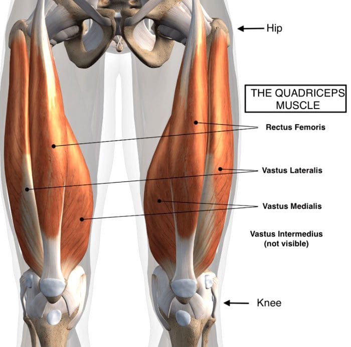
The quadriceps muscle consists of four muscles in front of the thigh that connects to the knee right below the knee cap. These muscles straighten the knee for walking, running, and jumping. They also help bend the knee for squatting. They move the leg forward when running and fire/transmit electrical impulses when the foot hits the ground to absorb shock. When jumping, the muscles provide stability coming down as well as when standing on one leg.
Quadriceps Strain
Thigh strains are common in sports. Most players are sidelined because of this injury when compared to strains in the hamstrings or groin. Factors that can increase the risk of injury include:
Exhaustion
Muscle weakness
The strength of the quadriceps to the hamstrings is uneven, causing one set to get overused.
Consistent sprinting and/or kicking
Previous strain and/or injury
The quadriceps is made up of four muscles. One is the rectus femoris, which gets injured the most. It’s the only muscle that crosses two joints – the hip joint and the knee joint.
Symptoms and Injury Grades
Individuals commonly report a pulling/stretching sensation in the front of the thigh. Common symptoms include:
Pain
Swelling
Bruising
Muscle tenderness
For minor quadriceps strains or tears, moderate to dull pain presents along with stiff movement.
Grades categorize the severity of the strain:
Grade 1 presents with mild discomfort in the thigh with no loss of strength.
Grade 2 presents with moderate pain, swelling, and some loss of strength.
Grade 3 is a complete rupture of the fibers. Individuals are in severe pain and unable to walk.
Grade 3 is where surgery is required.
Symptoms can vary depending on the type of injury that has been sustained and the severity. There is pain and localized swelling for both strains and contusions. If a muscle rupture has happened, there could be a bump/lump within the muscle or a gap in the muscle. If rupture of the Quadriceps Tendon has occurred, individuals often report hearing a pop when the injury happens. The swelling often makes straightening the leg difficult or impossible.
Injury causes
Thigh strains usually happen when slowing down/decelerating after a sprint. This can be because the individual takes too small or too large steps causing the muscles to overstretch, much like a rubber band that, if overstretched, tears, and if under stretched, it bunches up, which can cause spasms and tears.
Treatment
In the initial stages after a quadriceps strain, it is recommended to follow the RICE Procedure for 24 hours: This includes:
Rest
Ice
Compression
Elevate
The leg needs to be rested every 2-3 hours in 20-minute sessions.
A bandage can provide added support.
For slight tears and strains, it is recommended to stretch the quadriceps gently.
This helps prevent the muscles from experiencing shortening. This happens by the formation of scar tissue that pulls the muscle/s, making them shorter.
Gentle stretches allow the muscles to heal with minimal shortening. This helps prevent further and/or re-injury.
Chiropractic Physical Therapy Rehabilitation
After the acute stage of the injury, receiving regular chiropractic sports adjustments, physical therapy massage, coupled with strength training exercises will speed up recovery.
Physical therapy massage will remove scar tissue and keep the muscle/s loose and flexible.
Exercises for strengthening the muscles after injury will be recommended according to the individual’s condition/case.
Following correct post-injury-care, exercises, and physical therapy.
Healing time can be 4- 6 weeks.
Body Composition
Strength Training: The Inverted Row
This workout targets the back muscles, spine and scapular stabilizers, deep abdominals, and arms. Everyday activities that require various types of pulling motion, lifting, etc., become easier. To perform:
Lie flat on your back.
Grab a stable barbell or set of straps that are above you.
Pull your upper body up as high as possible while keeping the back straight.
Squeeze the shoulder blades together at the top.
Complete as many reps as possible.
Once enough strength and endurance have been built, try a pullup.
References
Kary, Joel M. “Diagnosis and management of quadriceps strains and contusions.” Current reviews in musculoskeletal medicine vol. 3,1-4 26-31. 30 Jul. 2010, doi:10.1007/s12178-010-9064-5
Hillermann, Bernd, et al. “A pilot study comparing the effects of spinal manipulative therapy with those of extra-spinal manipulative therapy on quadriceps muscle strength.” Journal of manipulative and physiological therapeutics vol. 29,2 (2006): 145-9. doi:10.1016/j.jmpt.2005.12.003
Wenban, Adrian B. “Influence of active release technique on quadriceps inhibition and strength: a pilot study.” Journal of manipulative and physiological therapeutics vol. 28,1 (2005): 73. doi:10.1016/j.jmpt.2004.12.015
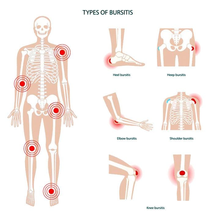
Bursitis types: This is a condition that affects the bursae, which are the small, fluid-filled sacs that provide cushion for the:
Muscles
Tendons
Bones near joints
The bursae make it easier for tissues to slide over each other. The body has around one hundred and sixty bursae. However, only a few become clinically affected. These include the:
Wrist
Elbow
Shoulder
Hips
Knees
The base of the big toe and heel
The condition typically presents near joints constantly being used repetitively, like a job, sports, house/yard chores, etc. What happens is one or more of the bursae sacs become inflamed, resulting in pain.
Causes
Inflamed or irritated bursae typically cause it from overuse or intense/vigorous activity.
It can also be caused by bacterial infection.
Arthritis and gout can also cause bursitis.
Another cause is age.
As tendons age, they can tear easily, lose their elasticity, and can’t take too much stress.
Intense physical activities can lead to bursitis. These include:
Gardening
Typing
Working with a computer mouse
Throwing
Golf
Tennis
Manual tasks
Carpentry
These types of activities can lead to incorrect posture, overuse, and injury/damage.
Symptoms
The main symptom is pain in and around the affected area that worsens with movement. Depending on the severity of the strain and the length of time it has been going on, the pain can be intense with active and passive movements. Other symptoms include:
Tenderness
Stiffness
For some individuals, it can present as acute, with the intensity increasing.
This happens when movement aggravates the condition.
Bursitis Types
Four major types include:
Prepatellar
Trochanteric
Olecranon
Retrocalcaneal
Prepatellar Bursitis
Prepatellar is an inflammation of the sac situated between the skin and the patella/kneecap. The most common causes are trauma from a fall and direct pressure/friction from repetitive kneeling. This is one of the bursitis types that can get infected. Overproduction of liquid places pressure on the other areas of the knee, causing swelling. Most individuals report swelling and knee pain just over the front of the knee.
Trochanteric Bursitis
This bursitis type goes over the lateral area of the hip. There is a distinctive tenderness and aching pain. This type is more common for individuals with arthritis conditions and fibromyalgia. This condition is also seen after surgery, mainly osteotomies. The bursa can become inflamed in case of injury or overuse. It tends to affect middle-aged or older folks. Common causes include:
Muscle tears
Hip injuries
Tight hip or leg muscles
Disc disease of the low back
Leg-length inequality
Improper walking technique from a minor injury or strain
Overuse of the gluteal muscles
Flat feet
Improper footwear
Olecranon Bursitis
Olecranon is a common bursitis type. It is diagnosed by the appearance of swelling over the elbow. The swelling happens just behind the olecranon process of the ulna. The bursa can become infected. This bursitis does cause blood to rupture out, and fluid could be present. Individuals are advised to avoid leaning or resting on the elbows.
Retrocalcaneal Bursitis
This is characterized by pain in the Achilles tendon. Chronic inflammation of the bursa is brought on by friction, supination, and overpronation. The flexibility of the calf muscles can be significantly reduced. Severe pain and swelling of the posterior soft tissue in front of the Achilles tendon are common symptoms. This bursitis type is often accompanied by mid-portion insertional tendinosis.
Risk Of Getting Bursitis
Anybody at any age can develop bursitis, but older individuals, specifically those in their forties and beyond, are more susceptible. This comes from all the wear and tear of the muscles and bones.
Risk Factors
Overpronation of the foot
Leg length deviation
Osteoarthritis
Obesity
Tight hamstring muscles
Incorrect physical training
Not stretching properly
Body Composition
When Inflammation Becomes Permanent
When white blood cells cause inflammation, it’s signaling that the body’s immune system works properly. The process works like this:
Inflammation activates
White blood cells attack the foreign invader
The invader is neutralized
The inflammation deactivates
This is how the body’s defense system naturally works. But, white blood cells are not the only type of cell that emit cytokines. Adipocytes or fat cells are another type of cell that can emit cytokines and cause inflammation. Scientists have learned that fat is an active endocrine organ that secretes various proteins and chemicals, including inflammatory cytokines. The body stores excess calories as fat to be used later for energy. When the body keeps adding more adipose tissue, cytokines are released by the fat cells, triggering inflammation. Obesity is characterized as a state of low-grade, chronic inflammation. Increased fat cells place the body in a constant state of stress activating immune responses. This means the body is in a constant state of inflammation with the immune system switch permanently on.
References
Aaron, Daniel L et al. “Four common types of bursitis: diagnosis and management.” The Journal of the American Academy of Orthopaedic Surgeons vol. 19,6 (2011): 359-67. doi:10.5435/00124635-201106000-00006
Coelho, Marisa et al. “Biochemistry of adipose tissue: an endocrine organ.” Archives of medical science: AMS vol. 9,2 (2013): 191-200. doi:10.5114/aoms.2013.33181
Khodaee, Morteza. “Common Superficial Bursitis.” American family physician vol. 95,4 (2017): 224-231.
IFM's Find A Practitioner tool is the largest referral network in Functional Medicine, created to help patients locate Functional Medicine practitioners anywhere in the world. IFM Certified Practitioners are listed first in the search results, given their extensive education in Functional Medicine