Individuals with hip bursitis often experience discomfort during physical activity, walking, and pain when lying on the affected side. What treatment options are available to control and manage the condition?
Hip Bursitis
Hip bursitis, also known as trochanteric bursitis, is a common condition that causes pain and discomfort in the hip and upper thigh along the outside of the hip joint. It occurs when one of the hip’s bursae, or fluid-filled sacs cushion joints, becomes inflamed. Treatment for hip bursitis is to control the inflammation caused by this condition.
Causes
Hip bursitis can be caused by injury or overuse of the hip, such as repetitive activities, twisting, or rapid joint movement. It can also be caused by a direct blow or fall to the side of the hip.
Symptoms
Pain from hip bursitis can be sharp at first and may feel dull and achy later.
It may be worse when standing up after sitting, moving, or using the hip.
Individuals may also notice pain when lying on the affected side or sitting for a long time.
Rest
This means a period of not participating in physical, exercise, and sports activities that aggravate symptoms. Any activity that causes hip pain should be avoided as this only contributes to inflammation of the bursa. (American Academy of Orthopaedic Surgeons, 2022) Modifying how particular activities are performed can help alleviate pressure on the inflamed bursa. Working with a physical therapist can also be recommended. They are experts in movement and alignment, and if certain muscles are overused compared to others, this can lead to unhealthy movement patterns, causing bursa irritation.
Anti-Inflammatory Medications
Nonsteroidal anti-inflammatory drugs, such as Motrin, Aleve, Naprosyn, etc., will help control inflammation (American Academy of Orthopaedic Surgeons, 2022). Anti-inflammatory medications can be extremely effective but should be taken cautiously. The instructions on the label need to be followed unless directed otherwise by a healthcare provider. Be aware of side effects and inform the healthcare provider if side effects present.
Cold Therapy
Applying ice to the hip area often helps alleviate the symptoms (National Library of Medicine, 2022). Ice can control inflammation by decreasing blood circulation to the area, especially after physical activity and exercise.
Aspiration
A needle is placed into the bursa to drain the fluid for those with a significant amount of fluid collected within the bursa. (National Library of Medicine, 2022) This is rarely needed in cases of hip bursitis, but when it is done, it can be combined with a cortisone injection.
Cortisone Injections
A cortisone injection may also be given into the bursa to alleviate pain. (American Academy of Orthopaedic Surgeons, 2022) The cortisone injection is helpful because it can be a diagnostic and therapeutic tool. In cases where hip bursitis may be one of several diagnoses being considered, cortisone can be given to see if it helps alleviate symptoms. Cortisone is a powerful anti-inflammatory medication that can be administered directly to the problem area. These injections are well-tolerated, but there can be possible side effects. Once the initial symptoms are under control, physical therapy strengthening and stretching exercises may be recommended.
Stretching
Most find relief by stretching the muscles and tendons over the outside of the hip, specifically the iliotibial band. The goal is for a better-conditioned muscle and tendon to glide more easily and not cause inflammation. Proper stretching techniques and posture are important in re-injury prevention.
Physical Therapy
Working with a physical therapist is an effective adjunct treatment for bursitis (American Academy of Orthopaedic Surgeons, 2022). Physical therapists correct muscle imbalances through stretching and exercise and improve alignment to prevent bursa irritation from reoccurring.
Surgery
Most patients get better with conservative treatment within about six weeks. Surgical treatment for hip bursitis is rarely needed (UCSF Health, 2024). Those who do not rest from their activities until the inflammation subsides often have a return of bursitis symptoms, and those who return too aggressively to activities and do not gradually build up also find that their symptoms return. In cases where surgery is needed, the healthcare provider may recommend an arthroscopic bursectomy. (American Academy of Orthopaedic Surgeons, 2022) The surgery is an outpatient minimally invasive procedure in which the bursa is removed through a small incision. After a short healing period, the individual can return to normal activity. Crutches may be used for a few days. Common complications are anesthetic-related complications and infection.
Injury Medical Chiropractic and Functional Medicine Clinic
As with any treatment program, always talk with your healthcare provider before initiating specific treatments. Fortunately, treatment of hip bursitis is generally accomplished with conservative therapies. Efforts to limit pressure directly on the bursa, alleviate inflammation, and restore normal movement to the hip joint will typically resolve symptoms. At Injury Medical Chiropractic and Functional Medicine Clinic, we focus on what works for you to relieve pain and restore function. Regarding musculoskeletal pain, specialists like chiropractors, acupuncturists, and massage therapists can help mitigate the pain through spinal adjustments that help the body realign itself. They can also work with other associated medical professionals to integrate into a treatment plan to improve the body’s flexibility and mobility, resolve musculoskeletal issues, and prevent future pain symptoms from reoccurring.
The Chiropractic Approach for Pain Relief
References
American Academy of Orthopaedic Surgeons. (2022). Hip bursitis. https://orthoinfo.aaos.org/en/diseases–conditions/hip-bursitis
National Library of Medicine. (2022). Bursitis: Learn More – How can bursitis be treated? InformedHealth.org [Internet]. Cologne, Germany: Institute for Quality and Efficiency in Health Care (IQWiG). https://www.ncbi.nlm.nih.gov/books/NBK525763/
Fans of almonds may have noticed that they can go stale and taste awful. Can knowing how to store almonds help extend their shelf life?
Almonds
Almonds are a healthy, satisfying snack rich in nutrients, including fiber and protein. However, they can go stale and should not be eaten. Whole almonds stay fresh the longest because chopped, roasted, or ground almonds release their oils, which are exposed to more oxygen and go rancid more quickly.
Storage
They should be stored in an airtight container in the refrigerator or freezer for extended life. They are not recommended to be stored at room temperature for long periods, so storing them in the pantry is not recommended. However, enough for a snack can be taken from storage for a day or two while keeping the rest safe and fresh.
Natural almonds can be stored in the refrigerator or freezer for two years or more.
Roasted almonds can stay fresh for up to a year in an airtight container in the refrigerator or freezer.
Almond paste is recommended to be refrigerated and can stay fresh for 2 to 2 1/2 years.
Rancidity
Almonds are rich in omega-3 and monounsaturated fats, so they are recommended for heart health. (Cleveland Clinic, 2023) However, the fats become rancid if the almonds are exposed to oxygen, especially at room temperature. Rancid oil gives the stale almonds a bad taste. Spoiled almonds are not poisonous, but the fats no longer benefit health. It is possible that rancid fat could contribute to chronic health problems if consumed regularly. (Estévez M. et al., 2017) Almonds last longer than other nuts because they contain some phytochemical antioxidants that protect the nuts. So, if they taste bad, it’s time to throw them out. (University of California Agriculture and Natural Resources, 2010)
Salmonella
Salmonella is a bacteria that causes symptoms of food-borne illness, including upset stomach, vomiting, diarrhea, and fever. Almonds are generally safe from salmonella. However, raw almonds were responsible for two salmonella outbreaks in the United States and Canada. Salmonella outbreaks have also been reported in Australia. In response to those cases, the nuts now must be roasted, blanched, processed, steamed, or treated with a gas called propylene oxide. And none of these treatments remove the nutritional benefits.
Injury Medical Chiropractic and Functional Medicine Clinic
Injury Medical Chiropractic and Functional Medicine Clinic providers use an integrated approach to create customized care plans for each patient and restore health and function to the body through nutrition and wellness, chiropractic adjustments, functional medicine, acupuncture, Electroacupuncture, and sports medicine protocols. If the individual needs other treatment, they will be referred to a clinic or physician best suited for them. Dr. Jimenez has teamed up with top surgeons, clinical specialists, medical researchers, nutritionists, and health coaches to provide the most effective clinical treatments.
Functional Nutrition
References
Almond Board of California. (2024). Shelf Stability and Shelf Life. https://www.almonds.com/tools-and-resources/food-safety-and-quality/shelf-stability-and-shelf-life
Estévez, M., Li, Z., Soladoye, O. P., & Van-Hecke, T. (2017). Health Risks of Food Oxidation. Advances in food and nutrition research, 82, 45–81. https://doi.org/10.1016/bs.afnr.2016.12.005
University of California Agriculture and Natural Resources. (2010). Nuts: Safe Methods for Consumers to Handle, Store, and Enjoy. https://ucfoodsafety.ucdavis.edu/sites/g/files/dgvnsk7366/files/inline-files/44384_0.pdf
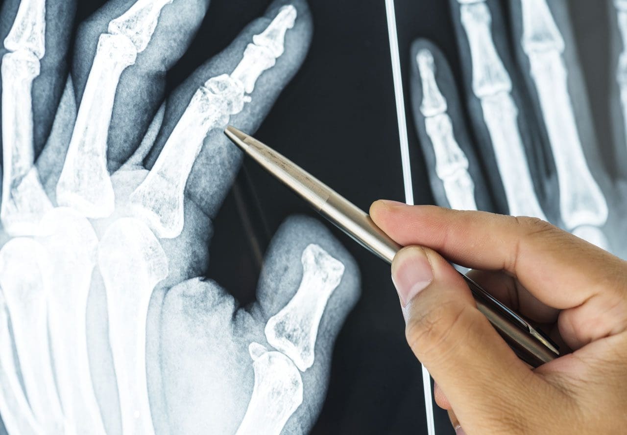
For individuals dealing with finger injuries, which can occur from various causes, including overuse, jobs, sports, and more, can knowing the cause of finger pain help healthcare providers determine what steps to take for treatment?
Finger Injuries
Finger injuries are common and can range from minor to serious. (van Veenendaal L. M. et al., 2014) Symptoms can result from an acute injury, including broken fingers and sprains, or chronic conditions like arthritis.
Fractures
Finger fractures can vary and can be serious and lead to permanent damage, deformity, and loss of function if not treated properly. What is important is that fractures are appropriately diagnosed so the proper treatment plan can be initiated. Most finger fractures can be addressed with simple treatments, while others may require surgery. (Oetgen M. E., and Dodds S. D. 2008)
Sprain and Dislocation
Sprains and dislocations are common finger injuries. (Prucz R. B. and Friedrich J. B. 2015) Both damage the ligaments that support the finger joints. In more severe injuries, a dislocation can occur, necessitating the finger to be put back into place or reduced. Individuals with a sprain or dislocation often notice finger swelling or stiffness for months after the injury.
Ligament Damage
Some call this injury skier’s or gamekeeper’s thumb, which results from a specific type of thumb dislocation. Here, the ulnar collateral ligament of the thumb is damaged. This ligament helps keep the thumb stable and supports grip and hand strength. However, this type of ligament injury often requires surgery. (Christensen T. et al., 2016)
Arthritis
Arthritis causes damage to normal joint surfaces where two bones come together. Fingers are one of the most common locations where arthritis occurs. (Spies C. K. et al., 2018) Two types of arthritis commonly affect the fingers: osteoarthritis and rheumatoid arthritis.
Arthritis of The Thumb
Arthritis of the thumb usually occurs at the joint where the thumb meets the wrist. This joint called the carpometacarpal/CMC joint, helps with gripping and pinching. Thumb arthritis is more common in women than men and increases in frequency over 40. (Deveza L. A. et al., 2017)
Trigger Finger
Trigger finger or stenosing tenosynovitis, is a common injury that causes pain and snapping of the fingers’ tendons, resulting in a sensation of locking or catching when bending and straightening the digits. (Makkouk A. H. et al., 2008) Other symptoms include pain and stiffness in the fingers and thumb. Treatments can vary from observation, rest, splinting, injections, and surgery.
Tendon Injuries
Mallet finger
A mallet finger is an injury to the tip of the finger. Usually, it occurs when the end of a straightened finger or thumb is hit, jamming the finger. After the injury, the individual may notice that they cannot fully straighten the tip of the finger. Treatment almost always uses a splint that has to stay on for about six weeks without removal. (Alla, S. R., Deal, N. D., and Dempsey, I. J. 2014) Very rarely is a surgical procedure necessary.
Jersey Finger
This is an injury to the finger flexor tendon. The flexor tendon pulls the finger into the palm when contracting the forearm flexor muscles. The injury occurs at the tip of the finger; typically, the tendon snaps back to the finger’s base or into the palm.
Ring Injuries
Injuries to the finger while wearing wedding bands or other finger jewelry can lead to serious complications. Even minor injuries can have devastating complications if the severity of the injury is not recognized and addressed. If an injury occurs while wearing the jewelry and there is soft tissue damage, including blood circulation being cut off, immediate medical attention is necessary.
Other Injuries
Bruises
The most common finger injury is caused by direct trauma to the skin and muscles. Symptoms include pain, swelling, tenderness, and discoloration of the skin.
Cuts and Scrapes
These can range from minor to more serious, such as injuries that cut through blood vessels, nerves, and tendons.
Injury Medical Chiropractic and Functional Medicine Clinic
After the initial inflammation and swelling have subsided, a doctor will recommend a treatment plan that usually involves physical therapy, self-performed physical rehabilitation, or supervision by a physical therapist or team. At Injury Medical Chiropractic and Functional Medicine Clinic, our areas of practice include Chronic Pain, Personal Injury, Auto Accident Care, Work Injuries, Back Injury, Low Back Pain, Neck Pain, Migraine Headaches, Sports Injuries, Severe Sciatica, Scoliosis, Complex Herniated Discs, Fibromyalgia, Chronic Pain, Complex Injuries, Stress Management, Wellness & Nutrition, Functional Medicine Treatments, and in-scope care protocols. We focus on what works for you to relieve pain and restore function. If other treatment is needed, individuals will be referred to a clinic or physician best suited to their injury, condition, and/or ailment.
Sports Injury Rehabilitation
References
van Veenendaal, L. M., de Klerk, G., & van der Velde, D. (2014). A painful finger as first sign of a malignancy. Geriatric orthopaedic surgery & rehabilitation, 5(1), 18–20. https://doi.org/10.1177/2151458514522125
Oetgen, M. E., & Dodds, S. D. (2008). Non-operative treatment of common finger injuries. Current reviews in musculoskeletal medicine, 1(2), 97–102. https://doi.org/10.1007/s12178-007-9014-z
Prucz, R. B., & Friedrich, J. B. (2015). Finger joint injuries. Clinics in sports medicine, 34(1), 99–116. https://doi.org/10.1016/j.csm.2014.09.002
Christensen, T., Sarfani, S., Shin, A. Y., & Kakar, S. (2016). Long-Term Outcomes of Primary Repair of Chronic Thumb Ulnar Collateral Ligament Injuries. Hand (New York, N.Y.), 11(3), 303–309. https://doi.org/10.1177/1558944716628482
Spies, C. K., Langer, M., Hahn, P., Müller, L. P., & Unglaub, F. (2018). The Treatment of Primary Arthritis of the Finger and Thumb Joint. Deutsches Arzteblatt international, 115(16), 269–275. https://doi.org/10.3238/arztebl.2018.0269
Deveza, L. A., Hunter, D. J., Wajon, A., Bennell, K. L., Vicenzino, B., Hodges, P., Eyles, J. P., Jongs, R., Riordan, E. A., Duong, V., Min Oo, W., O’Connell, R., & Meneses, S. R. (2017). Efficacy of combined conservative therapies on clinical outcomes in patients with thumb base osteoarthritis: protocol for a randomised, controlled trial (COMBO). BMJ open, 7(1), e014498. https://doi.org/10.1136/bmjopen-2016-014498
Makkouk, A. H., Oetgen, M. E., Swigart, C. R., & Dodds, S. D. (2008). Trigger finger: etiology, evaluation, and treatment. Current reviews in musculoskeletal medicine, 1(2), 92–96. https://doi.org/10.1007/s12178-007-9012-1
Alla, S. R., Deal, N. D., & Dempsey, I. J. (2014). Current concepts: mallet finger. Hand (New York, N.Y.), 9(2), 138–144. https://doi.org/10.1007/s11552-014-9609-y
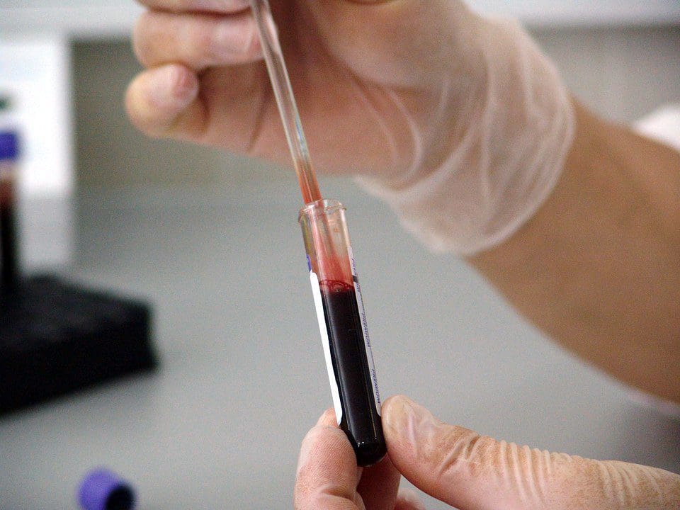
Are there blood tests for individuals with chronic and severe back pain symptoms that can help healthcare providers diagnose?
Blood Tests To Help Diagnose Back Pain
If a healthcare provider suspects an infection or inflammatory arthritis is the cause of back pain, blood tests may be used to diagnose. When trying to find the cause of back pain, a healthcare provider will examine the patient’s medical history, perform a physical examination, and, if necessary, order diagnostic tests. (Dansie E. J. and Turk D. C. 2013) For example, the National Institute of Arthritis and Musculoskeletal and Skin Diseases says that MRIs can reveal abnormalities in the spine. Still, a person may not feel pain or experience any other symptoms. The NIAMS also says healthy, pain-free individuals can have elevated SED levels. A high sedimentation rate or sed rate, also known as an erythrocyte sedimentation rate (ESR) test, can indicate inflammation in the body. (National Institute of Arthritis and Musculoskeletal and Skin Diseases, 2023)
Commonly Used Tests
Blood tests that can help diagnose back pain include:
Complete Blood Count – CBC
This test can indicate inflammation or infections.
Sed Rate or Erythrocyte Sedimentation Rate
This test measures inflammation by analyzing how red blood cells settle through plasma.
If the SED rate indicates that inflammation is present, the possibility of an underlying cause may be some form of arthritis or a tumor, which is rare.
A genetic marker in the blood that is more common in individuals with ankylosing spondylitis and reactive arthritis. (McMichael A. and Bowness P. 2002)
This marker may be tested if the healthcare provider suspects either disease.
Ankylosing spondylitis is an inflammatory arthritis affecting the sacroiliac joints, hips, and spine. (Sieper J. et al., 2002)
Injury Medical Chiropractic and Functional Medicine Clinic
At Injury Medical Chiropractic and Functional Medicine Clinic, we focus on what works for you to relieve pain and restore function. Regarding musculoskeletal pain, specialists like chiropractors, acupuncturists, and massage therapists can help mitigate the pain through spinal adjustments that help the body realign itself. Our areas of practice include Chronic Pain, Personal Injury, Auto Accident Care, Work Injuries, Back Injury, Low Back Pain, Neck Pain, Migraine Headaches, Sports Injuries, severe sciatica, Scoliosis, Complex Herniated Discs, Fibromyalgia, Chronic Pain, Complex Injuries, Stress Management, Wellness and nutrition, Functional Medicine Treatments, and in-scope care protocols. They can also work with other associated medical professionals to develop a personalized treatment plan to help relieve muscle pain, improve the body’s flexibility and mobility, resolve musculoskeletal issues, and prevent future pain symptoms from reoccurring.
Integrative Medicine Approach
References
Dansie, E. J., & Turk, D. C. (2013). Assessment of patients with chronic pain. British journal of anaesthesia, 111(1), 19–25. https://doi.org/10.1093/bja/aet124
National Institute of Arthritis and Musculoskeletal and Skin Diseases. (2023). Back Pain. Retrieved from https://www.niams.nih.gov/health-topics/back-pain
Harrison M. (2015). Erythrocyte sedimentation rate and C-reactive protein. Australian prescriber, 38(3), 93–94. https://doi.org/10.18773/austprescr.2015.034
Sproston, N. R., & Ashworth, J. J. (2018). Role of C-Reactive Protein at Sites of Inflammation and Infection. Frontiers in immunology, 9, 754. https://doi.org/10.3389/fimmu.2018.00754
McMichael, A., & Bowness, P. (2002). HLA-B27: natural function and pathogenic role in spondyloarthritis. Arthritis research, 4 Suppl 3(Suppl 3), S153–S158. https://doi.org/10.1186/ar571
Sieper, J., Braun, J., Rudwaleit, M., Boonen, A., & Zink, A. (2002). Ankylosing spondylitis: an overview. Annals of the rheumatic diseases, 61 Suppl 3(Suppl 3), iii8–iii18. https://doi.org/10.1136/ard.61.suppl_3.iii8
Hamdulay, S. S., Glynne, S. J., & Keat, A. (2006). When is arthritis reactive?. Postgraduate medical journal, 82(969), 446–453. https://doi.org/10.1136/pgmj.2005.044057
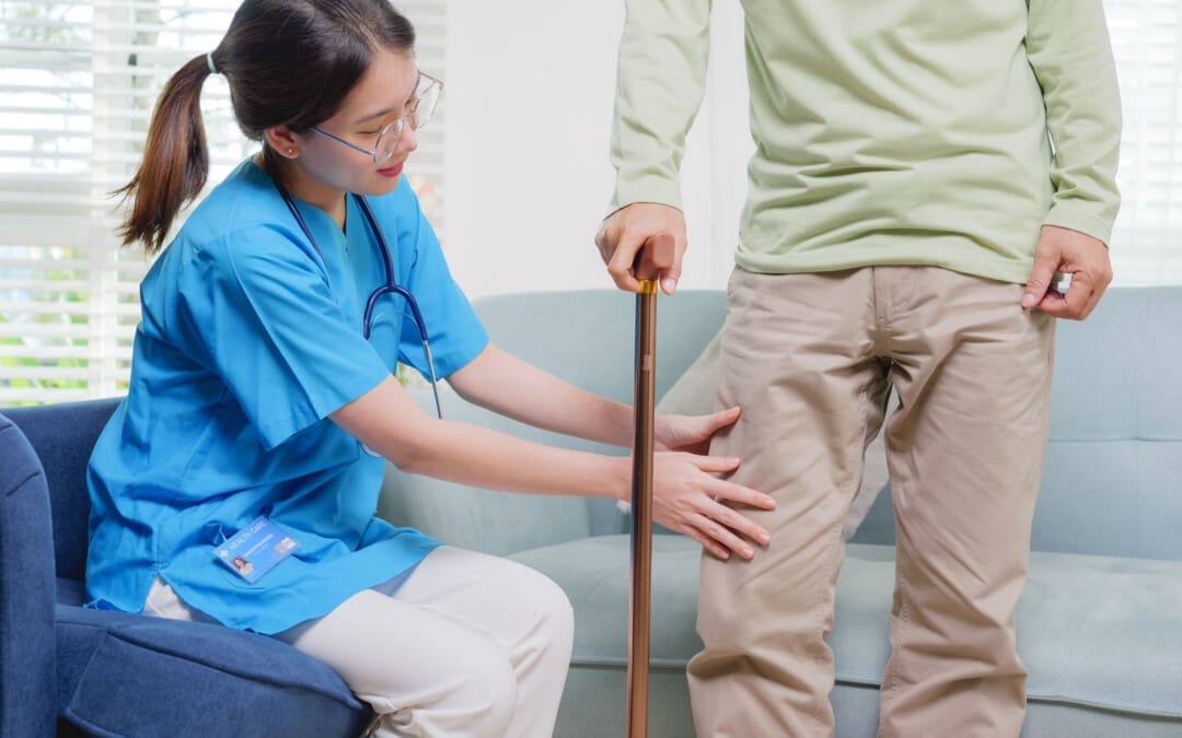
Can using a cane help individuals after an injury, living with chronic pain or balance issues, or post-surgery?
Walking With A Cane
A cane is an assistive device that can help individuals walk after injury or surgery and aids with balance and stability. It can be used for:
Balance or Stability Issues
Canes can help with minor balance or stability issues, such as weakness in the leg or trunk, or after an injury.
Pain
Canes can help reduce stress on painful joints or limbs.
Independence
Canes can help people continue living independently, especially the elderly.
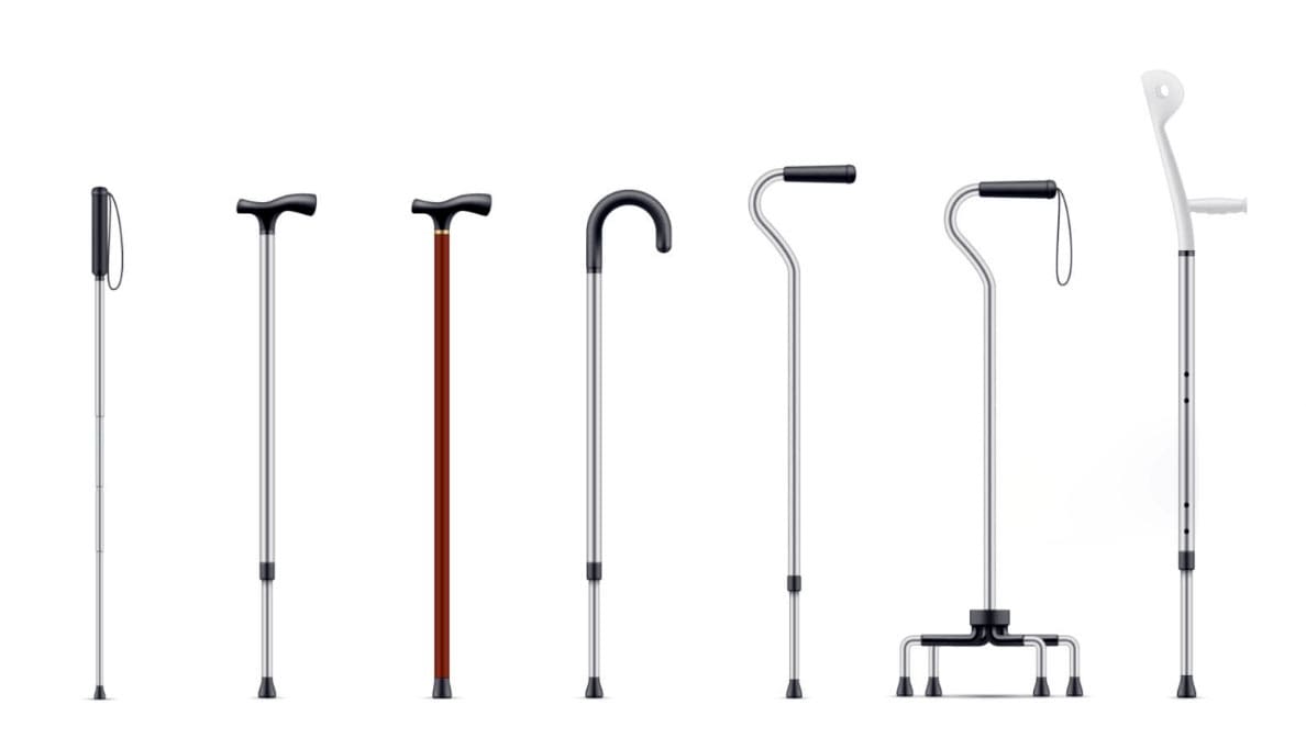
There are different types of canes, including single-point and quad canes. Single-point canes are generally the least expensive. Quad canes have four points and can provide more stability. It is important to use it correctly to prevent falls and injuries.
Post-surgery or Injury
A cane can help reduce pressure on the leg or back after surgery or injury. Healthcare providers may recommend a cane as a step-down device after using a walker or crutches. Before walking with the cane, ensure it is at the right height. Hold the cane in the hand on the opposite side of the injury. Hold the cane’s handle at the level of the bend in the wrist when standing with the elbow slightly bent. (American Academy of Orthopaedic Surgeons, 2020) If there are issues in both legs or a cane is used after back surgery, keep the cane in the hand with the most support.
Step forward with the cane and injured leg at the same time.
Step the non-injured leg up with the cane firmly on the ground to meet the injured leg.
The feet should be side by side.
Walking Normally
Once comfortable taking practice steps, try walking normally with the cane. Step forward with the cane and injured leg simultaneously. The cane should be off the ground when the wounded leg is in the air. Firmly plant the cane when stepping onto the injured leg. Step forward with the cane and the injured leg first, then step past the injured leg with the good leg.
Step down with the injured leg while lowering the cane to the step below.
Make sure the cane is firmly on the stairs.
Bring the good leg down to the same step.
If a handrail is available, use it. Although doing so may require moving the cane to the other hand, even if it’s on the same side as the injured leg, it will improve stability and reduce the risk of falling. Once proficient on the stairs, individuals may alternate placing one foot on each step.
Walking With Chronic Pain
Walking with a cane with a chronic pain condition is similar to using it with an injury. The location of the pain will determine which hand the cane is held in. If the pain is on the right side of the body, keep the cane on the left side or vice versa. If chronic pain is not in the legs but, for example, back pain makes it difficult to walk, hold the cane on either side, whichever feels more supportive and comfortable. If there is weakness on one side of the body or decreased sensation/numbness in one of the legs or feet, hold the cane on the opposite side of the pain, weakness, or numbness. Walking with a cane can also benefit individuals with other medical conditions. For example, assistive devices may be recommended for those with balance issues. (National Library of Medicine, 2023)
Cane Types
There are two primary types of canes, characterized by their points, and choosing the right one depends on the reason it’s needed. (Arthritis Foundation, N.D.)
Single-point
Single-point canes have one tip at the end.
These are recommended for those who need to relieve some pressure off an injured leg or need support due to occasional difficulties with balance.
Quad
Quad canes have four tips or feet to provide more stability.
They provide more support and are recommended for those with significant weakness in one leg or difficulty maintaining their balance while walking.
The traditional cane has a rounded C handle. Other types have contoured handles for a more secure grip. Talk to a doctor, physical therapist, or other health care professional for suggestions on which cane is right for you.
Losing Balance
A potential risk of using a cane is losing balance. If unable to maintain balance with a cane, individuals may want to consider a different walking device, such as a walker or crutches. To reduce the risk of falls, consider the following tips (American Academy of Orthopaedic Surgeons, 2020)
Wear shoes with nonskid soles.
Add lighting so you can see where you are walking.
Remove throw rugs or objects that can cause tripping.
Arrange furniture to allow for wide walking paths throughout the home.
Carry objects in a backpack or fanny pack rather than holding them.
Injury Medical Chiropractic and Functional Medicine Clinic
Regarding musculoskeletal pain, specialists like chiropractors, acupuncturists, and massage therapists can help mitigate the pain through spinal adjustments that help the body realign itself. They can also work with other associated medical professionals to develop a personalized treatment plan to help relieve muscle pain, improve the body’s flexibility and mobility, resolve musculoskeletal issues, and prevent future pain symptoms from reoccurring.
Osteoporosis
References
American Academy of Orthopaedic Surgeons. (2020). How to use crutches, canes, and walkers. https://orthoinfo.aaos.org/en/recovery/how-to-use-crutches-canes-and-walkers/
Hirayama, K., Otaka, Y., Kurayama, T., Takahashi, T., Tomita, Y., Inoue, S., Honaga, K., Kondo, K., & Osu, R. (2022). Efficiency and Stability of Step-To Gait in Slow Walking. Frontiers in human neuroscience, 15, 779920. https://doi.org/10.3389/fnhum.2021.779920
National Library of Medicine. (2023). Using a cane. Retrieved from https://medlineplus.gov/ency/patientinstructions/000343.htm
Arthritis Foundation. (N.D.). How to choose the right cane. https://www.arthritis.org/health-wellness/healthy-living/managing-pain/joint-protection/how-to-choose-the-right-cane
Individuals who have experienced spinal or back trauma, suffered fractures, are going through spinal degeneration, or are dealing with a spinal condition have an increased risk of anterolisthesis, where a vertebra slips forward relative to the vertebra below it. Can healthcare providers help prevent and treat the condition?
Anterolisthesis
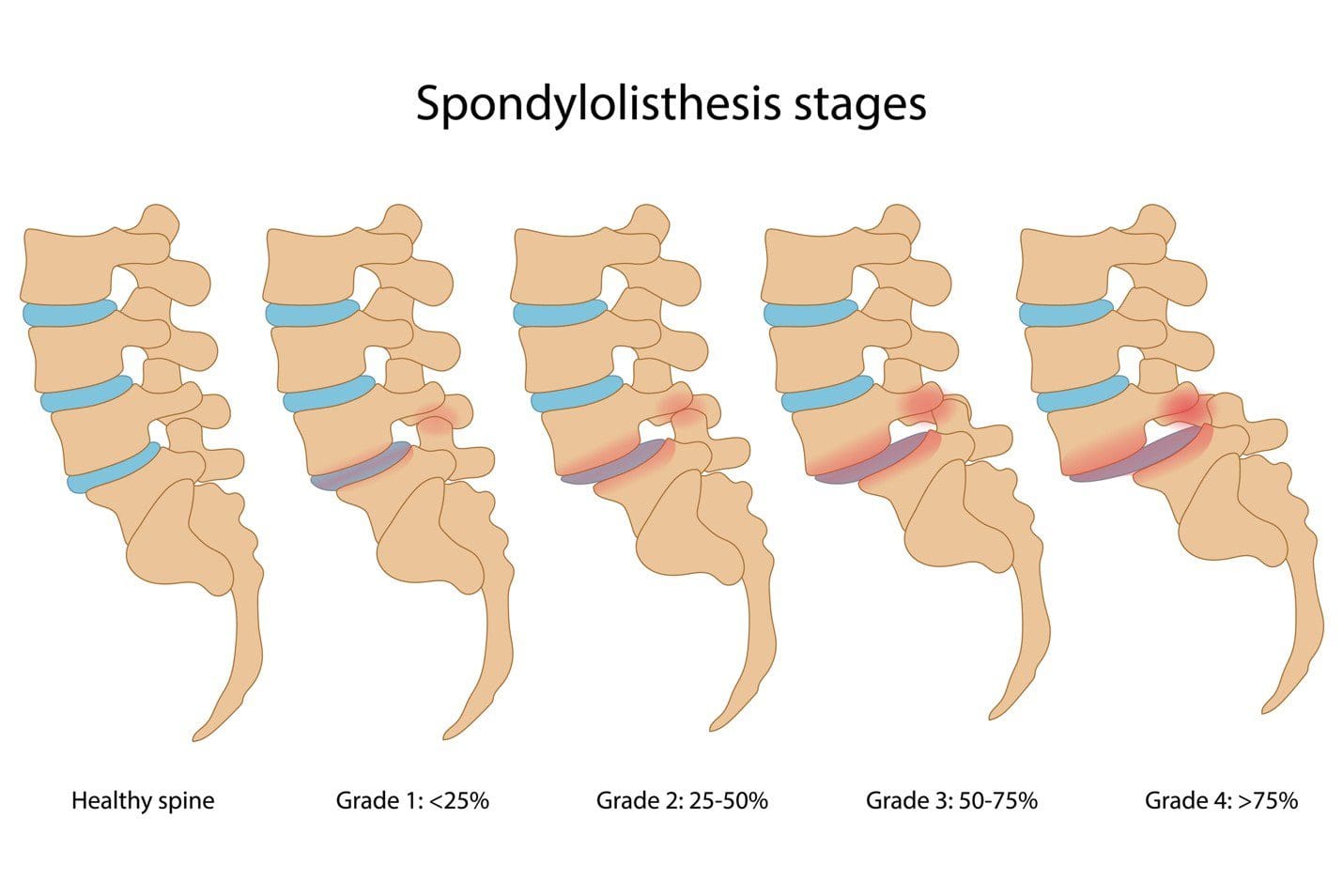
The spine consists of 33 individual bones or vertebrae stacked on one another. Anterolisthesis occurs when one vertebral segment slips forward over another. The condition can be mild, asymptomatic, or cause significant pain and neurological symptoms. Many different things, including osteoarthritis, osteoporosis, trauma, or a fracture, can cause this vertebral shifting. (Cedars Sinai, 2022) Spondylolisthesis is a general term for shifting a spinal vertebra over the one below it. It includes anterolisthesis, forward moving, and the less common retrolisthesis, or backward shifting.
Grades
Anterolisthesis is typically graded using the Meyerding scale, which assigns one of five grades according to how much slippage has occurred. These grades include:
Anterolisthesis can lead to various symptoms, depending on the severity and if the surrounding spinal nerves have been affected. The most common complaints include:
Diagnosis begins with a subjective evaluation and a physical examination. During these, the healthcare provider will assess sensation, strength, and reflexes and will order one of several diagnostic tests, including:
X-rays
Visualizes the vertebrae in the spine and their position relative to those above and below.
Also provides a clear picture of spinal arthritis or disc degeneration.
Magnetic Resonance Imaging – MRI
Allows the spinal cord, nerves, muscles, and discs to be assessed for compression or damage.
Several factors determine how the condition is treated, including:
The grade of the slippage.
The cause.
The symptoms.
The presence of instability on a diagnostic test such as an X-ray.
Stable and mildly symptomatic cases are usually treated with a combination that can involve:
Physical therapy
Activity modification
Bracing
Nonsteroidal anti-inflammatory medications/NSAIDs like ibuprofen.
Spinal injections
In more severe cases in which spinal instability or significant neurological symptoms are present, surgery may be recommended. This commonly involves a spinal decompression or fusion procedure. The technique varies based on the surgeon’s preferences and anatomy. (Koslosky E., and Gendelberg D. 2020)
Prognosis
Most individuals with this condition don’t know they have it until it is found accidentally on an X-ray or an MRI for something else. Mild cases can cause minimal symptoms and can be well-managed with conservative treatments. Cases of unstable anterolisthesis or those with neurological compression often require surgical intervention. These surgeries restore stability to the spine and alleviate any pressure on the nerves. More than 85% of individuals who need surgery have a successful outcome. (American Academy of Orthopaedic Surgeons, 2021)
Self-Care and Management
For individuals experiencing pain, numbness, or tingling from anterolisthesis, getting symptoms evaluated by a healthcare provider is an important first step. The healthcare provider may suggest one of several management strategies, which include:
Core Strengthening
To alleviate symptoms, exercises targeting the core muscles in the hips, pelvis, abdomen, and lower back are recommended.
Formal physical therapy may also be recommended.
Over-the-counter Meds
A healthcare provider may suggest pain-relieving medications like ibuprofen or naproxen to reduce soreness.
Activity Modification
Sticking to gentle, pain-free activities and avoiding excessive or repetitive extension of the spine can help prevent symptom aggravation. (American Academy of Orthopaedic Surgeons, 2021)
Injury Medical Chiropractic and Functional Medicine Clinic
At Injury Medical Chiropractic and Functional Medicine Clinic, our areas of practice include Chronic Pain, Personal Injury, Auto Accident Care, Work Injuries, Back Injury, Low Back Pain, Neck Pain, Migraine Headaches, Sports Injuries, Severe Sciatica, Scoliosis, Complex Herniated Discs, Fibromyalgia, Chronic Pain, Complex Injuries, Stress Management, Wellness & Nutrition, Functional Medicine Treatments, and in-scope care protocols. We focus on what works for you to relieve pain and restore function. If other treatment is needed, individuals will be referred to a clinic or physician best suited to their injury, condition, and/or ailment.
Koslosky, E., & Gendelberg, D. (2020). Classification in Brief: The Meyerding Classification System of Spondylolisthesis. Clinical orthopaedics and related research, 478(5), 1125–1130. https://doi.org/10.1097/CORR.0000000000001153
American Academy of Orthopaedic Surgeons. (2021). Adult spondylolisthesis in the low back. https://orthoinfo.aaos.org/en/diseases–conditions/adult-spondylolisthesis-in-the-low-back
Hospital for Special Surgery. (2023). Spondylolisthesis. https://www.hss.edu/condition-list_spondylolisthesis.asp
Can determining whether arm numbness occurs suddenly or gradually and whether there are other symptoms help healthcare providers diagnose and treat the condition?
Arm Numbness
Arm numbness or tingling are common symptoms that various medical conditions can cause. Numbness can be caused by a sudden health emergency, nerve disorder, or nutritional deficiency. (National Institute of Neurological Disorders and Stroke, 2024) Sometimes, this symptom results from an arm falling asleep and could resolve after just a few minutes. The sensation may be temporary, caused by something like sleeping in the wrong position. However, arm numbness and tingling may also be caused by neuropathy and chronic and progressive nerve damage and can also suddenly occur due to serious conditions, such as a heart attack or a stroke.
Circulation Issues
Deficient blood circulation in the arm could cause numbness and tingling as the nerves cannot receive enough oxygen. Conditions can interfere with normal blood flow and include: (Bryan L. and Singh A. 2024)
Atherosclerosis – plaque buildup in the arteries that may require medication or surgery.
Severe frostbite can damage the blood vessels but can be resolved with proper warming and wound care.
Vasculitis – is inflammation of the blood vessels that can be treated with medication.
Sleeping Position
A common example of sudden numbness and tingling is the feeling that the arm has fallen asleep. This usually occurs after sleeping awkwardly or leaning on the arm for a long time. Known as paresthesia, this sensation is related to the compression or irritation of nerves. (Bryan L. and Singh A. 2024) Sleeping in certain positions has been associated with nerve compression, especially when the hands or wrists are tucked or curled under the body, as well as maintaining proper spine alignment when sleeping, is the best way to prevent arm numbness.
Nerve Injuries and Conditions
Numbness that persists may be related to an injury or underlying health problem that affects the brachial plexus, a group of nerves that runs from the lower neck to the upper shoulders and controls movement and sensation in the arms. (Mount Sinai, 2022) Possible injuries that affect these nerves include: (Smith, S. M. et al., 2021)
A herniated disc caused by aging or trauma causes the disc to leak out and press on the nerve root.
Many medications, like gabapentin, can be used to alleviate the sensations of arm numbness.
Some medications can cause numbness as a side effect.
Some medications can cause complications, and arm numbness could be a symptom of those complications.
Inform healthcare providers about medications being taken to determine the relationship between them and any sensory changes.
Vitamin Deficiency
Peripheral neuropathy can also be caused by nutritional deficiencies and vitamin imbalances, which can damage nerves and cause sensation loss in the left or right arm. The most common sources are vitamin B12 deficiency and excess vitamin B6. (National Institute of Neurological Disorders and Stroke, 2024) Excessive alcohol consumption and other disorders that affect nutritional intake can also lead to nerve damage.
Injury Medical Chiropractic and Functional Medicine Clinic
At Injury Medical Chiropractic and Functional Medicine Clinic, our areas of practice include Chronic Pain, Personal Injury, Auto Accident Care, Work Injuries, Back Injury, Low Back Pain, Neck Pain, Migraine Headaches, Sports Injuries, Severe Sciatica, Scoliosis, Complex Herniated Discs, Fibromyalgia, Chronic Pain, Complex Injuries, Stress Management, Wellness & Nutrition, Functional Medicine Treatments, and in-scope care protocols. We focus on what works for you to relieve pain and restore function. If other treatment is needed, individuals will be referred to a clinic or physician best suited to their injury, condition, and/or ailment.
Chiropractic Care: The Natural Way to Recover From Injuries
References
National Institute of Neurological Disorders and Stroke. (2024). Paresthesia. Retrieved from https://www.ninds.nih.gov/health-information/disorders/glossary-neurological-terms#paresthesia
Bryan, L., Singh, A. Sleep Foundation. (2024). Numbness in Hands While Sleeping: Causes and Remedies. https://www.sleepfoundation.org/physical-health/numbness-in-hands-while-sleeping
Mount Sinai. (2022). Brachial plexopathy. https://www.mountsinai.org/health-library/diseases-conditions/brachial-plexopathy
Smith, S. M., McMullen, C. W., & Herring, S. A. (2021). Differential Diagnosis for the Painful Tingling Arm. Current sports medicine reports, 20(9), 462–469. https://doi.org/10.1249/JSR.0000000000000877
National Heart, Lung, and Blood Institute. (2023). Vasculitis. Retrieved from https://www.nhlbi.nih.gov/health/vasculitis/symptoms#:~:text=Nerve%20problems%2C%20including%20numbness%2C%20tingling,can%20also%20occur%20with%20vasculitis.
Centers for Disease Control and Prevention. (2024). Stroke signs and symptoms. Retrieved from https://www.cdc.gov/stroke/signs-symptoms/?CDC_AAref_Val=https://www.cdc.gov/stroke/signs_symptoms.htm
National Heart, Lung, and Blood Institute. (2022). What Is a Heart Attack? Retrieved from https://www.nhlbi.nih.gov/health/heart-attack
National Institute of Arthritis and Musculoskeletal and Skin Diseases. (2023). Spinal Stenosis Basics. Retrieved from https://www.niams.nih.gov/health-topics/spinal-stenosis/basics/symptoms-causes
Senderovich, H., & Jeyapragasan, G. (2018). Is there a role for combined use of gabapentin and pregabalin in pain control? Too good to be true?. Current medical research and opinion, 34(4), 677–682. https://doi.org/10.1080/03007995.2017.1391756
National Institute of Neurological Disorders and Stroke. (2024). Peripheral Neuropathy. Retrieved from https://www.ninds.nih.gov/health-information/disorders/peripheral-neuropathy
IFM's Find A Practitioner tool is the largest referral network in Functional Medicine, created to help patients locate Functional Medicine practitioners anywhere in the world. IFM Certified Practitioners are listed first in the search results, given their extensive education in Functional Medicine