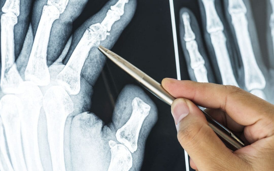
Finger sprains and dislocations are common hand injuries that can happen during work, physical/sports activities, or in automobile collisions and accidents. Can recognizing the symptoms help in developing an effective treatment strategy?
Finger Sprains and Dislocations
Finger sprains and dislocations are common injuries of the hand that cause pain and swelling.
A sprain happens when the finger tissue that supports a joint gets stretched beyond its limits in a way that stresses the ligaments and tendons.
The ligament tissue can be partially or completely torn. If the damage is bad enough, the joint comes apart.
This is a dislocation – A dislocation happens when the joint in the finger gets shifted out of its normal position.
Both injuries can cause pain and stiffness in the finger and hand.
Sprains
Finger sprains can happen any time the finger bends in an awkward or unusual way. This can happen from falling on the hand or getting hurt when engaged in physical activities like sports or household chores. Sprains can occur in any of the knuckle joints in the finger. However, most commonly, the joint in the middle of the finger gets sprained. It’s known as the proximal interphalangeal or PIP joint. (John Elfar, Tobias Mann. 2013) Symptoms of a finger sprain can include:
Other treatments to help a sprained finger include:
Elevate the hand if swelling and inflamed.
Gentle finger exercises/movements to prevent stiffness.
Icing the injured finger.
Take an anti-inflammatory medication.
Individuals who have not broken bones or dislocated the joint will probably be able to move their finger in about a week. A doctor will set a timeline for when to start using the finger normally.
Individuals who sprain their finger that feels swollen and stiff for longer than a few weeks are recommended to consult a doctor or specialist.
Thumb sprains and finger sprains in children may need to be splinted or taped for longer periods, as the ligament is not fully developed or as strong, which could lead to a tear.
Dislocations
A finger dislocation is a more severe injury involving the ligament, joint capsule, cartilage, and other tissues that causes misalignment of the finger. The ligaments and the joint capsule get torn when a joint is dislocated. The joint needs to be reset, which can be a simple process, or in severe cases, patients may need to be placed under anesthesia or undergo surgery to reset the joint properly.
In these cases, tendons or other tissues might be preventing the joint from getting into position.
Putting the finger back into the right position is known as”reduction.” Once reduced, the finger needs to be splinted.
Individuals also need an X-ray to ensure the joint is lined up correctly and that any bones were not broken or fractured when they sustained the injury. (James R. Borchers, Thomas M. Best. 2012)
Once reset, caring for a dislocated finger is basically the same as a sprained finger. Using ice on the finger, keeping the hand elevated to reduce swelling.
Elfar, J., & Mann, T. (2013). Fracture-dislocations of the proximal interphalangeal joint. The Journal of the American Academy of Orthopaedic Surgeons, 21(2), 88–98. doi.org/10.5435/JAAOS-21-02-88
OrthoInfo from the American Academy of Orthopaedic Surgeons. (2022) Hand fractures.
Hung, C. Y., Varacallo, M., & Chang, K. V. (2023). Gamekeeper’s Thumb. In StatPearls. StatPearls Publishing.
OrthoInfo from the American Academy of Orthopaedic Surgeons. (2022) Finger fractures.
Borchers, J. R., & Best, T. M. (2012). Common finger fractures and dislocations. American family physician, 85(8), 805–810.
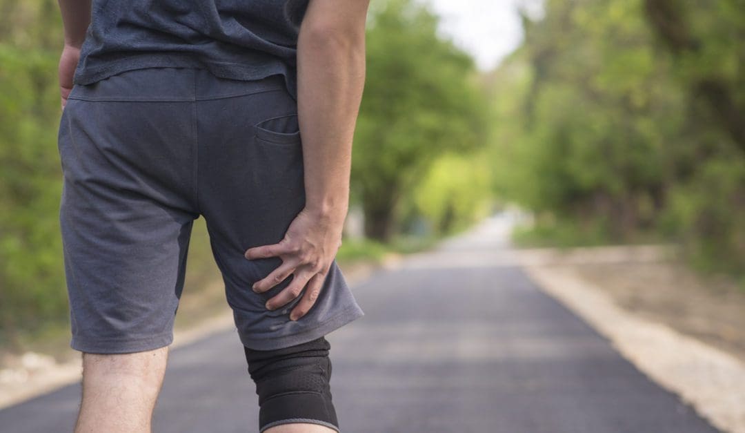
Hamstring muscle injuries are common, especially in athletes and individuals with physically demanding jobs. Is there a better chance of full recovery with surgical repair and post-op rehabilitation?
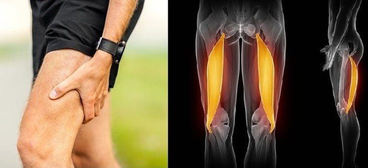
Hamstring Muscle Tear
Most often, hamstring muscle injuries are partial tears of the muscle. These types of injuries are muscle strains that occur when the muscle fibers are stretched beyond their normal limits. Complete tears of the hamstring muscle are unusual, but they do occur in both athletes and non-athletes. Determining the optimal treatment plan depends on:
The severity of the tendon tear
The expectations of the injured individual.
Incomplete tears are when the hamstring muscle is stretched too far, but not completely detached.
Complete tears usually occur at the top of the muscle where the tendon tears away from the pelvis.
A complete tear usually occurs when there is a sudden flexion of the hip and extension of the knee joint – when the muscle contracts in this position, it gets stretched beyond its limits.
Basic hamstring strains can be treated with simple steps – rest, ice, anti-inflammatory medications, and conservative therapies.
Symptoms
Symptoms of a hamstring muscle strain can include pain, bruising, swelling, and movement difficulty. (American Academy of Orthopaedic Surgeons. 2021) Individuals who sustain this injury typically experience sudden sharp pain. Signs of a tear can include:
Sharp pain where the buttock and thigh meet.
Difficulty walking.
Sitting can be difficult as the edge of a chair can place pressure directly on the injury.
Spasms and cramping sensations in the back of the thigh.
Weakness in the leg, specifically when bending the knee or lifting the leg behind the body.
Numbness or burning sensations as a result of sciatic nerve irritation.
Swelling and bruising in the back of the thigh – over time it can travel down to the back of the knee and calf and possibly into the foot.
With a complete hamstring tear, there is usually significant swelling and bruising that develops in the back of the thigh.
Diagnosis
The symptoms can be difficult to spot in the early stages which is why X-rays of the hip or thigh are usually obtained.
In some situations, a fragment of bone can get pulled off the pelvis along with the hamstring muscle attachment. MRI testing can be performed to evaluate the attachment and can define critical features of a complete hamstring muscle tear, including: (American Academy of Orthopaedic Surgeons. 2021)
The number of tendons involved.
Complete versus incomplete tearing.
The amount of retraction – the amount the tendons have pulled back.
This will guide the development of treatment.
Treatment
The treatment of a complete tear will depend on different factors. The other variable is the patient and their expectations.
Treatment is more aggressive in younger individuals like high-level athletes.
Treatment is less aggressive in middle-aged individuals.
Often a single tendon tear can be treated non-surgically.
When one tendon is involved, it is typically not pulled very far from its normal attachment and will develop scar tissue in a positive position.
Conversely, when three tendons have been torn, they usually pull more than a few centimeters away from the bone. These cases have better results with surgical repair. (UW Health. 2017)
Surgeons will use patient characteristics – high-level athletes or less physically active individuals – to guide treatment recommendations.
Rehabilitation
Rehabilitation following surgery can take 3-6 months or longer.
The first six weeks limit weight-bearing with the use of crutches.
Patients may be recommended to wear a brace to reduce tension on the repaired hamstring tendons.
Strengthening does not begin until three months post-op, and even light activities are usually delayed. (UW Health. 2017)
Because this injury can have a long recovery time, some individuals may choose nonsurgical treatment.
Sometimes these individuals experience symptoms of discomfort from sitting and may exhibit long-term weakness of the hamstring muscle.
Full recovery from a complete hamstring muscle injury takes time. Studies have shown high-level athletes are able to resume competitive sports after the repair and rehabilitation of an acute hamstring muscle injury. (Samuel K. Chu, Monica E. Rho. 2016)
Delaying surgical treatment may not always lead to optimal results.
When the tendon is torn away from its normal attachment, it begins to scar around the surrounding soft tissues.
When there is a delay of more than a few weeks following the initial injury, regaining the full length of the tendon and muscle can be challenging.
This could delay the rehabilitation process and may limit the potential for full recovery. (Ho Yoon Kwak, et al., 2011)
With severe injuries, there is a better chance of full recovery with surgical repair but could involve a long recovery and commitment to a post-op rehabilitation plan.
Chu, S. K., & Rho, M. E. (2016). Hamstring Injuries in the Athlete: Diagnosis, Treatment, and Return to Play. Current sports medicine reports, 15(3), 184–190. doi.org/10.1249/JSR.0000000000000264
Kwak, H. Y., Bae, S. W., Choi, Y. S., & Jang, M. S. (2011). Early surgical repair of acute complete rupture of the proximal hamstring tendons. Clinics in orthopedic surgery, 3(3), 249–253. doi.org/10.4055/cios.2011.3.3.249
Nowadays, individuals trying to avoid surgery have more therapy options. Can regenerative medicine help treat neuromusculoskeletal injuries?
Regenerative Medicine
Regenerative medicine utilizes the body’s raw cells and is used in cancer treatment and to reduce the risk of infections. (American Cancer Society. 2020) Researchers are looking for other ways to use these cells in medical therapies.
What are These Cells
Stem cells are unspecialized cells that can develop into any cell and in certain cases renew themselves an unlimited number of times. (National Institutes of Health. 2016)
Regenerative cell therapy uses these cells as a treatment for a disease or condition.
Regenerative cells are given to individuals to replace cells that have been destroyed or have died.
In the case of cancer, they may be used to help the body regain the ability to produce regenerative cells after treatment. (American Cancer Society. 2020)
For individuals with multiple myeloma and certain types of leukemia, regenerative cell therapy is used to eliminate cancer cells.
The therapy is called graft-versus-tumor effect/GvT, where a donor’s white blood cells/WBCs are used to eliminate the cancerous tumor. (American Cancer Society. 2020)
What They Can Treat
This is a new treatment that is still going through research. The Food and Drug Administration has only approved it for certain cancers and conditions that affect the blood and immune system. (Centers for Disease Control and Prevention. 2019) Regenerative cell therapy is FDA-approved to treat: (National Cancer Institute. 2015)
Leukemia
Lymphoma
Multiple myeloma
Neuroblastoma
It is also used to decrease the risk of infection after regenerative cell transplantation in individuals with blood cancers. (U.S. Food & Drug Administration. 2023)
Researchers are studying how these cells can treat other conditions. Clinical trials are analyzing how to use the therapy for neurodegenerative diseases like:
During regenerative cell therapy, the cells are given through an intravenous line. The three places where blood-forming cells can be obtained are bone marrow, the umbilical cord, and blood. Transplants can include: (American Cancer Society. 2020)
Autologous
The cells are taken from the individual who will be receiving the therapy.
Allogeneic
The cells are donated by another individual.
Syngeneic
The cells come from an identical twin, if there is one.
Safety
The therapy has shown to provide benefits but there are risks.
One risk is known as graft-versus-host disease – GVHD.
It occurs in one-third to half of allogeneic recipients.
This is where the body does not recognize the donor’s white blood cells and attacks them causing problems and symptoms throughout the body.
To treat GVHD medications are given to suppress the immune system to stop attacking the donor cells. (American Cancer Society. 2020)
The future of regenerative cell therapy is promising. Research is ongoing to find out how these cells can treat conditions and find new ways to treat and cure diseases.
Regenerative medicine has been researched for over twenty years for conditions like macular degeneration, glaucoma, stroke, and Alzheimer’s disease. (National Institutes of Health. 2022) This therapy is a new medical treatment that could be used in future therapies as part of a multidisciplinary approach to neuromusculoskeletal injuries and conditions.
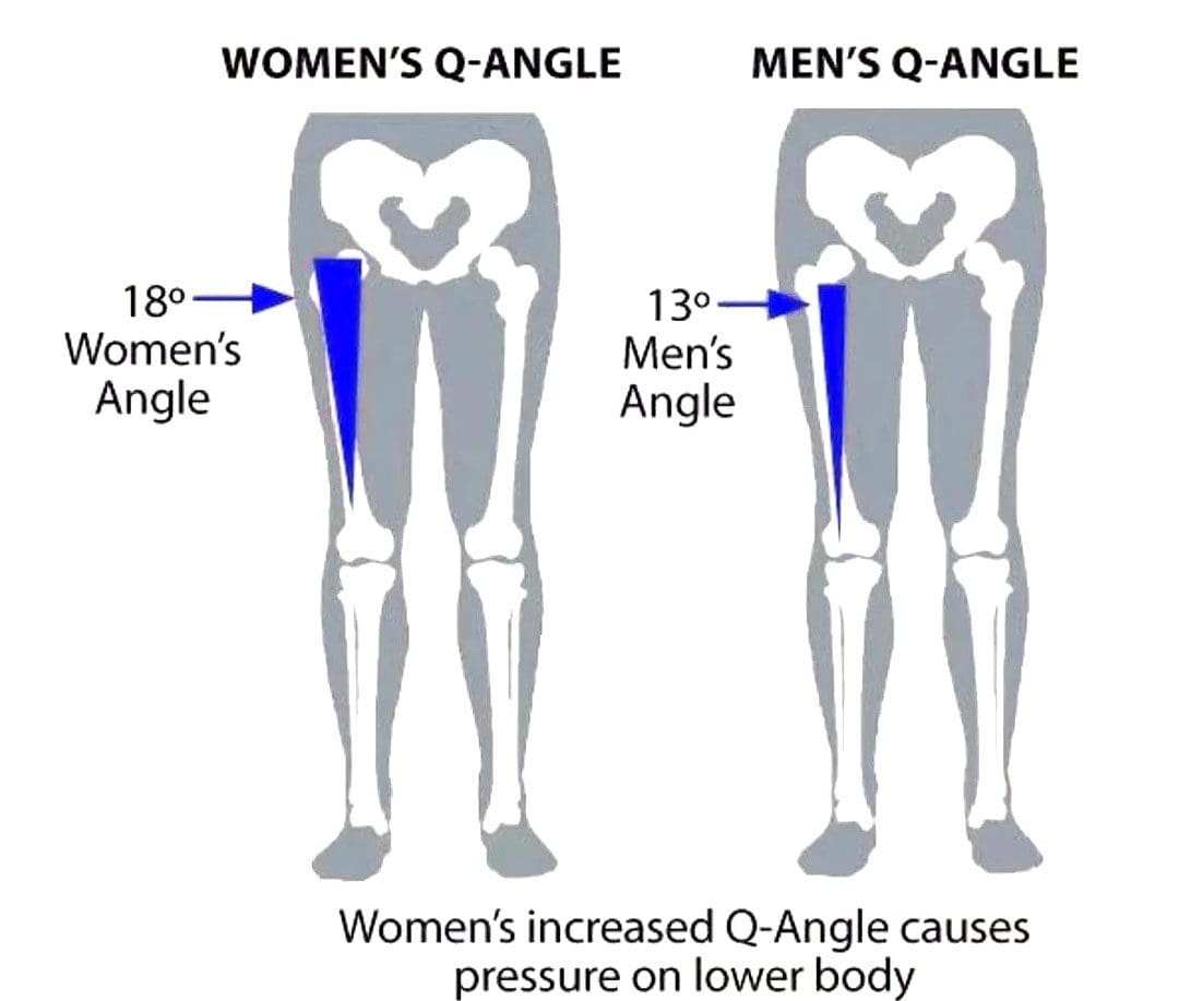
The Q or quadriceps angle is a measurement of pelvic width that is believed to contribute to the risk of sports injuries in women athletes. Can non-surgical therapies and exercises help rehabilitate injuries?
Quadriceps Q – Angle Injuries
The Q angle is the angle where the femur/upper leg bone meets the tibia/lower leg bone. It is measured by two intersecting lines:
One from the center of the patella/kneecap to the anterior superior iliac spine of the pelvis.
The other is from the patella to the tibial tubercle.
On average the angle is three degrees higher in women than men.
Women have biomechanical differences that include a wider pelvis, making it easier to give birth. However, this difference can contribute to knee injuries when playing sports, as an increased Q angle generates more stress on the knee joint, as well as leading to increased foot pronation.
Injuries
Various factors can increase the risk of injury, but a wider Q angle has been linked to the following conditions.
Patellofemoral Pain Syndrome
An increased Q angle can cause the quadriceps to pull on the kneecap, shifting it out of place and causing dysfunctional patellar tracking.
With time, this can cause knee pain (under and around the kneecap), and muscle imbalance.
Foot orthotics and arch supports could be recommended.
Some researchers have found a link, while others have not found the same association. (Wolf Petersen, et al., 2014)
Chondromalacia of the Knee
This is the wearing down of the cartilage on the underside of the kneecap.
An increased Q angle can be a factor that increases stress and causes the knee to lose its stability.
However, this remains controversial, as some studies have found no association between the Q angle and knee injuries.
Chiropractic Treatment
Strengthening Exercises
ACL injury prevention programs designed for women have resulted in reduced injuries. (Trent Nessler, et al., 2017)
The vastus medialis obliquus or VMO is a teardrop-shaped muscle that helps move the knee joint and stabilize the kneecap.
Strengthening the muscle can increase the stability of the knee joint.
Strengthening may require a specific focus on muscle contraction timing.
Closed-chain exercises like wall squats are recommended.
Glute strengthening will improve stability.
Stretching Exercises
Stretching tight muscles will help relax the injured area, increase circulation, and restore range of motion and function.
Muscles commonly found to be tight include the quadriceps, hamstrings, iliotibial band, and gastrocnemius.
Foot Orthotics
Custom-made, flexible orthotics decrease the Q angle and reduce pronation, relieving the added stress on the knee.
A custom orthotic ensures that the foot and leg dynamics are accounted for and corrected.
Motion-control shoes can also help correct overpronation.
Knee Rehabilitation
References
Khasawneh, R. R., Allouh, M. Z., & Abu-El-Rub, E. (2019). Measurement of the quadriceps (Q) angle with respect to various body parameters in young Arab population. PloS one, 14(6), e0218387. doi.org/10.1371/journal.pone.0218387
Petersen, W., Ellermann, A., Gösele-Koppenburg, A., Best, R., Rembitzki, I. V., Brüggemann, G. P., & Liebau, C. (2014). Patellofemoral pain syndrome. Knee surgery, sports traumatology, arthroscopy: Official journal of the ESSKA, 22(10), 2264–2274. doi.org/10.1007/s00167-013-2759-6
Vaienti, E., Scita, G., Ceccarelli, F., & Pogliacomi, F. (2017). Understanding the human knee and its relationship to total knee replacement. Acta bio-medica : Atenei Parmensis, 88(2S), 6–16. doi.org/10.23750/abm.v88i2-S.6507
Mitani Y. (2017). Gender-related differences in lower limb alignment, range of joint motion, and the incidence of sports injuries in Japanese university athletes. Journal of Physical Therapy Science, 29(1), 12–15. doi.org/10.1589/jpts.29.12
Nessler, T., Denney, L., & Sampley, J. (2017). ACL Injury Prevention: What Does Research Tell Us? Current reviews in musculoskeletal medicine, 10(3), 281–288. doi.org/10.1007/s12178-017-9416-5
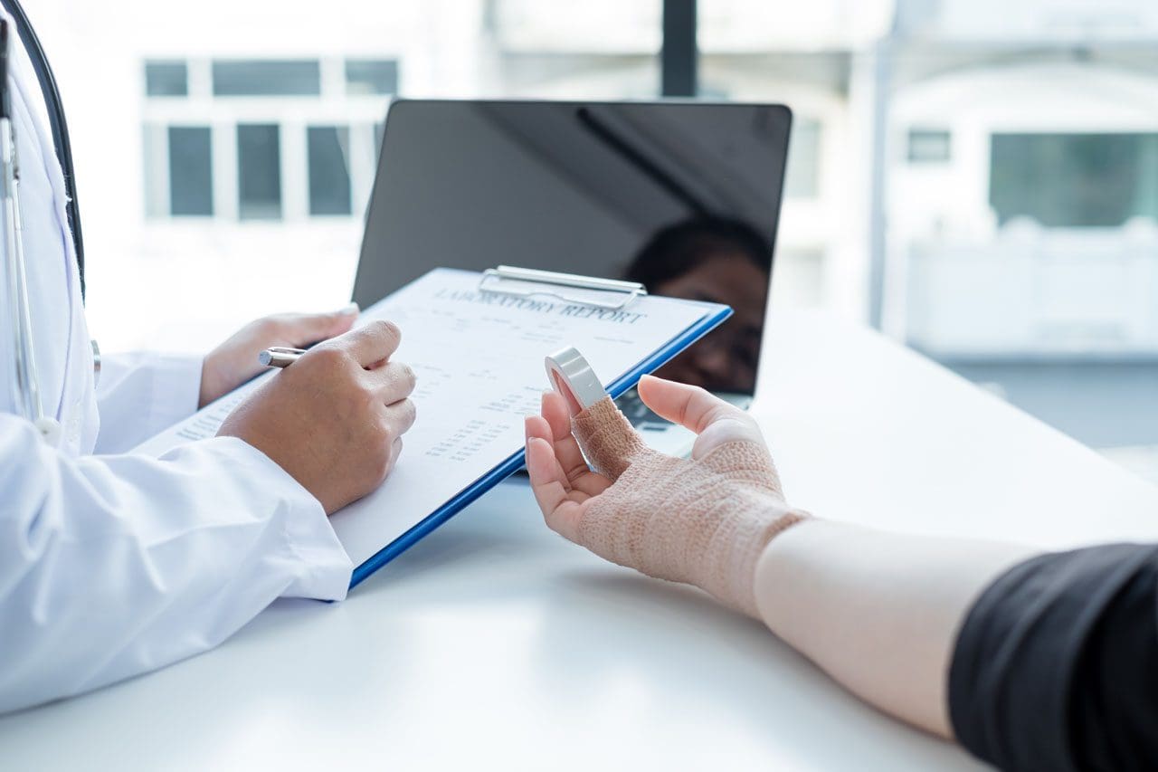
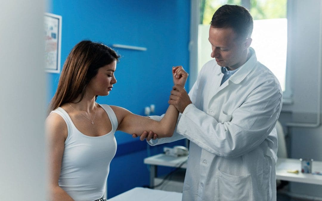
Golfing wrist injuries are common with treatment requiring 1-3 months of rest and immobilization and if tears are present surgery. Can chiropractic treatment help avoid surgery, expedite recovery, and rehabilitation?
Golfing Wrist Injuries
Golfing Wrist Injuries: According to a study, there are over 30,000 golf-related injuries treated in American emergency rooms every year. (Walsh, B. A. et al, 2017) Nearly a third are related to a strain, sprain, or stress fracture.
This causes pain and inflammation and is usually accompanied by a grinding sensation when moving the thumb and wrist.
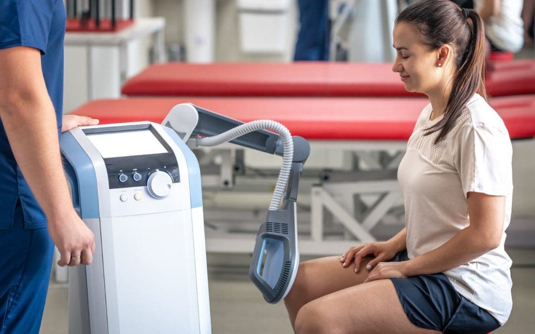
Chiropractic Treatment
Given the nature of these injuries, medical attention should be sought out for image scans to look at any damage and properly immobilize the wrist. Once a fracture has been ruled out or healed, golfing wrist injuries can benefit from chiropractic and physical therapy. (Hulbert, J. R. et al, 2005) A typical treatment may involve a multifaceted approach involving various therapies including:
Active release therapy, myofascial release, athletic taping, corrective exercise, and stretching.
A chiropractor will examine the wrist and its functioning to determine the nature of the injury.
A chiropractor may recommend using a splint to immobilize the wrist, particularly in cases of overuse.
They will relieve pain and swelling first, then focus on strengthening the joint.
They may recommend a regimen of icing the hand.
Adjustments and manipulations will relieve pressure on the nerves to reduce swelling and restore mobility.
Peripheral Neuropathy Successful Recovery
References
Walsh, B. A., Chounthirath, T., Friedenberg, L., & Smith, G. A. (2017). Golf-related injuries treated in United States emergency departments. The American journal of emergency medicine, 35(11), 1666–1671. doi.org/10.1016/j.ajem.2017.05.035
Moon, H. W., & Kim, J. S. (2023). Golf-related sports injuries of the musculoskeletal system. Journal of exercise rehabilitation, 19(2), 134–138. doi.org/10.12965/jer.2346128.064
Ray, G., Sandean, D. P., & Tall, M. A. (2023). Tenosynovitis. In StatPearls. StatPearls Publishing.
Zouzias, I. C., Hendra, J., Stodelle, J., & Limpisvasti, O. (2018). Golf Injuries: Epidemiology, Pathophysiology, and Treatment. The Journal of the American Academy of Orthopaedic Surgeons, 26(4), 116–123. doi.org/10.5435/JAAOS-D-15-00433
Tan, H. K., Chew, N., Chew, K. T., & Peh, W. C. (2014). Clinics in diagnostic imaging (156). Golf-induced hamate hook fracture. Singapore medical journal, 55(10), 517–521. doi.org/10.11622/smedj.2014133
Hulbert, J. R., Printon, R., Osterbauer, P., Davis, P. T., & Lamaack, R. (2005). Chiropractic treatment of hand and wrist pain in older people: systematic protocol development. Part 1: informant interviews. Journal of chiropractic medicine, 4(3), 144–151. doi.org/10.1016/S0899-3467(07)60123-2
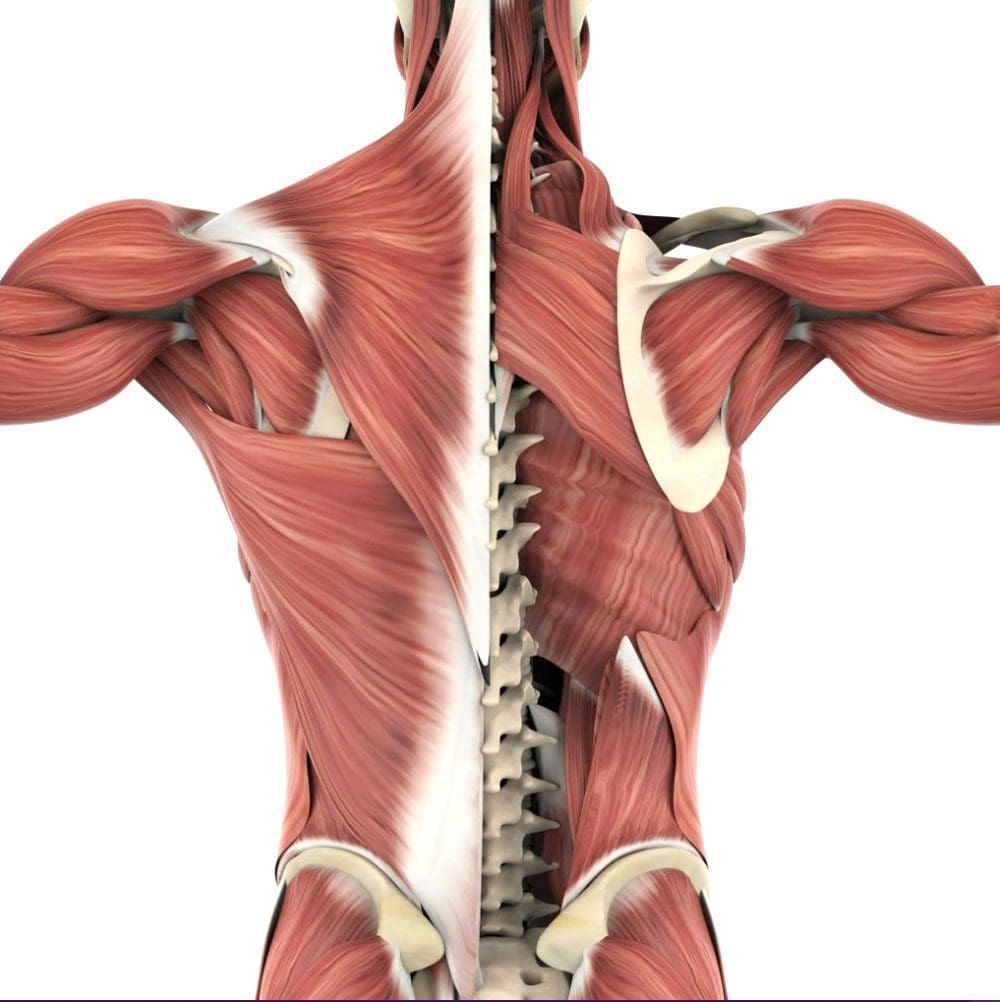
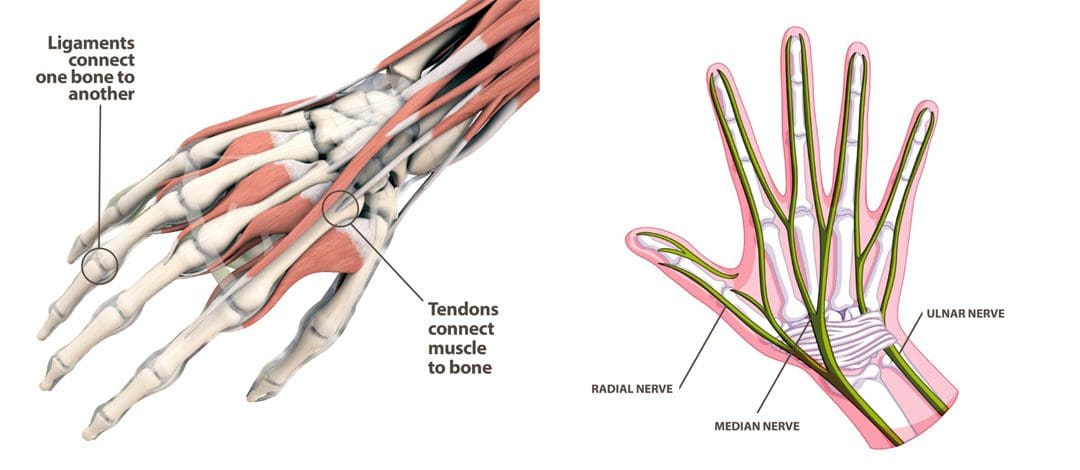
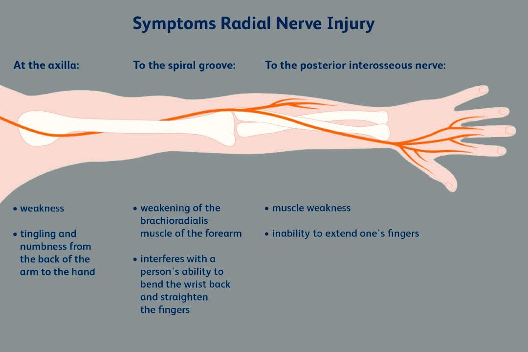
The brachial plexus is a network of nerves that begin in the cervical/neck spinal cord and travel down the cervicoaxillary canal into the armpit. Forming in the area of the shoulder joint at the branch junction of the brachial plexus, the radial nerve extends down the arm, through the elbow joint, into the forearm, across the wrist, and tips of the fingers. The nerves are susceptible to injury that can cause abnormal function leading to unusual sensations and impaired muscle function.
Radial Nerve
One of the major nerves of the upper extremity.
There is one brachial plexus on each side of the body that carries the nerves to each arm.
The radial nerve has two major functions.
One is to provide sensations in the hands, forearms, arms, and fingers.
The other is to deliver messages to muscles about when to contract.
Motor Function
The radial nerve transmits signals to the muscles of the back of the arm and forearm on when to contract.
Individuals who have abnormal radial nerve function can experience weakness of the muscles and symptoms like wrist drop.
A wrist drop occurs when the back forearm muscles cannot support the wrist, causing the individual to hold the wrist in a flexed posture.
Abnormal radial nerve function can cause symptoms of numbness or tingling in the back of the hand.
Conditions
Associated conditions to the radial nerve include lacerations, contusions, fractures, and palsies.
Nerve Contusion
A contusion typically occurs through blunt force trauma that can crush and smash the nerve area.
This causes abnormal or no function.
A nerve contusion can occur from a personal, work, or sports injury or other conditions that generate intense pressure on the nerve/s.
Nerve Lacerations
A laceration occurs when there is a penetrating injury that cuts and/or severs the nerve.
This injury can occur from stab wounds or sliced by broken glass, metal, etc.
Fractures
Broken bones of the upper extremity can lead to extended damage to the nerves near the damaged bone.
The most common type of fracture associated with radial nerve malfunction is fractures to the humerus bone.
The nerve wraps tightly around the humerus and can be injured with a fracture.
Most fracture-related radial nerve injuries heal on their own and do not require surgery.
However, the way the injury heals can be the difference between normal function and chronic pain.
Crutch Palsy
Crutch palsy is pressure on the radial nerve in the armpit resulting from using crutches incorrectly.
To use crutches properly, the individual needs to support their body weight through the hands.
However, many tend to place pressure around the armpit at the top of the crutch, causing irritation to the nerve in that area.
Padding the top of crutches and using the proper form can prevent the condition.
Saturday Night Palsy
Saturday night palsy is the abnormal function of the radial nerve after sleeping in a position that causes direct pressure against the nerve.
This often occurs when an individual falls asleep with their arm draped over an armrest on a chair.
The name comes from when individuals are intoxicated and fall asleep in a location other than the bed and in awkward positions.
Treatment
Nerve injuries often cause symptoms at different locations other than where the nerve damage is, complicating diagnosis. Determining the specific location of nerve damage is the first step in developing an appropriate treatment plan. Once the location has been identified, steps can be taken to prevent worsening damage to the nerve.
The objective is to relieve the pressure from the irritation or compression.
Massage to relax the area and increase blood circulation.
Decompression to physically restore alignment.
Adjustments to restore body balance.
Exercises and stretches to maintain treatment, strengthen the muscles, and prevent injuries.
In cases where there is structural damage, surgery may be necessary to remove pressure or repair damage.
Avoid Surgery
References
Ansari FH, Juergens AL. Saturday Night Palsy. [Updated 2023 Apr 24]. In: StatPearls [Internet]. Treasure Island (FL): StatPearls Publishing; 2023 Jan-. Available from: www.ncbi.nlm.nih.gov/books/NBK557520/
Barton, N J. “Radial nerve lesions.” The Hand vol. 5,3 (1973): 200-8. doi:10.1016/0072-968x(73)90029-6
Daly, Michael, and Chris Langhammer. “Radial Nerve Injury in Humeral Shaft Fracture.” The Orthopedic Clinics of North America vol. 53,2 (2022): 145-154. doi:10.1016/j.ocl.2022.01.001
DeCastro A, Keefe P. Wrist Drop. [Updated 2022 Jul 18]. In: StatPearls [Internet]. Treasure Island (FL): StatPearls Publishing; 2023 Jan-. Available from: www.ncbi.nlm.nih.gov/books/NBK532993/
Eaton, C J, and G D Lister. “Radial nerve compression.” Hand Clinics vol. 8,2 (1992): 345-57.
Glover NM, Murphy PB. Anatomy, Shoulder and Upper Limb, Radial Nerve. [Updated 2022 Aug 29]. In: StatPearls [Internet]. Treasure Island (FL): StatPearls Publishing; 2023 Jan-. Available from: www.ncbi.nlm.nih.gov/books/NBK534840/
Ljungquist, Karin L et al. “Radial nerve injuries.” The Journal of hand surgery vol. 40,1 (2015): 166-72. doi:10.1016/j.jhsa.2014.05.010
How do non-surgical treatments compare with traditional surgical treatments to improve mobility for individuals with hamstring injuries? The hamstrings are muscles in the lower extremities that provide mobility to the legs and stabilize the pelvis. Many athletes rely on their hamstrings to perform strenuous actions such as sprinting, jumping, squatting, and kicking during sporting events. However, the hamstrings are also very susceptible to injury. Athletes who repeatedly overstretch their hamstrings can experience muscle strain until microscopic tears form, which is common. Similarly, individuals who sit for long periods can also experience hamstring issues. When individuals are not physically active, their hamstrings can become weak and shortened, leading to symptoms such as muscle pain, trigger points, and strain on the accessory muscles. Hamstring injuries can also cause other issues that affect the lower body extremities. This article will explore how hamstring injuries affect mobility and how non-surgical treatments help people regain mobility. We work with certified medical providers who use our patients’ valuable information to treat individuals suffering from hamstring injuries and inform them about non-surgical treatments to regain mobility. We encourage patients to ask essential questions and seek education from our associated medical providers about their condition. Dr. Jimenez, D.C., provides this information as an educational service. Disclaimer
Hamstring Injuries Implementing Other Issues
Do you experience stiffness in the back of your thighs when warming up before exercising? Are you feeling radiating pain from the side of your hips and glutes due to prolonged sitting? Or do you tend to limp, affecting your gait and walking? Many people are unaware they are overexerting their hamstrings, which can cause pain. Those who engage in sports or work sedentary jobs may over or underuse their hamstrings, affecting their flexibility and mobility to the lower extremities. According to research studies, hamstring injuries are the most common non-contact muscle injuries caused by two mechanisms of injuries: stretch-type and sprint-type. Sprint-type injuries associated with hamstrings occur when the muscles are overexerted due to maximal or near-maximal action, causing muscle fatigue. To that point, hamstring injuries can also affect a person’s walking mobility.
Running without properly warming up the hamstring muscle can cause muscle fatigue. Stretch-type injuries associated with the hamstring muscles involve combination movements that include extreme hip flexion and knee extension. These injuries can also mimic sciatica, leading people to believe their sciatic nerve is acting up. However, treatments available can help reduce the pain associated with hamstring injuries and lengthen the shortened muscle to reduce pain.
Best Lower Body Stretches To Increase Flexibility-Video
If you want to reduce the pain associated with hamstring injuries, incorporating RICE can help prevent it from becoming chronic. This involves gently stretching the affected muscle to avoid cramps and pain while increasing flexibility. Hamstring injuries can also be linked to other chronic issues, which can cause inflammation in the surrounding muscles. Studies show that conditions like piriformis syndrome can cause nerve entrapment in the hamstrings, resulting in radiating pain down the leg that mimics low back pain and sciatica. As previously mentioned, hamstring injuries can limit mobility and be linked to chronic conditions. Thankfully, non-surgical treatments can help reduce pain and provide relief. Check out the video above to learn different stretches that can help reduce pain in the lower body and increase flexibility.
Treatments To Restore Mobility
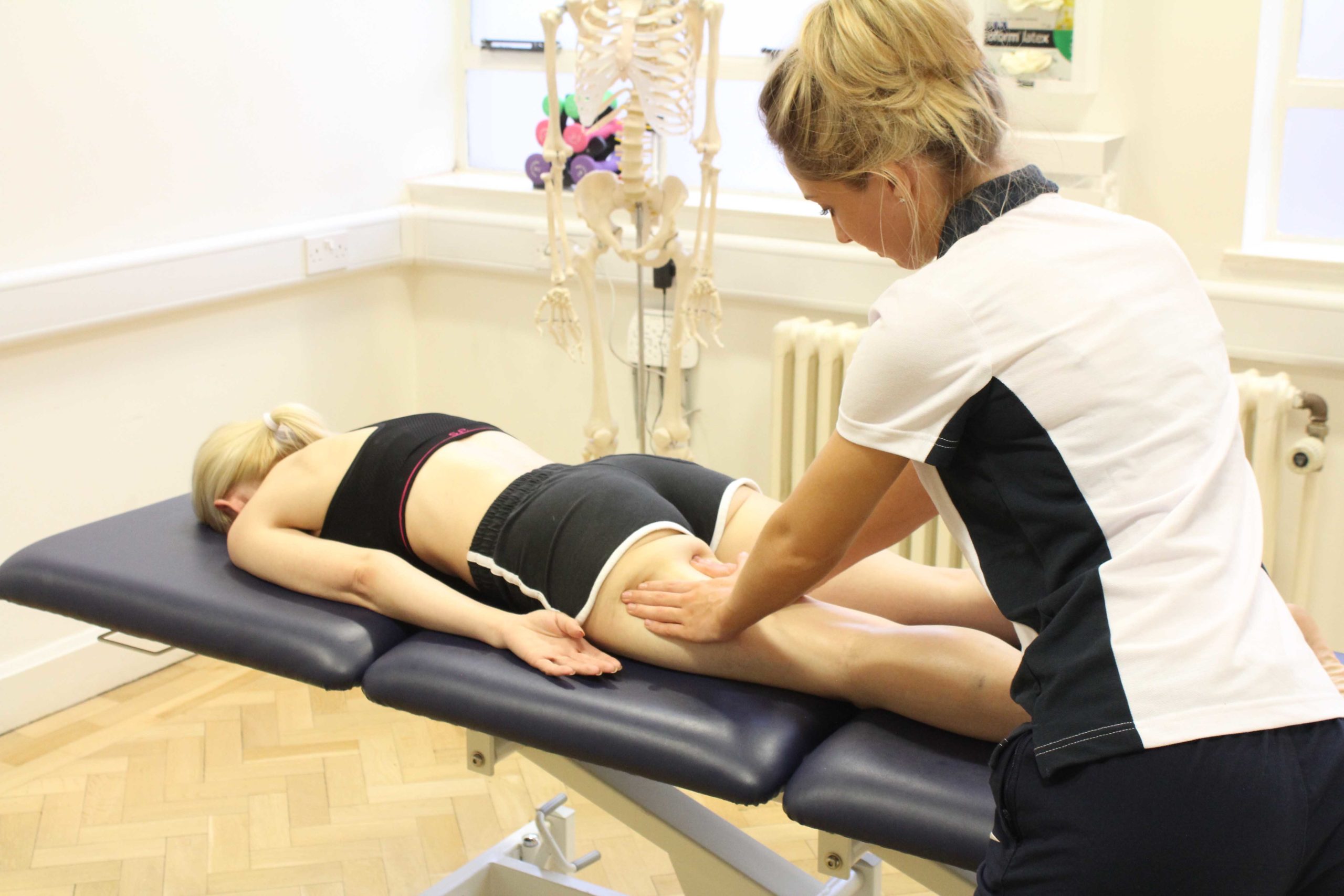
If rest, ice, compression, and gentle stretching do not provide relief, incorporating treatments for hamstring injuries to restore mobility can benefit many individuals. Seeking the help of a pain specialist, such as a massage therapist or chiropractor, to create a customized plan/program is recommended. There are various approaches that pain specialists can use to regain mobility and treat hamstring injuries.
MET Therapy
Many chiropractors and massage therapists incorporate MET (muscle energy techniques) therapy to gently stretch out the shortened hamstring muscle and help restore joint mobility in the lower extremities. In “Clinical Applications of Neuromuscular Techniques,” written by Leon Chaitow, N.D., D.O., and Judith Walker DeLany, L.M.T., stated that MET is crucial in stretching and strengthening the hamstring muscles through isometric contraction. At the same time, additional research studies reveal that the MET technique allows the hamstrings to have a greater increase in hip flexion ranges. MET therapy also helps strengthen the accessory muscles surrounding the hamstrings to restore mobility.
Spinal Decompression
If hamstring injuries are caused by nerve entrapment, then trying out spinal decompression can help restore mobility to the hips and lower extremities. According to “The Ultimate Spinal Decompression,” written by Dr. Eric Kaplan, D.C., FIAMA, and Dr. Perry Bard, D.C., stated that spinal decompression is safe and gentle on the spine as it provides gentle traction on the spinal disc to reduce pain and increase disc height. When hamstring injuries are associated with nerve entrapment, it could result from a herniated disc that aggravates the nerve root and causes referred pain to the hamstrings. Using gentle traction on the spine can help alleviate the pain caused by the aggravating nerve and reduce pain in the hamstrings. Many individuals can incorporate these treatments to reduce hamstring injuries and regain their mobility back to their legs.
References
Chaitow, L., & Delany, J. (2002). Clinical application of neuromuscular techniques. Vol. 2, The lower body. Churchill Livingstone.
IFM's Find A Practitioner tool is the largest referral network in Functional Medicine, created to help patients locate Functional Medicine practitioners anywhere in the world. IFM Certified Practitioners are listed first in the search results, given their extensive education in Functional Medicine