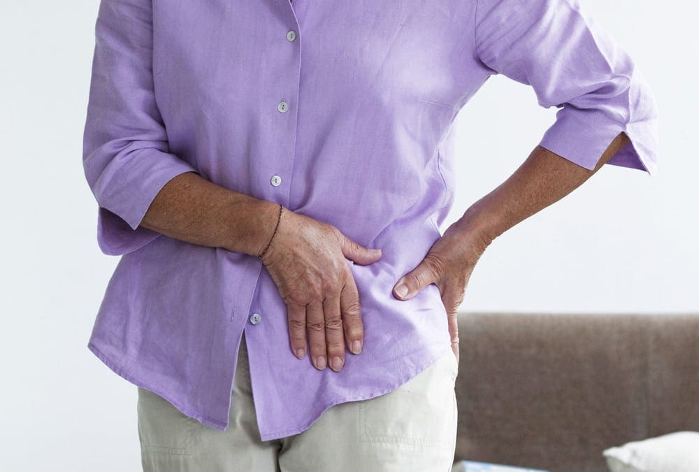
The hips are some of the most flexible structures in the human body, providing the necessary amount of strength and stability needed to support the human body when walking, running or jumping. However, the hip joint can also be vulnerable to damage or injury, resulting in debilitating hip pain. Trochanteric bursitis is hip pain brought on by the inflammation of the fluid-filled sac, or bursa, found on the outer border of the hip.
Trochanteric Bursitis Overview
There are about 160 bursae located around the entire body. Bursae act as a sort of “cushion” between soft tissues and bones, preventing bones from rubbing against tendons, ligaments, and muscles. Trochanteric bursitis can affect any of the bursae inside the human body. Trochanteric bursitis affects the outer part of the thighbone, or the femur, at the edge of the hip. This bony point is best known as the greater trochanter.
Another bursa, called the iliopsoas bursa, can be found on the inside of the hip. Inflammation of the iliopsoas bursa also triggers pain in the groin. Bursitis is considered to be one of the top causes of hip pain. Repetitive physical activities, such as climbing stairs, or even surgical interventions to the hip may cause inflammation in the bursa. Many doctors commonly refer to trochanteric�bursitis as greater trochanteric pain syndrome.
Signs and Symptoms of Trochanteric Bursitis
The main characteristic of trochanteric bursitis involves pain in the outer area of the hip or pain when laying on the affected side of the hip. The painful signs and symptoms will also generally become worse through certain physical activities, such as walking or climbing stairs. Pain may also�radiate down the�thigh and into the feet, or it may disperse. Pain can be sharp and fade into an ache, accompanied by swelling in the legs.
Causes of Trochanteric Bursitis
Common causes of trochanteric bursitis include�slip-and-fall accidents, strong blows to the hip, or lying on one side of the body for an extended period of time. Sports injuries involving�overuse from repetitive physical activities like running, bicycling, or climbing stairs, a ripped tendon or even standing may cause trochanteric�bursitis. Health issues, such as�bone spurs in the hip or thighbone, may consequently cause trochanteric bursitis.�
A variety of conditions and disorders may also lead to trochanteric bursitis, including spine problems, such as scoliosis or arthritis of the lumbar spine, even rheumatoid arthritis, and gout as well as thyroid disease. Moreover, legs of two different lengths,�hip surgery or prosthetic implants can create problems in the hips. Trochanteric bursitis is most common in middle-aged or elderly people and it is most prevalent in women than men.
�
Trochanteric Bursitis Treatment and Chiropractic Care
Avoiding the physical activities which caused trochanteric bursitis will allow time for the body to heal. After seeing a healthcare professional for diagnosis, the doctor may often recommend nonsteroidal anti-inflammatory drugs, or NSAIDs to help control pain and inflammation. The recommended amount should be used to avoid side effects. Some doctors may also use steroid injections to control pain and inflammation.
Many healthcare professionals may also recommend alternative treatment options,�such as chiropractic care and physical therapy to help improve trochanteric bursitis signs and symptoms. A chiropractor may utilize spinal adjustments�and manual manipulations to reduce pressure from the spine while a physical therapist may teach the patient exercises to maintain strength. A cane or crutches can also take the weight off a patient’s hip.
If pain relievers or alternative treatment options, such as chiropractic care or physical therapy, do not work for the patient, the healthcare professional might recommend surgery to remove the bursa. This procedure can be accomplished through very small incisions with a camera. Other treatment approaches should be considered before following through with surgery.� The scope of our information is limited to chiropractic as well as to spinal injuries and conditions. To discuss the subject matter, please feel free to ask Dr. Jimenez or contact us at�915-850-0900�.
Curated by Dr. Alex Jimenez
�
�
Additional Topics: Acute Back Pain
Back pain�is one of the most prevalent causes of disability and missed days at work worldwide. Back pain is the second most common reason for doctor office visits, outnumbered only by upper-respiratory infections. Approximately 80 percent of the population will experience back pain at least once throughout their life. The spine is a complex structure made up of bones, joints, ligaments, and muscles, among other soft tissues. Because of this, injuries and/or aggravated conditions, such as�herniated discs, can eventually lead to symptoms of back pain. Sports injuries or automobile accident injuries are often the most frequent cause of back pain, however, sometimes the simplest of movements can have painful results. Fortunately, alternative treatment options, such as chiropractic care, can help ease back pain through the use of spinal adjustments and manual manipulations, ultimately improving pain relief.
Athletic pubalgia is a debilitating health issue which affects the groin. The injury commonly happens through sports that use sudden changes of direction or intense twisting motions. Also referred to as a sports hernia, athletic pubalgia is characterized as a tear or strain in any soft tissue (muscle, tendon, ligament) of the abdominal or lower abdomen region.
Physiology of Athletic Pubalgia
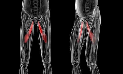
The soft tissues most often affected by athletic pubalgia are the oblique muscles found in the lower abdomen, especially in the tendons that attach the oblique muscles to the pubic bone. In many instances, the joints that connect the thigh muscles to the pubic bone,�known as the adductor muscles, are also stretched or torn as a result of athletic pubalgia.
Physical activities which involve planting the feet and twisting with maximum exertion can cause athletic pubalgia. A sports hernia is most prevalent in vigorous sports, such as hockey, soccer, wrestling, and football. Athletic pubalgia�causes pain and discomfort in the groin region which typically gets better with rest but comes back with physical activity.
A sports�hernia does not result in a visible bulge in the groin, such as the well-known inguinal hernia does. As time passes, athletic pubalgia can lead to an inguinal hernia, and abdominal organs can push against the diminished cells to form a visible bulge. Without treatment, this sports injury could lead to chronic, disabling pain and other symptoms.
Healthcare Professional Diagnosis
During the first consultation, a doctor will discuss the individual’s symptoms and how the injury happened. To�diagnose athletic pubalgia, the healthcare professional will look for tenderness in the groin or above the pubis. Although a sports hernia may be related to an inguinal hernia, the doctor may not find any hernias during a physical examination.
Furthermore, to help determine the presence of athletic pubalgia, the healthcare professional will probably ask the patient to perform a sit-up or to�bend the trunk against resistance. If you have a sports hernia, these tests will be painful. The doctor may also require�x-rays or magnetic resonance imaging (MRI) to help determine whether you have athletic pubalgia.�The scope of our information is limited to chiropractic as well as to spinal injuries and conditions. To discuss the subject matter, please feel free to ask Dr. Jimenez or contact us at�915-850-0900�.
Curated by Dr. Alex Jimenez
Additional Topics: Acute Back Pain
Back pain�is one of the most prevalent causes of disability and missed days at work worldwide. Back pain attributes to the second most common reason for doctor office visits, outnumbered only by upper-respiratory infections. Approximately 80 percent of the population will experience back pain at least once throughout their life. The spine is a complex structure made up of bones, joints, ligaments, and muscles, among other soft tissues. Because of this, injuries and/or aggravated conditions, such as�herniated discs, can eventually lead to symptoms of back pain. Sports injuries or automobile accident injuries are often the most frequent cause of back pain, however, sometimes the simplest of movements can have painful results. Fortunately, alternative treatment options, such as chiropractic care, can help ease back pain through the use of spinal adjustments and manual manipulations, ultimately improving pain relief.
Athletic pubalgia, also known as a hockey hernia,�hockey groin, Gilmore’s Groin,�sports hernia, or groin disruption, is a health issue of the pubic joint. It is a condition characterized by chronic groin pain in athletes and identified by a dilated ring of the inguinal canal. Soccer and ice hockey players are the athletes most commonly affected by athletic pubalgia, and both recreational and professional athletes can be impacted.
Athletic Pubalgia Symptoms
Symptoms of athletic pubalgia�generally manifest as pain following physical activity, most frequently through hip extension, and twisting and turning movements. The painful symptoms usually radiate into the adductor muscle region and the testicles, although it is often difficult for the individual to pinpoint the exact location of the�symptoms. Athletes with athletic pubalgia�experience soreness and stiffness after physical activity.
Any exertion which increases intra-abdominal pressure, such as sneezing or�coughing, as well as physical activity, can lead to pain. While pain in the stomach and pelvis can occur due to a variety of health issues, including injuries to the low back, or lumbar spine, the hip joint, the sacroiliac joint, and the abdomen, along with the genito-urinary system, diagnosis of athletic pubalgia demands skillful differentiation and evaluation.
Clinical Presentation of Athletic Pubalgia
The diagnosis of athletic pubalgia is based on the patient’s history, where healthcare professionals may also depend on the use�of magnetic resonance imaging,�or MRI. Symptoms can frequently be reproduced by certain movements, such as performing crunches or sit-ups. Pain associated with athletic pubalgia may also be elicited with the patient in a “frog posture,” in which the individual is supine with knees bent and heels together.
Many athletes experience concomitant fatigue or tearing of the�adductor muscles or labral tears of the hip. If there is stiffness in the adductor muscles post-injury, painful symptoms can manifest. Alternative treatment options should be to restore normal movement after the adductor has begun to heal, normally 6 to 8 weeks post-injury. Moreover, sleeping in a prone position with the hip on the affected side flexed and externally rotated can offer relief to some athletes with athletic pubalgia.
The precise prevalence of this health issue is unknown. Conservative therapies,�such as gentle stretching, may temporarily alleviate painful symptoms, however, definitive treatment options should be considered for long-term relief.�The scope of our information is limited to chiropractic as well as to spinal injuries and conditions. To discuss the subject matter, please feel free to ask Dr. Jimenez or contact us at�915-850-0900�.
Curated by Dr. Alex Jimenez
Additional Topics: Acute Back Pain
Back pain�is one of the most prevalent causes of disability and missed days at work worldwide. Back pain attributes to the second most common reason for doctor office visits, outnumbered only by upper-respiratory infections. Approximately 80 percent of the population will experience back pain at least once throughout their life. The spine is a complex structure made up of bones, joints, ligaments, and muscles, among other soft tissues. Because of this, injuries and/or aggravated conditions, such as�herniated discs, can eventually lead to symptoms of back pain. Sports injuries or automobile accident injuries are often the most frequent cause of back pain, however, sometimes the simplest of movements can have painful results. Fortunately, alternative treatment options, such as chiropractic care, can help ease back pain through the use of spinal adjustments and manual manipulations, ultimately improving pain relief.
The hip is commonly described as a “ball-and-socket” type joint. In a healthy hip, the ball at the top end of the thighbone, or femur, should fit firmly into the socket, which is part of the large pelvis bone. In babies and children with developmental dysplasia, or dislocation, of the hip, abbreviated as DDH, the hip joint may not have formed normally. As a result, the ball of the femur might easily dislocate and become loose from the socket.
Although DDH is often present from birth, it could also develop during a child’s first year of life. Recent research studies have demonstrated that infants whose thighs are swaddled closely with the hips and knees straight are at a higher risk for developing DDH. Because swaddling has become�increasingly popular, it is essential for parents to understand how to swaddle their babies safely, and they should realize that when done improperly, swaddling may cause health issues such as DDH.
Diagnosis for�Developmental Dysplasia of the Hip
In addition to visual cues, when�diagnosing for DDH, the healthcare professional will perform a careful evaluation, such as listening and feeling for “clunks” which indicates that the hip is placed in different positions. The doctor will also utilize other methods and techniques to determine if the hip is dislocated. Newborns recognized to be at higher risk for DDH are often tested using ultrasound. For babies and children, x-rays of the hip might be taken to provide further detailed images of the hip joint.
Treatment for�Developmental Dysplasia of the Hip
If DDH is discovered at birth, it can usually be treated with the use of a harness or brace. If the hip isn’t dislocated at birth, the condition might not be diagnosed until the child starts walking. At that point, treatment for DDH is much more complex, with less predictable results. If diagnosed and treated accordingly, children ought to have no restriction in function and develop the standard hip joint. DDH may result in atherosclerosis and other problems. It may produce a difference in agility or leg length.
In spite of proper treatment, hip deformity and osteoarthritis may develop later in life. This is particularly true when treatment starts after the age of 2 years. Therefore, diagnosis and treatment are essential in newborns and children with DDH. The scope of our information is limited to chiropractic as well as to spinal injuries and conditions. To discuss the subject matter, please feel free to ask Dr. Jimenez or contact us at�915-850-0900�.
Curated by Dr. Alex Jimenez
�
�
Additional Topics: Acute Back Pain
Back pain�is one of the most prevalent causes of disability and missed days at work worldwide. Back pain attributes to the second most common reason for doctor office visits, outnumbered only by upper-respiratory infections. Approximately 80 percent of the population will experience back pain at least once throughout their life. The spine is a complex structure made up of bones, joints, ligaments, and muscles, among other soft tissues. Because of this, injuries and/or aggravated conditions, such as�herniated discs, can eventually lead to symptoms of back pain. Sports injuries or automobile accident injuries are often the most frequent cause of back pain, however, sometimes the simplest of movements can have painful results. Fortunately, alternative treatment options, such as chiropractic care, can help ease back pain through the use of spinal adjustments and manual manipulations, ultimately improving pain relief.
Hip pain is a well-known health issue which can be caused by a wide array of problems, however, the site of the patient’s hip pain can provide valuable information regarding the underlying cause of this common health issue. Pain on the inside of the hip or groin can be due to problems within the hip joint itself while pain on the outside of the hip, upper thigh and outer buttocks may be due to problems with the ligaments, tendons and muscles, among other soft tissues, surrounding the hip joint. Furthermore, hip pain can be due to other injuries and conditions, including back pain.
Abstract
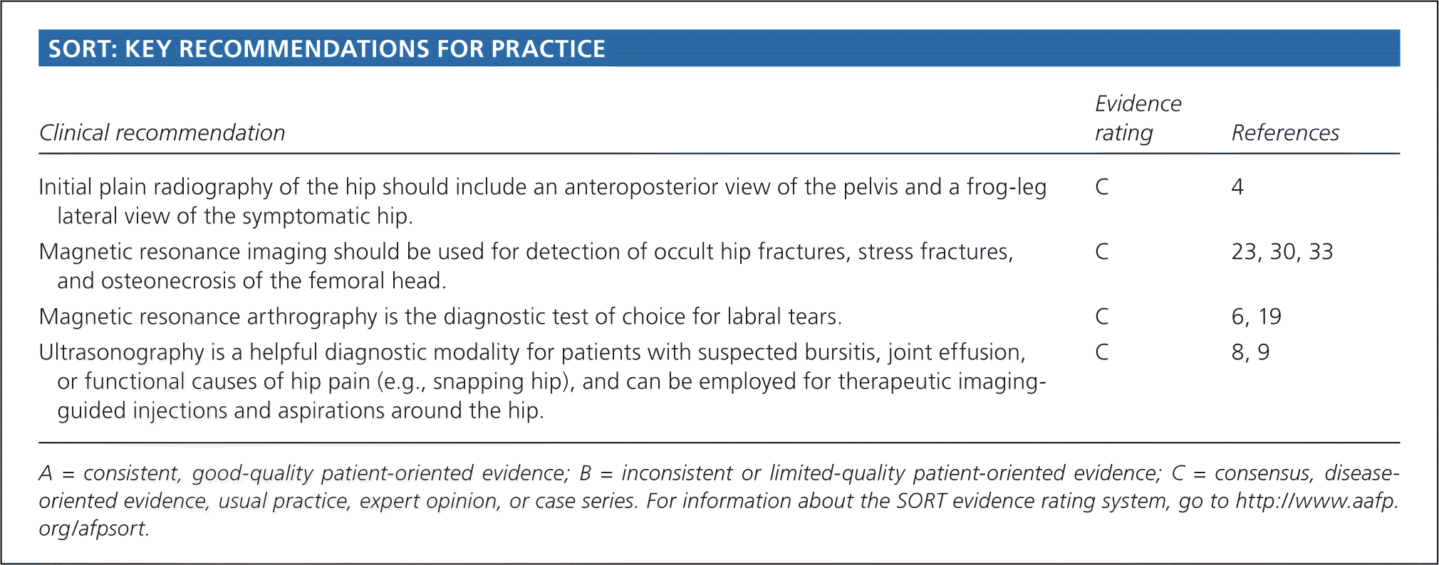
Hip pain is a common and disabling condition that affects patients of all ages. The differential diagnosis of hip pain is broad, presenting a diagnostic challenge. Patients often express that their hip pain is localized to one of three anatomic regions: the anterior hip and groin, the posterior hip and buttock, or the lateral hip. Anterior hip and groin pain is commonly associated with intra-articular pathology, such as osteoarthritis and hip labral tears. Posterior hip pain is associated with piriformis syndrome, sacroiliac joint dysfunction, lumbar radiculopathy, and less commonly ischiofemoral impingement and vascular claudication. Lateral hip pain occurs with greater trochanteric pain syndrome. Clinical examination tests, although helpful, are not highly sensitive or specific for most diagnoses; however, a rational approach to the hip examination can be used. Radiography should be performed if acute fracture, dislocations, or stress fractures are suspected. Initial plain radiography of the hip should include an anteroposterior view of the pelvis and frog-leg lateral view of the symptomatic hip. Magnetic resonance imaging should be performed if the history and plain radiograph results are not diagnostic. Magnetic resonance imaging is valuable for the detection of occult traumatic fractures, stress fractures, and osteonecrosis of the femoral head. Magnetic resonance arthrography is the diagnostic test of choice for labral tears.
Introduction
Hip pain is a common presentation in primary care and can affect patients of all ages. In one study, 14.3% of adults 60 years and older reported significant hip pain on most days over the previous six weeks.1 Hip pain often presents a diagnostic and therapeutic challenge. The differential diagnosis of hip pain (eTable A) is broad, including both intra-articular and extra-articular pathology, and varies by age. A history and physical examination are essential to accurately diagnose the cause of hip pain.
Anatomy
The hip joint is a ball-and-socket synovial joint designed to allow multiaxial motion while transferring loads between the upper and lower body. The acetabular rim is lined by fibrocartilage (labrum), which adds depth and stability to the femoroacetabular joint. The articular surfaces are covered by hyaline cartilage that dissipates shear and compressive forces during load bearing and hip motion. The hip’s major innervating nerves originate in the lumbosacral region, which can make it difficult to distinguish between primary hip pain and radicular lumbar pain.
The hip joint’s wide range of motion is second only to that of the glenohumeral joint and is enabled by the large number of muscle groups that surround the hip. The flexor muscles include the iliopsoas, rectus femoris, pectineus, and sartorius muscles. The gluteus maximus and hamstring muscle groups allow for hip extension. Smaller muscles, such as gluteus medius and minimus, piriformis, obturator externus and internus, and quadratus femoris muscles, insert around the greater trochanter, allowing for abduction, adduction, and internal and external rotation.
In persons who are skeletally immature, there are several growth centers of the pelvis and femur where injuries can occur. Potential sites of apophyseal injury in the hip region include the ischium, anterior superior iliac spine, anterior inferior iliac spine, iliac crest, lesser trochanter, and greater trochanter. The apophysis of the superior iliac spine matures last and is susceptible to injury up to 25 years of age.2
The hip joint is one of the larger joints found in the human body and it serves in locomotion as the thigh moves forward and backward. The hip joint also rotates when sitting and with changes of direction while walking. A variety of complex structures surround the hip joint. When an injury or condition affects these, it can ultimately lead to hip pain.
Dr. Alex Jimenez D.C., C.C.S.T.
Evaluation of Hip Pain
History
Age alone can narrow the differential diagnosis of hip pain. In prepubescent and adolescent patients, congenital malformations of the femoroacetabular joint, avulsion fractures, and apophyseal or epiphyseal injuries should be considered. In those who are skeletally mature, hip pain is often a result of musculotendinous strain, ligamentous sprain, contusion, or bursitis. In older adults, degenerative osteoarthritis and fractures should be considered first.
Patients with hip pain should be asked about antecedent trauma or inciting activity, factors that increase or decrease the pain, mechanism of injury, and time of onset. Questions related to hip function, such as the ease of getting in and out of a car, putting on shoes, running, walking, and going up and down stairs, can be helpful.3 Location of the pain is informative because hip pain often localizes to one of three basic anatomic regions: the anterior hip and groin, posterior hip and buttock, and lateral hip (eFigure A).
Physical Examination
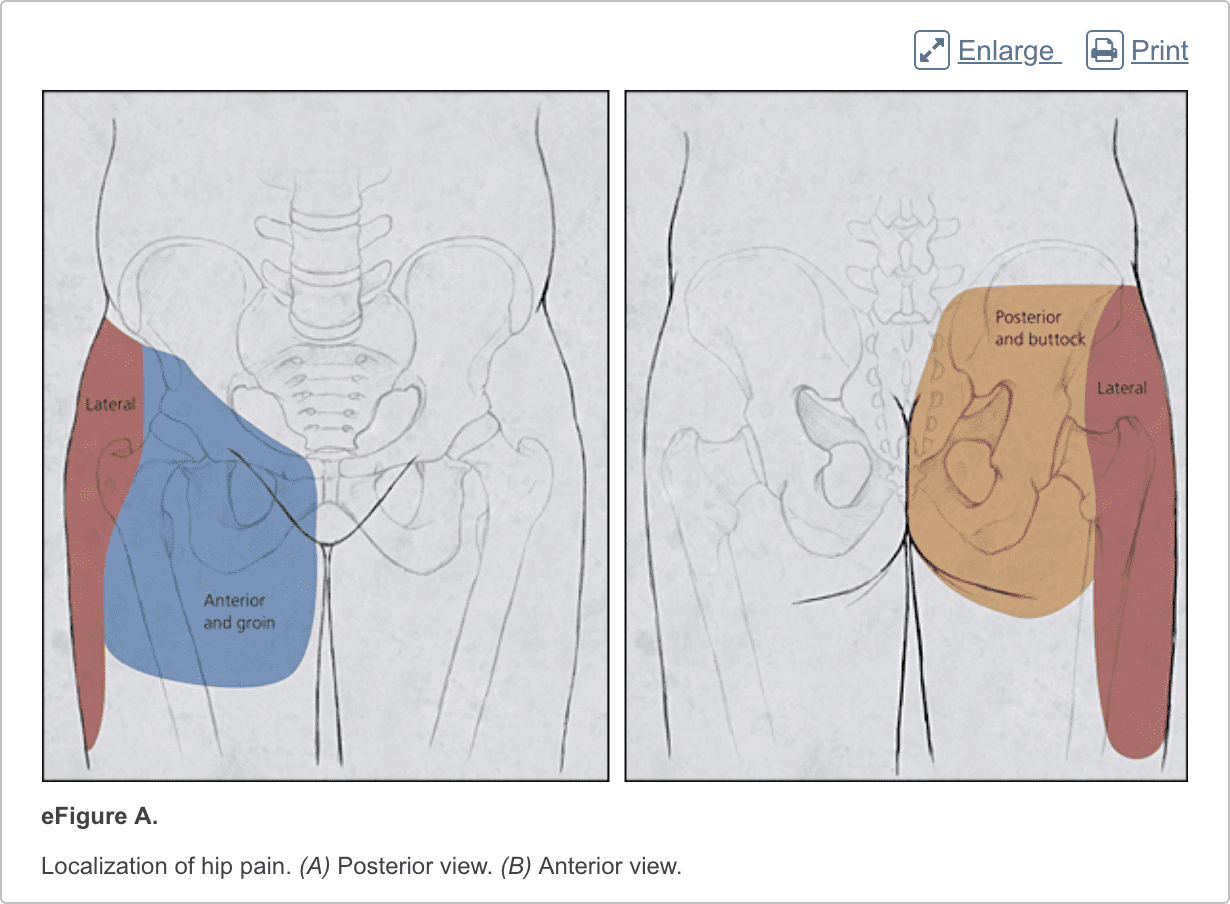
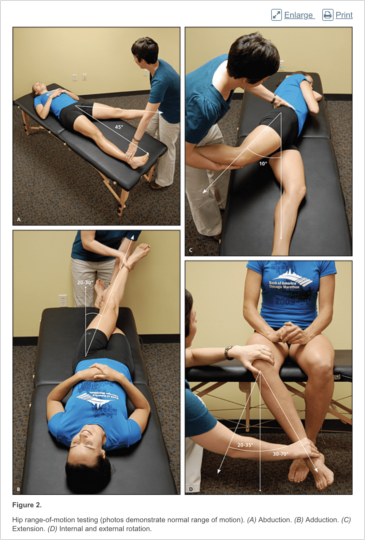
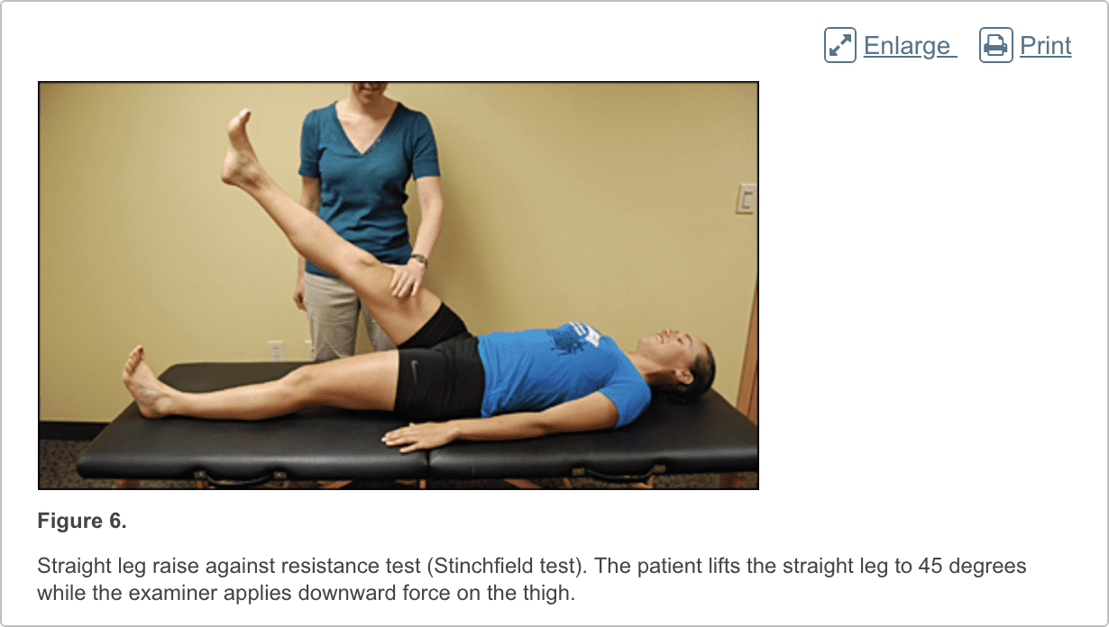
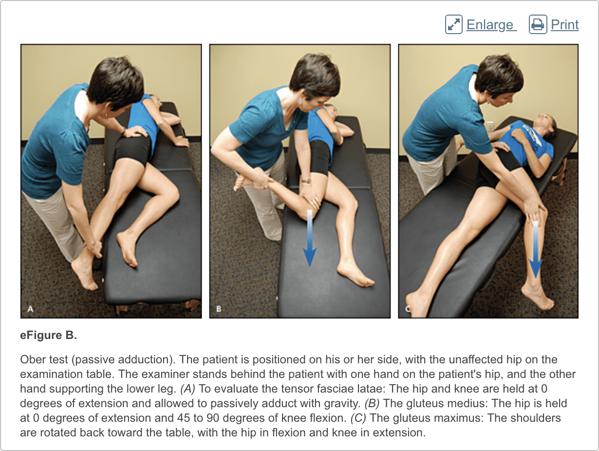
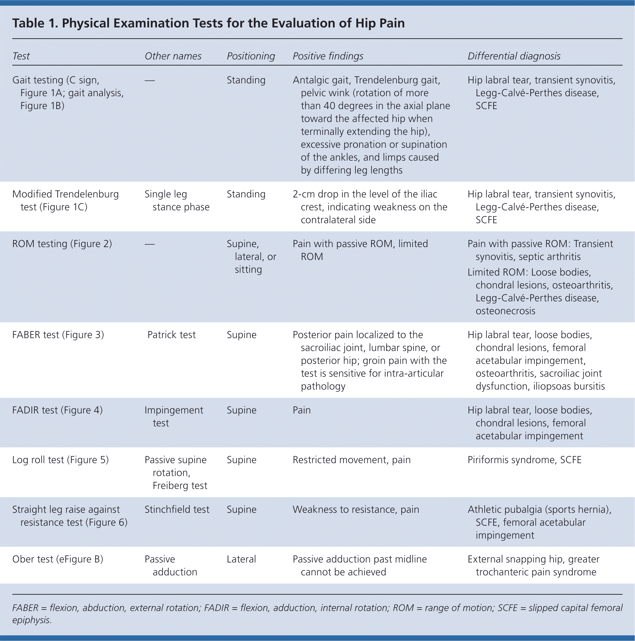
The hip examination should evaluate the hip, back, abdomen, and vascular and neurologic systems. It should start with a gait analysis and stance assessment (Figure 1), followed by evaluation of the patient in seated, supine, lateral, and prone positions (Figures 2 through 6, and eFigure B). Physical examination tests for the evaluation of hip pain are summarized in Table 1.
Imaging
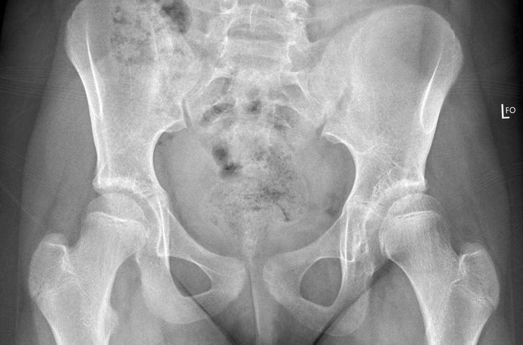
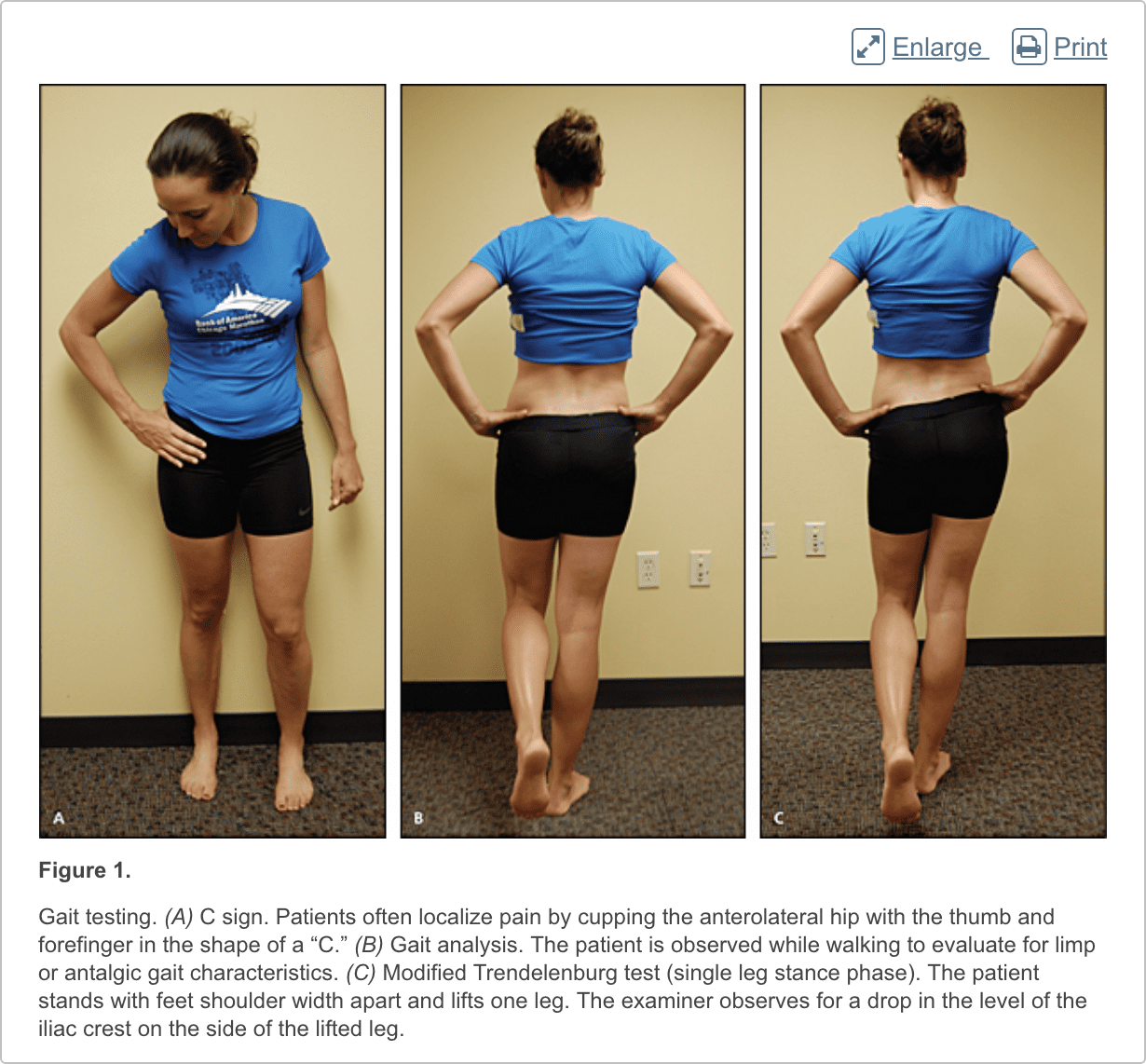
Radiography. Radiography of the hip should be performed if there is any suspicion of acute fracture, dislocation, or stress fracture. Initial plain radiography of the hip should include an anteroposterior view of the pelvis and a frog-leg lateral view of the symptomatic hip.4
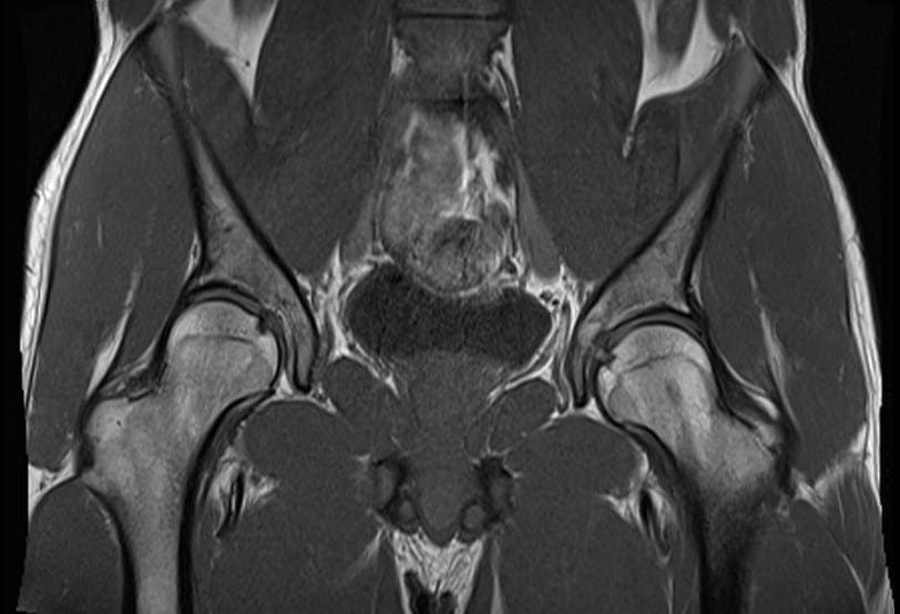
Magnetic Resonance Imaging and Arthrography. Conventional magnetic resonance imaging (MRI) of the hip can detect many soft tissue abnormalities, and is the preferred imaging modality if plain radiography does not identify specific pathology in a patient with persistent pain.5 Conventional MRI has a sensitivity of 30% and an accuracy of 36% for diagnosing hip labral tears, whereas magnetic resonance arthrography provides added sensitivity of 90% and accuracy of 91% for the detection of labral tears.6,7
Ultrasonography. Ultrasonography is a useful technique for evaluating individual tendons, confirming suspected bursitis, and identifying joint effusions and functional causes of hip pain.8 Ultrasonography is especially useful for safely and accurately performing imaging-guided injections and aspirations around the hip.9 It is ideal for an experienced ultrasonographer to perform the diagnostic study; however, emerging evidence suggests that less experienced clinicians with appropriate training can make diagnoses with reliability similar to that of an experienced musculoskeletal ultrasonographer.10,11
These are numerous causes for hip pain. Although some hip pain may only be temporary, other forms of hip pain can become chronic if left untreated for an extended period of time. Several common causes of hip pain include, arthritis, fracture, sprain, avascular necrosis, Gaucher’s disease, sciatica, muscle strain, iliotibial band syndrome or IT band syndrome and hematoma, among others described below.
Dr. Alex Jimenez D.C., C.C.S.T.
Differential Diagnosis of Anterior Hip Pain
Anterior hip or groin pain suggests involvement of the hip joint itself. Patients often localize pain by cupping the anterolateral hip with the thumb and forefinger in the shape of a �C.� This is known as the C sign (Figure 1A).
Osteoarthritis
Osteoarthritis is the most likely diagnosis in older adults with limited motion and gradual onset of symptoms. Patients have a constant, deep, aching pain and stiffness that are worse with prolonged standing and weight bearing. Examination reveals decreased range of motion, and extremes of hip motion often cause pain. Plain radiographs demonstrate the presence of asymmetrical joint-space narrowing, osteophytosis, and subchondral sclerosis and cyst formation.12
Femoroacetabular Impingement
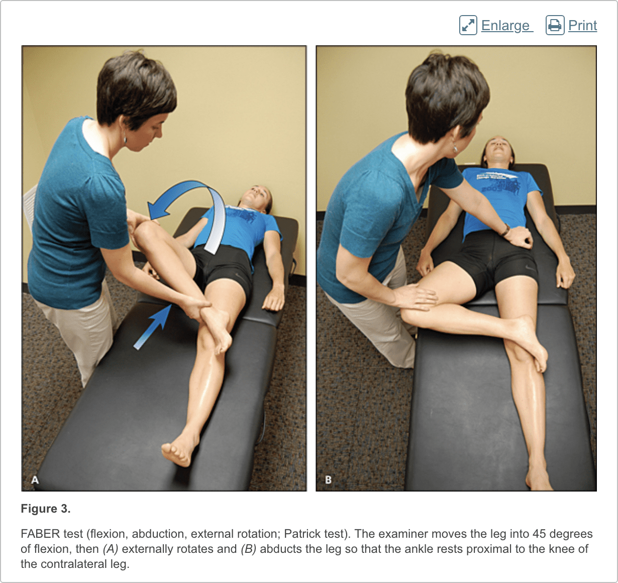
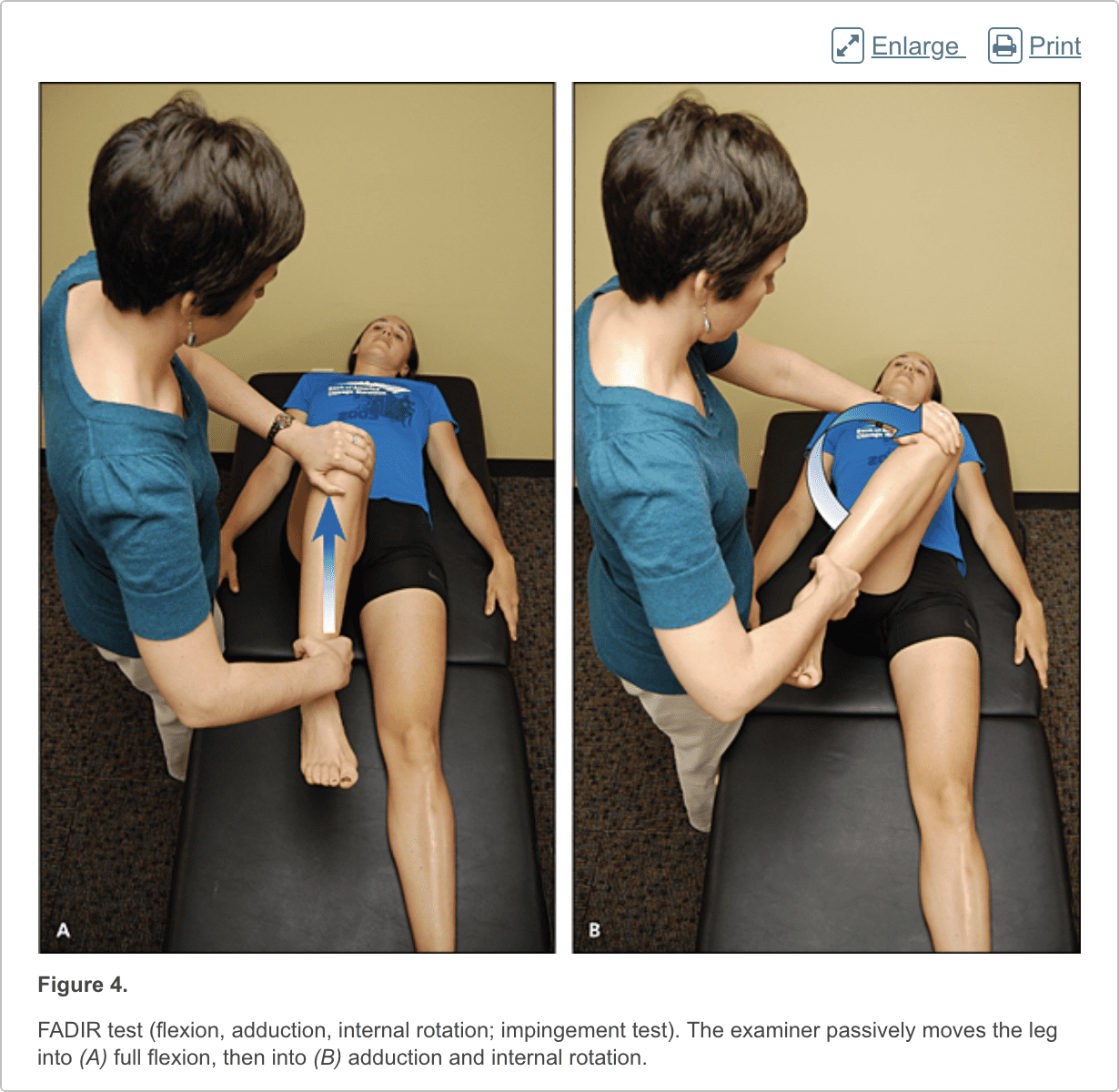
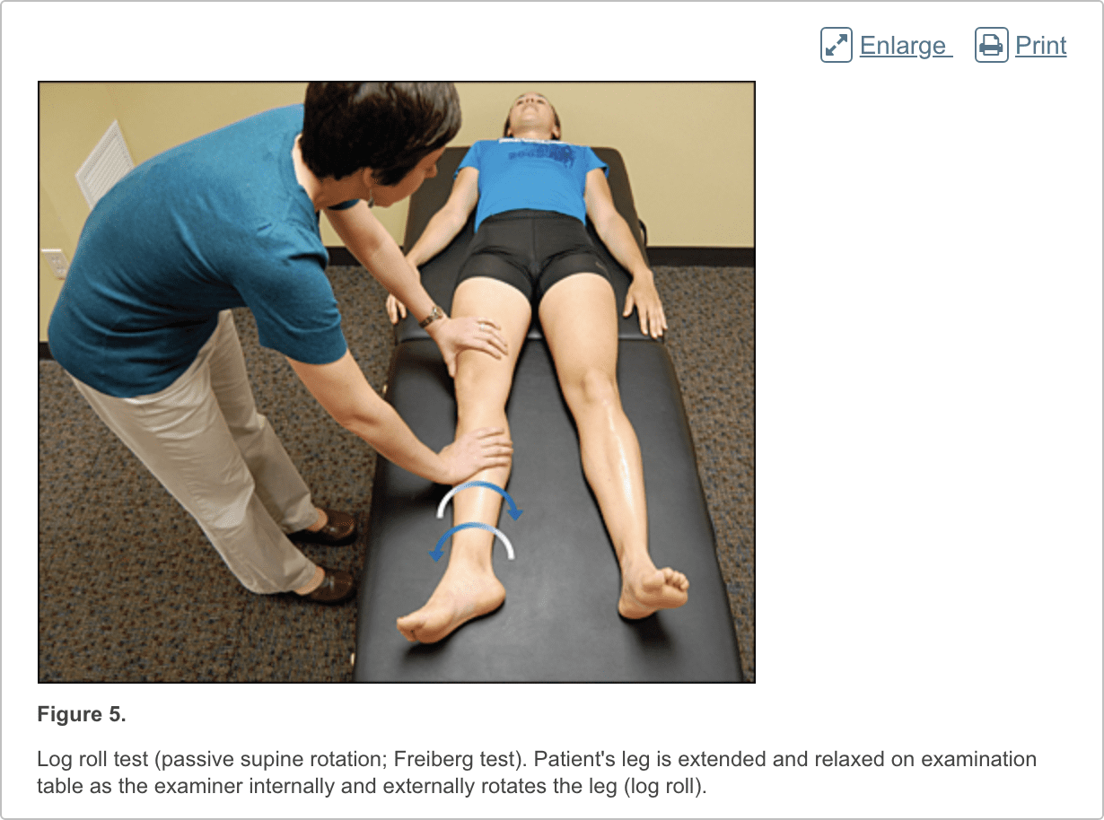
Patients with femoroacetabular impingement are often young and physically active. They describe insidious onset of pain that is worse with sitting, rising from a seat, getting in or out of a car, or leaning forward.13 The pain is located primarily in the groin with occasional radiation to the lateral hip and anterior thigh.14 The FABER test (flexion, abduction, external rotation; Figure 3) has a sensitivity of 96% to 99%. The FADIR test (flexion, adduction, internal rotation; Figure 4), log roll test (Figure 5), and straight leg raise against resistance test (Figure 6) are also effective, with sensitivities of 88%, 56%, and 30%, respectively.14,15 In addition to the anteroposterior and lateral radiograph views, a Dunn view should be obtained to help detect subtle lesions.16
Hip Labral Tear
Hip labral tears cause dull or sharp groin pain, and one-half of patients with a labral tear have pain that radiates to the lateral hip, anterior thigh, and buttock. The pain usually has an insidious onset, but occasionally begins acutely after a traumatic event. About one-half of patients with this injury also have mechanical symptoms, such as catching or painful clicking with activity.17 The FADIR and FABER tests are effective for detecting intra-articular pathology (the sensitivity is 96% to 75% for the FADIR test and is 88% for the FABER test), although neither test has high specificity.14,15,18 Magnetic resonance arthrography is considered the diagnostic test of choice for labral tears.6,19 However, if a labral tear is not suspected, other less invasive imaging modalities, such as plain radiography and conventional MRI, should be used first to rule out other causes of hip and groin pain.
Iliopsoas Bursitis (Internal Snapping Hip)
Patients with this condition have anterior hip pain when extending the hip from a flexed position, often associated with intermittent catching, snapping, or popping of the hip.20 Dynamic real-time ultrasonography is particularly useful in evaluating the various forms of snapping hip.8
Occult or Stress Fracture
Occult or stress fracture of the hip should be considered if trauma or repetitive weight-bearing exercise is involved, even if plain radiograph results are negative.21 Clinically, these injuries cause anterior hip or groin pain that is worse with activity.21 Pain may be present with extremes of motion, active straight leg raise, the log roll test, or hopping.22 MRI is useful for the detection of occult traumatic fractures and stress fractures not seen on plain radiographs.23
Transient Synovitis and Septic Arthritis
Acute onset of atraumatic anterior hip pain that results in impaired weight bearing should raise suspicion for transient synovitis and septic arthritis. Risk factors for septic arthritis in adults include age older than 80 years, diabetes mellitus, rheumatoid arthritis, recent joint surgery, and hip or knee prostheses.24 Fever, complete blood count, erythrocyte sedimentation rate, and C-reactive protein level should be used to evaluate the risk of septic arthritis.25,26 MRI is useful for differentiating septic arthritis from transient synovitis.27,28 However, hip aspiration using guided imaging such as fluoroscopy, computed tomography, or ultrasonography is recommended if a septic joint is suspected.29
Osteonecrosis
Legg-Calv�-Perthes disease is an idiopathic osteonecrosis of the femoral head in children two to 12 years of age, with a male-to-female ratio of 4:1.4 In adults, risk factors for osteonecrosis include systemic lupus erythematosus, sickle cell disease, human immunodeficiency virus infection, smoking, alcoholism, and corticosteroid use.30,31 Pain is the presenting symptom and is usually insidious. Range of motion is initially preserved but can become limited and painful as the disease progresses.32 MRI is valuable in the diagnosis and prognostication of osteonecrosis of the femoral head.30,33
Differential Diagnosis of Posterior Hip and Buttock Pain
Piriformis Syndrome and Ischiofemoral Impingement
Piriformis syndrome causes buttock pain that is aggravated by sitting or walking, with or without ipsilateral radiation down the posterior thigh from sciatic nerve compression.34,35 Pain with the log roll test is the most sensitive test, but tenderness with palpation of the sciatic notch can help with the diagnosis.35
Ischiofemoral impingement is a less well-understood condition that can lead to nonspecific buttock pain with radiation to the posterior thigh.36,37 This condition is thought to be a result of impingement of the quadratus femoris muscle between the lesser trochanter and the ischium.
Unlike sciatica from disc herniation, piriformis syndrome and ischiofemoral impingement are exacerbated by active external hip rotation. MRI is useful for diagnosing these conditions.38
Other
Other causes of posterior hip pain include sacroiliac joint dysfunction,39 lumbar radiculopathy,40 and vascular claudication.41 The presence of a limp, groin pain, and limited internal rotation of the hip is more predictive of hip disorders than disorders originating from the low back.42
Differential Diagnosis of Lateral Hip Pain
Greater Trochanteric Pain Syndrome
Lateral hip pain affects 10% to 25% of the general population.43 Greater trochanteric pain syndrome refers to pain over the greater trochanter. Several disorders of the lateral hip can lead to this type of pain, including iliotibial band thickening, bursitis, and tears of the gluteus medius and minimus muscle attachment.43�45 Patients may have mild morning stiffness and may be unable to sleep on the affected side. Gluteus minimus and medius injuries present with pain in the posterior lateral aspect of the hip as a result of partial or full-thickness tearing at the gluteal insertion. Most patients have an atraumatic, insidious onset of symptoms from repetitive use.43,45,46
In conclusion, hip pain is a common complaint which may occur due to a wide variety of health issues. Moreover, the precise location of the patient’s hip pain can provide valuable information to healthcare professionals regarding the underlying cause of the problem. The purpose of the article above was to demonstrate and discuss the evaluation of the patient with hip pain. The scope of our information is limited to chiropractic as well as to spinal injuries and conditions. To discuss the subject matter, please feel free to ask Dr. Jimenez or contact us at�915-850-0900�.
Curated by Dr. Alex Jimenez
Data Sources: We searched articles on hip pathology in American Family Physician, along with their references. We also searched the Agency for Healthcare Research and Quality Evidence Reports, Clinical Evidence, Institute for Clinical Systems Improvement, the U.S. Preventive Services Task Force guidelines, the National Guideline Clearinghouse, and UpToDate. We performed a PubMed search using the keywords greater trochanteric pain syndrome, hip pain physical examination, imaging femoral hip stress fractures, imaging hip labral tear, imaging osteomyelitis, ischiofemoral impingement syndrome, meralgia paresthetica review, MRI arthrogram hip labrum, septic arthritis systematic review, and ultrasound hip pain. Search dates: March and April 2011, and August 15, 2013.
Back pain�is one of the most prevalent causes of disability and missed days at work worldwide. Back pain attributes to the second most common reason for doctor office visits, outnumbered only by upper-respiratory infections. Approximately 80 percent of the population will experience back pain at least once throughout their life. The spine is a complex structure made up of bones, joints, ligaments, and muscles, among other soft tissues. Because of this, injuries and/or aggravated conditions, such as�herniated discs, can eventually lead to symptoms of back pain. Sports injuries or automobile accident injuries are often the most frequent cause of back pain, however, sometimes the simplest of movements can have painful results. Fortunately, alternative treatment options, such as chiropractic care, can help ease back pain through the use of spinal adjustments and manual manipulations, ultimately improving pain relief.
1.�Christmas C, Crespo CJ, Franckowiak SC, et al. How common is hip pain among older adults? Results from the Third National Health and Nutrition Examination Survey.�J Fam Pract. 2002;51(4):345�348.
2.�Rossi F, Dragoni S. Acute avulsion fractures of the pelvis in adolescent competitive athletes.�Skeletal Radiol. 2001;30(3):127�131.
3.�Martin HD, Shears SA, Palmer IJ. Evaluation of the hip.�Sports Med Arthrosc. 2010;18(2):63�75.
4.�Gough-Palmer A, McHugh K. Investigating hip pain in a well child.�BMJ. 2007;334(7605):1216�1217.
5.�Bencardino JT, Palmer WE. Imaging of hip disorders in athletes.�Radiol Clin North Am. 2002;40(2):267�287.
6.�Czerny C, Hofmann S, Neuhold A, et al. Lesions of the acetabular labrum: accuracy of MR imaging and MR arthrography in detection and staging.�Radiology. 1996;200(1):225�230.
7.�Czerny C, Hofmann S, Urban M, et al. MR arthrography of the adult acetabular capsular-labral complex.�AJR Am J Roentgenol. 1999;173(2):345�349.
8.�Deslandes M, Guillin R, Cardinal E, et al. The snapping iliopsoas tendon: new mechanisms using dynamic sonography.�AJR Am J Roentgenol. 2008;190(3):576�581.
9.�Blankenbaker DG, De Smet AA. Hip injuries in athletes.�Radiol Clin North Am. 2010;48(6):1155�1178.
10.�Balint PV, Sturrock RD. Intraobserver repeatability and interobserver reproducibility in musculoskeletal ultrasound imaging measurements.�Clin Exp Rheumatol. 2001;19(1):89�92.
11.�Ramwadhdoebe S, Sakkers RJ, Uiterwaal CS, et al. Evaluation of a training program for general ultrasound screening for developmental dysplasia of the hip in preventive child health care.�Pediatr Radiol. 2010;40(10):1634�1639.
12.�Altman R, Alarc�n G, Appelrouth D, et al. The American College of Rheumatology criteria for the classification and reporting of osteoarthritis of the hip.�Arthritis Rheum. 1991;34(5):505�514.
14.�Clohisy JC, Knaus ER, Hunt DM, et al. Clinical presentation of patients with symptomatic anterior hip impingement.�Clin Orthop Relat Res. 2009;467(3):638�644.
15.�Ito K, Leunig M, Ganz R. Histopathologic features of the acetabular labrum in femoroacetabular impingement.�Clin Orthop Relat Res. 2004;(429):262�271.
16.�Beall DP, Sweet CF, Martin HD, et al. Imaging findings of femoroacetabular impingement syndrome.�Skeletal Radiol. 2005;34(11):691�701.
17.�Burnett RS, Della Rocca GJ, Prather H, et al. Clinical presentation of patients with tears of the acetabular labrum.�J Bone Joint Surg Am. 2006;88(7):1448�1457.
18.�Leunig M, Werlen S, Ungersb�ck A, et al. Evaluation of the acetabular labrum by MR arthrography [published correction appears in�J Bone Joint Surg Br. 1997;79(4):693].�J Bone Joint Surg Br. 1997;79(2):230�234.
19.�Groh MM, Herrera J. A comprehensive review of hip labral tears.�Curr Rev Musculoskelet Med. 2009;2(2):105�117.
20.�Blankenbaker DG, De Smet AA, Keene JS. Sonography of the iliopsoas tendon and injection of the iliopsoas bursa for diagnosis and management of the painful snapping hip.�Skeletal Radiol. 2006;35(8):565�571.
21.�Egol KA, Koval KJ, Kummer F, et al. Stress fractures of the femoral neck.�Clin Orthop Relat Res. 1998;(348):72�78.
22.�Fullerton LR Jr, Snowdy HA. Femoral neck stress fractures.�Am J Sports Med. 1988;16(4):365�377.
24.�Margaretten ME, Kohlwes J, Moore D, et al. Does this adult patient have septic arthritis?�JAMA. 2007;297(13):1478�1488.
25.�Eich GF, Superti-Furga A, Umbricht FS, et al. The painful hip: evaluation of criteria for clinical decision-making.�Eur J Pediatr. 1999;158(11):923�928.
26.�Kocher MS, Zurakowski D, Kasser JR. Differentiating between septic arthritis and transient synovitis of the hip in children.�J Bone Joint Surg Am. 1999;81(12):1662�1670.
27.�Learch TJ, Farooki S. Magnetic resonance imaging of septic arthritis.�Clin Imaging. 2000;24(4):236�242.
28.�Lee SK, Suh KJ, Kim YW, et al. Septic arthritis versus transient synovitis at MR imaging.�Radiology. 1999;211(2):459�465.
29.�Leopold SS, Battista V, Oliverio JA. Safety and efficacy of intraarticular hip injection using anatomic landmarks.�Clin Orthop Relat Res. 2001; (391):192�197.
30.�Mitchell DG, Rao VM, Dalinka MK, et al. Femoral head avascular necrosis: correlation of MR imaging, radiographic staging, radionuclide imaging, and clinical findings.�Radiology. 1987;162(3):709�715.
31.�Mont MA, Zywiel MG, Marker DR, et al. The natural history of untreated asymptomatic osteonecrosis of the femoral head.�J Bone Joint Surg Am. 2010;92(12):2165�2170.
32.�Assouline-Dayan Y, Chang C, Greenspan A, et al. Pathogenesis and natural history of osteonecrosis.�Semin Arthritis Rheum. 2002;32(2):94�124.
33.�Totty WG, Murphy WA, Ganz WI, et al. Magnetic resonance imaging of the normal and ischemic femoral head.�AJR Am J Roentgenol. 1984;143(6):1273�1280.
35.�Hopayian K, Song F, Riera R, et al. The clinical features of the piriformis syndrome.�Eur Spine J. 2010;19(12):2095�2109.
36.�Torriani M, Souto SC, Thomas BJ, et al. Ischiofemoral impingement syndrome.�AJR Am J Roentgenol. 2009;193(1):186�190.
37.�Ali AM, Whitwell D, Ostlere SJ. Case report: imaging and surgical treatment of a snapping hip due to ischiofemoral impingement.�Skeletal Radiol. 2011;40(5):653�656.
38.�Lee EY, Margherita AJ, Gierada DS, et al. MRI of piriformis syndrome.�AJR Am J Roentgenol. 2004;183(1):63�64.
39.�Slipman CW, Jackson HB, Lipetz JS, et al. Sacroiliac joint pain referral zones.�Arch Phys Med Rehabil. 2000;81(3):334�338.
40.�Moore KL, Dalley AF, Agur AM.�Clinically Oriented Anatomy. 6th ed. Philadelphia, Pa.: Lippincott Williams & Wilkins; 2010.
41.�Adlakha S, Burket M, Cooper C. Percutaneous intervention for chronic total occlusion of the internal iliac artery for unrelenting buttock claudication.�Catheter Cardiovasc Interv. 2009;74(2):257�259.
42.�Brown MD, Gomez-Marin O, Brookfield KF, et al. Differential diagnosis of hip disease versus spine disease.�Clin Orthop Relat Res. 2004; (419):280�284.
43.�Segal NA, Felson DT, Torner JC, et al.; Multicenter Osteoarthritis Study Group. Greater trochanteric pain syndrome.�Arch Phys Med Rehabil. 2007;88(8):988�992.
44.�Strauss EJ, Nho SJ, Kelly BT. Greater trochanteric pain syndrome.�Sports Med Arthrosc. 2010;18(2):113�119.
I went through the physical therapy, and then I used chiropractic care, as well as crossfit, to kinda get me to that 100% mark. And I haven’t had a problem since, I continue to do crossfit and I use chiropractic care to make sure my body is aligned. It gives me that extra sense of feeling that I’m not gonna injure myself again, that my body is feeling good and 100%. I never have that feeling in the back of my mind, that, you know, I’m gonna injure myself. Sometimes I forget I even had a surgery and I think I can attribute that to a combination of chiropractic care and to the overall training aspect of crossfit. – Andrew Hutchison
There are a variety of causes for both hip pain and knee pain. While the hip joint can withstand a tremendous amount of wear-and-tear, it’s not indestructible. With age and usage, the hip cartilage can begin to degenerate, resulting in hip pain. Tendons and ligaments at the hip may also get excessive overused and can start to demonstrate signs of wear-and-tear over time. The anatomy of the knee, however, is more complex. The knee is collectively made up of bones, pads of cartilage and a joint capsule. Trauma or damage from an injury or degeneration associated with aging may cause knee pain.
Causes of Hip Pain
Hip pain is the general term used to define pain felt in or around the hip joint. Certain injuries and/or conditions may commonly cause hip pain. Arthritis, especially rheumatoid arthritis, are the primary culprits of hip pain in older individuals. Both may result in the breakdown of cartilage in the hip joint and can cause inflammation in the area. Combined with pain and discomfort, there’s generally also reduced range of motion in the hip as well as stiffness. Bursitis may also result in hip pain. The bursae are sacs of fluid which function by helping to reduce friction between the joints. If these become inflamed, however, they can lead to pain. Typically, it’s only repetitive movements that irritate the hip joint and result in pain.
Similar to bursitis, tendinitis can also lead to inflammation and is usually caused by repetitive stress from movements. Muscle or tendon strain can be a result of overuse. Repeated physical activities can additionally place unnecessary amounts of pressure on the ligaments, tendons and joints of the hip, especially in those which support the buttocks. If some of these are inflamed, the hip won’t be able to function normally and there will be painful symptoms as a result. Below, we will describe the various causes of hip pain as well as discuss their effects on the structure of the hip in detail.�Hip pain isn’t always felt in the hip itself as it may also be felt in the groin or thigh.
Tendonitis
The most frequent cause of severe hip pain is inflamed tendons, or tendonitis. This can generally be due to excessive exercise or physical activities. This health issue can be quite debilitating but it usually heals within a couple of days with proper care.
Arthritis
As mentioned above, one of the most common causes of chronic hip pain is arthritis. Arthritis can cause painful, stiff and tender joints, and it can cause walking problems. Various types of arthritis can cause hip pain, including:
Osteoarthritis might be the final result of age-related degeneration in the cartilage that surrounds the joints.
Trauma, damage or injury to a joint, like a fracture, may cause traumatic arthritis similar to atherosclerosis.
Infectious arthritis is a result of an infection in the joint caused by the degeneration of cartilage.
Rheumatoid arthritis is a result of the human body’s immune system attacking its own joints. This type of arthritis can ultimately destroy joint bones and cartilage.
Osteoarthritis is a a lot more commonly diagnosed than rheumatoid arthritis.It’s fundamental to understand the different types of arthritis as these can be powerful diagnostic tools to help effectively treat hip pain.
Trochanteric Bursitis
Another possible cause of hip pain involves a health issue medically referred to as trochanteric bursitis, as previously mentioned. This condition occurs when the bursa, which are liquid-filled sacs near the hip joint, become inflamed. Any number of variables can lead to trochanteric bursitis, such as hip injury, overuse of the joints, underlying health issues, or even the presence of other conditions like rheumatoid arthritis. This condition is reportedly much more common in females than in males.
Hip Fractures
Hip fractures are common causes of hip pain which most frequently occur in older adults and in people who have osteoporosis, which is a weakening of the bones associated with age and various other factors. Hip fractures cause very sudden and extreme hip pain, where they will require immediate medical attention. There are complications that can happen due a fractured hip, like a blood clot in the leg. A hip fracture usually requires surgical interventions to be corrected. Additionally, you may be required to seek further care from a qualified healthcare professional in order to engage in a rehabilitation program.
Less Common Causes of Hip Pain
There are additional, less common conditions that can result in hip pain. These include snapping hip syndrome and osteonecrosis, or avascular necrosis. Below, we will discuss these two health issues in detail.
Snapping Hip Syndrome
Snapping hip syndrome, which most commonly occurs in athletes, especially dancers, is characterized by a snapping noise or feeling from the hip. By way of instance, this snapping may happen when you’re walking or getting up from a chair. The problem is usually painless, but it can cause pain in several cases. Snapping hip with pain is generally an indication of a tear in the hip cartilage or other structure surrounding the hip joint.
Osteonecrosis
Osteonecrosis, also known as avascular necrosis, occurs when blood isn’t able to reach the bones, either permanently or temporarily. This can cause the reduction of bone. The cartilage of individuals with this condition is normal initially, however, it will eventually collapse as the disease evolves. Finally, bones may crack or crumble. It’s not always clear what triggers osteonecrosis. Joint harm, heavy usage of steroid drugs or alcohol, and cancer treatments could put you at greater risk of developing this condition, however, the cause is never determined in many osteonecrosis cases.
Causes of Knee Pain
Similar to hip pain, arthritis, especially rheumatoid arthritis and osteoarthritis, may commonly cause knee pain. With the breakdown of cartilage associated with osteoarthritis in the knee, the bones can begin to rub against one another, causing pain and discomfort which could eventually lead to other painful symptoms. Rheumatoid arthritis is a chronic inflammatory disorder of the joints and it can affect the soft tissue which lines the knee joints. The final result is inflammation, joint damage and joint pain at the knee. The knee joint is very prone to accidents. Typical knee injuries include: meniscal injuries, anterior cruciate ligament injuries and tendon injuries.
The meniscus might be damaged when the knee is bent and twisted in an unnatural way, where ligaments and tendons could also be overstretched and ultimately torn as a result. If it is not repaired, the probability of developing osteoarthritis increases. Any abrupt change in movement can additionally injure the anterior cruciate ligament. Nearly all causes of knee injury and knee pain are the result of a blow to the surface of the knee. Exercises or physical activities which could harm the complex structures of the knee include jogging and jumping. A dislocated kneecap is still another frequent source of knee pain. This occurs when the patella is moved from place and it can be very debilitating.
Temporary knee pain differs from chronic knee pain. Persistent knee pain is not always attributable to one incident. It the result of many causes or conditions.Persistent knee pain is characterized as long-term pain, swelling, or sensitivity in a single or both knees. The reason behind your knee pain can determine the symptoms you develop. Many conditions may cause or lead to chronic knee pain, and lots of treatments exist. Every person’s experience with chronic knee pain will most likely be different. Understanding the causes of knee pain can be a powerful diagnostic tool which can help healthcare professionals properly determine the proper treatment option for your specific health issue.�Common causes of knee pain include:
osteoarthritis: pain, inflammation and joint destruction brought on by degeneration of a joint.
tendinitis: pain in the knee which worsens when climbing, taking stairs, or walking up an incline.
bursitis: inflammation caused by repeated overuse or trauma of the knee.
chondromalacia patella: damaged cartilage under the kneecap.
gout: arthritis brought on by the buildup of uric acid.
Baker’s cyst: a buildup of synovial fluid, fluid which lubricates the joint, supporting the knee.
rheumatoid arthritis, or RA: a chronic autoimmune inflammatory disease that causes painful swelling, joint deformity and bone erosion.
dislocation: dislocation of the kneecap most commonly caused by trauma, damage or injury.
meniscus tear: a rupture in one or more of the soft tissues in the knee.
torn ligament: tear at one of the four ligaments in the knee, the most commonly injured ligament is the anterior cruciate ligament, or the ACL.
bone tumors: osteosarcoma, is the second most common bone cancer, which most commonly occurs in the knee.
Dr. Alex Jimenez’s Insight
While the hip and knee joints are capable of sustaining various degrees of stress when performing physical activities, trauma, damage or injury as well as the degeneration of these complex structures, can ultimately lead to knee and hip pain. The balance of the human body as a whole is fundamental towards overall health and wellness. Therefore, if an individual experiences hip or knee pain, the entire structure and function of their body can be tremendously affected. It’s important for a person with knee or hip pain to seek immediate medical attention from a qualified and experienced healthcare professional, such as a chiropractor or physical therapist, in order to restore the balance of their entire body.
Treatment for Knee and Hip Pain
The treatment of hip pain is based upon the cause. For many instances of trauma, damage or injury, rest is generally enough to enable the hip to heal. By way of instance, exercise-associated hip pain is normally eliminated within a couple of days. When you have arthritis, a healthcare professional may sometimes prescribe drugs and/or medications to help relieve stiffness and pain. Furthermore, your physician will refer you to a doctor who can provide additional information regarding the cause of your hip pain along with recommending an alternative treatment option, such as chiropractic care and physical therapeutics, that will explain to you how you can perform rehabilitation exercises to help maintain joint strength, mobility and flexibility.
For injuries, therapy typically involves bed rest and the use of drugs and/or medications, such as naproxen, to alleviate pain, swelling and stiffness. Hip fractures, malformation of the hip and some injuries may require surgical intervention to repair or replace the hip. In hip replacement surgery, a surgeon will replace a broken hip joint with an artificial one. Although hip replacement surgery will need rehabilitation to become accustomed to the new joint, this type of treatment option is often considered.
Alternative Treatment Options
Some holistic remedies can offer relief from hip pain. Make certain you discuss treatment options with your doctor before considering any treatment option. Potential alternative treatment options include visiting a chiropractor for a spinal adjustment or manual manipulation. Chiropractic care is a well-known treatment approach which focuses on the diagnosis, treatment and prevention of a variety of injuries and/or conditions associated with the musculoskeletal and nervous system. Chiropractic care can help carefully re-align the spine as well as help reduce pain and discomfort, improve swelling and inflammation and even increase strength, flexibility and mobility on other structures of the human body. Physical therapeutics can also help treat both hip and knee pain.
There are many benefits of physical therapeutics, including increased range of motion, reduction of pain, less inflammation and swelling, as well as an overall improvement of quality of life. The first steps a chiropractor or physical therapist will perform if you have hip pain will consist of tests, such as a gait evaluation, range of motion measurement and intensity measurements. Afterwards, the healthcare professional will create a personalized treatment program. Treatment techniques may also include ultrasound and ice. There will also be specific exercises and stretches to help boost hip strength, mobility and flexibility to decrease pain. For knee pain, tests are also done along with active and passive treatments. Chiropractic care and physical therapeutics can help promote healing without the need for drugs and/or medications, and surgical interventions.
Bursitis, a frequent cause of knee pain, can be treated in the following ways:
Ice the knee for 15 minutes once an hour for three of four hours. Do not apply the ice directly to the knee, instead, cover your knee with a cotton towel. Put ice in a plastic zip-close bag, then place the bag on the towel.
Wear cushioned, flat shoes that support your toes and do not worsen your pain.
Avoid sleeping on your side. Use cushions positioned on both sides of your body to keep you from rolling on your side. When lying on the side, keep a pillow between your knees.
Stay seated when possible. Should you need to stand, prevent hard surfaces and maintain your weight evenly distributed on both legs.
Participate or engage in weight loss programs and strategies to lose weight if you are overweight or obese, to reduce the amount of stress that is placed on the knees.
If you are experiencing hip pain, knee pain or some other type of joint pain, make sure to contact a qualified healthcare professional, in order to receive a proper diagnosis to begin the best treatment approach for your specific health issue. Trained and skilled doctors will take you on the path to better healing. Make sure you prepare a one-on-one consultation to acquire a comprehensive examination and say good-bye to your pain. Healthcare professionals are devoted to providing you with a healthy and wholesome lifestyle.�The scope of our information is limited to chiropractic as well as to spinal injuries and conditions. To discuss the subject matter, please feel free to ask Dr. Jimenez or contact us at�915-850-0900�.
Curated by Dr. Alex Jimenez
Additional Topics: Back Pain
Back pain is one of the most prevalent causes for disability and missed days at work worldwide. As a matter of fact, back pain has been attributed as the second most common reason for doctor office visits, outnumbered only by upper-respiratory infections. Approximately 80 percent of the population will experience some type of back pain at least once throughout their life. The spine is a complex structure made up of bones, joints, ligaments and muscles, among other soft tissues. Because of this, injuries and/or aggravated conditions, such as herniated discs, can eventually lead to symptoms of back pain. Sports injuries or automobile accident injuries are often the most frequent cause of back pain, however, sometimes the simplest of movements can have painful results. Fortunately, alternative treatment options, such as chiropractic care, can help ease back pain through the use of spinal adjustments and manual manipulations, ultimately improving pain relief.
Sometime after Kyle Gibson starts for the Twins in their home opener Monday afternoon, the durable young right-hander will connect with perhaps the most important member of his support team this year: his Chiropractor.
Gibson is still just 28, smack in the prime of his career, but there were times during the second half last season when his lower back started to bark at him. In early August in Toronto, for instance, he was shelled for eight earned runs in just 4 2/3 innings.
�I had a problem in Toronto,� Gibson said.
�There were a couple starts where I didn�t sit down in between innings,� Gibson said, �because if I sat down, my hips just got tight.�
Meanwhile, fellow Twins pitcher Trevor May, 26, was dealing with lower back issues of his own. In May�s case, the additional pounding of making multiple relief appearances without much recovery time had caused issues with the hip and lower-back area of his left (landing) leg, as well.
May�s physical woes left him unavailable for days at a time while the Twins chased their first postseason berth since 2010. Massage and electronic stimulation could only do so much to keep May on the mound.
A few sporadic sessions with a chiropractor didn�t provide immediate results, so May discontinued them.
Upon returning to Seattle this offseason, the yoga devotee decided to up the ante and visit a chiropractor weekly for hour-long sessions. This time, he began to see the benefits.
�A couple weeks before spring training, I felt it coming on a little bit again,� May said. �I was like, what is going on? I got it adjusted and my chiropractor said, �Man, you are way, way out of whack.� He explained to me where my pain was and why the hip was pressing against where it was and if we get that moved back, just lengthened out, it�s going to be really sore for a few weeks, but then it�s just going to go back to normal.�
OVERCOMING FEARS
In 2014, his first full season in the majors, Gibson saw a chiropractor a few times at the recommendation of Twins closer Glen Perkins.
Gibson missed a start in late July after getting shelled at home for six earned runs against the Tampa Bay Rays.
�One of Perk�s guys came in and adjusted me,� recalled Gibson, who threw seven shutout innings at Kansas City his next time out.
That never led to a regular appointment, partly because of Gibson�s relative youth but also because of a long-held fear of what a chiropractor might do to a young athlete�s spine.
�My view of them was, �OK, I want you to lay on a table and I�m going to pop your back and you can come back in a week,� � Gibson said. �Once you start doing it, you�ve got to keep doing it the rest of your life. That was my view.�
A conversation with May early in spring training this year left him more open to chiropractic manipulation.
Hoping to build on the gains of a breakthrough 2015 but still bothered by soreness in his lower back, Gibson asked May for feedback on his chiropractor. May, who by then was going once at week to Darin Stokke at Lifestyles Chiropractic, had nothing but good things to say about the sessions.
Dr. Stokke
�We found that baseball players get skeletally out of line,� May said. �They do one motion one way much harder (than most people), and my hips were really, really out of line. Seeing a chiropractor consistently has helped me make sure I�m getting readjusted and staying in line as much as possible.�
While initially there was some concern that the bullpen simply did not agree with May�s back, his chiropractic sessions convinced him (and the Twins) that he could manage the additional workload with proper preparation.
What derailed him in September 2015, as it turns out, was a problem with the set joint, where the left hip and lower back meet.
�It was all muscular,� May said. �It was just because one hip was closer to the spine than the other side. The other side was normal. (The left side) was just pressing so much and you get so much inflammation. It was just a perfect storm. It was just a little extra torque being in the �pen. That�s why it was bothering me. Now I�m on top of it.�
As May explained it to Gibson, realignment of the spine would allow the overtaxed areas of a pitcher�s core to meet the challenge of persistent pounding.
�Letting those muscles unflare and then heal and rebuild them back to where they�re supposed to be, that�s what we�re doing,� May said.
After doing some �normal treatment stuff� as a warmup, Stokke would check May�s alignment much the way a tire installer might need to check an automobile before sending it back out into traffic.
�He checks where you legs are,� May said. �If he sees you�re out of line, he puts you back in line, and the next day I try to do some exercises and heavy strength stuff, just to build those muscles back up. I�m seeing soreness go and I feel more in line and healthier.�
BELIEF SYSTEM
Despite taking the loss in his season debut in Baltimore, Gibson reports much the same results from his twice-weekly chiropractic sessions this spring.
�Toward the beginning of spring training my back started getting sore again,� Gibson said. �Going twice a week helped get things moving in the right direction.�
Now that he feels his lower-back problems are under control, Gibson plans to scale back to a single visit per homestand. That way he won�t have to find somebody to visit on the road, while also limiting those realignment sessions to perhaps two per month.
�It has made a big difference in my hips and just everything,� said Gibson, who set career highs for starts (32) and innings (194 2/3) last season. �My skeletal system was basically allowing my muscular system to stay tight and not function properly. That caused some nerve irritation.�
While May features the classic �drop and drive� delivery, Gibson is from the �tall and fall� school that should, in theory, produce less strain on a pitcher�s hips and back. That didn�t prove to be the case over Gibson�s first few seasons in the majors, so he finally realized adjustments were needed.
�Some of my problem was just that I had some tight hips pulling my pelvis out of line and causing some irritation in the nerve,� Gibson said. �There were certain things I realized I could pitch through. You find ways to get around certain sorenesses and aches and pains.�
If the Twins can get 200 innings out of Gibson and 65 to 75 relief appearances out of May, they won�t just have a better chance to end a postseason drought that has reached five years and counting. They could have additional members of their pitching staff lining up for realignment sessions.
Kyle Gibson, last season his lower back started to bark at him. The durable young right-hander connected with the most important member of his support team, his Chiropractor. While Trevor May, was dealing with lower back issues of his own. In May�s case, the additional pounding of making multiple relief appearances without much recovery time had caused issues with his�hip and lower-back area of his left (landing) leg, as well.
IFM's Find A Practitioner tool is the largest referral network in Functional Medicine, created to help patients locate Functional Medicine practitioners anywhere in the world. IFM Certified Practitioners are listed first in the search results, given their extensive education in Functional Medicine