ClickCease
+1-915-850-0900 [email protected]
Select Page
Functional Fitness & Chiropractic Care for Back Pain

Functional Fitness & Chiropractic Care for Back Pain

In association with professional experience in fitness and chiropractic care,�symptoms of back pain have been determined to improve in people who participate in physical activity and exercise, as directed by a physiotherapist, or physical therapist, or any other healthcare professional, such as a chiropractor. Various complementary and alternative treatment options for back pain also involve the use of functional fitness therapy, however, additional evidence-based research studies on the effectiveness of physical activity and exercise are still needed.


On a personal note, chiropractic care utilizes spinal adjustments and manual manipulations to carefully correct misalignments on the spine, or spinal subluxations. Along with chiropractic treatment, a doctor of chiropractor may also recommend a series of stretches and exercises to help increase flexibility, strength and mobility, improving the overall function of the spine. Physical activity for low back pain has been evaluated in the randomized controlled trial below.

Abstract


Objective


To evaluate effectiveness of an exercise programme in a community setting for patients with low back pain to encourage a return to normal activities.


Design


Randomised controlled trial of progressive exercise programme compared with usual primary care management. Patients� preferences for type of management were elicited independently of randomisation.


Participants


187 patients aged 18-60 years with mechanical low back pain of 4 weeks to 6 months� duration.


Interventions


Exercise classes led by a physiotherapist that included strengthening exercises for all main muscle groups, stretching exercises, relaxation session, and brief education on back care. A cognitive-behavioural approach was used.


Main Outcome Measures


Assessments of debilitating effects of back pain before and after intervention and at 6 months and 1 year later. Measures included Roland disability questionnaire, Aberdeen back pain scale, pain diaries, and use of healthcare services.


Results


At 6 weeks after randomisation, the intervention group improved marginally more than the control group on the disability questionnaire and reported less distressing pain. At 6 months and 1 year, the intervention group showed significantly greater improvement in the disability questionnaire score (mean difference in changes 1.35, 95% confidence interval 0.13 to 2.57). At 1 year, the intervention group also showed significantly greater improvement in the Aberdeen back pain scale (4.44, 1.01 to 7.87) and reported only 378 days off work compared with 607 in the control group. The intervention group used fewer healthcare resources. Outcome was not influenced by patients� preferences.


Conclusions


The exercise class was more clinically effective than traditional general practitioner management, regardless of patient preference, and was cost effective.


Key Messages


  • Patients with back pain need to return to normal activities as soon as possible but are often afraid that movement or activity may be harmful
  • An exercise programme led by a physiotherapist in the community and based on cognitive-behavioural principles helped patients to cope better with their pain and function better even one year later
  • Patients� preferences for type of management did not affect outcome
  • Patients in the intervention group tended to use fewer healthcare resources and took fewer days off work
  • This type of exercise programme should be more widely available


Introduction


Low back pain is common and, although it may settle quickly, recurrence rates are about 50% in the following 12 months. Recent management guidelines recommend that an early return to physical activities should be encouraged, but patients are often afraid of movement after an acute onset of back pain. Trials of specific exercise programmes for acute back pain have not shown them to be effective, but a specific exercise programme may have to be tailored to suit the individual patient and so is less likely to be effective for a heterogeneous group of patients.


However, there is some evidence that a general exercise programme, which aims to increase individuals� confidence in the use of their spine and overcome the fear of physical activity, can be effective for patients with chronic back pain (of more than six months� duration). A recent randomised trial of a supervised exercise programme in a hospital setting reported significantly better outcomes at six months and two years for the exercise group compared with the control group. Whether this approach would be effective and cost effective for patients with low back pain of less than six months� duration in a primary care setting is unknown.

 

Image 1 Exercise Classes for Back Pain


An important methodological problem occurs when it is not possible to blind subjects to the treatment they receive, since outcome is probably directly influenced by their preconceived ideas regarding the effectiveness of intervention. Thus, in trials where a double blind procedure is not feasible, participants who are not randomised to their treatment of choice may be disappointed and suffer from resentful demoralisation, whereas those randomised to their preferred treatment may have a better outcome irrespective of the physiological efficacy of the intervention. However, this problem may be partly ameliorated if patients� treatment preferences are elicited before randomisation, so that they can be used to inform the analysis of costs and outcomes.


In this paper, we report a fully randomised trial for the treatment of subacute low back pain in which the analysis was informed by patient preference.


Subjects and Methods


Recruitment of Subjects


Eighty seven general practitioners agreed to participate in the study, and the principal investigator (JKM) visited each practice to discuss participation. Selection of general practitioners was based in the York area and restricted by the need to provide easy access for patients to the classes. Only one invited practice declined to participate. Single handed practices were not invited. The general practitioners referred patients directly to the research team or sent a monthly list of patients who had consulted with back pain. Inclusion criteria were patients with mechanical low back pain of at least four weeks� duration but less than six months, aged between 18 and 60, declared medically fit by their general practitioner to undertake the exercise, and who had consulted one of the general practitioners participating in the study. Patients with any potentially serious pathology were excluded, as were any who would have been unable to attend or participate in the classes. The exclusion criteria were the same as described by Frost et al except that concurrent physiotherapy rather than previous physiotherapy was an exclusion criterion in this trial.


Evaluation


Patients who seemed eligible were contacted by telephone and if they were interested in participating in the study were invited to an initial interview, at which the study and its implications for participants were explained. Patients who met all the eligibility criteria and consented to participate attended a first assessment a week later.

 

Image 2 Physical Examination for Back Pain


This included a physical examination (to exclude possible serious spinal pathology) and collection of baseline data by means of validated measures of health status. The main outcome measures were the Roland back pain disability questionnaire, which measures functional limitations due to back pain, and the Aberdeen back pain scale, which is more a measure of clinical status. The Roland disability questionnaire consists of a 24 point scale: a patient scoring three points on the scale means that he or she reports, for example, �Because of my back I am not doing any of the jobs that I usually do around the house, I use a handrail to get upstairs, and I lie down to rest more often.� We also administered the EuroQoL health index (EQ-5D) and the fear and avoidance beliefs questionnaire (FABQ).


The second assessment was carried out at the patients� general practice six weeks after randomisation to treatment. The brief physical examination was repeated, and the patients were asked to complete the same outcome questionnaires.


In addition, patients were asked to complete pain diaries in the week before their first assessment and in the week before their second assessment. The diaries were used to assess subjective pain reports and asked �How strong is the pain?� and �How distressing is the pain?�


We also evaluated patients at six and 12 months� follow up by sending them outcome questionnaires to complete and return.


Randomisation and Treatments


A pre-prepared randomisation list was generated from a random numbers table and participants were stratified by practice in blocks of six. The trial coordinator ensured concealment of allocation from the clinical researchers by providing the research physiotherapist with a sealed envelope for a named patient before baseline assessment. A note inside the envelope invited the participant either to attend exercise classes or to continue with the current advice or treatment offered by his or her general practitioner. (One of the referring general practitioners used manipulation as usual treatment on most of his patients so that up to 37 patients in each arm of the study could also have received manipulation.) Each patient had an equal chance of being allocated to the intervention or the control group. Before patients were given their envelope they were asked whether they had any preference for the treatment assignment. The participants opened the envelope after leaving the surgery.


Intervention group�The exercise programme consisted of eight sessions, each lasting an hour, spread out over four weeks, with up to 10 participants in each class. The programme was similar to the Oxford fitness programme and included stretching exercises, low impact aerobic exercises, and strengthening exercises aimed at all the main muscle groups. The overall aim was to encourage normal movement of the spine. No special equipment was needed. Participants were discouraged from viewing themselves as invalids and from following the precept of �Let pain be your guide.� They were encouraged to improve their individual record and were selectively rewarded with attention and praise. Although partly based on a traditional physiotherapy approach, the programme used cognitive-behavioural principles. One simple educational message encouraging self reliance was delivered at each class. Participants were told that they should regard the classes as a stepping stone to increasing their own levels of activity.


Controls�Patients allocated to the control group continued under the care of their doctor and in some cases were referred to physiotherapy as usual. No attempt was made to regulate the treatment they received, but it was recorded.


Economic Analysis


We recorded patients� use of healthcare services using a combination of retrospective questionnaires and prospective diary cards, which they returned at 6 and 12 months� follow up. From this information we estimated the cost of each patient�s treatment. We compared the mean costs of treatment for the two groups by using Student�s t tests and standard confidence intervals. However, as cost data were highly positively skewed, these results were checked with a non-parametric �bootstrap.� The economic evaluation addressed both costs to the NHS and the costs to society. Participants were not charged for the classes, in line with any treatment currently available on the NHS.


Statistical Analysis


Our original intention was to recruit 300 patients, which, given a standard deviation of 4, would have provided 90% power at the 5% significance level to detect a 1.5 point difference between the two groups in the mean change on the Roland disability questionnaire. However, recruitment of patients to the study proved much slower than expected, and, because of the limitations of study resources, recruiting was stopped after 187 patients had been included into the study. This smaller sample reduced the power to detect such a difference to 72%, but there was still 90% power to detect a 2 point difference in outcome.


Our analysis was based on intention to treat. We estimated the effects of treatment on the outcome measures by means of analysis of covariance, with the change in scores as the dependent variable and adjustment being made for baseline score and patient preference. We used Student�s t tests to analyse the data from the pain diaries as the baseline scores were quite similar.


Dr. Alex Jimenez’s Insight

In consideration with the research study regarding a randomized controlled trial coordinated to determine the effectiveness of functional fitness towards the improvement of low back pain, we supplement our philosophies of overall health and wellness to our patients and we make sure to take their recovery and rehabilitation to the next level. Our fitness and chiropractic care treatment goes beyond many other medically advanced methods. The proprietary treatment methods offered at our clinic promote true well-being and fitness practices with a primary goal on the calibration of the human body. The outcome measures of the randomized controlled trial on exercise for low back pain involved two groups of participants, an intervention group and a control group. The results are recorded below.


Results


Study Population


Of the 187 patients included in the trial, 89 were randomised to the intervention and 98 to the control group. The figure shows their progress through the trial. In both groups those with the most severe back pain at randomisation were less likely to return follow up questionnaires: the mean Roland disability questionnaire score for responders at one year follow up was 5.80 (SD 3.48) compared with a mean score of 9.06 (4.58) for non responders respectively (P=0.002).


Baseline Characteristics


The clinical and demographic characteristics of the patients in the two groups were fairly well balanced at randomisation (Table 1), although those allocated to the intervention group tended to report more disability on the Roland disability questionnaire than did the control group. Most patients (118, 63%), when asked, would have preferred to be allocated to the exercise programme. Attendance of the classes was considered quite good, with 73% of the intervention group attending between six and eight of the classes. Four people failed to attend any classes and were included in the intention to treat analysis. No patients allocated to the control group took part in the exercise programme.
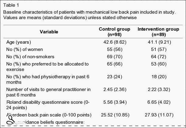
 

Table 1 Baseline Characteristics of Patients with Mechanical Low Back Pain Included in Study

Table 1: Baseline characteristics of patients with mechanical low back pain included in study. Values are means (standard deviations) unless stated otherwise.


Clinical Outcomes


Table ?2 shows the mean changes in outcome measures over time, from randomisation to final follow up at one year. After adjustment for baseline scores, the intervention group showed greater decreases in all measures of back pain and disability compared with the controls. At six weeks after randomisation, patients in the intervention group reported less distressing pain than the control group (P=0.03) and a marginally significant difference on the Roland disability questionnaire scores. Other variables were not significantly different, but the differences in change were all in favour of the intervention group. At six months the difference of the mean change scores of the Roland disability questionnaire was significant, and at one year the differences in changes of both the Roland disability questionnaire and the Aberdeen back pain scale were significant (Table ?2). Most of the intervention group improved by at least three points on the Roland disability questionnaire: 53% (95% confidence interval 42% to 64%) had done so at six weeks, 60% (49% to 71%) at six months, and 64% (54% to 74%) at one year. A smaller proportion of the control group achieved this clinically important improvement: 31% (22% to 40%) at six weeks, 40% (29% to 50%) at six months, and 35% (25% to 45%) at one year.
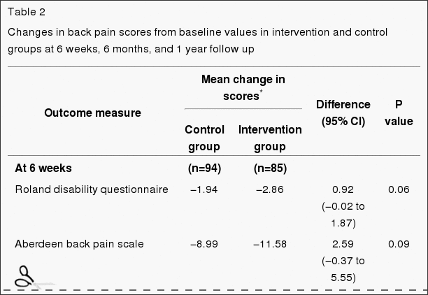
 

Table 2 Changes in Back Pain Scores from Baseline Values in Intervention and Control Groups

Table 2: Changes in back pain scores from baseline values in intervention and control groups at 6 weeks, 6 months, and 1 year follow up.


Patients� Preference


We examined the effect of patients� baseline preference for treatment on outcome after adjusting for baseline scores and main effects. Preference did not significantly affect response to treatment. The intervention had similar effects on both costs and outcomes regardless of baseline preference. For example, the change in the Roland disability questionnaire score at 12 months in the control group was ?1.93 for patients who preferred intervention and ?1.18 for those who were indifferent (95% confidence interval of difference ?1.05 to 2.55), and in the intervention group the change in score was ?3.10 for those who preferred intervention and ?3.15 for those who were indifferent ((95% confidence interval of difference ?1.47 to 3.08). As the interaction term (preference by random allocation) was non-significant, the results shown in Table ?2 exclude the preference term.


Economic Evaluation


Patients in the intervention group tended to use fewer healthcare and other resources compared with those in the control group (Table ?3). However, the mean difference, totalling �148 per patient, was not significant: the 95% confidence interval suggests there could have been a saving of as much as �442 per patient in the intervention group or an additional cost of up to �146. Patients in the control group took a total of 607 days off work during the 12 months after randomisation compared with 378 days taken off by the intervention group.
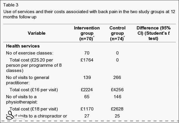
 

Table 3 Use of Services and their Costs Associated with Back Pain in the Two Study Groups

Table 3: Use of services and their costs associated with back pain in the two study groups at 12 months follow up.


Discussion


Our results support the hypothesis that a simple exercise class can lead to long term improvements for back pain sufferers. Studies have shown that a similar programme for patients with chronic back pain can be effective in the hospital setting. In this study we show the clinical effectiveness for patients with subacute or recurrent low back pain who were referred by their general practitioner to a community programme.


Current management guidelines for low back pain recommend a return to physical activity and taking exercise. In particular, they recommend that patients who are not improving at six weeks after onset of back pain, which may be a higher proportion than previously realised, should be referred to a reactivation programme. The programme we evaluated fits that requirement well. It shows participants how they can safely start moving again and increase their levels of physical activity. It is simple and less costly than individual treatment.


It seemed to have beneficial effects even one year later, as measured by functional disability (Roland disability questionnaire) and clinical status (Aberdeen back pain scale). The mean changes in scores on these instruments were small, with many patients reporting mild symptoms on the day of entry to the trial. However, a substantially larger proportion of participants in the exercise classes gained increases of over three points on the Roland disability questionnaire at six weeks, six months, and one year, which might be clinically important. At six weeks, participants in the exercise classes reported significantly less distressing pain compared with the control group, although the intensity of pain was not significantly different. This is consistent with findings from a study of chronic back pain patients in Oxford, in which changes in distressing pain were much greater than were the changes in intensity of pain.


People with back pain who use coping strategies that do not avoid movement and pain have less disability. In our study the participants in the exercise classes were able to function better according to Roland disability questionnaire scores than the control group at six months and one year after randomisation to treatment, and at one year they also showed a significantly greater improvement in clinical status as measured by the Aberdeen back pain scale. This increase in differences in effect between the intervention and control groups over time is consistent with the results from long term follow up in comparable back pain trials.


Study Design


The design of this study was a conventional randomised controlled trial in that all eligible patients were randomised. However, the participants were asked to state their preferred treatment before they knew of their allocation. A study of antenatal services showed that preferences can be an important determinant of outcome, but we did not find any strong effect of preference on the outcome, although a much larger sample size would be needed to confidently exclude any modest interaction between preference and outcome. This information may be useful to clinicians in that it suggests that exercise classes are effective even in patients who are not highly motivated. Our trial design, of asking patients for their preferences at the outset, has substantial advantages over the usual patient preference design, in which costs and outcomes cannot be reliably controlled for confounding by preference.


Conclusions


Our exercise programme did not seem to influence the intensity of pain but did affect the participants� ability to cope with the pain in the short term and even more so in the longer term. It used a cognitive-behavioural model, shifting the emphasis away from a disease model to a model of normal human behaviour, and with minimal extra training a physiotherapist can run it. Patients� preferences did not seem to influence the outcome.
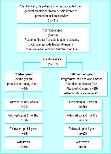
 

Figure 1 Flow Chart Describing Patient Progress Through the Trial

Figure 1: Flow chart describing patients’ progress through the trial.


Footnotes


Funding: This research was funded by the Arthritis Research Campaign, the Northern and Yorkshire Regional Health Authority, and the National Back Pain Association.


Competing interests: None declared.


In conclusion,�the participation of patients in functional fitness and/or exercise as recommended by a physiotherapist, or physical therapist, or any other healthcare professional, such as a chiropractor, is essential towards the improvement of their symptoms of low back pain. The exercise programme helped patients better cope with their symptoms of back pain where the intervention group showed that they used fewer healthcare resources and took fewer day off work, according to the outcome measures of the research study. Information referenced from the National Center for Biotechnology Information (NCBI). The scope of our information is limited to chiropractic as well as to spinal injuries and conditions. To discuss the subject matter, please feel free to ask Dr. Jimenez or contact us at 915-850-0900 .


Curated by Dr. Alex Jimenez

 

Green-Call-Now-Button-24H-150x150-2-3.png


Additional Topics: Sciatica


Sciatica is referred to as a collection of symptoms rather than a single type of injury or condition. The symptoms are characterized as radiating pain, numbness and tingling sensations from the sciatic nerve in the lower back, down the buttocks and thighs and through one or both legs and into the feet. Sciatica is commonly the result of irritation, inflammation or compression of the largest nerve in the human body, generally due to a herniated disc or bone spur.


blog picture of cartoon paperboy big news


IMPORTANT TOPIC: EXTRA EXTRA: Treating Sciatica Pain


Blank
References
1.�Croft P, editor.�Low back pain.�Oxford: Radcliffe Medical Press; 1997.
2.�Clinical Standards Advisory Group.�Back pain.�London: HMSO; 1994.
3.�Waddell G, Feder G, McIntosh A, Lewis M, Hutchinson A.�Low back pain evidence review.�London: Royal College of General Practitioners; 1996.
4.�Malmivaara A, Hakkinen U, Aro T, Heinrichs M, Koskenniemi L, Kuosma E, et al. The treatment of acute low back pain�bed rest, exercises or ordinary activity?�N Engl J Med.�1995;332:351�355.[PubMed]
5.�Faas A, Chavannes A, van Eijk JTM, Gubbels J. A randomized, placebo-controlled trial of exercise therapy in patients with acute low back pain.�Spine.�1993;18:1388�1395.�[PubMed]
6.�Frost H, Klaber Moffett J, Moser J, Fairbank J. Evaluation of a fitness programme for patients with chronic low back pain.�BMJ.�1995;310:151�154.�[PMC free article][PubMed]
7.�Frost H, Lamb S, Klaber Moffett J, Fairbank J, Moser J. A fitness programme for patients with chronic low back pain: 2 year follow-up of a randomised controlled trial.�Pain.�1998;75:273�279.�[PubMed]
8.�McPherson K, Britton A, Wennberg J. Are randomised controlled trials controlled? Patient preferences and unblind trials.�J R Soc Med.�1997;90:652�656.�[PMC free article][PubMed]
9.�Bradley C. Designing medical and educational studies.�Diabetes Care.�1993;16:509�518.�[PubMed]
10.�Clement S, Sikorski J, Wilson J, Candy B. Merits of alternative strategies for incorporating patient preferences into clinical trials must be considered carefully [letter]�BMJ.�1998;317:78.�[PubMed]
11.�Torgerson D, Klaber Moffett J, Russell I. Patient preferences in randomised trials: threat or opportunity?�J Health Serv Res Policy.�1996;1(4):194�197.�[PubMed]
12.�Roland M, Morris R. A study of the natural causes of back pain. Part 1: Development of a reliable and sensitive measure of disability in low-back pain.�Spine.�1983;8:141�144.�[PubMed]
13.�Ruta D, Garratt A, Wardlaw D, Russell I. Developing a valid and reliable measure of health outcome for patients with low back pain.�Spine.�1994;19:1887�1896.�[PubMed]
14.�Brooks R.with EuroQoL Group.�EuroQoL: the current state of play�Health Policy�19963753�72.[PubMed]
15.�Waddell G, Newton M, Henderson I, Somerville D, Main C. A fear-avoidance beliefs questionnaire (FABQ) and the role of fear-avoidance beliefs in chronic low back pain and disability.�Pain.�1993;52:157�168.�[PubMed]
16.�Jensen M, McFarland C. Increasing the reliability and validity of pain intensity measurement in chronic pain patients.�Pain.�1993;55:195�203.�[PubMed]
17.�Efron B, Tibshirani R.�An introduction to bootstrap.�New York: Chapman and Hall; 1993.
18.�Williams D, Keefe F. Pain beliefs and use of cognitive-behavioral coping strategies.�Pain.�1991;46:185�190.�[PubMed]
19.�Estlander A, Harkapaa K. Relationships between coping strategies, disability and pain levels in patients with chronic low back pain.�Scand J Behav Ther.�1989;18:56�69.
20.�Holmes J, Stevenson C. Differential effects of avoidant and attentional coping strategies on adaption to chronic and recent-onset pain.�Health Psychology.�1990;9:577�584.�[PubMed]
21.�Rosenstiel A, Keefe F. The use of coping strategies in chronic low back pain patients: relationship to patient characteristics and current adjustments.�Pain.�1983;17:33�44.�[PubMed]
22.�Slade P, Troup J, Lethem J, Bentley G. The fear avoidance model of exaggerated pain perception II Preliminary studies of coping strategies for pain.�Behav Res Ther.�1983;21:409�416.�[PubMed]
23.�Meade T, Dyer S, Browne W, Frank A. Randomised comparison of chiropractic and hospital outpatient management for low back pain: results from extended follow up.�BMJ.�1995;311:349�351.[PMC free article][PubMed]
24.�Cherkin D, Deyo R, Battie M, Street J, Barlow W. A comparison of physical therapy, chiropractic manipulation, and provision of an educational booklet for the treatment of patients with low back pain.�N Engl J Med.�1998;339:1021�1029.�[PubMed]
Close Accordion
Safe Physiotherapy Interventions in Cervical Disc Herniations

Safe Physiotherapy Interventions in Cervical Disc Herniations

Recognizing clinical and experimental evidence, physiotherapy is a healthcare profession that helps restore and maintain function to individuals affected by injury, disease or disability by using mechanical force and movements, manual therapy, exercise and electrotherapy, as well as through patient education and advice. The terms physiotherapy and physical therapy are used interchangeably to describe the same healthcare profession. Physiotherapy is recommended for a variety of injuries and conditions, and it can help support overall health and wellness for people of all ages.


For further notice,�physiotherapy services may be offered alongside chiropractic care, to provide a cautious and gentle manipulation and/or mobilization of the cervical and thoracic spine in the instance of a large cervical disc herniation. Cervical disc herniations can cause pain and discomfort, numbness and weakness in the neck, shoulders, chest, arms and hands.

Abstract


A 34-year-old woman was seen in a physiotherapy department with signs and symptoms of cervical radiculopathy. Loss of cervical lordosis and a large paracentral to intraforaminal disc prolapse (8?mm) at C5�C6 level was reported on MRI. She was taking diclofenac sodium, tramadol HCl, diazepam and pregabalin for the preceding 2?months and no significant improvement, except temporary relief, was reported. She was referred to physiotherapy while awaiting a surgical opinion from a neurosurgeon. In physiotherapy she was treated with mobilisation of the upper thoracic spine from C7 to T6 level. A cervical extension exercise was performed with prior voluntary extension of the thoracic spine and elevated shoulders. She was advised to continue the same at home. General posture advice was given. Signs and symptoms resolved within the following four sessions of treatment over 3?weeks. Surgical intervention was subsequently deemed unnecessary.


Background


Surgical interventions are commonly recommended in large cervical prolapsed discs and the importance of non-aggressive physiotherapy interventions is less recognised and poorly understood. We present interventions that were associated with resolution of symptoms of radiculopathy resulting from a larger cervical herniated disc. These interventions, if applied correctly, may help to reduce the number of surgeries required for cervical prolapsed discs.


Case Presentation


The patient was a 34-year-old woman. She was seen in the physiotherapy department with a complaint of left-sided neck and shoulder pain. The pain was radiating to her left arm and there was associated numbness. The duration of symptoms was more than 2?months with no history of trauma. The pain was present on waking in the morning and gradually increased during the day. She was otherwise a healthy woman. Neck movements were aggravating the symptoms. She was seen in the acute hospital accident and emergency department (A&E) twice since onset and had been taking diclofenac sodium, tramadol HCl, diazepam and pregabalin. An MRI was planned and a request was sent for physiotherapy during the MRI waiting period. A neurosurgical review was requested by the A&E consultant upon receipt of the MRI report 7?weeks later.


Patient examination in the physiotherapy department revealed a normal gait pattern, her left arm held in front of her chest with the left shoulder slightly elevated. Her active range of neck motion was restricted and was painful on the left side. Flexion and rotation to the left were aggravating her arm and shoulder pain. Strength deficits were noted in the left elbow flexors and wrist extensors (4/5) when compared with the right side. There was paraesthesia along the radial border of the forearm and thumb regions. The brachioradialis reflex was diminished and biceps reflex was sluggish. Triceps and plantar reflexes were normal. Passive intervertebral movements were tender at C5�C6 level and were reproducing the pain. Sustained pressure at C7 and below was easing the pain and also improving the neck range of motion. The patient was deemed to have C6 radiculopathy. The MRI report, available 2?weeks after the commencement of physiotherapy, confirmed the diagnosis.


Investigations


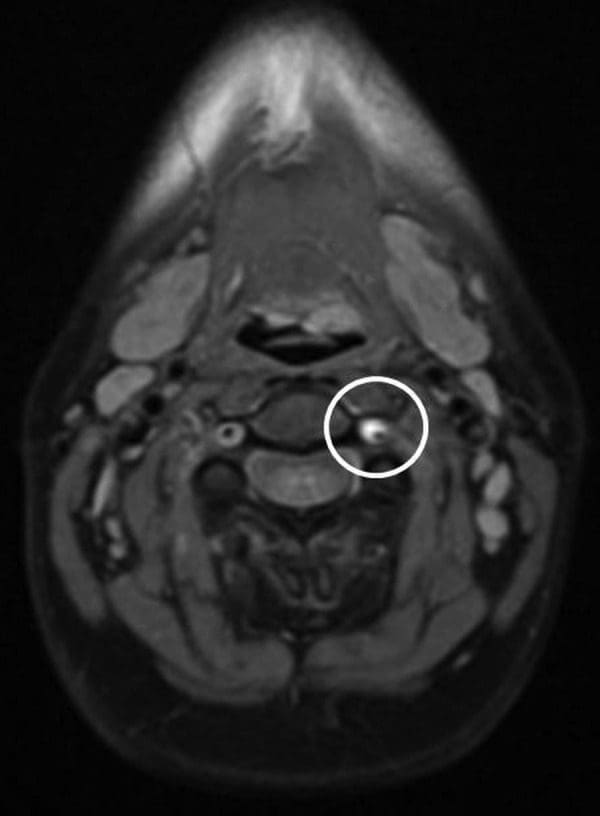
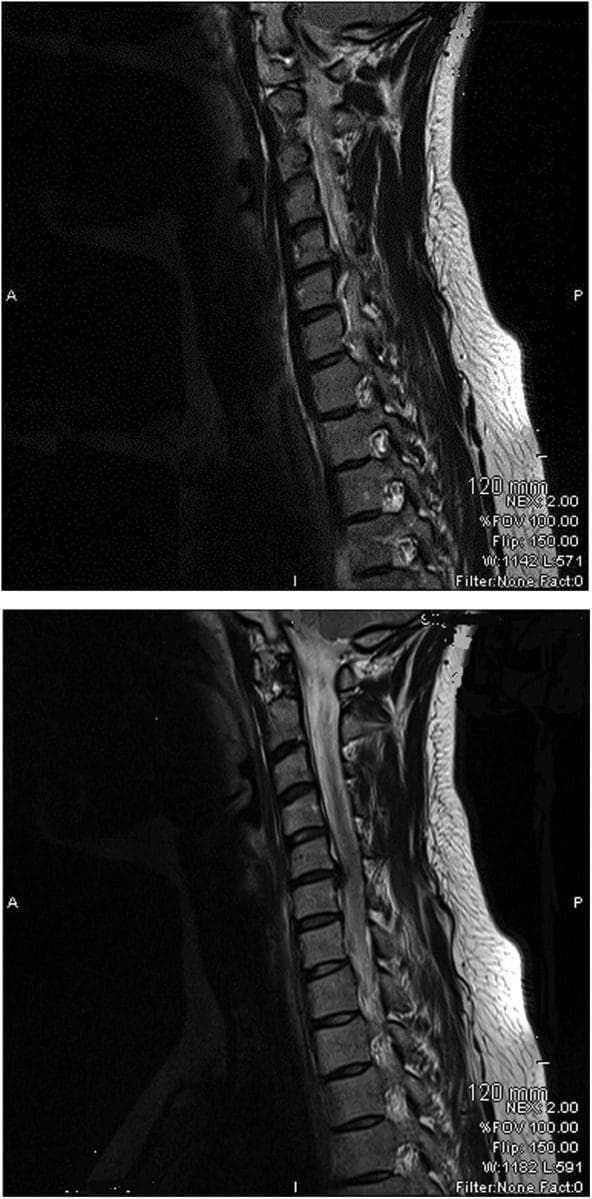
The findings from the plain cervical x-ray were unremarkable. MRI showed (Figure 1) loss of cervical spine lordosis, a left paracentral to intraforaminal lesion with 8?mm hernia, which indented the cord and obstructed the left paracentral recess and neural foramen.


Figure 1 Loss of Cervical Spine Lordosis and Large Disc Herniation at C5 and C6 on MRI

Figure 1: Loss of cervical spine lordosis and large disc herniation at C5 and C6 on MRI.


Differential Diagnosis


  • Cervical myelopathy.


Treatment


The patient received pharmacological treatment for the initial two symptomatic months, which included diclofenic sodium, tramadol, diazepam and pregabalin (lyrica) tablet. Physiotherapy was started after 2?months. Physiotherapy intervention consisted of mobilisation of the thoracic spine, resisted cervical extension exercises, a home programme of exercises and advice regarding the posture.


Mobilisation of the thoracic spine was administered in the prone lying position from C7 toT6 level. Mild intensity oscillations (15?reps) in an anterosuperior direction were directly applied to each of the spinal segments, through the thumb over the spinous processes, during the first visit. The applied force was enough to appreciate intervertebral movement in each segment and without significant pain. High-intensity oscillations (10�20) were applied during the subsequent treatment sessions. The patient was asked for symptom feedback during treatment.


Cervical spine extension exercises were carried out in a sitting position. The patient was asked to extend her thoracic spine with lungs fully inflated and shoulders elevated followed by extension of her cervical spine. Head extension was moderately resisted by the therapist near the end range of extension for 5�10?s and brought back to neutral after each resisted movement. The resisted movement was repeated at least three times with intervals of 30?s. The patient was asked to perform the same exercise at home every hour during the day.


The patient was educated regarding the rationale of extension exercises, sitting and lying posture and their effects on the spine. The duration of each session was approximately 20�25?min.


Dr. Alex Jimenez’s Insight

Surgical interventions are generally recommended and widely considered for large cervical disc herniations. Although less recognized and often misunderstood, however, physiotherapy can be just as effective towards improving herniated discs in the cervical spine, excluding the need for surgery, according to the research study. Pharmacological treatments are also commonly used to help temporarily reduce symptoms alongside physiotherapy interventions. Cautious and gentle, spinal manipulation and mobilization of the cervical spine should be performed in the case of large cervical disc herniations to avoid aggravating the injury and/or condition. As recommended by a physiotherapist, or other healthcare professional experienced in physiotherapy, proper exercise can restore the function of the cervical spine and prevent regression of large prolapsed discs along the spine. Through appropriate physiotherapy intervention as well as through patient safety and compliance, the retraction of the cervical herniated discs is possible.


Outcome and Follow-Up


Pharmacological interventions were helpful to reduce the patient’s pain on a temporary basis. Symptoms were recurring and resolution was not sustainable. The symptoms started improving after the first physiotherapy session and continued to improve during the subsequent sessions. It fully resolved in four sessions extended over 3?weeks. The patient was reviewed 4?months after the resolution of symptoms and there was no recurrence of symptoms. She was reviewed by a neurosurgeon and the surgical option was withdrawn.


Discussion


Stiffness of the thoracic spine has been linked to the painful pathologies of the cervical spine, and manipulation of the thoracic spine has been shown to improve painful symptoms and mobility of the cervical spine. However, cervical disc herniations of greater than 4?mm are considered inappropriate for physiotherapy interventions such as traction and manipulation. Spinal manipulation refers to a passive movement thrust of high velocity and low amplitude, usually applied at the end range of movement and is beyond the patient’s control. Manipulation of the cervical spine is an aggressive procedure, which carries various risks and is often associated with worsening of symptoms. Manipulation was not considered in the treatment options for this patient because of the risks associated with it, and also because of patient’s anxiety and lack of MRI-confirmed diagnosis.


Active extension of the thoracic spine increases the range of motion of the cervical spine and, in these authors� clinical experience, relieves minor neck symptoms. Conversely, thoracic spine kyphosis, such as slouch sitting, restricts the mobility of the cervical spine and aggravates the painful symptoms. A good sitting posture is constituted by a slightly extended thoracic spine. Therefore, active extension of the thoracic spine prior to cervical extension may improve cervical movements and restore cervical curvature.


It is believed that excessive pressure during flexion on the anterior aspect of the intervertebral discs pushes the nucleus pulposus posteriorly and causes herniations. Conversely, cervical lordosis might have the reverse effect�that is, decreases pressure on the anterior aspect of the discs and may create a suction effect which retracts the herniated contents. Therefore, a combination of short duration and repeated movements at the end of extension may serve as a suction pump and possibly retract the extruded content of the disc. Active cervical extension exercises, with an extended thoracic spine posture, may have been the key element in a home exercise programme to restore lordosis of the cervical spine and relieve radiculopathy symptoms in the current case. This may possibly have been due to the retraction of the herniated discs.


Spinal mobilisation refers to a gentle, oscillatory, passive movement of a spinal segment. These are applied to a spinal segment to gently increase the passive range of motion. It allows the patient to report aggravation of pain and to resist any unwanted movements. No mobilisation treatment was administered at C5�C6 level as palpation at this level was aggravating the symptoms. Segments below this level were mobilised with emphasis at C7�T1 level. Any treatment at the affected segment was likely to irritate the nerve root and thereby increase the inflammatory process.


Various interventions are reported for the treatment of prolapsed discs. Saal et al reported the use of traction, specific physical therapy exercise, oral anti-inflammatory medication and patient education in the treatment of 26 patients with herniated cervical discs (<4?mm) and reported significant improvement in outcomes for 24 patients. They observed that surgery for disc herniations occurs when a patient has significant myotomal weakness, severe pain or pain that persists beyond an arbitrary conservative treatment period of 2�8?weeks.


Spontaneous regressions of cervical disc protrusions are reported in the literature. However, spontaneous regressions of herniated cervical discs are speculated to be rare. Various factors related to regression are hypothesised and theorised. Pan et al summarised the factors related to the resorption of herniated disc as: the age of the patients; dehydration of the expanded nucleus pulposus; resorption of haematoma; revascularisation; penetration of herniated cervical disc fragments through the posterior longitudinal ligament; size of disc herniations; and existence of cartilage and annulus fibrosus tissue in the herniated material. Some studies on spontaneous regressions of discs reported that the patients were receiving physiotherapy. Physiotherapy interventions are not defined in any of these studies, however. Therefore, it is possible that disc regressions in these studies may be due to similar physiotherapy interventions as described here, or the patients were practising techniques and adopting postures as reported in the current case.


Learning Points


  • Thoracic spine mobilisation improves cervical spine biomechanics and can be considered in conjunction with other interventions in all painful conditions of the cervical spine.
  • Active extension of the thoracic spine facilitates movements of the cervical spine and may help regression of large prolapsed discs.
  • There is a possibility of retraction of herniated cervical discs through appropriate physiotherapy intervention.
  • Patient education ensures safety and compliance to therapist advice.
  • Meticulous assessment and patient feedback guides the therapist in selection of intensity of mobilisation.


Footnotes


Competing interests: None.


Patient consent: Obtained.


In conclusion,�physiotherapy, or physical therapy, is used to treat various injuries, diseases and disabilities, through the use of mechanical force and movements, manual therapy, exercise, electrotherapy, and through patient education and advice to restore and maintain function. As in the case above, physiotherapy can be recommended and considered as treatment before referring to surgical interventions of large cervical disc herniations. Information referenced from the National Center for Biotechnology Information (NCBI). The scope of our information is limited to chiropractic as well as to spinal injuries and conditions. To discuss the subject matter, please feel free to ask Dr. Jimenez or contact us at 915-850-0900 .


Curated by Dr. Alex Jimenez


Green-Call-Now-Button-24H-150x150-2-3.png


Additional Topics: Sciatica


Sciatica is referred to as a collection of symptoms rather than a single type of injury or condition. The symptoms are characterized as radiating pain, numbness and tingling sensations from the sciatic nerve in the lower back, down the buttocks and thighs and through one or both legs and into the feet. Sciatica is commonly the result of irritation, inflammation or compression of the largest nerve in the human body, generally due to a herniated disc or bone spur.


blog picture of cartoon paperboy big news


IMPORTANT TOPIC: EXTRA EXTRA: Treating Sciatica Pain


Blank
References
1.�Norlander S, Gustavsson BA, Lindell J, et al.�Reduced mobility in the cervico-thoracic motion segment�a risk factor for musculoskeletal neck-shoulder pain: a two-year prospective follow-up study.�Scand J Rehabil Med�1997;29:167�74.�[PubMed]
2.�Walser RF, Meserve BB, Boucher TR.�The effectiveness of thoracic spine manipulation for the management of musculoskeletal conditions: a systematic review and meta-analysis of randomized clinical trials.�J Man Manipulative Ther�2009;17:237�46.�[PMC free article][PubMed]
3.�Krauss J, Creighton D, Ely JD, et al.�The immediate effects of upper thoracic translatoric spinal manipulation on cervical pain and range of motion: a randomized clinical trial.�J Man Manipulative Ther2008;16:93�9.�[PMC free article][PubMed]
4.�Saal JS, Saal JA, Yurth EF.�Nonoperative management of herniated cervical intervertebral disc with radiculopathy.�Spine (Phila Pa 1976)�1996;21:1877�83.�[PubMed]
5.�Murphy DR, Beres JL.�Cervical myelopathy: a case report of a �near-miss� complication to cervical manipulation.�J Manipulative Physiol Ther�2008;31:553�7.�[PubMed]
6.�Leon-Sanchez A, Cuetter A, Ferrer G.�Cervical spine manipulation: an alternative medical procedure with potentially fatal complications.�South Med J�2007;100:201�3.�[PubMed]
7.�Scannell JP, McGill SM.�Disc prolapse: evidence of reversal with repeated extension.�Spine (Phila Pa 1976)�2009;34:344�50.�[PubMed]
8.�Gurkanlar D, Yucel E, Er U, et al.�Spontaneous regression of cervical disc herniations.�Minim Invasive Neurosurg�2006;49:179�83.�[PubMed]
9.�Mochida K, Komori H, Okawa A, et al.�Regression of cervical disc herniation observed on magnetic resonance images.�Spine (Phila Pa 1976)�1998;23:990�5; discussion 6�7.�[PubMed]
10.�Song JH, Park HK, Shin KM.�Spontaneous regression of a herniated cervical disc in a patient with myelopathy. Case report.�J Neurosurg�1999;90(1 Suppl):138�40.�[PubMed]
11.�Westmark RM, Westmark KD, Sonntag VK.�Disappearing cervical disc. Case report.�J Neurosurg1997;86:289�90.�[PubMed]
12.�Pan H, Xiao LW, Hu QF.�Spontaneous regression of herniated cervical disc fragments and its clinical significance.�Orthop Surg�2010;2:77�9.�[PubMed]
13.�Teplick JG, Haskin ME.�Spontaneous regression of herniated nucleus pulposus.�AJR Am J Roentgenol1985;145:371�5.�[PubMed]
Close Accordion
Electroacupuncture vs. Medium-Frequency Electrotherapy for Sciatica

Electroacupuncture vs. Medium-Frequency Electrotherapy for Sciatica

Electroacupuncture: Before reviewing the data below, it is important as a practicing doctor of chiropractic to inform that�sciatica and discogenic lower back pain are two closely related health issues which collectively, can be a source of painful symptoms and discomfort as well as a cause of limited mobility among affected individuals. Discogenic disease, also known as degenerative disc disease, is characterized as the naturally-occurring deterioration of the spinal intervertebral discs. While discogenic disease commonly develops with age, other factors, such as injury can also lead to degenerative disc disease. In addition, degenerative disc disease may cause other complications including bulging or herniated discs

Furthermore, from years of experience caring for patients with this health issue, bulging or herniated discs are not necessarily the cause of the individual’s pain and discomfort. Symptoms are instead the result of compression or irritation from displaced disc material against the spinal cord or an exiting nerve root. Sciatica occurs if the nerve being compressed or irritated is the sciatic nerve, the largest nerve in the human body which branches off the lower spine, down into the legs. Two methods of treatment, electroacupuncture and medium-frequency electrotherapy were used in the following research study to determine whether symptoms of sciatica would improve with one treatment therapy over the other.

Abstract

Objective. To investigate the short- and long-term effects of electroacupuncture (EA) compared with medium-frequency electrotherapy (MFE) on chronic discogenic sciatica. Methods. One hundred participants were randomized into two groups to receive EA (n = 50) or MFE (n = 50) for 4 weeks. A 28-week follow-up of the two groups was performed. The primary outcome measure was the average leg pain intensity. The secondary outcome measures were the low back pain intensity, Oswestry Disability Index (ODI), patient global impression (PGI), drug use frequency, and�electroacupuncture acceptance. Results. The mean changes in the average leg pain numerical rating scale (NRS) scores were 2.30 (1.86�2.57) and 1.06 (0.62�1.51) in the EA and MFE groups at week 4, respectively. The difference was significant (P < 0.001). The long-term follow-up resulted in significant differences. The average leg pain NRS scores decreased by 2.12 (1.70�2.53) and 0.36 (?0.05�0.78) from baseline in the EA and MFE groups, respectively, at week 28. However, low back pain intensity and PGI did not differ significantly at week 4. No serious adverse events occurred. Conclusions. EA showed greater short-term and long-term benefits for chronic discogenic sciatica than MFE, and the effect of EA was superior to that of MFE. The study findings warrant verification. This trial was registered under identifier ChiCTR-IPR-15006370.

Introduction: Electroacupuncture

Sciatica is defined as radicular leg pain localized to the dermatological distribution of a pathologically affected nerve root. Almost all discogenic sciatica is induced by lumbar disc herniation (LDH) and may be accompanied by neurological deficits, such as leg pain, leg paresthesia, disability, and low back pain. The estimated prevalence of sciatica ranges from 1.2 to 43% in various regions. Discogenic sciatica, which accounts for nearly 90% of sciatica, is a major cause of morbidity; moreover, it has a considerable impact on the economy due to both loss of work and the high costs of health care and societal support for the affected individual and his/her family. Current treatments for discogenic sciatica primarily include surgical and conservative treatments. Although discectomy is a more effective treatment than other treatments for patients with severe discogenic sciatica, in patients with less severe symptoms, surgery or conservative treatments appear to be equally effective. Discectomy should be avoided during initial treatment due to its high cost and its association with a higher incidence of postoperative complications, such as the loss of spine stability and extensive peridural fibrosis. Conservative measures comprise the first-line treatment strategy for managing radicular pain due to disc herniation. Regarding cost-effectiveness, the regimes that employ stepped approaches based on an initial treatment with conservative management have been recommended. However, many conservative treatments have no explicit curative effect, such as benzodiazepines, corticosteroids, traction, and spinal manipulation, which may be ineffective or less effective. Moreover, the long-term efficacy of analgesic drugs is not enduring, and intolerable side-effects, such as addiction, stomach ulcers, and constipation, occur frequently in patients with discogenic sciatica. Thus, based on recent information, the short- and long-term efficacy of conservative treatment should be evaluated.

Electroacupuncture (EA) has been used to treat sciatica for many decades in China. Several studies have reported that electroacupuncture EA may effectively treat neuropathic pain and relieve sciatica symptoms. However, no clear clinical evidence exists to support the application of acupuncture or�electroacupuncture in the treatment of discogenic sciatica according to the guideline for the diagnosis and treatment of lumbar disc herniation. Recently, two meta-analyses concerning sciatica treatment with acupuncture showed that previous studies on acupuncture were flawed and that the strength of the evidence was suboptimal; thus, studies of higher quality with longer-term follow-up are needed to clarify the long-term effect of acupuncture in sciatica patients.


Image of electroacupuncture being applied to patient.


Compared with manual acupuncture,�electroacupuncture treatment is capable of increasing the stimulation frequency and intensity in a controlled and quantifiable manner; moreover, its effect is superior to manual acupuncture for alleviating pain and improving paresthesia and dysfunction. Medium-frequency electrotherapy (MFE) is similar to transcutaneous electrical nerve stimulation (TENS) and may relieve pain and related symptoms. MFE works through electrostimulation of an electrode placed on the skin, and a battery powered device provided a small current to produce a tingling sensation. Several studies found that the effects obtained with 50?Hz EA were superior to those using 2?Hz EA. EA and MFE using the same frequency (50?Hz) at the same location were employed in another trial. The major difference between the two trial groups was the specific impact of needle penetration, with�electroacupuncture using needle penetration and MFE administered via nonpenetrating electrostimulation.


Image of medium frequency electrotherapy being applied to patient.


This study was a comparative trial that evaluated the effectiveness of electroacupuncture versus MFE for the treatment of chronic discogenic sciatica; these treatments are the most frequently used treatments for this disease in China. We explored the ability of�electroacupuncture to alleviate leg pain, low back pain, and dysfunction at various evaluation time points, which included an assessment of the long-term efficacy of electroacupuncture. We also assessed the patient global impression (PGI) and acceptance of�electroacupuncture compared with MFE and reports of adverse events.

Methods

Participants

The study commenced on May 28, 2015, and was completed by July 30, 2016, at the South Area of Guang’anmen Hospital, China Academy of Chinese Medical Sciences. Discogenic sciatica was diagnosed according to the criteria of the North American Spine Society. The inclusion criteria were as follows: (1) individuals aged 18 to 70 years; (2) participants whose sciatica symptoms correlated with magnetic resonance imaging (MRI) or computed tomography (CT) findings of lumbar disc herniation; (3) participants whose symptoms of leg pain lasted more than 3 months; (4) participants who agreed to follow the trial protocol; and (5) participants who could complete the study treatment and assessments. The exclusion criteria were as follows: (1) participants with severe progressive neurological symptoms (e.g., cauda equina syndrome and progressive muscle weakness); (2) participants who had undergone surgery for lumbar disc herniation within 6 months; (3) participants with symptoms caused by conditions other than lumbar disc herniation that might lead to radiating pain in the leg; (4) participants with pain in both legs; (5) participants with cardiovascular, liver, kidney, or hematopoietic system diseases, mental health disorders, or cancer for whom EA might be inappropriate or unsafe; (6) participants who had received EA or electrotherapy within the past week; (7) women who were pregnant or lactating; (8) participants who were participating in other clinical trials; and (9) participants with a pacemaker, metal allergy, or severe fear of needles.

Study Design

This was a single-center, prospective, controlled, randomized trial conducted in patients with chronic discogenic sciatica. This trial was approved by the Ethics Committee of Guang’anmen Hospital of China Academy of Chinese Medical Sciences (approval number 2015EC042) on May 26, 2015, and was registered on May 7, 2015, at http://www.chictr.org.cn/ (ref. ChiCTR-IPR-15006370). Written informed consent was obtained from each participant or their legal representative. All participants were required to be able to understand written instructions and able to complete the pain assessment forms.

Randomization and Allocation Concealment

The randomization was performed by the Drug Clinical Trial Office affiliated with Guang’anmen Hospital using a computerized random number generator. Opaque, sealed envelopes were numbered consecutively, and all the sealed envelopes were maintained by a researcher who was not involved in the treatment procedure or data analysis. After informed consent was obtained, an envelope was opened by the researcher according to the patient’s order of entry into the trial, and the assigned treatment was offered to the participant. The outcome assessors and statisticians were blinded to the allocation. Two copies of the envelopes were maintained to prevent the researchers from deviating from the randomization.

Intervention

The treatments were initiated one week after participant randomization. All participants received health education on sciatica, such as using a hard bed and losing weight. During the trial, the use of analgesic drugs or other treatments was not permitted. The details of prior drug use (including dose and time) were recorded in the medication record form. Huatuo Brand stainless steel needles (0.3 � 100?mm, Suzhou Medical Appliance Factory in China, CL) and a G6805-2 electric stimulator (Shanghai Huayi Medical Instrument in China Co., Ltd.) were used in the EA group, and the Quanrikang type J48A computerized intermediate-frequency therapy apparatus (Beijing Huayi New Technical Institute in China) was used in the MFE (control) group. The acupuncture procedures were performed in accordance with the Standards for Reporting Interventions in Clinical Trials of Acupuncture (STRICTA) guidelines. EA was performed by a trained clinician with more than 2 years of experience with acupuncture manipulation. The acupuncture regimen was based on our own pilot trial and specialist consensus. The acupoints of the affected side (DaChangShu, BL25) and the bilateral JiaJi (Ex-B2) corresponding to LDH were included in the EA group. The DaChangShu (BL25) acupoint was located according to the World Health Organization Standardized Acupuncture Point Location; JiaJi (Ex-B2) is located in the lumbar region 0.5 inches lateral to the posterior median line. After the participants assumed a prone position, the needle was vertically inserted rapidly into the JiaJi (Ex-B2) points. Then, the needle was inserted to a depth of approximately 1.5 inches. The participants were expected to experience soreness and distension transmitted to the leg. The needle was inserted straight into the DaChangShu on the BL25 point to a depth of 3 inches; then, the acupuncturist manipulated the needle with a lifting, thrusting, and twirling maneuver until feelings of soreness and distension were felt and radiated to the hips and lower limbs. The electric apparatus was applied to the JiaJi (Ex-B2) and DaChangShu (BL25) acupoints with a dilatational wave using a 50?Hz frequency and a comfortably tolerated maximum current intensity.

Participants assigned to the control group received MFE, which was administered by an experienced therapist different from the one delivering the EA. The acupoints and frequencies used in the MFE group were the same as those used in the EA group. After two pairs of 107 � 72?mm electrodes were placed on the acupoints, the MFE apparatus was turned on and muscle contractions were observed under the energizing electrode. The intensity was adjusted to the maximum current intensity tolerable at a comfortable level. The treatments in both groups were performed once daily for 5 sessions/week for the first 2 weeks and followed by 3 sessions/week for the following 2 weeks, with each session lasting 20 minutes.

Data Collection

The data in the trial were obtained from the case report forms recorded by the investigator. The participants’ demographic, clinical, and radiological characteristics were recorded. The diagnosis of lumbar disc herniation was confirmed after a review of the patient’s MRI or CT scan by two experienced musculoskeletal radiologists. Additionally, the diagnosis of discogenic sciatica was confirmed after a clinical examination by a consultant orthopedic physician. Investigators entered the collected data into the case report forms. At baseline and during the treatment period, the forms were completed by the participants under the guidance of a full-time staff member. During the follow-up period (16th and 28th weeks), the participants answered the questionnaire by phone.

Clinical Assessments

The primary outcome was the change from baseline in the average leg pain numerical rating scale (NRS) score at week 4. The secondary outcomes included average leg pain intensity at weeks 1, 2, 3, 16, and 28; low back pain intensity at weeks 2, 4, 16, and 28; Oswestry Disability Index (ODI) questionnaire results at weeks 2, 4, 16, and 28; PGI of improvement at weeks 2 and 4; drug use frequency at weeks 2 and 4; and EA acceptance evaluation at week 4. Adverse events were monitored and documented during the treatment and follow-up periods based on the investigator’s inquiry and reports by the participants themselves.

Primary Outcome Measure: The change from baseline in the average leg pain NRS score was measured using an 11-point numerical rating scale assessing leg pain, with 0 representing no pain and 10 representing the most severe pain. Participants were asked to rate their average leg pain intensity over the prior 24 hours. The average leg pain NRS score at week 4 was equal to the mean value of the NRS scores obtained at the three treatment sessions during the 4th week.

Secondary Outcome Measures: The following secondary outcome measures were determined. (1) The average leg pain intensity at other time points was measured by the NRS. The methods used to measure the secondary outcomes were the same as those used to measure the primary outcome except for the evaluation point. (2) Low back pain intensity was measured using an 11-point NRS. Participants rated their low back pain over the prior 24 hours with a pain NRS. The low back pain NRS score at the time of evaluation was equal to the mean value of the NRS scores in the previous 24 hours. (3) The ODI comprises 10 questions concerning the intensity of pain and daily activities. Each item contains 6 options. A higher score change in the ODI from baseline indicated more serious dysfunction. (4) The PGI improvement score was used to evaluate the improvement in pain and functional disability, and the improvement reported by patients was assessed using a 7-point scale (1 represents greatly improved and 7 represents marked worsening). (5) The frequency of drug use was recorded. The patients’ use of medications or nonprescription drugs during the trial was evaluated using a questionnaire to assess the influence of drugs. (6) To investigate which treatment was preferred, EA or MFE acceptance was assessed at week 4. A 4-point scale was used, with 1 representing �very difficult to accept� and 4 representing �very easy to accept.� (7) Adverse events were assessed using a questionnaire at the end of treatment and active reporting by the participants during treatment.

Sample Size and Statistical Analysis

The sample size calculation was based on the mean value of the leg pain intensity NRS score. According to our pilot trial, the decreases in the mean value of the leg pain intensity NRS scores in the EA and MFE groups at week 4 were 3.41 � 3.46 and 1.57 � 1.24, respectively. Our pilot study was an independent study conducted by our research team before this study, with no crossover participants between the previous study and the current study. We used PASS Version 11.0 (International Business Machines Corporation, China) software to calculate a sample size of 50 for each group to provide 90% power to detect a difference of 1.8 between the groups with a two-sided 5% level of significance, allowing for a 20% dropout rate and with the participants receiving the treatments and completing the follow-up.

The statistical analysis was performed using SPSS Version 22.0 (International Business Machines Corporation, China) software. Two-sided tests were used for all statistical analyses. The level of significance was established at 0.05. All patients who accepted randomization were included in the analysis. All data collected from the participants were included in the statistical analysis, and missing data were replaced by the last observed value. However, the outcomes for which no data except for the baseline assessment data were available were not included in the final analysis. The 100 participants included at least 1 treatment session. Thus, we analyzed the data of all the participants as the primary outcome, which was measured after the first treatment session. However, the secondary outcomes were evaluated at week 2, and 13 participants dropped out before week 2 without any data after treatment except for leg pain NRS scores. So the 13 participants were not included in the statistical analysis of secondary outcomes. Continuous data were represented by means and standard deviations (SD) if the data were normally distributed or by the medians and interquartile ranges if the data were skewed, or by means and 95% confidence intervals (CIs); categorical data were represented by percentages or 95% CIs. For comparisons with baseline data, a paired t-test was used for continuous data and a nonparametric test was used for categorical data. To compare the two independent samples, T tests or Mann�Whitney U tests were used to compare continuous variables, and chi-square tests or Fisher’s exact tests were used to compare categorical variables, as appropriate. A repeated measures analysis of variance or nonparametric test was used to compare differences in data between the groups at multiple time points.

Results

Dr. Alex Jimenez’s Insight

The short- and long-term effects of electroacupuncture (EA) versus medium-frequency electrotherapy (MFE) were evaluated to determine which of the two treatments, if not both, could most effectively be used to help improve symptoms of sciatica associated with degenerative disc disease. The research study was conducted with the participation of a variety of patients with symptoms of discogenic sciatica, over several types of interventions. Clinical assessments and data was collected throughout two different research study outcomes in order to gather the most valuable results. Sample size and statistical analysis were also considered before analyzing the data of all the participants and recording the results. The final outcome of the research study has been described in detail below.

Recruitment

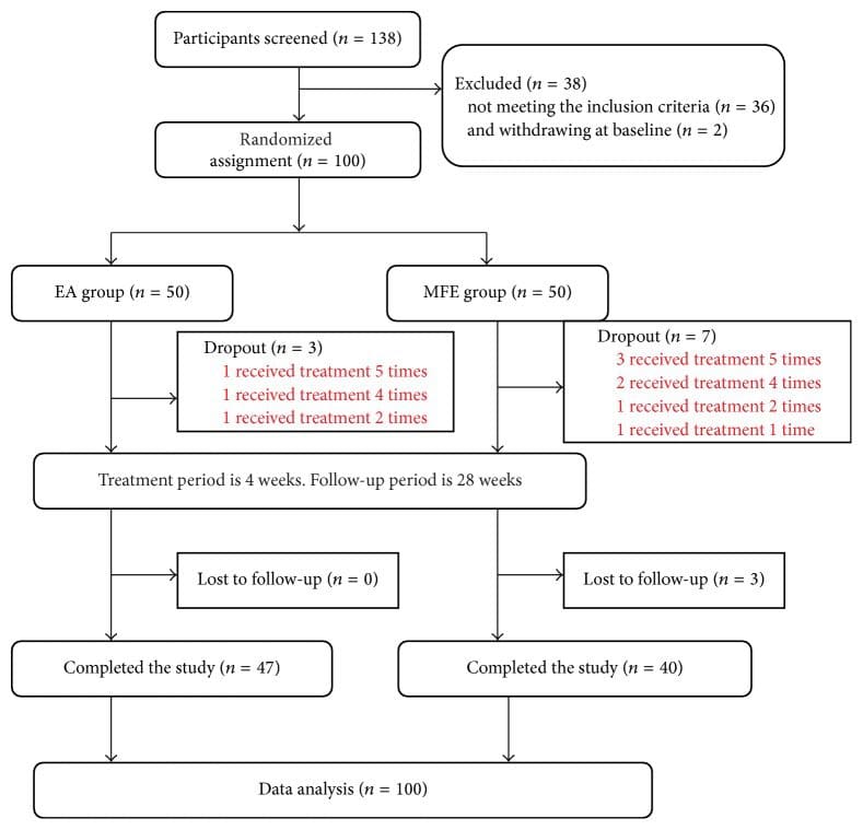
A total of 138 participants with chronic sciatica due to lumbar disc protrusion were screened, among whom 36 were rejected due to the exclusion criteria and 2 withdrew from the study. Therefore, 100 eligible patients were randomly assigned to the experimental (EA) group (n = 50) or the control (MFE) group (n = 50) at a ratio of 1?:?1. Eight participants withdrew from the study during the course of treatment due to the presence of aggravating symptoms, 1 participant exited the study due to travel, 1 participant withdrew due to an unsatisfactory curative effect, and 3 participants were lost to follow-up. In the dropout participants, no additional data except for the leg pain NRS scores were available because the evaluation period was not reached. According to the principle of ITT analysis, we analyzed the data of all 100 subjects for the leg pain NRS scores and then performed a sensitivity analysis of these 13 subjects to verify the reliability of the results. Details are provided in Figures ?1 and ?2.


Figure 1 Time Frame of Each Period

Figure 1: Time frame of each period. Figure 1 shows the time frame of baseline period, treatment period, and follow-up period.


Figure 2 Study Flow Diagram

Figure 2: Study flow diagram.


Characteristics of the Participants

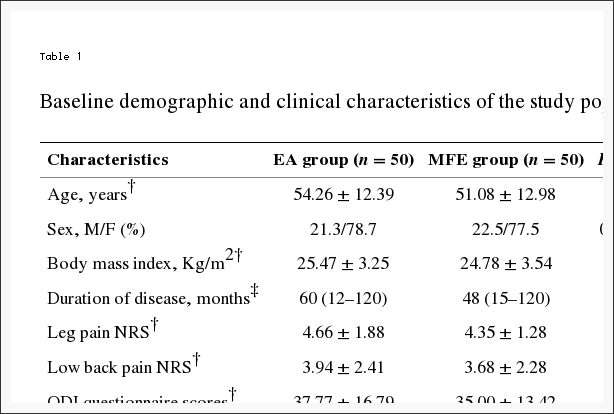
Table 1 shows the baseline data of the 100 participants. The mean age of all patients was 52.67 � 12.72 years. The mean duration was 48 (12�120) months. The duration of 2 participants in the electroacupuncture group was one month, and the duration of 1 participant was one month in the MFE group. The baseline demographics, body measurement data, and baseline outcomes are listed in Table 1. No significant differences in baseline demographics and clinical characteristics were observed (Table 1).

Table 1 Baseline Demographic and Clinical Characteristics of the Study Population

Table 1: Baseline demographic and clinical characteristics of the study population.

Primary Outcome

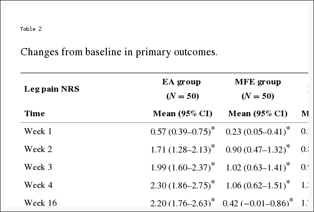
The decrease in the leg pain NRS scores from baseline to week 4 differed significantly between the EA group (n = 50) and the MFE group (n = 50) (P < 0.001). As shown in Table 2, the mean change from baseline to the 4th week in the average leg pain intensity NRS score was 2.30 (1.86�2.75) in the EA group and 1.06 (0.62�1.51) in the MFE group. At four weeks, the two groups both exhibited significantly greater reductions in NRS scores compared with baseline; however, the EA group showed a more significant decrease than the MFE group (Table 2).

Table 2 Changes from Baseline in Primary Outcomes

Table 2: Changes from baseline in primary outcomes.

Secondary Outcomes

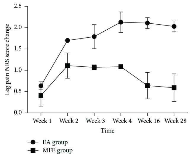
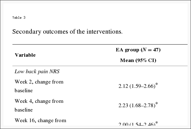
EA showed a more significant improvement in the leg pain scores at all the evaluation points compared with that observed in the MFE group (P < 0.001) (Figure 3 and Table 2). The EA group showed a significant decrease compared to the baseline in the leg pain, low back pain, and ODI scores at weeks 2, 4, 16, and 28 (all P < 0.05). Conversely, the MFE group did not show a significant improvement compared to the baseline in the low back pain score at weeks 16 and 28 (all P = 0.096). Significant reductions in the leg pain and ODI questionnaire scores were detected in the EA group at multiple time points compared with the MFE group (all P < 0.05). The EA group exhibited greater improvement. However, a negligible change was detected at multiple time points in the low back pain score and PGI between the two groups (all P > 0.05). Furthermore, no significant difference was detected in the frequency of drug use between the two groups at weeks 2 and 4 (all P > 0.05) in our trial. Consequently, an EA or MFE acceptance assessment administered after 4 weeks of intervention showed that EA was accepted as readily as MFE with no significant differences between the two groups (P = 0.055). The corresponding data are shown in Tables ?2 and ?3.


Figure 3 Change of Leg Pain Score in Two Groups

Figure 3: Change of leg pain score in two groups.


Table 3 Secondary Outcomes of the Interventions

Table 3: Secondary outcomes of the interventions.

A sensitivity analysis was performed based on the leg pain NRS score. We excluded 13 participants who received fewer treatment sessions (less than 10) and analyzed the data of the remaining 87 participants. This sensitivity analysis result showed that our original results were stable and reliable.

Adverse Events

No serious adverse events occurred in either group. One participant (2%) in the experimental group developed a subcutaneous hematoma. Two participants (4%) in the MFE group reported skin redness and itching. All adverse events disappeared without additional intervention.

Discussion

The results of this trial showed significant differences in the change in the leg pain NRS and ODI questionnaire scores in the EA group compared with those in the MFE group in the short-term treatment period and long-term follow-up. However, the EA group did not show a greater decrease in low back pain scores and PGI compared with the MFE group. These changes indicated that the effect of EA was superior to the effect of MFE in improving leg pain and dysfunction, whereas the effect of EA was not superior to that of MFE in relieving low back pain and systemic symptoms.

The leg pain NRS score showed a significant difference compared with the MFE group at week 4: a mean difference of 1.24 points was detected between the two groups. On average, a reduction of approximately 2�3.5 points in the NRS score represents a minimal clinically important difference (MCID) for acute and chronic pain. The change in the leg pain NRS score in the EA group at week 4 did not show a clinically important significant difference compared with the MFE group. However, our control group was not a placebo but a positive treatment. An effect size of 1.24 is generally considered as the large effect. The MCID of the ODI score ranged from 4 to 16 points, and the decline of the ODI score in the EA group reached the MCID criterion with a mean reduction of 5.69 compared with the MFE group. The results implied that the clinical effect of EA appears superior to the effect of MFE in improving dysfunction caused by sciatica. However, low back pain did not show a significant and clinically important difference, with a mean reduction in the NRS score of 0.58 at week 4 compared with the MFE group. It may be associated with a better response to pain around the electrodes by MFE. In our study, a long-term follow-up was performed. At week 28, the MFE group did not show significantly decreased leg pain compared to the baseline, whereas the EA group showed significantly decreased leg pain compared to the baseline. The difference between the two groups was significant. The results implied that the effect of EA but not MFE lasted at least 28 weeks. The low back pain and ODI scores also indicated that the long-term effects of EA were superior to those of MFE because the effects of EA persisted after the discontinuation of treatment.

In our trial, the leg pain NRS score was reduced by 49% compared with the baseline in the EA group at week 4; however, a greater increase in the response rate (69%) was reported in a trial comparing EA with TENS for sciatica during the treatment period. Another trial conducted in China demonstrated that the decrease in the mean value of the leg pain intensity NRS score in the EA group was 4.65 � 6.37 at week 4, which was higher than the value of 2.30 (1.86�2.75) obtained in our trial at the same time point. In a pilot trial comparing EA with physical therapy for symptomatic lumbar spinal stenosis (LSS), pain in the back and leg showed small improvements at 3 months. However, the ODI scores were different from the scores obtained in our study. No significant differences between the ODI scores of the two groups were observed at the 3-month follow-up time point in the study. The differences between the results of the two studies might be explained by the use of different acupoints, needling depth, manipulation methods, EA parameters, number and frequency, training and clinical experience level of the practitioners, missing data, and sample size.

Very few participants in either group took analgesics during the trial, and only anti-inflammatory drugs were used. This result might indicate that most of the participants believed that the analgesics would not alleviate pain and were concerned about adverse events. Most participants expected that EA or MFE would be beneficial and were aware that these techniques are relatively safe. According to the PGI, the participants perceived no difference between EA and MFE. Approximately 87.2% of the participants in the EA group reported that they were aided by EA at the 4th week, which was similar to the 83.5% of participants in the MFE group. The treatment acceptance assessment showed that none of the participants considered either treatment difficult to accept. Furthermore, 70.2% of the participants in the EA group reported that EA was easy or very easy to accept, similar to 72.5% of the participants in the MFE group. These results indicated that EA and MFE were both easy to accept and popular in China.

Leg pain is a typical symptom in sciatica patients, and the leg pain intensity NRS score reflects the improvement in this symptom in these patients. The leg pain NRS score may reasonably be used for the primary measurement of the therapeutic effect. Because studies have shown that most acupuncture therapy for sciatica lasts 1 to 4 weeks, we selected the change in the average leg pain intensity NRS score from baseline to the 4th week as the main measurement. In the previous literature, although primary outcome was generally measured at a certain time point, the average score reflected the average level of pain during the last week, which was thus more meaningful than other methods of measuring single time point due to recurrence of sciatica. The control group underwent MFE, which exerts its effect via the stimulation or activation of physiological events by applying energy, thereby producing therapeutic benefits that facilitate pain relief. Mechanisms leading to pain relief may be due to a variety of peripheral effects of control activity, on the spinal and spinal nervous system. The comparison between EA and MFE may reveal differences in response to needle penetration using the same electrostimulation. Because the stimulation parameters, particularly the frequency, are important factors that affect the outcome and because the effect of medium-frequency electrotherapy is better than the effect of low frequency electrotherapy, we used the same medium frequency and location in the study to ensure that the two groups were comparable.

Many studies have investigated the mechanism of EA. EA has been reported to relieve the symptoms of sciatica and increase the pain threshold in humans. Several previous studies showed that EA inhibited the primary afferent transmission of neuropathic pain and that deep EA stimulation improved the pathological changes and function of the injured sciatic nerve in rats. Other studies have suggested that descending inhibitory control, changes in nerve blood flow, or the inhibition of activity by nerve endings may be involved in the mechanism associated with the efficacy of EA. Long-lasting alleviation of pain has been suggested to be closely related to the muscle tension improvement provided by EA. A meta-analysis of patients with chronic pain showed that approximately 90% of the benefit of acupuncture was sustained at 12 months. The reason for the cumulative and sustained effects of acupuncture may be associated with the brain response and the cumulative duration of acupuncture stimulation.

This trial has several limitations. First, the participants and acupuncturists could not be blinded due to the significant difference between the two treatments. However, we followed rigorous quality control procedures in other aspects of the methodology. For example, a strict randomization and allocation concealment protocol was adopted. The outcome assessors and statisticians were blinded to the allocation. Second, some of the outcome measures of the trial were subjective. To address subjectivity, a short training session for the patients on the outcome reporting was held before they began the trial, and all subjective outcomes were based on the patient self-report forms. Third, we did not include a placebo control in the present preliminary study because several sham acupuncture randomized controlled trials (RCTs) have been performed to study acupuncture therapy in patients with sciatica. We considered that the use of a placebo did not provide sufficient sensitivity and may not have met ethical guidelines. Fourth, because we did not explore the effect of electroacupuncture EA on various degrees of pain severity, which degree of sciatica was most sensitive to EA was unclear. Subgroup analyses based on sciatica severity should be performed in a future multicenter, large-sample, randomized controlled study.

Conclusions: Electroacupuncture

This randomized controlled clinical trial demonstrated that the short-term and long-term effectiveness of electroacupuncture were superior to those of MFE in improving the symptoms of leg pain and dysfunction caused by chronic discogenic sciatica; moreover, the long-term effect of electroacupuncture was superior to that of MFE in improving low back pain. The results also suggested that the effect of electroacupuncture but not MFE lasted at least 28 weeks. No serious adverse events occurred in either group. Further studies are needed to examine the effectiveness of electroacupuncture relative to various physical therapy methods for patients with discogenic sciatica.

Acknowledgments

This trial was supported by the South Area of Guang’anmen Hospital science fund (Funding no. Y2015-07).

Conflicts of Interest

The authors have no conflicts of interest to declare.

In conclusion,�the effectiveness of electroacupuncture was superior to MFE towards improving the symptoms of discogenic sciatica. Although the research study concluded that electroacupuncture was superior to medium-frequency electrotherapy when treating for sciatica caused by degenerative disc disease, both types of treatments should still be used accordingly, depending on the patient’s condition and whether the specific treatment is best recommended by a healthcare professional. Electroacupuncture and medium-frequency electrotherapy are the most common types of treatment options for symptoms of sciatica associated with the deterioration of the spinal intervertebral discs. Information referenced from the National Center for Biotechnology Information (NCBI). The scope of our information is limited to chiropractic as well as to spinal injuries and conditions. To discuss the subject matter, please feel free to ask Dr. Jimenez or contact us at 915-850-0900 .

Curated by Dr. Alex Jimenez


Green-Call-Now-Button-24H-150x150-2-3.png

Additional Topics: Sciatica

Sciatica is referred to as a collection of symptoms rather than a single type of injury or condition. The symptoms are characterized as radiating pain, numbness and tingling sensations from the sciatic nerve in the lower back, down the buttocks and thighs and through one or both legs and into the feet. Sciatica is commonly the result of irritation, inflammation or compression of the largest nerve in the human body, generally due to a herniated disc or bone spur.

blog picture of cartoon paperboy big news


IMPORTANT TOPIC: EXTRA EXTRA: Treating Sciatica Pain


blank
References:

1. Konstantinou K., Dunn K. M. Sciatica: review of epidemiological studies and prevalence estimates. Spine. 2008;33(22):2464�2472. doi: 10.1097/brs.0b013e318183a4a2. [PubMed] [Cross Ref]
2. Ergun T., Lakadamyali H. CT and MRI in the evaluation of extraspinal sciatica. British Journal of Radiology. 2010;83(993):791�803. doi: 10.1259/bjr/76002141. [PMC free article] [PubMed] [Cross Ref]
3. Dagenais S., Caro J., Haldeman S. A systematic review of low back pain cost of illness studies in the United States and internationally. Spine Journal. 2008;8(1):8�20. doi: 10.1016/j.spinee.2007.10.005. [PubMed] [Cross Ref]
4. Asche C. V., Kirkness C. S., McAdam-Marx C., Fritz J. M. The societal costs of low back pain: data published between 2001 and 2007. Journal of Pain and Palliative Care Pharmacotherapy. 2007;21(4):25�33. doi: 10.1300/j354v21n04_06. [PubMed] [Cross Ref]
5. Saal J. A., Saal J. S. Nonoperative treatment of herniated lumbar intervertebral disc with radiculopathy. An outcome study. Spine. 1989;14(4):431�437. doi: 10.1097/00007632-198904000-00018. [PubMed] [Cross Ref]
6. Kreiner D. S., Hwang S. W., Easa J. E., et al. An evidence-based clinical guideline for the diagnosis and treatment of lumbar disc herniation with radiculopathy. Spine Journal. 2014;14(1):180�191. doi: 10.1016/j.spinee.2013.08.003. [PubMed] [Cross Ref]
7. Alexandre A., Cor� L., Azuelos A., Pellone M. Percutaneous nucleoplasty for discoradicular conflict. Acta Neurochirurgica. Supplement. 2005;92:83�86. [PubMed]
8. Zhu R. S., Ren Y. M., Yuan J. J., et al. Does local lavage influence functional recovery during lumber discectomy of disc herniation? One year’s systematic follow-up of 410 patients. Medicine. 2016;95(42) doi: 10.1097/md.0000000000005022.e5022 [PMC free article] [PubMed] [Cross Ref]
9. Nguyen C., Palazzo C., Grabar S., et al. Tumor necrosis factor-a? blockade in recurrent and disabling chronic sciatica associated with post-operative peridural lumbar fibrosis: results of a double-blind, placebo randomized controlled study. Arthritis Research and Therapy. 2015;17(1, article 330) doi: 10.1186/s13075-015-0838-4. [PMC free article] [PubMed] [Cross Ref]
10. Haghnegahdar A., Sedighi M. An outcome study of anterior cervical discectomy and fusion among Iranian population. Neuroscience Journal. 2016;2016:7. doi: 10.1155/2016/4654109.4654109 [PMC free article] [PubMed] [Cross Ref]
11. Lewis R., Williams N., Matar H. E., et al. The clinical effectiveness and cost-effectiveness of management strategies for sciatica: systematic review and economic model. Health Technology Assessment. 2011;15(39):1�578. doi: 10.3310/hta15390. [PMC free article] [PubMed] [Cross Ref]
12. Chou R., Deyo R., Friedly J., et al. Noninvasive Treatments for Low Back Pain. Rockville, Md, USA: Agency for Healthcare Research and Quality; 2016. AHRQ comparative effectiveness reviews.
13. Robaina-Padr�n F. J. Controversies of instrumented surgery and pain relief in degenerative lumbar spine. Results of scientific evidence. Neurocirugia. 2007;18(5):406�413. doi: 10.1016/S1130-1473(07)70266-X. [PubMed] [Cross Ref]
14. Qin Z., Liu X., Wu J., Zhai Y., Liu Z. Effectiveness of acupuncture for treating sciatica: a systematic review and meta-analysis. Evidence-Based Complementary and Alternative Medicine. 2015;2015 doi: 10.1155/2015/425108.425108 [PMC free article] [PubMed] [Cross Ref]
15. Ji M., Wang X., Chen M., Shen Y., Zhang X., Yang J. The efficacy of acupuncture for the treatment of sciatica: a systematic review and meta-analysis. Evidence-Based Complementary and Alternative Medicine. 2015;2015:12. doi: 10.1155/2015/192808.192808 [PMC free article] [PubMed] [Cross Ref]
16. Pang T., Lu C., Wang K., et al. Electroacupuncture at ST25 inhibits cisapride-induced gastric motility in an intensity-dependent manner. Evidence-based Complementary and Alternative Medicine. 2016;2016:7. doi: 10.1155/2016/3457025.3457025 [PMC free article] [PubMed] [Cross Ref]
17. MacPherson H., Vertosick E., Foster N., et al. The persistence of the effects of acupuncture after a course of treatment: a meta-analysis of patients with chronic pain. Pain. 2016 doi: 10.1097/j.pain.0000000000000747. [PMC free article] [PubMed] [Cross Ref]
18. MacPherson H., Altman D. G., Hammerschlag R., et al. Revised STandards for Reporting Interventions in Clinical Trials of Acupuncture (STRICTA): extending the CONSORT statement. Journal of Evidence-Based Medicine. 2010;3(3):140�155. doi: 10.1111/j.1756-5391.2010.01086.x. [PubMed] [Cross Ref]
19. Qiu T., Li L. Discussion on the Chinese edition of the WHO standard acupuncture point locations in the western Pacific region. Chinese Acupuncture and Moxibustion. 2011;31(9):827�830. [PubMed]
20. Selim A. J., Ren X. S., Fincke G., et al. The importance of radiating leg pain in assessing health outcomes among patients with low back pain: results from the veterans health study. Spine. 1998;23(4):470�474. doi: 10.1097/00007632-199802150-00013. [PubMed] [Cross Ref]
21. Fairbank J. C. T., Pynsent P. B. The oswestry disability index. Spine. 2000;25(22):2940�2953. doi: 10.1097/00007632-200011150-00017. [PubMed] [Cross Ref]
22. Lee J., Shin J.-S., Lee Y. J., et al. Effects of Shinbaro pharmacopuncture in sciatic pain patients with lumbar disc herniation: study protocol for a randomized controlled trial. Trials. 2015;16(1, article 455) doi: 10.1186/s13063-015-0993-6. [PMC free article] [PubMed] [Cross Ref]
23. Ostelo R. W. J. G., de Vet H. C. W. Clinically important outcomes in low back pain. Best Practice and Research. Clinical Rheumatology. 2005;19(4):593�607. doi: 10.1016/j.berh.2005.03.003. [PubMed] [Cross Ref]
24. Farrar J. T., Young J. P., Jr., LaMoreaux L., Werth J. L., Poole R. M. Clinical importance of changes in chronic pain intensity measured on an 11-point numerical pain rating scale. Pain. 2001;94(2):149�158. doi: 10.1016/S0304-3959(01)00349-9. [PubMed] [Cross Ref]
25. Wang Z.-X. Clinical observation on electroacupuncture at acupoints for treatment of senile radical sciatica. Chinese Acupuncture & Moxibustion. 2009;29(2):126�128. [PubMed]
26. Yuan W. A., Huang S. R., Guo K., et al. Integrative TCM conservative therapy for low back pain due to lumbar disc herniation: a randomized controlled clinical trial. Evidence-Based Complementary and Alternative Medicine. 2013;2013:8. doi: 10.1155/2013/309831.309831 [PMC free article] [PubMed] [Cross Ref]
27. Kim K. H., Kim Y. R., Baik S. K., et al. Acupuncture for patients with lumbar spinal stenosis: a randomised pilot trial. Acupuncture in Medicine. 2016;34(4):267�274. doi: 10.1136/acupmed-2015-010962. [PubMed] [Cross Ref]
28. Tiktinsky R., Chen L., Narayan P. Electrotherapy: yesterday, today and tomorrow. Haemophilia. 2010;16, supplement 5:126�131. doi: 10.1111/j.1365-2516.2010.02310.x. [PubMed] [Cross Ref]
29. Zhao X.-Y., Zhang Q.-S., Yang J., et al. The role of arginine vasopressin in electroacupuncture treatment of primary sciatica in human. Neuropeptides. 2015;52:61�65. doi: 10.1016/j.npep.2015.06.002. [PubMed] [Cross Ref]
30. Wang W.-S., Tu W.-Z., Cheng R.-D., et al. Electroacupuncture and A-317491 depress the transmission of pain on primary afferent mediated by the P2X3 receptor in rats with chronic neuropathic pain states. Journal of Neuroscience Research. 2014;92(12):1703�1713. doi: 10.1002/jnr.23451. [PubMed] [Cross Ref]
31. Liu Y.-L., Li Y., Ren L., et al. Effect of deep electroacupuncture stimulation of �Huantiao� (GB 30) on changes of function and nerve growth factor expression of the injured sciatic nerve in rats. Zhongguo Yi Xue Ke Xue Yuan Yi Xue Qing Bao Yan Jiu Suo Bian Ji. 2014;39(2):93�99. [PubMed]
32. Inoue M., Hojo T., Yano T., Katsumi Y. Electroacupuncture direct to spinal nerves as an alternative to selective spinal nerve block in patients with radicular sciatica�a cohort study. Acupuncture in Medicine. 2005;23(1):27�30. doi: 10.1136/aim.23.1.27. [PubMed] [Cross Ref]
33. Fan Y., Wu Y. Effect of electroacupuncture on muscle state and infrared thermogram changes in patients with acute lumbar muscle sprain. Journal of Traditional Chinese Medicine. 2015;35(5):499�506. [PubMed]
34. Li C., Yang J., Park K., et al. Prolonged repeated acupuncture stimulation induces habituation effects in pain-related brain areas: an fMRI study. PLoS ONE. 2014;9(5) doi: 10.1371/journal.pone.0097502.e97502 [PMC free article] [PubMed] [Cross Ref]
35. Khadilkar A., Odebiyi D. O., Brosseau L., Wells G. A. Transcutaneous electrical nerve stimulation (TENS) versus placebo for chronic low-back pain. Cochrane Database of Systematic Reviews. 2008;4(4)CD003008 [PubMed]

Close Accordion
Vertebral Artery Dissection Found During Chiropractic Examination

Vertebral Artery Dissection Found During Chiropractic Examination

Acknowledging the subsequent information below,�approximately more than 2 million people are injured in automobile accidents each year and among those incidents, the majority of the people involved are diagnosed with whiplash and/or neck injury by a healthcare professional. When the complex structure of the neck is subjected to trauma, tissue damage and other medical complications may occur. Vertebral artery dissection, or VAD, is characterized by a flap-like tear on the inner lining of the vertebral artery in charge of supplying blood to the brain. After the tear, blood can then enter the arterial wall and form a blood clot, thickening the artery wall and often impeding blood flow.


Through years of experience practicing chiropractic care,�VAD may often follow after trauma to the neck, such as that which occurs in an automobile accident, or whiplash injury. The symptoms of vertebral artery dissection include head and neck pain as well as intermittent or permanent stroke symptoms, such as difficulty speaking, impaired coordination and loss of vision. VAD, or vertebral artery dissection, is generally diagnosed with a contrast-enhanced CT or MRI scan.


Abstract


A 30-year-old woman presented to an emergency department with sudden onset of transient loss of left peripheral vision. Owing to a history of migraine headaches, she was released with a diagnosis of ocular migraine. Two days later, she sought chiropractic care for the chief symptom of severe neck pain. The chiropractor suspected the possibility of vertebral artery dissection (VAD). No manipulation was performed; instead, MR angiography (MRA) of the neck was obtained, which revealed an acute left VAD with early thrombus formation. The patient was placed on aspirin therapy. Repeat MRA of the neck 3?months later revealed resolution of the thrombus, without progression to stroke. This case illustrates the importance for all healthcare providers who see patients with neck pain and headache to be attentive to the symptomatic presentation of possible VAD in progress.


Background


Vertebral artery dissection (VAD) leading to stroke is an uncommon but potentially serious disorder. The incidence of stroke related to the vertebrobasilar system varies from 0.75 to 1.12/100?000 person-years. The pathological process in VAD typically involves dissection of the wall of the artery followed sometime later by thrombus formation, which may cause arterial occlusion or may lead to embolisation, causing occlusion of one or more of the distal branches off the vertebral artery, including the basilar artery, which can be catastrophic. VAD typically occurs in patients who have an inherent, transitory weakness in the arterial wall. In at least 80% of cases, the initial symptoms include neck pain with or without headache.


Many patients with VAD may in the early stages present to chiropractors seeking relief from neck pain and headache, without realising they are experiencing VAD. In many of these cases, the patient later develops a stroke. Until recently, it was assumed that the dissection (and subsequent stroke) was caused by cervical manipulative therapy (CMT). However, while early studies found an association between visits to a chiropractor and subsequent stroke related to VAD, recent data suggest that this relationship is not causal.


This case report is illustrative of the scenario in which a patient with an undiagnosed VAD in evolution consulted a chiropractor for neck pain and headache. After thorough history and examination, the chiropractor suspected VAD and did not perform CMT. Instead, the patient was referred for further evaluation, which detected a VAD in progress. Prompt diagnosis and anticoagulant treatment were thought to have averted progression to a stroke.


Case Presentation


A 30-year-old otherwise healthy woman consulted a chiropractor (DBF), reporting of right-sided neck pain in the suboccipital region. The patient reported that, 3?days previously, she had gone to the local hospital emergency department (ED) because of the sudden onset of loss of left peripheral vision. The visual symptoms interfered with her ability to see through her left eye; this was accompanied by �numbness� in her left eyelid. About 2?weeks prior to this ED visit, she had experienced an episode of acute left-sided neck pain with severe left-sided headache. She also related a history of migraine headache without prodrome. She was released from the ED with a tentative diagnosis of ocular migraine. She had never been previously diagnosed with ocular migraine, nor had she ever experienced any visual disturbances with her previous migraines.


Shortly after the left-sided ocular symptoms resolved, she suddenly developed right-sided neck pain without provocation, for which she sought chiropractic treatment. She also reported a transient episode of right-sided visual disturbance occurring that same day as well. This was described as sudden blurriness that was of short duration and resolved spontaneously earlier in the day of her presentation for chiropractic examination. When she presented for the initial chiropractic examination, she denied current visual disturbance. She said that she was not experiencing any numbness, paraesthesia or motor loss in the upper or lower extremities. She denied ataxia or difficulty with balance. Medical history was remarkable for childbirth 2� months prior to initial presentation. She stated that her migraine headaches were associated with her menstrual cycle. Family history was remarkable for a spontaneous ascending thoracic aortic aneurysm in her older sister, who was about 30?years of age when her aneurysm had occurred.


Investigations


Based on the history of sudden onset of severe upper cervical pain and headache with visual disturbance and ocular numbness, the DC was concerned about the possibility of early VAD. Urgent MR angiography (MRA) of the neck and head, along with MRI of the head, was ordered. No cervical spine examination or manipulation was performed because of the suspicion that the neck pain was related to VAD rather than to a �mechanical� cervical disorder.


MRA of the neck demonstrated that the left vertebral artery was small and irregular in calibre, extending from the C7 level cephalad to C2, consistent with dissection. There was a patent true lumen with a surrounding cuff of T1 hyper-intensity, consistent with dissection with subintimal thrombus within the false lumen (Figures 1 and ?2). MRI of the head with and without contrast, and MRA of the head without contrast, were both unremarkable. Specifically, there was no intracranial extension of dissection or evidence of infarction. MR perfusion of the brain revealed no focal perfusion abnormalities.


Figure 1 Axial Proton Density Image - Image 1

Figure 1: Axial proton density image demonstrates circumferential hyper-intensity surrounding the left cervical vertebral artery (representing the false lumen). Note decreased calibre of true lumen (black flow void) with respect to the right vertebral artery.


Figure 2 Axial Image from Three Dimensional Time of Flight MRA - Image 2

Figure 2: Axial image from three-dimensional time-of-flight MRA demonstrates T1 hypointense dissection flap separating the true lumen (lateral) from the false lumen (medial). MRA, MR angiography.


Differential Diagnosis


The ED released the patient with a tentative diagnosis of ocular migraine, due to her history of migraine headaches. However, the patient stated that the left-sided headache was atypical��like nothing I’ve ever experienced before.� Her previous migraines were associated with her menstrual cycle, but not with any vision changes. She had never been previously diagnosed with ocular migraine. MRA of the cervical region revealed that the patient actually had an acute dissection with thrombus formation in the left vertebral artery.


Treatment


Owing to the potential of impending stroke associated with an acute VAD with thrombus formation, the patient was admitted to the neurology stroke service for close neurological monitoring. During her admission, the patient did not experience any recurrence of neurological deficits and her headaches improved. She was discharged the following day with a diagnosis of left VAD and transient ischaemic attack. She was instructed to avoid vigorous exercise and trauma to the neck. Daily aspirin (325?mg) was prescribed, to be continued for 3�6?months after discharge.


Outcome and Follow-Up


After discharge from the stroke service, the patient had no recurrence of headache or visual disturbances, and her posterior neck pain symptoms resolved. Repeat imaging was performed 3?months after presentation, which demonstrated improved calibre of the cervical left vertebral artery with resolution of the thrombus within the false lumen (Figure 3). Imaging of the intracranial compartment remained normal, without evidence of interval infarction or perfusion asymmetry.


Figure 3 Maximum Intensity Projection MIP Images - Image 3

Figure 3: Maximum intensity projection (MIP) images from three-dimensional time-of-flight MRA (left image is at time of presentation and right image is at 3-month follow-up). The initial imaging demonstrates markedly diminutive calibre of the left vertebral artery


Discussion


The pathophysiological process of VAD is thought to start with degeneration of the tissues at the medial-adventitial border of the vertebral artery, leading to the development of microhaematomata within the wall of the artery and, eventually, arterial tear. This can lead to leakage of blood into the arterial wall, causing occlusion of the lumen with subsequent thrombus formation and embolisation, resulting in stroke related to one of the branches of the vertebral artery. This pathological process is similar to that of spontaneous carotid artery dissection, spontaneous thoracic aortic dissection and spontaneous coronary artery dissection. All these conditions tend to occur in younger adults and some have speculated that they may be part of a common inherited pathophysiological process. Notable in this case is the fact that the patient’s older sister had experienced a spontaneous thoracic aortic aneurysm (probably a dissection) at around the same age (30?years) as this patient was when she experienced her VAD.


While the dissection is often sudden, the luminal compromise and complications of VAD can develop gradually leading to variable symptoms and presentation, depending on the stage of the disease. The dissection itself, which develops some time before the onset of neural ischaemia, can cause stimulation of nociceptive receptors within the artery, producing pain that is most commonly felt in the upper cervical spine or head. Only after the pathophysiological process progresses to the point of complete arterial occlusion or thrombus formation with distal embolisation does the full manifestation of infarction occur. However, as illustrated in this case, neurological symptoms can develop early in the process, particularly in cases in which the true lumen demonstrates significant calibre decrease secondary to compression.


There are several interesting aspects to this case. First, it highlights the importance of spine clinicians being alert to the possibility that what may appear to be typical �mechanical� neck pain could be something potentially more sinister, such as VAD. The sudden onset of severe suboccipital pain, with or without headache, and accompanying brainstem related neurological symptoms, should alert the clinician to the possibility of VAD. As in the case reported here, patients with a history of migraine will typically describe the headache as different from their usual migraine. A careful neurological examination should be performed, looking for possible subtle neurological deficits, although the neurological examination will often be negative in the early stages of VAD.


Second, a triad of symptoms raised concern that the patient might be experiencing a VAD in progress. The symptom triad included: (1) spontaneous onset of severe upper cervical pain; (2) severe headache that was distinctly different from the patient’s usual migraine headaches; and (3) brainstem-related neurological symptoms (in the form of transient visual disturbance). Notably, careful neurological examination was negative. Nonetheless, the history was of sufficient concern to prompt immediate investigation.


When VAD is suspected but no frank signs of stroke are present, immediate vascular imaging is indicated. While the optimal imaging evaluation of VAD remains controversial, MRA or CTA are the diagnostic studies of choice given their excellent anatomic delineation and ability to evaluate for complications (including infarction and changes in brain perfusion). Some advocate the use of Doppler ultrasound; however, it has limited utility given the course of the vertebral artery in the neck and limited evaluation of the vertebral arteries cephalad to the origin. Additionally, ultrasound imaging is unlikely to allow visualisation of the dissection itself and thus can be negative in the absence of significant arterial occlusion.


Third, this case is interesting in light of the controversy about cervical manipulation as a potential �cause� of VAD. While case reports have presented patients who have experienced stroke related to VAD after cervical manipulation, and case�control studies have found a statistical association between visits to chiropractors and stroke related to VAD, further investigation has indicated that the association is not causal. Cassidy et al found that a patient who experiences stroke related to VAD is just as likely to have visited a primary care practitioner as to have visited a chiropractor prior to having the stroke. The authors suggested that the most likely explanation for the statistical association between visits to chiropractors and subsequent VAD is that a patient who experiences the initial symptoms of VAD (neck pain with or without headache) seeks medical attention for these symptoms (from a chiropractor, primary care practitioner, or another type of practitioner), then subsequently experiences the stroke, independent of any action taken by the practitioner.


It is important to note that, while there have been reported cases of carotid artery dissection after cervical manipulation, case�control studies have not found this association. The initial symptoms of carotid dissection (neurological symptoms, with neck and head pain less common than VAD), aortic dissection (sudden onset of severe, �tearing� pain) and coronary artery dissection (acute severe chest pain, ventricular fibrillation) are likely to cause the individual to immediately seek ED care, rather than seek chiropractic care. However, VAD has seemingly benign initial symptoms�neck pain and headache�which are symptoms that commonly cause patients to seek out chiropractic care. This may explain why only VAD is associated with visits to chiropractors, while these other types of dissections are not; patients with these other conditions, which have much more alarming symptoms, simply do not present to chiropractors.


This case is a good example of a patient with VAD in progress presenting to a chiropractor for the purpose of seeking relief from neck pain. Fortunately, the chiropractor was astute enough to ascertain that the patient’s symptoms were not suggestive of a �mechanical� cervical spine disorder, and appropriate diagnostic investigation was performed. However, if manipulation had been performed, the VAD that was already in progress from natural history may have been blamed on manipulation, after being detected on MRA imaging. Fortunately, in this case, the chiropractor was able to assist with early detection and treatment, and subsequently a stroke was likely averted.


Learning Points


  • A case is presented in which a patient saw a chiropractor, while seeking treatment for neck pain, and the history raised concern for possible vertebral artery dissection (VAD).
  • Rather than providing manipulative treatment, the chiropractor referred the patient for advanced imaging, which confirmed the diagnosis of VAD.
  • The case illustrates the importance of paying attention to subtle historical factors in patients with VAD.
  • It also serves as an example of a patient with a VAD in progress seeking the services of a chiropractor for the initial symptoms of the disorder.
  • In this case, early detection of the dissection occurred and the patient had a full recovery without any subsequent stroke.


Acknowledgments


The authors would like to acknowledge the assistance of Pierre Cote, DC, PhD, for his assistance with reviewing this manuscript.


Footnotes


Contributors: All the authors acknowledge that they have contributed the following to the submission of this manuscript: conception and design, drafting of the manuscript, critical revisions of the manuscript, literature review and references, and proof reading of the final manuscript.


Competing interests: None declared.


Patient consent: Obtained.


Provenance and peer review: Not commissioned; externally peer reviewed.


Information referenced from the National Center for Biotechnology Information (NCBI). The scope of our information is limited to chiropractic as well as to spinal injuries and conditions. To discuss the subject matter, please feel free to ask Dr. Jimenez or contact us at 915-850-0900 .


Cited by Dr. Alex Jimenez


Green-Call-Now-Button-24H-150x150-2-3.png


Additional Topics: Wellness


Overall health and wellness are essential towards maintaining the proper mental and physical balance in the body. From eating a balanced nutrition as well as exercising and participating in physical activities, to sleeping a healthy amount of time on a regular basis, following the best health and wellness tips can ultimately help maintain overall well-being. Eating plenty of fruits and vegetables can go a long way towards helping people become healthy.


blog picture of cartoon paperboy big news


IMPORTANT TOPIC: EXTRA EXTRA: Treating Sciatica Pain
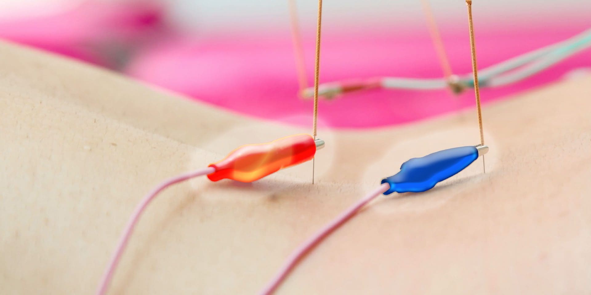


Blank
References
1.�Debette S, Leys D.�Cervical-artery dissections: predisposing factors, diagnosis, and outcome.�Lancet Neurol�2009;8:668�78.�doi:10.1016/S1474-4422(09)70084-5[PubMed]
2.�Boyle E, Cote P, Grier AR et al.�Examining vertebrobasilar artery stroke in two Canadian provinces.�Spine�2008;33(4 Suppl):S170�5.�doi:10.1097/BRS.0b013e31816454e0[PubMed]
3.�Lee VH, Brown RD Jr, Mandrekar JN et al.�Incidence and outcome of cervical artery dissection: a population-based study.�Neurology�2006;67:1809�12.�doi:10.1212/01.wnl.0000244486.30455.71[PubMed]
4.�Schievink WI.�Spontaneous dissection of the cartoid and vertebral arteries.�N Engl J Med�2001;344:898�906.�doi:10.1056/NEJM200103223441206[PubMed]
5.�Volker W, Dittrich R, Grewe S et al.�The outer arterial wall layers are primarily affected in spontaneous cervical artery dissection.�Neurology�2011;76:1463�71.�doi:10.1212/WNL.0b013e318217e71c[PubMed]
6.�Gottesman RF, Sharma P, Robinson KA et al.�Clinical characteristics of symptomatic vertebral artery dissection: a systematic review.�Neurologist�2012;18:245�54.�doi:10.1097/NRL.0b013e31826754e1[PMC free article][PubMed]
7.�Cassidy JD, Boyle E, Cote P et al.�Risk of vertebrobasilar stroke and chiropractic care: results of a population-based case-control and case-crossover study.�Spine�2008;33(4�Suppl):S176�83.�doi:10.1097/BRS.0b013e3181644600[PubMed]
8.�Rothwell DM, Bondy SJ, Williams JI.�Chiropractic manipulation and stroke: a population-based case-control study.�Stroke�2001;32:1054�60.�doi:10.1161/01.STR.32.5.1054[PubMed]
9.�Smith WS, Johnston SC, Skalabrin EJ et al.�Spinal manipulative therapy is an independent risk factor for vertebral artery dissection.�Neurology�2003;60:1424�8.�doi:10.1212/01.WNL.0000063305.61050.E6[PubMed]
10.�Volker W, Besselmann M, Dittrich R et al.�Generalized arteriopathy in patients with cervical artery dissection.�Neurology�2005;64:1508�13.�doi:10.1212/01.WNL.0000159739.24607.98[PubMed]
11.�Evangelista A, Mukherjee D, Mehta RH et al.�Acute intramural hematoma of the aorta: a mystery in evolution.�Circulation�2005;111:1063�70.�doi:10.1161/01.CIR.0000156444.26393.80[PubMed]
12.�Tweet MS, Hayes SN, Pitta SR et al.�Clinical features, management, and prognosis of spontaneous coronary artery dissection.�Circulation�2012;126:579�88.�doi:10.1161/CIRCULATIONAHA.112.105718[PubMed]
13.�Choi S, Boyle E, Cote P et al.�A population-based case-series of Ontario patients who develop a vertebrobasilar artery stroke after seeing a chiropractor.�J Manipulative Physiol Ther�2011;34:15�22.�doi:10.1016/j.jmpt.2010.11.001[PubMed]
14.�Naggara O, Louillet F, Touze E et al.�Added value of high-resolution MR imaging in the diagnosis of vertebral artery dissection.�AJNR Am J Neuroradiol�2010;31:1707�12.�doi:10.3174/ajnr.A2165[PubMed]
15.�Haynes MJ, Vincent K, Fischhoff C et al.�Assessing the risk of stroke from neck manipulation: a systematic review.�Int J Clin Pract�2012;66:940�7.�doi:10.1111/j.1742-1241.2012.03004.x[PMC free article][PubMed]
16.�Nebelsieck J, Sengelhoff C, Nassenstein I et al.�Sensitivity of neurovascular ultrasound for the detection of spontaneous cervical artery dissection.�J Clin Neurosci�2009;16:79�82.�doi:10.1016/j.jocn.2008.04.005[PubMed]
17.�Bendick PJ, Jackson VP.�Evaluation of the vertebral arteries with duplex sonography.�J Vasc Surg1986;3:523�30.�doi:10.1016/0741-5214(86)90120-5[PubMed]
18.�Murphy DR.�Current understanding of the relationship between cervical manipulation and stroke: what does it mean for the chiropractic profession?Chiropr Osteopat�2010;18:22�doi:10.1186/1746-1340-18-22[PMC free article][PubMed]
19.�Engelter ST, Grond-Ginsbach C, Metso TM et al.�Cervical artery dissection: trauma and other potential mechanical trigger events.�Neurology�2013;80:1950�7.�doi:10.1212/WNL.0b013e318293e2eb[PubMed]
20.�Peters M, Bohl J, Th�mke F et al.�Dissection of the internal carotid artery after chiropractic manipulation of the neck.�Neurology�1995;45:2284�6.�doi:10.1212/WNL.45.12.2284[PubMed]
21.�Nadgir RN, Loevner LA, Ahmed T et al.�Simultaneous bilateral internal carotid and vertebral artery dissection following chiropractic manipulation: case report and review of the literature.�Neuroradiology2003;45:311�14.�doi:10.1007/s00234-003-0944-x[PubMed]
22.�Dittrich R, Rohsbach D, Heidbreder A et al.�Mild mechanical traumas are possible risk factors for cervical artery dissection.�Cerebrovasc Dis�2007;23:275�81.�doi:10.1159/000098327[PubMed]
23.�Chung CL, Cote P, Stern P et al.�The association between cervical spine manipulation and carotid artery dissection: a systematic review of the literature.�J Manipulative Physiol Ther�2014; doi:10.1016/j.jmpt.2013.09.005�doi:10.1016/j.jmpt.2013.09.005[PubMed]
24.�Thomas LC, Rivett DA, Attia JR et al.�Risk factors and clinical features of craniocervical arterial dissection.�Man Ther�2011;16:351�6.�doi:10.1016/j.math.2010.12.008[PubMed]
25.�Klineberg E, Mazanec D, Orr D et al.�Masquerade: medical causes of back pain.�Cleve Clin J Med2007;74:905�13.�doi:10.3949/ccjm.74.12.905[PubMed]
Close Accordion
What are Case Reports & Case Series?

What are Case Reports & Case Series?

The diagnosis of a variety of diseases has been effectively determined through clinical and experimental data. Research studies provide valuable information on the pathogenesis of many conditions and are often the primary source of information regarding new diseases or conditions. Case reports and case series are first level research studies, offering the most initial insights on a particular health issue through the personal experience of one or more people with a disease or condition. The following article describes the purpose of case reports and case series, and how they provide clinical and experimental data.


Learning Objectives


1. Case reports and case series describe the experience of one or more people with a disease.
2. Case reports and case series are often the first data alerting to a new disease or condition.
3. Case reports and case series have specific limitations:

  • a. Lack of a denominator to calculate rates of disease
  • b. Lack of a comparison group
  • c. Selecting study populations
  • d. Sampling variation


Case Reports and Case Series


Case reports and case series represent the most basic type of study design, in which researchers describe the experience of a single person (case report) or a group of people (case series). Typically, case reports and case series describe individuals who develop a particular new disease or condition. Case reports and case series can provide compelling reading because they present a detailed account of the clinical experience of individual study subjects. In contrast, studies that evaluate large numbers of individuals typically summarize the data using statistical measures, such as means and proportions.


Example 3.1. A case series describes 15 young women who develop breast cancer; 9 of these women report at least once weekly ingestion of foods packaged with the estrogenic chemical bisphenol A (BPA). Urine testing confirms the presence of BPA among all nine case women.


It is tempting to surmise from these data that BPA might be causally related to breast cancer. However, case reports/case series have important limitations that preclude inference of a causal relationship.

First, case reports/case series lack denominator data that are necessary to calculate the rate of disease. The denominator refers to the population from which the diseased subjects arose. For example, to calculate the incidence proportion or incidence rate of breast cancer among women exposed to BPA, the total number of women who were exposed to BPA or the total number of person-years at risk is needed.


Table 1 - Incidence Proportion & Incidence Rate


Disease rates are needed for comparison with historically reported disease rates, or with rates from a selected comparison group. Unfortunately, obtaining the necessary denominator data may not be easy. In this example, additional data sources are needed to determine the total number of BPA-exposed women from whom the breast cancer cases arose. The case series data alone cannot be used to calculate the rate of breast cancer because they do not include the total number of women who were exposed to BPA.


A second problem with case report/case series report data is the lack of a comparison group. The 60% prevalence of BPA exposure among women with breast cancer seems unusually high, but what is prevalence of BPA exposure among women without breast cancer? This comparison is critical for addressing the hypothesis that BPA might be a cause of breast cancer.


A third limitation of case reports/case series is that these studies often describe highly select individuals who may not represent the general population. For example, it is possible that the 15 breast cancer cases originated from a single hospital in a community with high levels of air pollution or other potential carcinogens. Under these conditions, a fair estimate of breast cancer incidence among non-BPA exposed women from the same community would be required to make an inference that BPA causes breast cancer.


A fourth limitation of case reports/case series is sampling variation. This concept will be explored in detail later in this book. The basic idea is that there is tremendous natural variation in disease development in humans. The fact that 9 of 15 women with breast cancer reported BPA exposure is interesting; however, this number may be very different in the next case series of 15 women with breast cancer simply due to chance. A precise estimate of the rate of a disease, independent from chance, can be obtained only by increasing the number of diseased subjects.


Recall the list of factors that are used to judge whether a factor may be a cause of disease:


1. Randomized evidence
2. Strength of association
3. Temporal relationship between exposure and outcome
4. Dose-response association
5. Biological plausibility


In general, case reports/case series rely almost exclusively on biological plausibility to make their case for causation. For the BPA and breast cancer case series, there is no randomized evidence, no measure of the strength of association between BPA and breast cancer, no reported dose�response association, and no evidence that BPA exposure preceded the development of breast cancer. The inference for causation derives completely from previous biological knowledge regarding the estrogenic effects of BPA.


Despite limitations of case series data, they may be highly suggestive of an important new association, disease process, or unintended side effect of a medication or treatment.


Example 3.2. In 2007, a case series described three cases of male prepubertal gynecomastia. The report included detailed information on each subjects� age, body size, serum levels of endogenous steroids, and known exposures to exogenous hormones. It was discovered that all three otherwise healthy boys had been exposed to some product containing lavender oil (lotion, shampoo, soap), and that in each case, the gynecomastia resolved upon discontinuation of the product. Subsequent in vitro studies demonstrated endocrine-disrupting activity of lavender oil. This novel case series data may lead to further investigations to determine whether lavender oil, a common ingredient in commercially available products, may be a cause of gynecomastia.


Example 3.3. A vaccine designed to prevent rotavirus infection was found to cause weakening of the intestinal muscle layers in animals. Following release of the vaccine, a number of cases of intussusception (when one portion of the bowel slides into the next) were reported in children who received the vaccine, with some fatal cases. The strong biological plausibility underlying this initial association, and knowledge that intussusception is otherwise rare in infants, was highly suggestive of a causal relationship and the vaccine was removed from the market.


Information referenced from B. Kestenbaum, Epidemiology and Biostatistics: An Introduction to Clinical Research, DOI 10.1007/978-0-387-88433-2_3, � Springer Science+Business Media, LLC 2009. The scope of our information is limited to chiropractic as well as to spinal injuries and conditions. To discuss the subject matter, please feel free to ask Dr. Jimenez or contact us at 915-850-0900 .


Referenced by Dr. Alex Jimenez


Green-Call-Now-Button-24H-150x150-2-3.png


Additional Topics: Wellness


Overall health and wellness are essential towards maintaining the proper mental and physical balance in the body. From eating a balanced nutrition as well as exercising and participating in physical activities, to sleeping a healthy amount of time on a regular basis, following the best health and wellness tips can ultimately help maintain overall well-being. Eating plenty of fruits and vegetables can go a long way towards helping people become healthy.


blog picture of cartoon paperboy big news


IMPORTANT TOPIC: EXTRA EXTRA: Treating Sciatica Pain
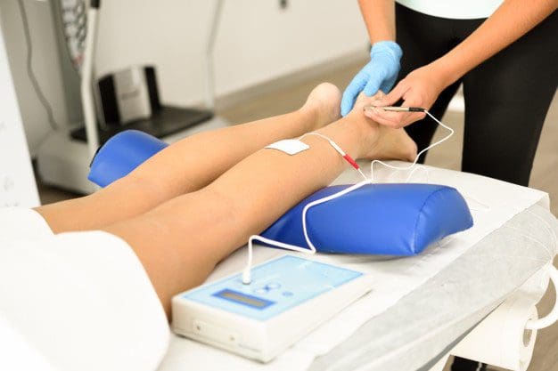


Management of Sciatica: Nonsurgical & Surgical Therapies

Management of Sciatica: Nonsurgical & Surgical Therapies

Consider the following, sciatica is a medical term used to describe a collective group of symptoms resulting from the irritation or compression of the sciatic nerve, generally due to an injury or aggravated condition. Sciatica is commonly characterized by radiating pain along the sciatic nerve, which runs down one or both legs from the lower back. The following case vignette discusses Mr. Winston’s medical condition, a 50-year-old bus driver who reported experiencing chronic, lower back and leg pain associated with sciatica during a 4-week time period. Ramya Ramaswami, M.B., B.S., M.P.H., Zoher Ghogawala, M.D., and James N. Weinstein, D.O., provide a comprehensive analysis of the various treatment options available to treat sciatica, including undergoing lumbar disk surgery and receiving nonsurgical therapy.


On a personal note, as a practicing doctor of chiropractic, choosing the correct treatment care for any type of injury or condition can be a personal and difficult decision. If the circumstances are favorable, the patient may determine what is the best form of treatment for their type of medical issue. While nonsurgical therapies, such as chiropractic care, can often be utilized to improve symptoms of sciatica, more severe cases of sciatica may require surgical interventions to treat the source of the issue. In most cases, nonsurgical therapies should be considered first, before turning to surgical therapies for sciatica.


Case Vignette


A Man with Sciatica Who is Considering Lumbar Disk Surgery


Ramya Ramaswami, M.B., B.S., M.P.H.


Mr. Winston, a 50-year-old bus driver, presented to your office with a 4-week history of pain in his left leg and lower back. He described a combination of severe sharp and dull pain that originated in his left buttock and radiated to the dorsolateral aspect of his left thigh, as well as vague aching over the lower lumbar spine. On examination, passive raising of his left leg off the table to 45 degrees caused severe pain that simulated his main symptom, and the pain was so severe that you could not lift his leg further. There was no leg or foot weakness. His body-mass index (the weight in kilograms divided by the square of the height in meters) was 35, and he had mild chronic obstructive pulmonary disease as a result of smoking one pack of cigarettes every day for 22 years. Mr. Winston had taken a leave of absence from his work because of his symptoms. You prescribed 150 mg of pregabalin per day, which was gradually increased to 600 mg daily because the symptoms had not abated.


Now, 10 weeks after the initial onset of his symptoms, he returns for an evaluation. The medication has provided minimal alleviation of his sciatic pain. He has to return to work and is concerned about his ability to complete his duties at his job. He undergoes magnetic resonance imaging, which shows a herniated disk on the left side at the L4�L5 root. You discuss options for the next steps in managing his sciatica. He is uncertain about invasive procedures such as lumbar disk surgery but feels limited by his symptoms of pain.


Treatment Options


Which of the following would you recommend for Mr. Winston?


  1. Undergo lumbar disk surgery.
  2. Receive nonsurgical therapy.


To aid in your decision making, each of these approaches is defended in a short essay by an expert in the field. Given your knowledge of the patient and the points made by the experts, which option would you choose?


Option 1: Undergo Lumbar Disk Surgery
Option 2: Receive Nonsurgical Therapy


1. Undergo Lumbar Disk Surgery


Zoher Ghogawala, M.D.


Mr. Winston�s case represents a common scenario in the management of symptomatic lumbar disk herniation. In this particular case, the patient�s symptoms and the physical examination are consistent with nerve-root compression and inflammation directly from an L4�L5 herniated disk on his left side. The patient does not have weakness but has ongoing pain and has been unable to work for the past 10 weeks despite receiving pregabalin. Two questions emerge: first, does lumbar disk surgery (microdiskectomy) provide outcomes that are superior to those with continued nonoperative therapy in patients with more than 6 weeks of symptoms; and second, does lumbar microdiskectomy improve the likelihood of return to work in patients with these symptoms?


The highest quality data on the topic come from the Spine Patient Outcomes Research Trial (SPORT). The results of the randomized, controlled trial are difficult to interpret because adherence to the assigned treatment strategy was suboptimal. Only half the patients who were randomly assigned to the surgery group actually underwent surgery within 3 months after enrollment, and 30% of the patients assigned to nonoperative treatment chose to cross over to the surgical group. In this study, the patients who underwent surgery had greater improvements in validated patient-reported outcomes. The treatment effect of microdiskectomy was superior to that of nonoperative treatment at 3 months, 1 year, and 2 years. Moreover, in an as-treated analysis, the outcomes among patients who underwent surgery were superior to those among patients who received nonoperative therapy. Overall, the results of SPORT support the use of microdiskectomy in this case.


Results of clinical trials are based on a comparison of treatment options in study populations and may or may not apply to individual patients. SPORT did not specify what type of nonoperative therapy was to be used. Physical therapy was used in 73% of the patients, epidural injections in 50%, and medical therapies (e.g., nonsteroidal antiinflammatory drugs) in more than 50%. In the case of Mr. Winston, pregabalin has been tried, but physical therapy and epidural glucocorticoid injections have not been attempted. Despite widespread use of physical therapy for the treatment of lumbar disk herniation, the evidence supporting its effectiveness is inconclusive, according to published guidelines of the North American Spine Society. On the other hand, there is evidence that transforaminal epidural glucocorticoid injection provides short-term relief (30 days) in patients with nerve-root symptoms directly related to a herniated disk. Overall, there is evidence, from SPORT and from a randomized trial from the Netherlands published in the Journal, that early surgery between 6 and 12 weeks after the onset of symptoms provides greater alleviation of leg pain and better overall pain relief than prolonged conservative therapy.


The ability to return to work has not been formally studied in comparisons of operative with nonoperative treatments for lumbar disk herniation. Registry data from the NeuroPoint-SD study showed that more than 80% of the patients who were working before disk herniation returned to work after surgery. The ability to return to work may be dependent on the type of vocation, since patients who are manual laborers may need more time to recover to reduce the risk of reherniation.


It is well recognized that many patients who have a symptomatic lumbar disk herniation will have improvement spontaneously over several months. Surgery can alleviate symptoms more quickly by immediately removing the offending disk herniation from the affected nerve root. The risk�benefit equation will vary among individual patients. In the case of Mr. Winston, obesity and mild pulmonary disease might increase the risk of complications from surgery, although in SPORT, 95% of surgical patients did not have any operative or postoperative complication. For Mr. Winston, a patient with pain that has persisted for more than 6 weeks, microdiskectomy is a rational option that is supported by high-quality evidence.


2. Receive Nonsurgical Therapy


James N. Weinstein, D.O.


This case involves a common presentation of low back pain radiating to the buttock and posterolateral thigh that might represent either referred mechanical pain or radiculopathy. Classic radiculopathy resulting from compression of a lower lumbar nerve root (L4, L5, or S1) results in pain that radiates distal to the knee and is often accompanied by weakness or numbness in the respective myotome or dermatome. In this case, the pain is proximal to the knee and is not associated with weakness or numbness. In SPORT, surgery resulted in faster recovery and a greater degree of improvement than nonoperative treatment in patients with pain that radiated distal to the knee and was accompanied by neurologic signs or symptoms. However, since Mr. Winston would not have met the inclusion criteria for SPORT, the results of diskectomy in this case would be somewhat unpredictable. He does not have radiculopathy that radiates below the knee, and he does not have weakness or numbness; nonoperative treatment should be exhausted before any consideration of a surgical procedure that in most cases has not been shown to be effective in patients with this type of presentation. In this issue of the Journal, Mathieson and colleagues report the results of a randomized, controlled trial that showed that pregabalin did not significantly alleviate pain related to sciatica. Mr. Winston has been treated only with pregabalin; therefore, other conservative options should be explored.


Saal and Saal reported that more than 80% of patients with radiculopathy associated with a lumbar disk herniation had improvement in a matter of months with exercise-based physical therapy. In the nonoperative SPORT cohort, patients had significant improvement from baseline, and approximately 60% of those with classic radiculopathy who initially received nonoperative treatment avoided surgery. Mr. Winston has had minimal treatment and has had symptoms for only 10 weeks. He should undergo a course of exercise-based physical therapy and a trial of a nonsteroidal antiinflammatory medication and may consider a lumbar epidural glucocorticoid injection. Although there is little evidence of the effectiveness of these nonoperative options alone, the combination of these treatments and the benign natural history of the patient�s condition could result in alleviation or resolution of symptoms. If these interventions � and time � do not resolve his symptoms, surgery could be considered as a final option, but it may not have long-term effectiveness and could in and of itself cause the possibility of more harm than good. Mr. Winston has risk factors, such as obesity and a history of smoking, that have been shown to contribute to poor surgical outcomes of certain spinal procedures.


Mr. Winston has symptoms of back pain that interfere with his quality of life. He would need to understand, through shared decision making, that a nonsurgical approach is likely to be more effective than surgery over time.


Information referenced from the National Center for Biotechnology Information (NCBI) and the New England Journal of Medicine (NEJM). The scope of our information is limited to chiropractic as well as to spinal injuries and conditions. To discuss the subject matter, please feel free to ask Dr. Jimenez or contact us at 915-850-0900 .


Cited by Dr. Alex Jimenez


Green-Call-Now-Button-24H-150x150-2-3.png


Additional Topics: Wellness


Overall health and wellness are essential towards maintaining the proper mental and physical balance in the body. From eating a balanced nutrition as well as exercising and participating in physical activities, to sleeping a healthy amount of time on a regular basis, following the best health and wellness tips can ultimately help maintain overall well-being. Eating plenty of fruits and vegetables can go a long way towards helping people become healthy.


blog picture of cartoon paperboy big news


IMPORTANT TOPIC: EXTRA EXTRA: Treating Sciatica Pain
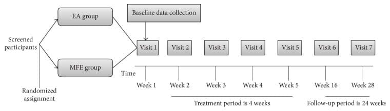


Blank
References


  • 1. Weinstein JN, Tosteson TD, Lurie JD, et al. Surgical vs nonoperative treatment for lumbar disk herniation: the Spine Patient Outcomes Research Trial (SPORT): a randomized trial. JAMA 2006;296:24412450

  • 2. Weinstein JN, Lurie JD, Tosteson TD, et al. Surgical vs nonoperative treatment for lumbar disk herniation: the Spine Patient Outcomes Research Trial (SPORT) observational cohort. JAMA 2006;296:24512459

  • 3. Kreiner DS, Hwang SW, Easa JE, et al. An evidence-based clinical guideline for the diagnosis and treatment of lumbar disc herniation with radiculopathy. Spine J 2014;14:180191

  • 4. Ghahreman A, Ferch R, Bogduk N. The efficacy of transforaminal injection of steroids for the treatment of lumbar radicular pain. Pain Med 2010;11:11491168

  • 5. Peul WC, van Houwelingen HC, van den Hout WB, et al. Surgery versus prolonged conservative treatment for sciatica. N Engl J Med 2007;356:22452256

  • 6. Ghogawala Z, Shaffrey CI, Asher AL, et al. The efficacy of lumbar discectomy and single-level fusion for spondylolisthesis: results from the NeuroPoint-SD registry: clinical article. J Neurosurg Spine 2013;19:555563

  • 7. Deyo RA, Weinstein JN. Low back pain. N Engl J Med 2001;344:363370

  • 8. Lurie JD, Tosteson TD, Tosteson AN, et al. Surgical versus nonoperative treatment for lumbar disc herniation: eight-year results for the spine patient outcomes research trial. Spine (Phila Pa 1976) 2014;39:316

  • 9. Mathieson S, Maher CG, McLachlan AJ, et al. Trial of pregabalin for acute and chronic sciatica. N Engl J Med 2017;376:11111120

  • 10. Saal JA, Saal JS. Nonoperative treatment of herniated lumbar intervertebral disc with radiculopathy: an outcome study. Spine (Phila Pa 1976) 1989;14:431437

  • 11. Pinto RZ, Maher CG, Ferreira ML, et al. Drugs for relief of pain in patients with sciatica: systematic review and meta-analysis. BMJ 2012;344:e497e497

  • 12. Pearson A, Lurie J, Tosteson T, et al. Who should have surgery for an intervertebral disc herniation? Comparative effectiveness evidence from the Spine Patient Outcomes Research Trial. Spine 2012;37:140149

  • 13. Weeks WB, Weinstein JN. Patient-reported data can help people make better health care choices. Harvard Business Review. September 21, 2015


Close Accordion
Assessment and Treatment of Upper Trapezius

Assessment and Treatment of Upper Trapezius

These assessment and treatment recommendations represent a synthesis of information derived from personal clinical experience and from the numerous sources which are cited, or are based on the work of researchers, clinicians and therapists who are named (Basmajian 1974, Cailliet 1962, Dvorak & Dvorak 1984, Fryette 1954, Greenman 1989, 1996, Janda 1983, Lewit 1992, 1999, Mennell 1964, Rolf 1977, Williams 1965).


Clinical Application of Neuromuscular Techniques: Upper Trapezius


Lewit (1999) simplifies the need to assess for shortness by stating, �The upper trapezius should be treated if tender and taut.� Since this is an almost universal state in modern life, it seems that everyone requires MET application to this muscle. Lewit also notes that a characteristic mounding of the muscle can often be observed when it is very short, producing the effect of �Gothic shoulders�, similar to the architectural supports of a Gothic church tower (see Fig. 2.13).


Assessment for Shortness of Upper Trapezius (13) (Fig. 4.30)


Figure 4 30 Assessment of the Relative Shortness of the Right Side Upper Trapezius Image 1


Figure 4.30 Assessment of the relative shortness of the right side upper trapezius. One side is compared with the other (for both the range of unforced motion and the nature of the end-feel of motion) to ascertain the side most in need of MET attention.


Test for upper trapezius for shortness (a) See scapulohumeral rhythm test (Ch. 5) which helps identify excessive activity or inappropriate tone in levator scapula and upper trapezius, which, because they are postural muscles, indicates shortness (Fig 5.13A, B). Greenman (1996) describes a functional �firing sequence� assessment which identifies general imbalance and dysfunction involving the upper and lower fixators of the shoulder (Fig. 4.31).


Figure 4 31 Palpation Assessment for Upper and Lower Fixators of the Shoulder Image 2


Figure 4.31 Palpation assessment for upper and lower fixators of the shoulder, including upper trapezius (Greenman 1996).


The patient is seated and the practitioner stands behind. The practitioner rests his right hand over the right shoulder area to assess firing sequence of muscles. The other hand can be placed either on the mid-thoracic region, mainly on the side being assessed, or spanning the lower back to palpate quadratus firing. The assessment should be performed at least twice so that various hand positions are used for different muscles (as in Fig. 4.31).


Greenman bases his description on Janda (1983), who notes the �correct� sequence for shoulder abduction, when seated, as involving: supraspinatus, deltoid, infraspinatus, middle and lower trapezius and finally contralateral quadratus. In dysfunctional states the most common substitutions are said to involve: shoulder elevation by levator scapulae and upper trapezius, as well as early firing by quadratus lumborum, ipsilateral and contralateral.


Inappropriate activity of the upper fixators results in shortness, and of the lower fixators in weakness and possible lengthening (see Ch. 2 for discussion of postural/phasic, etc. muscle characteristics).


Test for upper trapezius for shortness (b) The patient is seated and the practitioner stands behind with one hand resting on the shoulder of the side to be tested and stabilising it. The other hand is placed on the ipsilateral side of the head and the head/neck is taken into contralateral sidebending without force while the shoulder is stabilised (see Fig. 4.30).


The same procedure is performed on the other side with the opposite shoulder stabilised. A comparison is made as to which sidebending manoeuvre produced the greater range and whether the neck can easily reach 45� of side-flexion in each direction, which it should. If neither side can achieve this degree of sidebend, then both trapezius muscles may be short. The relative shortness of one, compared with the other, is evaluated.


Test for upper trapezius for shortness (c) The patient is seated and the practitioner stands behind with a hand resting over the muscle on the side to be assessed. The patient is asked to extend the arm at the shoulder joint, bringing the flexed arm/elbow backwards. If the upper trapezius is stressed on that side it will inappropriately activate during this movement. Since it is a postural muscle, shortness in it can then be assumed (see discussion of postural muscle characteristics in Ch. 3).


Test of upper trapezius for shortness (d) The patient is supine with the neck fully (but not forcefully) sidebent contralaterally (away from the side being assessed). The practitioner is standing at the head of the table and uses a cupped hand contact on the ipsilateral shoulder (i.e. on the side being tested) to assess the ease with which it can be depressed (moved caudally) (Fig. 4.32).


Figure 4 32 MET Treatment of Right Side Upper Trapezius Muscle Image 3


Figure 4.32 MET treatment of right side upper trapezius muscle. A Posterior fibres, B middle fibres, C anterior fibres. Note that stretching in this (or any of the alternative positions which access the middle and posterior fibres) is achieved following the isometric contraction by means of an easing of the shoulder away from the stabilised head, with no force being applied to the neck and head itself.


There should be an easy �springing� sensation as the practitioner pushes the shoulder towards the feet, with a soft end-feel to the movement. If depression of the shoulder is difficult or if there is a harsh, sudden end-point, upper trapezius shortness is confirmed.


This same assessment (always with full lateral flexion) should be performed with the head fully rotated away from the side being treated, half turned away from the side being treated, and slightly turned towards the side being treated, in order to respectively assess the relative shortness and functional efficiency of posterior, middle and anterior subdivisions of the upper portion of trapezius.


MET Treatment of Chronically Shortened Upper Trapezius


MET treatment of upper trapezius, method (a) (Fig. 4.32) In order to treat all the fibres of upper trapezius, MET needs to be applied sequentially. The upper trapezius is subdivided here into anterior, middle and posterior fibres. The neck should be placed into different positions of rotation, coupled with the sidebending as described in the assessment description above, for precise treatment of the various fibres.


The patient lies supine, arm on the side to be treated lying alongside the trunk, head/neck sidebent away from the side being treated to just short of the restriction barrier, while the practitioner stabilises the shoulder with one hand and cups the ear/mastoid area of the same side of the head with the other:


  • With the neck fully sidebent and fully rotated contralaterally, the posterior fibres of upper trapezius are involved in the contraction (see below). This will facilitate subsequent stretching of this aspect of the muscle.
  • With the neck fully sidebent and half rotated, the middle fibres are involved in the contraction.
  • With the neck fully sidebent and slightly rotated towards the side being treated the anterior fibres of upper trapezius are being treated.


The various contractions and subsequent stretches can be performed with practitioner�s arms crossed, hands stabilising the mastoid area and shoulder.


The patient introduces a light resisted effort (20% of available strength) to take the stabilised shoulder towards the ear (a shrug movement) and the ear towards the shoulder. The double movement (or effort towards movement) is important in order to introduce a contraction of the muscle from both ends simultaneously. The degree of effort should be mild and no pain should be felt. The contraction is sustained for 10 seconds (or so) and, upon complete relaxation of effort, the practitioner gently eases the head/neck into an increased degree of sidebending and rotation, where it is stabilised, as the shoulder is stretched caudally.


When stretching is introduced the patient can usefully assist in this phase of the treatment by initiating, on instruction, the stretch of the muscle (�as you breathe out please slide your hand towards your feet�). This reduces the chances of a stretch reflex being initiated. Once the muscle is being stretched, the patient relaxes and the stretch is held for 10�30 seconds.


CAUTION: No stretch should be introduced from the cranial end of the muscle as this could stress the neck. The head is stabilised at its side-flexion and rotation barrier.


Disagreement


There is some disagreement as to the head/neck rotation position as described in the treatment method above, which calls (for posterior and middle fibres) for sidebending and rotation away from the affected side.


Liebenson (1996), suggests that the patient �lies supine with the head supported in anteflexion and laterally flexed away and rotated towards the side of involvement�.


Lewit (1985b) suggests: �The patient is supine � the therapist fixes the shoulder from above with one hand, sidebending the head and neck with the other hand so as to take up the slack. He then asks the patient to look towards the side away from which the head is bent, resisting the patient�s automatic tendency to move towards the side of the lesion.� (This method is described below.)


The author has used the methods described above with good effect and urges readers to try these approaches as well as those of Liebenson and Lewit, and to evaluate results for themselves.


MET treatment of acutely shortened upper trapezius, method (b) Lewit suggests the use of eye movements to facilitate initiation of PIR before stretching, an ideal method for acute problems in this region.


The patient is supine, while the practitioner fixes the shoulder and the sidebent (away from the treated side) head and neck at the restriction barrier and asks the patient to look, with the eyes only (i.e. not to turn the head), towards the side away from which the neck is bent.


This eye movement is maintained, as is a held breath, while the practitioner resists the slight isometric contraction that these two factors (eye movement and breath) will have created.


On exhalation and complete relaxation, the head/neck is taken to a new barrier and the process repeated. If the shoulder is brought into the equation, this is firmly held as it attempts to lightly push into a shrug. After this 10 second contraction the muscle will have released somewhat and slack can again be taken out as the head is repositioned before a repetition of the procedure commences.


Dr. Alex Jimenez offers an additional assessment and treatment of the hip flexors as a part of a referenced clinical application of neuromuscular techniques by Leon Chaitow and Judith Walker DeLany. The scope of our information is limited to chiropractic and spinal injuries and conditions. To discuss the subject matter, please feel free to ask Dr. Jimenez or contact us at 915-850-0900 .


By Dr. Alex Jimenez


Green-Call-Now-Button-24H-150x150-2-3.png


Additional Topics: Wellness


Overall health and wellness are essential towards maintaining the proper mental and physical balance in the body. From eating a balanced nutrition as well as exercising and participating in physical activities, to sleeping a healthy amount of time on a regular basis, following the best health and wellness tips can ultimately help maintain overall well-being. Eating plenty of fruits and vegetables can go a long way towards helping people become healthy.


blog picture of cartoon paperboy big news


WELLNESS TOPIC: EXTRA EXTRA: Managing Workplace Stress


Blank
References
1.�Ballou SK, Keefer L. Multicultural considerations in the diagnosis and management of irritable bowel syndrome: a selective summary.�Eur J Gastroenterol Hepatol.�2013;25:1127�1133.�[PubMed]
2.�Gonzales Gamarra RG, Ruiz S�nchez JG, Le�n Jim�nez F, Cubas Benavides F, D�az V�lez C. [Prevalence of irritable bowel syndrome in the adult population of the city of Chiclayo in 2011]�Rev Gastroenterol Peru.�2012;32:381�386.�[PubMed]
3.�Gwee KA, Lu CL, Ghoshal UC. Epidemiology of irritable bowel syndrome in Asia: something old, something new, something borrowed.�J Gastroenterol Hepatol.�2009;24:1601�1607.�[PubMed]
4.�Krogsgaard LR, Engsbro AL, Bytzer P. The epidemiology of irritable bowel syndrome in Denmark. A population-based survey in adults ? 50 years of age.�Scand J Gastroenterol.�2013;48:523�529.�[PubMed]
5.�Ibrahim NK, Battarjee WF, Almehmadi SA. Prevalence and predictors of irritable bowel syndrome among medical students and interns in King Abdulaziz University, Jeddah.�Libyan J Med.�2013;8:21287.[PMC free article][PubMed]
6.�Lee YY, Waid A, Tan HJ, Chua AS, Whitehead WE. Rome III survey of irritable bowel syndrome among ethnic Malays.�World J Gastroenterol.�2012;18:6475�6480; discussion p. 6479.�[PMC free article][PubMed]
7.�Ghoshal UC, Abraham P, Bhatt C, Choudhuri G, Bhatia SJ, Shenoy KT, Banka NH, Bose K, Bohidar NP, Chakravartty K, et al. Epidemiological and clinical profile of irritable bowel syndrome in India: report of the Indian Society of Gastroenterology Task Force.�Indian J Gastroenterol.�2008;27:22�28.�[PubMed]
8.�Bures J, Cyrany J, Kohoutova D, F�rstl M, Rejchrt S, Kvetina J, Vorisek V, Kopacova M. Small intestinal bacterial overgrowth syndrome.�World J Gastroenterol.�2010;16:2978�2990.�[PMC free article][PubMed]
9.�Dibaise JK, Young RJ, Vanderhoof JA. Enteric microbial flora, bacterial overgrowth, and short-bowel syndrome.�Clin Gastroenterol Hepatol.�2006;4:11�20.�[PubMed]
10.�Bouhnik Y, Alain S, Attar A, Flouri� B, Raskine L, Sanson-Le Pors MJ, Rambaud JC. Bacterial populations contaminating the upper gut in patients with small intestinal bacterial overgrowth syndrome.�Am J Gastroenterol.�1999;94:1327�1331.�[PubMed]
11.�Dukowicz AC, Lacy BE, Levine GM. Small intestinal bacterial overgrowth: a comprehensive review.�Gastroenterol Hepatol (N Y)�2007;3:112�122.�[PMC free article][PubMed]
12.�Posserud I, Stotzer PO, Bj�rnsson ES, Abrahamsson H, Simr�n M. Small intestinal bacterial overgrowth in patients with irritable bowel syndrome.�Gut.�2007;56:802�808.�[PMC free article][PubMed]
13.�Ghoshal U, Ghoshal UC, Ranjan P, Naik SR, Ayyagari A. Spectrum and antibiotic sensitivity of bacteria contaminating the upper gut in patients with malabsorption syndrome from the tropics.�BMC Gastroenterol.�2003;3:9.�[PMC free article][PubMed]
14.�Carrara M, Desideri S, Azzurro M, Bulighin GM, Di Piramo D, Lomonaco L, Adamo S. Small intestine bacterial overgrowth in patients with irritable bowel syndrome.�Eur Rev Med Pharmacol Sci.�2008;12:197�202.�[PubMed]
15.�Ghoshal UC, Ghoshal U, Das K, Misra A. Utility of hydrogen breath tests in diagnosis of small intestinal bacterial overgrowth in malabsorption syndrome and its relationship with oro-cecal transit time.�Indian J Gastroenterol.�2006;25:6�10.�[PubMed]
16.�Ghoshal UC, Kumar S, Mehrotra M, Lakshmi C, Misra A. Frequency of small intestinal bacterial overgrowth in patients with irritable bowel syndrome and chronic non-specific diarrhea.�J Neurogastroenterol Motil.�2010;16:40�46.�[PMC free article][PubMed]
17.�Pimentel M, Chow EJ, Lin HC. Eradication of small intestinal bacterial overgrowth reduces symptoms of irritable bowel syndrome.�Am J Gastroenterol.�2000;95:3503�3506.�[PubMed]
18.�Sachdeva S, Rawat AK, Reddy RS, Puri AS. Small intestinal bacterial overgrowth (SIBO) in irritable bowel syndrome: frequency and predictors.�J Gastroenterol Hepatol.�2011;26 Suppl 3:135�138.�[PubMed]
19.�Park H. The role of small intestinal bacterial overgrowth in the pathophysiology of irritable bowel syndrome.�J Neurogastroenterol Motil.�2010;16:3�4.�[PMC free article][PubMed]
20.�Ghoshal UC, Shukla R, Ghoshal U, Gwee KA, Ng SC, Quigley EM. The gut microbiota and irritable bowel syndrome: friend or foe?�Int J Inflam.�2012;2012:151085.�[PMC free article][PubMed]
21.�Brown AC. Ulcerative colitis, Crohn�s disease and irritable bowel syndrome patients need fecal transplant research and treatment.�J Crohns Colitis.�2014;8:179.�[PubMed]
22.�Sampath K, Levy LC, Gardner TB. Fecal transplantation: beyond the aesthetic.�Gastroenterology.�2013;145:1151�1153.�[PubMed]
23.�Grace E, Shaw C, Whelan K, Andreyev HJ. Review article: small intestinal bacterial overgrowth–prevalence, clinical features, current and developing diagnostic tests, and treatment.�Aliment Pharmacol Ther.�2013;38:674�688.�[PubMed]
24.�Yakoob J, Abbas Z, Khan R, Hamid S, Awan S, Jafri W. Small intestinal bacterial overgrowth and lactose intolerance contribute to irritable bowel syndrome symptomatology in Pakistan.�Saudi J Gastroenterol.�2011;17:371�375.�[PMC free article][PubMed]
25.�Gwee KA, Bak YT, Ghoshal UC, Gonlachanvit S, Lee OY, Fock KM, Chua AS, Lu CL, Goh KL, Kositchaiwat C, et al. Asian consensus on irritable bowel syndrome.�J Gastroenterol Hepatol.�2010;25:1189�1205.�[PubMed]
26.�Lacy BE, Gabbard SL, Crowell MD. Pathophysiology, evaluation, and treatment of bloating: hope, hype, or hot air?�Gastroenterol Hepatol (N Y)�2011;7:729�739.�[PMC free article][PubMed]
27.�Harder H, Serra J, Azpiroz F, Passos MC, Aguad� S, Malagelada JR. Intestinal gas distribution determines abdominal symptoms.�Gut.�2003;52:1708�1713.�[PMC free article][PubMed]
28.�Kumar S, Misra A, Ghoshal UC. Patients with irritable bowel syndrome exhale more hydrogen than healthy subjects in fasting state.�J Neurogastroenterol Motil.�2010;16:299�305.�[PMC free article][PubMed]
29.�Hungin AP, Mulligan C, Pot B, Whorwell P, Agr�us L, Fracasso P, Lionis C, Mendive J, Philippart de Foy JM, Rubin G, Winchester C, de Wit N. Systematic review: probiotics in the management of lower gastrointestinal symptoms in clinical practice — an evidence-based international guide.�Aliment Pharmacol Ther.�2013;38:864�886.�[PMC free article][PubMed]
30.�Attar A, Flouri� B, Rambaud JC, Franchisseur C, Ruszniewski P, Bouhnik Y. Antibiotic efficacy in small intestinal bacterial overgrowth-related chronic diarrhea: a crossover, randomized trial.�Gastroenterology.�1999;117:794�797.�[PubMed]
31.�Marcelino RT, Fagundes-Neto U. [Hydrogen test (H2) in the air expired for the diagnosis of small bowel bacterial overgrowth]�Arq Gastroenterol.�1995;32:191�198.�[PubMed]
32.�Santavirta J. Lactulose hydrogen and [14C]xylose breath tests in patients with ileoanal anastomosis.�Int J Colorectal Dis.�1991;6:208�211.�[PubMed]
33.�Ghoshal UC. How to interpret hydrogen breath tests.�J Neurogastroenterol Motil.�2011;17:312�317.[PMC free article][PubMed]
34.�Yang CY, Chang CS, Chen GH. Small-intestinal bacterial overgrowth in patients with liver cirrhosis, diagnosed with glucose H2 or CH4 breath tests.�Scand J Gastroenterol.�1998;33:867�871.�[PubMed]
35.�Ghoshal UC, Ghoshal U, Ayyagari A, Ranjan P, Krishnani N, Misra A, Aggarwal R, Naik S, Naik SR. Tropical sprue is associated with contamination of small bowel with aerobic bacteria and reversible prolongation of orocecal transit time.�J Gastroenterol Hepatol.�2003;18:540�547.�[PubMed]
36.�Lu CL, Chen CY, Chang FY, Lee SD. Characteristics of small bowel motility in patients with irritable bowel syndrome and normal humans: an Oriental study.�Clin Sci (Lond)�1998;95:165�169.�[PubMed]
37.�Sciarretta G, Furno A, Mazzoni M, Garagnani B, Malaguti P. Lactulose hydrogen breath test in orocecal transit assessment. Critical evaluation by means of scintigraphic method.�Dig Dis Sci.�1994;39:1505�1510.�[PubMed]
38.�Rao SS, Camilleri M, Hasler WL, Maurer AH, Parkman HP, Saad R, Scott MS, Simren M, Soffer E, Szarka L. Evaluation of gastrointestinal transit in clinical practice: position paper of the American and European Neurogastroenterology and Motility Societies.�Neurogastroenterol Motil.�2011;23:8�23.[PubMed]
39.�Yu D, Cheeseman F, Vanner S. Combined oro-caecal scintigraphy and lactulose hydrogen breath testing demonstrate that breath testing detects oro-caecal transit, not small intestinal bacterial overgrowth in patients with IBS.�Gut.�2011;60:334�340.�[PubMed]
40.�Rana SV, Sharma S, Sinha SK, Kaur H, Sikander A, Singh K. Incidence of predominant methanogenic flora in irritable bowel syndrome patients and apparently healthy controls from North India.�Dig Dis Sci.�2009;54:132�135.�[PubMed]
41.�Dridi B, Henry M, El Kh�chine A, Raoult D, Drancourt M. High prevalence of Methanobrevibacter smithii and Methanosphaera stadtmanae detected in the human gut using an improved DNA detection protocol.�PLoS One.�2009;4:e7063.�[PMC free article][PubMed]
42.�Chatterjee S, Park S, Low K, Kong Y, Pimentel M. The degree of breath methane production in IBS correlates with the severity of constipation.�Am J Gastroenterol.�2007;102:837�841.�[PubMed]
43.�Lunia MK, Sharma BC, Sachdeva S. Small intestinal bacterial overgrowth and delayed orocecal transit time in patients with cirrhosis and low-grade hepatic encephalopathy.�Hepatol Int.�2013;7:268�273.[PubMed]
44.�Resmini E, Parodi A, Savarino V, Greco A, Rebora A, Minuto F, Ferone D. Evidence of prolonged orocecal transit time and small intestinal bacterial overgrowth in acromegalic patients.�J Clin Endocrinol Metab.�2007;92:2119�2124.�[PubMed]
45.�Hamilton I, Worsley BW, Cobden I, Cooke EM, Shoesmith JG, Axon AT. Simultaneous culture of saliva and jejunal aspirate in the investigation of small bowel bacterial overgrowth.�Gut.�1982;23:847�853.[PMC free article][PubMed]
46.�Corazza GR, Sorge M, Strocchi A, Benati G, Di Sario A, Treggiari EA, Brusco G, Gasbarrini G. Non-absorbable antibiotics and small bowel bacterial overgrowth.�Ital J Gastroenterol.�1992;24:4�9.�[PubMed]
47.�Kuwahara T, Ogura Y, Oshima K, Kurokawa K, Ooka T, Hirakawa H, Itoh T, Nakayama-Imaohji H, Ichimura M, Itoh K, et al. The lifestyle of the segmented filamentous bacterium: a non-culturable gut-associated immunostimulating microbe inferred by whole-genome sequencing.�DNA Res.�2011;18:291�303.�[PMC free article][PubMed]
48.�Beumer RR, de Vries J, Rombouts FM. Campylobacter jejuni non-culturable coccoid cells.�Int J Food Microbiol.�1992;15:153�163.�[PubMed]
49.�Fromm H, Sarva RP, Ravitch MM, McJunkin B, Farivar S, Amin P. Effects of jejunoileal bypass on the enterohepatic circulation of bile acids, bacterial flora in the upper small intestine, and absorption of vitamin B12.�Metabolism.�1983;32:1133�1141.�[PubMed]
50.�Yoshida T, McCormick WC, Swell L, Vlahcevic ZR. Bile acid metabolism in cirrhosis. IV. Characterization of the abnormality in deoxycholic acid metabolism.�Gastroenterology.�1975;68:335�341.[PubMed]
51.�Bj�rneklett A, Fausa O, Midtvedt T. Bacterial overgrowth in jejunal and ileal disease.�Scand J Gastroenterol.�1983;18:289�298.�[PubMed]
52.�Vanner S. The small intestinal bacterial overgrowth. Irritable bowel syndrome hypothesis: implications for treatment.�Gut.�2008;57:1315�1321.�[PubMed]
53.�Kinross JM, von Roon AC, Holmes E, Darzi A, Nicholson JK. The human gut microbiome: implications for future health care.�Curr Gastroenterol Rep.�2008;10:396�403.�[PubMed]
54.�Clauw DJ. Fibromyalgia: an overview.�Am J Med.�2009;122:S3�S13.�[PubMed]
55.�Pimentel M, Wallace D, Hallegua D, Chow E, Kong Y, Park S, Lin HC. A link between irritable bowel syndrome and fibromyalgia may be related to findings on lactulose breath testing.�Ann Rheum Dis.�2004;63:450�452.�[PMC free article][PubMed]
56.�Simr�n M, Stotzer PO. Use and abuse of hydrogen breath tests.�Gut.�2006;55:297�303.[PMC free article][PubMed]
57.�Gasbarrini A, Lauritano EC, Gabrielli M, Scarpellini E, Lupascu A, Ojetti V, Gasbarrini G. Small intestinal bacterial overgrowth: diagnosis and treatment.�Dig Dis.�2007;25:237�240.�[PubMed]
58.�Ghoshal UC, Srivastava D, Verma A, Misra A. Slow transit constipation associated with excess methane production and its improvement following rifaximin therapy: a case report.�J Neurogastroenterol Motil.�2011;17:185�188.�[PMC free article][PubMed]
59.�Bala L, Ghoshal UC, Ghoshal U, Tripathi P, Misra A, Gowda GA, Khetrapal CL. Malabsorption syndrome with and without small intestinal bacterial overgrowth: a study on upper-gut aspirate using 1H NMR spectroscopy.�Magn Reson Med.�2006;56:738�744.�[PubMed]
60.�Haboubi NY, Lee GS, Montgomery RD. Duodenal mucosal morphometry of elderly patients with small intestinal bacterial overgrowth: response to antibiotic treatment.�Age Ageing.�1991;20:29�32.�[PubMed]
61.�Shindo K, Machida M, Koide K, Fukumura M, Yamazaki R. Deconjugation ability of bacteria isolated from the jejunal fluid of patients with progressive systemic sclerosis and its gastric pH.�Hepatogastroenterology.�1998;45:1643�1650.�[PubMed]
62.�Wanitschke R, Ammon HV. Effects of dihydroxy bile acids and hydroxy fatty acids on the absorption of oleic acid in the human jejunum.�J Clin Invest.�1978;61:178�186.�[PMC free article][PubMed]
63.�Shanab AA, Scully P, Crosbie O, Buckley M, O�Mahony L, Shanahan F, Gazareen S, Murphy E, Quigley EM. Small intestinal bacterial overgrowth in nonalcoholic steatohepatitis: association with toll-like receptor 4 expression and plasma levels of interleukin 8.�Dig Dis Sci.�2011;56:1524�1534.�[PubMed]
64.�Ghoshal UC, Park H, Gwee KA. Bugs and irritable bowel syndrome: The good, the bad and the ugly.�J Gastroenterol Hepatol.�2010;25:244�251.�[PubMed]
65.�Barbara G, Stanghellini V, Brandi G, Cremon C, Di Nardo G, De Giorgio R, Corinaldesi R. Interactions between commensal bacteria and gut sensorimotor function in health and disease.�Am J Gastroenterol.�2005;100:2560�2568.�[PubMed]
66.�Cherbut C, Aub� AC, Blotti�re HM, Galmiche JP. Effects of short-chain fatty acids on gastrointestinal motility.�Scand J Gastroenterol Suppl.�1997;222:58�61.�[PubMed]
67.�Ramakrishna BS, Roediger WE. Bacterial short chain fatty acids: their role in gastrointestinal disease.�Dig Dis.�1990;8:337�345.�[PubMed]
68.�Dumoulin V, Moro F, Barcelo A, Dakka T, Cuber JC. Peptide YY, glucagon-like peptide-1, and neurotensin responses to luminal factors in the isolated vascularly perfused rat ileum.�Endocrinology.�1998;139:3780�3786.�[PubMed]
69.�Balsari A, Ceccarelli A, Dubini F, Fesce E, Poli G. The fecal microbial population in the irritable bowel syndrome.�Microbiologica.�1982;5:185�194.�[PubMed]
70.�Nobaek S, Johansson ML, Molin G, Ahrn� S, Jeppsson B. Alteration of intestinal microflora is associated with reduction in abdominal bloating and pain in patients with irritable bowel syndrome.�Am J Gastroenterol.�2000;95:1231�1238.�[PubMed]
71.�Cummings JH, Macfarlane GT. The control and consequences of bacterial fermentation in the human colon.�J Appl Bacteriol.�1991;70:443�459.�[PubMed]
72.�Camilleri M. Probiotics and irritable bowel syndrome: rationale, mechanisms, and efficacy.�J Clin Gastroenterol.�2008;42 Suppl 3 Pt 1:S123�S125.�[PubMed]
73.�Spiller R. Probiotics: an ideal anti-inflammatory treatment for IBS?�Gastroenterology.�2005;128:783�785.�[PubMed]
74.�Ford AC, Spiegel BM, Talley NJ, Moayyedi P. Small intestinal bacterial overgrowth in irritable bowel syndrome: systematic review and meta-analysis.�Clin Gastroenterol Hepatol.�2009;7:1279�1286.�[PubMed]
75.�Cuoco L, Salvagnini M. Small intestine bacterial overgrowth in irritable bowel syndrome: a retrospective study with rifaximin.�Minerva Gastroenterol Dietol.�2006;52:89�95.�[PubMed]
76.�Di Stefano M, Corazza GR. Treatment of small intestine bacterial overgrowth and related symptoms by rifaximin.�Chemotherapy.�2005;51 Suppl 1:103�109.�[PubMed]
77.�Pimentel M, Lembo A, Chey WD, Zakko S, Ringel Y, Yu J, Mareya SM, Shaw AL, Bortey E, Forbes WP. Rifaximin therapy for patients with irritable bowel syndrome without constipation.�N Engl J Med.�2011;364:22�32.�[PubMed]
78.�Hwang L, Low K, Khoshini R, Melmed G, Sahakian A, Makhani M, Pokkunuri V, Pimentel M. Evaluating breath methane as a diagnostic test for constipation-predominant IBS.�Dig Dis Sci.�2010;55:398�403.�[PubMed]
79.�Low K, Hwang L, Hua J, Zhu A, Morales W, Pimentel M. A combination of rifaximin and neomycin is most effective in treating irritable bowel syndrome patients with methane on lactulose breath test.�J Clin Gastroenterol.�2010;44:547�550.�[PubMed]
80.�Bengmark S. Colonic food: pre- and probiotics.�Am J Gastroenterol.�2000;95:S5�S7.�[PubMed]
81.�Quigley EM, Quera R. Small intestinal bacterial overgrowth: roles of antibiotics, prebiotics, and probiotics.�Gastroenterology.�2006;130:S78�S90.�[PubMed]
82.�Xiao SD, Zhang DZ, Lu H, Jiang SH, Liu HY, Wang GS, Xu GM, Zhang ZB, Lin GJ, Wang GL. Multicenter, randomized, controlled trial of heat-killed Lactobacillus acidophilus LB in patients with chronic diarrhea.�Adv Ther.�2003;20:253�260.�[PubMed]
83.�O’Mahony L, McCarthy J, Kelly P, Hurley G, Luo F, Chen K, O�Sullivan GC, Kiely B, Collins JK, Shanahan F, et al. Lactobacillus and bifidobacterium in irritable bowel syndrome: symptom responses and relationship to cytokine profiles.�Gastroenterology.�2005;128:541�551.�[PubMed]
84.�Tsuchiya J, Barreto R, Okura R, Kawakita S, Fesce E, Marotta F. Single-blind follow-up study on the effectiveness of a symbiotic preparation in irritable bowel syndrome.�Chin J Dig Dis.�2004;5:169�174.[PubMed]
85.�Kim YG, Moon JT, Lee KM, Chon NR, Park H. [The effects of probiotics on symptoms of irritable bowel syndrome]�Korean J Gastroenterol.�2006;47:413�419.�[PubMed]
86.�Park JS, Yu JH, Lim HC, Kim JH, Yoon YH, Park HJ, Lee SI. [Usefulness of lactulose breath test for the prediction of small intestinal bacterial overgrowth in irritable bowel syndrome]�Korean J Gastroenterol.�2010;56:242�248.�[PubMed]
87.�Mann NS, Limoges-Gonzales M. The prevalence of small intestinal bacterial vergrowth in irritable bowel syndrome.�Hepatogastroenterology.�2009;56:718�721.�[PubMed]
88.�Scarpellini E, Giorgio V, Gabrielli M, Lauritano EC, Pantanella A, Fundar� C, Gasbarrini A. Prevalence of small intestinal bacterial overgrowth in children with irritable bowel syndrome: a case-control study.�J Pediatr.�2009;155:416�420.�[PubMed]
89.�Nucera G, Gabrielli M, Lupascu A, Lauritano EC, Santoliquido A, Cremonini F, Cammarota G, Tondi P, Pola P, Gasbarrini G, et al. Abnormal breath tests to lactose, fructose and sorbitol in irritable bowel syndrome may be explained by small intestinal bacterial overgrowth.�Aliment Pharmacol Ther.�2005;21:1391�1395.�[PubMed]
90.�Reddymasu SC, Sostarich S, McCallum RW. Small intestinal bacterial overgrowth in irritable bowel syndrome: are there any predictors?�BMC Gastroenterol.�2010;10:23.�[PMC free article][PubMed]
91.�Lombardo L, Foti M, Ruggia O, Chiecchio A. Increased incidence of small intestinal bacterial overgrowth during proton pump inhibitor therapy.�Clin Gastroenterol Hepatol.�2010;8:504�508.�[PubMed]
92.�Parodi A, Dulbecco P, Savarino E, Giannini EG, Bodini G, Corbo M, Isola L, De Conca S, Marabotto E, Savarino V. Positive glucose breath testing is more prevalent in patients with IBS-like symptoms compared with controls of similar age and gender distribution.�J Clin Gastroenterol.�2009;43:962�966.[PubMed]
93.�Rana SV, Sinha SK, Sikander A, Bhasin DK, Singh K. Study of small intestinal bacterial overgrowth in North Indian patients with irritable bowel syndrome: a case control study.�Trop Gastroenterol.�2008;29:23�25.�[PubMed]
94.�Majewski M, McCallum RW. Results of small intestinal bacterial overgrowth testing in irritable bowel syndrome patients: clinical profiles and effects of antibiotic trial.�Adv Med Sci.�2007;52:139�142.[PubMed]
95.�Lupascu A, Gabrielli M, Lauritano EC, Scarpellini E, Santoliquido A, Cammarota G, Flore R, Tondi P, Pola P, Gasbarrini G, et al. Hydrogen glucose breath test to detect small intestinal bacterial overgrowth: a prevalence case-control study in irritable bowel syndrome.�Aliment Pharmacol Ther.�2005;22:1157�1160.[PubMed]
Close Accordion
Mastodon