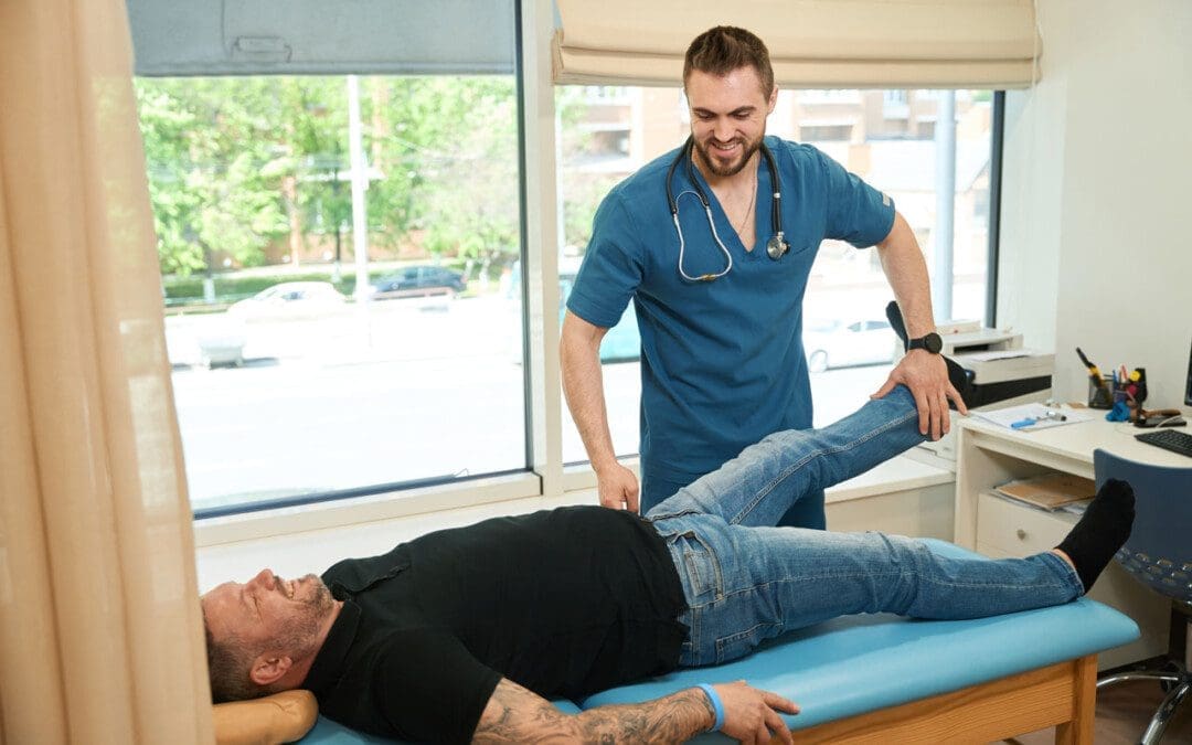
Individuals that have gone through a back injury may develop a synovial spinal cyst as a way to protect the spine that could cause pain symptoms and sensations. Can knowing the signs help healthcare providers develop a thorough treatment plan to relieve pain, prevent worsening of the condition and other spinal conditions?
Spinal Synovial Cysts
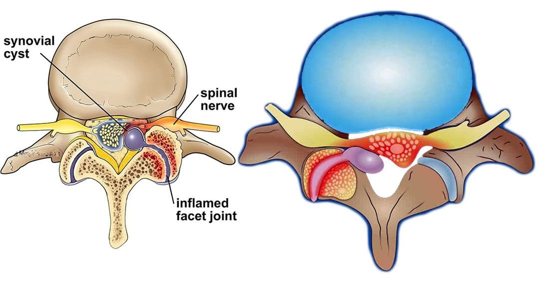
Spinal synovial cysts are benign fluid-filled sacs that develop in the spine’s joints. They form because of spinal degeneration or injury. The cysts can form anywhere in the spine, but most occur in the lumbar region/lower back. They typically develop in the facet joints or junctions that keep the vertebrae/spinal bones interlocked.
Symptoms
In most cases, synovial cysts don’t cause symptoms. However, the doctor or specialist will want to monitor for signs of degenerative disc disease, spinal stenosis, or cauda equina syndrome. When symptoms do present, they typically cause radiculopathy or nerve compression, which can cause back pain, weakness, numbness, and radiating pain caused by the irritation. The severity of symptoms depends on the size and location of the cyst. Synovial cysts can affect one side of the spine or both and can form at one spinal segment or at multiple levels.
Effects Can Include
Radiculopathy symptoms can develop if the cyst or inflammation caused by the cyst comes into contact with a spinal nerve root. This can cause sciatica, weakness, numbness, or difficulty controlling certain muscles.
Neurogenic claudication/impingement and inflammation of spinal nerves can cause cramping, pain, and/or tingling in the lower back, legs, hips, and buttocks. (Martin J. Wilby et al., 2009)
If the spinal cord is involved, it may cause myelopathy/severe spinal cord compression that can cause numbness, weakness, and balance problems. (Dong Shin Kim et al., 2014)
Symptoms related to cauda equina, including bowel and/or bladder problems, leg weakness, and saddle anesthesia/loss of sensation in the thighs, buttocks, and perineum, can present but are rare, as are synovial cysts in the middle back and neck. If thoracic and cervical synovial cysts develop, they can cause symptoms like numbness, tingling, pain, or weakness in the affected area.
Causes
Spinal synovial cysts are generally caused by degenerative changes like osteoarthritis that develop in a joint over time. With regular wear and tear, facet joint cartilage/the material in a joint that provides protection, a smooth surface, friction reduction, and shock absorption begins to waste away. As the process continues, the synovium can form a cyst.
Traumas, large and small, have inflammatory and degenerative effects on joints that can result in the formation of a cyst.
Around a third of individuals who have a spinal synovial cyst also have spondylolisthesis.
This condition is when a vertebrae slips out of place or out of alignment onto the vertebra underneath.
It is a sign of spinal instability.
Instability can occur in any spine area, but L4-5 are the most common levels.
This segment of the spine takes most of the upper body weight.
Epidural corticosteroid injections can reduce inflammation and could be an option to relieve pain.
Patients are recommended to receive no more than three injections per year.
Surgical Options
For severe or persistent cases, a doctor may recommend decompression surgery to remove the cyst and surrounding bone to relieve pressure on the nerve root. Surgical options range from minimally invasive endoscopic procedures to larger, open surgeries. The best surgical option varies based on the severity of the situation and whether associated disorders are present. Surgical options include:
Laminectomy – Removal of the bony structure that protects and covers the spinal canal/lamina.
Hemilaminectomy – A modified laminectomy where a smaller portion of the lamina is removed.
Facetectomy – The removal of part of the affected facet joint where the synovial cyst is located, usually following a laminectomy or hemilaminectomy.
Fusionof the facet joints and vertebra – Decreases vertebral mobility in the injured area.
Most individuals experience immediate pain relief following a laminectomy or hemilaminectomy.
Fusion can take six to nine months to heal completely.
If surgery is performed without fusion where the cyst originated, the pain could return, and another cyst could form within two years.
Surgery Complications include infection, bleeding, and injury to the spinal cord or nerve root.
How I Gained My Mobility Back With Chiropractic
References
Wilby, M. J., Fraser, R. D., Vernon-Roberts, B., & Moore, R. J. (2009). The prevalence and pathogenesis of synovial cysts within the ligamentum flavum in patients with lumbar spinal stenosis and radiculopathy. Spine, 34(23), 2518–2524. https://doi.org/10.1097/BRS.0b013e3181b22bd0
Kim, D. S., Yang, J. S., Cho, Y. J., & Kang, S. H. (2014). Acute myelopathy caused by a cervical synovial cyst. Journal of Korean Neurosurgical Society, 56(1), 55–57. https://doi.org/10.3340/jkns.2014.56.1.55
Epstein, N. E., & Baisden, J. (2012). The diagnosis and management of synovial cysts: Efficacy of surgery versus cyst aspiration. Surgical neurology international, 3(Suppl 3), S157–S166. https://doi.org/10.4103/2152-7806.98576
Ear problems like blockages or congestion can cause irritation and pain, as well as symptoms such as dizziness, ear discomfort, headaches, and sinus pain that can lead to infection. This condition can happen to anyone but is prevalent in children, individuals that live in high altitudes, and individuals who suffer from allergies. Spinal misalignments can cause interference to the nervous system that can create problems elsewhere in the body, like the ears.
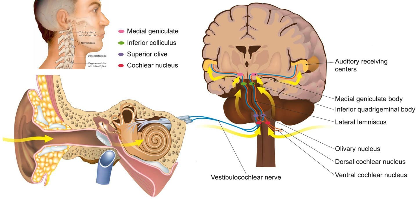
If there is neck misalignment along with pinched, tangled nerve/s signal transmissions can misfire or cut off messages disrupting the process of draining the Eustachian tube. This creates a buildup of bacteria and fluid, which can cause pain and pressure. Chiropractic decompression treatment uses gentle manipulation of the cervical spine to release the pressure affecting the ear.
Ear Problems
Bacteria or viruses cause ear infections in the middle ear. Infection often results from another illness like cold, sore throat, flu, respiratory disease, or allergies that causes congestion and swelling of the nasal passages, throat, and eustachian tubes.
Eustachian Tubes
The tubes functions include:
Regulating air pressure in the middle ear
Resupply fresh air in the ear
Drain the middle ear
The eustachian tubes are two canals that connect the middle ear to the throat and nasal cavity, known as the nasopharynx. (The eustachian tubes are more narrow in children, which makes them difficult to drain and more likely to get clogged.)When the lining of these canals comes under stress, they can become inflamed/swollen, blocking or filling with fluid causing excessive pressure and pain. This fluid can become infected and cause ear infection symptoms.
If the ear problem is connected to a misalignment of the cervical spine, the following symptoms may be experienced:
Fluid/effusion stays in the middle ear for an extended time.
It can build up over and over, despite no infection.
It can also affect hearing.
Misalignment in the upper cervical spine can cause muscles to flex awkwardly/irregularly, disrupting the opening and closing of the eustachian tubes and their positioning. This often causes inflammation along the eustachian canal, upper throat, and nasal cavity. If left untreated, the inflammation can develop into an infection, causing swelling and/or fluid buildup in the inner and middle ear. Common symptoms of middle ear infections in adults include:
Pain in one or both ears
Hearing is muffled
Sore throat
Fluid drainage from the ear
Chiropractic Realignment
Treatments are helpful for individuals who want to reduce taking antibiotics, which can minimize immunity by destroying the healthy bacteria in the gut. Chiropractic is a simple and effective way to treat ear problems. Realigning the vertebrae relieves tissue inflammation/swelling around the Eustachian tube to allow drainage, relieve pressure, and restore health.
Spinal Decompression Chiropractor
References
Collins, Rachael, et al. “Paralysis from an ear infection: a severe case of otitis externa leading to acute complete cervical cord syndrome.” BMJ case reports vol. 14,12 e245594. 1 Dec. 2021, doi:10.1136/bcr-2021-245594
Harmes, Kathryn M et al. “Otitis media: diagnosis and treatment.” American family physician vol. 88,7 (2013): 435-40.
Laulajainen Hongisto, Anu et al. “Severe Acute Otitis Media and Acute Mastoiditis in Adults.” The journal of international advanced otology vol. 12,3 (2016): 224-230. doi:10.5152/iao.2016.2620
Murphy, D R. “Chiropractic rehabilitation of the cervical spine.” Journal of manipulative and physiological therapeutics vol. 23,6 (2000): 404-8. doi:10.1067/mmt.2000.108143
Polkinghorn, B S. “Treatment of cervical disc protrusions via instrumental chiropractic adjustment.” Journal of manipulative and physiological therapeutics vol. 21,2 (1998): 114-21.
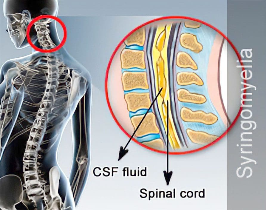
Syringomyelia is a disorder in which afluid-filled cyst/syrinx forms within the spinal cord. It is progressive, meaning that the cyst grows with time causing compression and damage to the spinal cord. The cyst usually begins in the neck/cervical spine but can develop in any area along the spinal cord. There are several possible causes; however, most are associated with a condition known as Chiari malformation. This is where the skull and neck come together, and either the skull is too small or shaped in a way that causes brain tissue to come out and settle in the spinal canal.
Syringomyelia Causes
Syringomyelia can be caused by or from complications of:
Chiari type I malformation develops during the fetal developmental stage and causes the lower part of the brain or cerebellum to stick out from its standard location.
Hemorrhage/bleeding
Inflammation of the spinal cord from virus or bacterial infection like meningitis
Spinal cord injury
Spinal cord tumor
Symptoms
A damaged spinal cord disrupts communication between the brain and the body. Symptoms differ for every individual, but common syringomyelia symptoms include:
Pain, stiffness, or weakness in the neck, arms, back, and/or legs
Symptoms usually develop slowly, but exercise, coughing, or some form of strain can cause sudden onset.
Diagnosis
Physical and neurological exams are performed to determine loss of feeling or inability to move around normally, like walking. Diagnostic tests of the spine will include a CT scan with contrast dye and/or an MRI. Early detection can help before it progresses, causing further damage, and delaying treatment can cause irreversible spinal cord injury. It is recommended at the first sign of symptoms to contact a doctor.
Treatment
Some individuals who have syringomyelia may have no symptoms. These individuals can go about their everyday lives but are recommended to be cautious with neck and back strain. For individuals experiencing symptoms, the primary treatment objectives are to:
Stop or control damage to the spinal cord
Preserve function
Prevent disability
Treatment options include:
Draining the cyst
Surgical removal of the cyst
Chiropractic and physical therapy could be included in the treatment plan to help the individual rebuild lost muscle strength and regain flexibility.
All too often, individuals with this disorder experience treatment delay/s because symptoms can be nonspecific or vague. Education is the key, and individuals can be diagnosed sooner by paying attention to the body’s warning signs.
Body Composition
Does too much protein hurt the kidneys?
While protein restriction can be appropriate for treating existing kidney disease, research shows that high protein intake in healthy individuals does not disrupt or cause damage to the kidneys or kidney function. The amino acids in protein are more likely to be excreted through urine when not being used. However, there are certain risks associated with consuming too much protein, and it is recommended to keep track of protein intake. Eating more protein:
Makes the body feel full longer
Can help curb overeating
Is essential for recovery and growth
When achieving daily caloric goals, maintaining a balance of nutrients like carbohydrates and healthy fats is essential for overall health.
References
Batzdorf, Ulrich. “Primary spinal syringomyelia. Invited submission from the joint section meeting on disorders of the spine and peripheral nerves, March 2005.” Journal of neurosurgery. Spine vol. 3,6 (2005): 429-35. doi:10.3171/spi.2005.3.6.0429
Di Lorenzo, N, and F Cacciola. “Adult syringomyelia. Classification, pathogenesis and therapeutic approaches.” Journal of neurosurgical sciences vol. 49,3 (2005): 65-72.
Fernández, Alfredo Avellaneda et al. “Malformations of the craniocervical junction (Chiari type I and syringomyelia: classification, diagnosis, and treatment).” BMC musculoskeletal disorders vol. 10 Suppl 1, Suppl 1 S1. 17 Dec. 2009, doi:10.1186/1471-2474-10-S1-S1
Naftel, Robert P et al. “Worsening or development of syringomyelia following Chiari I decompression: case report.” Journal of neurosurgery. Pediatrics vol. 12,4 (2013): 351-6. doi:10.3171/2013.7.PEDS12522
Roy, Anil K et al. “Idiopathic syringomyelia: retrospective case series, comprehensive review, and update on management.” Neurosurgical focus vol. 31,6 (2011): E15. doi:10.3171/2011.9.FOCUS11198
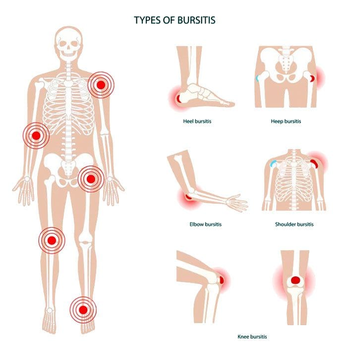
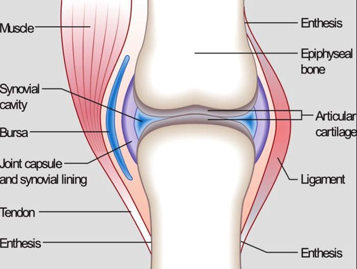
Bursitis types: This is a condition that affects the bursae, which are the small, fluid-filled sacs that provide cushion for the:
Muscles
Tendons
Bones near joints
The bursae make it easier for tissues to slide over each other. The body has around one hundred and sixty bursae. However, only a few become clinically affected. These include the:
Wrist
Elbow
Shoulder
Hips
Knees
The base of the big toe and heel
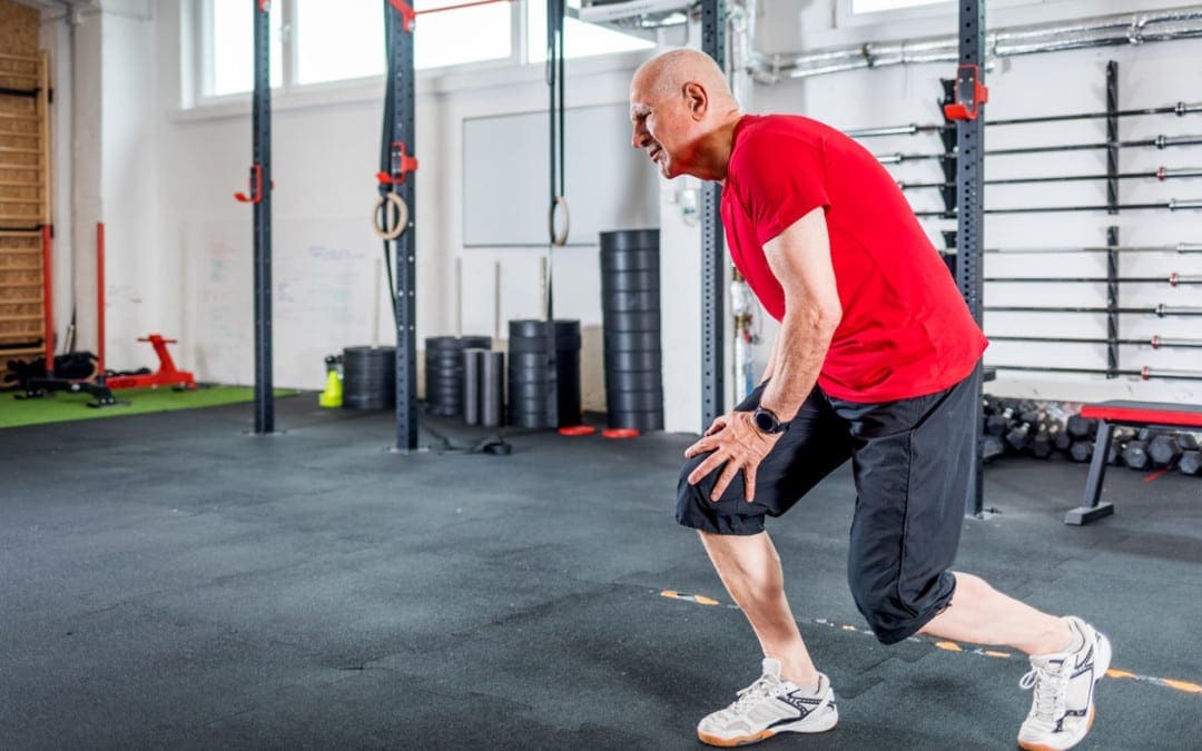
The condition typically presents near joints constantly being used repetitively, like a job, sports, house/yard chores, etc. What happens is one or more of the bursae sacs become inflamed, resulting in pain.
Causes
Inflamed or irritated bursae typically cause it from overuse or intense/vigorous activity.
It can also be caused by bacterial infection.
Arthritis and gout can also cause bursitis.
Another cause is age.
As tendons age, they can tear easily, lose their elasticity, and can’t take too much stress.
Intense physical activities can lead to bursitis. These include:
Gardening
Typing
Working with a computer mouse
Throwing
Golf
Tennis
Manual tasks
Carpentry
These types of activities can lead to incorrect posture, overuse, and injury/damage.
Symptoms
The main symptom is pain in and around the affected area that worsens with movement. Depending on the severity of the strain and the length of time it has been going on, the pain can be intense with active and passive movements. Other symptoms include:
Tenderness
Stiffness
For some individuals, it can present as acute, with the intensity increasing.
This happens when movement aggravates the condition.
Bursitis Types
Four major types include:
Prepatellar
Trochanteric
Olecranon
Retrocalcaneal
Prepatellar Bursitis
Prepatellar is an inflammation of the sac situated between the skin and the patella/kneecap. The most common causes are trauma from a fall and direct pressure/friction from repetitive kneeling. This is one of the bursitis types that can get infected. Overproduction of liquid places pressure on the other areas of the knee, causing swelling. Most individuals report swelling and knee pain just over the front of the knee.
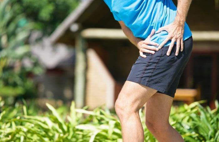
Trochanteric Bursitis
This bursitis type goes over the lateral area of the hip. There is a distinctive tenderness and aching pain. This type is more common for individuals with arthritis conditions and fibromyalgia. This condition is also seen after surgery, mainly osteotomies. The bursa can become inflamed in case of injury or overuse. It tends to affect middle-aged or older folks. Common causes include:
Muscle tears
Hip injuries
Tight hip or leg muscles
Disc disease of the low back
Leg-length inequality
Improper walking technique from a minor injury or strain
Overuse of the gluteal muscles
Flat feet
Improper footwear
Olecranon Bursitis
Olecranon is a common bursitis type. It is diagnosed by the appearance of swelling over the elbow. The swelling happens just behind the olecranon process of the ulna. The bursa can become infected. This bursitis does cause blood to rupture out, and fluid could be present. Individuals are advised to avoid leaning or resting on the elbows.
Retrocalcaneal Bursitis
This is characterized by pain in the Achilles tendon. Chronic inflammation of the bursa is brought on by friction, supination, and overpronation. The flexibility of the calf muscles can be significantly reduced. Severe pain and swelling of the posterior soft tissue in front of the Achilles tendon are common symptoms. This bursitis type is often accompanied by mid-portion insertional tendinosis.
Risk Of Getting Bursitis
Anybody at any age can develop bursitis, but older individuals, specifically those in their forties and beyond, are more susceptible. This comes from all the wear and tear of the muscles and bones.
Risk Factors
Overpronation of the foot
Leg length deviation
Osteoarthritis
Obesity
Tight hamstring muscles
Incorrect physical training
Not stretching properly
Body Composition
When Inflammation Becomes Permanent
When white blood cells cause inflammation, it’s signaling that the body’s immune system works properly. The process works like this:
Inflammation activates
White blood cells attack the foreign invader
The invader is neutralized
The inflammation deactivates
This is how the body’s defense system naturally works. But, white blood cells are not the only type of cell that emit cytokines. Adipocytes or fat cells are another type of cell that can emit cytokines and cause inflammation. Scientists have learned that fat is an active endocrine organ that secretes various proteins and chemicals, including inflammatory cytokines. The body stores excess calories as fat to be used later for energy. When the body keeps adding more adipose tissue, cytokines are released by the fat cells, triggering inflammation. Obesity is characterized as a state of low-grade, chronic inflammation. Increased fat cells place the body in a constant state of stress activating immune responses. This means the body is in a constant state of inflammation with the immune system switch permanently on.
References
Aaron, Daniel L et al. “Four common types of bursitis: diagnosis and management.” The Journal of the American Academy of Orthopaedic Surgeons vol. 19,6 (2011): 359-67. doi:10.5435/00124635-201106000-00006
Coelho, Marisa et al. “Biochemistry of adipose tissue: an endocrine organ.” Archives of medical science: AMS vol. 9,2 (2013): 191-200. doi:10.5114/aoms.2013.33181
Khodaee, Morteza. “Common Superficial Bursitis.” American family physician vol. 95,4 (2017): 224-231.
Although it is not officially summer, the past few weeks sure feels like it. Especially for those with joint discomfort and pain. As the body ages, individuals may notice their joints have some mobility/flexibility issues in the summer heat. Again, the heat and humidity are the culprits. The hotter it is, the more the body is susceptible to inflammation and swelling. The more prone an individual’s body is to swelling, the more pain can present. Barometric pressure can also have some form of impact on joint health. The pressure changes can cause the joints to become more sensitive. When the pressure changes, individuals often speak of their joints feeling tighter combined with stiffness, leading to a cycle of swelling and pain.
Joint Anatomy
Whether it’s the hip, knee, elbow, or hand, all of the body’s joints have fluid in them. It is a gel-like substance known as synovial fluid. This is what lubricates the joints and keeps them functioning smoothly. However, the temperature and humidity levels can change the thickness of the fluid in the joints. This means that the synovial fluid can become inflamed with the weather changes. This is a symptom when the joints begin to feel like they cannot move and/or are becoming stiff. Joint inflammation can become more common and chronic as the body gets older.
Weather and the joints
The summer heat and humidity can affect the joint because:
The tendons, ligaments, and muscles expand in this type of weather
The heat can restrict individuals from moving around. Non-use stiffens the joints
Joints that have worn down cartilage could have exposed nerves that are reacting to the temperature changes
Humidity causes the body to lose water by sweating. This can reduce the fluid around the joints leading to stiffness, immobility, and pain.
However, not everyone has joint problems in the summer heat. Many have joint issues when it’s cold, damp, or raining. Other’s are at their best in cool, dry weather. It depends on an individual’s body and how their joints react when the temperature changes.
Maintaining joint health for the summer heat
When joint discomfort or pain presents in the summer, there are a few easy ways to gain relief.
Properly Hydrate the Body
Water and sports drinks maintain the fluid levels in the body, specifically, it keeps the joints moving. One way to hydrate the body can be achieved by eating healthy fruits and vegetables. Water-rich fruits and vegetables include:
Watermelon
Oranges
Strawberries
Tomatoes
Cucumbers
Spinach
Celery
Over-The-Counter pain ointments and creams
Arthritis and anti-inflammatory creams/ointments can ease joint pain by allowing more blood circulation in the affected areas.
Dressing for the heat
Wear loose, natural fiber, breathable clothing that allows the body to move freely while maintaining a cool temperature.
Relax in the air conditioning
Get into the air conditioning. The cool air can help reduce joint inflammation.
Get in the Water
Swimming or just wading through doing some light exercise in the water cools the body’s core. In addition, the buoyancy of the water relieves pressure on the joints.
Body Composition Testing
Body Water
The body is made up of as much as 2/3’s water. Even though much of the body is made up of water, the percentage of body composition changes based on functional needs. Essential functions of water include:
Water is the building block to almost every cell in the body
It regulates the body’s temperature through sweating and respiration
Carbohydrates and proteins for energy are transported via the water in the blood
Water assists in the removal of metabolic waste through urination
It is part of the shock-absorbing system that protects the brain and spinal cord
Water is part of the saliva and fluid that lubricates the joints
The amount of water in the body depends on various factors. This includes:
Age
Gender
Physical activity
It is referred to as Total Body Water or TBW.
TBW is constantly changing with gains and losses of fluid in healthy adults. The body can detect irregularities and compensate for losses and/or gains to make sure that the systems are balanced.
The information herein is not intended to replace a one-on-one relationship with a qualified healthcare professional or licensed physician and is not medical advice. We encourage you to make your own health care decisions based on your research and partnership with a qualified health care professional. Our information scope is limited to chiropractic, musculoskeletal, physical medicines, wellness, sensitive health issues, functional medicine articles, topics, and discussions. We provide and present clinical collaboration with specialists from a wide array of disciplines. Each specialist is governed by their professional scope of practice and their jurisdiction of licensure. We use functional health & wellness protocols to treat and support care for the injuries or disorders of the musculoskeletal system. Our videos, posts, topics, subjects, and insights cover clinical matters, issues, and topics that relate to and support, directly or indirectly, our clinical scope of practice.* Our office has made a reasonable attempt to provide supportive citations and has identified the relevant research study or studies supporting our posts. In addition, we provide copies of supporting research studies available to regulatory boards and the public upon request.
We understand that we cover matters that require an additional explanation of how it may assist in a particular care plan or treatment protocol; therefore, to further discuss the subject matter above, please feel free to ask Dr. Alex Jimenez or contact us at 915-850-0900.
Morton, Darren, and Robin Callister. “Exercise-related transient abdominal pain (ETAP).” Sports medicine (Auckland, N.Z.) vol. 45,1 (2015): 23-35. doi:10.1007/s40279-014-0245-z
Peeler, Jason et al. “Managing Knee Osteoarthritis: The Effects of Body Weight Supported Physical Activity on Joint Pain, Function, and Thigh Muscle Strength.” Clinical journal of sports medicine: official journal of the Canadian Academy of Sports Medicine vol. 25,6 (2015): 518-23. doi:10.1097/JSM.0000000000000173
Quick, D C. “Joint pain and weather. A critical review of the literature.” Minnesota medicine vol. 80,3 (1997): 25-9.
Timmermans, Erik J et al. “The Influence of Weather Conditions on Joint Pain in Older People with Osteoarthritis: Results from the European Project on OSteoArthritis.” The Journal of rheumatology vol. 42,10 (2015): 1885-92. doi:10.3899/jrheum.141594
IFM's Find A Practitioner tool is the largest referral network in Functional Medicine, created to help patients locate Functional Medicine practitioners anywhere in the world. IFM Certified Practitioners are listed first in the search results, given their extensive education in Functional Medicine