Could incorporating standing lumbar flexion exercise into a daily routine help decrease pain and improve overall spinal mobility for individuals with low back pain?
Standing Lower Back Flexion Exercise
A chiropractic physical therapy team visit can help determine which exercises are best for an individual’s injury or condition and teach them what to stop doing if they have low back pain. Exercise and proper posture can decrease discomfort and improve mobility for individuals with low back pain. (Suh, J. H. et al., 2019) Sometimes, exercises that bend backward are recommended, while other times, flexion or forward bending movements are the best way to manage lower back pain. Many find the standing Williams lumbar flexion exercises maneuver helpful for low back pain. (Amila A, Syapitri H, Sembiring E. 2021)
Benefits
Individuals with certain diagnoses may benefit from spinal flexion. These diagnoses include:
Be sure to speak with a healthcare provider to understand the diagnosis and low back symptoms, and work with a physical therapist to be sure that forward flexion of the spine is the correct exercise for your back.
When To Avoid Lumbar Flexion
Some should avoid excessive forward bending, which could cause further damage or injury to the spine. Reasons to avoid flexion include:
Neurological signs such as difficulty urinating or controlling bowel movements (Howell E. R. 2012)
Before starting this or any other exercise program for your spine, check with a healthcare provider or physical therapist.
How to Perform
Gradually progressing with other gentle lumbar flexion exercises before full-standing lumbar flexion is recommended. These include performing a week or two of lumbar flexion lying down, followed by a couple weeks of lumbar flexion seated. Once these exercises are easy to perform and pain-free, progress with lumbar flexion standing postures.To perform, follow these steps:
Stand with your feet shoulder-width apart.
Slowly bend forward by sliding your hands down the front of your thighs.
Reach down as far as possible and let your lower back bend forward.
Grab your ankles and gently pull into more forward flexion to increase the backstretch.
Hold the end position for a second or two, then slowly return to the starting position.
As you exercise, be sure to monitor changes in symptoms. Pain worsening in the back or traveling down your leg indicates that you should stop the exercise (Spine-health, 2017). If the pain decreases in your leg or centralizes to your back, continue the exercise. Standing lumbar flexion can be repeated for 10 repetitions a couple of times daily. It can help decrease low back or leg pain symptoms and stretch tight hamstrings and back muscles. (Montefiore Pediatric Orthopedic and Scoliosis Center, 2003)
Injury Medical Chiropractic and Functional Medicine Clinic
Exercise can also prevent future lower back problems. Standing back flexion, postural correction, regular physical activity, and exercise are tools for keeping the spine healthy. Injury Medical Chiropractic and Functional Medicine Clinic works with primary healthcare providers and specialists to build optimal health and wellness solutions. We focus on what works for you to relieve pain, restore function, prevent injury, and help mitigate issues through adjustments that help the body realign itself. They can also work with other medical professionals to integrate a treatment plan to resolve musculoskeletal problems.
What Causes Disc Herniation?
References
Suh, J. H., Kim, H., Jung, G. P., Ko, J. Y., & Ryu, J. S. (2019). The effect of lumbar stabilization and walking exercises on chronic low back pain: A randomized controlled trial. Medicine, 98(26), e16173. https://doi.org/10.1097/MD.0000000000016173
Amila A, Syapitri H, Sembiring E. (2021). The effect of William Flexion Exercise on reducing pain intensity for elderly with low back pain. Int J Nurs Health Serv., 4(1), 28-36. https://doi.org/https://doi.org/10.35654/ijnhs.v4i1.374
Lurie, J., & Tomkins-Lane, C. (2016). Management of lumbar spinal stenosis. BMJ (Clinical research ed.), 352, h6234. https://doi.org/10.1136/bmj.h6234
Sfeir, J. G., Drake, M. T., Sonawane, V. J., & Sinaki, M. (2018). Vertebral compression fractures associated with yoga: a case series. European journal of physical and rehabilitation medicine, 54(6), 947–951. https://doi.org/10.23736/S1973-9087.18.05034-7
Howell E. R. (2012). Conservative management of a 31 year old male with left sided low back and leg pain: a case report. The Journal of the Canadian Chiropractic Association, 56(3), 225–232.
Spine-health. (2017). Exercise with lower back pain: Should you work through the pain? Spine-health
Knowledge from Veritas. https://www.spine-health.com/blog/exercising-lower-back-pain-should-you-work-through-pain
Montefiore Pediatric Orthopedic and Scoliosis Center. Center, M. P. O. a. S. (2003). Low Back Strain. https://www.cham.org/File%20Library/Global%20Navigation/Expertise%20And%20Programs/Pediatric%20Expertise/Orthopedics/Monte-LOW-BACK-STRAIN-WITH-EXERCISES.pdf
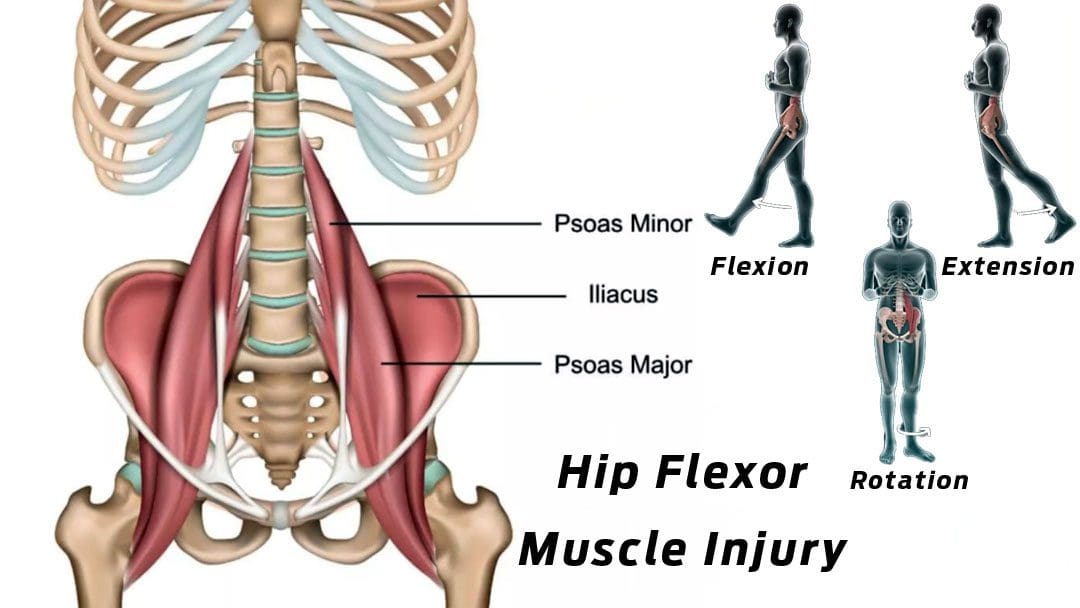
The iliopsoas muscle is a primary hip flexor that assists in the femur’s external rotation and maintains the hip joint’s strength and integrity. It also helps to stabilize the lumbar spine and pelvis. Athletes often overuse these muscles with all the sprinting, jumping, kicking, and changing directions when running, causing strains and/or tears. Repetitive hip flexion can result in chronic degenerative tendon changes. Chiropractic care and physical therapy can assist in the early phases of healing, safely transitioning to rehabilitation, and returning to physical activities.
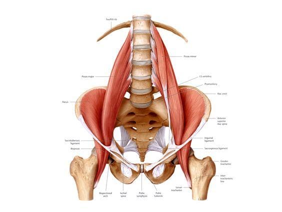
Iliopsoas Muscle
The hip flexors are the group of muscles, including the iliac and psoas major muscles/iliopsoas and the rectus femoris/quadriceps. One of the largest and thickest muscles in the body, the psoas, extends from the lumbar vertebrae, crosses in front of each hip, and attaches to the inside top of the thigh bone. The muscle works by flexing the hip joint and lifting the upper leg towards the body. These fibers can tear if tension is more than the muscle can bear. An iliopsoas strain occurs when one or more of these hip flexor muscles become overly stretched or begin to tear.
Injury
The injury can occur from sports or everyday physical activities. This leads to inflammation, pain, and scar tissue formation. An iliopsoas injury is commonly caused by sudden movements, including sprinting, kicking, and changing direction fast while running. Individuals participating in any sports, especially cycling, running, dance, tennis, martial arts, and soccer, are more likely to experience this injury. Other contributing factors include:
Muscle tightness
Joint stiffness
Muscle weakness
Inadequate core stability
Not warming up correctly
Improper biomechanics
Decreased fitness and conditioning
Individuals will feel a sudden stinging pain or pulling sensation, usually on the front of the hip, groin, or abdominal area. Other symptoms include:
Healing and recovery depend on the severity of the injury. A minor iliopsoas muscle injury can take around three weeks to recover fully. More serious strains and tears take six to eight weeks before returning to activity, as the tissue needs time to repair before starting rehabilitation.
Chiropractic Rehabilitation and Recovery
The first steps when dealing with this injury should be P.R.I.C.E. protection, rest, ice, compression, and elevation. It is important to rest and seek treatment immediately; if left untreated, the condition could worsen, lead to a chronic condition, and require surgery. A chiropractic treatment and rehabilitation plan will consist of the following:
Soft tissue massage
Joint mobilization
A chiropractor may recommend crutches to keep the weight off the hip.
A brace can help compress and stabilize the hip flexor to expedite healing.
A flexibility and strengthening program will be implemented to target the muscles around the hip.
Core strengthening exercises will improve the stability of the pelvis area to prevent any further overuse problems.
Wearing compression clothing could also be recommended, as the clothing helps maintain muscle temperature.
Labral Tear
References
Dydyk AM, Sapra A. Psoas Syndrome. [Updated 2022 Oct 24]. In: StatPearls [Internet]. Treasure Island (F.L.): StatPearls Publishing; 2022 Jan-. Available from: https://www.ncbi.nlm.nih.gov/books/NBK551701/
Lifshitz, Liran BPt, MSc, PT; Bar Sela, Shlomo BPt MPE; Gal, Noga BPt, MSc; Martin, RobRoy PhD, PT; Fleitman Klar, Michal BPt. Iliopsoas the Hidden Muscle: Anatomy, Diagnosis, and Treatment. Current Sports Medicine Reports 19(6):p 235-243, June 2020. | DOI: 10.1249/JSR.0000000000000723
Rauseo, Carla. “THE REHABILITATION OF A RUNNER WITH ILIOPSOAS TENDINOPATHY USING AN ECCENTRIC-BIASED EXERCISE-A CASE REPORT.” International journal of sports physical therapy vol. 12,7 (2017): 1150-1162. doi:10.26603/ijspt20171150
Rubio, Manolo, et al. “Spontaneous Iliopsoas Tendon Tear: A Rare Cause of Hip Pain in the Elderly.” Geriatric orthopedic surgery & rehabilitation vol. 7,1 (2016): 30-2. doi:10.1177/2151458515627309
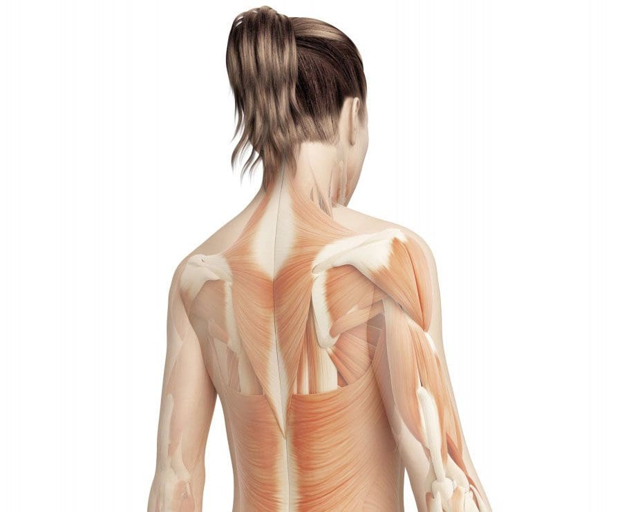
The spinal muscles and ligaments work in conjunction to help support the spine, maintain an upright posture, and control movements during activity and rest. The muscles are named based on shape, location, or a combination. Further categorization factors include muscle functions like flexion, extension, or rotation. Skeletal muscle is a form of striated muscle tissue that is voluntarily controlled by the somatic nervous system. Striated means it is striped in appearance. Most skeletal muscles are attached to bones by collagen fibers known as tendons.
Vertebral Muscle Types
Location
Forward flexors
Anterior
Lateral flexors
Lateral
Rotators
Lateral
Extensors
Posterior
It has the fastest contraction rate of all muscles. Before muscle/s contract, a nerve impulse starts in the brain and runs through the spinal cord to the muscle. For the muscles to contract and work properly they need energy/fuel. Mitochondria produce Adenosine triphosphate chemical cells that are needed for energy. Adenosine triphosphate is made as the mitochondria burn glucose or sugar. The blood vessels deliver the oxygen and nutrients that the mitochondria need to maintain a steady supply of adenosine triphosphate.
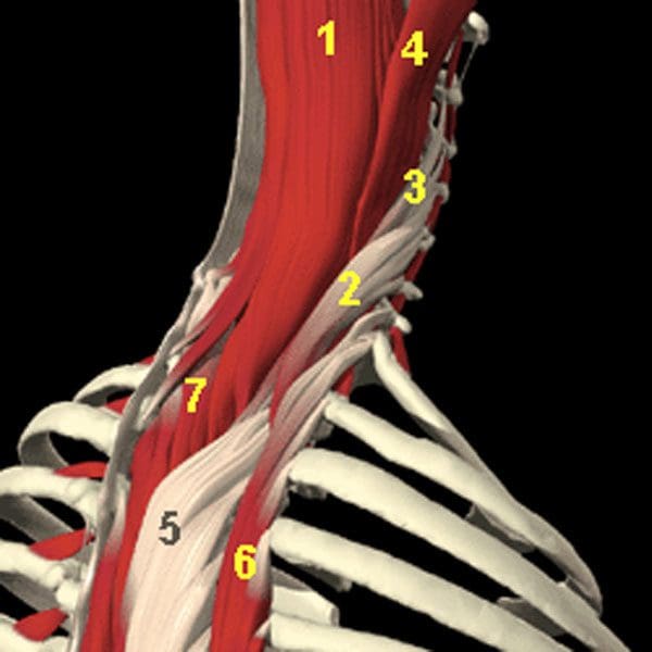
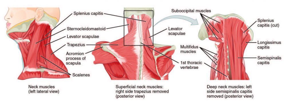
The Posterior Cervical and Upper Thoracic Spinal Muscles
Semispinalis Capitus – controls the head rotation and backward pulls
Iliocostalis Cervicis – extends the cervical vertebrae
The Longissimus Cervicus – extends the cervical vertebrae
Longissimus Capitus – controls the head’s rotation and backward pulls
Longissimus Thoracis – controls the extension/lateral flexion of the vertebral column and rib rotation
Iliocostalis Thoracis – controls the extension/lateral flexion of the vertebral column and rib rotation
Semispinalis Thoracis – extends and rotates the vertebral column
Muscles of the Spinal Column
Cervical muscles
Cervical Muscles
Function
Nerve
Sternocleidomastoid
Extends and rotates the head and flexes the vertebral column
C2, C3
Scalenus
Flexes and rotates the neck
Lower cervical
Spinalis Cervicis
Extends and rotates the head
Middle/lower cervical
Spinalis Capitus
Extends and rotates the head
Middle/lower cervical
Semispinalis Cervicis
Extends and rotates the vertebral column
Middle/lower cervical
Semispinalis Capitus
Rotates the head and pulls backward
C1-C5
Splenius Cervicis
Extends the vertebral column
Middle/lower cervical
Longus Colli Cervicis
Flexes the cervical vertebrae
C2-C7
Longus Capitus
Flexes the head
C1-C3
Rectus Capitus Anterior
Flexes the head
C2, C3
Rectus Capitus Lateralis
Bends the head laterally
C2, C3
Iliocostalis Cervicis
Extends the cervical vertebrae
Middle/lower cervical
Longissimus Cervicis
Extends the cervical vertebrae
Middle/lower cervical
Longissimus Capitus
Rotates the head and pulls backward
Middle/lower cervical
Rectus Capitus Posterior Major
Extends and rotates the head
Suboccipital
Rectus Capitus Posterior Minor
Extends the head
Suboccipital
Obliquus Capitus Inferior
Rotates the atlas
Suboccipital
Obliquus Capitus Superior
Extends and bends the head laterally
Suboccipital
Thoracic Muscles
Thoracic muscles
Function
Nerve
Longissimus Thoracis
Extension, lateral flexion of the vertebral column, and rib rotation
Perimysium is the sheath that groups the muscle fibers into bundles.
Endomysium is another type of connective tissue that sheaths each muscle fiber.
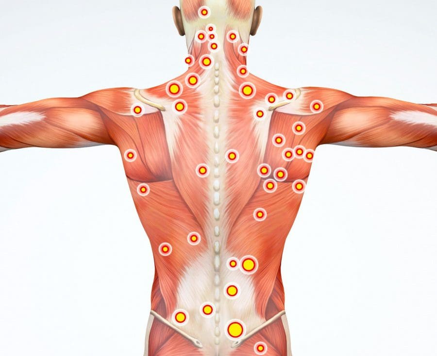
The cause of back pain and spinal muscle spasm/s can be caused by overuse, automobile accident, personal, work, or sports injury. The root cause of muscle spasm/s is usually a consequence of an injury to a structure within the lumbar spine. If there have been one or more episodes of muscle spasm in the low back, chances are it will re-occur. The muscles in the low back work together with the abdominal muscles. The spinal muscles add stability by maintaining an erect spine and maintain balance.
Back Pain Specialist
Dr. Alex Jimenez�s Blog Post Disclaimer
The scope of our information is limited to chiropractic, musculoskeletal, physical medicines, wellness, and sensitive health issues and/or functional medicine articles, topics, and discussions. We use functional health & wellness protocols to treat and support care for injuries or disorders of the musculoskeletal system. Our posts, topics, subjects, and insights cover clinical matters, issues, and topics that relate and support directly or indirectly our clinical scope of practice.*
Our office has made a reasonable attempt to provide supportive citations and has identified the relevant research study or studies supporting our posts. We also make copies of supporting research studies available to the board and or the public upon request. We understand that we cover matters that require an additional explanation as to how it may assist in a particular care plan or treatment protocol; therefore, to further discuss the subject matter above, please feel free to ask Dr. Alex Jimenez or contact us at 915-850-0900. The provider(s) Licensed in Texas& New Mexico*
IFM's Find A Practitioner tool is the largest referral network in Functional Medicine, created to help patients locate Functional Medicine practitioners anywhere in the world. IFM Certified Practitioners are listed first in the search results, given their extensive education in Functional Medicine