Low-level laser therapy (LLLT), also known as photobiomodulation, is the use of low-power lasers or light-emitting diodes (LEDs) for treatment purposes. When LLLT is used on the brain, it is known as transcranial LLLT or transcranial photobiomodulation. Many research studies have shown that LLLT can help treat a variety of brain health issues. �
Different from high-intensity surgical lasers, low-powered lasers do not cut or burn tissue. Instead, these lasers stimulate a biological reaction and promote cells to function properly. Moreover, it�s also easy to use LLLT utilizing red and near-infrared light on your own home. In the article below, we will discuss the brain health benefits of low-level laser therapy (LLLT). �
How Low-Level Laser Therapy Works
Research studies show that red and near-infrared light between the wavelengths of 632 nanometers (nm) and 1064 nm can have brain health benefits. For brain cells or neurons, the optimal range for the wavelengths seems to be between 800 nm and 1000 nm as these can penetrate the scalp and skull to reach the brain. Most devices ultimately fall within this range. �
The light given off from these devices stimulate a photochemical response within neurons or brain cells, which can increase the natural healing process and can also cause beneficial changes in their behavior by supporting the mitochondria. The mitochondria are the �powerhouses of the cell,� producing most of the energy in the human body in the form of adenosine-5- triphosphate (ATP). ATP is the cell’s main source of energy. The brain constantly needs to use it to function properly. �
Proper mitochondrial function and ATP production are fundamental for neuroprotection and cognitive enhancement as well as for the prevention and treatment of a variety of neurological diseases. Research studies have shown that transcranial LLLT promotes proper mitochondrial function and considerably improves the production of ATP in the human brain. �
The mitochondria have photoreceptors which absorb the photons from light and turn them into ATP or energy which can be utilized to perform cellular tasks and biological processes. This system is similar to that of plant photosynthesis where sunlight is absorbed by plants and turned into energy for the plants to grow. Furthermore, by stimulating the mitochondria and producing more ATP, LLLT gives brain cells or neurons even more ATP energy to better heal and repair themselves. �
On top of this, low-level laser therapy has also been shown to: �
Decrease free radicals and oxidative stress in the brain
Increase blood flow and circulation, including within the frontal cortex
Reduce pain by supporting the human body�s opioids or natural pain relievers
Increase rate of oxygen consumption in the frontal cortex
Increase serotonin
Many traumatic brain injuries and neurological diseases can be treated with LLLT, including anxiety, depression, post-traumatic stress disorder (PTSD), post-concussion syndrome, stroke, Alzheimer’s disease, and dementia. We will discuss how low-level laser therapy (LLLT) has been shown to help each of the brain health issues, among others, demonstrated below. �
LLLT for Traumatic Brain Injury
Traumatic brain injury (TBI) is a growing brain health issue where approximately 1.7 million people experience some type of TBI in the U.S. every year. Mild TBIs or concussions make up about 75 percent of all traumatic brain injuries. Military personnel frequently experience TBI and many of them often struggle with PTSD, anxiety, and depression. �
Several research studies have shown that patients with chronic mild TBI have experienced improved cognition, memory and sleep with LLLT. One research study also evaluated whether LLLT could help treat 11 patients with chronic mild TBI symptoms. Two patients had cognitive dysfunction and four patients had multiple concussions. �
After 18 LLLT sessions, the patient’s cognition, memory and verbal learning improved. Participants also said that they slept better and had fewer PTSD symptoms. Coworkers, friends, and family also reported improved social, interpersonal, and occupational functioning. In another research study, 10 people with chronic TBI were given 10 LLLT sessions and experienced reduced headaches, cognitive dysfunction, sleep problems, anxiety, depression and irritability. �
Several mice research studies also show that LLLT can prevent cell death and increase neurological performance after TBI. Researchers believe that LLLT improves TBI symptoms because the mitochondria in the brain can become dysfunctional after TBI, resulting in an inadequate supply of ATP. LLLT can support the mitochondria and increase ATP production. �
After traumatic brain injury (TBI) there is also poor blood flow and oxygenation, and increased inflammation and oxidative stress in the brain. This can ultimately cause brain damage, however, LLLT can help treat these brain health issues as well as help increase antioxidants, promote neurogenesis, and relieve chronic symptoms, among other brain health benefits. �
LLLT for Depression and Anxiety
Research studies in both rats and humans have shown that LLLT can improve mood and reduce symptoms of depression. In 2009, researchers took 10 patients with a history of major anxiety and depression, including PTSD and substance abuse, and utilized LLLT for four weeks. At the end of the research study, six of the 10 patients experienced remission of their depression and seven of the 10 patients experienced remission of their anxiety. There were no observable side-effects. �
Several research studies have shown that depression is associated with abnormal blood flow in the frontal cortex of the brain. LLLT increases blood flow and circulation. Other research studies have shown that participants report improved positive emotions and reduced depressive symptoms after LLLT treatment. Participants with TBI also experienced a decrease in anxiety, depression, irritability, and insomnia as well as an overall improvement in quality of life after LLLT. �
LLLT for Alzheimer’s Disease and Dementia
Research studies show that LLLT can boost performance and improve cognitive function, including attention and memory, in animals, young healthy people and elderly people. Preliminary research studies also show that LLLT may ultimately help slow down the progression of Alzheimer�s disease by decreasing a protein in the brain which is associated with dementia. �
The downregulation of brain-derived neurotrophic factor (BDNF) occurs early in the progression of Alzheimer’s disease and dementia. Research studies have shown that LLLT can also help prevent brain cell or neuron loss by upregulating BDNF. �
Researchers have also utilized LLLT in middle-aged mice and discovered that the memory and cognitive performance of the middle-aged mice improved so much that it became similar to that of young mice. The researchers concluded that LLLT should be utilized in cases of general cognitive impairment in elderly people or even for Alzheimer’s disease and dementia. �
Several other research studies have shown that LLLT increases alertness, awareness and sustained attention as well as improves short-term memory and reaction time. Research study participants also made fewer errors during tests. Another research study found that LLLT enhanced cognition by promoting neuroprotection and supporting the mitochondria. �
LLLT for Stroke
Numerous studies also show that LLLT reduces neurological problems and improves behavior in rats and rabbits after stroke. It also increases the growth of new brain cells or neurons, improving their overall recovery. Multiple other research studies also show that LLLT can considerably reduce brain damage and improve recovery outcome measures after a stroke. �
In one research study, researchers utilized LLLT on patients approximately 18 hours after they experienced a stroke. Five days after the stroke, they found considerably greater improvements in the LLLT-treated group. The improvements continued 90 days after the stroke. At the end of the research study, 70 percent of the patients treated with LLLT had successful outcome measures in comparison with only 51 percent of the control subjects in the research study. �
Follow up research studies with over 600 stroke patients found similar brain health benefits associated with low-level laser therapy (LLLT). Researchers believe that the increase in the production of ATP is responsible for the improvements. �
Low-level laser therapy, or LLLT, is a non-invasive treatment approach which utilizes low-power lasers or light-emitting diodes for the treatment of brain health issues and neurological diseases. Many research studies with both animal and human trial have demonstrated that LLLT provides many brain health benefits without harmful side-effects. Healthcare professionals can help improve the symptoms of brain health issues and neurological diseases with a variety of treatment methods and techniques. Proper diagnosis is fundamental for proper treatment. – Dr. Alex Jimenez D.C., C.C.S.T. Insight
Low-level laser therapy (LLLT), also known as photobiomodulation, is the use of low-power lasers or light-emitting diodes (LEDs) for treatment purposes. In the article above, we discussed the brain health benefits of low-level laser therapy (LLLT) on a variety of brain health issues and neurological diseases. The scope of our information is limited to chiropractic, musculoskeletal and nervous health issues as well as functional medicine articles, topics, and discussions. To further discuss the subject matter above, please feel free to ask Dr. Alex Jimenez or contact us at 915-850-0900 . �
Curated by Dr. Alex Jimenez �
Additional Topic Discussion: Chronic Pain
Sudden pain is a natural response of the nervous system which helps to demonstrate possible injury. By way of instance, pain signals travel from an injured region through the nerves and spinal cord to the brain. Pain is generally less severe as the injury heals, however, chronic pain is different than the average type of pain. With chronic pain, the human body will continue sending pain signals to the brain, regardless if the injury has healed. Chronic pain can last for several weeks to even several years. Chronic pain can tremendously affect a patient’s mobility and it can reduce flexibility, strength, and endurance.
Neural Zoomer Plus for Neurological Disease
�
Dr. Alex Jimenez utilizes a series of tests to help evaluate neurological diseases. The Neural ZoomerTM Plus is an array of neurological autoantibodies which offers specific antibody-to-antigen recognition. The Vibrant Neural ZoomerTM Plus is designed to assess an individual�s reactivity to 48 neurological antigens with connections to a variety of neurologically related diseases. The Vibrant Neural ZoomerTM Plus aims to reduce neurological conditions by empowering patients and physicians with a vital resource for early risk detection and an enhanced focus on personalized primary prevention. �
Formulas for Methylation Support
XYMOGEN�s Exclusive Professional Formulas are available through select licensed health care professionals. The internet sale and discounting of XYMOGEN formulas are strictly prohibited.
Proudly,�Dr. Alexander Jimenez makes XYMOGEN formulas available only to patients under our care.
Please call our office in order for us to assign a doctor consultation for immediate access.
If you are a patient of Injury Medical & Chiropractic�Clinic, you may inquire about XYMOGEN by calling 915-850-0900.
�
For your convenience and review of the XYMOGEN products please review the following link.*XYMOGEN-Catalog-Download �
* All of the above XYMOGEN policies remain strictly in force.
Central nervous system, or CNS, infections can be life-threatening if they are not diagnosed and treated early. Because CNS infections are non-specific, determining an accurate diagnosis can be challenging. The nucleic acid in vitro amplification-based molecular methods are starting to be utilized for routine microbial diagnosis. These molecular methods have improved beyond conventional diagnostic techniques with increased sensitivity and specificity. Moreover, molecular methods utilized on cerebrospinal fluid samples are considered the new standard for diagnosis of CNS infections caused by pathogens. �
Molecular methods for the diagnosis of CNS infections offers a variety of monoplex and multiplex PCR assays to diagnose several types of health issues. Pan-omic molecular platforms can also help diagnose CNS infections. Although molecular methods are utilized for the diagnosis of CNS infections, the outcome measures for these diagnostic techniques must be carefully identified by healthcare professionals. The following article discusses conventional diagnostic techniques and molecular methods utilized for the diagnosis of central nervous system infections, their application, and future approaches. �
Molecular Methods in the Diagnosis of CNS Infections
Because of increased sensitivity and specificity, nucleic acid in vitro amplification-based molecular methods has tremendously improved the ability to diagnose CNS infections in a reasonable and effective time frame. Several PCR-derived techniques have also ultimately increased the flexibility and rigor of currently available diagnostic techniques. �
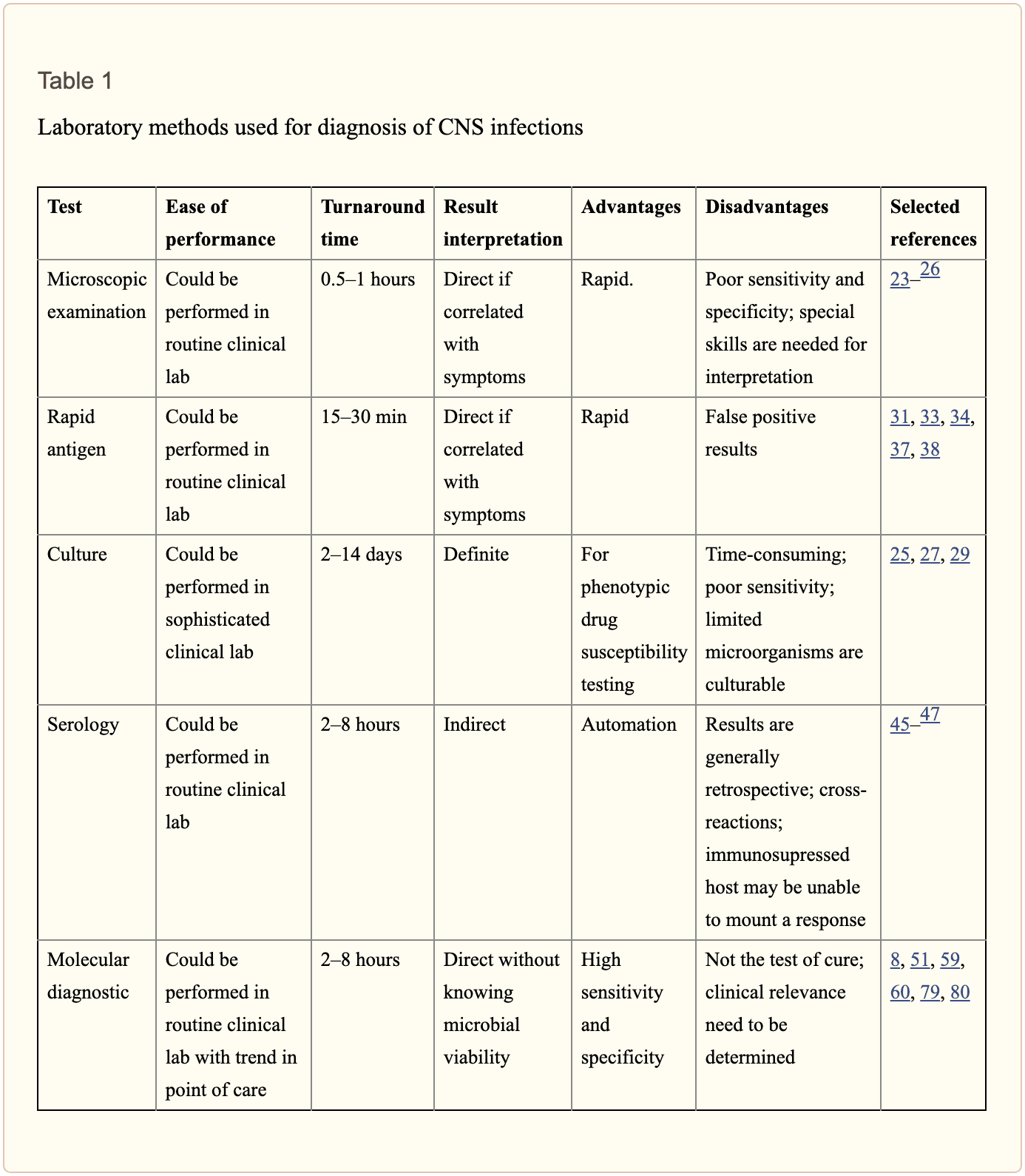
Reverse transcriptase, or RT,-PCR was developed to increase RNA targets. Its utilization plays a fundamental role in the diagnosis of RNA-virus infections as well as managing their reaction to treatment. Timely access to enterovirus RT-PCR outcome measures has demonstrated shorter hospital stays, reduced unnecessary antibiotic utilization, and decreased ancillary laboratory evaluations and tests. Broad-range rRNA PCR techniques, which utilize a single pair of primers targeting conserved regions of genes, have been utilized to diagnose bacterial pathogens and herpes viruses in the CSF. Isothermal amplification-based techniques. including loop-mediated isothermal amplification or LAMP, have been developed to offer a diagnosis within several minutes to hours. Table 2 demonstrates commercial molecular in vitro diagnostic devices, or IVD, which have been cleared by the US Food and Drug Administration, or FDA, for diagnosis of microbial pathogens in CSF. �
Monoplex Assays
A conventional molecular method involves three phases: sample extraction, target nucleic acid amplification, and amplicon detection. One of the first molecular assays successfully utilized for the diagnosis of CNS infections was utilized for the diagnosis of HSV in cerebrospinal fluid or CSF. PCR became the test of choice when research studies demonstrated that CSF PCR was similar to culture of brain tissue for diagnosis of HSV encephalitis and meningitis. Many PCR based methods for the diagnosis of herpes and enteroviruses have become available with increased sensitivity compared to viral culture. �
Real-time PCR with nucleic acid amplification and amplicon detection further improved the transition to molecular methods in clinical laboratories. Unlike conventional PCR, the real-time system is a �closed� system and it overcomes the fundamental problem of carryover contamination. At the time of manuscript preparation, three molecular assays utilized to help diagnose HSV and enteroviruses in CSF have ultimately been approved by the FDA as demonstrated in Table 2 of the previous article. � Real-time PCR-based methods are the main diagnostic technique utilized to help diagnose the Zika virus, which was first reported in Uganda in 1947, and is now a worldwide concern after the virus spread widely in Brazil and Central America. Research studies developed a one-step RT-PCR assay utilized to diagnose the Zika virus in human serum with a limited detection of 7.7pfu/reaction. Along with plasma, the Zika virus RNA can be diagnosed through urine and plasma within the first 2 weeks after symptoms have manifested. In March 2016, the FDA approved a trioplex-PCR assay under emergency use authorization for the simultaneous diagnosis of Zika, Chikungunya, and Dengue viruses in serum, urine, CSF and amniotic fluid. The RT-PCR assay utilizes dual labeled hydrolysis probes with a LOD of 1.54�10 4 GCE/ ml of Zika virus in serum. �
Introduction of real-time PCR based diagnostic assays have affected early and effective diagnosis of several bacterial infections. Isothermal amplification-based molecular assays have excellent performance characteristics and they don’t require any specialized equipment. These assays are fundamental for the utilization of on or near point-of-care testing. LAMP-based methods have been utilized to diagnose Neisseria meningitis, Streptococcus pneumoniae, Haemophilus influenzae type b, M. tuberculosis, and JEV in the CSF. The Xpert MTB/RIF assay has tremendously improved regulation of tuberculosis by offering an integrated and automated system which allows quick clinical decision making in a POC or near-care context. Several research studies have utilized the Xpert MTB/RIF to evaluate the diagnosis of M. tuberculosis in CSF from TB meningitis. In a meta-analysis of thirteen research studies, the pooled sensitivity of the Xpert assay was 80.5 percent, or 95 percent CI 59.0 percent to 92.2 percent, against culture and 62.8 percent, or 95 percent CI 47.7 percent to 75.8 percent, against composite standard. Utilizing a large volume of sample, of at least 8�10 ml, is necessary for testing CSF and centrifugation can cause considerable improvements in yield. Despite the lack of standardization for sample processing, WHO has allowed testing CSF with the automated Xpert MTB/RIF assay as the first-line test over conventional microscopy. �
Multiplex Assays
Simplicity makes multiplex molecular assays fundamental for the diagnosis of a panel of microbial targets. Several multiplex PCR assays have been developed to diagnose bacterial pathogens in CSF targeting the most common causes of meningitis: S. pneumoniae, N. meningitis, H. influenzae, L. monocytogenes, S. agalactiae, S. aureus, E. coli, and M. pneumoniae. A multiplex PCR followed by Luminex suspension array can simultaneously diagnose eight bacterial and viral pathogens in CSF, including N. meningitis, S. pueumoniae, E. coli, S. aureus, L. monocytogenes, S. agalactiae, HSV-1/2, and VZV, among others. �
Considering the variety of pathogens involved in CNS infection, application of comprehensive molecular panels with multiple bacterial and viral targets have improved the efficiency of diagnosis. The BioFire FilmArray Meningitis/Encephalitis panel is currently the only FDA cleared multiplex assay utilized for the diagnosis of six bacterial, such as Escherichia coli K1, Haemophilus influenzae, Listeria monocytogenes, Neisseria meningitides, Streptococcus agalactiae and Streptococcus pneumoniae, seven viral, such as cytomegalovirus, enterovirus, HSV-1, HSV-2, human herpesvirus 6 or HHV-6, human parechovirus and VZV, as well as a single fungal, such as Cryptococcus neoformans/gattii, target in CSF as demonstrated in Table 2. The integrated FilmArray system takes about an hour, with only 2 minutes of hands-on time. During the preparation of the manuscript, two research studies demonstrated the performance of this assay. Utilizing 48 samples from gram stain negative CSF samples from suspected cases of meningitis, research studies demonstrated that this system diagnosed more viral pathogens, such as EBV. Four cases of WNV and a single case of Histoplasma were not diagnosed by this assay. Among HIV infected patients in Uganda, the test performance demonstrated increased sensitivity and specificity for the diagnosis of Cryptococcus. Although the FilmArray Meningitis/Encephalitis panel offers a quick diagnosis of CNS infections, further research studies are needed to determine its performance for a variety of targets and other high-risk populations. �
Co-infections are frequently found among immunocompromised patients and can ultimately be challenging to diagnose for clinicians. The multiplex design allows simultaneous diagnosis of multiple targets on the same sample. One research study utilized a panel of monoplex and multiplex molecular assays to conduct a prospective cohort research study in Uganda to comprehensively evaluate the etiology of meningitis among HIV-infected adults. Among the 314 HIV-infected patients with meningitis, EBV co-infection was diagnosed with Cryptococcus, M. tuberculosis, or other viral pathogens. EBV in CSF in these settings is not completely understood although a single research study associated increased EBV viral load as a marker of poor outcome measures in patients with bacterial meningitis and EBV co-infection/ reactivation, among others. �
Pan-Omic Molecular Assays
Technological improvements in metagenomic deep sequencing have increased its utilization for clinical diagnosis of CNS infections. Several research studies have demonstrated its ability to solve diagnostic technique problems which challenge the limits of traditional laboratory testing. Due to sterile status and protection by BBB, CSF and brain biopsies are fundamental to further explore the utilization of this technology for pathogen diagnosis. Metagenomics was successfully utilized to establish a diagnosis of neuroleptospirosis in a 14-year-old boy with severe combined immunodeficiency who also suffered from recurrent bouts of fever, headache, and coma. Similarly, high-throughput RNA sequencing performed on brain biopsy from an 18-month-old boy with encephalopathy diagnosed a new Astrovirus as the cause. Despite the utilization of metagenomics for the diagnosis of infectious disease, there are many technological and practical concerns which need to be addressed before this form of diagnostic testing can become mainstream and part of the clinical standard of care. �
Other promising advances have occurred in transcriptomics, proteomics and metabolomics. Host and microbial microRNA or miRNA, profiles have been utilized for a variety of inflammatory and infectious diseases. Two miRNAs, miR-155 and miRNA-29b, were reported as potential biomarkers for JEV infection and treatment targets for anti-JEV therapy. Host neural epidermal growth factor, including 2 and apolipoprotein B in CSF, was able to diagnose tuberculous meningitis with 83.3 percent to 89.3 percent sensitivity and 75 percent to 92 percent specificity. CSF metabolite profiling has been reported to be useful in the classification, diagnosis, epidemiology, and treatment assessment of CNS infections in HIV patients. CSF metabolic profile analysis demonstrated bioenergetic adaptation in regulating shifts of HIV-infected patients. �
Outcome Measures Associated with Diseases
Diagnosis of an etiologic agent in patients with CNS infections needs consideration of the most common causative organisms, the available diagnostic techniques and molecular methods for these agents, and the highest-yield clinical specimens for evaluation and testing. Knowledge of the epidemiology and clinical presentation of specific agents is fundamental in selecting which diagnostic methods are appropriate for patients. Animal or vector exposures, geographic location, recent travel history, season of the year, exposure of ill contacts, and occupational exposures should be considered. �
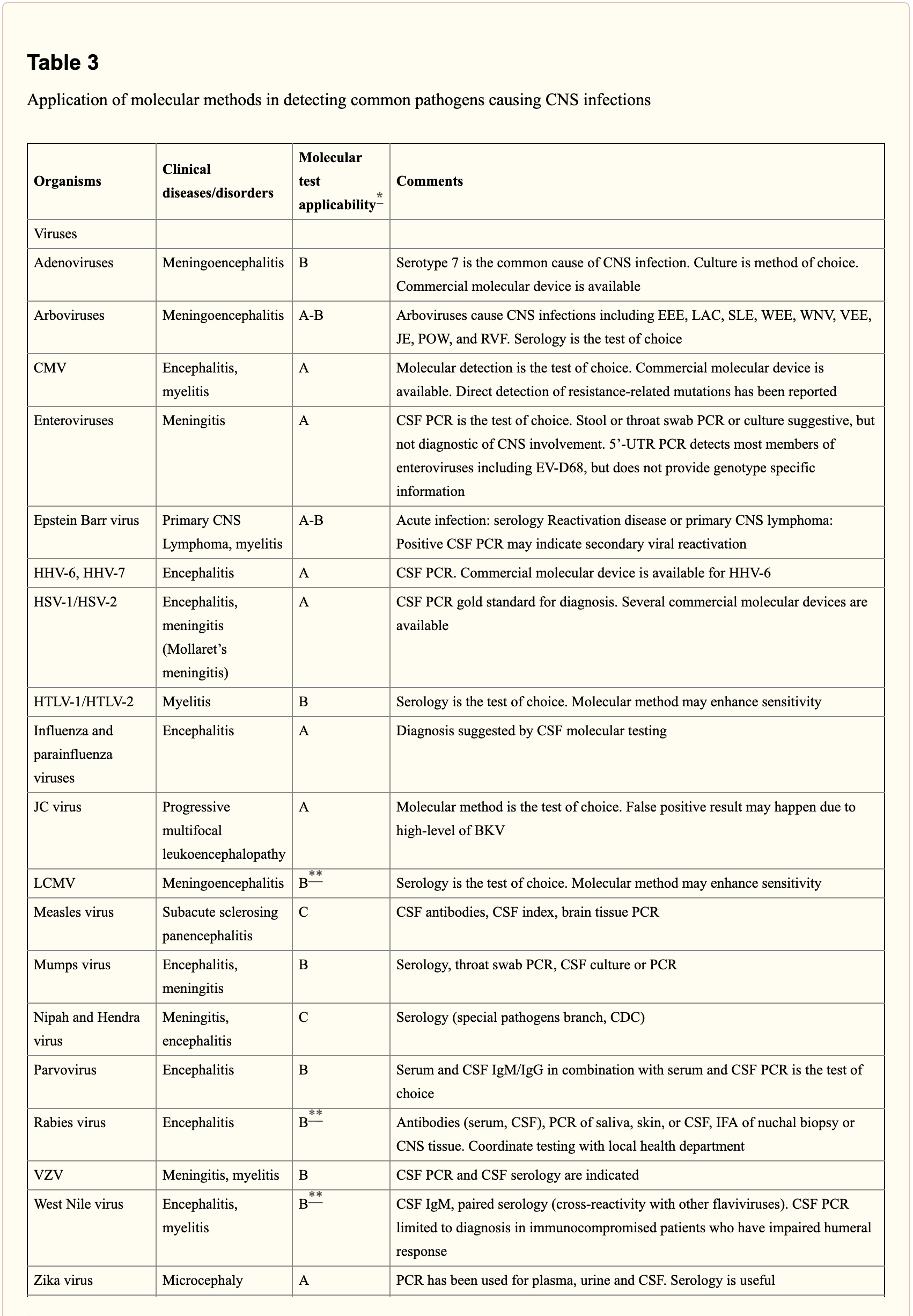
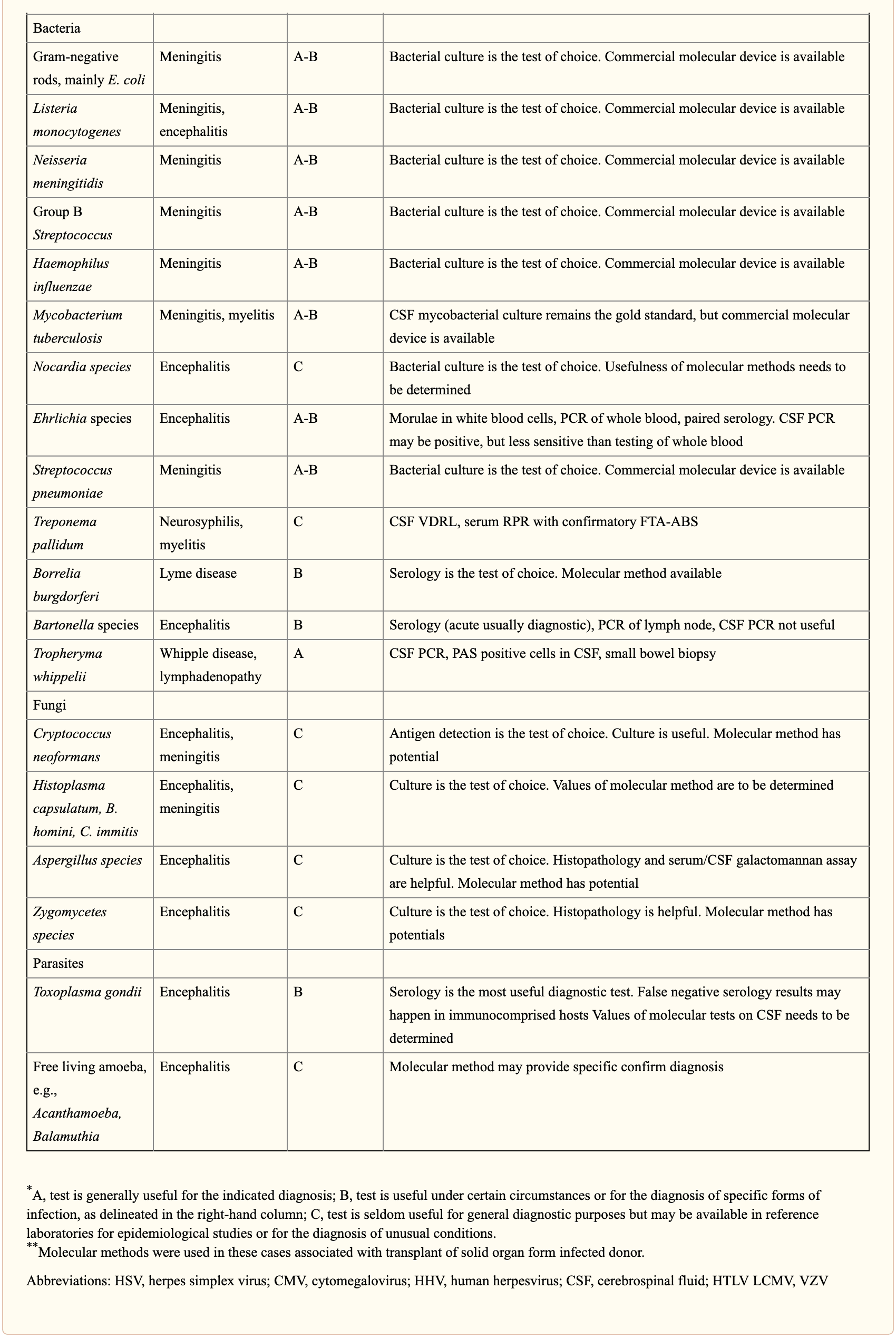
When selecting appropriate pathogen-specific molecular diagnostic methods, the following factors should be considered. CSF is the optimal specimen for PCR testing for patients with meningitis or meningoencephalitis. While indirect evidence can be determined by testing other specimen types, attempts should be made to obtain CSF samples early before treatment can compromise yield. Time of testing from the manifestation of symptoms is fundamental to understand and rule out false-negative results and recommend retesting within a certain time frame. By way of instance, HSV PCR can commonly render false-negative results if CSF sample is obtained very early or late in the process of HSE infection. Host health is also known to affect test performance characteristics. Immunocompromised patients are at risk for infection by a variety of opportunistic pathogens, by way of instance HHV-6, JC virus, Toxoplasma encephalitis in bone marrow transplant recipients and patients with HIV. Often, infection can be more severe, such as WNV, and challenging to diagnose in this population. Table 3 below demonstrates practical recommendations on application and pitfalls of molecular test for the diagnosis of CNS infections. �
Furthermore, a positive nucleic acid amplification testing results are considered to be complicated by the fact that some viruses survive latently in macrophages or neurologic tissues even if they’re incidentally diagnosed by sensitive molecular techniques without an actual pathogenic role which can potentially lead to overtreatment. Utilization of adjunctive biomarkers which help determine active replication is being explored to overcome this drawback in research studies. �
Historically, the diagnosis of microbiologic agents in patients with CNS infections has been hindered by the low yield of CSF culture for viral and fastidious bacterial organisms, delays in CNS production of organism-specific antibodies, and challenges in determining optimum samples for testing. The nucleic acid in vitro amplification-based molecular diagnostic methods and techniques have a wider and better application in clinical microbiology practice. The monoplex assay will likely be the main platform utilized for urgent, random-access, low throughput assays. Multiplex assays have the additional benefit of diagnosing multiple targets and mixed infections. As the volume of CSF sample retrieved is often small, multiplex assays enable comprehensive diagnostic analysis with a low amount of sample, obviating the need for repeated lumbar punctures. The clinical relevance and cost-effectiveness of simultaneous multi-pathogen diagnosis strategies need further research studies. Application of pan-omic techniques in challenging to diagnose CNS infections is the new exciting frontier, the technology is promising but routine implementation is expected to be slow due to various challenges, such as lack of applicable regulatory guidelines and adaptation in the clinical setting, although the outcome measures are promising. �
As previously mentioned, central nervous system, or CNS, infections can be life-threatening health issues if they are not accurately diagnosed and properly treated. However, determining a diagnosis of CNS infections can be challenging for many clinicians. Fortunately, a variety of diagnostic techniques and molecular methods can ultimately help determine the source of CNS infections and other health issues. These diagnostic techniques and molecular methods have tremendously improved over the years, as previously mentioned, and more of these evaluations are being utilized in clinical settings to accurately diagnose CNS infections for proper treatment. – Dr. Alex Jimenez D.C., C.C.S.T. Insight
In part 2 of our “Diagnosis of Central Nervous System Infections” article, we discussed the molecular methods and the pan-omic molecular assays which are utilized in the diagnosis of CNS infections as well as how specific testing outcome measures have ultimately been associated with clinical diseases and health issues. The scope of our information is limited to chiropractic, musculoskeletal and nervous health issues as well as functional medicine articles, topics, and discussions. To further discuss the subject matter above, please feel free to ask Dr. Alex Jimenez or contact us at 915-850-0900 . �
Curated by Dr. Alex Jimenez �
Additional Topic Discussion: Chronic Pain
Sudden pain is a natural response of the nervous system which helps to demonstrate possible injury. By way of instance, pain signals travel from an injured region through the nerves and spinal cord to the brain. Pain is generally less severe as the injury heals, however, chronic pain is different than the average type of pain. With chronic pain, the human body will continue sending pain signals to the brain, regardless if the injury has healed. Chronic pain can last for several weeks to even several years. Chronic pain can tremendously affect a patient’s mobility and it can reduce flexibility, strength, and endurance.
Neural Zoomer Plus for Neurological Disease
Dr. Alex Jimenez utilizes a series of tests to help evaluate neurological diseases. The Neural ZoomerTM Plus is an array of neurological autoantibodies which offers specific antibody-to-antigen recognition. The Vibrant Neural ZoomerTM Plus is designed to assess an individual�s reactivity to 48 neurological antigens with connections to a variety of neurologically related diseases. The Vibrant Neural ZoomerTM Plus aims to reduce neurological conditions by empowering patients and physicians with a vital resource for early risk detection and an enhanced focus on personalized primary prevention. �
Formulas for Methylation Support
XYMOGEN�s Exclusive Professional Formulas are available through select licensed health care professionals. The internet sale and discounting of XYMOGEN formulas are strictly prohibited.
Proudly,�Dr. Alexander Jimenez makes XYMOGEN formulas available only to patients under our care.
Please call our office in order for us to assign a doctor consultation for immediate access.
If you are a patient of Injury Medical & Chiropractic�Clinic, you may inquire about XYMOGEN by calling 915-850-0900.
�
For your convenience and review of the XYMOGEN products please review the following link.*XYMOGEN-Catalog-Download �
* All of the above XYMOGEN policies remain strictly in force.
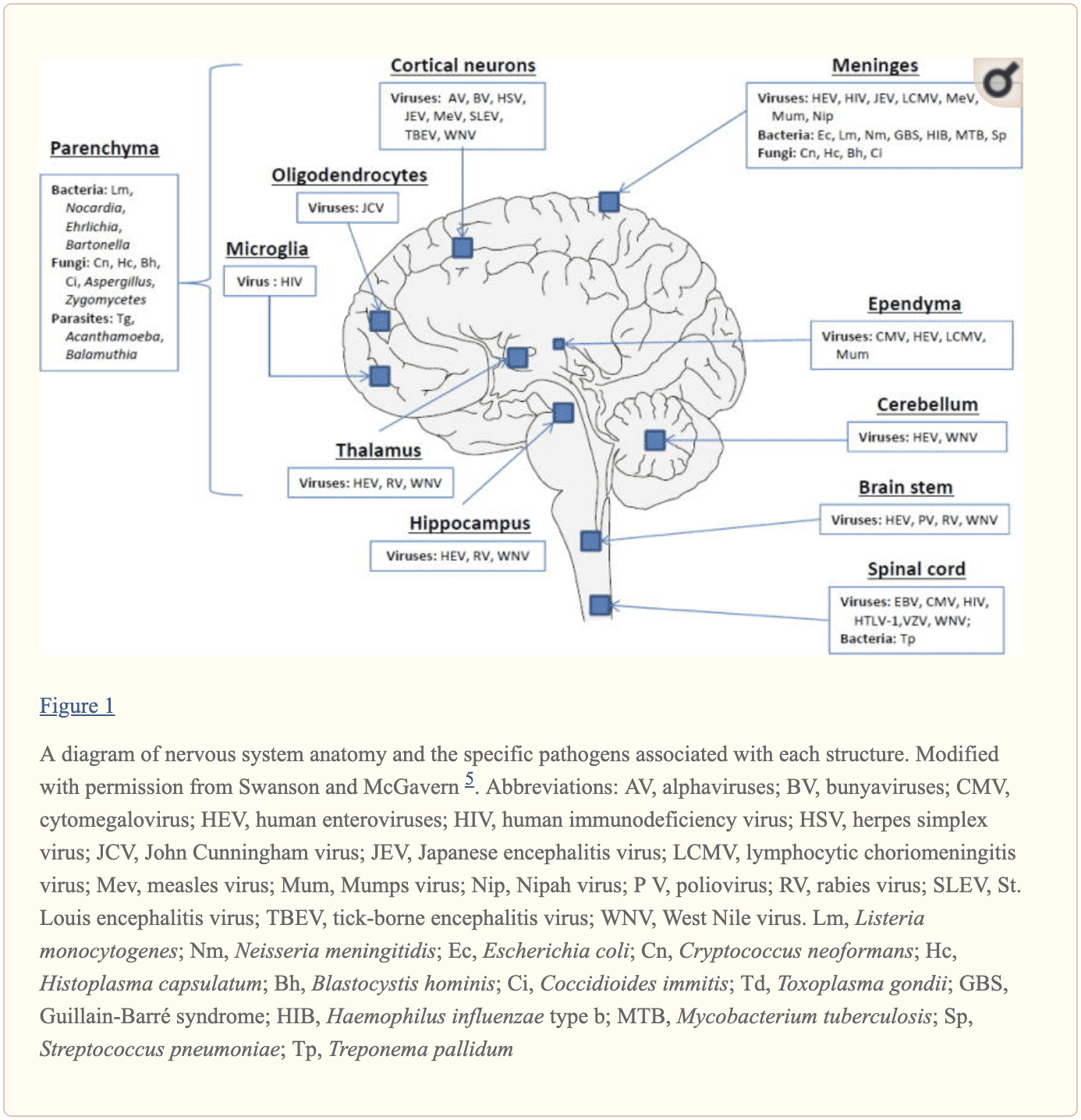
The central nervous system, or CNS, plays a fundamental role in the pathogenesis of infection. The CNS is regulated by the blood-brain barrier or BBB, however, it can still be exposed to a microbial invasion from a contiguous focus, hematogenous dissemination, or intraneural passage of organisms. A variety of environmental or commensal bacteria, viruses, fungi, protozoa, or parasites can enter the CNS and cause a variety of infections and health issues. Central nervous system infections can ultimately cause headache, stiff neck, vomiting, fever, photophobia, and focal neurological symptoms. �
What are Central Nervous System Infections?
CNS infections are characterized according to their affected region. Infection of the brain, spinal cord, and meninges results in meningitis, encephalitis, brain abscess, and myelitis. Infections can affect single or multiple regions of the brain, such as meningoencephalitis and encephalomyelitis. Moreover, CNS infections are characterized as acute, sub-acute, chronic, or recurrent based on their duration. Meningitis can cause headache, neck stiffness, fever, and photophobia over a period of hours to days. Encephalitis can cause brain parenchymal inflammation which can ultimately cause lethargy to coma. Last but not least, Myelitis can cause inflammation of the spinal cord including headache, fever, and paraparesis or paralysis. �
One of the most fatal CNS infections, acute bacterial meningitis, with three to five cases for every 100,000 people in the United States, has a mortality rate of 6 percent to 26 percent. Approximately 4,000 cases of acute bacterial meningitis occur in the U.S. every year with about 500 deaths. The frequent cause of acute bacterial meningitis includes Streptococcus pneumoniae, group B Streptococcus, Neisseria meningitides, Haemophilus influenzae, and Listeria monocytogenes. �
CNS infections caused by viruses are more common and are mostly mild and self-limited. However, these can manifest as meningitis and/or encephalitis. CNS infections caused by viruses can vary due to region and season. Non-polio enteroviruses are responsible for the majority of meningitis and/or encephalitis cases from late spring to fall. CNS infections due to herpes simplex viruses, or HSV, are associated with sporadic encephalitis and meningitis with severe sequelae if left untreated. �
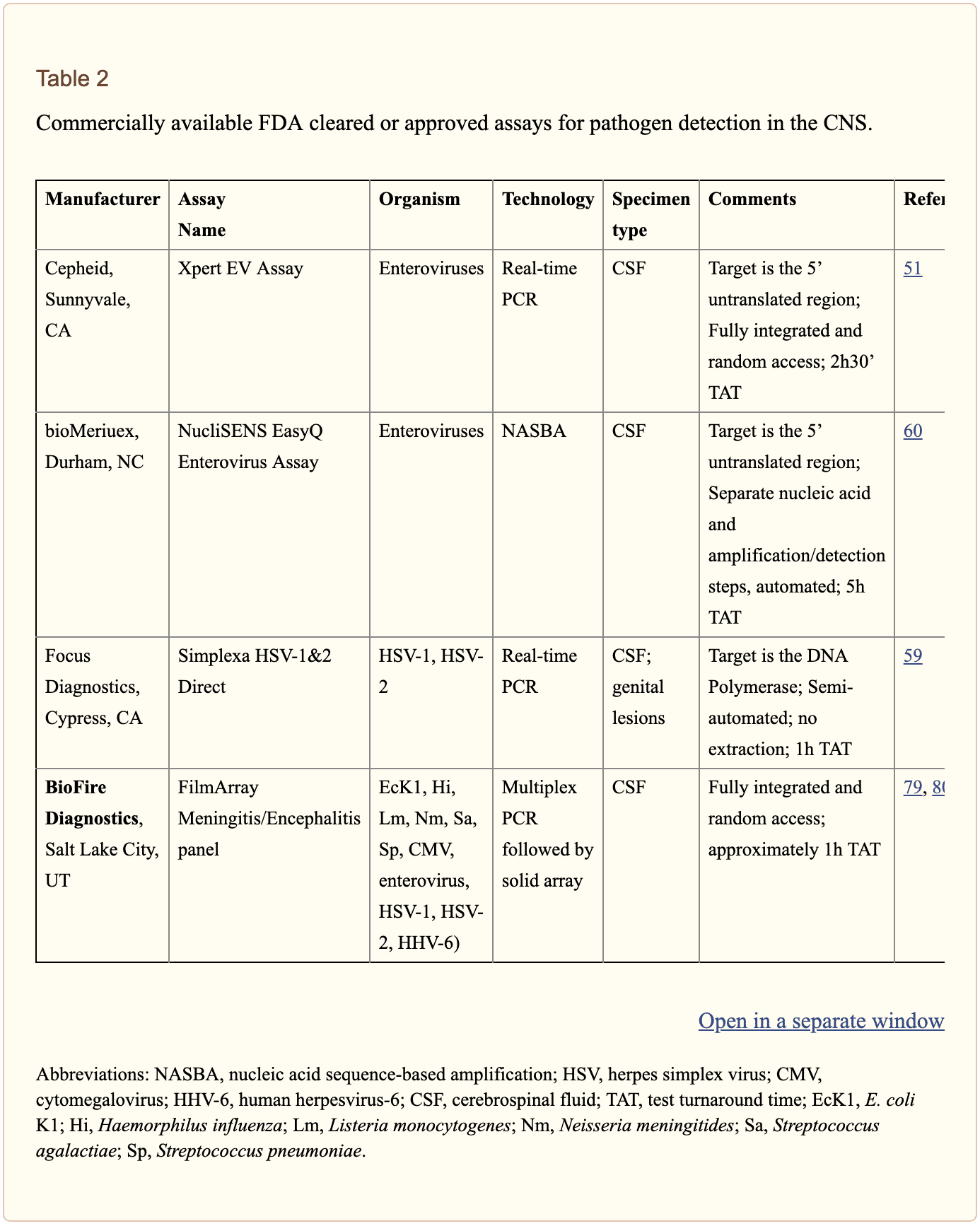
Diagnosis of CNS Infections
Diagnosis of microbial pathogens is fundamental for treatment. Methods and techniques utilized in clinical microbiology laboratories include direct microscopic examination, and culture techniques as well as antigen and antibody detection assays. However, each method and technique has several essential limitations. By way of instance, direct microscopic examination of CSF restricted sensitivity and specificity. The sensitivity of culture for enteroviruses is between 65 percent to 75 percent with average retrieval time of 3.7 to 8.2 days. Moreover, several serotypes of enteroviruses, especially Coxsackievirus A strains, are well-known to be non-cultivable and frequently grow poorly. Because enteroviruses are missing a common antigen found throughout a variety of serotypes, universal antigen and/or antibody diagnosis is impossible. Diagnosis of CNS HSV infections through methods and techniques utilized to determine culture sensitivity from CSF is tremendously poor. The presence of HSV IgG antibodies in CSF can ultimately be utilized to determine a diagnosis, however, the production is delayed until day 10 or day 12 after infection and it is not considered ideal for early diagnosis.
Diagnostic techniques, especially PCR based amplification, have developed a variety of mainstay tools to help determine the diagnosis of microbial pathogens in CSF. Molecular methods have demonstrated greater diagnosis rates than other diagnostic techniques. One research study demonstrated that 16S rRNA PCR-based assays were able to diagnose the causative organism in 65 percent of banked CSF samples compared to 35 percent when utilizing culture and microscopy. In another research study, diagnosis based on diagnostic techniques like molecular methods were utilized to optimize antibiotic treatment of patients with infectious meningitis when conventional methods and techniques demonstrated a negative outcome measure. Molecular methods and diagnostic techniques utilized on CSF samples are a fundamental standard when compared to the culture standard in the diagnosis of CNS infections caused by viruses which are challenging to diagnose. �
The diagnosis of CNS infections has tremendously changed over the last several years. PCR-based molecular methods have become a fundamental element in the clinical microbiology laboratory, providing tools for an accurate diagnosis. As demonstrated in Table 2, a variety of commercial molecular assays have been cleared by the Food and Drug Administration, or FDA, for the diagnosis of microbial pathogens. The approved assays for pathogen detection in the CNS are shown below. �
However, there are still several challenges in molecular diagnostic techniques and methods. Utilizing a combination of conventional diagnostic techniques and molecular methods, research studies demonstrated that in approximately 62 percent of patients with encephalitis, an etiologic organism could not be identified. Researchers have started to focus on developing advanced techniques and methods. In the following series of articles, we will demonstrate an update on the current conventional and molecular platforms utilized for the diagnosis of CNS infections. We will also demonstrate a preview on the potential clinical application of future technologies, including pan-omic assays. The emphasis of the following series of articles is to demonstrate optimal test selection in the clinical scenario for the diagnosis of CNS infection. �
Conventional Microbiology Methods and Techniques
Microscopic Examination
A positive CSF Gram stain confirms the diagnosis of bacterial meningitis. The sensitivity of the Gram stain for the diagnosis of bacterial meningitis is approximately 60 percent to 80 percent in patients not on antimicrobial treatment and approximately 40 percent to 60 percent in patients on antibacterial treatment. In one research study, Gram stain diagnosed as much as 90 percent Streptococcus pneumoniae and 50 percent Listeria monocytogenes in CSF collected from patients with bacterial meningitis confirmed by PCR 26 techniques and methods. Two organisms which are frequently diagnosed by microscopy include Mycobacterium tuberculosis by acid-fast bacillus, or AFB, staining and Cryptococcus neoformans by India ink or Gram stain. However,� the sensitivities of these techniques and methods are poor and culture is generally utilized instead. �
Culture
Culture of brain tissue can demonstrate a positive diagnosis of CNS infections, however, getting biopsies are tremendously invasive and frequently avoided unless a clinician determines that they are absolutely necessary. CSF sampling is most commonly performed to diagnose CNS infection. CSF viral, bacterial, including mycobacterial, and fungal cultures are fundamental in the diagnosis of infectious meningitis. However, CSF cultures in these cases are extremely low. Another disadvantage of CSF bacterial culture is that it generally requires up to 72 hours to determine a final diagnosis. A recent research study demonstrated that CSF mycobacterial culture had a sensitivity of 22 percent and a specificity of 100 percent in the diagnosis of tuberculosis meningitis. For viruses, utilizing monoclonal antibodies through culture increased the speed and specificity. However, due to time and sensitivity, CSF viral culture is frequently unable to determine a diagnosis. �
Rapid Antigen Detection
Cryptococcal antigen is the most commonly utilized antigen assay for CNS infections. The test utilizes Cryptococcus capsular polysaccharide antigens in CSF through enzyme immunoassay to determine a diagnosis. In a single research study which evaluated patients less than 35 years of age with CNS cryptococcosis, overall sensitivity and specificity of 93 percent to 100 percent and 93 percent to 98 percent were shown. Cryptococcus is a neurotropic fungus. Polysaccharide serum antigen titers with host immune status are frequently utilized to determine the need for a lumbar puncture to evaluate the patient for CNS health issues. The baseline peak titer of polysaccharide antigen in serum or CSF has demonstrated fundamental prognostic significance with an increased titer, or peak titer less than 1:1024, associated with antifungal therapy failure. �
The diagnosis of galactomannan, or GM, antigen and 1,3 ?-D-glucan, or BDG, in CSF, can help in the diagnosis of CNS aspergillosis or other invasive fungal infection such as fusariosis. Increased BDG in serum and CSF is associated with fungal infections. Measuring the levels of BDG is a beneficial biomarker in the evaluation of fungal CNS infection. It was recently demonstrated that patients receiving effective antifungal therapy demonstrated a decrease in CSF BDG concentrations with less than 31pg/ml and for this reason, BDG titers in CSF are a beneficial biomarker when monitoring response to treatment. �
For acute bacterial meningitis, a rapid antigen assay can help diagnose for a pneumococcal capsular antigen. Several research studies have demonstrated the utilization of M. tuberculosis-specific antigens in CSF for the diagnosis of tuberculosis meningitis. M. tuberculosis Early Secreted Antigenic Target 6, or ESAT-6, has been utilized for tuberculosis meningitis. �
Serology
Serological diagnosis of CNS infections is determined by identifying IgM antibodies or by demonstrating an increase in neutralizing antibody titers between acute- and convalescent-phase CSF. Due to a delay in antibody response when symptoms have manifested, a negative antibody test cannot be utilized to rule out infections and retesting may be required. Moreover, in specific populations, such as immunocompromised patients, the tests may not offer optimum sensitivity. In most instances, nucleic acid amplification tests have surpassed antibody-based detection as the test of choice. For several CNS infections, these assays play a fundamental role. CSF IgM is the most commonly utilized test for West Nile virus, or WNV, infections. Antibodies may manifest in as soon as 3 days and may continue for up to 3 months. However, its accuracy is challenged by high cross-reactivity with other flaviviruses and associated vaccines. Antibodies in recombinant WNV E proteins can determine where cross-reacting viruses co-circulate or determine which patients have been immunized. �
Fundamental serological assays for CNS infections are utilized for the diagnosis of neurosyphilis. Neurosyphilis is determined by a positive CSF venereal disease research laboratory, or VDRL, test. Diagnosis of varicella-zoster virus, or VZV, IgG in CSF is the most common technique and/or method for the diagnosis of VZV associated with CNS infection. �
Central nervous system, or CNS, infections can ultimately be life-threatening health issues if they are not diagnosed and treated early. Determining an accurate diagnosis of CNS infections can be challenging. Fortunately, a variety of diagnostic techniques and molecular methods can help determine the source of CNS infections. These diagnostic techniques and molecular methods have tremendously improved over the years and more and more of these evaluations are being utilized in clinical settings to accurately diagnose CNS infections for early treatment. – Dr. Alex Jimenez D.C., C.C.S.T. Insight
In part 2 of our “Diagnosis of Central Nervous System Infections” article, we will ultimately discuss the molecular methods and the pan-omic molecular assays which are utilized in the diagnosis of CNS infections as well as discuss how specific testing outcome measures are associated with clinical diseases and health issues. The scope of our information is limited to chiropractic, musculoskeletal and nervous health issues as well as functional medicine articles, topics, and discussions. To further discuss the subject matter above, please feel free to ask Dr. Alex Jimenez or contact us at 915-850-0900 . �
Curated by Dr. Alex Jimenez �
Additional Topic Discussion: Chronic Pain
Sudden pain is a natural response of the nervous system which helps to demonstrate possible injury. By way of instance, pain signals travel from an injured region through the nerves and spinal cord to the brain. Pain is generally less severe as the injury heals, however, chronic pain is different than the average type of pain. With chronic pain, the human body will continue sending pain signals to the brain, regardless if the injury has healed. Chronic pain can last for several weeks to even several years. Chronic pain can tremendously affect a patient’s mobility and it can reduce flexibility, strength, and endurance.
Neural Zoomer Plus for Neurological Disease
� �
Dr. Alex Jimenez utilizes a series of tests to help evaluate neurological diseases. The Neural ZoomerTM Plus is an array of neurological autoantibodies which offers specific antibody-to-antigen recognition. The Vibrant Neural ZoomerTM Plus is designed to assess an individual�s reactivity to 48 neurological antigens with connections to a variety of neurologically related diseases. The Vibrant Neural ZoomerTM Plus aims to reduce neurological conditions by empowering patients and physicians with a vital resource for early risk detection and an enhanced focus on personalized primary prevention. �
Formulas for Methylation Support
XYMOGEN�s Exclusive Professional Formulas are available through select licensed health care professionals. The internet sale and discounting of XYMOGEN formulas are strictly prohibited.
Proudly,�Dr. Alexander Jimenez makes XYMOGEN formulas available only to patients under our care.
Please call our office in order for us to assign a doctor consultation for immediate access.
If you are a patient of Injury Medical & Chiropractic�Clinic, you may inquire about XYMOGEN by calling 915-850-0900.
�
For your convenience and review of the XYMOGEN products please review the following link.*XYMOGEN-Catalog-Download �
* All of the above XYMOGEN policies remain strictly in force.
We often think of proteins are nutrients found in the food we eat and the main component of muscles, however, proteins are microscopic molecules located inside of cells which actually perform a variety of fundamental roles. The function of a protein depends on its shape, and when protein formation goes awry, the resulting misshapen proteins can cause numerous health issues, such as when proteins neglect their essential roles or when they form a sticky, clumpy clutter inside of cells. Protein formation is an error-prone procedure and mistakes along the way have been associated with neurological diseases. �
There are approximately 20,000 to over 100,000 unique types of proteins found inside a common human cell. Why so many? Proteins are the workhorses of the human cell. By way of instance, several of these proteins are structural, lending stiffness and rigidity to thin neurons or muscle tissues. Other proteins shuttle them to new places and bind to specific molecules and others catalyze responses. A property of proteins is possible through diversity and specificity in their role when they fold. �
Why Proteins Fold into a Functional Shape
A protein generally begins in the cell as a lengthy chain of about 300 building blocks known as amino acids. There are 22 different types of amino acids and their order decides what protein chain will fold onto itself. After folding, two types of structures will generally form. Several regions of the protein chain coil into slinky-like formations known as “alpha-helices,” while other regions fold into zigzag patterns known as “beta-sheets,” which resemble the folds of a paper fan. �
Both of these structures may interact to form complex structures. In one protein structure, many beta-sheets wrap themselves around to form a hollow tube. The tube is also generally short where the overall structure resembles snakes (alpha-helices) emerging out of a can (beta-sheet tubing ). Moreover, several other protein structures with descriptive names include the “beta-barrel,” that the”beta-propeller,” the”alpha/beta-horseshoe,” as well as the “jelly-roll fold”. �
These intricate structures allow proteins to perform their variety of roles in the cell. The “snakes in a can” protein, when embedded into a cell membrane, creates a tube which enables traffic in and out of cells. Other proteins form contours with pockets known as “active sites” which are perfectly shaped to bind to a certain molecule like a lock and key. By bending into different shapes, proteins can do different functions. To draw an analogy, all vehicles are made from steel, while a bus, dump truck, crane, or Zamboni are shaped to execute their very own tasks however races are won by the slick shape of a racecar. �
Why Protein Folding Sometimes Fails
Protein folding ultimately allows a protein to take a functional shape, however, it’s an intricate procedure which can sometimes fail. According to research studies, protein folding can go wrong due to three major reasons: �
A person may have a mutation which affects an amino acid in the protein chain, making it difficult for a specific protein to locate its favored fold or “native” state. This is how it is for mutations, such as those contributing to cystic fibrosis or sickle cell anemia. These mutations are found in the DNA sequence or “gene” which encodes one special protein. Therefore, these types of inherited mutations affect only that protein and its related function.
On the other hand, protein folding failure can be seen as an ongoing and much more general procedure which affects several proteins. When proteins are made, the structure which reads the instructions from DNA to produce the long chains of amino acids can make errors. Researchers estimate that the ribosome makes mistakes in as many as 1 in every 7 proteins. These mistakes can make the proteins which are resultantly inclined to continue to fold improperly.
Even though an amino acid chain does not have any mutations or mistakes, it may still not reach its own preferred folded shape because proteins don’t fold properly 100 percent of their time. Protein folding becomes much more difficult if the conditions in the cell change due to external factors such as temperature and acidity.
A collapse in protein folding can cause a variety of neurological diseases and researchers hypothesize that many health issues are associated with folding problems. There are two problems which exist in cells which don’t protein fold correctly. �
One type of problem, known as “loss of function,” results when not enough of a particular protein folds correctly, causing a lack of “specialized functions” necessary to perform a particularly important role. By way of instance, imagine a correctly folded protein is shaped to bind a toxin and split it into noxious compounds. Without enough of that protein accessible, the toxin will build-up to damaging levels. In another instance, a protein may be responsible for metabolizing sugar which can then be utilized by the cell for energy. The cell will grow due to lack of energy if not enough of this protein is accessible. The reason the cell becomes ill, in these cases, is because of a lack of one particular folded, functional protein. Cystic fibrosis, Tay-Sachs disease, Marfan syndrome, and some types of cancer are examples of health issues which result when one type of protein is unable to perform its role. Who knew that one type of protein out of thousands may be so significant? �
Proteins folding may also impact the overall health and wellness of the cell regardless of the utilization of the protein. When proteins fail to fold into their functional state, the consequent misfolded proteins could be contorted into shapes which are harmful to the crowded cell environment. Most proteins have sticky, “water-hating” amino acids which they bury deep inside their own core. Misfolded proteins utilize these parts on their exterior, such as a chocolate-covered candy which has been crushed to reveal a gooey center. These misfolded proteins commonly stick together to form clumps known as “aggregates.” Researchers discovered that the accumulation of misfolded proteins plays a fundamental role in several neurological diseases, including Alzheimer’s disease, Parkinson’s disease, Huntington’s disease, and Lou Gehrig’s (ALS) disease, however, researchers are still working to discover exactly how these misfolded molecules affect the well-being of the cells. �
One misfolded protein ultimately stands out from among the rest and it deserves particular attention. The “prion” protein in Creutzfeldt-Jakob disease, also known as mad cow disease, is an illustration of a misfolded protein gone rogue. This protein isn’t simply irreversibly misfolded, however, it also transforms other functional proteins into a similar twisted condition. �
How Cells Protect from Misfolded Proteins
Recent research studies have demonstrated that protein misfolding often occurs inside of cells. Fortunately, cells also have many systems in place and are accustomed to coping with this issue by refolding or destroying aberrant protein formations. � Appropriately known as chaperones, these structures accompany proteins throughout the folding procedure, enhancing a protein’s odds of folding properly and even allowing several misfolded proteins the opportunity to refold. Chaperones are proteins themselves. There are many distinct types of chaperones. A few chaperones supply safety to proteins, isolated from other molecules. Production of many chaperones is fostered when a cell encounters high temperatures or other states which can ultimately make protein folding more difficult, therefore, providing these chaperones the alias, “heat shock proteins.” �
The following line of cell defense against misfolded proteins is known as the proteasome. If misfolded proteins linger in the cell, they will be targeted for destruction by this structure, which chews proteins up and spits them out. The proteasome is similar to a center, permitting the cell to reuse amino acids to create proteins. The proteasome itself is not a single protein but many acting collectively. Proteins frequently interact to form larger structures. By way of instance, a human sperm’s tail is a structure made of various types of proteins which work together to produce a rotary engine which propels the sperm. �
Protein Folding and Misfolding Overview
Why is it that some misfolded proteins can evade systems such as chaperones and the proteasome? How can the neurological diseases previously mentioned above be caused by sticky misfolded proteins? Do some proteins misfold more often than others? These questions are at the forefront of research studies seeking to understand the health issues which ultimately result if protein fold goes awry as well as protein biology. The broad world of proteins, using its great assortment of shapes, bestows cells with capacities that allow for life to exist and allow for its diversity (e.g., the differences between eye, skin, lung or heart cells, and also the differences between species). But perhaps this is one of the many reasons why the word “protein” comes from the Greek word “protas,” meaning “of primary significance” and indeed they seem to be. �
Protein folding is a complex, physiochemical process by which a protein “folds” or assumes a functional shape to be able to perform their biological function. Proteins are nutrients we obtain from the food we eat and they are considered to be one of the main components of muscles, however, proteins play a wide variety of fundamental roles in the human body. According to research studies, protein misfolding can cause a variety of health issues, including neurological diseases. – Dr. Alex Jimenez D.C., C.C.S.T. Insight
The purpose of the article above is to describe protein folding and how it’s associated with neurological diseases. Neurological diseases are associated with the brain, the spine, and the nerves. The scope of our information is limited to chiropractic, musculoskeletal and nervous health issues as well as functional medicine articles, topics, and discussions. To further discuss the subject matter above, please feel free to ask Dr. Alex Jimenez or contact us at 915-850-0900 . �
Curated by Dr. Alex Jimenez �
Additional Topic Discussion: Chronic Pain
Sudden pain is a natural response of the nervous system which helps to demonstrate possible injury. By way of instance, pain signals travel from an injured region through the nerves and spinal cord to the brain. Pain is generally less severe as the injury heals, however, chronic pain is different than the average type of pain. With chronic pain, the human body will continue sending pain signals to the brain, regardless if the injury has healed. Chronic pain can last for several weeks to even several years. Chronic pain can tremendously affect a patient’s mobility and it can reduce flexibility, strength, and endurance.
Formulas for Methylation Support
XYMOGEN�s Exclusive Professional Formulas are available through select licensed health care professionals. The internet sale and discounting of XYMOGEN formulas are strictly prohibited.
Proudly,�Dr. Alexander Jimenez makes XYMOGEN formulas available only to patients under our care.
Please call our office in order for us to assign a doctor consultation for immediate access.
If you are a patient of Injury Medical & Chiropractic�Clinic, you may inquire about XYMOGEN by calling 915-850-0900.
�
For your convenience and review of the XYMOGEN products please review the following link.*XYMOGEN-Catalog-Download �
* All of the above XYMOGEN policies remain strictly in force.
Mitochondria are the “energy factory” of the human body. Several thousand mitochondria can be found in nearly every cell. Mitochondria also play several fundamental roles in the body, such as converting chemicals from the foods we eat into energy as well as to process oxygen. Mitochondria produce 90 percent of the energy the human body requires to function accordingly. The purpose of the following article is to describe an overview of mitochondrial disease and well-being. �
What are Mitochondrial Diseases?
Mitochondrial diseases are characterized as chronic, genetic, and often inherited health issues which ultimately occur when mitochondria fail to produce enough energy for the human body to function properly. Mitochondrial diseases may develop from birth however they can frequently develop at any age. Mitochondrial disease can affect any region of the human body, including the cells of the brain, muscles, heart, liver, kidneys, pancreas, eyes, ears, and nerves, among other structures. �
When the mitochondria don’t function as well as they should because of another health issue, mitochondrial dysfunction occurs. Furthermore, many health issues can cause secondary dysfunction and result in other neurological diseases, such as Alzheimer’s disease, Lou Gehrig’s disease, and muscular dystrophy. People with secondary dysfunction don’t have genetic mitochondrial disease and do not need to be concerned about the ongoing development or worsening of symptoms. �
What are the Symptoms of Mitochondrial Disease?
Symptoms of mitochondrial disease depend on which cells of the human body are affected. Symptoms can develop at any age, involve one or more organs, and may range from mild to severe. Even patients within the same household, having the exact same mitochondrial disease can have gaps in symptoms, severity, and age of onset or beginning of symptoms. �
Symptoms of mitochondrial diseases can include: �
Poor growth
Muscle pain, muscle weakness, exercise intolerance, low muscle tone
Vision and/or hearing problems
Learning disabilities, delays in development, mental retardation
In many people, primary mitochondrial disease is a genetic health issue which can be inherited in several ways. To understand inheritance types, it is helpful to find out more about genes and DNA. Genes are substances which provide us our traits, like brown eyes or blue eyes. Genes contain DNA, which is the “blueprint” which gives each person their distinctive make-up. �
In normal circumstances, a child inherits one gene from the father and one gene from the mother. A child with a mitochondrial disease doesn’t receive the pair of genes from the parents. The gene has mutated or has become defective. Learning how the mitochondrial disease is inherited helps predict the prospect of passing the disease(s) to children. �
Inheritance types of mitochondrial disease are: �
Autosomal recessive inheritance: The child receives one mutated copy of a gene from each parent. There is a 25 percent chance that each child in the family will inherit a mitochondrial disease.
Autosomal dominant inheritance: The child receives one mutated copy of a gene from either parent. There is a 50 percent chance that each child in the family will inherit a mitochondrial disease.
Mitochondrial inheritance: In this unique type of inheritance, the mitochondria contain their own DNA. Only mitochondrial disorders caused by mutations in the mitochondrial DNA are exclusively inherited from mothers. There is a 100 percent chance that each child in the family will inherit a mitochondrial disease.
Random mutations: Occasionally, genes develop a mutation of their own which is not inherited from a parent.
How are Mitochondrial Diseases Diagnosed?
Mitochondrial diseases can be difficult to diagnose by a healthcare professional because mitochondrial diseases can ultimately affect a variety of organs and tissues in the human body and patients can also have a variety of symptoms. There is currently no single lab test or diagnostic test which can confirm the identification of mitochondrial disease. That is why a referral to a medical facility with healthcare professionals who focus on these diseases is essential to making the diagnosis. �
Diagnosis begins with a series of evaluations and tests which may include: �
A review of a patient�s family history
A complete physical evaluation
A neurological evaluation
A metabolic evaluation which includes blood and urine tests, and, if needed, a cerebral spinal fluid test
Other evaluations, determined by the regions of the human body and the patient’s symptoms which may include: �
Magnetic resonance imaging (MRI) or spectroscopy (MRS) for neurological symptoms
Retinal exam or electroretinogram (ERG) for vision symptoms
Electrocardiogram (EKG) or echocardiogram for symptoms of heart disease
Audiogram or auditory-brainstem evoked responses (ABER) for hearing symptoms
Blood test to detect thyroid dysfunction if the patient has thyroid problems
Blood test to perform genetic DNA testing
Testing may include biochemical testing. Biopsies of skin and muscle tissue may also be utilized for diagnosis. �
How are Mitochondrial Diseases Treated?
Unfortunately, there is no cure for mitochondrial disease, however, treatment can help reduce symptoms or slow the decline of overall well-being. Treatment varies from patient to patient and depends on the severity and the mitochondrial disease characterized. There is absolutely no way to predict a patient’s reaction or forecast how that person will be affected in the long-term. No two people respond the same way to the same treatment even if they have the same mitochondrial disease. �
Treatments for mitochondrial disease may include: �
Vitamins and supplements, including Coenzyme Q10; B complex vitamins, such as thiamine (B1) and riboflavin (B2), Alpha lipoic acid, L-carnitine (Carnitor), Creatine, and L-Arginine.
Exercise and physical activity, including endurance exercises and resistance/strength training to increase muscle strength. Endurance exercises include walking, running, swimming, dancing, cycling and others. Resistance/strength training includes exercises such as sit-ups, arm curls, knee extensions, weight lifting and others.
Conserving energy. Don�t try to do too much in a short period of time. Pace yourself.
Other treatments including speech therapy, respiratory therapy, physical therapy, and chiropractic care, among others.
Avoid situations which can make the health issue worse. This includes exposure to cold and/or warmth, starvation, lack of sleep, stressful situations, and usage of alcohol, smokes and monosodium glutamate or MSG, a flavor enhancer commonly added to Chinese foods, canned vegetables, soups, as well as processed meats, among other processed foods. �
Mitochondrial diseases are long-term, genetic, and frequently inherited health issues which occur when the mitochondria fail to produce enough energy for the human body to function accordingly. According to research studies, approximately one in 5,000 people has a genetic mitochondrial disease. Chiropractic care is an alternative treatment option which can help relieve symptoms associated with a variety of health issues, including mitochondrial diseases. Many chiropractors are qualified and experienced in the treatment of neurological diseases. – Dr. Alex Jimenez D.C., C.C.S.T. Insight
The purpose of the article above is to describe mitochondrial disease and its effect on overall health and wellness. Neurological diseases are associated with the brain, the spine, and the nerves. The scope of our information is limited to chiropractic, musculoskeletal and nervous health issues as well as functional medicine articles, topics, and discussions. To further discuss the subject matter above, please feel free to ask Dr. Alex Jimenez or contact us at 915-850-0900 . �
Curated by Dr. Alex Jimenez �
Additional Topic Discussion: Chronic Pain
Sudden pain is a natural response of the nervous system which helps to demonstrate possible injury. By way of instance, pain signals travel from an injured region through the nerves and spinal cord to the brain. Pain is generally less severe as the injury heals, however, chronic pain is different than the average type of pain. With chronic pain, the human body will continue sending pain signals to the brain, regardless if the injury has healed. Chronic pain can last for several weeks to even several years. Chronic pain can tremendously affect a patient’s mobility and it can reduce flexibility, strength, and endurance.
Formulas for Methylation Support
XYMOGEN�s Exclusive Professional Formulas are available through select licensed health care professionals. The internet sale and discounting of XYMOGEN formulas are strictly prohibited.
Proudly,�Dr. Alexander Jimenez makes XYMOGEN formulas available only to patients under our care.
Please call our office in order for us to assign a doctor consultation for immediate access.
If you are a patient of Injury Medical & Chiropractic�Clinic, you may inquire about XYMOGEN by calling 915-850-0900.
�
For your convenience and review of the XYMOGEN products please review the following link.*XYMOGEN-Catalog-Download �
* All of the above XYMOGEN policies remain strictly in force. �
Our brain is one of our most important organs that controls everything that we do. From learning how to walk at our earlier stages, learning new motor skills, to remembering nostalgic events in our lives. However, when tragedy strikes, our brain is the first one to get impacted.
The brain has many functions in the past that were structured, fixed, and therefore, hard-wired. That changed in the 1970s when neuroscientists discovered that the brain was the opposite of what they originally thought. It turns out that the brain is continuously changing and gathering information for many life events called neuroplasticity.
Our brain�s neuroplasticity has helped us re-learned simple motor skills by training our bodies to do these functions through rehabilitation from any brain injuries that anyone has been through. However, for some people, when they are recovering from any tragic events can encounter many mental struggles and have a hard time to bounce back. The most common mental struggles are apparently stress.
Good Stress: Increases energy, strengthens the immune system, immune to other stressful situations.
Bad Stress: High blood pressure, mental health problems, weaker immune system.
These two categories can make our brain go into overdrive, however, once you find out what stresses you out; you can actually find many ways to de-stress and relax. Some examples are taking up a hobby to make your brain learn a new technique, while others are either exercising or talking to someone.
When you�re exercise, not only your whole body feels good, but also you can let out whatever is frustrating you when you put the work in. And when you are done exercising for thirty minutes to an hour, you will feel a whole lot better with a clear head. When you�re talking to someone, it feels pretty good to have somebody there to listen to your problems and sometimes they will give you some advice and maybe something to drink so you can feel relaxed a bit and let your worries slip away.
Other times when you want to keep your brain healthy is to eat some really good food. Some of the food we eat have been known to keep our brain�s motor skill running and making your body feel good. Omega-3s, antioxidants, L-theanine supplements are consumed to calm down the neurotransmitters that are in our brain.
Neurotransmitters
This leads to our neurotransmitters, GABA and Glutamate, to be monitored by MRS (magnetic resonance spectroscopy). When these two neurotransmitters are being monitored, doctors have found out that the patient�s glutamate is in overdrive and that they need to increase the patient�s GABA in order to lower the excitotoxicity and protecting the brain�s grey matter or else the brain will get destroyed.
Some of the best ways to ease an anxious mind are to figure out what is causing our brains to be extremely anxious in any situations that are thrown to us. Our brain is like the CPU of a computer that we programmed and managed so we can have these thoughts, passion, and desires that are wired into our minds. The brain is an intricated network of neurons and receptors that co-exist to various internal and external stimulations.
So, if we were to find the �virus� that is causing our brains to be overwork and anxious, we can change our mind to make it mellow out and tell ourselves that we are fine. Our brain has six brainwaves that are well known and here is a very quick outline of what each wave does.
Infra-low: The �reset� wavelength helps our brain slow down and reset our thought process.
Delta: These waves help us go into a deep meditative state.
Theta: These waves benefit our memories, intuition and learning process.
Alpha: These waves make us feel calm and be at a resting state.
Beta: These waves are split into three sections and each section deals with our waking state: Idling, calculated thoughts and learning new experiences.
Gamma: These waves make us have a quiet, calm healthy mind when we need peace and quiet.
The first five brain waves are key for us to have a calm, collected healthy mind when we have to go to sleep. We all know that having 8 hours of sleep is essential for us to have a healthy mind. When we don�t get enough sleep, we feel grouchy or even more tired when we have to get up to go to school or work. So, we have a bit of caffeine to lift our spirits up, and of course, go through the day. Even if we have some time to spare a quick nap for about thirty minutes seems to help our brain process what we learned and then feel refreshed after that nap.
Proper Sleep = Healthy Mind
Like the last paragraph stated, when we don�t get enough sleep, we feel more tired when we have to get up and start our day. However, let�s say someone is very anxious or has depression can suffer from hypersomnia. When a person suffers from hypersomnia, it takes that person�s willpower to actually get up and go out of their bedroom.
What they think is that �I don�t feel well� but; it is actually their brain producing so much glutamate and have less GABA that may be a factor to these triggers. But when we find supplements that can help our brain rewired itself naturally with these supplements that we find in food. As Hippocrates stated, �Let food be thy medicine and let medicine be thy food.�
All in all, our brain is one of the most valuable organs that we must take care of. Whether it be taking up a new hobby, going to eat some good food to fuel our brain cells and protect it at the same time, or even finding a quiet place to meditate. We have to have to make sure our brain�s neurochemistry is doing okay and that it is healthy enough to experience new things that we encounter throughout our lives.
El Paso, TX Neck Pain Chiropractic Treatment
Sandra Rubio discusses the symptoms, causes, and treatments of neck pain. Headaches, migraines, dizziness, confusion, and weakness in the upper extremities are a few of the typical symptoms. Trauma from an accident, such as that from an automobile accident or a sports injury, or an aggravated illness because of improper posture can commonly cause neck pain and other ailments. Dr. Alex Jimenez uses spinal alterations and manual manipulations, one of other chiropractic treatment techniques like deep-tissue massage, to reestablish the alignment of the cervical spine and improve neck pain. Chiropractic care with Dr. Alex Jimenez is your non-surgical choice for restoring general patient well-being.
Neck pain is a frequent health issue, with roughly two-thirds of the people being influenced by neck pain at any time throughout their lifetimes. Numerous other health issues can cause pain arising in the upper back, or the spine. Neck pain can result emanating from the vertebrae, or because of muscular tightness in both the neck and the upper back. Joint disruption in the neck causes migraines, and headache, as does joint disturbance at the trunk, or can generate a variety of other symptoms. Neck pain affects about 5 percent of the worldwide population as of 2010, based on figures.
NCBI Resources
The relationship between the body and the mind is still far from being fully understood. However, there is no denying the significant connection between our physical health and our mental health. When your body is healthier, your mood is more level and positive. Just like keeping a food diary can help you identify a food allergy, keeping an anxiety diary can help you see what things in your life are triggering your anxiety. Triggers for anxiety�can include a wide range of things, not all of them related to human interactions. All for a healthy mind!
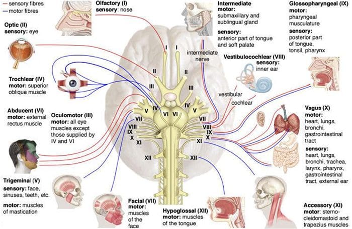
The spinal cord and brain make up the central nervous system while the spinal nerves that branch to the spinal cord and cranial nerves that branch to the brain makes up the peripheral nervous system.
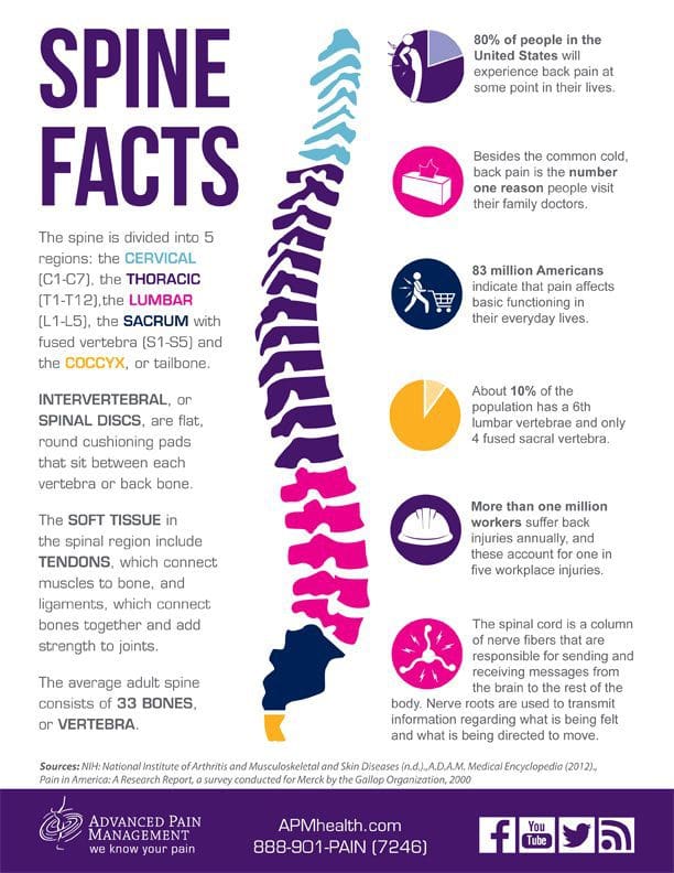
There are thirty-one sets of nerves that extend out of the spinal cord and are connected to it by the nerve root. Each nerve branches out about a half inch from the spinal cord before dividing into smaller branches. The dorsal rami are on the posterior side of the branch while the larger ventral rami are on the anterior side.
The dorsal rami provide nerve function for the skin of the trunk and posterior muscles. The ventral rami from T1 to T12 provide nerve function to the skin of the trunk as well as the lateral and anterior muscles. The anterior divisions that remain for plexuses, networks that provide nerve function to the body. Each plexus has specific areas on the body for skin sensitivity as well as certain muscles. Their point where they exit the spine determines how they are numbered. The four primary plexuses are:
Cervical plexus, C1 � C4, innervates the diaphragm, shoulder, and neck
Brachial plexus, C5 � T1, innervates the upper limbs
Lumbar plexus, T12/L1 � L4, innervates the thigh
Sacral plexus, L4 � S4, innervates the leg and foot.
These spinal nerves have two sets of fibers: motor and sensory. Motor fibers facilitate movement and provide nerve function to the muscles. Sensory fibers facilitate sensitivities to touch, temperature and other stimuli. They provide nerve function to the skin.
What are Myotomes and Dermatomes?
A group of muscles that are innervated by the motor fibers that stem from a specific nerve root is called a myotome. An area of the skin that is innervated by the sensory fibers that stem from a specific nerve root is called a dermatome. These patterns of myotome and dermatome are almost always identical from person to person. There are occasionally variances, but that is rare.
This consistency allows doctors to treat nerve pain in patients. If a specific area is hurting, they know that it is attributed to a certain myotome or dermatome, whichever the case may be, and its corresponding nerve root. Problems with nerve damage are often the result of stretching the nerve or compressing it.
When the nerves are injured in specific areas like the lumbosacral or brachial plexus, it presents as sensory and motor deficits in the limbs that correspond to them. Myotomes and dermatomes are used to assess the extent of the damage.
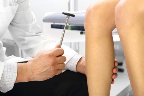
How are Myotomes and Dermatomes used to Assess Nerve Damage?
When a doctor tests for nerve root damage in a patient, he or she will often test the myotomes or dermatomes for the nerves assigned to that location. A dermatome is examined for abnormal sensation, such as hypersensitivity or lack of sensitivity.
This is done by using stimulus inducing tools such as a pen, paper clip, pinwheel, fingernails, cotton ball, or pads of the fingers. The patient is instructed to provide feedback regarding their response. Some of the abnormal sensation responses include:
A myotome is tested for nerve damage in the muscles which presents as muscle weakness. This grading scale, which assigns a rating to the degree of muscle weakness, is often used:
5 � Normal � Complete range of motion against gravity with full resistance
4 � Good � Complete range of motion against gravity with some resistance
3 � Fair � Complete range of motion against gravity with no resistance, active ROM
2 � Poor � Complete range of motion with some assistance and gravity eliminated
1 � Trace � Evidence of slight muscular contraction, no joint motion evident
0 � Zero � No evidence of muscle contraction
During a typical chiropractic exam, your chiropractor will assess both dermatomes and myotomes for potential neurological problems. This gives them additional insight on how to treat your condition, whether it’s related to a subluxation of vertebral bodies or other, other disease processes.
IFM's Find A Practitioner tool is the largest referral network in Functional Medicine, created to help patients locate Functional Medicine practitioners anywhere in the world. IFM Certified Practitioners are listed first in the search results, given their extensive education in Functional Medicine

 �
�