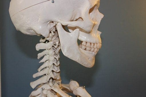
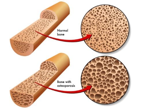
Osteopenia and osteoporosis, two very similar conditions, that are defined as decreased bone density, but osteopenia is far less. However, it is still a problem due to an increase of breaking a bone because of bone fragility.
Symptoms
Osteopenia usually doesn’t cause symptoms unless a bone is broken. However, some patients who present with osteopenia complain of dull back pain.
Symptoms associated with osteoporosis include the following:
Back pain, caused by a fractured or collapsed vertebra
Loss of height over time
A stooped posture
A bone fracture that occurs much more easily than expected
The causes and those at risk?
Women (primarily small-boned Caucasian and Asian) are most at risk for both conditions, primarily those who are age 65 or older as well as women who are postmenopausal. However, men can also be affected.
Anyone who meets any of the criteria for being at risk for either of the bone conditions should be evaluated. Often, catching the conditions early can make a significant difference in the effects that they have on the body and in some cases, can even be arrested so that they don�t progress.
Some of the common causes of both conditions include:
Lifestyle habits
Smoking
Insufficient calcium
Sedentary lifestyle
Excessive alcohol consumption
Vitamin D deficiency
Carbonated beverages
Medical Situations
Bulimia, anorexia, and other eating disorders
Estrogen deficiency in women
Certain hormone imbalances
Overactive thyroid
Certain treatments including radiation and chemotherapy
Low testosterone in men
Medications including anti-seizure, hydrocortisone, and steroids
Health issues
Tumors
Cystic fibrosis
Crohn�s disease
Digestive issues
It should also be noted that certain types of diets, particularly those that advocate extremely low fat, or no fat can also cause problems. Vitamin D is necessary for calcium absorption in the body, but vitamin D is a fat-soluble vitamin meaning the body requires some fat in order to make use of it. When there is inadequate fat, the vitamin cannot be absorbed and in turn, calcium cannot be absorbed.
A family history of osteopenia, osteoporosis, or low bone mass can increase a person risk by 50% to 85%.
A Diagnosis
Bone mineral density (BMD) tests are used to diagnose both osteopenia and osteoporosis by measuring the calcium levels in bone. This type of test can also provide an estimate of how much at risk a person is for bone fractures.
This test is painless and non-invasive. It is usually performed on the heel, shin bone, wrist, spine, finger, or hip.
Two common types of these tests are radiographs, a standard diagnostic tool for osteopenia, and Dual Energy X-ray Absorptiometry (DEXA). A DEXA scan is essentially a low energy x-ray so patients are not exposed to as much radiation as they would be if they had a regular x-ray. The results are attained by comparing the score (measurements were taken) to scans of individuals who do not have the condition.
Once the score is measured and compared, it is assessed using a chart that identifies the level or risk:
+1.0 to -1.0 – Normal bone density
-1.0 to -2.5 – Low bone density
-2.5 or higher – At risk for osteoporosis
What Treatments Is Available?
As with most conditions, prevention is the most effective treatment. If you have a family history or fall under any of the risk factors, there are things you can do to minimize the effects or prevent the conditions completely.
Your chiropractor can talk to you about lifestyle changes, exercise, and diet as well as supplements that you can take. Chiropractic adjustments can also be effective for many patients with osteopenia and osteoporosis as long as the chosen technique is a low force technique like Activator.
Many patients find these natural treatments preferable to any medications that may be prescribed. The most important thing you should do, though, is get a bone density test if you are in an at-risk category, are a woman who is postmenopausal or age 65 or older.
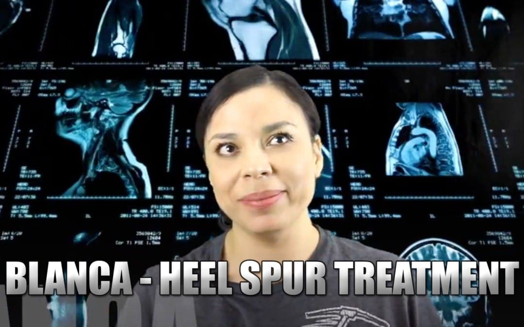
Heel Spur: Blanca, born and raised in El Paso, TX, has been suffering from heel spurs for about two years. As a registered nurse, her symptoms significantly affected her ability to work and her overall quality of life. Determined to improve her health, Blanca considered chiropractic care. Once she started treatment with Dr. Alex Jimenez, however, Blanca experienced tremendous relief from her heel spurs, almost instantly. Blanca highly recommends chiropractic care with Dr. Alex Jimenez as the non-surgical choice for treatment of heel spurs.
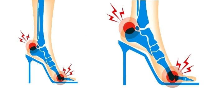
Chiropractic Heel Spur Treatment
A heel spur is a calcium residue resulting in a bony protrusion on the bottom of the heel bone. Although heel spurs are often painless, they can lead to heel pain. They are often associated with plantar fasciitis, a painful inflammation of the fibrous band of connective tissue (plantar fascia) that runs across the bottom of the foot and also connects the heel bone to the ball of the foot. Heel spurs are usually caused by strains on foot muscles and ligaments, stretching of the plantar fascia, and repeated tearing of the membrane which covers the heel bone. Heel spurs are particularly common among athletes.
We are blessed to present to you�El Paso�s Premier Wellness & Injury Care Clinic.
As El Paso�s Chiropractic Rehabilitation Clinic & Integrated Medicine Center,�we passionately are focused treating patients after frustrating injuries and chronic pain syndromes. We focus on improving your ability through flexibility, mobility and agility programs tailored for all age groups and disabilities.
If you have enjoyed this video and/or we have helped you in any way please feel free to subscribe and share us.
Dr. Alex Jimenez collaborates with top rated diagnosticians and imaging specialists. We are blessed to have in our association, imaging specialists that provide fast, courteous & premiere board certified specialists. In collaboration with our offices we can provide the quality of service our patients mandate and deserve.
Who We Are
Diagnostic Outpatient Imaging (DOI) is a state-of-the-art Radiology center in El Paso, TX. It is the only center of its kind in El Paso, owned and operated by a Radiologist.
This means when you come to DOI for a radiologic exam, every detail, from the design of the rooms, the choice of the equipment, the hand-picked technologists, and the software which runs the office, is carefully chosen or designed by the Radiologist and not by an accountant.
Our market niche is one center of excellence. Our values related to patient care are: We believe in treating patients the way we would treat our family and we will do our best to ensure that you have a good experience at our clinic.
Dear Doctors,
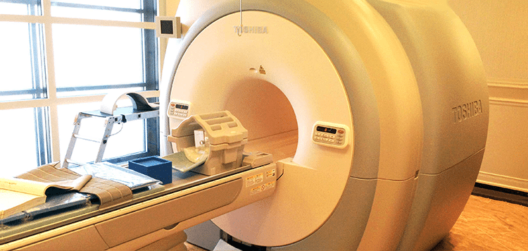
We are pleased to inform you of the arrival of our Titan 3-Tesla MRI at Diagnostic Outpatient Imaging. This is El Paso’s only radiology imaging center that offers this technology. Patients do not always realize how important image quality is: It can make the difference in the diagnosis.
3-Tesla MRI is like HD TV and once you try it, you will not want to go back. The increased magnet strength gives us many benefits at no additional expense to the patient. It gives us the ability to scan faster or to scan with higher detail. An MRI of the brain can take 20 minutes and have exceptional quality, or we can perform the scan in less time, with better quality that is achieved on most 1.5 Tesla “high field” MRIs. This is incredibly useful for children.
Our 3T MRI can perform Diffusion Tensor Imaging, MRI Spectroscopy and CSF flow studies to name just a few of its possibilities.
This scanner is not only very fast, it is very large. Our open MRI has a clearance of 35 cm. The 3T has a diameter of 71 cm! This is welcome news for nervous or claustrophobic patients, and combined with its speed, it can actually eliminate the need for sedation for some patients. 3T MRI is faster, clearer, and has more diagnostic possibilities. We are certain you and your patients will notice the difference.
Our Services
MRI’s:
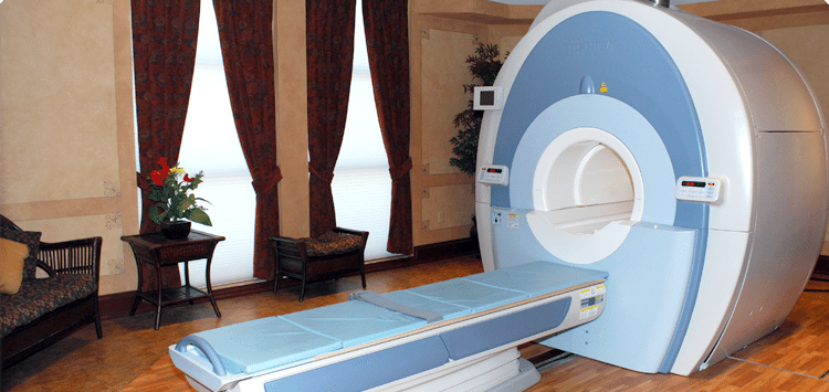
DOI has three MRI’s under one roof. All are American College of Radiology (ACR) Certified.
Good
Open MRI (0.35 Tesla): This MRI perfect for claustrophobic and very large patients. There is no table weight limit on this MRI
Better
High Field 1.5 Tesla MRI- This is a eight channel MRI with high end image quality. It is in a beautiful room and has ‘pianissimo’ technology, which makes the MRI relatively quiet. This machine has been the best MRI in private practice in El Paso for years. It will soon be eclipsed by our new 3.0 Tesla MRI.
Best
High Field 3.0 Tesla MRI- This is the only 3.0 Tesla MRI in private practice in El Paso. This technology can deliver stunning image quality, which can actually make a difference in your diagnosis. The increased magnet strength gives us many benefits at no additional expense to the patient.�??It gives us the ability to scan faster, or to scan with higher detail. This is welcome news for nervous or claustrophobic patients, and as well as for children as it can actually eliminate the need for sedation in some patients. 3T is faster, clearer, more diagnostic for a better for MRI. It is like HD TV. Once you have tried it, you won’t want to go back. This MRI effectively doubles our MRI capacity. If needed most exams can be completed in under 5 minutes, instead of the normal 30-45 minutes.
Breast MRI:
DOI began Breast MRI in July 2007, being the first facility in El Paso to perform the exam. We have now performed over 2500 breast MRI’s and many MRI-guided breast biopsies. All have been interpreted and/or performed by Dr. Boushka, making him the most experienced radiologist in the city with this exam. This is the most powerful tool for the detection of Breast cancer to date.
Hours: Monday to Thursday 7 am to 9 pm Friday 7 am to 5 pm Saturday 8 am to 4 pm
Prostate MRI:
Guys, you need great medical care also. We are the only facility in El Paso performing this leading edge exam. MRI can see cancers when other imaging methods cannot. Not only can we see prostate cancers with MRI, we can perform MRI-guided prostate biopies for pathologic (definitive) diagnosis.
Monday to Thursday 7 am to 9 pm Friday 7 am to 5 pm Saturday 8 am to 4 pm
CT:
We have a 16 slice Toshiba Aquillion CT scanner, with newly updated in Dec 2013. The upgrade allows for reduced X-ray dose, higher resolution, more patient comfort, shorter breath holds and doubles the speed of the scanner. This scanner performs CT X-ray exams as helical volume acquisitions in 3D from a single patient exam. Most exams are finished in under 60 seconds, unless delayed images with contrast are indicated. Additionally we have a powerful 3D post processing workstation.
Hours Monday to Friday 7 am to 6 pm
Ultrasound:
DOI has just doubled our Ultrasound capacity with newly purchased Philips 34 XRL scanner. We have Three certified Ultrasonographers with cumulative experience of 45 years. We are confident you will find them professional and compassionate. Beverly Bruner RDMS, Sonographer, formally of Desert Imaging has joined our team.
3D OB Ultrasounds:
You better believe it. Available whenever our US department is open. No referral necessary. Images are reviewed by an actual radiologist.
Ultrasound Hours: Monday, Tuesday, Thursday 8 am to 5 pm Wednesday 8 am to 8 pm Friday 8 am to 5 pm Saturday 8 am to 12 pm
Digital Mammography
DOI was the first facility in El Paso to acquire Hologic Full Field Digital Mammography and thus we have more experience with this technology than any facility in El Paso. Our Mammographer has 20 years of experience and has her own following of patents who seek her out to perform their mammograms because of her excellent and compassionate care. Our private pay screening mammography price of $90, including the interpretation is an unbeaten price in El Paso.
Hours Mon – Fri 8am to 4pm Extended hours Wednesday until 8pm) Saturdays 8am to 12pm
Bone Denisity (DEXA)
We have a brand new, Hologic Discovery CI bone densitometer scanner. This is the latest technology.
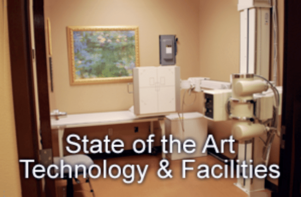
X-Ray
Our digital computed radiography was just updated February 2014. No appointments are necessary.
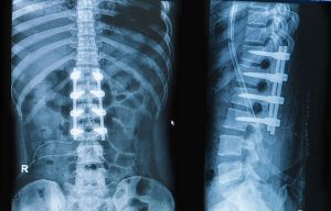
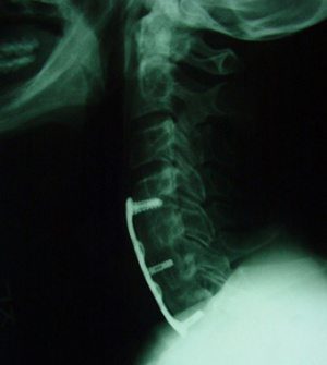
Bone growth stimulation (BGS) is a therapy your surgeon may prescribe following a spinal fusion procedure. A bone growth stimulator is an auxiliary device worn following cervical (neck) or lumbar (low back) spine surgery. BGS may be used to assist spinal bone fuse after a fusion procedure or as a treatment for failed fusion. Naturally, you’ve questions about this technology.
Spinal column with implant, screw placement and fusion
The info provided in this patient guide can assist you to learn:
Bone heals
Risk factors for a poor or failed fusion
Role of bone growth stimulation in spine fusion aftercare
Questions to ask your back surgeon
“Bone growth stimulation to be used in both the cervical and lumbar spine has demonstrated to substantially help fusion results. Having been a study centre for this particular technology, I’ve used bone growth stimulation in most my post-operative cervical and lumbar patient instances. The patient assessment standards I use contains:
Multi-level fusions; more than one degree of the back is fused
Co-morbidities (risk factors) that could hinder bone healing and growing”
�Gerard J. Girasole, MD
Orthopaedic Surgeon
Orthopaedic & Sports Medicine Center
About Spinal Fusion
Spinal fusion is done to stop motion of neurologic deficit and the spine. During the procedure two or more vertebral bodies are joined together using instrumentation and bone graft. Spinal instrumentation includes poles, screws, plates, and interbody devices (implants). Bone graft may comprise your own bone (autograft), donor bone (allograft), or alternative forms of graft.
Bone graft helps stimulate new bone to grow through three stages:
Inflammatory period: cells start to form new tissue
Repair period: small blood vessel ingrowth begins
Remodeling phase: bone structure becomes powerful
Spinal instrumentation creates an internal cast, allowing the inflammatory procedure to stimulate bone healing. With time, new bone grows into and about the implanted instrumentation healing into a construct that is sound.
Some patients are at risk for spinal fusion to fail. A failed fusion is called pseudarthrosis or nonunion. Pseudarthrosis and nonunion are medical terms your surgeon may utilize to identify a fusion dilemma.
Common Spinal Issues Treated Surgically With Fusion Include:
Degenerative disk disease
Fracture
Herniated disc
Spinal stenosis
Lumbar
Adult degenerative scoliosis
Spondylolisthesis
How Does A Bone Growth Stimulator Help Spinal Fusion?
A BGS sends electric signals to the fusion site. The electrical signals activate the body’s natural bone healing process, which may be impaired in at-risk patients.
Bone Growth Stimulation Has Been Put To Use For Decades To Help Bone Heal
Over 50 years ago scientists found that low-level electrical fields arouse the entire body’s bone-healing process. Other improvements included finding several types of energy that stimulate bone development, electromagnetic coil technology and only better devices � supported by clinical and scientific research�have enhanced bone healing in patients who undergo spinal fusion.
Different Types Of Bone Growth Stimulators
All bone growth stimulators are different. Certain types are designed to be surgically implanted (internal BGS) and other stimulators are worn outside the body (external BGS). Other differences include how stimulation is transmitted to the back and the kind of magnetic field or electric current created by the apparatus.
IFM's Find A Practitioner tool is the largest referral network in Functional Medicine, created to help patients locate Functional Medicine practitioners anywhere in the world. IFM Certified Practitioners are listed first in the search results, given their extensive education in Functional Medicine


 Please note that we can answer general questions, but anything specific to your medical case should be discussed with your physician.