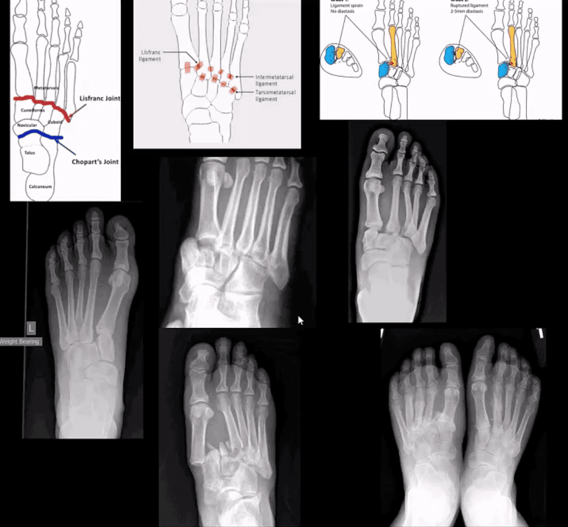
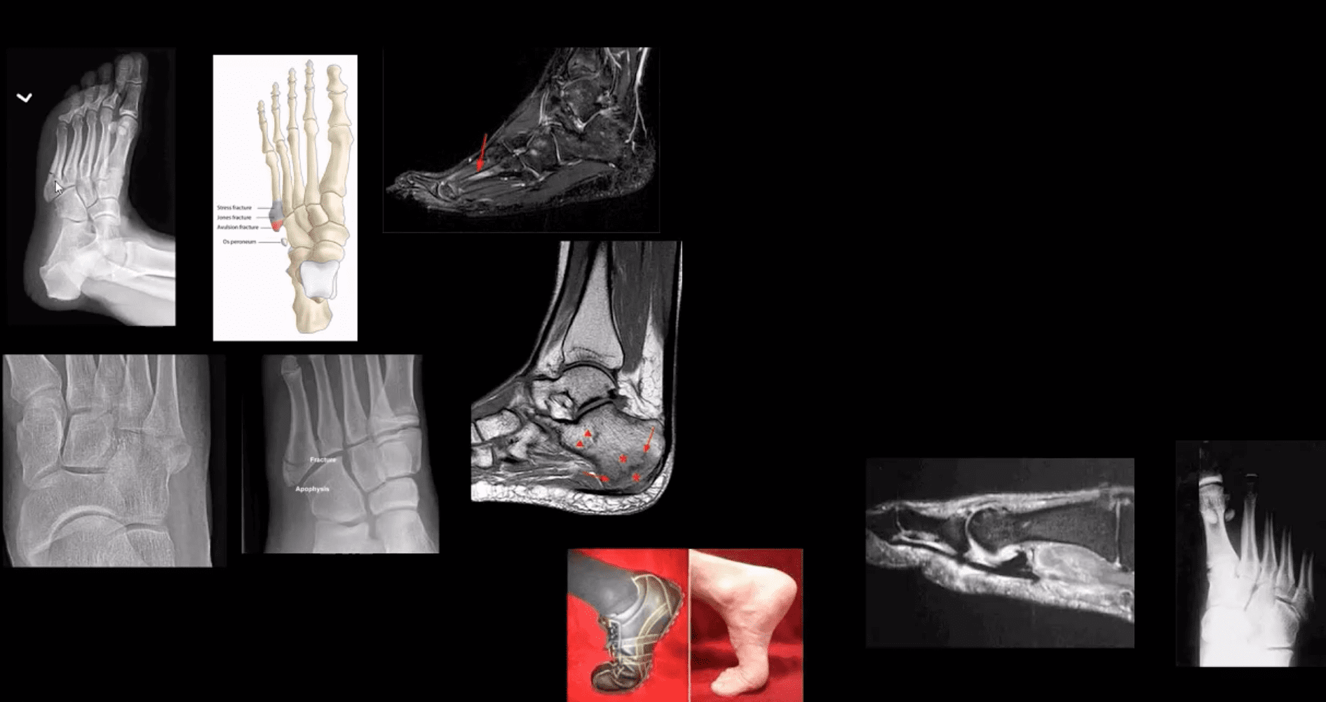
M/C dislocation of the foot at tarsal-metatarsal articulation (Lisfranc joint). Direct impact or landing and plantar or dorsal flexing the foot. Lisfranc ligament holding 2nd MT base and 1st Cu is torn. Manifests with or w/o fracture-avulsion.
Imaging: 1st step: foot radiography in most cases sufficient to Dx. MSK US may help: show disrupted Cu1-Cu2. Ligament and widened space > 2.5mm. MRI may help but not essential. Weight-bearing view aids Dx.
2-types: homolateral (1st MTP joint in contact) and divergent (2-5 MT displaced laterally and 1st MT medially)
Management: operative fixation is crucial
N.B. Atraumatic Lisfranc dislocation is a frequent complication of a diabetic Charcot foot
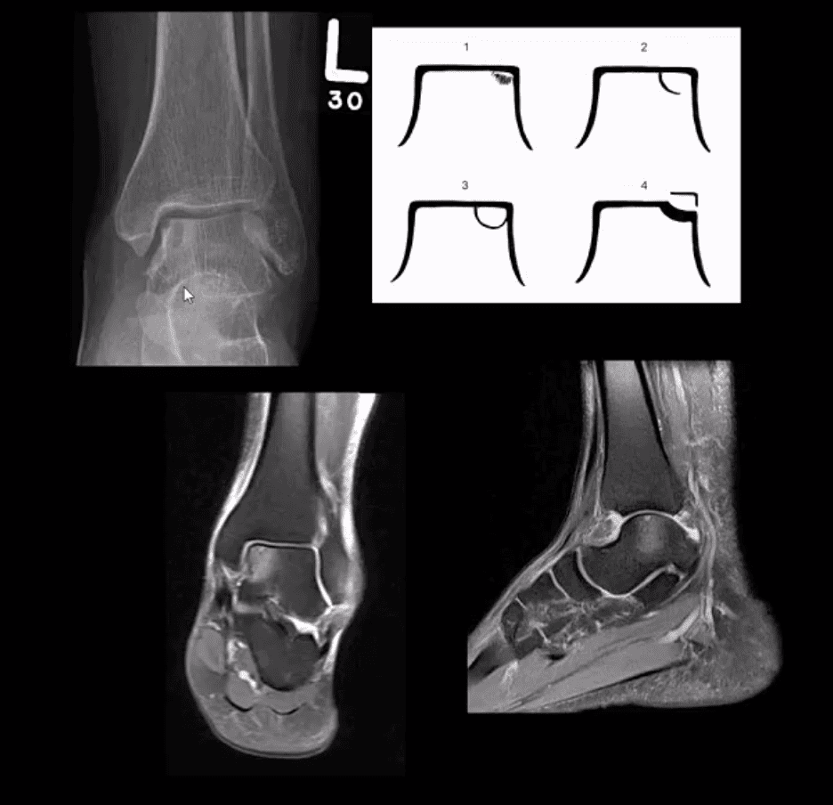
Osteochondral Injury of the Talus (OCD)
Common. Non-traumatic found in superior-medial talar dome. Traumatic may affect supero-lateral dome.
Clinically: pain/effusion/locking. Imaging is crucial.
1st step: radiography may reveal focal radiolucent concavity/halo, fragment.
MRI helpful esp. if OCD is cartilaginous and to demonstrate bone edema.
Jones Fx: extra-articular Fx of proximal metaphysis of the 5th MT. Prone to non-union. Often fixed operatively.
Pseudo-Jones: intra-articular avulsion of 5th MT styloid/base by eccentric contraction of Peroneus Brevis M. Managed conservatively: boot-cast immobilization. Both Jones & Pseudo-Jones Dx by foot series radiography.
Stress Fx. Calcaneus, 2nd, 3rd, 5th MTs. Repeated loading (running) or “March foot” 2nd/3rd MT. Clinically: pain on activity, reduced by rest. Dx: x-rays often unrewarding earlier. MRI or MSK US may help. Managed: Conservatively. Complications; progress into complete Fx
Turf toe: common athletic hyperextension of 1st MTP-sesamoid/plantar plate complex is tearing. 1st MTP unstable/loose. Managed operatively.
Arthritis of the Foot & Ankle
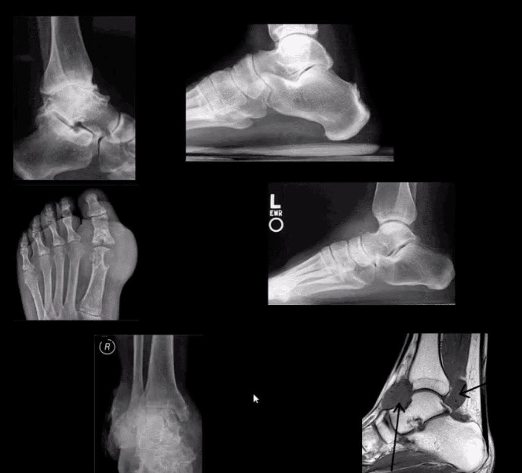
DJD of the ankle: uncommon a primary OA. Typically develops as 2nd to trauma/AVN, RA, CPPD, Hemophilic arthropathy, Juvenile Idiopathic Arthritis, etc. manifests as DJD: osteophytes, JSL, subchondral cysts all seen on x-rays
Inflammatory Arthritis: RA may develop in the ankle or any synovial joint. Will typically presents with symmetrical Hands/feet RA initially (2nd, 3rd MCP, wrists, MTPs in feet) usually with erosion, uniform JSL, juxta-articular osteopenia, and delayed subluxations.
HLA-B27 spondyloarthropathies: commonly affect lower extremity: heel, ankle esp in Reactive (Reiter). Erosive-productive bone proliferation is a crucial Dx.
Gouty Arthritis: common in the lower extremity. Ankle, mid-foot foot esp 1st MTPs. Initial onset: acute gouty arthritis with ST effusion and no erosions/tophi. Chronic tophaceous gout: peri-articular, intra-osseous punched-out erosions with over-hanging edges, no initial JSL/osteopenia, ST. Tophi may be seen.
Miscellaneous arthropathy: PVNS. Not common. Affects 3-4th decades of life. The result of synovial proliferation with Macrophages and multi-nucleated Giant Cells filled with hemosiderin and fatty accumulation may lead to inflammation, cartilage damage, extrinsic bone erosions. Dx: x-rays are insensity, MRI modality of choice. Synovial biopsy. Management: operative, can be difficult.
Neuropathic Osteoarthropathy
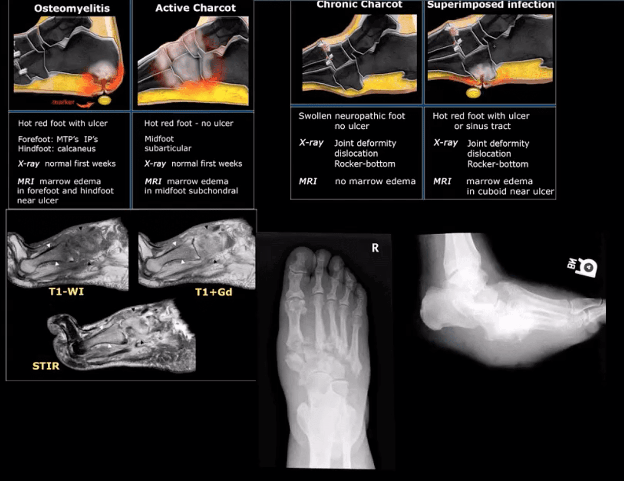
(Charcot’s joint) Common and on the rise d/t epidemic in type 2 DM. May present with pain initially (50% of cases) and painless destructive arthropathy as a late manifestation. Early Dx: delayed. Imaging is crucial: x-rays: initially unrewarding, some SF effusion is seen. MRI helps with early Dx and extremity off-loading. Late Dx: irreversible dislocations, collapse, disability. Note: Lisfrance dislocation in Charcot joint
M/C mid-foot (TM joint) in 40% of cases, ankle 15%. Progression: Rocker-bottom foot, ulcerations, infections, increased morbidity, and mortality.
Early Dx: by MRI is crucial. Suspect it in patients with type 2 DM especially if early non-traumatic foot/ankle pain reported.
10% of all fractures. 2nd m/c following femoral neck Fx. Demographics: active young males and older osteoporotic females
Stable Fx: overall prognosis is good
Unstable Fx: require ORIF. 15%-20% chances of 2nd OA.
Role of imaging is to determine the complexity, stability and care planning (i.e., operative vs. conservative)
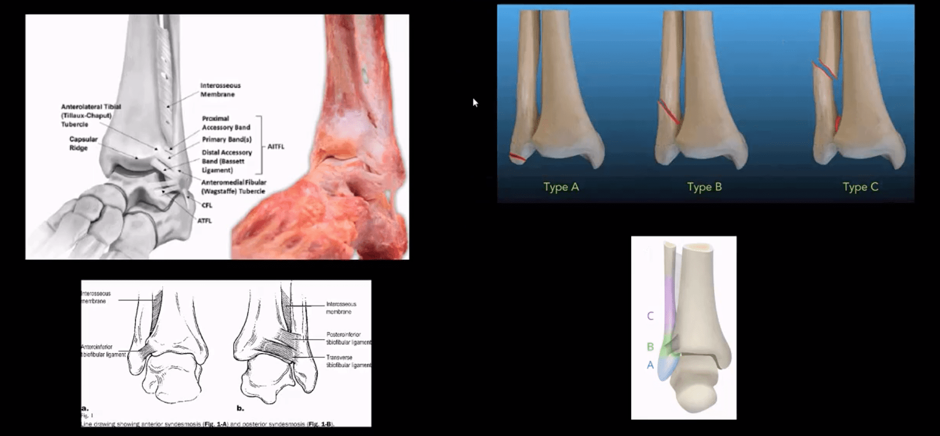
Weber classification considers tearing of distal tibial-fibular syndesmosis and potential instability
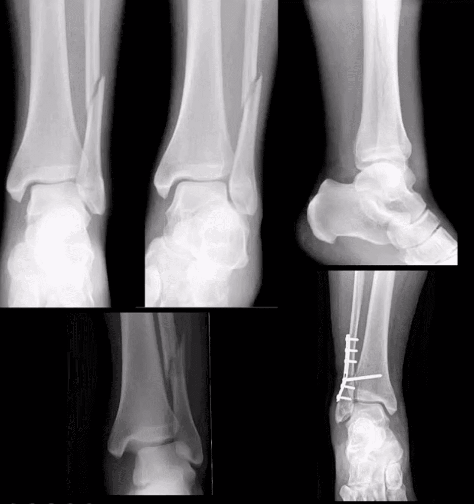
Weber A – below syndesmosis. Stable, typically avulsion of the distal fibular malleolus
Weber B – at the level of syndesmosis: may be outside syndesmosis and stable or tearing syndesmosis and unstable
Weber C – above syndesmosis. Always unstable d/t tearing of syndesmosis
Variations of fractures may involve the position/role of the talus bone during Fx (e.g., abduction, adduction, rotation, etc.) this is known as Lauge-Hanson classification
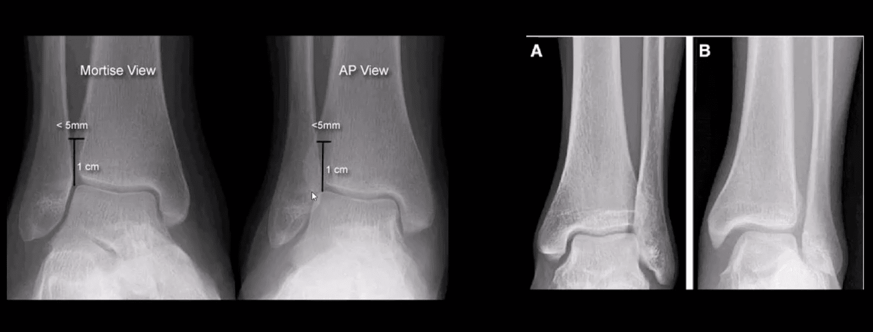
Reveal infrasyndesmotic Fx of fibular malleolus (Weber A)
Stable Injury
Conservative care in the form of short-leg walking cast/boot can be used. Good recovery. If no evidence of osteochondral injury, relatively low chances of post-traumatic OA
No further imaging required. MRI may help to reveal bone contusion and osteochondral injury
Weber B at Level of Syndesmosis
Can be stable or unstable. On occasions, the decision is made during operative exploration.
CT scanning may help with further evaluation
Management: depends on stability. Additional stabilization required if syndesmosis is ruptured
Weber C
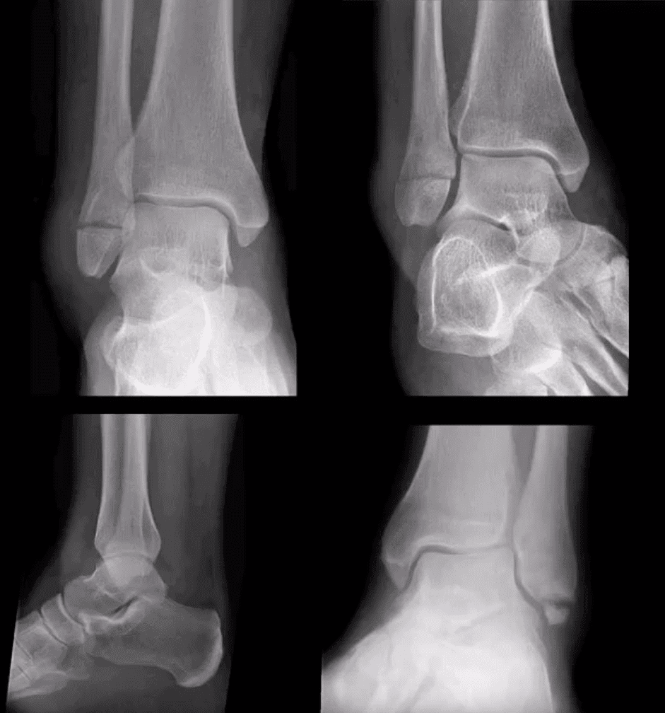
AP, medial oblique and lateral views reveal Weber C – suprasyndesmotic injury with abnormal joint widening d/t disruption of the tib-fib syndesmosis. Very unstable injury.
Occasionally, when Weber C Fx positioned 6-cm from the tip of the lateral malleolus, it may be termed as Pott’s ankle Fx (name after Percival Pott’s who has proposed the original classification of ankle fractures based on their stability and degree of rotation). The term is somewhat outdated.
Management: operative with additional stabilization of the syndesmosis
Maisonneuve Fracture
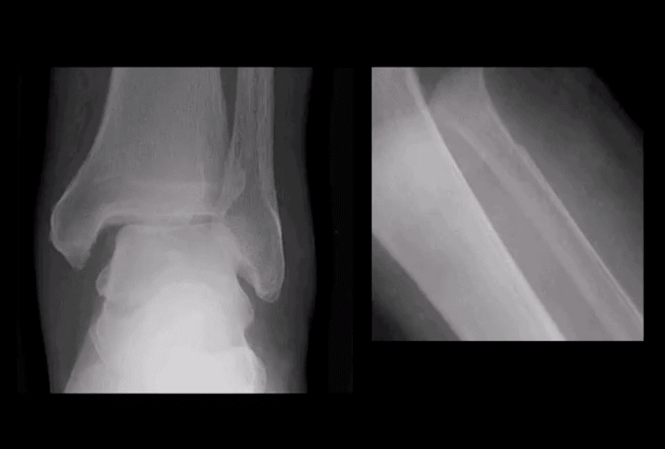
Often spiral fracture of the proximal fibula combined with an unstable ankle injury
No immediate ankle fracture is noted radiographically, thus can be missed on ankle views and require tibia and fibula views
Rad features: widening of the ankle d/t syndesmosis tear and sometimes deltoid ligament disruption. Interosseous membrane is torn with proximal fibular Fx caused by pronation with external-rotation force
Management: operative
Bimalleolar & Trimalleolar Fx
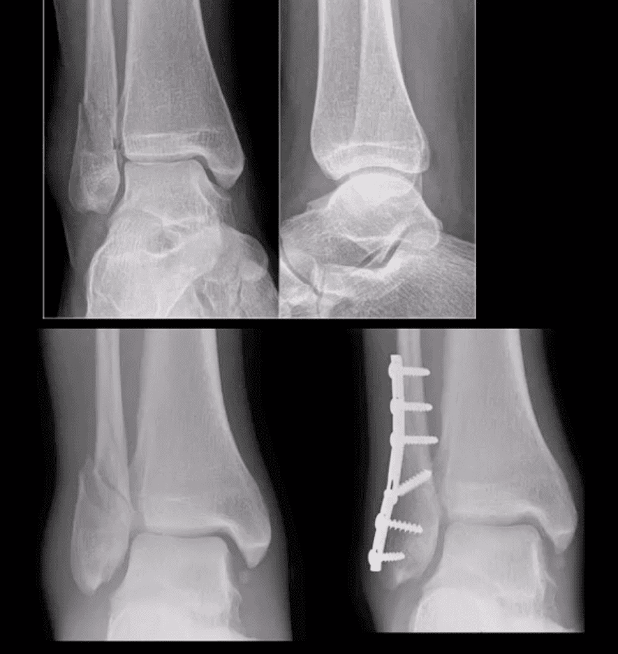
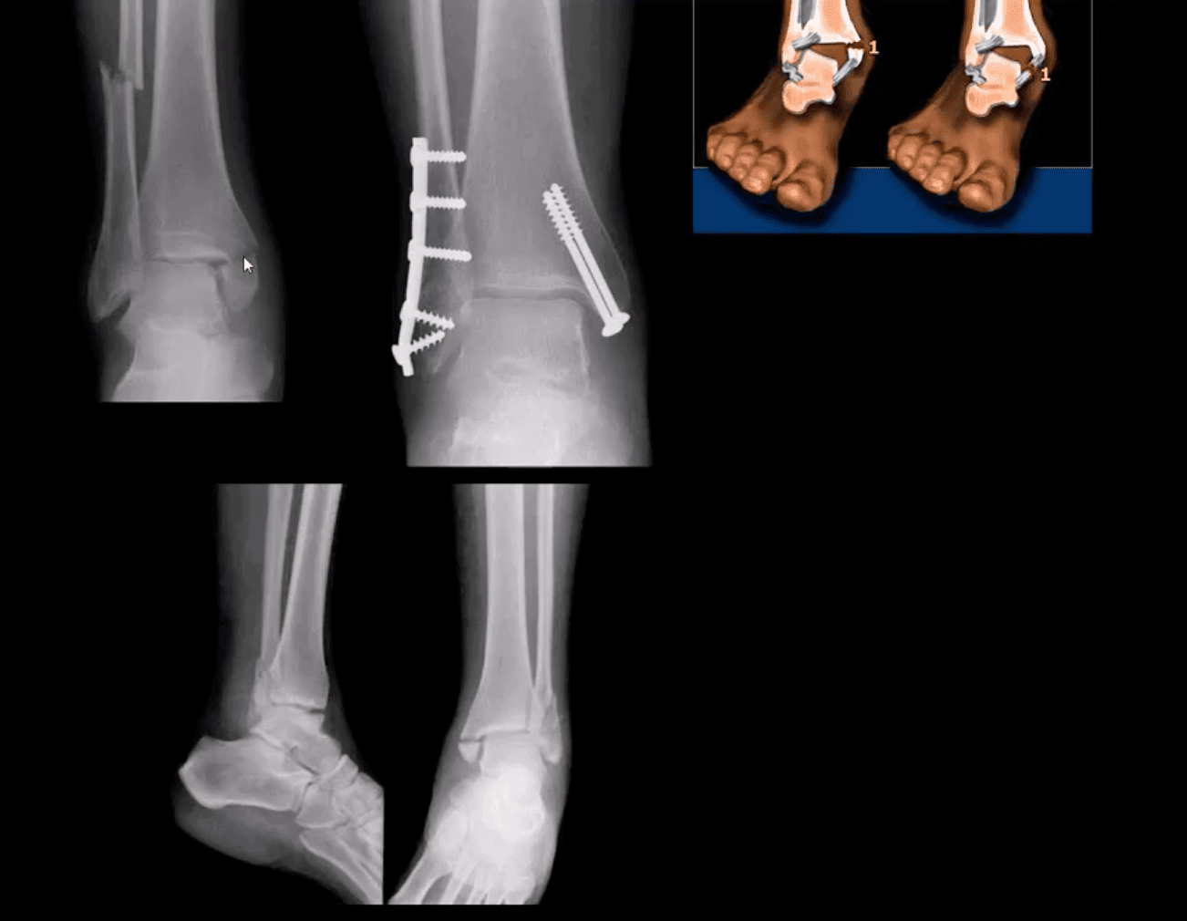
Above top images Bimalleolar Fx v. unstable, the result of pronation and abduction/external rotation. Rx: ORIF.
Trimalleolar Fx: 3-parts ankle Fx. Medial and lateral malleolus and avulsion of the posterior aspect of tibial plafond. More unstable. Rx: operative
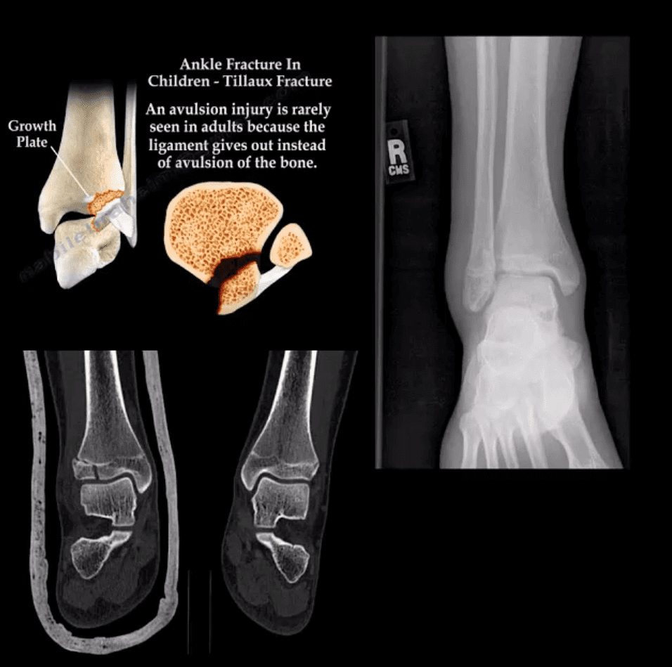
Tillaux Fx
Pediatric Fx affecting older child when the medial side of the physis is closed or about to close with lateral side till open. Avulsion by the anterior tibi-fibular ligament. Complications: 2nd dry/premature OA. Rx: can be conservative if stable by boot cast immobilization.
Pediatric Growth Plate Injuries
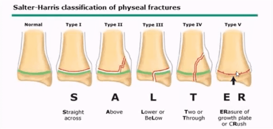
Salter-Harris classification helps to diagnose and prognosticate physeal injuries.
Helpful mnemonic: SALTR
S: type 1-slip through the growth plate
A: type 2-above, Fx extends into the metaphysis
L: type 3-lower, intra-articular Fx extends through the epiphysis
T: type4, “through” Fx extends through all: physis, metaphysis, and epiphysis.
R: type 5, “ruined.” Crush injury to physis leading to complete death of the growth plate
Type 1 and 5: present with no fracture
Type 2: has the best prognosis and considered the most common.
Management: referral to a pediatric orthopedic surgeon
Complications: early physis closure, limb shortening, premature OA and others.
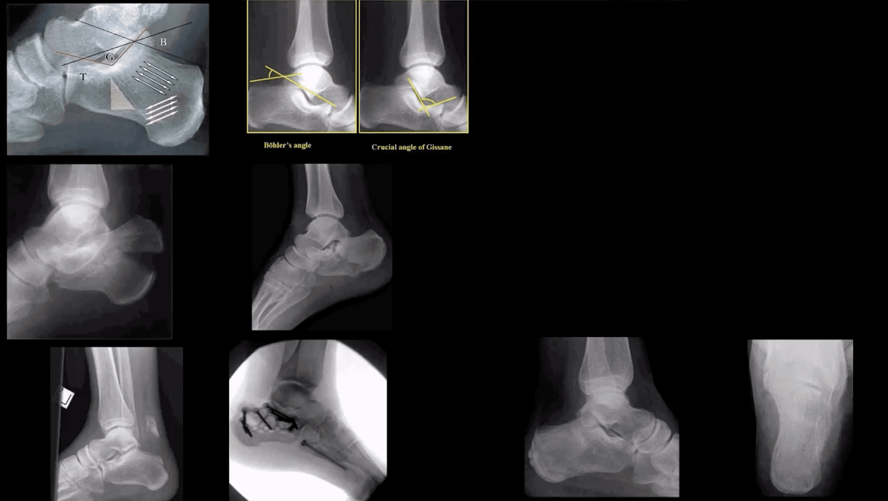
Calcaneal Fracture
Most frequent tarsal Fx. 17% open Fx
Mechanisms: axial loading (intra-articular Fx into sub-talar and calcaneal-cuboid joints in 75% cases). Avulsion by Achilles tendon (m/c in osteoporotic bone). Stress (fatigue) Fx.
Intra-articular Fx carries a poor prognosis. Typically comminuted. Rx: operative.
B/I calcaneal intra-articular fx with associated vertebra compression Fx with associated vertebral compression Fx (T10-L2) often termed Casanova aka Don Juan (Lover’s) fx.
Imaging: x-radiography with added “heel view” 1st step. CT scanning is best for Dx and pre-op planning.
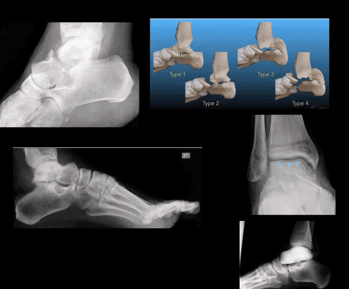
M/C fractured tarsal bone is the Talus. M/C region: talar neck (30-50%). Mechanism: Axial loading in dorsiflexion. Complications: Ischemic osteonecrosis (AVN) of the talus. Premature (2nd OA). Imaging: 1st step: radiographs, CT can be helpful with further delineation
Hawkins classification helps with Dx, prognosis & treatment. “Hawkins sign’ on plain film/CT scan may help with AVN Dx. (above blue arrows indicate good prognosis d/t radiolucent line indicating no AVN because the bone is vascularized and hence resorbed)
Rx: Type 1: conservative with short leg cast or boot (risk of AVN-0-15%), Type 2-4-ORIF (risk of AVN 50%-100%)
James Hill, father and teacher to two older sons and a younger daughter called Madison Hill, belong to an athletic family. Madison began participating in several sports because she had been a young woman, but she consequently experienced numerous sports injuries. Luckily, James Hill and his daughter Madison Hill went to go to Dr. Alex Jimenez and he has tremendously helped her recover after every injury she experiences. Collectively, they’ve learned to trust in chiropractic care, particularly following Dr. Alex Jimenez’s innovative treatment techniques and procedures. Mr. Hill expresses his gratitude in addition to how far Dr. Alex Jimenez’s understanding in sports injury therapy has greatly enlarged his general comprehension of the human body’s healing process. After Madison suffered a recent ankle sprain, she was instantly reassured by Dr. Alex Jimenez regarding how much faster she’d return-to-play during chiropractic care. James Hill and Madison Hill highly recommend Dr. Alex Jimenez as the non-surgical selection for volleyball sports injuries, among others..
Chiropractor Ankle Sprain Treatment
Every year, millions of young athletes engage in high school sports. However, once an injury occurs, it might develop into an issue that might result in them having to discontinue their particular sport or physical activity if not treated properly. The pressure to continue participating in the sport or physical activity may cause young athletes not to follow proper recovery guidelines, which might subsequently result in more injury with long-term consequences. Sports injuries among young athletes fall into two key categories: overuse injuries and acute injuries. Both kinds include injuries to the soft tissues and bones. Whether an injury is acute or due to overuse, a young athlete who develops a symptom which impacts their athletic performance should be diagnosed appropriately by a health care professional. Sports accidents which are untreated can lead to permanent disability. Many high school sports injuries can be avoided through appropriate instruction and conditioning.
We are blessed to present to you�El Paso�s Premier Wellness & Injury Care Clinic.
As El Paso�s Chiropractic Rehabilitation Clinic & Integrated Medicine Center,�we passionately are focused treating patients after frustrating injuries and chronic pain syndromes. We focus on improving your ability through flexibility, mobility and agility programs tailored for all age groups and disabilities.
If you have enjoyed this video and/or we have helped you in any way please feel free to subscribe and recommend�us.
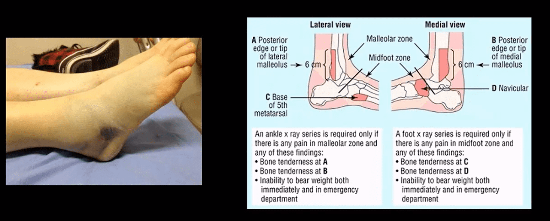
Ankle pain and injury are not issues reserved solely for athletes. In the United States, more than 25,000 people deal with ankle sprains or pain every day. Studies show that somewhere around 40 percent of ankle sprains are not treated correctly or are misdiagnosed, which leads to disability or chronic ankle pain.
When the ankle does not function properly, it can affect the entire body. The muscles on either side of the leg or even under the foot can become sore or painful. This can lead to loss of mobility, an uneven gait, and hip and back pain.
Anatomy Of The Ankle
Three bones connect to the joint that is the ankle. The lower ends of the tibia (shinbone) and the fibula (lower leg small bone) meet to form a socket that the talus (ankle bone) sits in.
The bottom of the talus rests on the calcaneus (heel bone). There is about an inch-thick lining of somewhat soft cartilage in the joint, which provides shock absorption for carrying body weight, but it is tough and durable so that, provided there is no injury, it will last for a lifetime.
The bones are held together by ligaments and the muscles are attached to the bones by tendons. When there is an injury, it can impact the bone, muscles, tendons and ligaments.
Treatment For Ankle Injury & Pain
The typical treatment for pain, such as with a sprain, is R.I.C.E., which is rest, ice, compression and elevation. A somewhat newer treatment approach replaces the R with an M, meaning that instead of rest, movement is required instead.
However, it is important that the movement is done safely and carefully. Certain types of ankle injuries can be exacerbated by movement so it should be approached with care.
Other types of traditional treatment include varying methods of pain control from ibuprofen to opioids. Severe injuries, such as a torn ligament, may require surgery. When a patient experiences pain, an x-ray is often used to see if there is an injury and to determine the extent of that injury. Sometimes, though an x-ray is not able to see the injury. In such cases, an MRI may be used.
Chiropractic For Ankle Pain
Chiropractic is very effective for treating foot and ankle pain. The chiropractor will begin by assessing the patient�s source of pain and determine what is causing it. They may use x-rays, MRI, CT scan, and other types of diagnostic tools to help them select the best course or treatment.
When an injury is new and the area is inflamed and tender, the course of treatment may include ultrasound, iontophoresis and whirlpool baths. This is in addition to rest, ice, compression and elevation. As it heals, the inflammation subsides and it becomes more stable, chiropractic adjustments to the foot may be introduced.
Chiropractic can help reduce pain without prescription medication and the associated side effects. This alone is often a great draw for many patients. However, there are other benefits that chiropractic can provide for ankle pain.
Regular chiropractic treatment can help strengthen the ankle and increase its stability while increasing mobility and flexibility. Often nerves and soft tissue can become damaged. Chiropractic treatments done on a consistent basis facilitates blood flow, which speeds healing and reduces the chance of injury. It also uses a whole body approach so that the patient can get recommendation on diet and lifestyle changes, such as losing weight or exercising.
Chiropractic care is a very effective therapy for treating ankle pain and injury. It is non-invasive and a natural approach to healing that allows the body to heal itself.
Injury Medical Chiropractic Clinic: Migraine Treatment & Recovery
An ankle can be injured as glamorously as falling off a $400 platform stiletto heel, stepping off a Parisian curb, or as mundanely as tripping over a toy truck, or falling over a rock on your way to the mailbox. No matter the cause, ankle injuries are painful and problematic, and cause recurring problems if left untreated.
The poor ankle sure has it rough. It supports a person’s entire body weight, twists and turns many times a day, and maintains proper balance. This heavy responsibility takes its toll. Emergency rooms treat approximately one million patients ever year for ankle injuries.
Ankles are technically “the joint where the foot joins the leg.”� In reality, there are more moving parts involved than that simplified definition allows. Multiple bones and two separate joints actually converge in the ankle area, which increases the chance of an ankle injury.
Ankle Injuries:
Sprains:
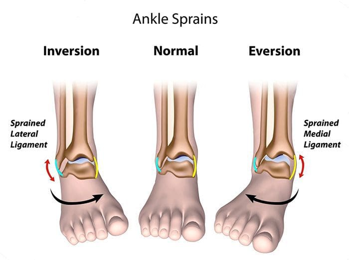
When you roll your ankle outward, the movement damages the ligaments on the outside of the ankle. This is a common sports injury and, unfortunately, once you have sprained your ankle it’s more likely to recur. Up to half of the people who suffer from a sprained ankle will sprain it again.
Strains:
There are two tendons in the ankle that are commonly strained, usually over stretching from overuse or trauma.
Fractures:
This injury happens when one or more of the three bones in the ankle is injured. While less common than a sprain or strain, a fractured ankle may also involve damaged ligaments and require surgery.
Many instances of ankle injuries are avoidable. Be sure to wear proper shoes when exercising or participating in sports, avoid uneven walking surfaces, and keep stairways and floors in your home clear of clutter
And, ladies, avoid the really high heels. We know, we know, they are just so cute! 🙂
Even with ankle-protecting precautions, you still may end up on your rump in the grass nursing your swollen ankle. What should you do if you injure your ankle? There are several forms of treatment for an ankle injury depending on its severity.
Rest and ice: For mild injuries, stay off your ankle and use ice packs to reduce the swelling. Rest allows the injured area to heal faster.
Visit a doctor: If you experience severe pain, swelling, and are unable to put weight on your ankle, see a doctor, as some ankle injuries grow worse without treatment. Injuries may require a brace, cast, or even surgery.
See a chiropractor: Patients frequently see strongly positive results in ankle injuries from a series of chiropractic treatments. Chiropractors understand the way the ankle is built, and use chiropractic adjustments to reduce pain and inflammation and promote faster healing.
Exercise rehab: Once you are healed, it’s vital to build up the ankle’s strength to avoid re-injury. Your chiropractor can lay out an exercise routine that you can employ into your regular workouts that will improve your balance and increase mobility. Performing these moves helps dramatically decrease dealing with this again down the road.
Ankle injuries are common and, whether or not you maintain an active lifestyle, you may end up suffering from one. By visiting a chiropractor on the front end, you can better plan a course of treatment that will heal your ankle quickly, reduce the pain effectively, and minimize the chance of a recurrence.
I have been travelling through Athens and now Istanbul. My 11 year old is a Percy Jackson nut and has been filling me in with the who’s who of Greek mythology and I am learning Latin words every day. Quite an education!
I looked up the word syndesmosis and the Latin translation is “(New Latin, from Greek sundesmos) bond, ligament, from sundein, meaning to bind together”. As sports injury professionals, we know syndesmosis to be the joint articulation between the tibia and the fibula bones around the ankle. These two bones are �bound’ together with very firm and strong ligaments.
Syndesmosis comes to mind after I saw a girl sprain a syndesmosis at the Archaeological Museum in Istanbul today. This poor girl was preoccupied by the hundreds of cats and kittens running all over the place and did not see the uneven cobblestones on which she placed her foot. At the same time, she turned to change direction. This is a common mechanism of injury for a syndesmosis – a forced dorsiflexion and rotation on a fixed foot.
Rehab Masterclass Issue 140 Of Sports Injury Bulletin
Of all the ankle injuries, injury to the syndesmosis is the biggest pest to sports physios and the like. And unlike simple garden variety ankle sprains that heal quickly, the syndesmosis takes a LONG time to heal properly. If you deal with athletes that are susceptible to syndesmosis sprains, I’m sure you will agree that these are harder injuries to manage because of the severe consequences if done badly.
I go into a fair bit of detail in my Sports Injury Bulletin piece about syndesmosis injuries, detailing how they happen, how to identify them and then manage them. What I would like to highlight here are the implications of mismanaging a syndesmosis sprain.
In the current issue of The Journal of Sports and Physical Therapy, a group of Japanese researchers discovered that individuals who had chronic ankle instability (CAI) had a distal fibula that was positioned more lateral compared with healthy individuals with no CAI. In effect, those who had suffered serious syndesmosis injuries in the past and ended up with a wider distance between the fibula and the tibia, suffered more ongoing ankle pain than those without a tibfib separation.
Research shows that even a 1mm displacement of the talus within the mortise (due to a wider placed fibula) can reduce the contact area in the talocrural joint by 42% (Ramsey and Hamilton 1976). Mismanaged syndesmosis injuries, resulting in an excessive amount of opening, can lead to early onset arthritic changes and chronic ankle instability. The talus bone bounces around in the now wider tibfib articulation.
A Widening Of The Fibula Is Due To One Of The Following:
Poor initial management, whereby the athlete is allowed to weight bear too early and this weight bearing forces the fibula away from the tibia as the syndesmosis ligaments are trying to heal.
The degree of damage is so severe that proper tightening of these ligaments is not possible without surgical intervention such as a screw or similar being placed between the two bones to �force’ them together.
The key for a sports injury practitioner, is to properly identify a regular ankle sprain from a more serious syndesmosis injury. If you get this part wrong and allow the athlete to get back to weight bearing too early, then expect some complaints about a chronically painful ankle some time down the track.
Call Today!
Kobayashi et al (2014). �Fibular malalignment in individuals with chronic ankle instability.’ JOPST. 44(11); pp 841-910.
Ramsey and Hamilton (1976). J Bone and J Surgery Am. 58(3); 356-357.
IFM's Find A Practitioner tool is the largest referral network in Functional Medicine, created to help patients locate Functional Medicine practitioners anywhere in the world. IFM Certified Practitioners are listed first in the search results, given their extensive education in Functional Medicine




 Chiropractic For Ankle Pain