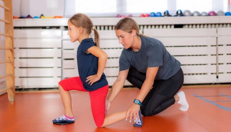
Children walking with their toes pointed in may be pigeon-toed. What are the causes, conditions associated with it, and treatments?
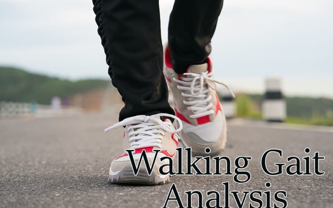
Pigeon-toed Walking
If a child walks with their feet turned inward at the toes, it is usually described as being pigeon-toed. This pointing inward of the feet occasionally occurs as a child learns to walk and may continue through toddlerhood. It is noticed more often in children than adults, but older individuals can experience it. Pigeon-toed walking is rarely a major orthopedic problem and usually disappears without treatment. However, there are times when it may impact a child’s lower extremities and hips. Bracing or surgery may be necessary in these cases to correct the problem. (Paramanandam V. et al., 2019) This condition is common and typically is caused by abnormal birth positions in utero. Sometimes, slight issues may lead to noticeable functional characteristics. Mild changes in bone shape and positioning usually cause pigeon toes. Often, it subsides in a few years as the child continues to develop.
What Does It Mean?
There is usually no need to worry, as this condition is likely not permanent and will go away in a few years. (Paramanandam V. et al., 2019) However, it is recommended that you check in with your healthcare provider to ensure the child is developing normally. Some adults walk with their toes turned in. This may be due to a birth defect, a weakness, or a rare case of pigeon-toed walking as a youth that was left untreated or did not go away.
Causes
There are various reasons for pigeon-toed walking. To determine the cause, a healthcare provider can assess the child’s condition and make a diagnosis, including:
Metatarsus Adductus
A condition where the front part of the foot is turned inward.
The metatarsals are the long bones of the forefoot.
This is when the bones of the foot point inward, leading to pigeon-toed walking.
A clinical examination and X-ray can confirm the metatarsus adducts as a cause of pigeon-toed walking.
Tibial Torsion
A twisted shinbone (tibia) can cause the feet to turn inward in younger children.
The shinbone/tibia in some children may be slightly twisted.
The tibia can either turn outward or inward.
When it twists inward, it may manifest as a pigeon-toed gait.
Tibial torsion may accompany femoral anteversion.
It is diagnosed with an X-ray.
Children with tibial torsion typically grow out of the problem, and the pigeon-toed disappears by age 4. (Uden H., & Kumar S. 2012)
Femoral Anteversion
A common cause, especially in older children, is when the thighbone/femur is twisted inward.
If the femur turns inward and forward unnaturally, where the femoral neck meets the body of the femur, it is called femoral anteversion.
An outward and backward rotation of the femur is called femoral retroversion.
Many children with femoral anteversion appear knock-kneed with a large gap between their feet when standing with knees together, and when they walk, they appear pigeon-toed.
A clinical examination and X-ray diagnose it.
Symptoms
In most cases, the child does not complain of any pain. However, if pain is felt, it can include:
Tightness in the calf muscles
Aching on the outer edges of the feet
Knee pain
Usually, parents will notice pigeon-toes when their child is first learning to walk. Rest assured, the child most likely is not experiencing pain. They have feet and knees that turn inward when they walk and run. (Uden H., & Kumar S., 2012)
A pediatrician or primary care provider can assess the situation and make recommendations. Most pigeon-toed children begin walking and running normally after age 3 or 4, so a watch-and-wait approach is used. Parents may have to take their child to a specialist, like an orthopedic surgeon, if they complain of pain while walking. A specialist may be referred if the child cannot walk due to the inward turn of their feet.
Risk Factors
Pigeon-toed walking is not a preventable condition but rather one that develops during pregnancy. Causes may include: (Scorcelletti M. et al., 2020)
A pregnancy with twins or multiple births
Large fetus
Breech position in utero when the baby is positioned feet first.
Typically, a normal gait will appear by age 3 or 4. Other treatments may include:
Physical Therapy Exercises and Gait Training
Exercises to stretch tight lower extremity muscles and strengthen hip and leg muscles can help improve walking gait.
See a pediatric specialist before starting, as research shows that parental stretching of a newborn with metatarsus adductus offers little benefit. (Eamsobhana P. et al., 2017)
Bracing or Casting
Braces
Serial casting is a procedure that helps children improve their range of movement and may be done to place their lower extremities in an optimum position as they develop. (Uden H., & Kumar S., 2012)
Surgery
For cases in which tibial torsion is the cause, osteotomy surgery, which involves cutting and/or removing bone, may be recommended to correct the structural deformity of the shinbone.
Injury Medical Chiropractic & Functional Medicine Clinic
Injury Medical Chiropractic and Functional Medicine Clinic works with primary healthcare providers and specialists to develop an optimal health and wellness solution. We focus on what works for you to relieve pain, restore function, and prevent injury. Regarding musculoskeletal pain, specialists like chiropractors, acupuncturists, and massage therapists can help mitigate the pain through spinal adjustments that help the body realign itself. They can also work with other medical professionals to integrate a treatment plan to resolve musculoskeletal issues.
Foot Pronation
References
Paramanandam, V., Lizarraga, K. J., Soh, D., Algarni, M., Rohani, M., & Fasano, A. (2019). Unusual gait disorders: a phenomenological approach and classification. Expert review of neurotherapeutics, 19(2), 119–132. https://doi.org/10.1080/14737175.2019.1562337
Uden, H., & Kumar, S. (2012). Non-surgical management of a pediatric “intoed” gait pattern – a systematic review of the current best evidence. Journal of Multidisciplinary Healthcare, 5, 27–35. https://doi.org/10.2147/JMDH.S28669
Scorcelletti, M., Reeves, N. D., Rittweger, J., & Ireland, A. (2020). Femoral anteversion: significance and measurement. Journal of Anatomy, 237(5), 811–826. https://doi.org/10.1111/joa.13249
Eamsobhana, P., Rojjananukulpong, K., Ariyawatkul, T., Chotigavanichaya, C., & Kaewpornsawan, K. (2017). Does the parental stretching programs improve metatarsus adductus in newborns?. Journal of Orthopaedic Surgery (Hong Kong), 25(1), 2309499017690320. https://doi.org/10.1177/2309499017690320
For individuals experiencing musculoskeletal issues and pain symptoms, can learning about biomechanics and how it applies to movement, physical training, and performance, help in injury treatment and prevention?
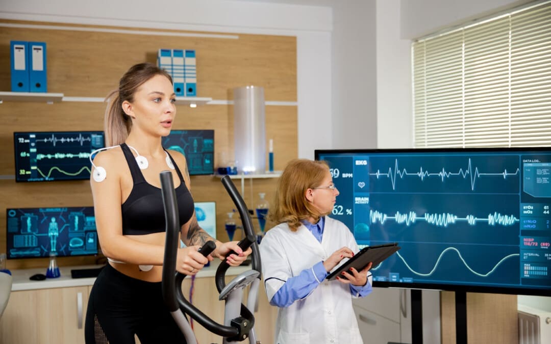
Biomechanics
Biomechanics studies all life forms and their mechanical workings. Many think of biomechanics in sports and athletic performance, but biomechanics helps create and improve technologies, equipment, and injury rehabilitation techniques. (Tung-Wu Lu, Chu-Fen Chang 2012) Scientists, sports medicine doctors, physiotherapists, chiropractors, and conditioning specialists utilize biomechanics to help develop training protocols and techniques to improve therapy outcomes.
Body Movement
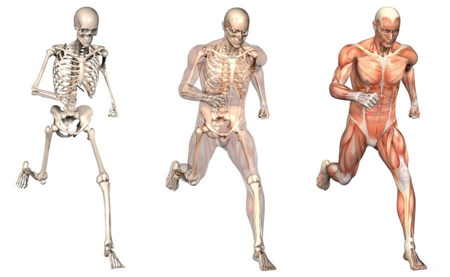
Biomechanics studies the movement of the body, including how muscles, bones, tendons, and ligaments work together, especially when movement is not optimal or correct. It is part of the larger field of kinesiology, specifically focusing on motion mechanics and analysis of how all the individual parts of the body work together to make up athletic and normal movements. (José M Vilar et al., 2013) Biomechanics includes:
Structure of bones and muscles.
Movement ability.
Mechanics of blood circulation, renal function, and other functions.
The study of forces and the effects of these forces on the tissues, fluid, or materials used for diagnosis, treatment, or research. (Jose I. Priego-Quesada 2021)
Sports
Sports biomechanics studies motion in exercising, training, and sports, which incorporates physics and the laws of mechanics. For example, the biomechanics of a specific exercise looks at:
Body position.
Movement of the feet, hips, knees, back, shoulders, and arms.
Knowing the correct movement patterns helps make the most of the exercise while preventing injuries, correcting form mistakes, informing training protocols, and increasing positive results. Understanding how the body moves and why it moves the way it does helps medical professionals prevent and treat injuries, alleviate pain symptoms, and improve performance.
Equipment
Biomechanics is used in the development of physical and sports equipment to improve performance. For example, a shoe can be designed for optimal performance for a skateboarder, long-distance runner, or soccer player. Playing surfaces are also studied for this purpose, such as how the surface stiffness of artificial turf affects athletic performance. (Jose I. Priego-Quesada 2021)
Individuals
Biomechanics can analyze an individual’s movements for more effective movement during training and games.
For example, an individual’s running gait or swing can be filmed with recommendations on what to change to improve.
Injuries
The science studies the causes, treatment, and prevention of neuromusculoskeletal injuries.
The research can analyze the forces that cause injuries and provide information for medical professionals on how to reduce the risk of injury.
Training
Biomechanics studies sports techniques and training systems to develop ways to improve efficiency.
This can include research on positioning, release, follow-through, etc.
It can analyze and help design new training techniques based on the mechanical demands of the sport, aimed at resulting in better performance.
For example, muscle activation is measured in cycling using electromyography and kinematics, which helps researchers analyze factors like posture, components, or exercise intensity that affect activation. (Jose I. Priego-Quesada 2021)
Motions
In biomechanics, the body’s motions are referred to from anatomical positioning:
Standing upright, with the gaze straight ahead
Arms at the sides
Palms facing forward
Feet spaced slightly apart, toes forward.
The three anatomical planes include:
Sagittal – median – Dividing the body into right and left halves is the sagittal/median plane. Flexion and extension occur in the sagittal plane.
Frontal – The frontal plane divides the body into front and back sides but also includes abduction, or moving a limb away from the center, and adduction, or moving a limb towards the center in the frontal plane.
Transverse – horizontal. – The upper and lower parts of the body are divided by the transverse/horizontal plane. Rotating movements occur here. (American Council on Exercise 2017)
Moving the body in all three planes occurs with daily activity. This is why performing exercises in each plane of motion to build strength, function, and stability is recommended.
Tools
Various tools are used to study biomechanics. Studies are usually performed using a device known as electromyography or EMG sensors. Sensors are placed on the skin and measure the amount and degree of muscle fiber activation in certain muscles during test exercises. EMGs can help:
Researchers understand which exercises are more effective than others.
Therapists know whether patients’ muscles are properly operating and functioning.
Dynamometers are another tool that helps measure muscle strength.
They measure the force output generated during muscle contractions to see if the muscles are sufficiently strong.
They are used to measure grip strength, which can be an indicator of overall strength, health, and longevity. (Li Huang et al., 2022)
Beyond Adjustments: Chiropractic and Integrative Healthcare
References
Lu, T. W., & Chang, C. F. (2012). Biomechanics of human movement and its clinical applications. The Kaohsiung journal of medical sciences, 28(2 Suppl), S13–S25. https://doi.org/10.1016/j.kjms.2011.08.004
Vilar, J. M., Miró, F., Rivero, M. A., & Spinella, G. (2013). Biomechanics. BioMed research international, 2013, 271543. https://doi.org/10.1155/2013/271543
Priego-Quesada J. I. (2021). Exercise Biomechanics and Physiology. Life (Basel, Switzerland), 11(2), 159. https://doi.org/10.3390/life11020159
American Council on Exercise. Makeba Edwards. (2017). Planes of Motion Explained (Exercise Science, Issue. https://www.acefitness.org/fitness-certifications/ace-answers/exam-preparation-blog/2863/the-planes-of-motion-explained/
Huang, L., Liu, Y., Lin, T., Hou, L., Song, Q., Ge, N., & Yue, J. (2022). Reliability and validity of two hand dynamometers when used by community-dwelling adults aged over 50 years. BMC geriatrics, 22(1), 580. https://doi.org/10.1186/s12877-022-03270-6
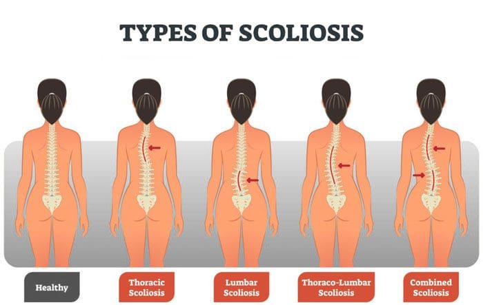
Scoliosis in adolescents and teens can be corrected with proper bracing, adjustments and lead a normal life. For adult scoliosis correcting the problem is more difficult. Fortunately, cases of adult scoliosis are rare. Scoliosis cases that follow from childhood into adulthood require a comprehensive diagnosis to determine severity. Thoracolumbar scoliosis adult-onset scoliosis requires an understanding of the catalysts to develop an effective treatment plan. Chiropractors use a full range of diagnostic tools to measure the severity of adult scoliosis.
Diagnosis
Adult scoliosis is the presentation of abnormal curvature of the spine. It can happen in the thoracic, lumbar spine, or both. This can have varying degrees of severity. Severe adult scoliosis can be apparent through visual assessment and examination. Cases that are not as obvious require utilizing diagnostic tools. These include:
Imaging
X-rays will show any asymmetry that is associated with scoliosis. This asymmetry can be present in the hips or shoulder and is usually qualified by spinal misalignment.
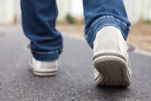
Walking Gait Examination
Inspecting how worn out an individual’s shoe/s are and having them perform various walking tests can reveal problems with gait. In adults, this can present instability. For example, having problems with balance or fast-twitch muscle response.
Neuromotor Exams
These exams are general and first performed to get a baseline diagnosis for the presence of adult scoliosis. Tests look at the left and right coordination along with the sense of touch capabilities. This measures the severity of the improper spinal curvature and how much it has affected the development of an individual’s motor functions. It is also done in the context of how it’s affecting the body’s biomechanics. Following these exams are quantitative tools/techniques for measuring the severity of adult thoracolumbar scoliosis. These include:
Cobb Angle Measurement
This tool determines the maximum degree of spinal curvature variation and provides a context for severity.
King Classification Tool
This examines the vertebral alignment to determine the spinal variance in specific vertebrae from the neutral center position.
Lenke Classification Tool
This spinal exam relies on measurements of three positions and looks for flexibility.
Combined Approaches
When assessing adult scoliosis, this is important to understand and helps determine how to proceed with treatment. The body is no longer in development as an adolescent. This means bracing does not come with a one-size-fits-all approach. Chiropractic can help with the assessment modalities used to investigate adult scoliosis cases. These measurement and analyses tools are often used in combination to develop a complete picture of what is going on.
Body Composition
Fill Up With Prebiotics
Individuals can help their gut bacteria thrive in the digestive tract by consuming prebiotics. Prebiotics are a form of soluble fiber. The body cannot digest these prebiotics, but gut bacteria can. Recommended sources of fiber-rich prebiotics can be found in nutrient-dense foods like:
Leeks
Garlic
Onions
Fruits
Legumes
Raw chicory
A diet with various fiber types has been shown to reduce the risk of obesity and prevent weight gain. Resistant starches like plantains, green bananas, and cooled potatoes have increased beneficial bacteria in the colon. Barley, oats, and wheat bran are insoluble high-fiber grains that are also recommended sources.
References
Aebi, Max. “The adult scoliosis.” The European spine journal: official publication of the European Spine Society, the European Spinal Deformity Society, and the European Section of the Cervical Spine Research Society vol. 14,10 (2005): 925-48. doi:10.1007/s00586-005-1053-9
Haenen, Daniëlle et al. “A diet high in resistant starch modulates microbiota composition, SCFA concentrations, and gene expression in pig intestine.” The Journal of nutrition vol. 143,3 (2013): 274-83. doi:10.3945/jn.112.169672
Lowe, Thomas et al. “The SRS classification for adult spinal deformity: building on the King/Moe and Lenke classification systems.” Spine vol. 31,19 Suppl (2006): S119-25. doi:10.1097/01.brs.0000232709.48446.be
Although it is not officially summer, the past few weeks sure feels like it. Especially for those with joint discomfort and pain. As the body ages, individuals may notice their joints have some mobility/flexibility issues in the summer heat. Again, the heat and humidity are the culprits. The hotter it is, the more the body is susceptible to inflammation and swelling. The more prone an individual’s body is to swelling, the more pain can present. Barometric pressure can also have some form of impact on joint health. The pressure changes can cause the joints to become more sensitive. When the pressure changes, individuals often speak of their joints feeling tighter combined with stiffness, leading to a cycle of swelling and pain.
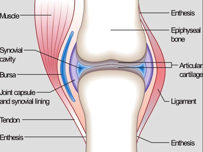
Joint Anatomy
Whether it’s the hip, knee, elbow, or hand, all of the body’s joints have fluid in them. It is a gel-like substance known as synovial fluid. This is what lubricates the joints and keeps them functioning smoothly. However, the temperature and humidity levels can change the thickness of the fluid in the joints. This means that the synovial fluid can become inflamed with the weather changes. This is a symptom when the joints begin to feel like they cannot move and/or are becoming stiff. Joint inflammation can become more common and chronic as the body gets older.
Weather and the joints
The summer heat and humidity can affect the joint because:
The tendons, ligaments, and muscles expand in this type of weather
The heat can restrict individuals from moving around. Non-use stiffens the joints
Joints that have worn down cartilage could have exposed nerves that are reacting to the temperature changes
Humidity causes the body to lose water by sweating. This can reduce the fluid around the joints leading to stiffness, immobility, and pain.
However, not everyone has joint problems in the summer heat. Many have joint issues when it’s cold, damp, or raining. Other’s are at their best in cool, dry weather. It depends on an individual’s body and how their joints react when the temperature changes.
Maintaining joint health for the summer heat
When joint discomfort or pain presents in the summer, there are a few easy ways to gain relief.
Properly Hydrate the Body
Water and sports drinks maintain the fluid levels in the body, specifically, it keeps the joints moving. One way to hydrate the body can be achieved by eating healthy fruits and vegetables. Water-rich fruits and vegetables include:
Watermelon
Oranges
Strawberries
Tomatoes
Cucumbers
Spinach
Celery
Over-The-Counter pain ointments and creams
Arthritis and anti-inflammatory creams/ointments can ease joint pain by allowing more blood circulation in the affected areas.
Dressing for the heat
Wear loose, natural fiber, breathable clothing that allows the body to move freely while maintaining a cool temperature.
Relax in the air conditioning
Get into the air conditioning. The cool air can help reduce joint inflammation.
Get in the Water
Swimming or just wading through doing some light exercise in the water cools the body’s core. In addition, the buoyancy of the water relieves pressure on the joints.
Body Composition Testing
Body Water
The body is made up of as much as 2/3’s water. Even though much of the body is made up of water, the percentage of body composition changes based on functional needs. Essential functions of water include:
Water is the building block to almost every cell in the body
It regulates the body’s temperature through sweating and respiration
Carbohydrates and proteins for energy are transported via the water in the blood
Water assists in the removal of metabolic waste through urination
It is part of the shock-absorbing system that protects the brain and spinal cord
Water is part of the saliva and fluid that lubricates the joints
The amount of water in the body depends on various factors. This includes:
Age
Gender
Physical activity
It is referred to as Total Body Water or TBW.
TBW is constantly changing with gains and losses of fluid in healthy adults. The body can detect irregularities and compensate for losses and/or gains to make sure that the systems are balanced.
The information herein is not intended to replace a one-on-one relationship with a qualified healthcare professional or licensed physician and is not medical advice. We encourage you to make your own health care decisions based on your research and partnership with a qualified health care professional. Our information scope is limited to chiropractic, musculoskeletal, physical medicines, wellness, sensitive health issues, functional medicine articles, topics, and discussions. We provide and present clinical collaboration with specialists from a wide array of disciplines. Each specialist is governed by their professional scope of practice and their jurisdiction of licensure. We use functional health & wellness protocols to treat and support care for the injuries or disorders of the musculoskeletal system. Our videos, posts, topics, subjects, and insights cover clinical matters, issues, and topics that relate to and support, directly or indirectly, our clinical scope of practice.* Our office has made a reasonable attempt to provide supportive citations and has identified the relevant research study or studies supporting our posts. In addition, we provide copies of supporting research studies available to regulatory boards and the public upon request.
We understand that we cover matters that require an additional explanation of how it may assist in a particular care plan or treatment protocol; therefore, to further discuss the subject matter above, please feel free to ask Dr. Alex Jimenez or contact us at 915-850-0900.
Morton, Darren, and Robin Callister. “Exercise-related transient abdominal pain (ETAP).” Sports medicine (Auckland, N.Z.) vol. 45,1 (2015): 23-35. doi:10.1007/s40279-014-0245-z
Peeler, Jason et al. “Managing Knee Osteoarthritis: The Effects of Body Weight Supported Physical Activity on Joint Pain, Function, and Thigh Muscle Strength.” Clinical journal of sports medicine: official journal of the Canadian Academy of Sports Medicine vol. 25,6 (2015): 518-23. doi:10.1097/JSM.0000000000000173
Quick, D C. “Joint pain and weather. A critical review of the literature.” Minnesota medicine vol. 80,3 (1997): 25-9.
Timmermans, Erik J et al. “The Influence of Weather Conditions on Joint Pain in Older People with Osteoarthritis: Results from the European Project on OSteoArthritis.” The Journal of rheumatology vol. 42,10 (2015): 1885-92. doi:10.3899/jrheum.141594
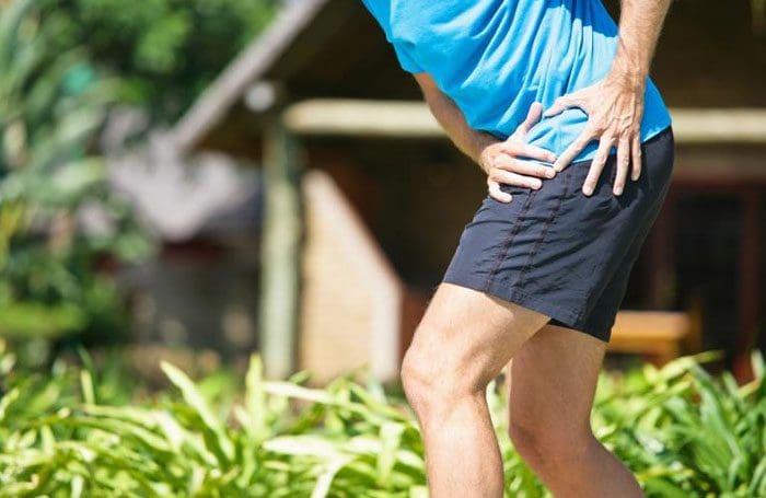
Determining if sciatica pain and symptoms are showing improvement can be as simple as the pain significantly reducing or it could be a bit more complex, depending on the severity of the condition. Chiropractic treatment keeps track of the location and movement of the pain as a reliable indicator that complete relief is getting closer. When the pain retreats up the leg, it is a sign of improvement even with back and buttock aches/pain that feel like it’s worsening.
Various Symptoms
The most common symptoms include:
Tingling
Numbness
Sharp pain
Dull pain
Radiating/spreading pain
A feeling like insects crawling or water trickling down the leg
Changing sensations in the buttock, back, leg, or foot
Does It Get Worse Before Getting Better?
Sciatica can get worse before it gets better. This is known as centralization where the pain moves or retreats back towards the midline of the spine after repeated movements or guided/chiropractic positioning and adjusting. It can be misleading, making the individual think the sciatica is worsening, or that something has caused a sciatica flare-up. However, healing is taking place.The furthest location away from the low back is the area to pay attention to. It’s different for individuals. It could be the:
Foot
Calf
Back of the thigh
No matter where the pain is pay attention to that particular area. If it feels like the sciatica is getting worse, take a moment to determine where the pain is presenting. If the pain has retreated and there is no pain in the foot, calf, or leg, the sciatica is getting better. What happens is the retreating pain going up the spine increases the pain in the back and buttocks. This means there is an improvement.
Sciatica Getting Worse
How to tell if it’s getting worse? An increase in pain could indicate that it is getting worse. But, the key is to pay attention to the location and movement of the pain. When it gets worse the pain is advancing, for example, if there was pain only in the back and buttock yesterday, and today the pain is radiating down the back of the leg into the calf, then the sciatica is getting worse.
Length of Time Sciatic Nerve Pain Lasts
For most individuals, sciatic nerve pain lasts from two to six weeks.
The acute pain lasts around 1 to 2 weeks, with lingering discomfort as the condition heals
There are factors that can cause sciatica to remain longer, or increase the chances of returning This includes:
Tight hamstrings
Weight gain
Pregnancy
Poor posture
Improper lifting
Sciatica that lasts more than six weeks is considered chronic. Medical intervention should be sought out if it lasts this long. Non-invasive treatment like chiropractic or physical therapy is recommended to help speed the healing process and reduce pain.
Permanent Cure
Most sciatica cases are caused by a spinal disc disorder in the lower back. Around 85% of sciatica cases are disc-related. There is a chance that sciatica can return. For most individuals, it only takes a small amount of work to keep sciatica at bay. Staying healthy and flexible are two ways to prevent sciatica from returning. This can be done through:
Healthy diet
Maintain a healthy weight
Staying active with 2 ½ hours of physical activity/exercise a week
Maintain proper posture
Regular stretching
Quitting smoking
If overweight it is highly recommended to lose weight. One study showed that obesity increased the risk of hospitalization by 36%. Other factors that increase the potential for sciatica are frequent intense physical activity levels in sports, exercise, DIY projects, etc.
Chiropractic Improvement
Whether dealing with sciatica during pregnancy, from tight hamstrings, or piriformis syndrome, chiropractic can help. A chiropractor can bring relief through:
Surgery is rarely needed and only as a last resort. Chiropractic care will generate improvement and will educate the individual on what to do to prevent sciatica from flaring up.
Body Composition Improvement
Essential Fat vs Storage Fat
There is essential fat in the body. It has a significant role in overall health and is essential for survival. Essential fat is present in the:
Organs
Bone marrow
Nerve cells
Brain
Essential fat helps with:
Maintaining sufficient energy reserves that function as a metabolic fuel
Conserves body heat and functions as an insulator
Protects the internal organs and joints acting as a soft cushion
Non-essential/storage fat is adipose tissue that accumulates as an energy reserve. Storage fat affects body shape and appearance.
Disclaimer
The information herein is not intended to replace a one-on-one relationship with a qualified health care professional, licensed physician, and is not medical advice. We encourage you to make your own health care decisions based on your research and partnership with a qualified health care professional. Our information scope is limited to chiropractic, musculoskeletal, physical medicines, wellness, sensitive health issues, functional medicine articles, topics, and discussions. We provide and present clinical collaboration with specialists from a wide array of disciplines. Each specialist is governed by their professional scope of practice and their jurisdiction of licensure. We use functional health & wellness protocols to treat and support care for the musculoskeletal system’s injuries or disorders. Our videos, posts, topics, subjects, and insights cover clinical matters, issues, and topics that relate to and support, directly or indirectly, our clinical scope of practice.* Our office has made a reasonable attempt to provide supportive citations and has identified the relevant research study or studies supporting our posts. We provide copies of supporting research studies available to regulatory boards and the public upon request. We understand that we cover matters that require an additional explanation of how it may assist in a particular care plan or treatment protocol; therefore, to further discuss the subject matter above, please feel free to ask Dr. Alex Jimenez or contact us at 915-850-0900.
Dr. Alex Jimenez DC, MSACP, CCST, IFMCP, CIFM, CTG* email: [email protected] phone: 915-850-0900 Licensed in Texas & New Mexico
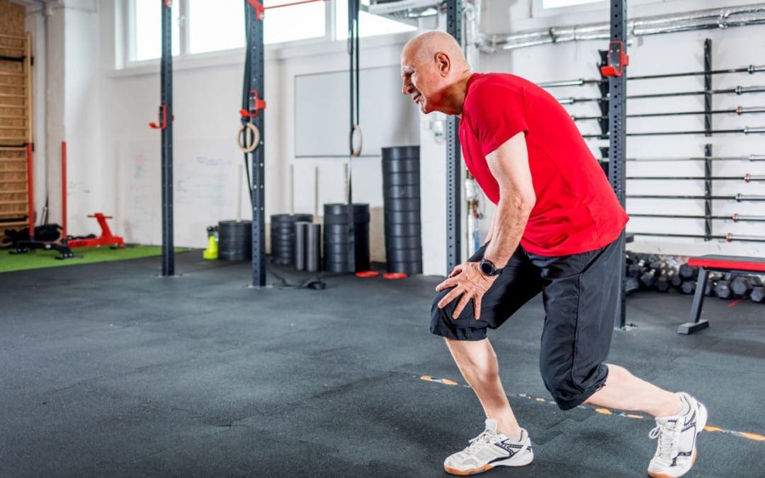
The way a person walks is known as gait. A problem with gait can indicate pain in a patient as well as serious conditions like diabetes and arthritis. An individual’s gait can be very telling revealing problems in the:
It is a diagnostic tool for a variety of conditions, injuries, and syndromes including autism. When it comes to chiropractic, an individual’s gait can offer important information regarding the root of the complaints, allowing for a more well-rounded, whole-body approach. If you think that the way you walk or moves does not matter, think again, as it could save you from developing back problems in the long run.
Gait Analysis
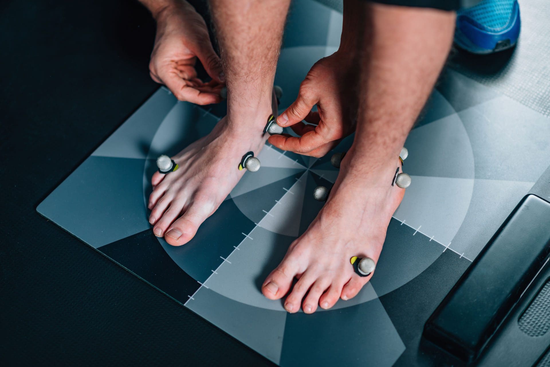
Gait analysis consists of a set of tests designed to provide a comprehensive evaluation of an individual’s gait. It is a study of human movement/motion that involves observing the individual and using specialized instruments that measure body movements, muscle activity, and body mechanics.
It is an assessment tool to provide insight into creating a treatment plan for people who have conditions or injuries that affect their ability to walk. It is utilized in sports biomechanics to help athletes gain optimal movement and for identifying problems with movement or posture, especially those with injuries. During the analysis, the individual may be asked to walk in a certain pattern or on a treadmill, that is usually connected to a computer, while the chiropractor/therapist looks at them from different angles.
Cameras are placed at different points to capture various views/perspectives including the front, back, and sides. The individual might have markers placed at certain points on the body like the knee, ankle, pelvis, etc. As the person moves, the computer captures data of the movement with a three-dimensional calculation of each marker. It generates a model to assess the movement of the skeletal structure, resulting in a detailed analysis of each joint�s movement.
Factors that Affect Gait
There are factors that affect an individual’s gait and that information is necessary for the analysis to be accurate. Age, gender, height, and weight of the individual is vital because men and women move differently and as individual age their body structure changes. Excess weight or physique can affect an individual’s posture and gait.
Shoes or not wearing shoes will also affect gait, as the terrain individuals walk on, and articles they carry, like a purse or backpack, changes how we walk. Other factors include:
Body proportion
State of mind
Emotions
Stress level
Personality type
Pathological factors such as
Neurological diseases
Psychiatric disorders
Trauma
Musculoskeletal issues
This is also measured and factored into the analysis data that includes the patients:
Length of stride
Cadence
Hip angle
Foot angle
Step length
Walking
Motion speed
Advantages of a Gait Analysis
Getting a gait analysis can be very helpful because it can provide invaluable insight into how your body is aligned and how it moves. It is a great diagnostic tool for identifying health issues related to the gait, spine, and feet and can also help provide early detection of health issues before the onset of symptoms.
If your chiropractor recommends you get a gait analysis it could be they suspect that something is going on, or it could mean they want a more thorough examination to provide optimal care. If you have any concerns talk with your chiropractor and ask them any questions that you have prior to the analysis, as stress and anxiety can put tension on the muscles and body, thus affecting the results.
Reduce Excessive Foot Pronation with Custom Foot Orthotics
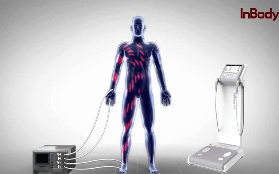
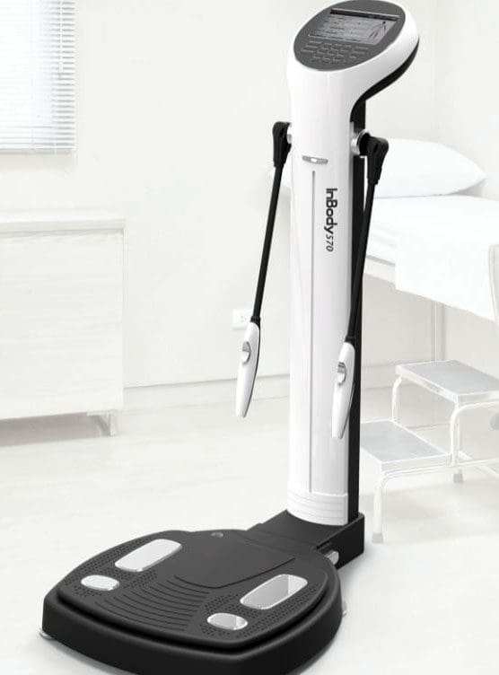
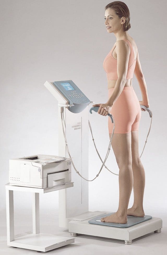
InBody devices use a method called Bio-electrical Impedance Analysis (BIA) to measure body composition. This divides your weight into different components, e.g., lean body mass and fat mass are utilized in the assessment of health and nutrition.
InBody Technology
Resistance Concept
An illustration of how this works:
Imagine cars in traffic
Your car is the voltage-current
The highway is body water
If no other cars were around, you could roll right through
If the human body was only water and nothing else, there would be no resistance.
But water is not the only element
You are not the only car on the freeway
The more traffic gets onto the freeway, the longer it takes for you. This is resistance.
Other elements:
Fat
Muscle
Bone
Minerals
Create resistance to the current going through the body
In BIA testing, the more water in the body equals less resistance
The muscle in the body contains water
The more muscle you have, the more body water
The more body water, the less the resistance on the current
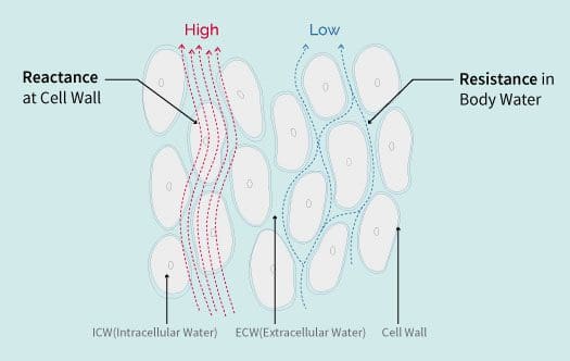
Bringing It Together
Impedance is the vector sum of the resistance
Reactance is the measurement BIA devices use to determine body composition
BIA applies cylinder model for the relationship between impedance and a body
Impedance is calculated by using two formulas:
The volume of a cylinder (Volume = Length x Area)
Impedance is inversely proportional to cross-sectional area and directly proportional to length.
Knowing the impedance and length of the cylinder, a measurement can be made of the volume of total body water.
In the body, the same formula applies, where the length is the height.
Calculation of the volume of the total body water can be made by knowing the impedance and height.
This is why it’s imperative to have correct height.
BIA Technology Has Been Revolutionized With InBody
Measuring impedance with electrodes creates contact resistance.
InBody accounts for this by strategically placing electrodes to accurately measure.
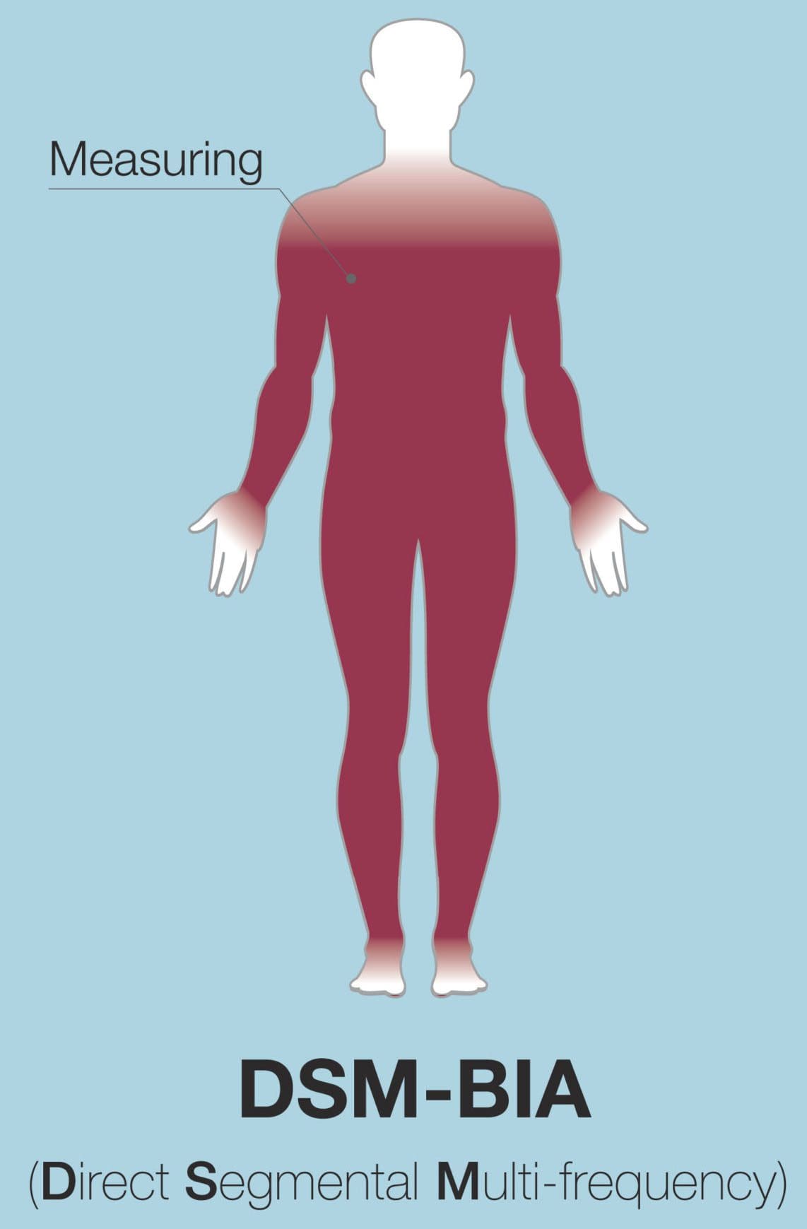
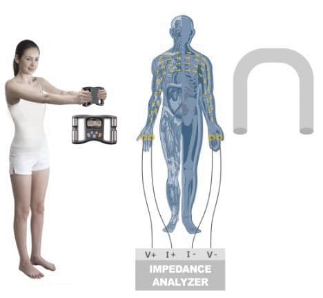
InBody provides independent measurements for the body�s 5 cylinders:
Left Arm
Right Arm
Left Leg
Right Leg
The Torso
InBody uses multiple currents and varying frequencies.
No empirical estimations are used to calculate body composition.
InBody measures impedance independently, so results are not affected by age, ethnicity, or gender.
Direct Segmental Multi-frequency Bioelectrical Impedance Analysis
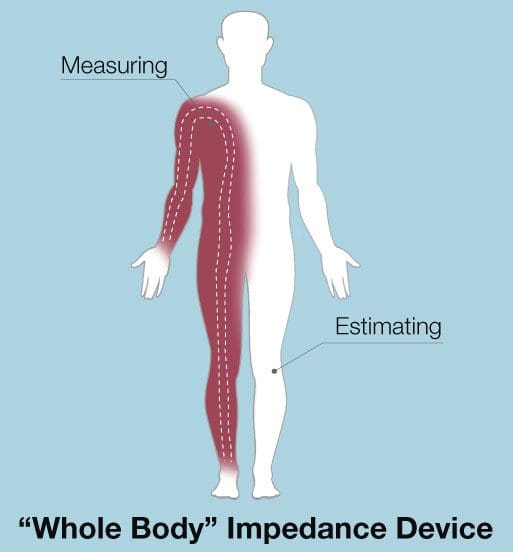
Traditional BIA systems view the body as a single cylinder and use whole-body impedance to determine total body water.
This method has a number of flaws:
It assumes the distribution of lean body mass and body fat are constant.
The shape & length of the arms, legs, and torso differ so the body cannot be seen as just one, but five separate parts.
Impedance is based on length and cross-sectional area, the calculation of TBW is inaccurate because each segment has different length and cross-section.
One major problem with the one-cylinder method is the lack of a torso measurement.
The torso has the lowest length and highest cross-section area.
This results in a very low impedance (10-40 ohms).
However, the trunk comprises about 50% of an individual�s lean body mass (LBM).
In the whole-body impedance measurement, the torso impedance is ignored and so changes the body torso impedance.
If the body torso is not measured separately then the impedance of the torso could be overlooked.
Because the body torso contains more water and muscles than the limbs, 1 ohm of torso impedance and 1 ohm of limb impedance can be completely different.
A difference of even 1-2 ohms can lead to a significant error in the determination of TBW.
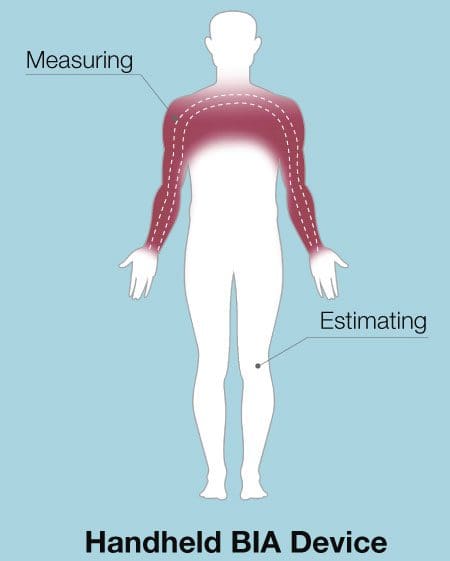
Some BIA devices only measure the impedance values of two cylinders and estimate the rest.
Some BIA scales only measure the legs.
For BIA handheld devices, only the arms are measured.
Some devices say they measure the whole body when they only measure one arm, one leg, and estimate the rest.
When using a BIA device, find one that actually measures the torso and measures it separately.
Otherwise, the estimations can lead to large errors.
InBody devices do not estimate through Direct Segmental Multi-frequency BIA, which in simpler terms, which means that each segment of the body right arm, left arm, torso, right leg, left leg are measured separately.
History of Bioimpedance Technology
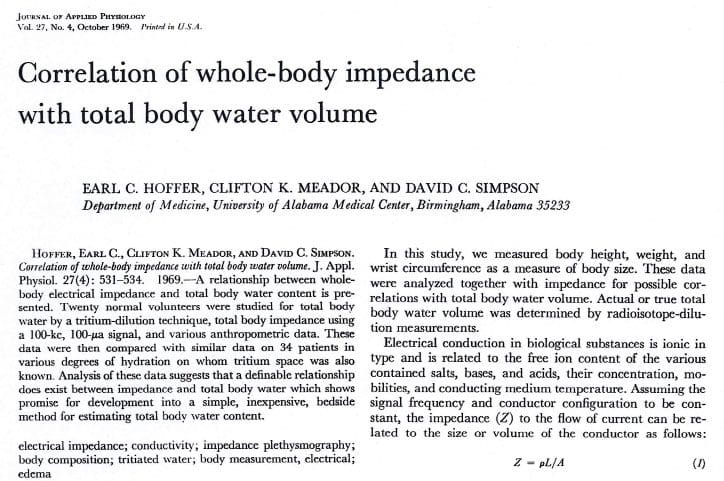
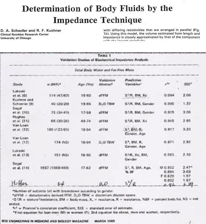
In 1969 came The Hoffer and Impedance Index
In 1969, Hoffer experimented to prove that total body water and biological impedance were highly interconnected. This meant that impedance measurement could be used to determine total body water.
He showed the squared value of height divided by impedance was highly correlated with total body water.
He took impedance measurements of the right half of the body. These included the right arm, torso, and right leg.
The equation he proved is the impedance index used in Bioelectrical analysis today.
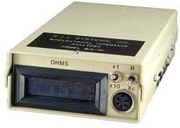
In 1979 came the RJL System and First Impedance Meter
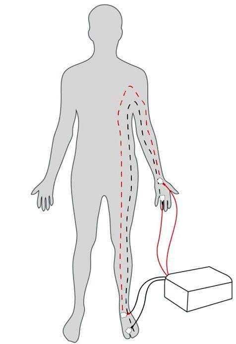
In 1979, RJL Systems brought the impedance meter and the BIA method began.
The device measured impedance through attachment of electrodes on the back of the right hand and on top of the right foot. A current of 50kHz was run through the right half of the body.
Before this, body composition could only be measured with calipers or underwater weighing.
These techniques had to be carried out by skilled people and was not easy.
Only specific types of patients were able to benefit from them, as well.
However, this was fast, less expensive and less intrusive. Thus, body composition analysts, nutritionists, and medical experts started using BIA technology.
In the 1980’s BIA limitations Emerged along with…
Lukaski, Segal and other scholars are the ones that accelerated the evolution of BIA.
Their studies proved that BIA had a high correlation with top standard methods, e.g., Underwater Weighing and DEXA.
However, there were technical limitations with BIA which surfaced towards the end of 1980s.
A common limitation was that BIA would assumed the human body was a cylinder shape and so only used a single frequency of 50 kHz.
Through research, various equations evolved (along with the impedance index). This complemented the technical limitation of BIA and was able to achieve greater accuracy for patients of different age, gender etc.
Lukaski & Kushner Develop Empirical Data Equation
This increased the accuracy of the results.
These equations utilized empirical data:
Gender and
Age to calculate a person�s body composition.
Empirical data is defined, as knowledge acquired by means of observation or experimentation.
By collecting data on a sample population that (hopefully) represents the variance of the entire population, researchers attempt to derive trends that may be used to predict outcomes.
In body composition, researchers identify these trends in muscle and fat mass; they use this data to predict body composition based on specific variables (age, gender, ethnicity, etc.)
Although empirical estimations could give you an accurate estimate of a general user�s body composition, there are significant problems when they are used for medical purposes.
Suppose there is a device that uses empirical equations to calculate TBW.
And there are two individuals who have same amount of lean body mass, however, one is 30 years old and the other is 40 years old.
Even though they have the same amount of LBM, the empirical equation will calculate that both will have 0.8 L difference in their TBW. This only because of age, which is not fair or accurate.
Home BIA Devices Start Showing Up
Because of technological limitations, BIA devices turned into home devices instead of hospital devices.
Then Japanese manufacturers released a variety of BIA body composition devices that the general public could easily use.
Some measured the impedance between two feet while standing on a scale. Others would hold the device and then measure the impedance between the hands.
Then In 1992 Kushner Proposed Multi-Frequencies & Segmental Analysis
Kushner claimed the human body to be made from five cylinders
Right Arm
Left Arm
Torso
Right Leg
Left Leg
Since the torso makes up 50% of lean body mass, Kushner emphasized measuring the impedance of the torso separately would be very significant.
Measuring total impedance alone is not sufficient. However, when all five parts are measured separately at different frequencies, then a distinction between extracellular water and intracellular water can be made.
In 1996 Dr. Cha Creates InBody Composition Analyzer
In 1996, Dr. Kichul Cha, a bioengineer from Harvard Medical School, develops the first 8-point electrode system with direct segmental analysis, which measures impedance for the five cylinders of the body at multiple frequencies.
This allows separate checking of torso impedance.
With this technology highly accurate results, without using empirical data, were able to be yielded.
InBody body composition analyzers became precision based medical devices. Impedance values for all of the body’s cylinders can be found on the InBody Result Sheet.
Many BIA products today provide muscle mass for each section of the body.
However, you can see the impedance values of all five parts of the body with the use of both high and low frequencies.
InBody Spotlight – Rachel Cosgrove of Results Fitness
History Body Model
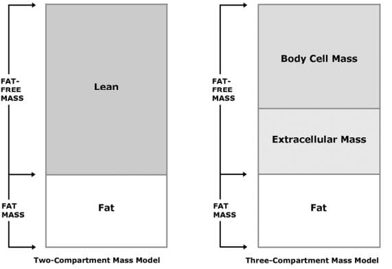
Body composition analysis lends itself to a number of techniques depending upon the specific needs of your patients.
Two-Compartment Mass Model: The two-compartment mass model divides the body into Fat-Free Mass and Fat Mass.
This simple model is useful when evaluating basic nutritional, fitness, and weight management needs of patients.
Three-Compartment Mass Model: The three-compartment model divides the body into:
Body Cell Mass
Extracellular Mass
Fat Mass
This model is often used in support of nutritional counseling and monitoring changes associated with aging.
Appropriate for full range of patients.
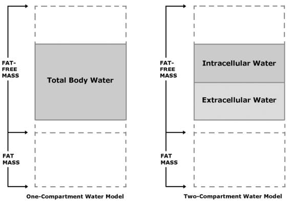
One-Compartment Water Model The one-compartment water model accounts for Total Body Water (TBW).
Total Body Water is the sum of Intracellular Water plus Extracellular Water and is wholly contained within Fat-Free Mass.
Normally, about 73% of Fat-Free Mass is water.
This model is handy for evaluating basic hydration status of patients.
Two-Compartment Water Model The two-compartment water model divides:
Total Body Water into
Intracellular Water and
Extracellular Water
This model is often used for the assessment of fluid balance associated with the treatment of conditions in a clinical setting.
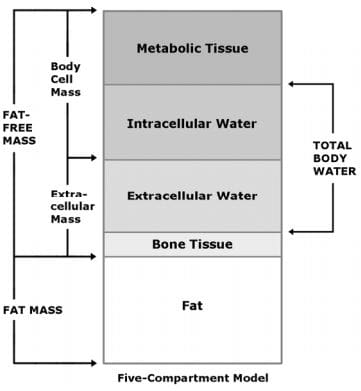
Five-Compartment Model divides the body into:
Metabolic Tissue
Intracellular Water
Extracellular Water
Bone Tissue
Fat Mass
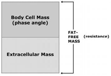
Impedance Model Application
When monitoring:
Compartment
Fat-Free Mass Resistance
Body Cell Mass
Total Body Water
Intracellular Water
Chart
Resistance
Phase Angle Resistance
Phase Angle
Application Guide
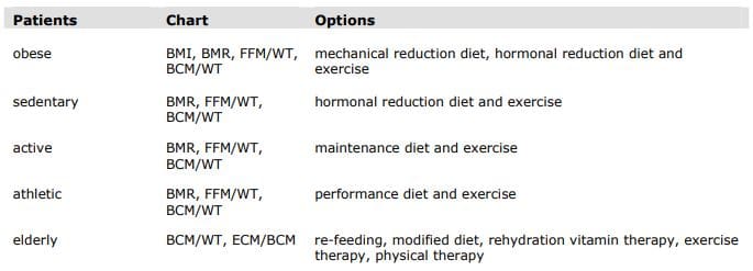
Obesity
The United States has long recognized obesity as a serious health condition.
In February 1985, the National Institutes of Health (NIH) in its Consensus Development Conference Statement (1) declared “The evidence is now overwhelming that obesity, defined as excessive storage of fat, has adverse effects on health and longevity.”
Obesity has long been associated with health risks.
While the specific mechanisms linking obesity to health risks are not fully understood, recent research focused on genes that express only in fat tissue has shown promise.
These genes code for hormones associated with insulin resistance and cardiovascular plaques.
The obesity epidemic continues to grow unabated in the United States (2,3).
Now, it has become a serious health problem in “both developed and developing countries in the Western Pacific Region of the World Health Organization” (4).
A useful definition of obesity is “excess fat mass resulting in mechanical or hormonal stress on the cardiovascular system, organs, and muscular-skeletal system.”
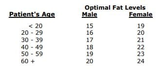
Diagnostic Criteria Body Mass Index (BMI) of 30 or greater. or
Fat Mass that is greater than 25 percent for males or greater than 30 percent for females.
Mechanical Reduction We recommend a two-step approach to normal function.
The first step is weight reduction to relieve mechanical stress on body systems.
Estimate total daily caloric expenditure = BMR * 1.2.
Set dietary intake = caloric expenditure – 700 calories per day.
Continue diet until BMI = 30.
NOTE: The initial dietary intake for obese patients will appear to be very high.
For example, a patient weighing 300 lb with 40 percent body fat will have a basal metabolic rate of 2550 calories, a total caloric expenditure of 3060 calories, and dietary intake of 2360 calories per day.
Hormonal Reduction
The second step is to decrease the ratio of fat mass to fat-free mass to reduce the incidence of fat-related hormones.
Measure the basal metabolic rate and fat-free mass.
Estimate caloric expenditure = BMR * 1.2.
Set dietary intake = caloric expenditure – 500 calories per day.
Continue until percentage fat mass reaches optimal level.
NOTE: Exercise is important in BIA because weight loss from dieting alone is comprised of 45 percent fat-free mass and 55 percent fat mass per pound. Exercise can alter this ratio to 25 percent fat-free mass and 75 percent fat mass.
References:
1. NIH Consensus Conference Statement, Health Implications of Obesity. Annals of Internal Medicine, 1985; 103 (6 pt 2):1073-1077.
2. Mokdad AH, et al. The spread of the obesity epidemic in the United States, 1991 – 1998. Journal of American Medical Association, 1999; 282:1519-1522.
3. Blackburn GL. Managing obesity in America: An overview. Advanced Studies in Medicine 2002;2(2):40-49.
4. Regional Office for the Western Pacific of the World Health Organization, the International Association for the Study of Obesity and the International Obesity Task Force. The Asia-Pacific perspective: Redefining obesity and its treatment. Health Communications Australia Pty. Limited, February 2000.
IFM's Find A Practitioner tool is the largest referral network in Functional Medicine, created to help patients locate Functional Medicine practitioners anywhere in the world. IFM Certified Practitioners are listed first in the search results, given their extensive education in Functional Medicine