by Dr Alex Jimenez DC, APRN, FNP-BC, CFMP, IFMCP | Gluten Free Recipes
I’ve been baking quite a bit of bread lately, and I thought it was high time to share some new bread recipes. Almost a year ago, I posted a top-rated recipe for a traditional two-step, 24-hour sourdough bread. I love that recipe, and I think that it makes a really delicious, sour bread. However, sometimes I want my bread to come out less sour, or I don’t have the time to do the two-stage sourdough process. This recipe I use for a bread that only takes one rise – then it’s shaped and baked.
1-Step Sourdough Bread Recipe
First mix: 10 minutes
First rise: 6-12 hours
Bake time: 45 minutes
Whisk together until blended in the bowl of a stand mixer with the paddle attachment or in a large bowl with a fork:
460 g Spring Water (don’t use tap water or any chlorinated water)
30g whole psyllium husk (or 20g finely ground psyllium husk)
Mix into the liquid with the paddle attachment or by hand with a wooden spoon:
400gBread Flour
100g wild yeast sourdough Starter (@120% hydration)
12g (1 TBSP) sugar
1 1/4 tsp salt
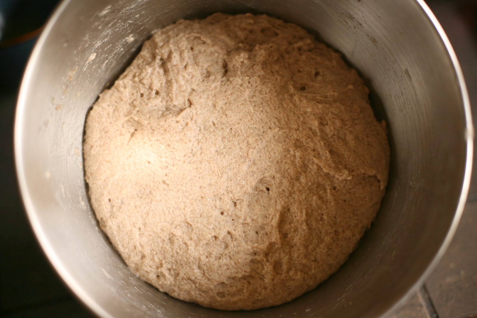
Pre-shape the dough into a ball and keep it seam-side up in the bowl. Cover the bowl with plastic wrap and let stand at room temperature for 6-12 hours. Keep an eye on it starting at the 6-hour mark.
When the bread has risen significantly, and you think it’s getting close to time, heat your oven to 450 degrees F with a cast-iron dutch oven inside. You will know the bread is ready to bake when it has risen quite a bit, and a fingermark gently poked against the surface of the dough doesn’t fill in immediately anymore. Once it passes the “finger test” and the oven is hot, you can shape the loaf, although it’s better to under-proof a little than over-proof. (If you need to go longer than 12 hours on the rise, put the dough in the refrigerator after the bread shows a significant rise. You can leave it in the fridge for up to a day or maybe three, then shape and bake.)
Carefully invert the bread onto a piece of parchment paper. Shape the bread into a slightly tighter ball by tucking the sides of the dough underneath all around the edge. Dust the top with flour if desired. Score the loaf with slashes 1/2 inch deep.
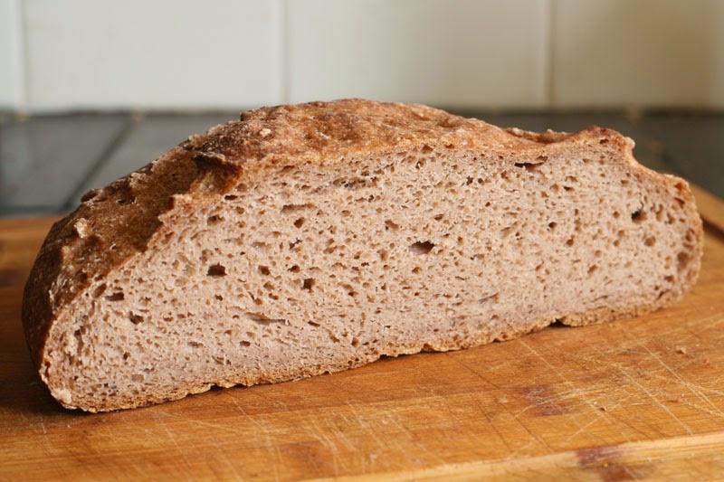
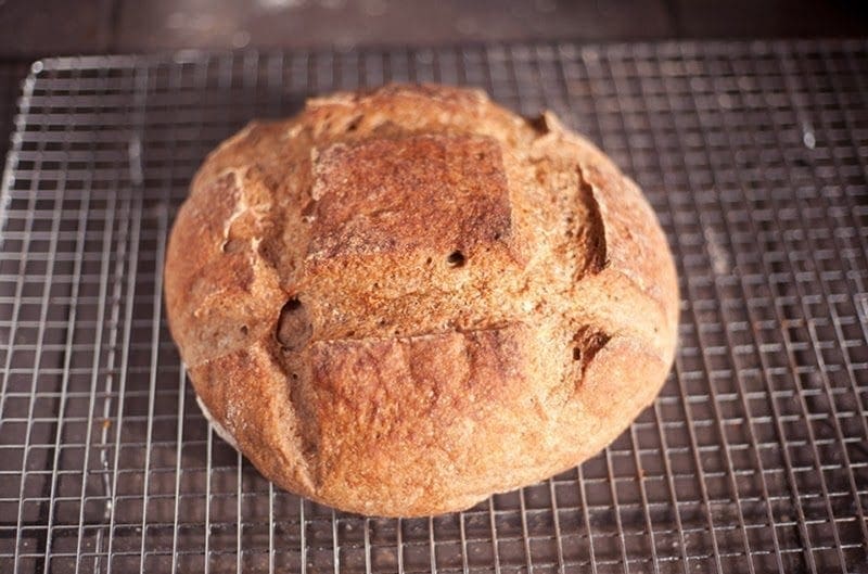
Using the parchment paper to lift it, carefully place the shaped loaf inside the hot dutch oven. Spritz the bread and all around the cast-iron pan before covering it with the lid. Bake the bread for 25 minutes inside the dutch oven, remove it to the rack, and bake another 20 minutes or until deeply browned. Remove the bread to cool on a rack, or for a crispier crust, let it cool in the oven with the door propped ajar.
Enjoy some authentic sourdough bread!
by Dr Alex Jimenez DC, APRN, FNP-BC, CFMP, IFMCP | Fibromyalgia
A gluten free diet can work for fibromyalgia patients and provide relief from common symptoms. According to the research, it is important to note that the gluten free (GF) diet may help both fibromyalgia (FM) patients with and without celiac disease. Before you start a new diet, you should consult a doctor, but first consider the following studies.
Gluten free diet for fibromyalgia helps non-celiac gluten sensitivity
A study published in Rheumatology International reveals that fibromyalgia patients who have not been diagnosed with celiac disease may still benefit from going on the gluten free diet. Researchers point out that it is possible for these patients to have non-celiac gluten sensitivity. This means that the celiac disease tests come back negative, but the patients are still reacting to gluten.
According to the researchers, when fibromyalgia patients adhered to the gluten free diet, they were able to reduce or eliminate many FM symptoms. They point out that 90 patients out of the 246 who participated in the study responded well to the gluten free diet. All of these patients reported a reduction in pain, and some were able to return to work and normal life. In addition, the patients noted that their fatigue, depression, gastrointestinal symptoms and migraines improved on the gluten free diet.
Gluten free diet for fibromyalgia and celiac disease
It is possible to have both fibromyalgia and celiac disease. A study published in BMC Gastroenterology reveals that patients who have fibromyalgia and celiac disease benefit from the gluten-free diet. After one year of following the gluten free diet, the patients reported an improvement in the quality of their lives and a reduction in fibromyalgia symptoms.
The patients had fewer tender points and better scores on the health assessment questionnaire. In addition, both their celiac disease, fibromyalgia pain and other symptoms decreased. Furthermore, they were able to reduce the number of prescribed drugs they took.
Should you try the gluten-free diet for fibromyalgia?
It is important to consider the diet advice from experts about fibromyalgia. Several studies have shown that the gluten free diet can help patients with this medical condition. However, each case is unique, so you have to consult your doctor before making significant diet changes.
If you have fibromyalgia, you may want to consider being tested for celiac disease. Some of the main symptoms of celiac disease are abdominal pain and gastrointestinal problems such as bloating, diarrhea, constipation, gas, indigestion and nausea. You may also experience cramps, itchy rashes, weight loss, fatigue and many other symptoms.
The gluten free diet requires you to eliminate all wheat, barley and rye products. Gluten is a protein, and it can appear as an ingredient in many products. In addition, cross-contamination is a big issue, so many products can be contaminated with gluten and not safe on this diet. If you decide to follow this diet, you must start by reading labels carefully.
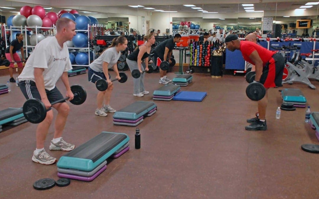
by Dr Alex Jimenez DC, APRN, FNP-BC, CFMP, IFMCP | Fitness
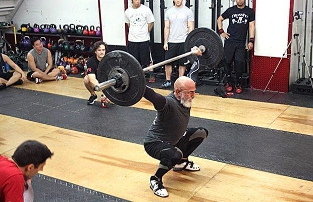
TUESDAY, March 28, 2017 (HealthDay News) — High-intensity exercise may help older adults reverse certain aspects of the “cellular” aging process, a new study suggests.
It’s no secret that regular exercise is healthy for young and old alike. But researchers said the new findings point to particular benefits from “high-intensity interval training” for older adults.
That’s the type of workout that combines brief bursts of vigorous exercise with periods of moderate activity: A person might, for example, go all-out on a stationary bike for a few minutes, ease up for the next few, and then start again.
In this study, older adults who performed that type of exercise showed greater changes at the cellular level, compared to those who worked out more moderately.
Specifically, interval training gave a bigger boost to mitochondrial function in the muscle. Mitochondria are the “powerhouses” within body cells that break down nutrients to be used for energy.
The training also revved up activity in more genes related to mitochondrial function and muscle growth.
What does it all mean?
The study findings suggest that interval training can turn back the clock in ways that moderate aerobic exercise and strength training do not, according to lead researcher Dr. K. Sreekumaran Nair.
But, he stressed, the findings do not mean older adults should jump into a vigorous exercise regimen.
“If you’re sedentary, you should talk to your doctor before you start exercising,” said Nair. He’s an endocrinologist at the Mayo Clinic in Rochester, Minn.
“And then,” he said, “you can start with walking, and build yourself up to a fast pace.”
For older adults who want to progress to a more-intense regimen, Nair said, it’s best to start with supervision. But he also stressed that intense exercise is not a must. “Any regular exercise will bring health benefits — absolutely,” he added.
This study demonstrated that, he pointed out. Even though interval training had the biggest effects on aspects of cellular aging, other types of exercise boosted older adults’ fitness levels and muscle strength.
The study, published recently in Cell Metabolism, involved 72 younger and older adults who were sedentary.
Nair’s team randomly assigned each of them to one of three supervised exercise groups.
One group did high-intensity interval training three days a week: They pedaled on an exercise bike at their maximum speed for 4 minutes, before easing up for 3 minutes; they repeated that process four times. They also worked out more moderately — walking on a treadmill — twice a week.
A second group performed moderate aerobic exercise — using an exercise bike at a less-intense pace — five days a week, for 30 minutes. They also did some light strength-training four days a week.
The third group performed strengthening exercises only, two days a week.
After 12 weeks, all of the groups were showing positive changes — younger and older exercisers alike, the researchers found.
People who performed moderate aerobic exercise boosted their fitness levels — the body’s ability to supply blood and oxygen to working muscles. And the improvement was greater for older adults, who generally started out with lower fitness levels than younger people.
Meanwhile, people who performed strength-training — alone or with aerobic exercise — increased their muscle strength.
The interval-training group showed only small gains in strength. But the training improved mitochondrial function in the muscles, especially among older adults.
Dr. Chip Lavie is medical director of cardiac rehabilitation and prevention at the John Ochsner Heart and Vascular Institute in New Orleans.
He said this is a “great” study that demonstrates the benefits of different forms of exercise.
According to Lavie, it adds to other evidence that high-intensity interval training is “probably the best form of exercise.”
Many studies, he said, have found that interval training beats moderate aerobic exercise when it comes to improving fitness and the heart’s structure and function.
“It would be ideal to get more people to do high-intensity interval training,” Lavie said, “and it’s possible for more-motivated individuals.”
But, he added, the reality is, many people may not have the motivation or ability.
In that case, Lavie advised finding a moderate regimen you can live with — such as 30 to 40 minutes of walking or using an exercise bike or elliptical machine most days of the week.
SOURCES: K. Sreekumaran Nair, M.D., Ph.D., professor, medicine, Mayo Clinic, Rochester, Minn.; Chip Lavie, M.D., medical director, cardiac rehabilitation and prevention, and director, exercise laboratories, John Ochsner Heart and Vascular Institute, New Orleans; March 7, 2017, Cell Metabolism
News stories are written and provided by HealthDay and do not reflect federal policy, the views of MedlinePlus, the National Library of Medicine, the National Institutes of Health, or the U.S. Department of Health and Human Services.
by Dr Alex Jimenez DC, APRN, FNP-BC, CFMP, IFMCP | UTEP (Local) RSS
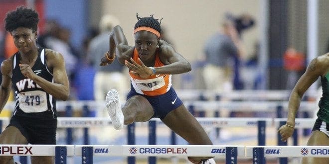
Tobi Amusan continues to dominate the 100m hurdles as she clocked the fastest qualifying time in the 100m hurdles (12.75) Friday morning at the 90th Clyde Littlefield Texas Relays, presented by Spectrum.
The sophomore will compete at 1:17 p.m. (MT) and will be televised on the Longhorn Network.
The women�s 4x400m relay team also qualified for Saturday�s final, advancing with the sixth-fastest time (3:41.20). Despite a shaky handoff, the team comprised of Dreshanae Rolle, Florence Uwakwe, Madison Gibson and Ada Benjamin were able to stay composed and win their heat.
The 4x100m relay team of Israel Ramsay, (Tobi) Amusan, (Florence) Uwakwe and(Madison) Gibson just missed out of reaching the final by less than a second. The Miners clocked a time of 45.40. The last qualifying time came from Virginia Tech at 45.37.
Saturday�s slate will feature Samantha Hall in the women�s discus, which has a 1:30 p.m. (MT) start. Lilian Koech will participate in the 1,500m final set to start at 2:40 p.m., followed by the men�s mile run featuring Jonah Koech and 800m champion Michael Saruni. The meet will conclude with the 4x400m relay at 3:40 p.m.
by Dr Alex Jimenez DC, APRN, FNP-BC, CFMP, IFMCP | Fitness, Health, Wellness
Ashley Graham is definitely about that gym life. And we know this because the 29-year-old model has been posting her sweat sessions on Instagram as of late. Her workout of choice: hitting it hard with Dawin Peña, co-founder and trainer at The DogPound, a boutique training studio in NYC.
Thanks to a recent Instagram story posted by the America’s Next Top Model judge, we happened to get a glimpse into one of Graham’s evening exercise routines. Let me tell you, it is a killer upper-body circuit that hit the triceps, biceps, chest, and back. And she finished off with some core work.
The best part: it only took Graham 2 minutes and 48 seconds to get through these moves. (Yep, I timed her). Granted, you have to take into account that she probably rested here and there, and did a few more sets and reps than she let us in on, but even with those considerations, this is still a great workout option for when you are short on time.
Our suggestion: Cycle through this 11-move circuit 3 times. If you do that, you are looking at about a 10- to 12-minute upper body blaster that you can knock out the next time you are in the gym. Oh, and you also might want to download the playlist Graham was rocking too—Sean Paul’s “Gimme The Light,” Mr. Probz’s “Waves,” Kid Cudi’s “The Pursuit of Happiness,” Eddie Money’s “Take Me Home Tonight” and Future’s “Real Sisters”—because it was kind of fire!
Now get that upper body (and those abs) in shape…because summer is coming!
Rope Triceps Extension
Stand with feet hip-width apart, hinge forward slightly and grab each end of the cable ropes; palms face in. Keeping upper arms straight and close to body, pull down using forearms, lowering the rope until arms are fully extended and at either side of legs. Pause and then slowly return back to start. (Graham did 10 reps.)
Overhead Triceps Extension
Stand with back to cable machine, feet staggered, one foot in front of the other, and knees slightly bent; hinge forward. With arms overhead and bent backwards to about 90-degrees, hold cable rope in each hand; palms face in. Keeping elbows close to ears, pull ropes down until arms are fully extended. Pause and then return to start. (Graham did 9 reps.)
Rope Biceps Curl
Stand with feet shoulder-width apart, legs bent slightly and end of cable ropes grasped between hands; palms face in. Pull ropes up toward shoulders using forearms; upper arms stay fixed. Lower back down to start, and then repeat. (Graham did 9 reps.)
RELATED: Love Ashley Graham? Here are 9 Other Body-Positive Activists You Should Follow
Seated Cable Row
Start seated on a bench with legs wider than hip-width apart, feet planted, and arms extended up on a diagonal with ends of cable ropes grasped between hands; palms face in. Squeezing back, pull ropes down towards torso; keep arms close to body. Pause and then slowly return to start. (Graham did 8 reps.)
Seated One Arm Row
Start seated on a bench with legs wider than hip-width apart and feet planted. Place left hand on hip while right arm is extended up on a diagonal with cable handle grasped in hand; palm face down. Squeezing back, pull rope down towards torso, twisting hand out so that palm faces in; keep arm close to body. Hold, and then slowly return back to start. Repeat on opposite side. (Graham did 7 reps.)
Seated Lat Pulldown
Start seated with back straight, knees under knee pad and bar in hands (overhand grip) slightly wider than shoulder-width apart. Without moving torso, pull bar down to chest while squeezing shoulder blades together. Pause, and then slowly return to start. (Graham did 7 reps.)
RELATED: 11 Best Exercises to Get Strong, Toned Arms
Dumbbell Fly
Lie faceup on a bench with feet planted on floor and a dumbbell in each hand. Extend arms straight up over chest; palms face in. Keeping a slight bend in elbows, slowly open arms out until they are in line with chest and hands are parallel to floor. Pause and then raise arms back up to start. (Graham did 6 reps.)
Standing Biceps Curls
Stand with feet wider than hip-width apart and knees slightly bent. Hold a dumbbell in each hand in front of body; palms face up. Bend elbows and curl the right hand up toward the right shoulder. As you slowly lower the right hand back down, begin repeating the motion with the left hand. Continue alternating. (Graham did 4 reps per arm.)
In & Out
Start seated with arms behind you and hands on floor; fingers facing feet. Lean back, raise legs and bend knees. With abs tight, extend legs straight out as you lower back down slightly. Pull legs back in and lift torso back up. Continue repeating. (Graham did 17 reps.)
RELATED: 7 Upper-Body Exercises That Banish Bra Bulge
Jacknife
Lie faceup with arms and legs extended straight out. Simultaneously raise your right leg and left arm as you crunch up, bringing the two together over the stomach. Lower back to start. After desired number of reps, repeat with opposite arm and leg. (Graham did 10 reps.)
Crunches
Lie faceup with legs extended straight up, a light bend in knees, feet crossed at the ankles (left over right) and hands lightly on back of head. Crunch up and then lower back to start. Repeat. (Graham did 15 reps.)

by Dr Alex Jimenez DC, APRN, FNP-BC, CFMP, IFMCP | UTEP (Local) RSS
Tobi Amusan continues to dominate the 100m hurdles as she clocked the fastest qualifying time in the 100m hurdles (12.75) Friday morning at the 90th Clyde Littlefield Texas Relays, presented by Spectrum.
The sophomore will compete at 1:17 p.m. (MT) and will be televised on the Longhorn Network.
The women’s 4x400m relay team also qualified for Saturday’s final, advancing with the sixth-fastest time (3:41.20). Despite a shaky handoff, the team comprised of Dreshanae Rolle, Florence Uwakwe, Madison Gibson and Ada Benjamin were able to stay composed and win their heat.
The 4x100m relay team of Israel Ramsay, (Tobi) Amusan, (Florence) Uwakwe and(Madison) Gibson just missed out of reaching the final by less than a second. The Miners clocked a time of 45.40. The last qualifying time came from Virginia Tech at 45.37.
Saturday’s slate will feature Samantha Hall in the women’s discus, which has a 1:30 p.m. (MT) start. Lilian Koech will participate in the 1,500m final set to start at 2:40 p.m., followed by the men’s mile run featuring Jonah Koech and 800m champion Michael Saruni. The meet will conclude with the 4x400m relay at 3:40 p.m.

by Dr Alex Jimenez DC, APRN, FNP-BC, CFMP, IFMCP | Diets, Health, Wellness
A stomach ache can strike for all kinds of reasons, from contaminated food to chronic disease. It passes, sure, but the pain, headache, diarrhea, vomiting and other classic symptoms of stomach flu ensure a crummy couple of days
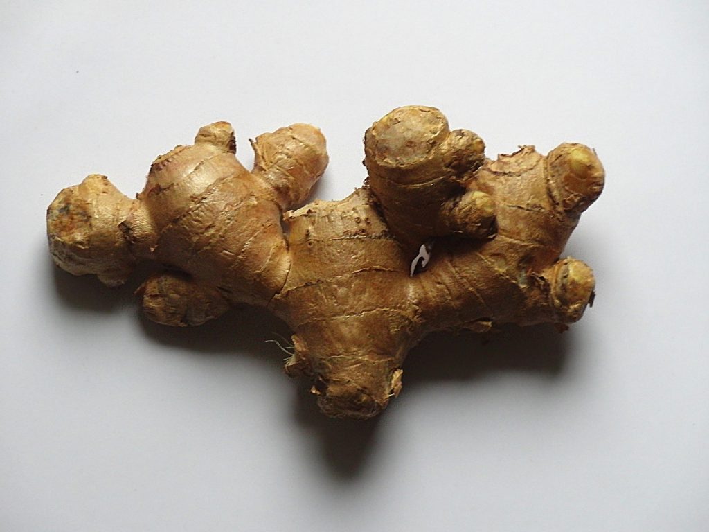
It can be tough to know what to put in your body when you’re dealing with an upset stomach, but there are a few surefire foods. Ginger, scientifically, is a good place to start. �Ginger and also turmeric, which is a member of the ginger family, seem to be anti-inflammatory,� says Dr. Emeran Mayer, a professor of digestive diseases at UCLA. Both ginger and turmeric are roots, he says, and may have developed special antibacterial properties in order to withstand contamination from microorganisms in soil. Skip the sugary commercial ginger ales, which contain little real ginger, and sip water infused with ginger or turmeric instead, he advises.
You won�t want to eat in the throes of vomiting, but starting to sip water and other beverages right away is a good idea, says Dr. Joseph Murray, a gastroenterologist at Mayo Clinic. Because you�re getting rid of essential vitamins and nutrients with every trip to the bathroom, it�s important to replenish your body�s electrolytes�namely salt, but also potassium and glucose (sugar), he says. If the word �electrolytes� makes you think of Gatorade, you�re not far off. But Gatorade and other sports drinks may not contain enough salt to replenish your depleted stores. �Diluted tomato juice is pretty good, mostly because it�s salty,� Murray says.
Once you�ve stopped vomiting and your stomach feels a bit better, you will want to eat. But don’t sit down for a big meal; nibble food throughout the day instead, Murray explains.
Research from Penn State University�s Hershey Medical Center recommends what every parent knows as the BRAT foods: bananas, white rice, applesauce and toast. Eating only these four foods may be too restrictive (and could lead to malnourishment, especially among kids). But foods like these are good choices, because the harder your inflamed stomach has to work to digest something, the more likely it is to act up, Murray says. Foods that are easy for the body to break down�simple, minimally seasoned carbohydrates like saltine crackers, as opposed to hardier fare like whole grains and leafy greens�are less likely to trigger stabs of pain or a dash to the toilet.
There are plenty of foods you should avoid. Pass on dairy foods, because an upset stomach is likely to have problems digesting and absorbing lactose, Murray explains. �Even in the days or weeks after you�ve recovered, you may experience a temporary bout of lactose intolerance while your gut recovers,� he says. Also, skip high-fat foods (like nuts, oils and avocado), spicy dishes, alcohol and coffee, which may all aggravate a recovering stomach, says Dr. Joel Mason, a gastroenterologist and professor of medicine and nutrition at Tufts University.
What about probiotics? While Mason and other experts say there�s promising research on probiotics for relief of gut-related conditions, there�s still not good evidence to support swallowing probiotic-rich foods to cure a stomach ache. One problem with probiotics is that the micro-organic makeup of your gut is different from everyone else�s. �There are also hundreds of probiotic strains, and the effect each has may be determined by your [gut�s] microbiome composition,� UCLA�s Mayer explains. �In the future, we may be able to map your microbiome simply and inexpensively, and make appropriate probiotic recommendations.� But we�re just not there yet.
Another issue is that nearly all the research linking probiotics to relief of gut-related issues has looked at freeze-dried probiotics in capsules or tablets, Mason says. �Eating yogurt or Kefir or other probiotic foods to relieve symptoms may be effective, but that hasn�t yet been shown.�
While probiotic supplements are likely safe for most people, Mason says ingesting probiotics could in some cases be risky. �When you consume a probiotic, you�re consuming billions of bacterial or fungal spores,� he explains. In �the vast majority of instances,� that won�t hurt you. �But if you have an impaired immune system, there�s pretty good documentation that ingesting these organisms can set off very serious infections�even life-threatening infections,� he explains.
If you want to roll the dice with probiotics, you�re best off sticking to those found in traditional food sources like sauerkraut, kefir, and kombucha. �Eat those three, and you�ll get a wide range of probiotics,� Mayer says. There may not be strong evidence yet to show they can relieve an achy stomach, “but they�re what I would give to my own family,� he says.

Call Today!
by Dr Alex Jimenez DC, APRN, FNP-BC, CFMP, IFMCP | Athletes, UTEP (Local) RSS
Related Articles
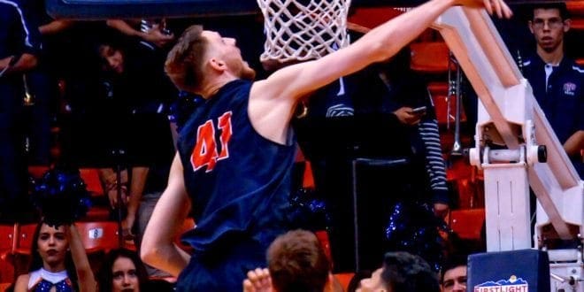
UTEP Athletics officials have been notified by the NCAA that 7-1 center Matt Willms has been granted a sixth year of eligibility and will return for the 2017-18 season.
�We think this is a big deal,� UTEP coach Tim Floyd said. �It gives Matt his first offseason in five years to actually be able to work out, lift and improve his game. This is when players get better. He has enormous potential and getting him back is huge, both for him and for us. I�d like to thank the NCAA for their thorough review of his case and for making the right decision.�
Willms has played only three full seasons (2013-14, 2014-15, 2016-17) in a career wracked by injuries. He has undergone two major surgeries, one for a torn labrum in his shoulder and another for a fracture of the nevicular bone in his foot.
Last year, coming off the foot surgery, Willms enjoyed his finest season as a Miner, averaging 11.4 points, 5.5 rebounds and 1.3 blocks while leading Conference USA in field goal percentage (.591). He scored in double figures 18 times and was named Conference USA Player of the Week on Jan. 30 after averaging 26.0 points and making 24-of-31 shots (77.4 percent) on the Miners� road trip to WKU and Marshall.
Willms will enter his senior year with career totals of 693 points, 416 rebounds and 98 blocked shots with a .571 (274-for-480) field goal percentage. He has played in 95 games and ranks ninth in school history in blocks.
NCAA rules permit players five years to play four. Willms redshirted in 2012-13, meaning he has been on campus for five years already. UTEP officials had to submit paperwork to the NCAA detailing Willms� situation and requesting the sixth year, but nothing was a given.
�When I first found out that I may not get my eligibility back next year, it started going through my head. What if I can�t play? Where do I go from here? Do I go overseas? Would I even have an option there,� Willms said. �Once the process started, they said it�s difficult [to get the sixth year] and it put more doubt in my mind. I got a call from coach today telling me that I�ve been accepted. The first thing I thought about is the amount of talent we have next year, and being a senior and being able to lead the team. It means a lot to me to come back and wear the Miner jersey for one last season.�
Willms is already mapping out his offseason goals.
�One thing that I want to do is put on at least 15 pounds,� he said. �Another thing is to work on my foot speed, get my foot speed back, work a little more on my post moves, moves I can counter and go from there. Those are my goals, just to try to get better and once the new guys come in, give them some pointers on what to work on and what to expect.�
by Dr Alex Jimenez DC, APRN, FNP-BC, CFMP, IFMCP | Fitness, Health, Wellness
Thanks to the Google’s�many clever features, we no longer get lost (as often), bungle dates and double-book, or choose mediocre restaurants. And now�Google is back at it again, with yet another way to simplify your life:�Reserve with Google�is a new platform that makes it super easy�to discover and book fitness classes.
The site�allows you to search your area for upcoming classes, and reserve�and pay for�a spot instantly. If you’re signed into your Google account, some of your info (like your name, email, and phone number) will pre-load to make booking even faster. The service�kicked off in New York, Los Angeles, and San Francisco, and is now available across�the�country, thanks to partnerships with booking services you may already know and love, such as Mindbody,�Genbook, and MyTime.

Reserve with Google�also makes recommendations to help you discover new ways to get your sweat on, complete with�class descriptions, pricing, and reviews�so you know exactly what you’re signing up for. And the �Discover more around you� section groups suggestions into categories like��Yoga this evening� and �Pilates before work.� Really want to mix it up? You can choose Google’s�notorious �I�m feeling lucky” option.
To help you fit your workouts into your schedule,�the service works with Google Maps (so you get a�visual of the closest gyms and studios)�and Google�Calendar (so you can block off precious time).
Whether�you’re looking to try a new activity, or schedule out a full week of exercise, this platform can help you do so seamlessly. And once you’re sufficiently sore, it’ll help�you search�for massage appointments (and other spa services) nearby to help your body bounce back.

by Dr Alex Jimenez DC, APRN, FNP-BC, CFMP, IFMCP | UTEP (Local) RSS
AUSTIN �� Lucia Mokrasova set the school record with 5,671 points in the heptathlon while freshman All-American Michael Saruni broke a meet standard to win the 800m at the 90th Clyde Littlefield Texas Relays, presented by Spectrum on Thursday afternoon at Mike A. Myers Stadium.
�Lucy didn�t have the start that we wanted today, but I think she recovered well and finished strong to break her record from last year,� UTEP head coach Mika Laaksonen said. �She competed well and looks like she has a lot more left in the tank.�
Heading into day two of competition with 3,517 points, Mokrasova opened the day by leaping out to 5.30m (17-4.75) in the long jump, where she earned 643 points. She tallied 666 points after throwing for 39.94m (131-0).
After two events the junior notched 4,942 points and needed to run her time from a year ago (2:22.59) to tie her previous record of 5,615 points. The junior ran a 2:18.44 in the 800m to earn 845 points, breaking her previous record by 56 points.
Mokrasova finished in eighth place in a field of 23 and her total score ranks first in Conference USA.
The other big highlight of day two at the Texas Relays for UTEP Saruni�s star-studded performance. The Kenyan competed in the Invitational (including professionals) 800m run and finished by setting a Texas Relays meet record with the fastest time in the nation (1:48.82). In his heat was the 2012 Olympic silver medialist (Leonel Manzano) and the collegiate record holder (Donavan Brazier). Saruni will participate in the mile run scheduled for Saturday at 2:45 p.m. MT.
�Michael, what can you say a freshman running that times, considering the competition he faced,� Laaksonen said. �For him to come in and break the record is impressive.�
In the women�s Invitational 800m run, Lilian Koech finished in eighth place with a time of 2:11.42. She was the fourth collegiate athlete to finish.
Yanique Bennett clocked a time of 59.92 in the 400m hurdles on her way to a 20th-place finish in a field of 73. Teammate Dreshanae Rolle followed with a time of 1:00.23 to a 24th-place finish.
Cosmas Boit garnered a silver medal in the 1,500m run with a time of 3:49.90 and Daniel Cheruiyot clocked a 9:18.57 to finish in fifth place in the 3,000m steeplechase.
Winny Koech, Gladys Jerotich, Linda Cheruiyot and Antony Kosgei closed out the night in the 5,000m run. Koech (16:22.26) led the way to a gold-place finish with the fifth-fastest time in the nation, Cheruiyot (16:44.76) finished fourth and Jerotich (17:34.28) finished 17th overall. In the men�s race, Kosgei paced to a bronze showing with a time of 14:16.60.