Low back pain is a common complaint that generally goes away on its own, however, what should a person do if their LBP becomes chronic and/or persistent? How is an individual’s quality of life affected and how does their pain intensity impact their physical capacity? Is there any type of treatment which can help improve low back pain? Many different types of treatment options can be used to safely and effectively treat low back pain. The purpose of the following research study is to determine the influence of the McKenzie method and endurance exercises on low back pain. The article demonstrates evidence-based information on the improvement of the quality of life of patients with LBP after receiving the treatment protocol mentioned below.
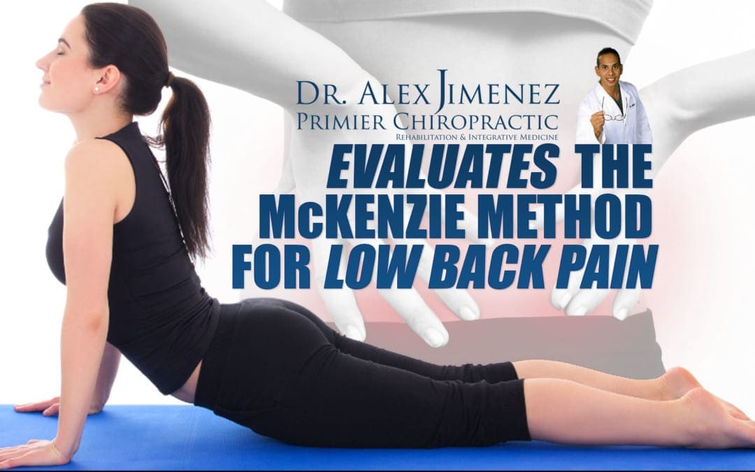
Influence of Mckenzie Protocol and Two Modes of Endurance Exercises on Health-Related Quality of Life of Patients with Long-Term Mechanical Low Back Pain
Abstract
Introduction
Long-term Mechanical Low-Back Pain (LMLBP) negatively impacts on patients� physical capacity and quality of life. This study investigated the relationship between Health-Related Quality of Life (HRQoL) and pain intensity, and the influence of static and dynamic back extensors� endurance exercises on HRQoL in Nigerian patients with LMLBP treated with the McKenzie Protocol (MP).
Methods
A single-blind controlled trial involving 84 patients who received treatment thrice weekly for eight weeks was conducted. Participants were assigned to the MP Group (MPG), MP plus Static Back Endurance Exercise Group (MPSBEEG) or MP plus Dynamic Endurance Exercise Group (MPDBEEG) using permuted randomization. HRQoL and pain was assessed using the Short-Form (SF-36) questionnaire and Quadruple Visual Analogue Scale respectively.
Results
Sixty seven participants aged 51.8 � 7.35 years completed the study. A total drop-out rate of 20.2% was observed in the study. Within-group comparison across weeks 0-4, 4-8 and 0-8 of the study revealed significant differences in HRQoL scores (p < 0.05). Treatment Effect Scores (TES) across the groups were significantly different (p = 0.001). MPSBEEG and MPDBEEG were comparable in TES on General Health Perception (GHP) at week 4; and GHP and Physical Functioning at week 8 respectively (p > 0.05). However, MPDEEG had significantly higher TES in the other domains of the SF-36 (p = 0.001).
Conclusion
HRQoL in patients with LMLBP decreases with pain severity. Each of MP, static and dynamic back extensors endurance exercises significantly improved HRQoL in LMLBP. However, the addition of dynamic back extensors endurance exercise to MP led to greater improvement in HRQoL.
Keywords:Mckenzie protocol, endurance exercises, quality of life, back pain
Background
Low-Back Pain (LBP) is described as the constellation of symptoms of pain or discomfort originating from impairments in the structures in the low back [1�2]. LBP is one of the most common ailments afflicting mankind [3]. It is a complicated condition which affects the physiological and psychosocial aspects of the patient [4, 5]. Epidemiological reports indicate that 70 to 85% of all people have LBP at some time in their life [1, 6]. The World Health Organization predicted that the greatest increases in LBP prevalence in the next decade will be in developing nations [7]. In line with this, a systematic review by Louw et al [8] concluded that the global burden and prevalence of LBP among Africans is rising.
It is estimated that 80-90% of patients with LBP will recover within six weeks, regardless of treatment [9]. However, 5-15% of all people that have LBP will develop long-term LBP (i.e. LBP of 12 weeks and longer) [10, 11]. The patient subgroup with long-term LBP accounts for 75-90% of the socioeconomic cost of LBP [12] and over 30% of these patients with long-term LBP seek healthcare for their back complaints. Long-term LBP significantly impacts on patients� physical [13], psychological and social functioning [14] and can affect well-being and quality of life [15]. Reduced quality of life in patients with long-term LBP is associated with poor prognosis [16], intermittent or recurrent episodes of LBP [17], disability [18] and psychosocial dysfunction [19, 20].
Assessment of Health-Related Quality of Life (HRQoL) in relation to LBP has been recommended in LBP management [21, 22]. Several HRQoL instruments have been developed to assess self-perceived general health status [21, 22]. The SF-36 Health Status Questionnaire, though a generic instrument, has been recommended in the assessment of HRQoL of patients with long-term LBP [22] and it assesses eight domains such as physical functioning, role limitations due to physical problems, bodily pain, general health perceptions, vitality, social functioning, role limitation due to emotional problems and general mental health [23, 24].
Consequent to the foregoing, treatment intervention that may help improve the HRQoL of patients with long-term LBP has been advocated. Although, physiotherapy plays an important role in the management of patients with LBP, the traditional approach based on biomedical model, which is centered on the treatment of impairments and patho-physiological variables, may not fully addressed the wider range of factors including psychosocial impairments associated with long-term LBP [25, 26]. However, long-term LBP is considered to be a multi-factorial bio-psychosocial problem which has an impact on both social life [27, 28] and quality of life [29] and thus requires a multi-dimensional approach based on a bio-psychosocial model (a model that includes physical, psychological and social elements) in its assessment and treatment [30, 31].
Based on empirical recommendations from research, recent decades have witnessed tremendous advances in preventive, pharmacological and physiotherapy management for a limited number of patients with LBP especially in developed countries. However, the improvement in health outcomes observed in most Western countries over the past few decades has not been achieved in Africa [32] and therefore, the health of Africans is of global concern [8]. Compared with Australians [33], Europeans [34] and North Americans [35], the use of exercise as medicine in Africans is poor. Exercise is the central element in the physical therapy management of patients with long-term LBP [9, 36]. Exercise often does not require expensive instruments and probably the cheapest intervention and one in which the patient has some measure of direct control [37]. Nonetheless, it remains inconclusive which exercise regimen will significantly influence the quality of life of patients with long-term LBP. The McKenzie Protocol (MP) is one of the most commonly used physical therapy interventions in long-term mechanical LBP with documented effectiveness [38�41]. However, there is a dearth of studies that have investigated the influence of the MP on HRQoL in patients with long-term mechanical LBP. Therefore, this study was intended to answer the following questions: (1). Will pain intensity significantly influence HRQoL? (2) Will static and dynamic back extensors� endurance exercises significantly influence HRQoL in Nigerian patients with long-term mechanical LBP (LMLBP) treated with the MP?
Methods
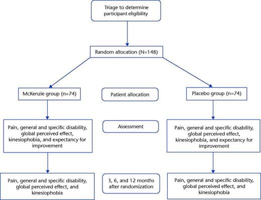
Eighty four patients with LMLBP participated in this single-blind randomized trial. The participants were consecutively recruited from the physiotherapy department, Obafemi Awolowo University (OAU) Teaching Hospitals Complex and the OAU Health Centre, Ile-Ife, Nigeria. The McKenzie Institute’s Lumbar Spine Assessment Format (MILSAF) [3] was used to determine eligibility to participate in the study. Based on the MILSAF, patients who demonstrated Directional Preference (DP) for extension only were recruited to ensure homogeneity of samples. DP is described as the posture or movement that reduces or centralizes radiating pain that emanates from the spine. Exclusion criteria were red flags indicative of serious spinal pathology with signs and symptoms of nerve root compromise (with at least two of dermatomal sensory loss, myotomal muscle weakness and reduced lower limb reflexes), individuals with any obvious spinal deformity or neurological disease; pregnancy; previous spinal surgery; previous experience of static and dynamic endurance exercise and having DP for flexion, lateral or no DP. Long-term low-back pain was defined as a history of LBP of not less than 3 months [42].
Based on the sample size table by Cohen [43] with alpha level set at 0.05, degree of freedom at 2, effect size at 0.25, and power at 80, the study found a minimum sample size of 52. However, in order to accommodate for possible attrition or loss during the study, a total of 75 patients (25 per group) was included. The participants were randomly assigned to one of three treatment groups using permuted block randomization; the McKenzie Protocol (MP) Group (MPG) (n = 29), MP plus Static Back Endurance Exercise Group (MPSBEEG) (n = 27) and MP plus Dynamic Back Endurance Exercise Group (MPDBEEG) (n = 28). Sixty seven (32 males (47.8%) and 35 females (52.2%) participants completed the eight week study. Twenty five participants completed the study in MPG, 22 in MPSBEEG and 20 in MPDBEEG. A total drop-out rate of 20.2% was observed in the study. Fourteen percent of participants in MPG were lost to follow-up. Nineteen percent of the participants in MPSBEEG dropped out (out of these, 40% were lost to follow-up while 60% absconded due to improvement in their health condition). In the MPDBEEG, 28.6% of the participants dropped out (37.5% were lost to follow-up while 62.5% absconded due to improvement in their health condition).
Treatment was given thrice weekly for eight weeks and outcomes were assessed at the end of the fourth and eighth week of study. Ethics and Research Committee of the Obafemi Awolowo University Teaching Hospitals Complex and the joint University of Ibadan /University College Hospital Institutional Review Committee respectively gave approval for the study.
Instruments
A height meter calibrated from 0-200cm was used to measure the height of each participant to the nearest 0.1cm. A weighing scale was used to measure the body weight of participants in kilograms to the nearest 1.0Kg. It is calibrated from 0 – 120kg. A metronome (Wittner Metronom system Maelzel, Made in Germany) was used to set a uniform tempo for dynamic back endurance muscles endurance test, which involves repeated contraction or movements over a period of time performed synchronously to the metronome beat. Patients lay on a plinth for the MP, static and dynamic back endurance exercise respectively.
General Health Status Questionnaire – Short Form -36 (SF-36) was used to assess the quality of life of the participants. The SF-36 has been recommended in the assessment of patients with long-term LBP [24, 44, 45]. A Yoruba translated version of the Health Status Questionnaire (SF-36) was used for participants who were literate in the Yoruba language and preferred the Yoruba version. The translation was done at the department of linguistics and African languages of Obafemi Awolowo University, Ile Ife. Pearson product moment correlation coefficient (r) of 0.84 was obtained for the criterion validity of the back translation of the Yoruba version. Quadruple Visual Analogue Scale (QVAS) was used to assess pain intensity of participants. QVAS is a reliable and valid method for pain measurement [46, 47]. A Yoruba translated version of the QVAS was used for participants who were literate in the Yoruba language and prefers the Yoruba version. The translation was done at the department of linguistics and African languages of Obafemi Awolowo University, Ile Ife. Pearson product moment correlation coefficient (r) of 0.88 was obtained for the criterion validity of the back translation of the Yoruba version.
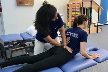
Treatment
Treatment for the different groups (MPG, MPSBEEG and MPDBEEG) comprised three phases including warm up, main exercise and cool down. Prior to treatment, the participants were instructed in details on the study procedures. This was followed by a low intensity warm-up phase of five minutes duration comprising active stretching of the upper extremities and low back and strolling at self-determined pace around the research venue. Treatment also ended with a cool-down phase comprising of the same low intensity exercise as the warm-up for about five minutes.
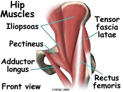
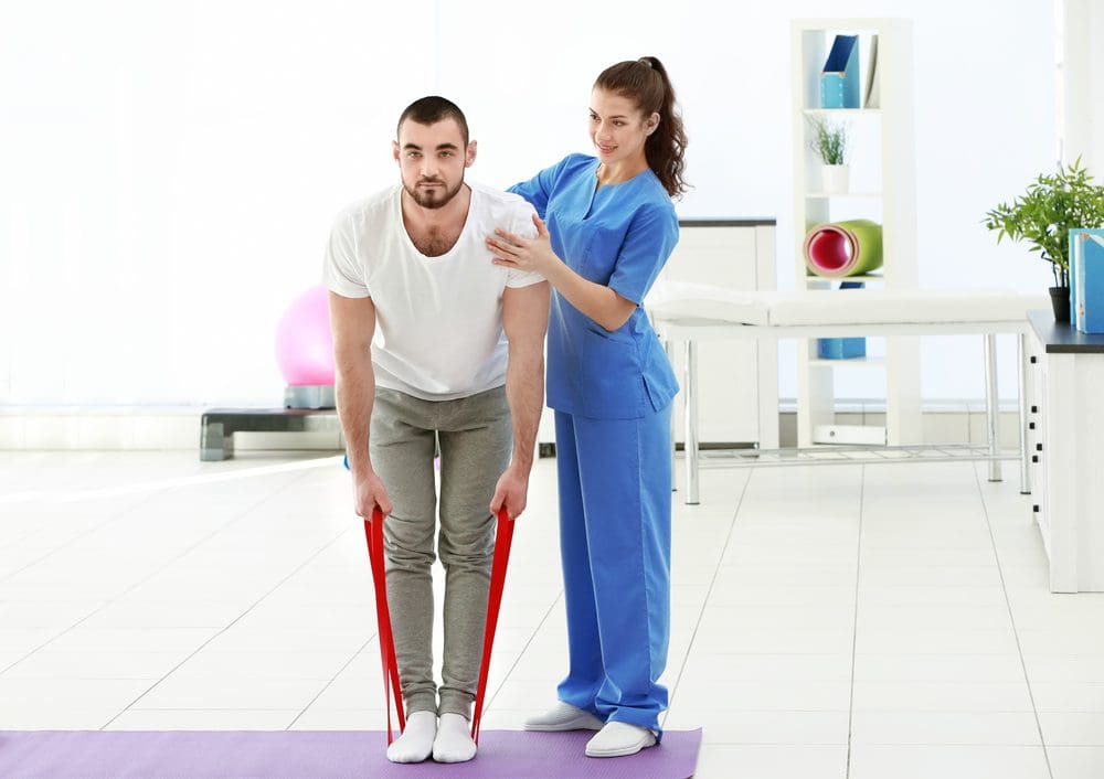
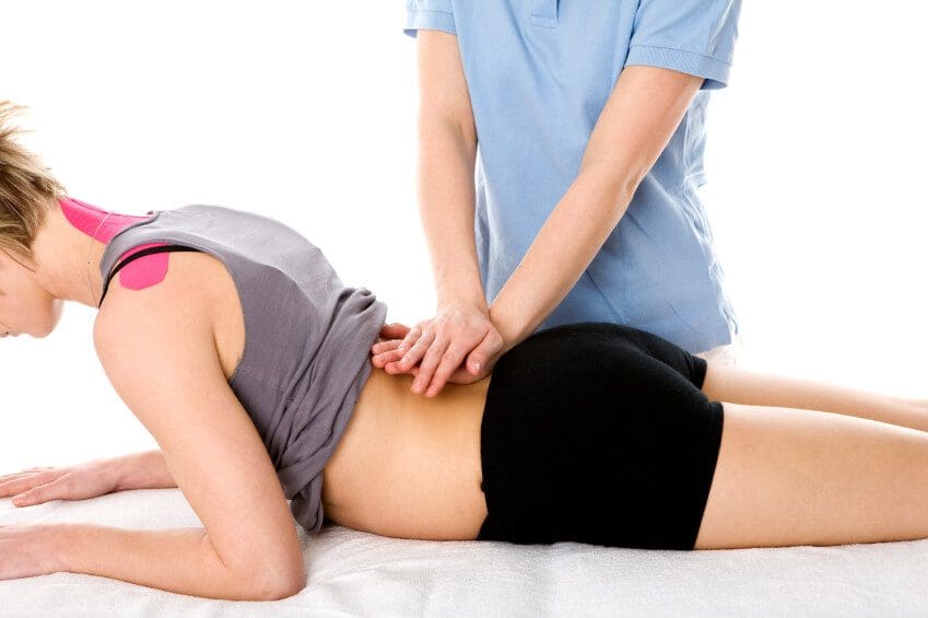
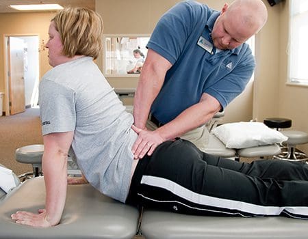
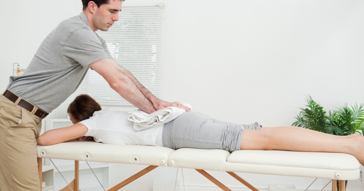
The McKenzie Protocol (MP) involved a course of specific lumbosacral repeated movements in extension that cause the symptoms to centralize, decrease or abolish. The determination of the direction preference for extension was followed by the main MP activities including �Extension lying prone�, �Extension In Prone� and �Extension in standing�. The MP also included a set of back care education instructions which comprised a 9 item instructional guide on standing, sitting, lifting and other activities of daily living for home exercise for all the participants (Appendix).
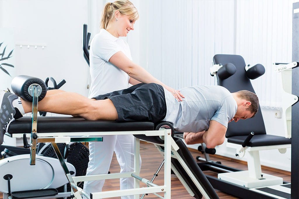
In addition to completing the MP (i.e., back extension exercises plus the back care education), static back extensors endurance exercise which included five different static exercises differentiated by the alteration of the positions of the upper and lower limbs with the patient in prone lying on a plinth was carried out [48]. The participants began the exercise training programme with the first exercise position, but progressed to the next exercises at their own pace when they could hold a given position for 10 seconds. On reaching the fifth progression, they continued with the fifth progression until the end of the exercise programme [48, 49]. The following were the five exercise progressions:
Participant lay in prone position with both arms by the sides of the body and lifting the head and trunk off the plinth from neutral to extension;
Participant lay in prone position with the hands interlocked at the occiput so that shoulders were abducted to 90� and the elbows flexed, and lifting the head and trunk off the plinth from neutral to extension;
Participant lay in prone position with both arms elevated forwards, and lifting the head, trunk and elevated arms off the plinth from neutral to extension;
Participant lay in prone position and lifting the head, trunk and contralateral arm and leg off the plinth from neutral to extension; and
Participant lay in prone position with both shoulders abducted and elbows flexed to 90�, and lifting the head, trunk and both legs (with knees extended) off the plinth.
If pain was aggravated during the exercise, the participant was asked to stop. If the pain diminished within 5 minutes after the exercise, he/she was asked to continue the exercise but to hold the exercise position for only 5 seconds. The participant was asked to progress to 10 seconds if there was no adverse response. Each exercise was repeated 9 times. After 10 repetitions, the participant was instructed to rest for between 30 seconds to 1 minute. Static holding time in the exercise position was gradually increased to 20 seconds to provide a greater training stimulus [50, 51]. The dosage of series of 10 repetitions was adopted from a previous protocol for participants with sub-acute LBP [52].
In addition to completing the MP, dynamic back extensors endurance exercise which included five different isokinetic exercises differentiated by the alteration of the positions of the upper and lower limbs with the patient in prone lying on a plinth was carried out. The dynamic back endurance exercise was an exact replica of the static back extensors endurance exercise protocol in terms of exercise positions, progressions and duration. However, instead of static posturing of the trunk in the prone lying position and holding the positions of the upper and lower limbs suspended in the air during all the five exercise progressions for the 10 seconds, the participant was asked to move the trunk and the suspended limbs 10 times.
If pain was aggravated during the exercise, participant was asked to stop. If the pain diminished within 5 minutes after the exercise, the participant was asked to continue the exercise but to carry out only 5 movements in the exercise position. The participant was asked to progress to 10 movements if there is no adverse response. Each exercise was repeated 9 times. After 10 repetitions, the participants were instructed to rest for between 30 seconds to 1 minute. The number of movements of the trunk in the exercise position was gradually increased to 20 seconds to provide a greater training stimulus.
In order to achieve adequate training effect based on recommendation of previous studies, a 30 to 45 minute exercise duration, thrice weekly and eight weeks exercise; and training load of 10 seconds static hold or 10 repetitions per exercise position was adopted [53, 54].
The researchers (CEM and OA) were credentialed in the McKenzie method and supervised the exercises. The researchers were blinded to the recruitment, randomization and assessment procedures which were carried out by an assistant who was blinded to the treatment protocols of the different groups. The research assistant was also credentialed in McKenzie method. The questionnaires used in this study were self- administered.
Data Analysis
Data were analyzed using descriptive of mean and standard deviation; and inferential statistics. One-way ANOVA was used to compare the participants� general characteristics and pain intensity by treatment groups. Pearson’s Product Moment Correlation Analysis was used to test the relationship between HRQoL and intensity of pain. The Kruskal Wallis test was used to compare the treatment outcomes (mean change) on HRQoL across group at week four and eight of the study respectively. Friedman’s ANOVA and Wilcoxon signed ranked tests for multiple comparisons were used to compare within group changes in across the three study time points Alpha level was set at p = 0.05. The data analyses were carried out using SPSS 13.0 version software (SPSS Inc., Chicago, Illinois, USA).
Dr. Alex Jimenez’s Insight
How can the McKenzie method improve an individual’s quality of life? With years of experience working alongside patients to help them recover from a variety of spinal health issues, I’ve seen how debilitating low back pain can be if left untreated for an increased amount of time. Although spinal adjustments and manual manipulations can efficiently help improve symptoms of low back pain, other alternative treatment options may help patients recover faster. The McKenzie method and endurance exercises are used by many healthcare professionals to safely and effectively rehabilitate patients with LBP. The results of the research study ultimately demonstrate how the treatment protocol can help improve an individual’s quality of life.
Results
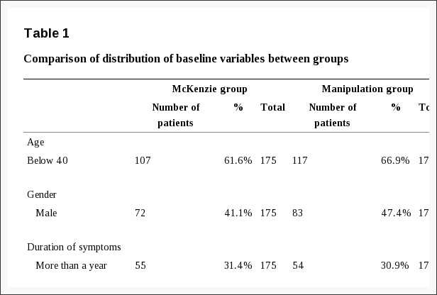
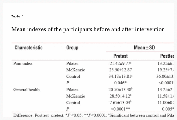
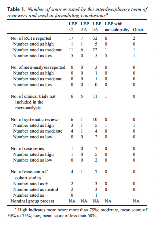
The mean age, height, weight and BMI of all the participants was 51.8 � 7.35 years, 1.66 � 0.04m, 76.2�11.2 Kg and 27.2 � 4.43 kg/m2 respectively. Comparison of the participants� general characteristics by treatment groups revealed that the participants in the different groups were comparable in their general characteristics (p > 0.05) (Table 1).
Table 1: One-way ANOVA comparison of the participants� general characteristics and pain intensity by treatment groups
The mean pain intensity score (VAS) reported by the participants was 6.55 � 1.75. The relationship between each of the eight domains of HRQoL and intensity of pain (VAS score) is presented in Table 2.
Table 2: Relationship between Health-Related Quality of Life and intensity of pain (VAS score) (n = 67)
From the result, correlation co-efficient (r) ranged between-0.603 to-0.878 at p = 0.001. Table 3 shows the comparison of the participants� baseline measure of HRQoL.
Table 3: Kruskal Wallis comparison of the participants� baseline assessment of HRQoL
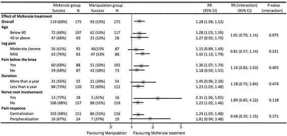
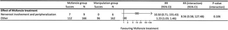
The results indicate that the participants in the different treatment groups were comparable in all the domains of HRQoL (p > 0.05). Within-group comparison of HRQoL in MPG, MPSBEEG and MPDBEEG across the 3 time points (weeks 0-4, 4-8 and 0-8) of the study showed that there were significant improvements (p < 0.05) (Table 4). Comparison of treatment outcomes (mean change score (MCS)) at week four and eight of the study are presented in Table 5. There were significant differences in SF-36 scores across the group (p > 0.05) at the end of the 4th and 8th week of the study respectively. The Tukey multiple comparisons post-hoc analysis was used to elucidate where the differences within between groups lie. The result indicated that MPSBEEG and MPDBEEG had significantly higher MCS on all domains of SF-36 compared with MPG at week four and eight respectively (p < 0.05). There was no significant difference between the MPSBEEG and MPDBEEG in the MCS of General Health Perception domain of SF-36 at week four; and on General Health Perception and Physical Functioning Domains of SF-36 at week eight respectively. However, MPDBEE had significantly higher treatment effects on other domains of HRQoL (p = 0.001).
Table 4: Friedman’s ANOVA and Wilcoxon signed ranked test multiple comparisons of HRQoL among MPG, MPSBEEG and MPDBEEG across the 3 time points of the study.
Table 5: Kruskal Wallis comparison of the participants� treatment outcomes (mean change) at week four of the study.
Discussion
This study evaluated the relationship between HRQoL and pain intensity, and the influence of static and dynamic back extensors� endurance exercises on HRQoL in Nigerian patients with LMLBP treated with the MP. The mean age of the patients in this study was 51.8 � 7.35 years. This age falls within the age bracket during which LBP is reported to be a more common problem [55]. From the result of this study, no significant difference in physical characteristics and pain intensity was found in the different treatment groups at baseline. Baseline characteristics are believed to be predictors of response to treatment in clinical trials for LBP [56]. Comparability in baseline measure in clinical trials is reported to reduce the chances of co-founders other than the intervention in predicting outcomes. Therefore, it is implied that the results obtained at different point in the course of this study could have been largely due to the effects of the various treatment regimens.
This study investigated the relationship between HRQoL and the intensity of pain. From the result, significant moderate to high inverse relationships were found between pain intensity and the different domains of HRQoL. General health perception showed the least correlation (r = -0.603; p = 0.001) while social functioning had the highest correlation with pain intensity (r = -0.878; p = 0.001). It is inferred from the study’s result that HRQoL of patients with long-term LBP decreases with severity of pain. Previous studies have reported an association between LBP and psychosocial factors [26, 57]. Specifically, significant inverse correlation has been reported between severity of pain and quality of life in patients with chronic LBP [57�59]. Pain is believed to have a profound effect on HRQoL [59] and the degree, to which the patients believe that they are disabled by it, is a powerful factor in the extent of their quality of life impairments [60]. Therefore, quality of life is an indicator of the level of endurance of people to pain [61].
Within-group comparison of each of MP, MP plus Static Back Endurance Exercise (MPSBEE) and MP plus Dynamic Back Endurance Exercise (MPDBEE) across the 3 time-points (weeks 0-4, 4-8 and 0-8) of the study revealed that each treatment regimen led to significant improvement in HRQoL. Patients in this study displayed baseline values of the SF-36 comparable to those described in other studies on chronic LBP [62]. The baseline values of all domains of the SF-36 observed in this study were lower than those of adult normative data reported by Jenkinson et al [63] leaving room for any improvement accruable to treatment regimens to be assessed. From this study, all the eight domains of the SF-36 significantly improved at the 4th and 8th week assessment. However, on the final assessment, social functioning, general health perception and bodily pain improved more than the other domains of SF-36 in the MPG. General health perception, physical functioning, social functioning, bodily pain and energy vitality improved more than the other domains of SF-36 in the MPSBEEG while general health perception, physical functioning, social functioning, bodily pain and energy vitality improved more than the other domains of SF-36 in the MPDBEEG. Role physical, role emotional and mental health were the least improved domains of the SF-36 among the treatment groups. Though significant improvements were observed in the different domains by treatment groups on final assessment, the values were still lower than the adult normative data for general health status assessed using the SF-36 questionnaire [63]. A previous study by Smeets and colleagues [64] found that active physical therapy regimen primarily designed to improve physiological aspects of LBP such as aerobic fitness level, low back muscle strength and endurance can also reduce the impact of psychosocial factors that it did not deliberately target. In view of current evidence, Hill and Fritz [57] suggest that it may not necessarily follow that a psychologist is better placed to improve treatment outcomes than a physical therapist, even when a goal of treatment is the mediation of a psychosocial factor. Hill and Fritz [57] also argue that psychosocial factors including fear of movement, anxiety, a faulty coping strategy and quality of life have a strong influence on the success of treatment for patients with back pain at a group level. Literature suggests that exercise generally has a potential benefit on psychosocial aspect of patient with long-term LBP. Long-term LBP leads to deconditioning [65] and many problems associated with deconditioning are believed to be reversible through general and specific exercise regimens [66]. Harding and Watson [66] note that improvement in overall physical function is linked with improvement in psychosocial function. Unfortunately, there is a dearth of studies on the effect of the MP and back extensors endurance exercises on HRQoL in patients with long-term mechanical LBP.
From the result of this study, comparison of the different treatment regimens indicate that MPSBEE and MPDBEE had significantly higher treatment effect on all domains of HRQoL compared with MP at week four and eight respectively. MPSBEE and MPDBEE were comparable in their effect on general health perception domain at week four; and on health perception and physical functioning domains of the HRQoL at week eight. However, MPDBEE had significantly higher treatment effects on other domains of HRQoL. Generally, exercise seems to leads to improved wellness and quality of life. Still, there does not appear to be a consensus of opinion on the most effective programme designed to maintain exercise benefits. The McKenzie method is a popular and promising classification-based treatment for LBP among physical therapists [3] in addition to delivering theoretical information in order to educate patients about their condition, so that patients are better able to understand their condition and how to change their behaviour towards an episode of LBP [67]. However, few studies have investigated the effect of the MP on HRQoL in patients with LMLBP. Udermann et al [68] found significant improvements in HRQoL measures in chronic LBP patients treated with MP but reported that the addition of resistance training for the lumbar extensors provided no additional benefit. In recent times, endurance training of the low-back extensors aimed at improving physical performance and psychosocial health in patients with LBP has increased in popularity [69, 48, 52, 70], yet their effectiveness in enhancing quality of life remains unclear [71].
The observed efficacy of the MP, MPSBEE and MPDBEE in this study could be as a result of the fact that each of the regimen contained active exercise carried out in extension positions. Active exercise can be described as functional exercise performed by the patient or client. Previous studies have shown that active exercise, irrespective of the type is more effective in the management of patients with long-term LBP than passive therapy [72, 73]. The MP utilizes a system of patient self generated force to mobilize or manipulate the spine through a series of active repeated movements or static positioning and it is based on the patient’s pain response to certain movements and postures during assessment [3]. Similarly, endurance exercises are active exercises that require static posturing or repeated movements in order to initiate overload stimuli on the musculature. The different treatment regimen in this study had movement components, either from the MP which is the baseline treatment for all the groups or from the back extensors endurance exercise protocols. It is postulated from the results of this study that the significant higher treatment outcome of MPDBEE might be due to the combined effects of movements and overload stimulus on the back extensor muscles. MPDBEE seems to contain movement ingredients, firstly, from the MP which is the baseline treatment for this group and it involved a series of active repeated movements. Secondly, the dynamic back extensors endurance exercise also involved repeated movements of the trunk and limbs in the sagittal plane. It seems that extension exercise with movement elements carried out in patterns similar to the daily tasks motions might help to improve psychosocial aspects of long-term LBP as observed in this study.
Limitations of the Study
The generalizability of the findings of this study is limited by the fact that a generic quality of life tool was employed because of the scarcity of standard HRQoL tools with documented psychometric properties specific for patients with LBP. Theoretically, specific HRQoL measures are opined to be more responsive than generic HRQL measures [74]. Like all other self-reported assessment, it is possible that the patients in this study might have given exaggerated responses or overestimated the effect of exercise on their HRQoL. Furthermore, individuals� perception of psychosocial construct such as HRQoL is believed to be influenced by subjective interpretation and cultural bias [75, 76]. The high drop-out rate observed in this study is also a potential limitation and source of bias which may limit the interpretation and generalizability of study results. Finally, the treatment outcomes of the different regimens were only measured over such a short period of time of eight weeks.
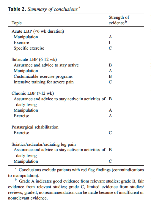
Conclusion
Health-related quality of life of patients with long-term LBP decreases with severity of pain. The McKenzie Protocol, static and dynamic back extensors endurance exercises had significant therapeutic effect on HRQoL in patients with LMLBP. However, the addition of dynamic back extensors endurance exercise to MP led to higher improvement on HRQoL. It is recommended that static or dynamic endurance exercise be combined with MP in patients with LMLBP to derive maximum improvement in general health status.
Acknowledgements
This research was funded by an African Doctoral Dissertation Research Fellowship award offered by the African Population and Health Research Center (APHRC) in partnership with the International Development Research Centre (IDRC). We would like to thank the management and clinicians of the department of physiotherapy OAUTHC, Ile-Ife, Nigeria for their support in carrying out the study. We will also like to thank all the patients who participated in this study.
Competing Interests
The authors declare no competing interests.
Authors� Contributions
All the authors have contributed in this study in ways that comply to the ICMJE authorship criteria. All the authors have read and approved the final version of the manuscript.
In conclusion,�the quality of life of patients with chronic and/or persistent low back pain improved and the pain intensity of the symptoms of LBP appeared to decrease with the use of McKenzie therapy and endurance exercises, according to the study. Furthermore, under the McKenzie treatment protocol, static and dynamic back extensor endurance exercises were recorded to significantly improve symptoms as compared to endurance exercises alone. Information referenced from the National Center for Biotechnology Information (NCBI). The scope of our information is limited to chiropractic as well as to spinal injuries and conditions. To discuss the subject matter, please feel free to ask Dr. Jimenez or contact us at 915-850-0900 .
Curated by Dr. Alex Jimenez
Additional Topics: Sciatica
Sciatica is referred to as a collection of symptoms rather than a single type of injury or condition. The symptoms are characterized as radiating pain, numbness and tingling sensations from the sciatic nerve in the lower back, down the buttocks and thighs and through one or both legs and into the feet. Sciatica is commonly the result of irritation, inflammation or compression of the largest nerve in the human body, generally due to a herniated disc or bone spur.
1. Waddell G. London: Churchill Livingstone; 1998. The back pain revolution.
2. Burton AK, Balague F, Cardon G, Eriksen HR, Henrotin Y, Lahad A, et al. On behalf of the COST B13 Working Group on Guidelines for Prevention in Low Back Pain. European guidelines for prevention in low back pain – November 2004. Eur Spine J. 2006;15:s136�168. [PMC free article][PubMed]
3. Mckenzie RA. Waikanae, New Zealand: Spinal Publication Limited; 1990. Treat Your Own Back. Spinal Publication. Pu.
4. Sikorski JM, Stampfer HG, Cole RM, Wheatley AE. Psychological aspects of chronic low back pain. Aust N Zeal J Surg. 1996;66(5):294�7. [PubMed]
5. Filho IT, Simmonds MJ, Protas EJ, Jones S. Back pain, physical function, and estimates of aerobic capacity: what are the relationships among methods and measures? Am J Phys Med Rehabil. 2002;81(12):913�20. [PubMed]
6. Anderson GBJ. Epidemiologic features of chronic low-back pain. Lancet. 1999;354(9178):581�585. [PubMed]
7. World Health Organization (WHO) Scientific Group on the Burden of Musculoskeletal Conditions of the Start of the New Millennium. Geneva: WHO; 2003. The burden of musculoskeletal conditions at the start of the new millennium. [PubMed]
8. Louw QA, Morris LD, Grimmer-Somers K. The prevalence of low back pain in Africa: a systematic review. BMC Musculoskelet Disord. 2007;8:105. [PMC free article][PubMed]
9. van Tulder MW, Koes BW, Bouter LM. Conservative treatment of acute and chronic nonspecific low back pain. A systematic review of randomized controlled trials of the most common interventions. Spine. 1997;22(18):2128�56. [PubMed]
10. Quittan M. Management of Back Pain. Disabil Rehabil. 2002;24(8):423�34. [PubMed]
11. Bigos SJ, McKee J, Holland JP, Holland CL, Hildebrandt J. Back pain; the uncomfortable truth-assurance and activity paradigm. Der Schmertz. 2001;15(6):430�434. [PubMed]
12. Deyo RA, Tsui-Wu YJ. Functional disability due to low-back pain: a population-based study indicating the importance of socioeconomic factors. Arthritis Rheum. 1987;30(11):1247�1253. [PubMed]
13. Coste J, Delecoeuillerie G, Cohen de Lara A, Le Parc JM, Paolaggi JB. Clinical course and prognostic factors of acute low-back pain: an inception cohort study in primary care practice. BMJ. 1994;308(6928):577�80. [PMC free article][PubMed]
14. Picavet HS, Schouten JS. Musculoskeletal pain in the Netherlands: prevalences; consequences and risk groups; the DMC 3-study. Pain. 2003;102(1-2):167�78. [PubMed]
15. Tuzun EH. Quality of life in chronic musculoskeletal pain. Best Pract Res Clin Rheumatol. 2007;21(3):567�579. [PubMed]
17. Linton SJ. A review of psychological risk factors in back and neck pain. Spine. 2000;25(9):1148�56. [PubMed]
18. Scholich SL, Hallner D, Wittenberg RH, Hasenbring MI, Rusu AC. The relationship between pain, disability, quality of life and cognitive-behavioural factors in chronic back pain. Disabil Rehabil. 2012;34(23):1993�2000. [PubMed]
19. Geisser ME, Robinson ME, Miller QL, Bade SM. Psychosocial factors and functional capacity evaluation among persons with chronic pain. J Occup Rehabil. 2003;13(4):259�76. [PubMed]
20. Lam� IE, Peters ML, Vlaeyen JW, Kleef M, Patijn J. Quality of life in chronic pain is more associated with beliefs about pain, than with pain intensity. Eur J Pain. 2005;9(1):15�24. [PubMed]
21. Deyo RA, Andersson G, Bombardier C, Cherkin DC, Keller RB, Lee CK, et al. Outcome measures for studying patients with low back pain. Spine. 1994;19(Suppl 18):2032S�6. [PubMed]
22. Bombardier C. Outcome assessments in the evaluation of treatment of spinal disorders. Spine. 2000;25(24):3100�3. [PubMed]
23. Ware JE, Snow KK, Kosinski M, Gandek B. SF-36 Health Survey – Manual and Interpretation Guide. Boston: The Health Institute; New England Medical Center. 1993;4:3.
24. Ware JE, Jr, Sherbourne CD. The MOS 36-item shortform health survey (SF-36) I. Conceptual framework and item selection. Med Care. 1992;30(6):473�483. [PubMed]
25. Main CJ, George SZ. Psychosocial Influences on Low Back Pain: Why Should You Care? Phys Ther. 2011;91(5):609�13. [PubMed]
26. Vlaeyenm JWS, Kole-Snijders AM, Boeren RG, van Eek H. Fear of movement/(re)injury in chronic low back pain and its relation to behavioral performance. Pain. 1995;62:363�372. [PubMed]
27. Gatchel RJ, Polatin PB, Mayer TG. The dominant role of psychosocial risk factors in the development of chronic low back pain disability. Spine. 1995;20(24):2702�2709. [PubMed]
28. George SZ, Joel E Bialosky, Julie M Fritz. Beliefs Acute Low Back Pain and Elevated Fear-Avoidance Physical Therapist Management of a Patient With. Phys Ther. 2004;84(6):538�549. [PubMed]
29. H�gg O, Burckhardt C, Fritzell C, Nordwall A. Quality of Life in Chronic Low Back Pain: A Comparison with Fibromyalgia and the General Population. J Muscoskel Pain. 2003;11(1):31�38.
30. Woby SR, Watson PJ, Roach NK, Urmston M. Are changes in fear-avoidance beliefs, catastrophizing, and appraisals of control, predictive of changes in chronic low back pain and disability? Eur J Pain. 2004;8(3):201�210. [PubMed]
31. Weiner BK. Spine Update – The Biopsychosocial Model and Spine Care. Spine. 2008;33(2):219�223. [PubMed]
32. Lopez A, Mathers C, Ezzati M, Jamison D, Murray J. Global and regional burden of disease and risk factors, : Systematic analysis of population health data 2001. Lancet. 2006;367(9524):1747�57. [PubMed]
33. Australian Bureau of Statistics (ABS) Canberra: ABS; 2006. Physical activity in Australia: a snapshot, 2004-05. ABS cat. no. 4835.0.55.001.
36. Hayden JA, van Tulder MW, Tomlinson G. Systematic Review: Strategies for using exercise therapy to improve outcomes in chronic low-back pain. Ann Int Med. 2005;142(9):776�785. [PubMed]
38. Cherkin DC, Deyo RA, Battla MC, Street JH, Hund M, Barlow W. A comparison of Physical therapy chiropractice manipulation or an educational booklet for the treatment of low back pain. New Eng J Med. 1998;339(15):1021�1029. [PubMed]
39. McKenzie R, May S. Mechanical diagnosis & therapy. 2nd edition. Vol. 1. Waikanae, New Zealand: Spinal Publications New Zealand Ltd.; 2003. The lumbar spine.
40. Machado LA, de Souza MS, Ferreira PH, Ferreira ML. The McKenzie method for low back pain: a systematic review of the literature with a meta-analysis approach. Spine. 2006;31:254�262. [PubMed]
41. Ayanniyi O, Lasisi OT, Adegoke BOA, Oni-Orisan MO. Management of low back pain: Attitudes and treatment preferences of physiotherapists in Nigeria. Afr J Biomed Res. 2007;10(1):41�49.
42. Mbada CE, Ayanniyi O, Ogunlade SO. Effect of static and dynamic back extensor muscles endurance exercise on pain intensity, activity limitation and participation restriction in patients with long-term mechanical low-back pain. Med Rehabil. 2011;15(3):11�20.
43. Cohen J. In Statistical Power Analyses for Behavioural Sceinces 2nd Ed Chapter 8. New Jersey: Lawrence Erlbaum Associates; 1988. The analysis of variance and covariance: Sample size tables.
44. Bronfort G, Bouter LM. Responsiveness of general health status in chronic low back pain: a comparison of the COOP charts and the SF-36. Pain. 1999;83(2):201�9. [PubMed]
45. Taylor SJ, Taylor AE, Foy MA, Fogg AJB. Responsiveness of common outcome measures for patients with low back pain. Spine. 2001;24(17):1805�1812. [PubMed]
46. Jensen MP, McFarland CA. Increasing the reliability and validity of pain intensity measurement in chronic pain patients. Pain. 1993;55(2):195�203. [PubMed]
47. Von Korff M, Deyo RA, Cherkin D, Barlow SF. Back pain in primary care: Outcomes at 1 year. Spine. 1993:55�862. [PubMed]
48. Moffroid MT, Haugh LD, Haig AJ, Henry SM, Pope MH. Endurance training of trunk extensor muscles. Phys Ther. 1993;73:10�17. [PubMed]
49. Adegoke BOA, Babatunde FO. Effect of an exercise protocol on the endurance of trunk extensor muscles: a RCT. Hong Kong Physiother J. 2007;25:2�9.
50. Petrofsky JS, Lind AR. Aging, isometric strength and endurance; and cardiovascular responses to static effort. J Appl Physiol. 1975;38(1):91�95. [PubMed]
51. Bonde-Petersen F, Mork AL, Nielsen E. Local muscle blood flow and sustained contractions of human arm and back muscles. Eur J Appl Physiol Occup Physiol. 1975;34(1):43�50. [PubMed]
52. Chok B, Lee R, Latimer J, Beng Tan S. Endurance training of the trunk extensor muscles in people with sub acute low back pain. Phys Ther. 1999;79(11):1032�1042. [PubMed]
53. Fox EL, Bowers RW, Foss ML. 4th Ed. Philadelphia: Saunders College; 1988. The physiological basis of physical education and athletics.
54. Liddle SD, Baxter GD, Gracey JH. Exercise and chronic low back pain – what works? Pain. 2004;107(1-2):176�190. [PubMed]
55. Leboeuf-Yde C, Kyvik KO. At what age does low back pain become a common problem? A study of 29;4 24 individuals aged 12-41 years. Spine. 1998;23(2):228�34. [PubMed]
56. Underwood MR, Morton V, Farrin A, UK BEAM trial team Do baseline characteristics predict response to treatment for low back pain? Secondary analysis of the UK BEAM dataset. Rheumatology. 2007;46(8):1297�1302. [PubMed]
57. Hill JC, Fritz JM. Psychosocial influences on low back pain; disability; and response to treatment. Phys Ther. 2011;91(5):712�21. [PubMed]
58. Sengul Y, Kara B, Arda MN. The relationship between health locus of control and quality of life in patients with chronic low back pain. Turk Neurosurg. 2010;20(2):180�185. [PubMed]
59. Tavafian SS, Eftekhar H, Mohammad K, Jamshidi AR, Montazeri A, Shojaeezadeh D, Ghofranipour F. Quality of Life in Women with Different Intensity of Low Back Pain. Iran J Public Health. 2005;34(2):36�39.
60. Turner JA, Jensen MP, Romano JM. Do beliefs, coping, and catastrophizing independently predict functioning in patients with chronic pain. Pain. 2000;85(1-2):115�25. [PubMed]
61. Lyons RA, Lo SV, Littlepage BNC. Comparative health status of patients with 11 common illnesses in Wales. J Epidemiol Community Health. 1994;48(4):388�390. [PMC free article][PubMed]
62. Lurie J. A review of generic health status measures in patients with low back pain. Spine. 2000;25(24):3125�9. [PubMed]
63. Jenkinson C, Coulter A, Wright L. Short form 36 (SF 36) health survey questionnaire: normative data for adults ofworking age. BMJ. 1993;306(6890):143740. [PMC free article][PubMed]
64. Smeets RJ, Vlaeyen JW, Kester AD, Knottnerus JA. Reduction of pain catastrophizing mediates the outcome of both physical and cognitive-behavioral treatment in chronic low back pain. J Pain. 2006;7:261�271. [PubMed]
65. Verbunt JA, Seelen HA, Vlaeyen JW, van de Heijden GJ, Heuts PH, Pons K, Knottnerus JA. Disuse and deconditioning in chronic low back pain: concepts and hypotheses on contributing mechanisms. Eur J Pain. 2003;7(1):9�21. [PubMed]
66. Harding VR, Watson PJ. Increasing Activity & Improving Function In Chronic Pain Management. Physiotherapy. 2000;86(12):619�630.
67. Garcia AN, Gondo FLB, Costa RA, Cyrillo FN, Silva TM, Costa LCM, Costa LOP. Effectiveness of the back school and McKenzie techniques in patients with chronic non-specific low back pain: a protocol of a randomised controlled trial. BMC Musculoskelet Disord. 2011;12:179. [PMC free article][PubMed]
68. Udermann BE, Mayer JM, Donelson RG, Graves JE, Murray SR. Combining lumbar extension training with McKenzie therapy: Effects on pain; disability; and psychosocial functioning in chronic low back pain patients. GLMJ. 2004;3(2):7�12.
69. Kovascs FM, Abraira V, Zamora J, Fernandez C. The transition from acute to subacute and chronic low back pain: A study based on determinants of quality of life and prediction of chronic disability. Spine. 2005;30:1786�1792. [PubMed]
70. Johnson OE, Adegoke BOA, Ogunlade SO. Comparison of four physiotherapy regimens in the treatment of long-term mechanical low back pain. JJPTA. 2010;13(1):9�16. [PMC free article][PubMed]
71. Shaughnessy M, Caulfield B. A pilot study to investigate the effect of lumbar stabilisation exercise training on functional ability and quality of life in patients with chronic low back pain. Int J Rehabil Res. 2004;27(4):297�301. [PubMed]
72. Kank��np�� M, Taimela S, Airaksien OJ, Hannnien O. The efficacy of active rehabilitation in chronic low back pain. Effect on pain intensity; self-experienced disability and lumbar fatigability. Spine. 1999;24(10):1034�42. [PubMed]
73. Rainville J, Hartigan C, Martinez E, Limke J, Jouve C, Finno M. Exercise as a treatment for chronic low back pain. Spine J. 2004;4(1):106�115. [PubMed]
74. Guyatt Gordon. Insights and Limitations from Health-Related Quality-of-Life Research. Gen Intern Med. 1997;12(11):720�721. [PMC free article][PubMed]
75. Kleinman A, Eisenberg L, Good B. Culture, illness and care: clinical lessons from anthropologic and cross-cultural research. Ann Intern Med. 1978;88:251�258. [PubMed]
76. Carr AJ, Higginson IJ. Are quality of life measures patient centred? BMJ. 2001;322(7298):1357�1360. [PMC free article][PubMed]
Approximately 15 to 40% of those injured in automobile accidents will struggle with chronic pain for the rest of their life. Journal of the American Academy of Orthopedic Surgeons, 2007
Whiplash injuries not only increase your chances of chronic neck and shoulder pain, they also increase the probability of other seemingly unrelated health problems. Journal of Clinical Epidemiology, 2001
Chronic Pain does bad things to people. According to standardized assessment tests, 100% of those struggling with chronic pain caused by whiplash injuries have abnormal psychological profiles. The only way to resolve these abnormal psychological profiles is to relieve / remove the chronic back pain, neck pain and headaches. Counseling / Psychiatry has not been shown to improve the pain nor the psychological profiles of people suffering from the effects of their automobile accident. Pain, 1997
The longest-running study ever done on whiplash patients looked at the overall health of whiplash patients almost twenty years after their automobile accident. Nearly two decades after their accident, 55% of those patients still deal with chronic pain. Accident Analysis and Prevention, 2002
Unless you have a fracture or specific ligament tear, Cervical Collars are no longer recommended for treating patients with whiplash injuries. When cervical collars are used as a whiplash injury treatment, there is a 90% probability that you will still have chronic neck pain in six months. Spine, 2000
One in one hundred people around the world (1% of the population, or just over 70 million people) suffer from ongoing chronic neck pain due to an automobile-induced whiplash injury. Injury, 2005
One in fifty people injured in Whiplash-like accident deal with chronic pain severe enough to need diagnostic testing, medications, and doctor visits, on an ongoing basis —– nearly eight years after the accident occured. Pain, 1994
“Statistically, every American can expect to be in a motor vehicle collision once every ten years. Motor vehicle collisions have been the number one cause of death of our children for decades. Since 9/11 (September 11, 2001), about 3,000 Americans have died as a consequence of terrorism; about 360,000 Americans have died in motor vehicle crashes. Since the start of the American Revolution in 1775, about a million Americans have died in our wars. Since Henry Ford introduced the mass-produced motorcar in 1913, more than 2.5 million Americans have met their deaths on the road. And millions of Americans who did not die from motor vehicle collisions were injured.” Orthopedist and one of the world’s foremost experts on whiplash, Dr. Dan Murphy. There are 3,000,000 new cases of whiplash in the US every year.
Whiplash Injuries Explained
The word �whiplash� is a layperson�s term —- and although it is typically associated with Car Crashes, crashes are certainly not the only way to get a whiplash injury. Whiplash Associated Disorders (WAD) are typically referred to in the medico-legal literature as �Acceleration / Deceleration� injuries, or “Hyperflexion / Hyperextension” injuries. And, as many of you have come to understand the hard way, they can be incredibly violent � even in seemingly minor accidents that had surprisingly little vehicular damage. With over three million new cases of Acceleration / Deceleration injuries occurring each year, and over 50% of those progressing to at least some degree of unresolved or �chronic� symptoms, it is clear that Whiplash Associated Disorders are taking a massive toll on our country financially, physically, and emotionally.
When people think of �whiplash� they tend to think of motor vehicle accidents (MVA�s). Although MVA is probably the single most common cause of the symptoms most frequently associated with and experienced by those suffering with Whiplash Associated Disorders (neck pain, upper back pain, shoulder pain, fuzzy thinking, numbness, tingling and / or weakness of the hands, dizziness, etc), whiplash can occur in about a thousand and one different ways. And while there are certain symptoms that we see over and over and over in our clinic (neck pain and headaches, for instance), whiplash can seemingly cause about a thousand and one different symptoms as well. Some of the most common causes of WAD that I see in my office include sports injuries, work injuries (think logging here), spousal abuse, fights, horse accidents (falls), and almost anything else that has the capacity to �snap� your head suddenly and violently.
Although the most common problems associated with Whiplash Associated Disorders are related to the neck (neck pain, numb hands, headaches), scientific research shows that Acceleration / Deceleration injuries routinely cause all sorts of other injuries as well. For instance, I commonly see people whose low back pain started with an MVA. I even see people whose FIBROMYALGIA was brought on by the emotional and physical stress of an MVA! One of the most shocking conclusions concerning Whiplash Associated Disorders, was written by a pair of the most well known whiplash researchers on the planet � medical researchers, not chiropractic researchers. Drs. Gargan & Bannister stated in a study that was done in the 1990?s, that whiplash-like injuries frequently result in a whole host of, �bizarre and seemingly unrelated symptoms�. Although there are plenty of malingerers, fakers, scam artists, money-grubbers, and drug seekers out there; far too many people are lumped into these categories simply because their problems do not show up on traditional medical tests such as MRI / CT.
Even though there are literally scores of scientific studies concluding that Whiplash Associated Disorders are difficult (often to the point of being impossible) to image on x-rays, CT’s, or MRI�s, these are still the chief method the medical community is using to determine whether or not you were injured, and just how serious this injury might be. The problem is, if the vast majority of soft-tissue injuries (injuries to LIGAMENTS, TENDONS, MUSCLES, FASCIA, etc) do not image well with advanced imaging techniques, and imaging is the medical community�s chief method of diagnosis; unless you have a herniated disc, you will invariably be treated like nothing is really wrong with you � like you are a scam artist trying to extort a huge settlement from an insurance company. Stop and think for a moment about how problematic that fascia, arguably the single most pain-sensitive tissue in your entire body, will not show up on any tests —- including MRI.
When you are taken the the ER, you will have some tests run and the doctor will look at you and say, �Thank God Mrs. Smith. Nothing is broken! Now, go home and rest, and call your family doctor tomorrow. In the mean time, wear this collar, and take these Anti-Inflammatory Medications, pain pills, and muscle relaxers. Oh, and don�t forget to use a heat pack as well.� Is this good advice? Sure it is � if you own a medical clinic! Follow this advice and you are certain to become a lifetime ARTHRITIC! The truth is, when it comes to the evaluation and treatment of injuries to fascia and other elastic, collagen-based connective tissues, all of our hi-tech equipment with its bells and whistles is simply not helping diagnose or help most injured people. You are reading a page on whiplash —- my guess is that you completely understand this concept because you have been there, and done that! The Old Model of tissue injury evaluation and treatment went out the door about 25 years ago. It just seems like no one has remembered to tell treating physicians about the NEW MODEL.
Brain Based Injury
Your short drive to work was no different than any other day —- until you began slowing down for the school bus stopping in front of you. Just as you’re coming to a complete stop, BAM; your world explodes as someone plows into your car from behind, knocking you into the bus. Turns out the kid driving the full-sized crew cab pickup truck that hit you was texting, and never even hit his brakes. You’re having a hard time remembering exactly what happened. You remember a flash of light and your head being slammed backwards over the top of your headrest. You vaguely recall that your head rocketed forward as you hit the bus — almost hitting the windshield. You step out of your 1997 Toyota Camry to take stock of the situation. There is no blood or guts. In fact, you don’t even have a bruise to show for your trouble. But by the time the State Troopers arrive to work the accident, you not only have a neck pain unlike anything you have ever felt before, you have a banging headache as well. You’re having trouble putting the pieces in order for them. They ask if you need an ambulance, but you do not want to go to the Emergency Room. But a few weeks later, you’re still having trouble with your memory. Work is not going well because on top of the pain and exhaustion (yeah, since the accident you can’t sleep either), everything seems fuzzy, foggy, and hazy. Who would have thought that whiplash could cause these sorts of symptoms —– particularly without any overt / obvious injuries?
Whiplash Injuries are particularly dangerous because they are a common cause of MTBI (Mild Traumatic Brain Injury). MTBI results from the brain bouncing off the inside of the skull during the hyperextension / hyperflexion of the neck. As you can imagine, this damages / destroys nerve cells. Depending on which part of the brain is injured, a person might have problems in some of the following areas…
Walking / Moving
Balance
Coordination
Strength / Endurance
Ability to Communicate
Ability to Understand
Ability to Think
Memory
Strange or Unexplainable Pain Patterns or Symptoms (these are some of the “bizarre and seemingly unrelated symptoms” talked about by whiplash researchers Gargan and Bannister.)
Altered Psychological Profiles
Because these symptoms are often subtle, not very specific, and do not show up on standard medical tests such as x-rays or MRI’s, it�s common for patients with MTBI not to complain about them — at least initially. For many people it can be embarrassing “complaining” to the chiropractor or doctor about these vague and difficult-to-describe symptoms that have no external findings to relate them to (bruising, abrasions, broken bones, etc). Believe it or not, many patients are relieved to find out that there is a physiological reason that they feel the way they do, and that it is not “all in their head”. The good news is that with the correct kind of care, most of the patients who are struggling with these injuries will recover within a year’s time. But unfortunately, not all do. It is for this group of people that the term MTBI or “Post Concussive Syndrome” is used.
Factors That Worsen Whiplash Injury
The �old� model of whiplash said that WAD was simply caused by stretched or torn tissue, which was solely the result of the head flying around upon impact. That model simply did not explain the injuries being reported in low-speed collisions (15 mph and under). The most current whiplash models shows that a wave is �shot� through the spine upon impact —- quite similar to the wave you create to move the garden hose a couple of feet to the left. This wave, which occurs in a fraction of a second, can tear both connective tissue and nerve tissue microscopically. It also momentarily induces a tremendous amount of pressure in the smallest blood vessels (capillaries) which is known as �blood hammer�. Blood Hammer, FASCIAL TEARING, and subsequent Neurological Damage, helps to explain some of these “bizarre and seemingly unrelated symptoms” that are almost epidemic in those who have suffered whiplash injuries due to MVA’s.
What Can Make Whiplash Injury Worse?
FACTORS THAT POTENTIALLY INCREASE WHIPLASH SEVERITY
Unaware of approaching impact
Being Female (less muscle mass)
Incorrectly positioned headrest (too low)
Wet, Icy, or Slick roads (or gravel)
Automatic Transmission
Your vehicle is small and light or struck by a larger vehicle
Elderly or arthritic spine (or history of previous whiplash injury)
Head turned at impact
Angled or side-impact accidents (rear-enders are particularly bad)
FACTORS THAT POTENTIALLY DECREASE WHIPLASH SEVERITY
Aware of approaching impact
Being Male (more muscle mass)
Headrest positioned at mid-ear
Dry Pavement
Manual Transmission
Your vehicle is large, heavy, or struck by a much smaller vehicle
Younger or more flexible and healthy spine (no previous injury)
Head facing forward at impact
Straight impacts
Relationship: Severity Of Injury & Amount Of Vehicle Damage
“Different parts of the human body have different inertial masses. The mechanism of injury from a rear-end motor vehicle collision, is, as a rule, an inertial injury. This means the injury does not occur as a consequence of direct contact of vehicle parts to the patient�s body; rather, injury occurs as a consequence of different inertial masses moving independently from one another.” Dr. Daniel Murphy, Board Certified Orthopedist and Leading Expert in Whiplash Diagnosis and Treatment
In 1687, famed astronomer / mathematician / physicist / philosopher / and theologian, Sir Issac Newton, wrote his still-renowned Philosophiae Naturalis Principia Mathmatica (now referred to as Principia or simply “Principles”), that is still considered to be the greatest scientific textbook in human history.
In Principia, Newton laid out his three Laws of Motion. These laws are able to explain whiplash and the subsequent injury that follows better than anything else I have seen thus far. For understanding whiplash injuries and their relationship to vehicle damage, Newton’s first law is the most important —- The Law of Inertia. Channel your 8th grade science class and stay with me here as we take a brief science / physics review. Newton’s First Law: Objects at rest remain at rest unless they are acted on by an outside force. Likewise, objects in motion stay in motion unless they are acted on by an outside force. And remember this; Like Dr. Murphy described above, whiplash injuries occur because different parts of your body can and will have different inertias — sometimes very different inertias.
Let’s say that you are sitting at a stoplight and minding your own business. You’re humming along to Manfred Mann’s Blinded by the Light, when all of a sudden —- BAM! You are slammed from behind and launched across the intersection like you were shot from a cannon! You are not sure what happened, but you feel like you just got knocked into next week. PHYSICS LESSON: When your vehicle was struck from behind, it shot forward. Much of this had to do with the fact that you were driving a 1992 Toyota Corolla, and the kid that hit you (he was texting of course) was headed to the sale barn for his dad, driving a F-350 Supercab, and pulling a stock trailer loaded with eight steers. When he hit you, there was a huge instantaneous change in momentum. In a fraction of a second, your Corolla was accelerated from zero to over 50 mph. Let’s look at this event in frame-by-frame fashion.
As the Corolla shot forward, so did your torso that was sitting in the seat. Follow me, because here is the precise point where whiplash occurs. As your body was accelerated forward, your head (at least in the initial milliseconds) did not move. The head is much smaller (and lighter) than your torso, and attached by a thin column of muscles, tissues, and tiny vertebrate we call the neck or Cervical Spine. Because of the weight difference between the head and the body, as well as the fact that the connector between them (the neck) is stretchy and relatively thin; the head has a completely different inertia than the body. This was magnified by the fact that the seat back kept your torso from moving very far backwards, but did nothing to stop your neck — and unfortunately, your head restraint was not adjusted to the proper height. In other words, your body was essentially driven out from under your head; then a fraction of a second later, your head not only caught up with your body, it actually accelerated to a greater velocity than your body, and overshot it as your head slammed forward.
Let’s review: As the vehicle, the seat, and your body rocketed forward with the explosive energy and momentum shift from the impact, your head remained stationary for a split second. Your body was essentially driven out from under your head, making it appear that your head slammed backwards. As your head’s momentum began catch up to that of your body, the tissues in your neck began to stretch and deform. Unfortunately, when the force of the accident is greater than the forces holding your tissues together, these tissues begin to tear —- at least on a microscopic basis (remember, most of the time this tearing and SCAR TISSUE will not show up on an MRI). The result was a whiplash injury —- an inertial injury to the SPINAL LIGAMENTS, SPINAL DISCS, FASCIA, TENDONS, and other soft tissues of the neck and upper back. In fact, there are studies showing that even though they are too small to be effectively imaged with current MRI technology, there are often (usually) microscopic fractures of the FACET JOINTS present with intense whiplash injuries. Frequently, there is also sub-clinical brain injury as well.
Interestingly enough, one of the things that make muscles contract with greater intensity is to maximally stretch them (think of the windup and cocked arm of a baseball pitcher here). When the neck is stretched to such a great degree, it’s muscles contract to an equally intense degree. When coupled with the acceleration and subsequent deceleration of the vehicle, this causes the neck to slam forward causing still more tissue tearing in the neck and upper back. And the most important thing to grasp is that your neck and head never hit anything throughout the entire process. The injury to the neck itself (which happened in a matter of milliseconds) occurred because of a huge momentary shift in momentum, energy, and inertia between your body and your head —- just like what you see in Shaken Baby Syndrome.
Although you are slightly dazed, you get out of your Corolla and begin to appraise the situation. You look at your limbs. They look intact. You can move. You are breathing. There’s no blood. Nothing looks bruised or feels broken. In fact, you do not have as much as a scratch on you. You do not want to go to the Emergency Room, but the State Trooper working the accident talks you in to it. You have several spinal x-rays and a CT of your neck. Everything is negative. The ER doctor comes in, pokes you, prods you a couple times, and has you move a bit. He then delivers a short monologue — one he has delivered hundreds of times previously, “Wow Mr. Jones. Sounds like you were born under a lucky star. Thank God nothing is broken. Neurologically you check out fine. You’ll be sore, but just go see your family doctor tomorrow. You’ll get some PAIN PILLS, NSAIDS, CORTICOSTEROIDS, and MUSCLE RELAXERS. Don’t worry. You’ll be just fine.”
But that’s just it. You saw your doctor, and as the weeks go by, you’re not fine. Far from it. You are in pain, and it’s getting worse. But you have nothing to show for it. Like I said, there were no broken bones and no bruises. Heck, there was not even a cut or scratch. There is nothing that would alert anyone (let alone a doctor who is not up on the most current research) that you are in pain —- and that it’s getting worse. And on top of that, the damage to the rear end of your Corolla looked surprisingly light compared to how hard you were hit and the way that you feel (for Pete’s sake, the car is actually drivable). The other fellow’s insurance company paid you $2,000 for your Toyota, which was over double the Kelly Blue Book value. They took care of the ambulance ride and Emergency Room visit, and even offered you $1,500 for pain and suffering. You hired an attorney, but he acts like he does not really believe how much you hurt either. What’s going on here?
Almost half a century ago (1964), the prestigious medical journal, American Journal of Orthopedics revealed a still well-concealed fact — that there is no relationship (none, nada, zilch, zero) between the damage done to the vehicle and the amount of injury to the vehicle’s occupants. Since that time, the medical and scientific communities have proved this fact over and over and over again via research. It is a fact that I have heard verified over and over and over again by the Law Enforcement Officers and Paramedics that I adjust on a regular basis. Although most of the time, Insurance Companies and the Attorneys that represent them would have you believe just the opposite (there was not enough vehicle damage to have an injury), it’s just not true. Decades worth of scientific studies tell us that the severity of the vehicle damage cannot predict….
If patients will suffer whiplash injuries.
How severe those injuries might be.
How long it will take to effectively treat / heal the injury — or whether they will ever really heal at all.
Whether or not the injured party will end up with Chronic Pain and / or Arthritis as a direct result of the accident.
Dozens upon dozens of studies on Motor Vehicle Accidents have shown that vehicles that do not crumple upon impact will be accelerated with a far greater force and momentum. The faster that your vehicle is accelerated upon impact, the greater the inertial stresses to the neck and upper back. This is why today’s vehicles are made with “crumple zones”. You are much better off if the force of impact is absorbed by vehicular deformation, than by deformation of your body, particularly the soft tissues and discs of your neck. The larger the inertial stresses to the neck and upper back, the greater the damage to the soft tissues of the cervical spine / neck.
So, it stands to reason that harder impacts and greater amounts of vehicle damage lead to greater amounts of bodily injury. Not only is this not true, but most of the medical research on whiplash injuries today is being done on the effects of low speed impacts (those under 15 mph). Here are a few of the Scientific / Medical / Legal profession’s journals saying that there is no relationship between the amount of vehicular damage and the amount of injury to the vehicle’s occupants.
The Spine, 1982
Orthopedic Clinics of North America, 1988
Society of Automotive Engineers, 1990
Injury, 1993
Trial Talk, 1993
Injury, 1994
American Journal of Pain Management, 1994
Society of Automotive Engineers, 1995
Society of Automotive Engineers, 1997
Archives of Physical Medicine and Rehabilitation, 1998
Journal Of Whiplash & Related Disorders, 2002
Spine, 2004
Journal of Neurology, Neurosurgery, and Psychiatry, 2005
Spine, 2005
Whiplash Injuries, 2006
One of the problems, however, with whiplash injuries is that they frequently end up causing DEGENERATIVE ARTHRITIS. This has to do with the fact that these inertial injuries damage tissues in ways that cannot be imaged using even the most advanced technologies. Because most doctors are not up on current whiplash research, and feel you are looking for a big settlement, they frequently treat you like a malingerer (faker). However, these injuries cause the microscopic fibrosis that causes abnormal joint motion over time. This leads to arthritis so frequently, that I can often predict with a great deal of accuracy when a person’s injury occurred — just by looking at a current x-ray of their neck.
Arthritis After An Automobile Accident
X-rays taken an average of seven years after a whiplash injury revealed that arthritis in the neck’s spinal discs in almost 40% of the patients. The study’s uninjured group showed only a 6% rate of arthritis. What did the authors conclude? �Thus, it appeared that the injury had started the slow process of disc degeneration.� The Cervical Spine Research Society, 1989
Whiplash patients who already had degenerative arthritis of their cervical spine (neck), showed evidence of degenerative arthritis at previously non-arthritic discs and vertebrates in 55% of cases. The Cervical Spine Research Society, 1989
Compared to the necks of uninjured patients, a single incidence of whiplash increases the occurance of neck arthritis by 10 years. The Journal of Orthopedic Medicine, 1997
Pre-exisiting arthritis of the neck / Cervical Spine, greatly worsens the effects of a whiplash injury. Numerous studies show how this slows recovery times and increases the probability of ending up with Chronic Pain and even more arthritis than you started with. British Journal of Bone and Joint Surgery, 1983; The American Academy of Orthopedic Surgeons, 1987; Orthopedic Clinics of North America, 1988; Spine, 1994; British Journal of Bone and Joint Surgery, 1996
A great example of Inertia Injuries involves the sport of soccer. Soccer players who regularly “head” soccer balls, speed up degenerative arthritis of the neck by as much as twenty years. European Spine Journal, 2004 This is not new information, however. I wrote a newspaper column on the subject clear back in 1993. We saw that professional soccer players had double the amount of neck arthritis as their non-soccer playing peer group.
Whiplash Disorders: Difficult To Diagnose Despite Advanced Imaging
WAD is difficult to properly diagnose or evaluate using standard medical tests. X-rays do not ever show soft connective tissues, and dozens of studies show that MRIs, contrary to popular belief, do a poor job of imaging injured soft tissues — ESPECIALLY FASCIA. This is why you might feel like you are �dying�, but all of the tests are negative. People go through this experience over and over. They are then sent home from the E.R. or doctor�s office with pain killers, muscle-relaxers, and anti-inflammation drugs which can actually cause injured tissue to heal approximately 1/3 weaker and less elastic than it otherwise would, and told that in time it will heal. Just like a broken arm that is cocked off at a funny angle but never set or put in a cast; it will heal�.. It just won�t heal the right way or with the proper amount of joint function / motion.
So just how should a problem like this be addressed? The key to a functional recovery is controlled motion. CHIROPRACTIC ADJUSTMENTS, specific stretches, and strengthening exercises are the number one way to accomplish this! Because FASCIAL ADHESIONS are usually part of the whiplash equation, you will probably need to undergo some form of Tissue Remodeling as well. Restoring movement, function, and strength (both to individual joints or vertebrate, and to the spine or limb as a whole) is the only proven method that is effective in truly reducing the symptoms of whiplash. Contrary to popular belief, using drugs to simply cover symptoms, is never a good option.
If the only treatment you receive for your whiplash injury is palliative (meaning covering symptoms with drugs, without addressing the underlying cause of those symptoms), then any relief achieved is temporary, and the end product of this process will likely be dysfunction, degeneration, and chronic pain!
Doctor/s Cannot Find Anything Wrong: What To Do
I would seriously consider getting a new doctor. As you have already read, whiplash is frequently a “clinical” diagnosis. This simply means that it is not going to show up well on standard imaging tests such as x-rays, CT, and even MRI. If your doctor is not up on the most current whiplash research, you lose — in more ways than one. Let me show you the results of one study that wanted to determine if the effects of whiplash were real (“organic”) or in the patient’s head (“psychometric”). By the way, this study comes from a 1997 issue of one of the planet’s most prestigious medical journals, The Journal of Orthopedic Medicine. They compared a large control group to a large whiplash group, ten years after the accident. Not only does this give us a long-term look at the effects of whiplash, it also removes the potential effects of litigation on the research as any legal issues would have been long settled.
NON-WHIPLASH INJURED GROUP
Neck Pain
Headaches
Numbness, Tingling, Pain, Paresthesia in Arms / Hands
Combined Back and Neck Pain
Neck Degeneration as Seen on X-rays
WHIPLASH INJURED GROUP
Eight Times more Neck Pain
Eleven Times more Headaches
Sixteen Times more Numbness, Tingling, Pain, Paresthesia in Arms / Hands
Thirty Two Times more Combined Back and Neck Pain
Neck Degeneration was Ten Years Advanced when Compared to the Control Group
Hyperflexion/Hyperextension Of The Cervical Spine
Hyperflexion
Hyperextension
With Hyperflexion, the spine goes forward, which drives the Nucleus of the disc to the back. This is why Herniated Discs are a frequent result of Whiplash Injuries. In Hyperextension, the spine is slammed backward. Although this rarely if ever results in frontal Disc Herniations, it jams the facets (the two little joints to the rear and on either side of the disc). This can lead to a degenerative condition called Facet Syndrome.
Notice in this Flexion / Extension X-ray that there is Spinal Degeneration occurring at the level of the C5-C6 Spinal Disc. This means that either this X-ray is being taken years (maybe decades) after an injury, or that this person had pre-existing degeneration (bone spurs, thin discs, and calcium deposits) prior to this latest injury. Either way, the individual being X-rayed had a Flexion / Extension injury of some sort probably 20 years ago or so. How can we predict this. Although there is a certain degree of “guesswork” that goes into knowing this, we know that DEGENERATIVE ARTHRITIS occurs due to loss of joint motion over time, and that whiplash tends to strike worst at C5-C6.
Soft Tissue Injuries?: How Long Do They Take To Heal?
That the spine and its supporting Connective Tissues can take up to two years to heal is not really new information. It can be found at least as far back as a 1986 issue of the Canadian Family Physician. More recent studies showing these longer healing times include a 1994 issue of the journal Pain, a 1994 issue of the journal Spine, and a 2005 issue of the medical journal Injury. In fact, the 1994 issue of Spine said that appropriately treated whiplash patients took an average time of over seven months to heal. This means that for every person who took 4-6 weeks to heal from their injuries, someone else is taking well over a year.
For people injured in Automobile Accidents, falls, Horse Accidents, Motorcycle Crashes, or any number of other ways that people end up with “Whiplash Injuries”, this is a commonly-asked question.� But it’s also a commonly asked question for those whose soft tissue injury was not traumatic, but was due to chronic, repeated, sub-maximal loading.� It’s more than understandable.� No matter how the injury occurred or what it is, everyone wants to know how long it is going to take to get better.� Just bear in mind that healing takes time.� And although you will often hear “6-8 weeks” bantered around, this is only partially true.� If you will notice the chart below, you can see that after about 3-4 weeks, the only thing going on is “Maturation and Remodeling”.� Do not be fooled!� This phase is not only critical, but far too often ignored by those who have a financial interest in your injury.
Tissue Repair & Healing Phases
STAGE I (Inflammatory Phase): This phase lasts from 12-72 hours, and is characterized by a release of inflammatory chemicals by injured cells. When cells are injured and die, they rupture and release their contents into the extracellular fluid (WHAT IS INFLAMMATION). These �Inflammatory Chemicals� that are released from ruptured cells are a necessary and vital component of the healing process. However, in excessive amounts, they can cause a great deal of pain. They also promote excessive microscopic scarring. Be aware that if you visit your doctor for a soft tissue injury, you will be given anti-inflammatory medications. These have serious side-effects (heart, liver, kidneys, etc). However, the real kick in the teeth is the fact that this class of drug has been scientifically proven to cause injured connective tissues to heal significantly weaker and with less elasticity than they otherwise would. Nowhere is this more true tha with Corticosteroids. Do a quick search of the Medico-Scientific Literature on Corticosteroids and soft tissue injuries. You will see over and over again that they are detrimental to the healing process and should play no part in the treatment of these injuries (HERE is an example from the field of Sports Injuries).
STAGE II (Passive Congestion): In this phase that begins by the 2nd to 4th day, we begin to see swelling (sometimes we do not see it, because it is not on the body�s surface). Remember; �inflammation� is not synonymous with swelling. Inflammatory Chemicals released by dying cells attract the fluid that causes swelling. This is why using cold therapy (ice) to control both inflammation and swelling is such an important part of the healing process � particularly in its earliest stages. However, the best method for moving out this “Congestive Swelling” is via controlled motion if possible. Oh, and your doctor may tell you to use heat during these initial two phases of soft tissue healing; don’t do it. Use ICE to control the inflammation!
STAGE III (Regeneration & Repair Phase): The Repair Phase is where new collagen fibers are made by fibroblasts. The body then uses these collagen fibers as a sort of soft tissue �patch�. Just like with your old blue jeans, a patch is not ideal. But once those old Levis tear or rip, what else are you going to do? In the body, this collagen patch (scar tissue) tends to be different than the tissue around it in a number of ways. Scar Tissue is weaker, less elastic, MUCH MORE PAIN SENSITIVE, has SEVERELY DIMINISHED PROPRIOCEPTIVE ABILITIES, etc). Be aware that the Repair Phase of tissue healing only lasts about 6 weeks, with the majority being completed in half that time. WARNING: This 3rd stage of healing is where many of the so-called �experts� want you to believe the process of Tissue Healing & Repair ends because this phase ends within a month of injury. But that’s not where the story ends. Dr. Dan Murphy uses dozens of studies to, “document that the best management of soft tissue injuries during this phase of healing is early, persistent, controlled mobilization. In contrast, immobilization is harmful, leading to increased risk of slowed healing and chronicity”.
STAGE IV (Maturation / Remodeling Phase): Not only is it the longest, but the Remodeling Phase is by far the most critical of the four stages of Connective Tissue healing. Yet it is the phase that most often gets overlooked. It is also where people most often get duped (sometimes inadvertently, but more often than not, purposefully) by doctors, insurance companies, and attorneys. Many of you reading this know exactly what I am talking about. The most current research shows that in case of serious Connective Tissue Injury, the Remodeling Phase can last up to two years; making the old �6-8 weeks� sound ridiculous (gulp)! The Remodeling Phase is characterized by a �realignment� (REMODELING) of the individual fibers that make up the injured tissue (the collagen �patch� that we call Scar Tissue). What is interesting is that each study that comes out on this topic, seems to be saying that this phase of healing lasts longer than what the study that came out before it said. This is a good thing. However, bear in mind that if you have not improved within 90 days after injury, standard forms of treatment become much less likely to help you. Phase IV can also be risky because although a person’s pain may have dissipated, the injury itself has not completely healed and is vulnerable to re-injury.
As Controlled Loading / Tensile Loading is applied to the healing tissues via CHIROPRACTIC ADJUSTMENTS, Scar Tissue Remodeling, STRETCHING and strengthening exercises, Proprioceptive Re-education, Massage Therapy, TRIGGER POINT THERAPY, PNF, etc; the individual tissue fibers move from a more random, tangled, and twisted wad of unorganized collagen fibrils; to a tissue that is much more organized, parallel, and orderly as far as its microscopic configuration is concerned. Again, this takes time! Although our Scar Tissue Remodeling Therapy can frequently bring immediate relief (just look at our VIDEO TESTIMONIALS), it is obvious from the medical literature that there is a healing processes that cannot be bypassed. Because numerous Scientific Studies have proved Cold Laser Therapy to be effective in regenerating Collagen (SEE HERE), we highly recommend it for our more seriously injured patients as well.
Everyone has heard the old cliche that is still used by doctors, “You�d have been better off to break the bone than to tear the ligaments”. Knowing what we know about the healing of the Collagen-Based, Elastic Connective Tissues; this statement makes a lot of sense! Soft tissues heal much slower than other tissues (including bones). Do not let anyone try and convince you otherwise! This is why following the complete stretching and strengthening protocol that goes hand-in-hand with our �Tissue Remodeling� treatment, is the one and only way that it will work properly over the long haul. By the way, we have dealt extensively with the fact that whiplash injuries heal best with forms of therapy that employ controlled motion such as does chiropractic. Now I want to explore what the scientific literature says about using medications for whiplash injuries explained.
Whiplash Injuries Explained: Relationship Of Inflammation To Pain & Scar Tissue
In 2007, the renowned pain researcher Dr. Sota Omoigui, published an article in the medical journal Medical Hypothesis called, “The Biochemical Origin of Pain: The Origin of All Pain is Inflammation and the Inflammatory Response”. In it, he showed the relationship between pain, inflammation, and fibrosis (Scar Tissue). Most people tend to think of Inflammation as a “local” phenomenon. You know; sprain an ankle, and it swells — sometimes a whole bunch. But it is critical to remember that the terms “swelling” and “inflammation” are in no ways synonymous. When cells of soft tissues are seriously injured (like in Whiplash Injuries), they die. These dead then rupture their contents into the surrounding extra-cellular fluid. In response to this, the Immune System makes a group of chemicals that we collectively refer to as “Inflammation”, which in small amounts, are normal and good. Their local presence is indicated by five well known signs and symptoms. The classical names for the various signs of Local Inflammation come from Latin and include:
Dolar (Pain)
Calor (Heat)
Rubor (Redness)
Tumor (Swelling) Chemicals we collectively call “Inflammation” are not synonymous with swelling, but they attract swelling.
Functio Laesa (Loss of Function)
Although these chemicals can remain in a local area (I stub my toe, the toe gets red and inflamed), they can invade the blood stream and have a systemic (whole body) effect as well. But inflammation does not end there. These immune system chemicals that we refer to collectively as “inflammation” (prostaglandins, leukotrienes, thromboxanes, cytokines, chemokines, certain enzymes, kinnins, histamines, eicosanoids, substance P, and dozens of others) are being touted by the medical community as the primary cause of a whole host of physical ailments, when there are too many of them in the body. Some of the other problems that Inflammation is known to cause includes;
Disc Injuries, Slipped Disc, Disc Herniation, and Disc Rupture
Heart Disease and virtually all forms of Cardiovascular Problems
Skin conditions including Eczema and Psoriasis
Arthritis & Fibromyalgia
Asthma
ADD, ADHD, Depression, and various forms of Dementia
Neurological Conditions
Female Issues
Cancer
Inflammatory Bowel Disease / Leaky Gut Syndrome
Diabetes, Insulin Resistance, Hypoglycemia, and other Blood Sugar Regulation Problems
Obesity
Inflammation causes pain, ill health, and eventually, death. But this list is not the thrust of this section. To understand is the way that inflammation is related to Scar Tissue, Adhesion, and Fibrosis.
Born in 1904, Dr. James Cyriax, a Cambridge-educated M.D. widely known as the “The Einstein of Physical Medicine” wrote his Magnum Opus, Orthopaedic Medicine, Diagnosis of Soft Tissue Lesions, in 1982 shortly before he passed away. Cyriax is still considered one of the brilliant pioneers of soft tissue research. One of Dr. Cyriax’ groundbreaking discoveries is that Scar Tissue / Fibrosis can and will generate an Inflammatory Response long after the Fourth Stage of Healing (Maturation & Remodeling) is over. Pay attention to what Cyriax wrote over three decades ago.
�Fibrous tissue appears capable of maintaining an inflammation, originally traumatic, as the result of a habit continuing long after the cause has ceased to operate…… It seems that the inflammatory reaction at the injured fibers continues, not merely during the period of healing, but for an indefinite period of time afterwards, maintained by the normal stresses to which such tissues are subject.�
Why would what Cyriax refers to as “normal mechanical stresses” cause an “indefinite period” of inflammation? This one is easy. Scar Tissue and Fibrosis are so dramatically different from normal tissue. One of the most obvious ways that this can be seen is by looking at any good Pathology Textbook. Scar Tissue and Fibrosis is far weaker and much less elastic than normal Connective Tissue. What does this mean? Only that it is easily re-injured. This starts the whole vicious cycle over again. Injury —-> Inflammation —> Pain —> Fibrosis & Scar Tissue Formation —> Re-injury —> Repeat indefinitely. Just remember that the end result of this cycle is degeneration of the affected bones and spinal discs!
HEALTHY CONNECTIVE TISSUE
SCAR TISSUE & FIBROSIS
Notice how the Connective Tissue on the left is uniformly wavy. This is due to the collagen fibrils that provide stretchiness and elasticity. Now notice how the cells of the Scar Tissue and Fibrosis run and swirl in many different ways. This decreases both elasticity and strength of the Scar Tissue.
Scar Tissue & Fibrosis: Different From Normal Tissue, 3 Ways
SCAR TISSUE IS WEAKER
Repaired soft tissues are weaker than the body’s undamaged soft tissues. The diameter of the collagen fibers of scar tissue are smaller than those of normal tissue. Also, as you can see from the pictures above, the structure has been physically changed. This weakness leads to a viscous cycle of instability, re-injury, and degeneration.
SCAR TISSUE IS LESS ELASTIC
Repaired soft tissues are always less elastic and “stiffer” than the body’s undamaged soft tissues. This has to do with the fact that the individual collagen fibers will never identically align themselves quite like the original uninjured soft tissue. This is all easy to see because range of motion testing on injured individuals will always show areas of decreased ranges of motion.
SCAR TISSUE IS MORE PAIN-SENSITIVE
Repaired soft tissues have a strong tendency to be more pain-sensitive than their uninjured counterparts. In fact, for reasons that are not completely understood, Scar Tissue has the neurological capability of going into something called “super-sensitivity”, and can end up 1,000 times more sensitive to pain than normal tissue.
Dr. Soto Omoigui had this to say about the relationship between pain, inflammation, and fibrosis, “The origin of all pain is inflammation and the inflammatory response…. Irrespective of the type of pain, whether it is acute or chronic pain, peripheral or central pain, nociceptive or neuropathic pain, sharp, dull, aching, burning, stabbing, numbing or tingling, the underlying origin is inflammation and the inflammatory response.” Fellow pain researcher Doctor Manjo stated in the “Chronic Inflammation” chapter of his 2004 pathology textbook that (slightly paraphrased for patients), “After a day or two of acute inflammation, the connective tissue�in which the inflammatory reaction is unfolding�begins to react, producing more fibroblasts, more capillaries, more cells�more tissue, but it cannot be mistaken for normal connective tissue. Fibrosis means an excess of fibrous connective tissue. It implies an excess of collagen fibers. When fibrosis develops in the course of inflammation it may contribute to the healing process. By contrast, an excessive or inappropriate stimulus can produce severe fibrosis and impair function. Why does fibrosis develop? In most cases the beginning clearly involves chronic inflammation. Fibrosis is largely secondary to inflammation.”
It is not difficult to connect the dots! Chronic Inflammation of a whiplash injury leads to Scar Tissue Formation, and Scar Tissue Formation leads to even more pain. And like I mentioned earlier, the whole mess leads to Spinal Degeneration. How can you break free? Dr. Cyriax goes on to say in his book that immobilization of injured soft tissues is a bad thing, and mobilization of injured soft tissues is not only good, but necessary for proper healing to take place. But under the umbrella of America’s PHARMACEUTICAL DRUG CULTURE, functional restoration frequently takes a back seat to different kinds of medicines. Don’t get me wrong; if you need something for the pain after a whiplash injury, there is no dishonor in doing something on a short-term basis. However, this is never the solution. It is masking symptoms to get you through a rough place. As long as you understand this, OK. However, there is one class of drugs that should play no part in the healing of your Whiplash Injury…
Inflammation Medications For Whiplash & Soft Tissue Injuries
The most prestigious medical school on the planet, John’s Hopkins proved that 1,000 200 mg capsules of Tylenol consumed over the course of a person’s lifetime doubles that person’s chances of dialysis. Furthermore, 5,000 pills increase kidney failure by nearly nine times. New England Journal of Medicine, 1994
Regular use of Tylenol and other similar medications is a top cause of liver disease / liver failure. New England Journal of Medicine, 1997
NSAID’s (Non-Steroidal Anti-Inflammatory Drugs) used by arthritis sufferers causes 16,500 Americans to die of bleeding ulcers each year. Fatal GI bleeds are the 15th most common cause of death in America. New England Journal of Medicine, 1999
Gastrointestinal (GI) toxicity caused by NSAID use is one of the most commonly seen and serious drug side effects in modern cultures. Spine, 2003 & Surgical Neurology, 2006
Regular use of Tylenol doubles one’s chances of developing high blood pressure. Hypertension, 2005
All NSAIDs (Non-Steroidal Anti-Inflammatory Drugs) increase chances of Myocardial Infarction (heart attack) by about 40%. This risk starts the first day the drug is consumed. European Heart Journal, 2006
Celebrex increases your chances of intestinal bleeding by four times (nearly 400%). Vioxx increases your chances of bleeding ulcers and other GI Bleeds by over three times (nearly 330%). Medications taken for pain increase your chances of GI Bleeds by nearly 140%. Drug Safety, 2009
Vioxx was removed from the market in 2004 because it increased one’s chances of a heart attack by 230% (exponentially more if you already had a congestive heart). Celebrex increased the risk of heart attack by 44%. Pain Medications, on average, increase your chances of a heart attack by nearly half 50%. While Vioxx was pulled from the market, the others are considered to be “acceptably safe” and they were allowed to stay on the market. Drug Safety, 2009
Those who took the greatest amounts of NSAID pain medications increased their chances of all types of dementia —– Alzheimer�s included. The increase was a whopping 2/3 (66%). Neurology, 2009
So, what is a person supposed to do? Despite decades of research saying that NSAID’s are not “therapeutic” (actually helps you get better), but are instead, “palliative” (makes you feel better without any therapeutic benefits), the medical community continues to hand these and other dangerous drugs out almost like candy. Just remember that any pain relief achieved without addressing the underlying components of the Whiplash Injury, are temporary. And that’s not all. When joints and tissues heal in RESTRICTED FASHION, they always end up with copious amounts of decay, degeneration, and deterioration. And the final kick in the teeth for those of you who have been on this MEDICAL MERRY-GO-ROUND is that much of this research is at least two decades old. As I have said for a very long time, much of the medical community is caught in a time warp. They are treating whiplash injuries using outdated models, often times very outdated models.
Chiropractic Benefits: Whiplash, Neck/Back Pain
Over 70 years ago, the best available research said that soft tissue injuries require early and regular joint motion in order to heal properly. American Journal of Anatomy, 1940
Over 50 years ago, research pointed out that the most effective treatment for whiplash injury does not involve medication, but instead needs mobilization, manipulation and traction to heal. The best results for patients with whiplash injuries require early and regular joint mobilization. Furthermore, it must be done by someone expertly trained in rehabilitation of injured joints. Journal of the American Medical Association, 1958
For injured soft tissues to heal properly requires joint movement / motion. Joint immobilization should be avoided. Textbook of Orthopedic Medicine, 1982 & Continuous Passive Motion, 1993
Chiropractic spinal adjustments fix over 4/5 of disabled patients suffering from chronic low back and sciatica. This is true despite the failure of other approaches. Canadian Family Physician, 1985
Chiropractic spinal adjustments have been proven superior in the treatment of chronic and acute low back pain, when compared to hospital outpatient treatment. These benefits of chiropractic adjustments were still seen 3 years post-treatment. British Medical Journal, 1991
Chiropractic spinal adjustments have been shown to be more effective than physical therapy mobilizations and manipulations. Lancet, 1991
93% of those struggling with chronic pain due to whiplash injury —- who have already failed medical care and physical therapy —- improve significantly under chiropractic care. Injury, 1996
When it comes to chronic neck pain, manual manipulation of the neck has been shown to be significantly better than pain meds and exercise. Annals of Internal Medicine, 2002
Chiropractic spinal adjustments have been clinically proven to be over five times more effective than NSAID’s (Non-steroidal Anti-Inflammatory Drugs) for chronic neck and low back pain. In this study, the chiropractic group suffered from no adverse reactions, but the the NSAID group had more patients reporting adverse drug reactions than were actually helped. Half the NSAIDS used in the study are now off the market. Spine, 2003
For chronic neck and back pain, chiropractic spinal adjustments proved significantly better than both acupuncture and pain medicines. Furthermore, chiropractic adjustments were the only treatment studied that showed therapeutic benefit one year post-treatment. Journal of Manipulative and Physiological Therapeutics, 2005
In patients with chronic pain from DEGENERATIVE ARTHRITIS, 59% can eliminate their pain meds by taking omega-3 fatty acids found in fish oil (EPA & DHA). Surgical Neurology, 2006
In the recent medical publication called, �A Review of the Evidence for the American Pain Society and the American College of Physicians Clinical Practice Guideline�, only spinal manipulation was touted as effective for the treatment of both acute and chronic low back pain. Annals of Internal Medicine, 2007
A joint research effort from the University of California, San Francisco, and Harvard Medical School, showed that �Chiropractic care is more effective than other modalities for treating low back and neck pain�. Do Chiropractic Services for the Treatment of Low Back and Neck Pain Improve the Value of Health Benefits Plans? An Evidence-Based Assessment of Incremental Impact on Population Health and Total Health Care Spending, 2009
Long Term Prognosis: Whiplash
Despite the fact that you can see from the current scientific literature how successful chiropractic care is at helping people with severe, debilitating, whiplash injuries; not everyone injured in an MVA will recover. Unfortunately, many will never recover —- even after several decades. It seems that whiplash caused by Motor Vehicle Accidents is the portal whereby numerous people enter into the realm of Chronic Pain and dysfunction. The truth is that there is a great deal of scientific research done of this particular topic. And furthermore, as you can see from the small comments in red made by the authors of each individual study, litigation seems to have little or no effect on clinical outcomes.
The Journal of Bone and Joint Surgery published research in 1964 showing that of 145 patients involved in a study of whiplash injuries; as many as 83% of the injured patients continued to suffer from pain two years after the accident. The study’s authors said this, “If the symptoms resulting from an extension-acceleration injury of the neck are purely the result of litigation neurosis, it is difficult to explain why [at least] 45%of the patients should still have symptoms two years or more after settlement of their court action.”
A 1989 issue of Neuro-Orthopedics published a study was carried out on patients suffering with whiplash for well over a decade. Despite the length of time involved, nearly two thirds still struggled with moderate to severe pain symptoms due to their accident. The study’s authors said this, “If symptoms were largely due to impending litigation it might be expected that symptoms would improve after settlement of the claim. Our results would seem to discount this theory, with the long-term outcome seeming to be determined before the settlement of compensation.”
A 7-year study on whiplash-injured patients published in a 2000 issue of the Journal of Clinical Epidemiology showed that 40% of those suffering an accident-induced whiplash injury continued to suffer from neck and shoulder pain seven years post-accident.
A 2005 research project published in the medical journal Injury, showed that over 20% of those injured in a whiplash injury struggled with Chronic Pain nearly 8 years post-injury. Furthermore, almost half of those in the study suffered from “Nuisance Pain” during the same time frame.
An 11 year study published in a 1990 issue of the British Journal of Bone and Joint Surgery showed 40% of the whiplash patients struggling with Chronic Pain over a decade after the fact. 40% of the remainder of the study’s people dealt with “Nuisance Pain” during the same period. The study’s authors said this, “The fact that symptoms do not resolve even after a mean 10 years supports the conclusion that litigation does not prolong symptoms.”
A fifteen and a half year study published in a 1996 issue of the British Journal of Bone and Joint Surgery reported that well over 40% of whiplash-injured patients struggled with Chronic Pain from the accident over a decade and a half after the fact. Almost 30% of the rest dealt with “Nuisance Pain” over the course of the study. The study’s authors said this, “Symptoms did not improve after settlement of litigation, which is consistent with previous published studies”.
The European Spine Journal published a nearly two decade long study on whiplash-injured patients in 2002. Well over half (55%) of those studied had pain seventeen years post-accident. One quarter of these dealt with daily neck pain, and almost one quarter had radiating arm pain on a daily basis. The study’s authors said this, “It is not likely that the patients exposed to motor vehicle accidents would over-report or simulate their neck complaint at follow-up 17 years after the accident, as all compensation claims will have been settled.”
In one of the longest studies done to date on whiplash injured patients, a 2006 issue of the British Journal of Bone and Joint Surgery looked at whiplash-injured patients three decades after their initial injury. 15% of these patients struggled with daily pain severe enough to require treatment. Four out of ten of the remainder dealt with “Nuisance Pain” over the same time frame.
Attorney’s, Insurance, Fees & Medical Pay
After 20 years of practice, I can almost say that I have seen it all. Almost. One thing that I have not seen is an improvement in the way that the financial responsibility for Motor Vehicle Accidents (MVA) is handled by insurance companies. This is a big part of the reason that I do not accept automobile insurance (yours or the other party�s) for the treatment of injuries sustained in MVA�s. Attorneys tend to get involved, and I have found that in most cases, attorneys don’t really work for you, they work for themselves.
WHERE DOES THIS ALL LEAD?
Although, I do not treat huge numbers of MVA cases acutely (they tend to go wherever their attorney sends them usually whoever can run up the highest bills), I treat scores of MVA victims once they have reached the chronic stage. After their attorney reaches a settlement for their injured client, any treatment they were receiving typically ends. As you can tell from both our Patient Testimonial Page, as well as our Blog Post called the WEEKLY TREATMENT DIARY, the treatment frequently ends without ever effectively dealing with the underlying scar tissue and Fibrotic Adhesions that leave so many people in Chronic Pain, long after they have settled their injury claim.
These folks enter the miserable world of CHRONIC NECK / BACK PAIN and HEADACHES, and then wonder what the heck they are going to do because their $3,000 settlement check is long gone. The patient is then left with a choice. They can climb back on the Medical Merry-Go-Round and continue to spin in circles. Tests, blood work, MRI�s, CT scans, drugs, drugs, and more drugs; and therapy � more of the same (expensive) stuff you went through before you settled your case, with more of the same crappy results. Or they can do something different.
Prevent Whiplash Injuries & Lessen The Effects
There are several ways to go about preventing or at the very least, lessening the potential effects of a whiplash-like accident / injury. one of the most effective would be driving a vehicle that is highly rated in crash tests. What is the safest vehicle on the road today? Without a doubt, the Volvo and Saab brands have out-performed every other auto maker in the market today as far as safety is concerned. However, there are a number of things you can do to protect yourself besides trading your Chevy in for a Volvo.
DRIVE A SAFE VEHICLE: Make sure that the vehicle you drive is highly rated by the organizations that rank automobile safety. This information can be found HERE.
DRIVE SAFELY AND DEFENSIVELY: This is common sense. Because I rode a motorcycle for many years, I learned how to drive defensively. I always thought that by paying attention and trying to think one step ahead of everything going on around me, crashes with other vehicles could be avoided. That was until I hit a drunk who ran a stop sign (I was in a full-sized Chevy Silverado). Things happen quickly, that you have no control over. However, driving your automobile in an unsafe manner definitely puts you at a higher risk for suffering a Whiplash Injury.
WEAR YOUR SEAT BELTS: The simple truth of the matter is that seat belts will probably not lessen the “Whiplash” component of an Automobile Accident. In fact, by holding your body in place while your head flies around, they can potentially worsen a neck injury to the soft tissues. However, seat belts will help to keep you alive.
MAKE SURE YOUR HEAD RESTRAINT IS ADJUSTED PROPERLY: This is by far the most important thing you can do diminish your chances of Whiplash Injury should you end up in an MVA. The truth is, most of us refer to these things that stick out of the top of our seats as “Head Rests” instead of “Head Restraints”, and actually have them adjusted improperly (all the way down). The purpose of these devices is not to “rest” your head because you are tired, it is to “restrain” your head from flying backwards during a rear-ender accident. The top of the Head Restraint should be level with the top of your head, and the gap between the two should not be more than about two inches. For the record; if you recline your seat more than 20 degrees, all bets are off. A serious rear-ender will cause you to ramp up in your seat rendering the Head Restraint useless.
These assessment and treatment recommendations represent a synthesis of information derived from personal clinical experience and from the numerous sources which are cited, or are based on the work of researchers, clinicians and therapists who are named (Basmajian 1974, Cailliet 1962, Dvorak & Dvorak 1984, Fryette 1954, Greenman 1989, 1996, Janda 1983, Lewit 1992, 1999, Mennell 1964, Rolf 1977, Williams 1965).
Clinical Application of Neuromuscular Techniques: Sternocleidomastoid (SCM)
Assessment for Shortness of Sternocleidomastoid�(see also Box 4.10)
Assessment for SCM is as for the scalenes � there is no absolute test for shortness but observation of posture (hyperlordotic neck, chin poked forward) and palpation of the degree of induration, fibrosis and trigger point activity can all alert to probable shortness of SCM. This is an accessory breathing muscle and, like the scalenes, will be shortened by inappropriate breathing patterns which have become habitual. Observation is an accurate assessment tool.
Box 4.10 Notes on Sternocleidomastoid
Sternocleidomastoid (SCM) is a prominent muscle of the anterior neck and is closely associated with the trapezius. SCM often acts as postural compensator for head tilt associated with postural distortions found elsewhere (spinal, pelvic or lower extremity functional or structural inadequacies, for instance) although they seldom cause restriction of neck movement.
SCM is synergistic with anterior neck muscles for flexion of the head and flexion of the cervical column on the thoracic column, when the cervical column is already flattened by the prevertebral muscles. However, when the head is placed in extension and SCM contracts, it accentuates lordosis of the cervical column, flexes the cervical column on the thoracic column, and adds to extension of the head. In this way, SCM is both synergist and antagonist to the prevertebral muscles (Kapandji 1974).
SCM trigger points are activated by forward head positioning, �whiplash� injury, positioning of the head to look upwardly for extended periods of time and structural compensations. The two heads of SCM each have their own patterns of trigger point referral which include (among others) into the ear, top of head, into the temporomandibular joint, over the brow, into the throat, and those which cause proprioceptive disturbances, disequilibrium, nausea and dizziness. Tenderness in SCM may be associated with trigger points in the digastric muscle and digastric trigger points may be satellites of SCM trigger points (Simons et al 1998).
Simons et al (1998) report: When objects of equal weight are held in the hands, the patient with unilateral trigger point [TrP] involvement of the clavicular division [of SCM] may exhibit an abnormal Weight Test. When asked to judge which is heaviest of two objects of the same weight that look alike but may not be the same weight (two vapocoolant dispensers, one of which may have been used) the patient will [give] evidence [of] dysmetria by underestimating the weight of the object held in the hand on the same side as the affected sternocleidomastoid muscle. Inactivation of the responsible sternocleidomastoid TrPs promptly restores weight appreciation by this test. Apparently, the afferent discharges from these TrPs disturb central processing of proprioceptive information from the upper limb muscles as well as vestibular function related to neck muscles.
Lymph nodes lie superficially along the medial aspect of the SCM and may be palpated, especially when enlarged. These nodes may be indicative of chronic cranial infections stemming from a throat infection, dental abscess, sinusitis or tumour. Likewise, trigger points in SCM may be perpetuated by some of these conditions (Simons et al 1998).
Lewit (1999) points out that tenderness noted at the medial end of the clavicle and/or at the transverse process of the atlas is often an indication of SCM hypertonicity. This will commonly accompany a forward head position and/or tendency to upper chest breathing, and will almost inevitably be associated with hypertonicity, shortening and trigger point evolution in associated musculature, including scalenes, upper trapezius and levator scapula (see crossed syndrome notes in Ch. 2).
Since SCM is only just observable when normal, if the clavicular insertion is easily visible, or any part of the muscle is prominent, this can be taken as a clear sign of tightness of the muscle.�If the patient�s posture involves the head being held forward of the body, often accompanied by cervical lordosis and dorsal kyphosis (see notes on upper crossed syndrome in Ch. 2), weakness of the deep neck flexors and tightness of SCM is suspected.
Functional SCM Test (see Fig. 5.14A, B)
The supine patient is asked to �very slowly raise your head and touch your chin to your chest�. The practitioner stands to the side with his head at the same level as the patient. At the beginning of the movement of the head, as the patient lifts this from the table, the practitioner would (if SCM were short) note that the chin was lifted first, allowing it to jut forwards, rather than the forehead leading the arc-like progression of the movement. In marked shortness of SCM the chin pokes forward in a jerk as the head is lifted. If the reading of this sign is unclear then Janda (1988) suggests that a slight resistance pressure be applied to the forehead as the patient makes the �chin to chest� attempt. If SCM is short this will ensure the jutting of the chin at the outset.
MET Treatment of Shortened SCM (Fig. 4.35)
The patient is supine with the head supported in a neutral position by one of the practitioner�s hands. The shoulders rest on a cushion or folded towel, so that when the head is placed on the table it will be in slight extension. The patient�s contralateral hand rests on the upper aspect of the sternum to act as a cushion when pressure is applied during the stretch phase of the operation (as in scalene and pectoral treatment). The patient�s head is fully but comfortably rotated, contralaterally.
Figure 4.35 MET of sternocleidomastoid on the right.
The patient is asked to lift the fully rotated head a small degree towards the ceiling, and to hold the breath. When the head is raised there is no need for the practitioner to apply resistance as gravity effectively provides this.
After 7�10 seconds of isometric contraction (ideally with breath held), the patient is asked to slowly release the effort (and the breath) and to place the head (still in rotation) on the table, so that a small degree of extension occurs.
The practitioner�s hand covers the patient�s �cushion� hand (which rests on the sternum) in order to apply oblique pressure/stretch to the sternum, to ease it away from the head and towards the feet.
The hand not involved in stretching the sternum caudally should gently restrain the tendency the head will have to follow this stretch, but should not under any circumstances apply pressure to stretch the head/neck while it is in this vulnerable position of rotation and slight extension.
The degree of extension of the neck should be slight, 10�15� at most.
This stretch, which is applied as the patient exhales, is maintained for not less than 20 seconds to begin the release/stretch of hypertonic and fibrotic structures. Repeat at least once. The other side should then be treated in the same manner.
CAUTION: Care is required, especially with middle aged and elderly patients, in applying this useful stretching procedure. Appropriate tests should be carried out to evaluate cerebral circulation problems. The presence of such problems indicates that this particular MET method should be avoided.
Dr. Alex Jimenez offers an additional assessment and treatment of the hip flexors as a part of a referenced clinical application of neuromuscular techniques by Leon Chaitow and Judith Walker DeLany. The scope of our information is limited to chiropractic and spinal injuries and conditions. To discuss the subject matter, please feel free to ask Dr. Jimenez or contact us at 915-850-0900 .
By Dr. Alex Jimenez
Additional Topics: Wellness
Overall health and wellness are essential towards maintaining the proper mental and physical balance in the body. From eating a balanced nutrition as well as exercising and participating in physical activities, to sleeping a healthy amount of time on a regular basis, following the best health and wellness tips can ultimately help maintain overall well-being. Eating plenty of fruits and vegetables can go a long way towards helping people become healthy.
Muscular energy techniques, or METs, are considered to be some of the most valuable tools any healthcare professional can have and there are several reasons for it. METs have a wide application range and essential modifications can be made for each of them for a variety of injuries and/or conditions. Muscular energy techniques also represent an important aspect of rehabilitation. Furthermore, METs are both gentle and effective. But most importantly, METs actively involve the patient in the recovery process. Unlike other types of treatment therapies, the patient is involved in every step, contracting at the appropriate time, relaxing at the appropriate time, engaging in eye movement, and even breathing when instructed by the healthcare professional.
Muscular energy techniques have been used with other treatment modalities, such as the McKenzie method, to improve the outcome measures of injuries or conditions. The following research study demonstrates clinical and experimental evidence on the impact of the McKenzie method with METs for low back pain, one of the most common complaints affecting spine health. The purpose of the article is to educate and advice patients with low back pain on the use of METs with the McKenzie method.
Impact of McKenzie Method Therapy Enriched by Muscular Energy Techniques on Subjective and Objective Parameters Related to Spine Function in Patients with Chronic Low Back Pain
Abstract
Background: The high incidence and inconsistencies in diagnostic and therapeutic process of low back pain (LBP) stimulate the continuing search for more efficient treatment modalities. Integration of the information obtained with various therapeutic methods and a holistic approach to the patient seem to be associated with positive outcomes.The aim of this study was to analyze the efficacy of combined treatment with McKenzie method and Muscle Energy Technique (MET), and to compare it with the outcomes of treatment with McKenzie method or standard physiotherapy in specific chronic lumbar pain.
Material/Methods: The study included 60 men and women with LBP (mean age 44 years). The patients were randomly assigned to 1 of 3 therapeutic groups, which were further treated with: 1) McKenzie method and MET, 2) McKenzie method alone, or 3) standard physiotherapy for 10 days. The extent of spinal movements (electrogoniometry), level of experienced pain (Visual Analogue Scale and Revised Oswestry Pain Questionnaire), and structure of the spinal discs (MRI) were examined prior to the intervention, immediately thereafter, and 3 months after the intervention.
Results: McKenzie method enriched with MET had the best therapeutic outcomes. The mobility of cervical, thoracic, and lumbar spine normalized at levels corresponding to 87.1%, 66.7%, and 95% of respective average normative values. Implementation of McKenzie method, both alone and combined with MET, was associated with a significant decrease in Oswestry Disability Index, significant alleviation of pain (VAS), and significantly reduced size of spinal disc herniation.
Conclusions: The combined method can be effectively used in the treatment of chronic LBP.
MeSH Keywords:Low Back Pain, Manipulation, Chiropractic, Manipulation, Spinal
Background
Low back pain (LBP) is the most prevalent form of musculoskeletal disorder. According to published statistical data, 70�85% of people experience LBP at some stage of their lives [1�7]. Only 39�76% of the patients recover completely after an acute episode of pain, suggesting that a considerable fraction of them develop a chronic condition [8].
The goals of physiotherapy in patients with chronic LBP include elimination of pain, restoration of the lost extent of movements, functional improvement, and improvement of the quality of life. These objectives are achieved by various protocols of exercise, manipulation, massage, relaxation techniques, and counselling. Although numerous previously published studies have dealt with various therapeutic modalities of LBP, the evidence of their efficacy is highly inconclusive [9�12]. At present the management of chronic LBP still raises many controversies. Inconsistency of established diagnoses and implemented protocols of management points to the importance of the problem in question. Despite extensive research, the issue of spinal pain management still constitutes a challenge for physicians, physiotherapists, and researchers [8,13].
McKenzie method is 1 of many treatment modalities of LBP. It is a system of mechanical diagnosis and management of spinal pain syndromes, based on comprehensive and reproducible evaluation, knowledge of symptoms patterns, directional preference, and centralization phenomenon. This method is focused on the spinal disc disorders [14]. McKenzie method is based on the phenomenon of movement of the nucleus pulposus inside the intervertebral disc, depending on the adopted position and the direction of the movements of the spine. The nucleus pulposus that is exposed to the pressure from both surfaces of the vertebral bodies takes the shape of a spherical joint. This means that it has the ability to perform 3 rotary movements in all directions and has 6 degrees of freedom of movement. The nucleus pulposus performs the movements of flexion, extension, lateral bend (left and right), rotation (right and left), linear displacement (slip) along the sagittal axis, linear displacement along the transverse axis and the separation or approximation along the vertical axis [15].Numerous studies have shown that during forward bend of the spine it is possible to observe extension of the rear surface of the fibrous ring, compressing of the front part of the intervertebral disc and the shift of nucleus pulposus to the dorsal side. When stretching, the mechanism is the opposite [16].
The musculoskeletal system is vital for the maintenance of the balanced tension of the body. Musculofascial disorders can be associated with various problems, pain, or even loss of some motor function. Muscle Energy Techniques (MET) are among the most popular therapeutic modalities aimed at the improvement of elasticity in contractile and non-contractile tissues [17].
High incidence, inconsistencies in diagnostic and therapeutic process, and huge costs associated with the management of chronic spinal disorders stimulate the continuing search for more efficient treatment modalities. This requires the knowledge of neurophysiological processes, proper interpretation of pain, identification of unfavorable motor and postural patterns, holistic approach to the patient, and integration of the information obtained with various therapeutic methods [18].
The aim of this study was to analyze the efficacy of combined treatment with McKenzie method and MET, and to compare it with the outcomes of treatment with McKenzie method or standard physiotherapy in chronic lumbar pain. We evaluated the effect exerted by each of the interventions on the extent of movements, level of experienced pain, and structure of the spinal discs as assessed by means of magnetic resonance imaging.
Material and Methods
Patients
The randomized study included 60 men and women with mean age of 44 years. All individuals were diagnosed by a specialist physician and referred for rehabilitation. The protocol of the study was approved by the Local Bioethical Committee of the Poznan University of Medical Sciences (decision no. 368/0). All patients were diagnosed with chronic spinal pain persisting for longer than 1 year. The inclusion criteria of the study were: 1) documented magnetic resonance imaging (MRI) of the spine, 2) confirmed protrusion or bulging in the lumbosacral spine, 3) intermittent lumbosacral pain, 4) projection of pain to the buttock or thigh, 5) unilateral character of the symptoms. The exclusion criteria were: 1) confirmed extrusion or sequestration of nucleus pulposus of the spinal disc, 2) symptoms manifesting below the knee, 3) history of spinal surgery, 4) structural disorders of spinal discs in more than 2 spinal segments, 5) evident stenosis of the spinal canal, 6) focal lesions of the spinal cord, and 7) spondylolisthesis.
Patients showed great interest and all completed the study.
Protocol
The following tests were used to determine the baseline (i.e. pre-intervention) parameters of the studied patients: 1) electrogoniometric determination of the extent of movement in all spinal segments and angular values of physiological curvatures, 2) Oswestry questionnaire, and 3) Visual Analogue Scale (VAS). Subsequently, the patients were randomly assigned to 1 of 3 therapeutic groups (20 persons each), which were further treated with: 1) McKenzie method and MET, 2) McKenzie method alone, 3) standard physiotherapy. Each of the 3 therapeutic protocols included 10 daily sessions, performed during 5 consecutive weekdays. 24 hours following the last therapeutic session, the same parameters as at the baseline were determined by the investigator blinded to the treatment assignment. Moreover, all patients were subjected to repeated magnetic resonance.
Therapeutic Intervention
McKenzie group One session lasted 30 minutes. On the basis of the McKenzie spinal pain classification, the derangement syndrome was diagnosed in all patients [14]. The therapy included hyperextension techniques, hyperextension with self-pressure or pressure by the therapist, and hyperextensive mobilization. These techniques were applied in the sagittal plane, following the rule of force progression [14]. Moreover, the patients were asked to self-perform the therapeutic procedure at home (5 cycles per day with 2-hour intervals, 15 repetitions each).
McKenzie + MET group The classic McKenzie method enriched with Muscle Energy Technique was implemented. McKenzie protocol in both groups (McKenzie McKenzie + MET) was the same. All patients in this therapeutic group were also diagnosed with the derangement syndrome. A technique of post-isometric relaxation was used at the end of each therapeutic session. It was characterized by the following parameters: 1) time of contraction equal to 7�10 seconds, 2) intensity of contraction corresponding to 20�35%, 3) beginning in the intermediate extent of movement for a given patient, 4) 3 seconds of interval between consecutive contraction phases, 5) 3 repetitions, 6) contraction of antagonist muscle at the terminal phase of the procedure, 7) passive return to the baseline position. The procedure involved relaxation of the erector spinae muscle group and was performed in a sitting position. The exercise was performed in an anterior and lateral flexion, and in rotation. The therapy involved bilateral parts of the erector spinae so as to balance the muscular tension [17]. The duration of 1 combined session was 40 minutes. Patients treated with the combined method were also asked to exercise at home (5 cycles per day with 2-hour intervals, 15 repetitions each).
Standard treatment group Individuals randomized to this therapeutic group were treated with classical massage, laser therapy, and transcutaneous electrical nerve stimulation (TENS) applied to the lumbosacral region. Additionally, the patients were asked to perform general exercises strengthening spinal and abdominal muscles (once a day at home). The exercises were to be performed for 15 minutes, in a prone, supine, and lateral position. The aim of the training was to strengthen the muscles stabilizing the pelvic girdle, i.e. the erector spinae, quadratus lumborum, rectus abdominis, oblique abdominal, gluteal, and iliopsoas muscles. The classical massage lasted 20 minutes. The laser therapy was conducted with a contact technique with Lasertronic LT-2S device. The duration of laser therapy was 80 seconds (2�40 s). The treatment was applied on both sides of the spinous processes of the lumbar spine. The parameters of the procedure were as follows: energy 32 J, power of radiation 400 mW, wavelength 810 nm, continuous mode. TENS electrotherapy was performed with Diatronic DT-10B device. The electrodes were placed on both sides of the lumbosacral spine. The parameters of the TENS procedure were as follows: duration 15 minutes, frequency 50 Hz, current 20�30 mA (subjectively adjusted), duration of a single impulse 50 microseconds. The total time per session=36 min 20 sec + 15 min as home exercises once a day.
Evaluation of Therapeutic Effect
Electrogoniometry The extent of movements and the angles of spinal curvatures were determined with tensiometric Penny & Giles electrogoniometer in Boocok�s modification [19], which prevents potential measurement bias associated with shifting skin and soft tissues in relation to bones. The electrogoniometer enables linear measurement with a bias no greater than 1�. The measurements were taken according to Lewandowski�s methodology [20]. The reliability of these measurements was previously verified by Szulc et al.21 The reference values used in our study were calculated on the basis of Lewandowski�s measurements taken in a group of about 20 000 individuals [20].
Revised Oswestry pain questionnaire The degree to which the dysfunction of the lumbar spine limited the performance of the activities of daily living was determined with the Revised Oswestry Pain Questionnaire [22,23]. We used the revised version of the questionnaire as it is the only variant of this instrument which examines the changes in the level of lumbar pain. The survey was conducted twice, prior to and after the therapy.
Visual analogue scale (VAS) To verify the efficacy of the therapy, the participants were examined with the visual analogue scale (VAS) at the baseline (prior to the intervention) and 24 hours after completing the treatment [24].
Magnetic resonance imaging The degree of degeneration of the spinal discs and the therapeutic outcome were verified on magnetic resonance imaging performed prior to and after the intervention, at the same time of the day. The examination was conducted in sagittal and axial planes, and used T1- and T2-weighted images. The displacement of the nucleus pulposus was expressed in mm. The methodology of examination was described previously by Fazey et al. [25].
Statistical Analysis
Statistical analysis was conducted with Statistica 10.0 software. Bivariate analysis of variance (AVOVA) with 1 intergroup factor (type of intervention) and 1 intragroup factor (measurement prior to intervention, 24 hours and 3 months after the intervention) was used to analyze the differences in studied parameters resulting from the type of the implemented therapy, and to verify the efficacy of various therapeutic protocols. The significance of differences in multiple comparisons was verified with the Scheff�s post-hoc test.
Dr. Alex Jimenez’s Insight
Low back pain is a common symptom that can be treated in a number of ways. Chiropractic care is one of the most common alternative treatment options for LBP, however, healthcare professionals have started using other treatment modalities to help improve symptoms of low back pain. Physical therapy and exercise have commonly been used together, alongside well-known treatment modalities, to help speed up the patient’s recovery process. The research study aims to determine how the McKenzie method and muscular energy techniques can improve low back pain and promote overall health and wellness. As a doctor of chiropractic, the positive effects of physical therapy and exercise is reflected on the recovery of patients.
Results
The significant effects of bivariate interaction (method � time) suggest that the implemented therapeutic methods exerted variable time-dependent effect on the functional parameters of the spine, Oswestry questionnaire scores, values of visual analog scale, and the results of magnetic resonance imaging in patients with chronic low back pain.
Data on the mobility of various spinal segments prior to the intervention, and 24 hours and 3 months after the intervention suggests that the implementation of McKenzie method enriched with MET was reflected by better therapeutic outcome compared to classical McKenzie method and standard physiotherapy. Mobility of various spinal segments in all axes and planes improved significantly as a result of the therapy with McKenzie method enriched in MET. In contrast, the least pronounced improvement of spinal mobility was documented in the case of standard physiotherapy (Tables 1?�3).
Table 1: Basic statistical characteristics and significance of differences between the angular values of the cervical spine mobility depending on the phase of the study and type of implemented therapeutic method.
Table 2: Basic statistical characteristics and significance of differences between the angular values of the thoracic spine mobility depending on the phase of the study and type of implemented therapeutic method.
Table 3: Basic statistical characteristics and significance of differences between the angular values of the lumbar spine mobility depending on the phase of the study and type of implemented therapeutic method.
The analysis of the anterior flexion of the cervical spine revealed that the improvement of mobility was most pronounced in McKenzie + MET group (?%=42.02). The lack of significant difference between the measurement taken immediately after the intervention and 3 months thereafter suggests that the therapeutic effect was persistent. Less pronounced, albeit significant, improvement of the mobility was also documented in the case of McKenzie method alone (?%=14.79); also this effect persisted after 3 months. In contrast, no significant changes in the extent of anterior flexion of the cervical spine were documented in the group subjected to standard physiotherapy (Figure 1).
Figure 1: Mean angular values of the anterior flexion of the cervical spine determined at various phases of the study in patients treated with three different therapeutic methods (McKenzie method + MET, McKenzie method alone, standard physiotherapy).
Also, the analysis of changes in the degree of thoracic and lumbar spine anterior flexion revealed variability in the outcomes of the studied methods (Figures 2, ?3).
Figure 2: Mean angular values of the anterior flexion of the thoracic spine determined at various phases of the study in patients treated with three different therapeutic methods (McKenzie method + MET, McKenzie method alone, standard physiotherapy).
Figure 3: Mean angular values of the anterior flexion of the lumbar spine determined at various phases of the study in patients treated with three different therapeutic methods (McKenzie method + MET, McKenzie method alone, standard physiotherapy).
The greatest improvement of the mobility, equal to ?%=80.34 and ?%=40.43 in the thoracic and lumbar segment, respectively, was documented in the McKenzie + MET group. The lack of significant difference between the measurements of both the segments taken immediately after the intervention and 3 months thereafter suggests that the therapeutic effect was persistent (Tables 2, ?3). The changes in the remaining functional spinal parameters followed a similar pattern and are summarized in Tables 1?�3.
The degree of mobility in various spinal segments observed after implementation of studied therapeutic methods was compared with respective average normative values published by Lewandowski [20[ (Figures 4?�6). Implementation of McKenzie method enriched with MET was reflected by the most pronounced improvement in the spinal mobility, which fit within the respective normative ranges. The functional parameters of cervical, thoracic, and lumbar spine normalized at levels corresponding to 87.1%, 66.7%, and 95% of respective average normative values.
Figure 4: Functional parameters of the cervical spine (CL � cervical lordosis; CAF � cervical anterior flexion; CPF � cervical posterior flexion; CRF � cervical right flexion; CLF � cervical left flexion; CRR � cervical right rotation; CLR � cervical left rotation) � comparison between values determined in patients treated with three different therapeutic methods and respective normative values published by Lewandowski.
Figure 5: Functional parameters of the thoracic spine (ThK � thoracic kyphosis; ThAF � thoracic anterior flexion; ThPF � thoracic posterior flexion; ThRF � thoracic right flexion; ThLF � thoracic left flexion; ThRR � thoracic right rotation; ThLR � thoracic left rotation) � comparison between values determined in patients treated with three different therapeutic methods and respective normative values published by Lewandowski.
Figure 6: Functional parameters of the lumbar spine (LL � lumbar lordosis; LAF � lumbar anterior flexion; LPF � lumbar posterior flexion; LRF � lumbar right flexion; LLF � lumbar left flexion; LRR � lumbar right rotation; LLR � lumbar left rotation) � comparison between values determined in patients treated with three different therapeutic methods and respective normative values published by Lewandowski.
Irrespective of the therapeutic method and timing of measurement, the angular values of all spinal curvatures fit within the respective normative values and no significant inter- and intragroup differences were documented (Table 4).
Table 4: Basic statistical characteristics and significance of differences between the angular values of the physiological spinal curvatures depending on the phase of the study and type of implemented therapeutic method.
The scores of Oswestry questionnaire also differed depending on the type of implemented intervention. Implementation of McKenzie method, both alone and combined with MET, was reflected by a significant decrease in Oswestry Disability Index. No significant differences were documented between the outcomes of these 2 methods. In contrast, standard physiotherapy had the least pronounced effect on the Oswestry Disability Index (Table 5).
Table 5: Basic statistical characteristics and significance of differences between the Oswestry questionnaire scores, values of visual analogue scale, and magnetic resonance imaging findings depending on the phase of the study and type of implemented therapeutic method.
The analysis of visual analogue scale values suggests that both McKenzie method enriched with MET and classical McKenzie method produced the strongest therapeutic effects, i.e. alleviation of pain. Implementation of both these methods was reflected by marked augmentation of experienced pain, without any significant intergroup differences. In contrast, standard physiotherapy reduced pain to a minimal extent, and no significant differences were observed between VAS scores obtained prior to and after this intervention (Table 5).
Magnetic resonance imaging performed prior to and after the intervention confirmed that McKenzie method enriched with MET produced the best therapeutic outcome manifested by a reduced size of spinal disc herniation. Smaller, albeit significant, improvement of this parameter was also documented in the case of classical McKenzie method. These 2 therapeutic methods did not differ significantly in terms of the post-intervention size of the spinal disc herniation. In contrast, no reduction in the size of the spinal disc herniation was documented after implementation of standard physiotherapy (Table 5).
Discussion
The number of studies validating the efficacy of combined therapeutic methods and techniques is sparse [3,21,26,27]. Wilson et al. [26] concluded that MET is an optimal adjunct technique for other therapeutic modalities [26].
Many studies confirmed the positive effects of McKenzie method [28�36]. Similarly, a body of evidence confirms the therapeutic value of MET [37�44]. Moreover, positive outcomes of both these techniques were documented in patients with spinal pain, including LBP [45,46]. However, to the best of our knowledge, none of the previous studies verified whether the combination of these methods improves the therapeutic outcome.
Noticeably, both the therapies are based on different concepts and involve different therapeutic techniques. The McKenzie method is oriented at the management of all structural abnormalities of the spinal discs. The aim of this therapy is to eliminate pain and normalize function of the affected spinal segment [14]. Therefore, McKenzie method focuses on the treatment of spinal disc pathologies as the principal cause of pain. Takasaki et al. [35] documented positive changes in the spinal disc, i.e. the resolution of herniation, in patient treated with McKenzie method.
However, various injuries and other medical conditions, as well as repetitive negative motor pattern, are also reflected by the disorders of the musculofascial system. This can be reflected by the development of certain compensatory mechanisms, accumulation of muscular tension, motor limitation, and functional disorders [17,40,42]. In contrast, the treatment of the musculofascial system is not included in the concept of McKenzie method. Therefore, the aim of including the muscle energy techniques in the proposed protocol of combined therapy was to potentiate its therapeutic effect through the relaxation and stretching of contracted musculature, strengthening of weakened muscles, reduction of passive muscular tension, improvement of joint mobility, and normalization of motor function [26,43].
The differences observed with regards to the mobility of various spinal segments prior to and after the intervention point to better therapeutic outcome of the combined methods. Noticeably, improved mobility was documented not only in the lumbar spine but also in the cervical and thoracic segment. Therefore, the implementation of MET improved the scope of the combined method (McKenzie + MET) as compared to the classical McKenzie method. Our findings suggest that musculofascial disorders may to a large extent be responsible for limited spinal mobility in patients with chronic LBP. In their papers on the therapeutic effects of manual therapy, Pool et al. [12] and Zaproudina et al. [47] emphasize the importance of limitations in spinal mobility as a sensitive marker of pathological changes.
The magnetic resonance findings documented in patients treated with combined McKenzie method and MET suggest that this combination has no negative effect on the size of spinal disc herniation (Figure 7). This confirms the safety of MET and plausibility of its application in patients with spinal disc pathologies [26]. Of note, relatively large subjective and objective improvements were achieved despite the short duration of the treatment, which included only 10 sessions throughout a 2-week period.
Figure 7: Magnetic resonance images of the structural changes of the L5�S1 spinal disc: (A) prior to, and (B) after the combined therapy (McKenzie method + MET).
Furthermore, control electrogoniometry conducted 3 months after the intervention confirmed the persistent effect of the combined treatment. Moreover, a slight improvement was documented in the case of some functional parameters examined immediately after the intervention and 3 months thereafter. Perhaps, this phenomenon reflected proper education of our patients and further prophylactic self-exercising according to McKenzie method.
Chronic low back pain (CLBP) has a multifactorial etiology [18], and as such requires multimodal treatment. The evidence of therapeutic effects should not be limited to the diagnostic imaging, but mostly be reflected by functionality of a patient, level of experienced pain, extent of movements, and normalization of motor function.
Conclusions
The following conclusions can be formulated on the basis of our findings:
Comparison of the subjective and objective outcomes of 3 therapeutic methods � standard physiotherapy, McKenzie method alone, and McKenzie method combined with MET � in patients with chronic low back pain suggests that the combined method is the most effective.
The use of the combined method (McKenzie + MET) exerts a positive effect on structural (resolution of spinal disc herniation documented on MRI) and functional parameters (improved mobility of various spinal segments), improves the quality of life, and reduces the level of experienced pain.
Acknowledgements
The study was conducted under the auspices of the University School of Physical Education in Poznan. The authors express their gratitude to the owners of the Private Rehabilitation Practice �Antidotum� for consent to perform the study in their facility.
Footnotes
Source of support: The study was supported by the resources from the Ministry of Science and Higher Education for the statutory activity of the Department of Anatomy of the University School of Physical Education in Poznan
Conflict of interest: None declared.
In conclusion, the research study demonstrating clinical and experimental evidence on the impact of the McKenzie method with METs for low back pain, one of the most common complaints affecting spine health, concluded that the combined treatment modalities were effectively used in the improvement of chronic low back pain. The purpose of the article was to educate and advice patients with low back pain on the use of METs with the McKenzie method. Furthermore, the use of the combined treatment modalities demonstrated a positive effect on structural and functional parameters, improving the patient’s quality of life and reducing the level of pain they experienced. Information referenced from the National Center for Biotechnology Information (NCBI). The scope of our information is limited to chiropractic as well as to spinal injuries and conditions. To discuss the subject matter, please feel free to ask Dr. Jimenez or contact us at 915-850-0900 .
Curated by Dr. Alex Jimenez
[accordions title=”References”]
[accordion title=”References” load=”hide”]1. Drozda K, Lewandowski J, G�rski P. Back pain in lower and upper secondary school pupils living in urban areas of Poland. The case of Poznan. Ortopedia, Traumatologia, Rehabilitacja. 2011;13(5(6)):489�503. [PubMed]
2. Drozda K, Lewandowski J. Epidemiology of back pain among secondary school pupils in Poznan. Fizjoterapia Polska. 2011;4(1):31�40.
3. Dunsford A, Kumar S, Clarke S. Integrating evidence into practice: use of McKenzie-based treatment for mechanical low back pain. J Multidiscip Healthc. 2011;4:393�402. [PMC free article] [PubMed]
4. Joud A, Petersson IF, Englund M. Low back pain: epidemiology of consultations. Arthritis Care Res (Hoboken) 2012;64:b1084�88. [PubMed]
5. Lewandowski J, Szulc P, Boch-Kmieciak J, et al. Epidemiology of low back pain in students of physical education and physiotherapy. Studies in Physical Culture and Tourism. 2011;18(3):265�69.
6. Pereira LM, Obara K, Dias JM, et al. Comparing the Pilates method with no exercise or lumbar stabilization for pain and functionality in patients with chronic low back pain: systematic review and meta-analysis. Clin Rehabil. 2012;26:10�20. [PubMed]
7. Werneke MW, Hart D, Oliver D, et al. Prevalence of classification methods for patients with lumbar impairments using the McKenzie syndromes, pain pattern, manipulation, and stabilization clinical prediction rules. J Man Manip Ther. 2010;18:197�204. [PMC free article] [PubMed]
8. da C Menezes Costa L, Maher CG, Hancock MJ, et al. The prognosis of acute and persistent low-back pain: a meta-analysis. CMAJ. 2012;184:E613�24. [PMC free article] [PubMed]
9. Borges TP, Greve JM, Monteiro AP, et al. Massage application for occupational low back pain in nursing staff. Rev Lat Am Enfermagem. 2012;20:511�19. [PubMed]
10. Cherkin DC, Sherman KJ, Kahn J, et al. A comparison of the effects of 2 types of massage and usual care on chronic low back pain: a randomized, controlled trial. Ann Intern Med. 2011;155:1�9. [PMC free article] [PubMed]
11. Kilpikoski S, Al�n M, Paatelma M, et al. Outcome comparison among working adults with centralizing low back pain: Secondary analysis of a randomized controlled trial with 1-year follow-up. Adv Physiother. 2009;11:210�17.
12. Pool JJ, Ostelo RW, Knol DL, et al. Is a behavioral graded activity program more effective than manual therapy in patients with subacute neck pain? Results of a randomized clinical trial. Spine. 2010;35:1017�24. [PubMed]
13. Frankel BS, Moffett JK, Keen S, et al. Guidelines for low back pain: changes in GP management. Fam Pract. 1999;16:216�22. [PubMed]
14. McKenzie R, May S. The lumbar spine: mechanical diagnosis and therapy. 2nd ed. Waikanae: Spinal Publications; 2003.
15. Kanpandji AI. Anatomia funkcjonalna staw�w. Tom 3.6 ed. Wroc?aw: Elsevier Urban & Partners; 2010. [in Polish]
16. Alexander LA, Hancock E, Agouris I, et al. The response of the nucleus pulposus of the lumbar intervertebral discs to functionally loaded positions. Spine. 2007;32(14):1508�12. [PubMed]
17. Chaitow L. Muscle energy techniques. 3rd ed. Edinburgh: Churchill Livingstone; 2006.
18. O�Sullivan P. It�s time for change with the management of non-specific chronic low back pain. Br J Sports Med. 2012;46:224�27. [PubMed]
19. Boocock MG, Jackson JA, Burton AK, et al. Continuous measurement of lumbar posture using flexible electrogoniometers. Ergonomics. 1994;37:175�85. [PubMed]
20. Lewandowski J. Formation of physiological curvatures and segmental mobility of the human spine aged from 3 to 25 years in electrogoniometric studies. 1st ed. Poznan: AWF Poznan; 2006.
21. Szulc P, Lewandowski J, Marecki B. Verification of selected anatomic landmarks used as reference points for universal goniometer positioning during knee joints mobility range measurements. Med Sci Monit. 2001;7:312�15. [PubMed]
22. Fairbank JC, Pynsent PB. The Oswestry disability index. Spine. 2000;25:2940�52. [PubMed]
23. Hicks GE, Manal TJ. Psychometric properties of commonly used low back disability questionnaires: are they useful for older adults with low back pain? Pain Med. 2009;10:85�94. [PMC free article] [PubMed]
24. Mudgalkar N, Bele SD, Valsangkar S, et al. Utility of numerical and visual analog scales for evaluating the post-operative pain in rural patients. Indian J Anaesth. 2012;56:553�57. [PMC free article] [PubMed]
25. Fazey PJ, Takasaki H, Singer KP. Nucleus pulposus deformation in response to lumbar spine lateral flexion: an in vivo MRI investigation. Eur Spine J. 2010;19(11):1115�20. [PMC free article] [PubMed]
26. Wilson E, Payton O, Donegan-Shoaf L, et al. Muscle energy technique in patients with acute low back pain: a pilot clinical trial. J Orthop Sports Phys Ther. 2003;33:502�12. [PubMed]
27. Bronfort G, Goldsmith CH, Nelson CF, et al. Trunk exercise combined with spinal manipulative or NSAID therapy for chronic low back pain: a randomized, observer-blinded clinical trial. J Manipulative Physiol Ther. 1996;19:570�82. [PubMed]
28. Bybee RF, Olsen DL, Cantu-Boncser G, et al. Centralization of symptoms and lumbar range of motion in patients with low back pain. Physiother Theory Pract. 2009;25:257�67. [PubMed]
29. Chen J, Phillips A, Ramsey M, et al. A case study examining the effectiveness of mechanical diagnosis and therapy in a patient who met the clinical prediction rule for spinal manipulation. J Man Manip Ther. 2009;17:216�20. [PMC free article] [PubMed]
30. Garcia AN, Gondo FL, Costa RA, et al. Effects of two physical therapy interventions in patients with chronic non-specific low back pain: feasibility of a randomized controlled trial. Rev Bras Fisioter. 2011;15:420�27. [PubMed]
31. Hosseinifar M, Akbari M, Behtash H, et al. The effects of stabilization and Mckenzie exerciseson transverse abdominis and multifidus muscle thickness, pain, and disability: A randomized controlled trial in nonspecific chronic low back pain. J Phys Ther Sci. 2012;25:1541�45. [PMC free article] [PubMed]
32. Mbada CE, Ayanniyi O, Ogunlade SO, et al. Influence of Mckenzie protocol and two modes of endurance exercises on health-related quality of life of patients with long-term mechanical low-back pain. Pan Afr Med J. 2014;17(Supp 1):5. [PMC free article] [PubMed]
33. Garcia AN, da Cunha Menezes Costa L, Hancock MJ, et al. Efficacy of the McKenzie method in patients with chronic nonspecific low back pain: a protocol of randomized placebo-controlled trial. Phys Ther. 2015;95:267�73. [PubMed]
34. Schenk RJ, Jozefczyk C, Kopf A. A randomized trial comparing interventions in patients with lumbar posterior derangement. J Man Manip Ther. 2003;11:95�102.
35. Takasaki H, May S, Fazey PJ, et al. Nucleus pulposus deformation following application of mechanical diagnosis and therapy: a single case report with magnetic resonance imaging. J Man Manip Ther. 2010;18:153�58. [PMC free article] [PubMed]
36. Williams B, Vaughn D, Holwerda T. A mechanical diagnosis and treatment (MDT) approach for a patient with discogenic low back pain and a relevant lateral component: a case report. J Man Manip Ther. 2011;19:113�18. [PMC free article] [PubMed]
37. Chugh R, Kalra S, Sharma N, et al. Effects of muscle energy techniques and its comparison to self stretch of bilateral ankle plantarflexors on performance of balance scores in healthy elderly subjects. Physiother Occup Ther J. 2011;4:61�71.
38. Fryer G, Ruszkowski W. The influence of contraction duration in muscle energy technique applied to the atlanto-axial joint. J Osteopath Med. 2004;7:79�84.
39. Fryer G, Pearce AJ. The effect of muscle energy technique on corticospinal and spinal reflex excitability in asymptomatic participants. J Bodyw Mov Ther. 2013;17(4):440�47. [PubMed]
40. Gugliotti M. The use of mobilization, muscle energy technique, and soft tissue mobilization following a modified radical neck dissection of a patient with head and neck cancer. Rehabil Oncol. 2011;29:3�8.
41. K???k?en S, Yilmaz H, Sall? A, U?urlu H. Muscle energy technique versus corticosteroid injection for management of chronic lateral epicondylitis: Randomized controlled trial with 1-year follow-up. Arch Phys Med Rehabil. 2013;94:2068�74. [PubMed]
42. Moore SD, Laudner KG, McLoda TA, et al. The immediate effects of muscle energy technique on posterior shoulder tightness: a randomized controlled trial. J Orthop Sports Phys Ther. 2011;41:400�7. [PubMed]
43. Rajadurai V. The effect of muscle energy technique on temporomandibular joint dysfunction: a randomized clinical trail. Asian J Sci Res. 2011;4:71�77.
44. Shadmehr A, Hadian MR, Naiemi SS, et al. Hamstring flexibility in young women following passive stretch and muscle energy technique. J Back Musculoskelet Rehabil. 2009;22:143�48. [PubMed]
45. Day JM, McKeon P, Nitz A. The efficacy of cervical/thoracic active range of motion for detecting changes associated with individuals receiving muscle energy techniques. Phys Ther Rev. 2010;15:453�61.
46. Day JM, Nitz AJ. The effect of muscle energy techniques on disability and pain scores in individuals with low back pain. J Sport Rehabil. 2012;21:194�98. [PubMed]
47. Zaproudina N, Hietikko T, Hanninen OO, et al. Effectiveness of traditional bone setting in treating chronic low back pain: a randomised pilot trial. Complement Ther Med. 2009;17:23�28. [PubMed][/accordion]
[/accordions]
Additional Topics: Sciatica
Sciatica is referred to as a collection of symptoms rather than a single type of injury or condition. The symptoms are characterized as radiating pain, numbness and tingling sensations from the sciatic nerve in the lower back, down the buttocks and thighs and through one or both legs and into the feet. Sciatica is commonly the result of irritation, inflammation or compression of the largest nerve in the human body, generally due to a herniated disc or bone spur.
These assessment and treatment recommendations represent a synthesis of information derived from personal clinical experience and from the numerous sources which are cited, or are based on the work of researchers, clinicians and therapists who are named (Basmajian 1974, Cailliet 1962, Dvorak & Dvorak 1984, Fryette 1954, Greenman 1989, 1996, Janda 1983, Lewit 1992, 1999, Mennell 1964, Rolf 1977, Williams 1965).
Clinical Application of Neuromuscular Techniques: Scalenes
Box 4.9 Notes on Scalenes
The scalenes are a controversial muscle since they seem to be both postural and phasic (Lin et al 1994), their status being modified by the type(s) of stress to which they are exposed (see Ch. 3 for discussion of this topic).
Janda (1988) reports that �spasm and/or trigger points are commonly present in the scalenes as also are weakness and/or inhibition�.
The attachment sites of the scalene muscles vary, as does their presence. The scalene posterior is sometimes absent, and sometimes blends with the fibres of medius.
Scalene medius is noted to frequently attach to the atlas (Gray 1995) and sometimes extend to the 2nd rib (Simons et al 1998).
The scalene minimus (pleuralis), which attaches to the pleural dome, is present in onethird (Platzer 1992) to three-quarters (Simons et al 1998) of people, on at least one side and, when absent, is replaced by a transverse cupular ligament (Platzer 1992).
The brachial plexus exits the cervical column between the scalenus anterior and medius. These two muscles, together with the 1st rib, form the scalene hiatus (also called the �scalene opening� or �posterior scalene aperture�) (Platzer 1992). It is through this opening�that the brachial plexus and vascular structures for the upper extremity pass. When scalene fibres are taut, they may entrap the nerves (scalene anticus syndrome) or may elevate the 1st rib against the clavicle and indirectly crowd the vascular, or neurologic, structures (simultaneous compromising of both neural and vascular structures is rare) (Stedman 1998). Any of these conditions may be diagnosed as �thoracic outlet syndrome�, which is �a collective title for a number of conditions attributed to compromise of blood vessels or nerve fibers (brachial plexus) at any point between the base of the neck and the axilla� (Stedman 1998).
Assessment of Shortness in Scalenes (14)
Assessment of cervical sidebending (lateral flexion) strength. This involves the scalenes and levator scapulae (and to a secondary degree the rectus capitis lateralis and the transversospinalis group).
The practitioner places a stabilising hand on the top of the shoulder to prevent movement and the other on the head above the ear, as the seated patient attempts to flex the head laterally against this resistance. Both sides are assessed.
Observation assessment (a) There is no easy test for shortness of the scalenes apart from observation, palpation and assessment of trigger point activity/tautness and a functional observation as follows:
In most people who have marked scalene shortness there is a tendency to overuse these (and other upper fixators of the shoulder and neck) as accessory breathing muscles.
There may also be a tendency to hyperventilation (and hence for there to possibly be a history of anxiety, phobic behaviour, panic attacks and/or fatigue symptoms).
These muscles seem to be excessively tense in many people with chronic fatigue symptoms.
The observation assessment consists of the practitioner placing his relaxed hands over the patient�s shoulders so that the fingertips rest on the clav-icles, at which time the seated patient is asked to inhale deeply. If the practitioner�s hands noticeably rise towards the patient�s ears during inhalation then there exists inappropriate use of scalenes, which indicates that they are stressed, which also means that, by definition, they will have become shortened and require stretching treatment.
Observation assessment (b) (Fig. 4.33) Alternatively, during the history taking interview, the patient can be asked to place one hand on the abdomen just above the umbilicus and the other flat against the upper chest.
Figure 4.33 Observation assessment of respiratory function. Any tendency for the upper hand to move cephalad, or earlier than the caudad hand, suggests scalene overactivity.
On inhalation, the hands are observed: if the upper hand initiates the breathing process and rises significantly towards the chin, rather than moving forwards, a pattern of upper chest breathing can be assumed, and therefore stress, and therefore shortness of the scalenes (and other accessory breathing muscles, notably sternomastoid).
MET Treatment of Short Scalenes (Fig. 4.34A, B, C)
Patient lies supine with a cushion or folded towel under the upper thoracic area so that, unless supported by the practitioner�s contralateral hand, the head would fall into extension. The head is rotated contralaterally (away from the side to be treated). There are three positions of rotation required:
Full contralateral rotation of the head/neck produces involvement of the more posterior fibres of the scalenes
A contralateral 45� rotation of the head/neck involves the middle fibres
A position of only slight contralateral rotation involves the more anterior fibres.
The practitioner�s free hand is placed on the side of the patient�s head to restrain the isometric contraction which will be used to release the scalenes. The patient�s head is in one of the above degrees of rotation, supported by the practitioner�s contralateral hand.
Figure 4.34A MET for scalenus posticus. On stretching, following the isometric contraction, the neck is allowed to move into slight extension while a mild stretch is introduced by the contact hand which rests on the second rib, below the lateral aspect of the clavicle.
Figure 4.34B MET treatment for the middle fibres of scalenes. The hand placement (thenar or hypothenar eminence of relaxed hand) is on the 2nd rib below the centre of the clavicle.
Figure 4.34C MET treatment of the anterior fibres of the scalenes; hand placement is on the sternum
The patient is instructed to try to lift the forehead a fraction and to attempt to turn the head towards the affected side, with appropriate breathing cooperation, while resistance is applied by the practitioner�s hand to prevent both movements (�breathe in and hold your breath as you �lift and turn�, and hold this for 7�10 seconds�). Both the effort and, the counter-pressure should be modest and painless at all times.
After a 7�10 second contraction, the head is placed into extension and one hand remains on it to prevent movement during the scalene stretch.
The patient�s contralateral hand is placed (palm down) just inferior to the lateral end of the clavicle on the affected side (for full rotation of the head, posterior scalenes). The practitioner�s hand which was acting to produce resistance to the isometric contraction is now placed onto the dorsum of the patient�s �cushion� hand.
As the patient slowly exhales, the practitioner�s contact hand, resting on the patient�s hand, which is itself resting on the 2nd rib and upper thorax, pushes obliquely away and towards the foot on that same side, following the rib movement into its exhalation position, so stretching the attached musculature and fascia. This stretch is held for at least 20 seconds after each isometric contraction. The process is then repeated at least once more.
The head is rotated 45� contralaterally and the �cushion� hand contact, which applies the stretch of the middle scalenes, is placed just inferior to the middle aspect of the clavicle. When the head is in the almost upright facing position for the anterior scalene stretch, the �cushion� hand contact is on the upper sternum itself.
In all other ways the methodology is as described for the first position above.
NOTE: It is important not to allow heroic degrees of neck extension during any phase of this treatment. There should be some extension, but it should be appropriate to the age and condition of the individual.
A degree of eye movement can assist scalene treatment and may be used as an alternative to the �lift and turn� muscular effort described above. If the patient makes the eyes look caudally (towards the feet) and towards the affected side during the isometric contraction, she will increase the degree of contraction in the muscles. If during the resting phase, when stretch is being introduced, she looks away from the treated side, with eyes looking towards the top of the head, this will enhance the stretch of the muscle.
This whole procedure should be performed bilaterally several times in each of the three head positions. Scalene stretches, with all their variable positions, clearly also influence many of the anterior neck structures.
Dr. Alex Jimenez offers an additional assessment and treatment of the hip flexors as a part of a referenced clinical application of neuromuscular techniques by Leon Chaitow and Judith Walker DeLany. The scope of our information is limited to chiropractic and spinal injuries and conditions. To discuss the subject matter, please feel free to ask Dr. Jimenez or contact us at 915-850-0900 .
By Dr. Alex Jimenez
Additional Topics: Wellness
Overall health and wellness are essential towards maintaining the proper mental and physical balance in the body. From eating a balanced nutrition as well as exercising and participating in physical activities, to sleeping a healthy amount of time on a regular basis, following the best health and wellness tips can ultimately help maintain overall well-being. Eating plenty of fruits and vegetables can go a long way towards helping people become healthy.
The hips joints join the legs to the trunk of the body, and are formed by the femurs and pelvic bones. The hips are ball-and-socket type joints, where the femoral head (ball) fits into the cup-shaped acetabulum (socket) of the pelvis (Figure 1). When compared to the shoulder, which is also a ball-and-socket joint, the acetabulum is a deeper socket, and encompasses a greater area of the ball, or femoral head. This accommodation is necessary to provide stability for the hip, as it is a major weight-bearing joint, and one of the largest joints in the body. When not weight-bearing, the ball and socket of the hip joint are not perfectly fitted. However, as the hip joint bears more weight, the surface area contact increases, and the joint becomes more stable. When in a standing position, the body�s center of gravity passes through the center of the acetabula. While walking, weight-bearing stresses on the hips can be five times a person�s body weight. Healthy hips can support your weight and allow for pain-free movement. Hip injuries or disease can cause changes that affect your gait, as well as changes that affect the ability of the hips to distribute weight bearing. Abnormal stress is then placed on the joints that are above and below the hips.
The three fused hips or innominate bones that form the acetabulum include the ilium, pubis, and ischium. The ilium forms the superior aspect, the pubis forms the inferior and anterior aspect, and the ischium forms the inferior and posterior aspect. The depth of the acetabulum socket is further increased by the attached fibrocartilaginous labrum (Figure 2). In addition to providing stability to the hip joint, the labrum allows flexibility and motion. Hip joint stability can be hampered by injuries resulting from playing sports, running, overuse, or falling, as well as by disease or tumor. MRI of the hips may be ordered to assess the joint(s) for internal derangement, fracture, or degenerative joint disease. A blow to the hip joint or a fall can result in dislocation of the hip, or a hip fracture. Osteoporosis or low bone density can also lead to hip fractures. Successful prevention and/or treatment of osteoporosis may be achieved through nutrition (adequate amounts of calcium, vitamin D and phosphorus), exercise, safety measures, and medications.
Articular cartilage covers the femoral head and the acetabulum (Figure 3). This cartilage is thin but tough, flexible, smooth and slippery, with a rubbery consistency. It absorbs shock, and allows the bones to move against each other easily and without pain. It is kept lubricated by synovial fluid, which is made in the synovial membrane (joint lining). Synovial fluid is both viscous and sticky. This fluid is what allows us to flex our joints under great pressure without wear. The articular cartilage of the hip is typically about � inch thick, except in the posterior aspect of the hip socket (Figure 4). Here, the cartilage is thicker, as this area absorbs most of the force during walking, running, and jumping. MRI of the hip joint can detect problems involving both the articular cartilage and the fibrocartilaginous ring, or labrum. Cartilage has minimal blood vessels, so it is not good at repairing itself. Fraying, fissuring, and other abnormalities or defects of the cartilage can lead to arthritis in the hip joint. Contrast can be directly injected in the hip joint for a detailed look at the cartilage and labrum.
The femurs are the longest bones in the body, with large round heads that rotate and glide within the acetabula of the pelvis. The femoral head is particularly subject to pathologic changes if there is any significant alteration of blood supply (avascular necrosis). The femoral neck connects the head of the femur to the shaft. The neck ends at the greater and lesser trochanters, which are sites of muscle and tendon attachments. A disease characterized by an inadequate blood supply to the femoral head is Legg-Calve-Perthes disease, also known as LCP or simply Perthes disease. This is a degenerative disease of the hip joint that affects children, most commonly seen in boys ages two through twelve. One of the growth plates of the femoral head, the capital femoral epiphysis, is inside the joint capsule of the hip. Blood vessels that feed this epiphysis run along the side of the femoral neck, and are in danger of being torn or �pinched off� if the growth plate is damaged. This can result in a loss of blood supply to the epiphysis, leading to a deformity of the femoral head (Figure 5). The femoral head may become unstable and break easily, which can lead to incorrect healing and deformities of the entire hip joint (Figure 6). Treatment of Perthes disease is centered on the goal of returning the femoral head to a normal shape. Surgical and non-surgical treatments are used, based on the idea of �containment�- holding the femoral head in the acetabulum as much as possible, while still allowing motion of the hip joint for cartilage nutrition and healthy growth of the joint.
High level athletes and active individuals may be susceptible to a hip condition known as Femoro-Acetabular Impingement, or FAI. FAI is characterized by excessive friction in the hip joint. The femoral head and acetabulum rub abnormally, and can create damage to the articular or labral cartilage. FAI is also associated with labral tears, early hip arthritis, hyperlaxity and low back pain. FAI generally occurs in two forms: Cam and Pincer. The Cam form results in abnormal contact between the femoral head and the socket of the hip because the femoral head and neck relationship is aspherical (Figure 7). Males and those involved in significant contact sports typically display Cam impingement. Pincer impingement occurs when the acetabulum covers too much of the femoral head, resulting in the labral cartilage being pinched between the rim of the socket and the anterior femoral head-neck junction (Figure 8). Pincer impingement may be more common in women. Typically, these two forms exist together, and are labeled as �mixed impingement� (Figure 9).
Ewing�s sarcoma is a malignant bone tumor that may affect the pelvis and/or femur, thereby also affecting the stability of the hips. Like Perthes disease, Ewing�s sarcoma is more common in males, typically presenting in childhood or early adulthood. MRI is routinely used in the work-up of these malignant tumors to show bony and soft tissue extent of the tumor, and its relation to nearby anatomic structures (Figure 10). Contrast may be used to help determine the amount of necrosis within the tumor, which aids in determining the response to treatment before surgery.
Figure 10. MRI demonstrating Ewing�s sarcoma.
Ligaments Of The Hips
Hip stability is further increased by three strong ligaments that encompass the hip joint and form the joint capsule. These ligaments connect the femoral head to the acetabulum, with names suggestive of the bones they connect. They include the pubofemoral and iliofemoral ligaments anteriorly, and the ischiofemoral ligament posteriorly (Figure 11). The iliofemoral ligament is the strongest ligament in the body. However, sports and overuse can still result in sprains of these sturdy ligaments of the joint capsules of the hips. A smaller ligament, the ligamentum teres, is an intracapsular ligament that connects the tip of the femoral head to the acetabulum (Figure 12). A small artery within this ligament brings some of the blood supply to the femoral head. Damage to the ligamentum teres, and its enclosed artery, can result in avascular necrosis.
Muscles & Tendons Of The Hips
The muscles of the thigh and lower back work together to keep the hip stable, in alignment, and able to move. The hip gains stability because the hip muscles do not attach right at the joint. Hip muscles allow the movements of flexion, extension, abduction, adduction, and medial and lateral rotation. To better understand the functions of the muscles surrounding the hip, they can be divided into groups based on their locations- anterior, posterior, and medial.
The anterior thigh muscles are the main hip flexors, and are located anterior to the hip joint. Seventy percent of the thigh�s muscle mass is made up of the quadriceps femoris muscle, so named because it arises from four muscle heads- the rectus femoris, vastus medialis, vastus intermedius, and vastus lateralis (Figures 13, 14). The rectus femoris is the only one of the �quad� muscles to cross the hip joint. The sartorius muscle is found anterior to the quadriceps, and also serves as an abductor and lateral rotator of the hip. The most powerful of the anterior thigh hip flexors is the iliopsoas, which originates in the low back and pelvis and attaches at the lesser trochanter.
Posterior hip muscles include those of both the thigh and gluteal regions. The posterior thigh muscles are also known as the hamstrings- semimembranosus, semitendinosus, and biceps femoris (Figure 15). These muscles originate at the inferior pelvis, and are the extensors for the hip. They are active in normal walking motions. When the hamstrings are �tight�, they limit hip flexion when the knee joint is extended (bending forward from the waist with knees straight), and can limit lumbar movement, leading to back pain. The gluteal muscles include the gluteus maximus, medius, and minimus, six deep muscles that serve as lateral rotators, and the tensor fasciae latae. The three gluteals and the anterior sartorius muscle are all involved in abduction. The gluteus maximus is the main hip extensor, and is the most superficial of the gluteal muscles. It is involved in running and walking uphill, and assists with normal tone of the iliotibial band, which lies lateral to it. The gluteus medius and minimus both insert at the greater trochanter of the femur. The minimus is the deepest of the three gluteal muscles. Anterior to the gluteus minimus is the tensor fasciae latae muscle. It is a flexor and medial rotator of the hip, originating from the anterior superior iliac spine (ASIS) and inserting on the iliotibial band. The term �tensor fasciae latae� defines this muscle�s job- �muscle that stretches the band on the side�. This muscle helps the iliopsoas, gluteus medius, and gluteus minimus muscles during flexion, abduction and medial rotation of the thigh by making the iliotibial band taut, thereby steadying the trunk and stabilizing the hip (Figure 16). The iliotibial band or tract is not a muscle, but a thickened, fibrous band of deep fascia, or connective tissue. It is found at the lateral aspect of the thigh, and runs from the ilium to the tibia. It encloses the muscles and helps with lateral stabilization of the knee joint, as well as helping to maintain both hip and knee extension. Tightening of the iliotibial (IT) band typically causes more problems at the knee as opposed to the hip, but hip pain can result from the IT band rubbing as it passes over the greater trochanter.
The medial thigh (groin) muscles include five muscles of adduction, and one lateral rotator (Figures 17, 18). The lone lateral rotator is the obturator externus, which covers the external surface of the obturator foramen in the deep upper medial thigh. The adductors include the gracilis, the pectineus, and the adductor brevis, longus and magnus. The gracilis is the longest adductor, extending from the medial inferior aspect of the pubic bone, to the medial aspect of the tibia. The adductor magnus is the most massive of the medial muscles of the thigh.
The tendons and muscles of the hips are very powerful and create great forces, making them prone to inflammation and irritation. Tendonitis of the hip can result from repetitive movements involving the soft tissues surrounding the hip joint. Overuse of the hip joint in fitness workouts can lead to tendonitis. Tendons lose their elasticity as we age, resulting in swelling and irritation when the tendons are no longer �gliding� on their normal paths. Iliopsoas tendonitis plays a major role in snapping hip syndrome, or dancer�s hip. A snapping sensation when the hip is flexed and extended may be accompanied by an audible snapping or popping noise, as well as pain. This can be both an extra-articular and an intra-articular occurrence. Extra-articular snapping is often found in those patients with a leg length difference (the longer leg is symptomatic), those with tightness of the iliotibial band on the involved side, and those with weak hip abductors and external rotators. Lateral extra-articular snapping can be caused by the iliotibial band, tensor fascia latae or gluteus medius tendon as they slide back and forth across the greater trochanter (Figure 19). If any of these connective tissue bands thickens, they can �catch� on the greater trochanter during the motion of hip extension, thereby creating the �snapping� sensation and sound. Medial extra-articular snapping, which is less common, can occur when the iliopsoas tendon catches on the anterior inferior iliac spine, lesser trochanter, or iliopectineal ridge during hip extension. Intra-articular snapping hip syndrome is similar in many ways to the extra-articular type, but often involves an underlying mechanical problem in the lower extremity, and more intense pain. Intra-articular snapping may be indicative of a torn acetabular labrum, recurrent hip subluxation, a tear of the ligamentum teres, loose bodies, articular cartilage damage, or synovial chondromatosis (cartilage formations in the synovial membrane of the joint). Snapping hip syndrome is usually found in those ages 15-40, often in those in training for the military. It can also affect athletes, especially those involved in dance, gymnastics, soccer, and track and field. These athletes will all be performing repeated hip flexions, which can lead to tendonitis in the hip area. The repetitive motions of those involved in weightlifting and running generally lead to a thickening of the tendons in the hip region, rather than snapping hip syndrome. Prevention, or at least a lessening, of this syndrome may be found with increased stretching of the iliopsosas muscle or the iliotibial band. Surgery is usually not required, unless intra-articular pathology is present.
Figure 19. Hip muscles.
Tendon or muscle strains can occur suddenly, as in sports injuries, or they can develop over time, with symptoms including pain, swelling, muscle spasms, and difficulty moving certain muscles. MRI can be used to detect tendon and muscle tears and strains, as well as bone tumors and infection. MRI has shown good accuracy for the diagnosis of tears of the gluteus medius and gluteus minimus tendons, which are both abductor tendons of the hip. An association was found between these tears and areas of high signal intensity superior or lateral to the greater trochanter on T2-weighted images, tendon elongation in the gluteus medius, and tendon discontinuity (Figure 20). STIR and fat-suppressed T2-weighted coronal images are very sensitive for detection of areas of high signal intensity superior to the greater trochanter. Coronal T1-weighted images demonstrate tendon elongation in the gluteus medius (Figure 21). Axial images may prove superior for localizing involvement to individual abductor tendons and confirming tendon discontinuity (Figure 22). Tears of the abductor tendons may be the leading cause of greater trochanteric pain syndrome.
Figure 20. Sag. T2 shows high signal intensity superior to greater trochanter (gt) corresponding to swollen bursa (*).
Figure 21. Coronal STIR shows high signal intensity superior to greater trochanter in bursa (*) between gluteus medius (me) and gluteus minimus (mi) tendons.
Figure 22. Axial T2 shows high signal intensity corresponding to fluid replacing distal rt. gluteus medius tendon (black arrow); normal left tendon (white arrow).
Nerves Of The Hips
The nerves of the hip supply the various muscles in the hip area. The major nerves include the femoral, obturator, and lateral femoral cutaneous nerves anteriorly, and the large sciatic nerve posteriorly (Figure 23). The femoral nerve innervates the quadriceps femoris and sartorius, and is the sensory nerve to the anterior thigh. Trauma to this nerve usually occurs in the pelvis, as it passes through or near the psoas muscle. The obturator nerve passes along the lateral pelvic wall and through the obturator foramen, then splits into branches that supply the adductor muscle group. This nerve can also be subject to trauma in the pelvis due to its passage through the obturator foramen. The lateral femoral cutaneous nerve is a sensory nerve that travels along the anterolateral aspect of the thigh. It supplies sensation to the skin surface of the thigh. This is the single nerve involved in a painful condition called meralgia paresthetica, which is characterized by tingling, numbness, and burning pain in the outer part of the thigh. Meralgia paresthetica results from focal entrapment of the lateral femoral cutaneous nerve as it passes through the tunnel formed by the lateral attachment of the inguinal ligament and the ASIS. The posterior sciatic nerve passes deep to the gluteus maximus into the posterior thigh, where it innervates the hamstring muscles, on its way down to the lower leg and foot. The sciatic nerve is approximately as big around as the thumb, and is the largest single nerve in the human body. It can be injured in cases of posterior hip dislocation. Pressure on this nerve can cause nerve pain, numbness, tingling and weakness (sciatica symptoms) in the buttocks, leg, or foot, depending on the site of origin of the sciatic nerve compression.
Figure 23. Anterior and posterior views of the nerves of the hip.
Arteries & Veins Of The Hips
The arterial blood vessels that supply the hips are branches of the internal and external iliacs. The internal iliac artery gives off the superior and inferior gluteals, and the obturator artery. The inferior gluteal flows to the posterior aspect of the hip joint and proximal femur, where it joins a branch of the femoral artery. The obturator artery runs through the obturator foramen, and sends its acetabular branch to the ligamentum teres as part of the blood supply to the femoral head. The external iliac becomes the femoral artery, which has numerous branches that supply the hip and proximal femur. The largest femoral branch is the profunda femoris, which branches superiorly into the medial and lateral circumflex femorals (Figure 24). The circumflex femorals and the inferior gluteal artery contribute to the anastomoses to supply the femoral head, femoral neck, and the hip joint. The medial circumflex also has an acetabular branch to the ligamentum teres. Congenital anomalies in the hip anastomoses, degenerative processes, and trauma can all compromise the blood supply to the hip joint area.
Figure 24. Anterior and posterior views of the arteries of the hip.
Venous flow in the hip and proximal femur typically follows the arterial flow, including the same names for the vessels. The deep veins of the hip and thigh can be the origination of a deep vein thrombosis, which can result in a pulmonary embolus. This can be caused by immobility after hip surgery, sitting in cars or airplanes for extended trips, being overweight, or slow or low blood flow. These blood clots can break off, travel through the larger veins of the thigh and hip, continue through the heart, and become lodged in the smaller vessels of the lung. MRI is being used more frequently to diagnose this very serious condition.
Bursae Of The Hips
The hip joint is surrounded by bursae, similar to the shoulder. These fluid-filled sacs are lined with a synovial membrane, which produces synovial fluid. Their function is to lessen the friction between tendon and bone, ligament and bone, tendons and ligaments, and between muscles. There may be as many as 20 bursae around the hip. If they become infected or inflamed, the result is a painful condition called bursitis. Common hip bursae that may become inflamed include the greater trochanteric bursa, the iliopsoas bursa, and the ischial bursa (Figure 25). The greater trochanteric bursa is sandwiched between the greater trochanter of the femur, and the muscles and tendons that cross over it. If this bursal sac becomes inflamed, patients experience pain with every step they take, as each step requires the tendon to move over the femur at the hip joint. A tight iliotibial band can also cause irritation of the greater trochanteric bursa. Iliopsoas bursitis can result from irritation of the bursa found between the hip joint and the iliopsoas muscle that passes in front of it. Another common site for bursitis is the ischial bursa, which acts as a lubricating pad between tendons and the ischial tuberosity, which is the bony prominence of the pelvis that you sit on. The ischial bursa acts to prevent destruction of the tendons as they move over the ischial tuberosity. Prolonged sitting can cause ischial bursitis. Inflammation around the ischial tuberosity can irritate the sciatic nerve, and trigger symptoms similar to sciatica. Hip bursitis is seen in runners and athletes in sports that involve excessive running (soccer, football, etc.). It can also be caused by an injury (traumatic bursitis), and is seen in post-op hip replacement and hip surgery patients. Treatment for hip bursitis typically includes rest, anti-inflammatory medications, and ice. It may become necessary to aspirate the bursa, which can be combined with a cortisone injection. MRI may be needed if the diagnosis is unclear, or if the problem does not resolve with normal treatments.
Figure 25. Bursae of the hip.
Axial Scans
When positioning unilateral axial slices for the hip, a coronal image can be used to ensure inclusion of all pertinent anatomy. The slices should extend superiorly to include the entire femoral head and acetabulum, and inferiorly to include anatomy below the lesser trochanter. The slices should be aligned perpendicular to the shaft of the femur, as seen in the coronal image in Figure 39.
Figure 39. Axial slice setup using sagittal and coronal images.
For bilateral axial hip slice setup, parameters may have to be altered to maintain adequate resolution with the larger FOV that is required (Figure 40). The slice group may require angulation to maintain alignment of the femoral heads on the resultant images.
Figure 40. Bilateral axial slice setup using a coronal image.
Coronal Scans
Coronal slices of the hip should cover the area from the posterior margin to the anterior margin of the femoral head. The area from the proximal margin of the femoral shaft to the greater sciatic notch should be included in the image (Figure 41). Slices may be angled so that they are parallel to the femoral neck. Thinner slices may be requested for coronal scanning.
Figure 41. Coronal slice setup using axial and sagittal images.
Sagittal Scans
Sagittal slices of the hip should extend past the greater trochanter laterally, and through the acetabulum medially. The slices should be aligned along the long axis of the femur, and perpendicular to the coronal slices, as seen in the coronal image in Figure 42. Two different slice groups will be necessary when performing bilateral sagittal scans.
Figure 42. Sagittal slice setup using coronal and axial images.
Hips Arthrography
MR hip arthrography is often times referred to as the gold standard for assessment of the labrum of the hip. The most clinically significant abnormal findings that result from hip arthrography are labral detachments and tears. Detachment of the labrum, which is more common than a labral tear, can be diagnosed from the appearance of the injected contrast at the acetabular-labral interface (Figure 43). A labral tear can result in injected contrast appearing within the substance of the labrum (Figure 44). Contrast injection is necessary to differentiate torn or detached labra from other pathologic conditions, which may have separate signal intensities. The sensitivity and accuracy for the diagnosis of labral tears and detachment with MR arthrography vs. nonarthrographic MR is 90%. Hip arthrography with MR can also depict intrarticular loose bodies, osteochondral abnormalities, and abnormalities of soft-tissue structures.
Hip arthrography can be performed under fluoro in the x-ray dept., with the patient being moved to the MRI dept. for further imaging, or the entire procedure can be performed in the MRI suite, if MR compatible supplies are available for interventional techniques. The patient should be securely positioned with the hips in internal rotation.
T1-weighted imaging is performed post-contrast to visualize the high signal of the intraarticular contrast. T1 gradient echo sequences offer the benefits of thin sections, elimination of partial volume averaging, and increased detection of small tears. Fatsat sequences are helpful in increasing the contrast between the injected contrast and the adjacent soft tissue. STIR or fatsat T2 sequences performed in the coronal plane may help to detect unsuspected pathologic conditions in the soft tissue and adjacent osseous structures.
Post-contrast axial oblique images have been shown to optimize the detection of the most common sports-related acetabular labral tears, which are anterior or anterosuperior in location. Using a mid-coronal localizer, the axial oblique slices should be prescribed parallel to the long axis of the femoral neck.
Figure 43. Labral detachment as seen in a fat-suppressed T1-wtd. sag. image; arrowheads indicate involvement of anterior and anterosuperior labrum.
Figure 44. Labral tear as seen in a T1-wtd. image; arrowheads indicate enlarged labrum; short arrow indicates linear intralabral collection of contrast material; long arrow indicates communication between the joint and the iliopsoas bursa.
blank
References:
Kapit, Wynn, and Lawrence M. Elson. The Anatomy Coloring Book. New York: HarperCollins, 1993.
Hip Anatomy, Function, and Common Problems. (Last updated 28July2010). Retrieved from http://healthpages.org/anatomy-function/hip-structure-function-common-problems/
Cluett, J. M.D. (Updated 22May2012). Labral Tear of the Hip Joint. Retrieved from http://orthopedics.about.com/od/hipinjuries/qt/labrum.htm
Hughes, M. D.C. (15July2010). Diseases of the Femur Bone. Retrieved from http://www.livestrong.com/article/175599-diseases-of-the-femur-bone/
A Patient�s Guide to Perthes Disease of the hip. (n.d.). Retrieved from http://www.orthopediatrics.com/docs/Guides/perthes.html
Hip Injuries and Disorders. (Last reviewed 10February2012). Retrieved from http://nlm.nih.gov/medlineplus/hipinjuriesanddisorders.html
Ligament of head of femur. (Updated 20December2011). Retrieved from http://en.wikipedia.org/wiki/Ligament_of_head_of_femur
Ewing�s sarcoma. (Last modified 06January2012). Retrieved from http://en.wikipedia.org/wiki/Ewing%27s_sarcoma
Hip Anatomy. (n.d.). Retrieved from http://www.activemotionphysio.ca/Injuries-Conditions/Hip
Iliotibial Band Friction Syndrome. (n.d.). Retrieved from http://www.physiotherapy-treatment.com/iliotibial-band-friction-syndrome.html
Snapping hip syndrome. (Last modified 09November2011). Retrieved from http://en.wikipedia.org/wiki/Snapping_hip_syndrome
Sekul, E. (Updated 03February2012). Meralgia Paresthetica. Retrieved from http://emedicine.medscape.com/article/1141848-overview
Yeomans, S. D.C. (Updated 07July2010). Sciatic Nerve and Sciatica. Retrieved from http://www.spine-health.com/conditions/sciatica/sciatic-nerve-and-sciatica
Mayo Clinic staff. (26July2011). Meralgia paresthetica. Retrieved from http://www.mayoclinic.com/health/meralgia-paresthetica/DS00914
Deep Vein Thrombosis (DVT)-Blood Clots in the Legs. (n.d.). Retrieved from http://catalog/nucleusinc.com/displaymonograph.php?MID=148
Petersilge, C. M.D. (03May2000). Chronic Adult Hip Pain: MR Arthrography of the Hip. Retrieved from http://radiographics.rsna.org/content/20/suppl_1/S43.full
Acetabular branch of medial circumflex femoral artery. (Last modified 17November2011). Retrieved from http://en.wikipedia.org/wiki/Acetabular_branch_of_medial_circumflex_femoral_artery
Cluett, J. M.D. (Updated 26March2011). Hip Bursitis. Retrieved from http://orthopedics.about.com/cs/hipsurgery/a/hipbursitis.htm
Steinbach, L. M.D., Palmer, W. M.D., Schweitzer, M. M.D. (10June2002). Special Focus Session MR Arthrography. Retrieved from http://radiographics.rsna.org/content/22/5/1223.full
Schueler, S. M.D., Beckett, J.M.D., Gettings, S.M.D. (Last updated 05August2010). Ischial Bursitis/Overview. Retrieved from http://www.freemd.com/ischial-bursitis/overview.htm
Hwang, B., Fredericson, M., Chung, C., Beaulieu, C., Gold, G. (29October2004). MRI Findings of Femoral Diaphyseal Stress Injuries in Athletes. Retrieved from http://www.ajronline.org/content/185/1/166.full.pdf
The Femur (Thigh Bone). (n.d.). Retrieved from http://education.yahoo.com/reference/gray/subjects/subject/59
Norman, W. PhD, DSc. (n.d.). Joints of the Lower Limb. Retrieved from http://home.comcast.net/~wnor/lljoints.htm
Femur. (Last modified 24September2012). Retrieved from http://en.wikipedia.org/wiki/Femur
Wheeless, C. III, M.D. (Last updated 25April2012). Ligaments of Humphrey and Wrisberg. Retrieved from http://wheelessonline.com/ortho/ligaments_of_humphrey_and_wrisberg
Muscle Strains in the Thigh. (Last reviewed August2007). Retrieved from http://orthoinfo.aaos.org/topic.cfm?topic=A00366
Shiel, W. Jr., M.D. (Last reviewed 23July2012). Hamstring Injuries. Retrieved from http://www.medicinenet.com/hamstring_injury/article.htm
Hamstring Muscle Injuries. (Last reviewed July 2009). Retrieved from http://orthoinfo.aaos.org/topic.cfm?topic=a00408
Knee. (Last modified 19September2012). Retrieved from http://en.wikipedia.org/wiki/Knee
DeBerardino, T. M.D. (Updated 30March2012). Quadriceps Injury. Retrieved from http://emedicine.medscape.com/article/91473-overview
Kan, J.H. (n.d.). Osteochondral Abnormalities: Pitfalls, Injuries, and Osteochondritis Dissecans. Retrieved from http://www.arrs.org/shopARRS/products/s11p_sample.pdf
Nerves of the Lower Limb. (Last updated 30March2006). Retrieved from http://download.videohelp.com/vitualis/med/lowrnn.htm
The Adductor Canal. (Last updated 30March2006). Retrieved from http://download.videohelp.com/vitualis/med/addcanal.htm
Nabili, S. M.D. (n.d.). Varicose Veins & Spider Veins. Retrieved from http://www.medicinenet.com/varicose_veins/article.htm
Basic Venous Anatomy. (n.d.). Retrieved from http://vascular-web.com/asp/samples/sample104.asp
Femoral nerve. (Last modified 23September2012). Retrieved from http://en.wikipedia.org/wiki/Femoral_nerve
Peron, S. RDCS. (Last modified 16October2010). Anatomy � Lower Extremity Veins. Retrieved from http://www.vascularultrasound.net/vascular-anatomy/veins/lower-extremity-veins
Medical Multimedia Group, L.L.C. (n.d.). Knee Anatomy. Retrieved from http://www.eorthopod.com/content/knee-anatomy
Knee Joint Anatomy, Function and Problems. (Last updated 06July2010). Retrieved from http://healthpages.org/anatomy-function/knee-joint-structure-function-problems/
Coronary ligament of the knee. (Last modified 09May2010). Retrieved from http://en.wikipedia.org/wiki/Coronary_ligament_of_the_knee
Walker, B. (n.d.). Patellar Tendonitis Treatment � Jumper�s Knee. Retrieved from http://www.thestretchinghandbook.com/archives/patellar-tendonitis.php
Osgood-Schlatter disease. (Last reviewed 12November2010). Retrieved from http://www.ncbi.nlm.nih.gov/pubmedhealth/PMH0002238/
Grelsamer, R. M.D. (n.d.). The Anatomy of the Patella and the Extensor Mechanism. Retrieved from http://kneehippain.com/patient_pain_anatomy.php
Oblique popliteal ligament. (Last modified 24March2012). Retrieved from http://en.wikipedia.org/wiki/Oblique_popliteal_ligament
Shiel, W. Jr., M.D. (Last reviewed 27July2012). Chondromalacia Patella (Patellofemoral Syndrome). Retrieved from http://www.medicinenet.com/patellofemoral_syndrome/article.htm
Knee. (Last modified 19September2012). Retrieved from http://en.wikipedia.org/wiki/Knee
Mosher, T. M.D. (Last updated 11April2011). MRI of Knee Extensor Mechanism Injuries Overview of the Knee Extensor Mechanism. Retrieved from http://emedicine.medscape.com/article/401001-overview
Carroll, J. M.D. (December 2007). Oblique Menisco-meniscal Ligament. Retrieved from http://radsource.us/clinic/0712
DeBerardino, T. M.D. (Last updated 30March2012). Medial Collateral Knee Ligament Injury. Retrieved from http://emedicine.medscape.com/article/89890-overview#a0106
Farr, G. (Last updated 31December2007). Joints and Ligaments of the Lower Limb. Retrieved from http://becomehealthynow.com/article/bodyskeleton/951/
Knee anatomy overview. (02March2008). Retrieved from http://www.kneeguru.co.uk/KNEEnotes/node/741
Dixit, S. M.D., Difiori, J. M.D., Burton, M. M.D., Mines, B. M.D. (15January2007). Management of Patellofemoral Pain Syndrome. Retrieved from http://www.aafp.org/afp/2007/0115/p194.html
Knee Muscles. (Last updated 05September2012). Retrieved from http://www.knee-pain-explained.com/kneemuscles.html
Popliteus muscle. (Last updated 20February2012). Retrieved from http://en.wikipedia.org/wiki/Popliteus_muscle
Kneedoc. (10February2011). Nerves. Retrieved from http://thekneedoc.co.uk/neurovascular/nerves
Wheeless, C. III, M.D. (Last updated 15December2011). Popliteal Artery. Retrieved from http://wheelessonline.com/ortho/popliteal_artery
The Popliteal Artery. (n.d.) Retrieved from http://education.yahoo.com/reference/gray/subjects/subject/159
Knee bursae. (Last updated 09May2012). Retrieved from http://en.wikipedia.org/wiki/Bursae_of_the_knee_joint
Hirji, Z., Hunjun, J., Choudur, H. (02May2011). Imaging of the Bursae. Retrieved from http://www.ncbi.nlm.nih.gov/pmc/articles/PMC3177464/
Kimaya Wellness Limited. (n.d.). Organ>Popliteal Artery. Retrieved from http://kimayahealthcare.com/OrganDetail.aspx?OrganID=103&AboutID=1
Total Vein Care. (Last updated 24February2012). Varicose Vein Anatomy and Function for Patients. Retrieved from http://www.veincare.com/education/
Tibia. (Last updated 01April2012). Retrieved from http://en.wikipedia.org/wiki/Tibia
Norkus,S., Floyd, R. (Published 2001). The Anatomy and Mechanisms of Syndesmotic Ankle Sprains. Retrieved from http://www.ncbi.nlm.nih.gov/pmc/articles/PMC155405/
Soleus muscle. (Last updated 10April2012). Retrieved from http://en.wikipedia.org/wiki/Soleus_muscle
Achilles Tendinitis. (Last reviewed June2010). Retrieved from http://orthoinfo.aaos.org/topic.cfm?topic=A00147
Wheeless, C. III,M.D. (Last updated 11April2012). Sural Nerve. Retrieved from http://wheelessonline.com/ortho/sural_nerve
Medical Multimedia Group, L.L.C. (Last updated 26July2006). Ankle Syndesmosis Injuries. Retrieved from http://www.orthogate.org/patient-education/ankle/ankle-syndesmosis-injuries.html
Cluett, J. M.D. (Last updated 16September2008). Exertional Compartment Syndrome. Retrieved from http://orthopedics.about.com/od/overuseinjuries/a/compartment.htm
Leg Veins (Thigh, Lower Leg) Anatomy, Pictures and Names. (Last updated 21November2010). Retrieved from http://www.healthype.com/leg-veins-thigh-lower-leg-anatomy-pictures-and-names.html
Cluett, J.M.D. (Last updated 6October2009). Stress Fracture. Retrieved from http://orthopedics.about.com/cs/otherfractures/a/stressfracture.htm
Ostlere, S. (1December2004). Imaging the ankle and foot. Retrieved from http://imaging.birjournals.org/content/15/4/242.full
Inverarity, L. D.O. (Last updated 23January2008). Ligaments of the Ankle Joint. Retrieved from http://physicaltherapy.about.com/od/humananatomy/p/ankleligaments.htm
Golano, P., Vega, J., DeLeeuw, P., Malagelada, F.,Manzanares, M., Gotzens, V., van Dijk, C. (Published online 23March2010). Anatomy of the ankle ligaments:a pictorial essay. Retrieved from http://www.ncbi.nlm.nih.gov/pmc/articles/PMC2855022/
Numkarunarunrote, N., Malik, A., Aguiar, R.,Trudell, D., Resnick, D. (11October2006). Retinacula of the Foot and Ankle: MRI with Anatomic Correlation in Cadavers. Retrieved from http://www.ajronline.org/content/188/4/W348.full
Medical Multimedia Group, L.L.C. (n.d.). A Patient�s Guide to Ankle Anatomy. Retrieved from http://www.eorthopod.com/content/ankle-anatomy
The Anterior Tibial Artery. (n.d.). Retrieved from http://education.yahoo.com/reference/gray/subjects/subject/160
Foot and Ankle Anatomy. (Last updated 28July2011). Retrieved from http://northcoastfootcare.com/pages/Foot-and-Ankle-Anatomy.html
Donnelly, L., Betts, J., Fricke, B. (1July2009). Skimboarder�s Toe: Findings on High-Field MRI. Retrieved from http://www.ajronline.org/content/184/5/1481.full
Foot. (Last updated 28August2012). Retrieved from http://en.wikipedia.org/wiki/Foot
Wiley, C. (n.d.). Major Ligaments in the Foot. Retrieved from http://www.ehow.com/list_6601926_major-ligaments-foot.html
Turf Toe: Symptoms, Causes, and Treatments. (Last reviewed 9August2012). Retrieved from http://www.webmd.com/fitness-exercise/turf-toe-symptoms-causes-and-treatments
Cluett, J. M.D. (Last updated 02April2012). Turf Toe. Retrieved from http://orthopedics.about.com/od/toeproblems/p/turftoe.htm
Neurology and the Feet. (n.d.) Retrieved from http://footdoc.ca/www.FootDoc.ca/Website%20Nerves%20Of%20The%20Feet.htm
The Veins of the Lower Extremity, Abdomen, and Pelvis. (n.d.). Retrieved from http://education.yahoo.com/reference/gray/subjects/subject/173
Corley, G., Broderick, B., Nestor, S., Breen, P., Grace, P., Quondamatteo, F., O�Laighin, G. (n.d.). The Anatomy and Physiology of the Venous Foot Pump. Retrieved from http://www.eee.nuigalway.ie/documents/go_anatomy_of_the_plantar_venous_plexus_manuscript.pdf
Morton�s neuroma. (Last modified 8August2012). Retrieved from http://en.wikipedia.org/wiki/Morton%27s_metatarsalgia
Acknowledging statistical data, low back pain can be the result of a variety of injuries and/or conditions affecting the lumbar spine and its surrounding structures. Most cases of low back pain, however, will resolve on their own in a matter of weeks. But when symptoms of low back pain become chronic, its essential for the affected individual to seek treatment from the most appropriate healthcare professional. The McKenzie method has been used by many healthcare specialists in the treatment of low back pain and its effects have been recorded widely throughout various research studies. The following two articles are being presented to evaluate the McKenzie method in the treatment of LBP in comparison to other types of treatment options.
Efficacy of the McKenzie Method in Patients With Chronic Nonspecific Low Back Pain: A Protocol of Randomized Placebo-Controlled Trial
Presented Abstract
Background: The McKenzie method is widely used as an active intervention in the treatment of patients with nonspecific low back pain. Although the McKenzie method has been compared with several other interventions, it is not yet known whether this method is superior to placebo in patients with chronic low back pain.
Objective: The purpose of this trial is to assess the efficacy of the McKenzie method in patients with chronic nonspecific low back pain.
Design: An assessor-blinded, 2-arm, randomized placebo-controlled trial will be conducted.
Setting: This study will be conducted in physical therapy clinics in S�o Paulo, Brazil.
Participants: The participants will be 148 patients seeking care for chronic nonspecific low back pain.
Intervention: Participants will be randomly allocated to 1 of 2 treatment groups: (1) McKenzie method or (2) placebo therapy (detuned ultrasound and shortwave therapy). Each group will receive 10 sessions of 30 minutes each (2 sessions per week over 5 weeks).
Measurements: The clinical outcomes will be obtained at the completion of treatment (5 weeks) and at 3, 6, and 12 months after randomization. The primary outcomes will be pain intensity (measured with the Pain Numerical Rating Scale) and disability (measured with the Roland-Morris Disability Questionnaire) at the completion of treatment. The secondary outcomes will be pain intensity; disability and function; kinesiophobia and global perceived effect at 3, 6, and 12 months after randomization; and kinesiophobia and global perceived effect at completion of treatment. The data will be collected by a blinded assessor.
Limitations: Therapists will not be blinded.
Conclusions: This will be the first trial to compare the McKenzie method with placebo therapy in patients with chronic nonspecific low back pain. The results of this study will contribute to better management of this population.
Subject:Therapeutic Exercise, Injuries and Conditions: Low Back, Protocols
Issue Section:Protocol
Low back pain is a major health condition associated with a high rate of absenteeism from work and a more frequent use of health services and work leave entitlements.[1] Low back pain recently was rated by the Global Burden of Disease Study as one of the 7 health conditions that most affect the world’s population,[2] and it is considered a debilitating health condition that affects the population for the greatest number of years over a lifetime.[2] The point prevalence of low back pain in the general population is reported to be up to 18%, increasing to 31% in the last 30 days, 38% in the last 12 months, and 39% at any point in life.[3] Low back pain also is associated with high treatment costs.[4] It is estimated that in European countries, the direct and indirect costs vary from �2 to �4 billion a year.[4] The prognosis of low back pain is directly related to the duration of the symptoms.[5,6] Patients with chronic low back pain have a less favorable prognosis compared with patients with acute low back pain[5,7] and are responsible for most of the costs for management of back pain, generating the need for research aimed at finding better treatments for these patients.
There is a great variety of interventions for the treatment of patients with chronic low back pain, including the McKenzie method developed by Robin McKenzie in New Zealand in 1981.[8] The McKenzie method (also known as Mechanical Diagnosis and Therapy [MDT]) is an active therapy that involves repeated movements or sustained positions and has an educational component with the purpose of minimizing pain and disability and improving spinal mobility.[8] The McKenzie method involves the assessment of symptomatic and mechanical responses to repeated movements and sustained positions. Patients’ responses to this assessment are used to classify them into subgroups or syndromes called derangement, dysfunction, and posture.[8�10] Classification according to one of these groups guides the treatment principles.
Derangement syndrome is the largest group and characterized by patients who demonstrate centralization (transition of pain from distal to proximal) or disappearance of pain[11] with repeated movement testing in one direction. These patients are treated with repeated movements or sustained positions that could reduce pain. Patients classified as having dysfunction syndrome are characterized by pain that occurs only at the end of the range of motion of only one movement.[8] The pain does not change or centralize with repeated movement testing. The treatment principle for patients with dysfunction is repeated movements in the direction that generated the pain. Finally, patients classified as having postural syndrome experience intermittent pain only during sustained positioning at the end of the range of motion (eg, sustained slumped sitting).[8] The treatment principle for this syndrome consists of posture correction.[11]
The McKenzie method also includes a strong educational component based on the books titled The Lumbar Spine: Mechanical Diagnosis & Therapy: Volume Two[11] and Treat Your Own Back.[12] This method, unlike other therapeutic methods, aims to make the patients as independent of the therapist as possible and thus capable of controlling their pain through postural care and the practice of specific exercises for their problem.[11] It encourages patients to move the spine in the direction that is not harmful to their problem, thus avoiding movement restriction due to kinesiophobia or pain.[11]
Two previous systematic reviews have analyzed the effects of the McKenzie method[9,10] in patients with acute, subacute, and chronic low back pain. The review by Clare et al[9] demonstrated that the McKenzie method showed better results in short-term pain relief and improvement of disability compared with active interventions such as physical exercise. The review by Machado et al[10] showed that the McKenzie method reduced pain and disability in the short term when compared with passive therapy for acute low back pain. For chronic low back pain, the 2 reviews were unable to draw conclusions about the effectiveness of the McKenzie method due to the lack of appropriate trials. The randomized controlled trials that have investigated the McKenzie method in patients with chronic low back pain[13�17] compared the method with other interventions such as resistance training,[17] the Williams method,[14] unsupervised exercises,[16] trunk strengthening,[15] and stabilization exercises.[13] Better results in reducing pain intensity were obtained with the McKenzie method compared with resistance training,[17] the Williams method,[14] and supervised exercise.[16] However, the methodological quality of these trials[13�17] is suboptimal.
It is known from the literature that the McKenzie method yields beneficial results when compared with some clinical interventions in patients with chronic low back pain; however, to date, no studies have compared the McKenzie method against a placebo treatment in order to identify its actual efficacy. Clare et al[9] highlighted the need to compare the McKenzie method with placebo therapy and to study the effects of the method in the long term. In other words, it is not known whether the positive effects of the McKenzie method are due to its real efficacy or simply to a placebo effect.
The objective of this study will be to assess the efficacy of the McKenzie method in patients with chronic nonspecific low back pain using a high-quality randomized placebo-controlled trial.
Method
Study Design
This will be an assessor-blinded, 2-arm, randomized placebo-controlled trial.
Study Setting
This study will be conducted in physical therapy clinics in S�o Paulo, Brazil.
Eligibility Criteria
The study will include patients seeking care for chronic nonspecific low back pain (defined as pain or discomfort between the costal margins and the inferior gluteal folds, with or without referred symptoms in the lower limbs, for at least 3 months[18]), with a pain intensity of at least 3 points as measured with the 0- to 10-point Pain Numerical Rating Scale, aged between 18 and 80 years, and able to read Portuguese. Patients will be excluded if they have any contraindication to physical exercise[19] or ultrasound or shortwave therapy, evidence of nerve root compromise (ie, one or more motor, reflex, or sensation deficits), serious spinal pathology (eg, fracture, tumor, inflammatory and infectious diseases), serious cardiovascular and metabolic diseases, previous back surgery, or pregnancy.
Procedure
First, the patients will be interviewed by the study’s blinded assessor, who will determine eligibility. Eligible patients will be informed about the objectives of the study and asked to sign a consent form. Next, the patient’s sociodemographic data and medical history will be recorded. The assessor will then collect the data related to the study outcomes at the baseline assessment, after completion of 5 weeks of treatment, and 3, 6, and 12 months after randomization. With the exception of baseline measurements, all other assessments will be collected over the telephone. All data entry will be coded, entered into an Excel (Microsoft Corporation, Redmond, Washington) spreadsheet, and double-checked prior to the analysis.
Outcome Measures
The clinical outcomes will be measured at the baseline assessment, after treatment, and 3, 6, and 12 months after random allocation. The primary outcomes will be pain intensity (measured with the Pain Numerical Rating Scale)[20] and disability (measured with the Roland-Morris Disability Questionnaire)[21,22] after completion of 5 weeks of treatment. The secondary outcomes will be pain intensity and disability 3, 6, and 12 months after randomization and disability and function (measured by the Patient-Specific Functional Scale),[20] kinesiophobia (measured with the Tampa Scale of Kinesiophobia),[23] and global perceived effect (measured with the Global Perceived Effect Scale)[20] after treatment and 3, 6, and 12 months after randomization. On the day of the baseline assessment, each patient’s expectancy for improvement also will be assessed using the Expectancy of Improvement Numerical Scale,[24] followed by assessment using the McKenzie method.[8] Patients may experience an exacerbation of symptoms after the baseline assessment due to the MDT physical examination. All measurements were previously cross-culturally adapted into Portuguese and clinimetrically tested and are described below.
Pain Numerical Rating Scale
The Pain Numerical Rating Scale is a scale that assesses the levels of pain intensity perceived by the patient using an 11-point scale (varying from 0 to 10), in which 0 represents �no pain� and 10 represents the �worst possible pain.�[20] The participants will be instructed to select the average of pain intensity based on the last 7 days.
Roland-Morris Disability Questionnaire
This questionnaire consists of 24 items that describe daily activities that patients have difficulty performing due to low back pain.[21,22] The higher the number of affirmative answers, the higher the level of disability associated with low back pain.[21,22] The participants will be instructed to complete the questionnaire based on the last 24 hours.
Patient-Specific Functional Scale
The Patient-Specific Functional Scale is a global scale; therefore, it can be used for any part of the body.[25,26] The patients will be asked to identify up to 3 activities that they feel unable to perform or that they have difficulty performing due to their low back pain.[25,26] Measurement will be taken using Likert-type, 11-point scales for each activity, with higher average scores (ranging from 0 to 10 points) representing better ability to perform the tasks.[25,26] We will calculate the average of these activities based on the last 24 hours, with a final score ranging from 0 to 10.
Global Perceived Effect Scale
The Global Perceived Effect Scale is a Likert-type, 11-point scale (ranging from ?5 to +5) that compares the patient’s current condition with his or her condition at the onset of symptoms.[20] Positive scores apply to patients who are better and negative scores apply to patients who are worse in relation to the onset of symptoms.[20]
Tampa Scale of Kinesiophobia
This scale assesses the level of kinesiophobia (fear of moving) by means of 17 questions that deal with pain and intensity of symptoms.[23] The scores from each item vary from 1 to 4 points (eg, 1 point for �strongly disagree,� 2 points for �partially disagree,� 3 points for �agree,� and 4 points for �strongly agree�).[23] For the total score, it is necessary to invert the scores of questions 4, 8, 12, and 16.[23] The final score can vary from 17 to 68 points, with higher scores representing a higher degree of kinesiophobia.[23]
Expectancy of Improvement Numerical Scale
This scale assesses the patient’s expectancy for improvement after treatment in relationship to a specific treatment.[24] It consists of an 11-point scale varying from 0 to 10, in which 0 represents �no expectancy for improvement� and 10 represents �expectancy for the greatest possible improvement.�[24] This scale will be administered only on the first day of assessment (baseline) before the randomization. The reason for including this scale is to analyze whether the expectation of improvement will influence the outcomes.
Random Allocation
Before the treatment begins, the patients will be randomly allocated to their respective intervention groups. The random allocation sequence will be implemented by one of the researchers not involved with recruiting and assessing the patients and will be generated on Microsoft Excel 2010 software. This random allocation sequence will be inserted into sequentially numbered, opaque, sealed envelopes (to ensure that allocation is concealed from the assessor). The envelopes will be opened by the physical therapist who will treat the patients.
Blinding
Given the nature of the study, it is not possible to blind the therapists to the conditions of treatment; however, the assessor and the patients will be blinded to the treatment groups. At the end of the study, the assessor will be asked whether the patients were allocated to the real treatment group or to the placebo group in order to measure assessor blinding. A visual representation of the study design is presented in the Figure.
Figure 1: Flow Diagram of the Study.
Interventions
The participants will be allocated to groups receiving 1 of 2 interventions: (1) placebo therapy or (2) MDT. Participants in each group will receive 10 sessions of 30 minutes each (2 sessions per week over 5 weeks). The studies on the McKenzie method do not have a standard number of sessions given that some studies propose low doses of treatment,[16,17,27] and others recommend higher doses.[13,15]
For ethical reasons, on the first day of treatment, patients from both groups will receive an information booklet called The Back Book,[28] based on the same recommendations as the existing guidelines.[29,30] This booklet will be translated into Portuguese so that it can be completely understood by the study’s participants, who will receive additional explanations regarding the content of the booklet, if needed. Patients will be asked in each session if they have felt any different symptom. The chief investigator of the study will periodically audit the interventions.
Placebo Group
The patients allocated to the placebo group will be treated with detuned pulsed ultrasound for 5 minutes and detuned shortwave diathermy in pulsed mode for 25 minutes. The devices will be used with the internal cables disconnected to obtain the placebo effect; however, it will be possible to handle them and adjust doses and alarms as if they were connected to simulate the pragmatism of clinical practice as well as to increase credibility of use of these devices on the patients. This technique has been used successfully in previous trials with patients with low back pain.[31�35]
McKenzie Group
The patients of the McKenzie group will be treated according to the principles of the McKenzie method,[8] and the choice of therapeutic intervention will be guided by the physical examination findings and classification. Patients also will receive written instructions from the Treat Your Own Back[12] book and will be asked to perform home exercises based on the principles of McKenzie method.[11] The descriptions of the exercises that will be prescribed in this study are published elsewhere.[27] Adherence to home exercises will be monitored by means of a daily log that the patient will fill in at home and bring to the therapist at each subsequent session.
Statistical Methods
Sample Size Calculation
The study was designed to detect a difference of 1 point in pain intensity measured with the Pain Numerical Rating Scale[20 ](estimate for standard deviation=1.84 points)[31] and a difference of 4 points in disability associated with low back pain measured with the Roland-Morris Disability Questionnaire[21,22] (estimate for standard deviation=4.9 points).[31] The following specifications were considered: statistical power of 80%, alpha level of 5%, and follow-up loss of 15%. Therefore, the study will require a sample of 74 patients per group (148 in total).
Analysis of the Effects of Treatment
The statistical analysis of our study will follow intention-to-treat principles.[36] The normality of the data will be tested by visual inspection of histograms, and the characterization of the participants will be calculated using descriptive statistical tests. The between-group differences (effects of treatment) and their respective 95% confidence intervals will be calculated by constructing mixed linear models[37] using interaction terms of treatment groups versus time. We will conduct a secondary exploratory analysis to assess whether patients classified as having derangement syndrome have a better response to the McKenzie method (compared with placebo) than those with other classifications. For this assessment, we will use a 3-way interaction for group, time, and classification. For all of these analyses, we will use the IBM SPSS software package, version 19 (IBM Corp, Armonk, New York).
Ethics
This study was approved by the Research Ethics Committee of the Universidade Cidade de S�o Paulo (#480.754) and prospectively registered at ClinicalTrials.gov (NCT02123394). Any protocol modifications will be reported to the Research Ethics Committee as well as to the trial registry.
Dr. Alex Jimenez’s Insight
Low back pain is one of the most common reasons people seek immediate medical attention for every year. Although many healthcare professionals are qualified and experienced in the diagnosis of the source of the patient’s low back pain, finding the right healthcare specialist who can provide the proper treatment for the individual’s LBP can be the real challenge. A variety of treatments can be used to treat low back pain, however, a wide array of healthcare professionals have started utilizing the McKenzie method in the treatment of patients with nonspecific low back pain. The purpose of the following article is to evaluate the effectiveness of the McKenzie method for low back pain, carefully analyzing the data of the research study.
Discussion
Potential Impact and Significance of the Study
The existing randomized controlled trials investigating the McKenzie method in patients with chronic low back pain have all used an alternative intervention as the comparison group.[14�17] To date, no study has compared the McKenzie method with a placebo treatment in patients with low back pain in order to identify its real efficacy, which is an important gap in the literature.[9] Interpretation of the previous comparative effectiveness studies is limited by the lack of knowledge of the efficacy of the McKenzie method for people with chronic low back pain. This study will be the first to compare McKenzie method with placebo therapy in patients with chronic nonspecific low back pain. A proper comparison against a placebo group will provide more unbiased estimates of the effects of this intervention. This type of comparison has already been done in trials aiming to assess the efficacy of motor control exercises for patients with chronic low back pain,[31] spinal manipulative therapy and diclofenac for patients with acute low back pain,[38] and exercise and advice for patients with subacute low back pain.[39]
Contribution to the Physical Therapy Profession and for Patients
The McKenzie method is one of the few methods used in physical therapy that advocates for the independence of patients.[8,12] This method also provides patients with tools to promote their autonomy in managing the current pain and even future recurrences.[12] We expect that patients treated with the McKenzie method will benefit more than the patients treated with the placebo treatment. If this hypothesis is confirmed in our study, the results will contribute to better clinical decision making of physical therapists. Moreover, the approach has the potential to reduce the burden associated with the recurrent nature of low back pain if patients can better self-manage future episodes.
Strengths and Weaknesses of the Study
This trial contemplates a substantial number of patients to minimize bias, and it was prospectively registered. We will use true randomization, concealed allocation, blinded assessment, and an intention-to-treat analysis. The treatments will be conducted by 2 therapists who were extensively trained to perform the interventions. We will monitor the home exercise program. Unfortunately, due to the interventions, we will not be able to blind the therapists to the treatment allocation. It is known from the literature that the McKenzie method yields beneficial results when compared with some clinical interventions in patients with chronic low back pain.[14�17] To date, however, no studies have compared the McKenzie method with a placebo treatment in order to identify its actual efficacy.
Future Research
The intention of this study group is to submit the results of this study to a top-level, international peer-reviewed journal. These published results may provide a basis for future trials that investigate the effectiveness of the McKenzie method when delivered at different doses (different numbers of sets, repetitions, and sessions), which is still unclear in the literature. Our secondary exploratory analysis aims to assess whether patients classified as having derangement syndrome have a better response to the McKenzie method (compared with placebo treatment) than those with other classifications. This assessment will contribute to a better understanding of possible subgroups of patients with chronic low back pain who respond best to specific interventions. This is an important issue, as exploring subgroups is currently considered the most important research priority in the field of low back pain.[40]
This study was fully funded by S�o Paulo Research Foundation (FAPESP) (grant number 2013/20075-5). Ms Garcia is funded by a scholarship from the Coordination for the Improvement of Higher Education Personnel/Brazilian Government (CAPES/Brazil).
The study was prospectively registered at ClinicalTrials.gov (trial registration: NCT02123394).
Predicting a Clinically Important Outcome in Patients with Low Back Pain Following McKenzie Therapy or Spinal Manipulation: A Stratified Analysis in a Randomized Controlled Trial
Presented Abstract
Background: Reports vary considerably concerning characteristics of patients who will respond to mobilizing exercises or manipulation. The objective of this prospective cohort study was to identify characteristics of patients with a changeable lumbar condition, i.e. presenting with centralization or peripheralization, that were likely to benefit the most from either the McKenzie method or spinal manipulation.
Methods: 350 patients with chronic low back pain were randomized to either the McKenzie method or manipulation. The possible effect modifiers were age, severity of leg pain, pain-distribution, nerve root involvement, duration of symptoms, and centralization of symptoms. The primary outcome was the number of patients reporting success at two months follow-up. The values of the dichotomized predictors were tested according to the prespecified analysis plan.
Results: No predictors were found to produce a statistically significant interaction effect. The McKenzie method was superior to manipulation across all subgroups, thus the probability of success was consistently in favor of this treatment independent of predictor observed. When the two strongest predictors, nerve root involvement and peripheralization, were combined, the chance of success was relative risk 10.5 (95% CI 0.71-155.43) for the McKenzie method and 1.23 (95% CI 1.03-1.46) for manipulation (P?=?0.11 for interaction effect).
Conclusions: We did not find any baseline variables which were statistically significant effect modifiers in predicting different response to either McKenzie treatment or spinal manipulation when compared to each other. However, we did identify nerve root involvement and peripheralization to produce differences in response to McKenzie treatment compared to manipulation that appear to be clinically important. These findings need testing in larger studies.
Electronic supplementary material: The online version of this article (doi:10.1186/s12891-015-0526-1) contains supplementary material, which is available to authorized users.
Keywords:Low back pain, McKenzie, Spinal manipulation, Predictive value, Effect modification
Background
The most recent published guidelines for the treatment of patients with persistent non-specific low back pain (NSLBP) recommend a program focusing on self-management after initial advice and information. These patients should also be offered structured exercises tailored to the individual patient and other modalities such as spinal manipulation [1,2].
Previous studies have compared the effect of the McKenzie-method, also known as Mechanical Diagnosis and Therapy (MDT), with that of spinal manipulation (SM) in heterogeneous populations of patients with acute and subacute NSLBP and found no difference in outcome [3,4].
Recently, the need for studies testing the effect of treatment strategies for subgroups of patients with NSLBP in primary care has been emphasized in consensus-papers [5,6] as well as the current European guidelines [7], based on the hypothesis that subgroup analyses, preferably complying with the recommendations of �Prognostic Factor Research�[8], will improve decision making towards the most effective management strategies. Although initial data show promising results, there is presently insufficient evidence to recommend specific methods of subgrouping in primary care [1,9].
Three randomized studies, comprising patients with predominantly acute or subacute low back pain (LBP), have tested the effects of MDT versus SM in a subgroup of patients that presented with centralization of symptoms or directional preference (favorable response to end range motions) during physical examination [10-12]. The conclusions drawn from these studies were not in concurrence and the usefulness was limited by a low methodological quality.
Our recent randomized study, comprising patients with predominantly chronic LBP (CLBP), found a marginally better overall effect of MDT versus SM in an equivalent group [13]. In order to pursue the idea of subgrouping further, it was part of the study plan to explore predictors based on patient characteristics that could assist the clinician in targeting the most favorable treatment to the individual patient.
The objective of this study was to identify subgroups of patients with predominantly CLBP, presenting with centralization or peripheralization, which were likely to benefit from either MDT or SM two months after the completion of treatment.
Methods
Data Collection
The present study is a secondary analysis of a previously published randomized controlled trial [13]. We recruited 350 patients from September 2003 through May 2007 at an outpatient back care centre in Copenhagen, Denmark.
Patients
Patients were referred from primary care physicians for treatment of persistent LBP. Eligible patients were between 18 and 60 years of age, suffering from LBP with or without leg pain for a period of more than 6 weeks, able to speak and understand the Danish language, and fulfilled the clinical criteria for centralization or peripheralization of symptoms during initial screening. Centralization was defined as the abolition of symptoms in the most distal body region (such as the foot, lower leg, upper leg, buttocks, or lateral low back) and peripheralization was defined as the production of symptoms in a more distal body region. These findings have previously been found to have acceptable degree of inter-tester reliability (Kappa value 0.64) [14]. The initial screening was performed prior to randomization by a physical therapist with a diploma in the MDT examination system. Patients were excluded if they were free of symptoms at the day of inclusion, demonstrated positive non-organic signs [15], or if serious pathology, i.e. severe nerve root involvement (disabling back or leg pain in combination with progressive disturbances in sensibility, muscle strength, or reflexes), osteoporosis, severe spondylolisthesis, fracture, inflammatory arthritis, cancer, or referred pain from the viscera, was suspected based on physical examination and/or magnetic resonance imaging. Other exclusion criteria were application for disability pension, pending litigation, pregnancy, co-morbidity, recent back surgery, language problems, or problems with communication including abuse of drugs or alcohol.
The trial population had predominantly CLBP lasting on average 95 weeks (SD 207), mean age was 37 years (SD10), mean level of back and leg pain was 30 (SD 11.9) on a Numeric Rating Scale ranging from 0 to 60, and mean level of disability was 13 (SD 4.8) on Roland Morris Disability Questionnaire (0-23). Our method of pain measurement reflects that back pain is often a fluctuating condition where pain location and severity might vary on a daily basis. Therefore, a validated comprehensive pain questionnaire [16] was used in order to guarantee that all aspects of back and leg pain intensity were recorded. The scales are outlined in the legend to Table 1.
After baseline measures were obtained, randomisation was carried out by a computer-generated list of random numbers in blocks of ten using sealed opaque envelopes.
Ethics
Ethical approval of the study was granted by Copenhagen Research Ethics Committee, file no 01-057/03. All patients received written information about the study and gave their written consent prior to participation.
Treatments
The practitioners performing the treatments had no knowledge of the results of the initial screening. The treatment programs were designed to reflect daily practice as much as possible. Detailed information on these programs have been published earlier [13].
The MDT treatment was planned individually following the therapist�s pre-treatment physical assessment. Specific manual vertebral mobilization techniques including high velocity thrust were not allowed. An educational booklet describing self care [17] or a �lumbar roll� for correction of the seated position was sometimes provided to the patient at the discretion of the therapist. In the SM treatment, high velocity thrust was used in combination with other types of manual techniques. The choice of combination of techniques was at the discretion of the chiropractor. General mobilizing exercises, i.e. self-manipulation, alternating lumbar flexion/extension movements, and stretching, were allowed but not specific exercises in the directional preference. An inclined wedged pillow for correction of the seated position was available to the patients if the chiropractor believed this to be indicated.
In both treatment groups, patients were informed thoroughly of the results of the physical assessment, the benign course of back pain, and the importance of remaining physically active. Guidance on proper back care was also given. In addition, all patients were provided with a Danish version of �The Back Book� which previously has been shown to have beneficial effect on patients� beliefs about back pain [18]. A maximum of 15 treatments for a period of 12 weeks were given. If considered necessary by the treating clinician, patients were educated in an individual program of self-administered mobilizing, stretching, stabilizing, and/or strengthening exercises at the end of the treatment period. Treatments were performed by clinicians with several years of experience. Patients were instructed to continue their individual exercises at home or at a gym for a minimum of two months after completion of the treatment at the back center. Because the patients suffered predominantly from CLBP we expected this period of self administered exercises to be necessary for the patients to experience the full effect of the intervention. Patients were encouraged not to seek any other kind of treatment during this two months period of self-administered exercises.
Outcome Measures
The primary outcome was the proportion of patients reporting success at follow-up two months after end of treatment. Treatment success was defined as a reduction of at least 5 points or a final score below 5 points on the 23-item modified Roland Morris Disability Questionnaire (RMDQ) [19]. A validated Danish version of RMDQ was used [20]. The definition of treatment success was based on the recommendations by others [21,22]. A sensitivity analysis using 30% relative improvement on RMDQ as definition of success was also performed. In accordance with the protocol [13], we considered a relative between-group difference of 15% in the number of patients with successful outcome to be minimal clinically important in our analysis of interaction.
Prespecified Predictor Variables
In order to reduce the likelihood of spurious findings [23], we restricted the number of candidate effect modifiers in the dataset to six. To increase the validity of our findings, a directional hypothesis was established for each variable according to the recommendations of Sun et al. [24] Four baseline variables have previously been suggested in randomized studies to be predictive of long term good outcome in patients with persistent LBP following MDT in comparison with strengthening training: centralization [25,26], or following SM in comparison to physiotherapy or treatment chosen by a general practitioner: age below 40 years [27,28], duration of symptoms more than 1 year [27], and pain below the knee [29]. As recommended by others [30], another two variables were added based on the participating experienced clinicians� judgments of which characteristics they would expect to predict good outcome from their treatment compared to the other. The additional variables prioritized by the physiotherapists in the MDT group were signs of nerve root involvement and substantial leg pain. The additional variables prioritized by the chiropractors in the SM group were no signs of nerve root involvement and not substantial leg pain.
In a supplementary analysis, we took the opportunity to explore whether the inclusion of further six baseline variables, assumed to have prognostic value for good outcome in either of the treatment groups, would appear to have an effect modifying effect as well. To our knowledge, no further variables from previous one arm studies have been reported to have prognostic value of long term good outcome in patients with persistent LBP following MDT, whereas three variables have been reported to have prognostic value following SM: male gender [28], mild disability [28], and mild back pain [28]. Another three variables were agreed upon by the clinicians to be included in the supplementary analysis as they were assumed by experience from clinical practice to have prognostic value for good outcome regardless of treatment with MDT or SM: low number of days on sick leave past year, high patient expectations to recovery, and high patient expectations about coping with work tasks six weeks after initiation of treatment.
Dichotomization of possible predictor variables were made to allow for comparisons to be made with those of earlier studies. In cases where no cut off values could be found in the literature, dichotomization was performed above/below the median found in the sample. Definitions of variables are presented in the legend to Table 1.
Statistics
The entire intention-to-treat (ITT) population was used in all the analyses. The last score was carried forward for subjects with missing two months RMDQ scores (7 patients in the MDT group and 14 patients in the SM group). In addition, a post hoc per protocol analysis was carried out comprising only those 259 patients that completed the full treatment. The analysis plan was agreed in advance by the trial management group.
The possible predictors were dichotomized and the chance of success was investigated by estimating the relative risk (RR) of success in each of the two strata. The impact of the investigated predictors was estimated by comparing the chance of success between the treatment groups when divided into the two strata. To test for treatment effect modification of the predictors we performed chi-squared tests for interaction between intervention and the two different strata for each of the predictors. This is basically the same as an interaction from a regression model. Confidence intervals were also inspected for potential clinically important effects.
Following the univariate analysis, a multivariate analysis was planned including effect modifiers with a p-value below 0.1.
Dr. Alex Jimenez’s Insight
Low back pain can occur due to several types of injuries and/or conditions and its symptoms may be acute and/or chronic. Patients with low back pain can benefit from a variety of treatments, including chiropractic care. Chiropractic treatment is one of the most common alternative treatment options utilized to treat low back pain. According to the article, the results of the improvement of LBP with spinal adjustments and manual manipulations, along with the use of exercise, vary considerably among the participants. The focus of the following research study is to determine which patients are most likely to benefit from the McKenzie method as compared to spinal adjustments and manual manipulations.
Results
Participants were similar with respect to socio-demographic and clinical characteristics at baseline in the treatment groups. An overview of the distribution of the included dichotomized variables at baseline is provided in Table 1. No differences were found between the treatment groups.
Overall, the post hoc per protocol analysis did not produce outcome results that were different from the results of the ITT analysis and therefore only the results of the ITT analysis will be reported.
Figure 1 presents the distribution of predictors with regards to effect modification in the MDT group versus SM. In all subgroups, the probability of success with MDT was superior to that of SM. Because of low sample size, confidence intervals were wide and none of the predictors had a statistically significant treatment modifying effect. The predictors with a clinically important potential effect in favor of MDT compared to SM were nerve root involvement (28% higher proportion of patients with success when nerve root involvement was present than when absent) and peripheralization of symptoms (17% higher proportion of patients with success in case of peripheralization than in case of centralization). If present, nerve root involvement increased the chance of success following MDT 2.31 times compared to that of SM and 1.22 times if not present. This means that for the subgroup of patients with nerve root involvement receiving MDT, compared to those receiving SM, the relative effect appeared to be 1.89 times (2.31/1.22, P?= 0.118) higher than for the subgroup with no nerve root involvement.
Figure 1: Treatment effect modified by predictors. The top point estimate and confidence intervals indicate overall effect without subgrouping. Subsequent pairs of point estimates and confidence intervals show the chances of treatment success.
Figure 2 presents the modifying effect of a composite of the two predictors with a clinically important potential effect. If signs of nerve root involvement and peripheralization were present at baseline, the chance of success with MDT compared to SM appeared 8.5 times higher than for the subgroup with no centralization and nerve root involvement. The number of patients was very small and the differences were not statistically significant (P?=?0.11).
Figure 2: Impact of the two clinically important predictors combined on treatment effect. RR?=?Relative Risk with Yates correction.
None of the prognostic candidate variables explored in the supplementary analysis appeared to have any clinically important modifying effect (Additional file 1: Table S1).
The results from the sensitivity analysis using 30% relative improvement on RMDQ as definition of success were not markedly different from those presented above (Additional file 2: Table S2).
Discussion
To our knowledge, this is the first study trying to identify effect modifiers when two mobilizing strategies, i.e. MDT and SM, are compared in a sample of patients with as changeable condition characterized by centralization or peripheralization.
Our study found that none of the potential effect modifiers were able to statistically significantly increase the overall effect of MDT compared to that of SM. However, the between-group difference for two of the variables exceeded our clinically important success-rate of 15% in number of patients with successful outcome, so our study is likely to have missed a true effect and, in that sense, did not have a large enough sample size.
The most apparent finding is that in our small subgroup of patients with signs of nerve root involvement, the relative chance of success appeared 1.89 times (2.31/1.22) higher than in patients with no nerve root involvement when treated with MDT, compared to those treated with SM. The difference was in the expected direction.
Although not statistically significant in our small sample, the variable peripheralization exceeded our clinically important success-rate of 15%, but was found not to be in the expected direction. No previous studies have assessed the effect modification of centralization or peripheralization in patients with CLBP. The RCT by Long et al. [25,26] concluded that patients with directional preference, including centralization, fared better 2 weeks after baseline than patients with no directional preference when treated with MDT in comparison with strengthening training. However, the outcome among peripheralizers was not reported, so the poor outcome reported in patients with no directional preference might be related to the subgroup of patients who responded with no change in symptoms during initial examination and not to those that responded with peripheralization. An alternative explanation might be that the effect modifying impact of centralization or peripheralization on MDT is dependent on the control treatment. Our findings suggest that future studies in this area need to involve predictive value of peripheralization as well as centralization.
When a composite of the two most promising predictors, peripheralization and signs of nerve root involvement, were present at baseline, the relative chance of success with MDT compared to SM appeared 8.5 times higher than for the subgroup with no centralization and nerve root involvement. The number of patients was very small and the confidence interval was wide. Therefore only a preliminary conclusion about interaction can be drawn and it calls for a validation in future studies.
In our study, there appeared to be no characteristic by which SM had better results compared to MDT. Thus, we could not support the results of two studies with a similar design as ours (two arms, sample of patients with persistent LBP, and outcome reported in terms of reduction of disability at long term follow up) [27,29]. In those studies, Nyiendo et al. [29] found a modifying effect of leg pain below knee on treatment by SM compared to that of the general practitioner six months after baseline, and Koes et al. [27] found a modifying effect of age below 40 years and symptom duration more than a year on treatment by SM compared to that of physiotherapy 12 months after baseline. However, results from those, as well as other previous RCTs comprising patients with persistent LBP, have supported our findings regarding the lack of effect modification of age [27,29,31], sex [29,31], baseline disability [27,29,31], and duration of symptoms [31], on SM when measured on reduction of disability 6-12 months after randomization. So, although evidence is emerging in patients with acute LBP regarding subgroup characteristics predictive of better results from SM compared to other types of treatment [32], we are still in the dark with respect to patients with persistent LBP.
The usefulness of choosing a criterion for success by combining an improvement of at least 5 points or an absolute score below 5 points on RMDQ is debatable. A total of 22 patients were considered successful based on score below 5 at follow up without having an improvement of at least 5 points. We therefore performed a sensitivity analysis using a relative improvement of at least 30% as criterion of success as recommended by others [22] (see Additional file 2: Table S2). As a result, the percentage of patients with successful outcome in the MDT group remained the same whereas 4 more patients were defined as successes in the SM group. Overall the sensitivity analysis did not produce outcome results that were markedly different from those of the primary analysis and therefore only those have been discussed above.
Strengths and Limitations
This study used data from a RCT, whereas many others have used single arm designs not suitable for the purpose of evaluating treatment effect modification [33]. In accordance with the recommendations by the PROGRESS group [8] we prespecified the possible predictors and also the direction of the effect. Furthermore, we limited the number of predictors included in order to minimize the chance of spurious findings.
The main limitation in secondary studies to previously conducted RCTs is that they are powered to detect overall treatment effect rather that effect modification. In recognition of the post hoc nature of our analysis, reflected in wide confidence intervals, we must emphasize that our findings are exploratory and require formal testing in a larger sample size.
Conclusions
In all subgroups, the probability of success with MDT was superior to that of SM. Although not statistically significant, the presence of nerve root involvement and peripheralization appear promising effect modifiers in favour of MDT. These findings need testing in larger studies.
Acknowledgements
The authors thank Jan Nordsteen and Steen Olsen for clinical expert advice, and Mark Laslett for comments and language correction.
This study was in part supported by grants from The Danish Rheumatism Association, The Danish Physiotherapy Organization, The Danish Foundation for Chiropractic Research and Continuous Education, and The Danish Institute for Mechanical Diagnosis and Therapy. RC/The Parker Institute acknowledge the funding support from the Oak Foundation. The funds were independent of the management, analyses, and interpretation of the study.
Footnotes
Competing interests: The authors declare that they have no competing interests.
Authors� contributions: All authors were involved in the data analysis and the writing process, and the requirements for authorship have been met. All analyses were conducted by TP, RC, and CJ. TP conceived and led the study and was responsible for writing the first draft of the paper, but the other authors have participated throughout the writing process and have read and approved the final version.
In conclusion,�the above two articles were presented in order to evaluate the McKenzie method in the treatment of LBP in comparison to other types of treatment options. The first research study compared the McKenzie method with placebo therapy in patients with low back pain, however, the results of the study still need additional evaluations. In the second research study, no significant results could predict a different response in the use of the McKenzie method. Information referenced from the National Center for Biotechnology Information (NCBI). The scope of our information is limited to chiropractic as well as to spinal injuries and conditions. To discuss the subject matter, please feel free to ask Dr. Jimenez or contact us at 915-850-0900 .
Curated by Dr. Alex Jimenez
[accordions title=”References”]
[accordion title=”References” load=”hide”]1
Waddell
G
. The Back Pain Revolution
. 2nd ed
. New York, NY
: Churchill Livingstone
; 2004
.
2
Murray
CJ
, Lopez
AD
. Measuring the global burden of disease
. N Engl J Med
. 2013
;369
:448
�457
.
Google Scholar
CrossRef
PubMed
3
Hoy
D
, Bain
C
, Williams
G
, et al.
. A systematic review of the global prevalence of low back pain
. Arthritis Rheum
. 2012
;64
:2028
�2037
.
Google Scholar
CrossRef
PubMed
4
van Tulder
MW
. Chapter 1: European guidelines
. Eur Spine J
. 2006
;15
:134
�135
.
Google Scholar
CrossRef
5
Costa Lda
C
, Maher
CG
, McAuley
JH
, et al.
. Prognosis for patients with chronic low back pain: inception cohort study
. BMJ
. 2009
;339
:b3829
.
Google Scholar
CrossRef
PubMed
6
da C Menezes Costa
, Maher
CG
, Hancock
MJ
, et al.
. The prognosis of acute and persistent low-back pain: a meta-analysis
. CMAJ
. 2012
;184
:E613
�E624
.
Google Scholar
CrossRef
PubMed
7
Henschke
N
, Maher
CG
, Refshauge
KM
, et al.
. Prognosis in patients with recent onset low back pain in Australian primary care: inception cohort study
. BMJ
. 2008
;337
:154
�157
.
Google Scholar
CrossRef
8
McKenzie
R
, May
S
. The Lumbar Spine: Mechanical Diagnosis & Therapy: Volume One
. 2nd ed
. Waikanae, New Zealand
: Spinal Publications
; 2003
.
9
Clare
HA
, Adams
R
, Maher
CG
. A systematic review of efficacy of McKenzie therapy for spinal pain
. Aust J Physiother
. 2004
;50
:209
�216
.
Google Scholar
CrossRef
PubMed
10
Machado
LA
, de Souza
MS
, Ferreira
PH
, Ferreira
ML
. The McKenzie method for low back pain: a systematic review of the literature with a meta-analysis approach
. Spine (Phila Pa 1976)
. 2006
;31
:254
�262
.
Google Scholar
CrossRef
PubMed
11
McKenzie
R
, May
S
. The Lumbar Spine: Mechanical Diagnosis & Therapy: Volume Two
. 2nd ed
. Waikanae, New Zealand
: Spinal Publications
; 2003
.
12
McKenzie
R
. Trate Noc� Mesmo a sua Coluna [Treat Your Own Back]
. Crichton, New Zealand
: Spinal Publications New Zealand Ltd
; 1998
.
13
Miller
ER
, Schenk
RJ
, Karnes
JL
, Rousselle
JG
. A comparison of the McKenzie approach to a specific spine stabilization program for chronic low back pain
. J Man Manip Ther
. 2005
;13
:103
�112
.
Google Scholar
CrossRef
14
Nwuga
G
, Nwuga
V
. Relative therapeutic efficacy of the Williams and McKenzie protocols in back pain management
. Physiother Theory Pract
. 1985
;1
:99
�105
.
Google Scholar
CrossRef
15
Petersen
T
, Larsen
K
, Jacobsen
S
. One-year follow-up comparison of the effectiveness of McKenzie treatment and strengthening training for patients with chronic low back pain: outcome and prognostic factors
. Spine (Phila Pa 1976)
. 2007
;32
:2948
�2956
.
Google Scholar
CrossRef
PubMed
16
Sakai
Y
, Matsuyama
Y
, Nakamura
H
, et al.
. The effect of muscle relaxant on the paraspinal muscle blood flow: a randomized controlled trial in patients with chronic low back pain
. Spine (Phila Pa 1976)
. 2008
;33
:581
�587
.
Google Scholar
CrossRef
PubMed
17
Udermann
BE
, Mayer
JM
, Donelson
RG
, et al.
. Combining lumbar extension training with McKenzie therapy: effects on pain, disability, and psychosocial functioning in chronic low back pain patients
. Gunders Lutheran Medical Journal
. 2004
;3
:7
�12
.
18
Airaksinen
O
, Brox
JI
, Cedraschi
C
, et al.
. Chapter 4: European guidelines for the management of chronic nonspecific low back pain
. Eur Spine J
. 2006
;15
:192
�300
.
Google Scholar
CrossRef
19
Kenney
LW
, Humphrey
RH
, Mahler
DA
. ACSM’s Guidelines for Exercise Testing and Prescription
. Baltimore, MD
: Williams & Wilkins
; 1995
.
20
Costa
LO
, Maher
CG
, Latimer
J
, et al.
. Clinimetric testing of three self-report outcome measures for low back pain patients in Brazil: which one is the best?
Spine (Phila Pa 1976)
. 2008
;33
:2459
�2463
.
Google Scholar
CrossRef
PubMed
21
Costa
LO
, Maher
CG
, Latimer
J
, et al.
. Psychometric characteristics of the Brazilian-Portuguese versions of the Functional Rating Index and the Roland-Morris Disability Questionnaire
. Spine (Phila Pa 1976)
. 2007
;32
:1902
�1907
.
Google Scholar
CrossRef
PubMed
22
Nusbaum
L
, Natour
J
, Ferraz
MB
, Goldenberg
J
. Translation, adaptation and validation of the Roland-Morris questionnaire: Brazil Roland-Morris
. Braz J Med Biol Res
. 2001
;34
:203
�210
.
Google Scholar
CrossRef
PubMed
23
de Souza
FS
, Marinho Cda
S
, Siqueira
FB
, et al.
. Psychometric testing confirms that the Brazilian-Portuguese adaptations, the original versions of the Fear-Avoidance Beliefs Questionnaire, and the Tampa Scale of Kinesiophobia have similar measurement properties
. Spine (Phila Pa 1976)
. 2008
;33
:1028
�1033
.
Google Scholar
CrossRef
PubMed
24
Devilly
GJ
, Borkovec
TD
. Psychometric properties of the credibility/expectancy questionnaire
. J Behav Ther Exp Psychiatry
. 2000
;31
:73
�86
.
Google Scholar
CrossRef
PubMed
25
Chatman
AB
, Hyams
SP
, Neel
JM
, et al.
. The Patient-Specific Functional Scale: measurement properties in patients with knee dysfunction
. Phys Ther
. 1997
;77
:820
�829
.
Google Scholar
PubMed
26
Pengel
LH
, Refshauge
KM
, Maher
CG
. Responsiveness of pain, disability, and physical impairment outcomes in patients with low back pain
. Spine (Phila Pa 1976)
. 2004
;29
:879
�883
.
Google Scholar
CrossRef
PubMed
27
Garcia
AN
, Costa
LCM
, da Silva
TM
, et al.
. Effectiveness of Back School versus McKenzie exercises in patients with chronic nonspecific low back pain: a randomized controlled trial
. Phys Ther
. 2013
;93
:729
�747
.
Google Scholar
CrossRef
PubMed
28
Manchester
MR
, Glasgow
GW
, York
JKM
, et al.
. The Back Book: Clinical Guidelines for the Management of Acute Low Back Pain
. London, United Kingdom
: Stationery Office Books
; 2002
:1
�28
.
29
Delitto
A
, George
SZ
, Van Dillen
LR
, et al.
. Low back pain
. J Orthop Sports Phys Ther
. 2012
;42
:A1
�A57
.
Google Scholar
CrossRef
PubMed
30
van Tulder
M
, Becker
A
, Bekkering
T
, et al.
. Chapter 3: European guidelines for the management of acute nonspecific low back pain in primary care
. Eur Spine J
. 2006
;15
:169
�191
.
Google Scholar
CrossRef
31
Costa
LO
, Maher
CG
, Latimer
J
, et al.
. Motor control exercise for chronic low back pain: a randomized placebo-controlled trial
. Phys Ther
. 2009
;89
:1275
�1286
.
Google Scholar
CrossRef
PubMed
32
Balthazard
P
, de Goumoens
P
, Rivier
G
, et al.
. Manual therapy followed by specific active exercises versus a placebo followed by specific active exercises on the improvement of functional disability in patients with chronic non specific low back pain: a randomized controlled trial
. BMC Musculoskelet Disord
. 2012
;13
:162
.
Google Scholar
CrossRef
PubMed
33
Kumar
SP
. Efficacy of segmental stabilization exercise for lumbar segmental instability in patients with mechanical low back pain: a randomized placebo controlled crossover study
. N Am J Med Sci
. 2012
;3
:456
�461
.
34
Ebadi
S
, Ansari
NN
, Naghdi
S
, et al.
. The effect of continuous ultrasound on chronic non-specific low back pain: a single blind placebo-controlled randomized trial
. BMC Musculoskelet Disord
. 2012
;13
:192
.
Google Scholar
CrossRef
PubMed
35
Williams
CM
, Latimer
J
, Maher
CG
, et al.
. PACE�the first placebo controlled trial of paracetamol for acute low back pain: design of a randomised controlled trial
. BMC Musculoskelet Disord
. 2010
;11
:169
.
Google Scholar
CrossRef
PubMed
36
Hollis
S
, Campbell
F
. What is meant by intention to treat analysis? Survey of published randomised controlled trials
. BMJ
. 1999
;319
:670
�674
.
Google Scholar
CrossRef
PubMed
37
Twisk
JWR
. Applied Longitudinal Data Analysis for Epidemiology: A Practical Guide
. New York, NY
: Cambridge University Press
; 2003
.
38
Hancock
MJ
, Maher
CG
, Latimer
J
, et al.
. Assessment of diclofenac or spinal manipulative therapy, or both, in addition to recommended first-line treatment for acute low back pain: a randomised controlled trial
. Lancet
. 2007
;370
:1638
�1643
.
Google Scholar
CrossRef
PubMed
39
Pengel
LH
, Refshauge
KM
, Maher
CG
, et al.
. Physiotherapist-directed exercise, advice, or both for subacute low back pain: a randomized trial
. Ann Intern Med
. 2007
;146
:787
�796
.
Google Scholar
CrossRef
PubMed
40
Costa Lda
C
, Koes
BW
, Pransky
G
, et al.
. Primary care research priorities in low back pain: an update
. Spine (Phila Pa 1976)
. 2013
;38
:148
�156
.
Google Scholar
CrossRef
PubMed[/accordion]
[accordion title=”References” load=”hide”]1. Chou R, Qaseem A, Snow V, Casey D, Cross JT, Jr, Shekelle P, et al. Diagnosis and treatment of low back pain: a joint clinical practice guideline from the American College of Physicians and the American Pain Society. Ann Intern Med. 2007;147(7):478�91. doi: 10.7326/0003-4819-147-7-200710020-00006. [PubMed] [Cross Ref]
2. NHS Early management of persistent non-specific low back pain. NICE Clinical Guideline. 2009;88:1�30.
3. Cherkin DC, Battie MC, Deyo RA, Street JH, Barlow W. A comparison of physical therapy, chiropractic manipulation, and provision of an educational booklet for the treatment of patients with low back pain. N Engl J Med. 1998;339(15):1021�9. doi: 10.1056/NEJM199810083391502. [PubMed] [Cross Ref]
4. Paatelma M, Kilpikoski S, Simonen R, Heinonen A, Alen M, Videman T. Orthopaedic manual therapy, McKenzie method or advice only for low back pain in working adults. A randomized controlled trial with 1 year follow-up. J Rehabil Med. 2008;40(10):858�63. doi: 10.2340/16501977-0262. [PubMed] [Cross Ref]
5. Foster NE, Dziedzic KS, van Der Windt DA, Fritz JM, Hay EM. Research priorities for non-pharmacological therapies for common musculoskeletal problems: nationally and internationally agreed recommendations. BMC Musculoskelet Disord. 2009;10:3. doi: 10.1186/1471-2474-10-3. [PMC free article] [PubMed] [Cross Ref]
6. Kamper SJ, Maher CG, Hancock MJ, Koes BW, Croft PR, Hay E. Treatment-based subgroups of low back pain: a guide to appraisal of research studies and a summary of current evidence. Best Pract Res Clin Rheumatol. 2010;24(2):181�91. doi: 10.1016/j.berh.2009.11.003. [PubMed] [Cross Ref]
7. Airaksinen O, Brox JI, Cedraschi C, Hildebrandt J, Klaber-Moffett J, Kovacs F, et al. Chapter 4. European guidelines for the management of chronic nonspecific low back pain. Eur Spine J. 2006;15(Suppl 2):S192�300. doi: 10.1007/s00586-006-1072-1. [PMC free article] [PubMed] [Cross Ref]
8. Hingorani AD, Windt DA, Riley RD, Abrams K, Moons KG, Steyerberg EW, et al. Prognosis research strategy (PROGRESS) 4: Stratified medicine research. BMJ. 2013;346:e5793. doi: 10.1136/bmj.e5793. [PMC free article] [PubMed] [Cross Ref]
9. Fersum KV, Dankaerts W, O�Sullivan PB, Maes J, Skouen JS, Bjordal JM, et al. Integration of sub-classification strategies in RCTs evaluating manual therapy treatment and exercise therapy for non-specific chronic low back pain (NSCLBP): a systematic review. Br J Sports Med. 2010;44(14):1054�62. doi: 10.1136/bjsm.2009.063289. [PubMed] [Cross Ref]
10. Erhard RE, Delitto A, Cibulka MT. Relative effectiveness of an extension program and a combined program of manipulation and flexion and extension exercises in patients with acute low back syndrome. Phys Ther. 1994;74(12):1093�100. [PubMed]
11. Schenk RJ, Josefczyk C, Kopf A. A randomized trial comparing interventions in patients with lumbar posterior derangement. J Man Manipul Ther. 2003;11(2):95�102. doi: 10.1179/106698103790826455. [Cross Ref]
12. Kilpikoski S, Alen M, Paatelma M, Simonen R, Heinonen A, Videman T. Outcome comparison among working adults with centralizing low back pain: Secondary analysis of a randomized controlled trial with 1-year follow-up. Adv Physiol Educ. 2009;11:210�7. doi: 10.3109/14038190902963087. [Cross Ref]
13. Petersen T, Larsen K, Nordsteen J, Olsen S, Fournier G, Jacobsen S. The McKenzie method compared with manipulation when used adjunctive to information and advice in low back pain patients presenting with centralization or peripheralization. A randomized controlled trial. Spine (Phila Pa 1976) 2011;36(24):1999�2010. doi: 10.1097/BRS.0b013e318201ee8e. [PubMed] [Cross Ref]
14. Petersen T, Olsen S, Laslett M, Thorsen H, Manniche C, Ekdahl C, et al. Inter-tester reliability of a new diagnostic classification system for patients with non-specific low back pain. Aust J Physiother. 2004;50:85�94. doi: 10.1016/S0004-9514(14)60100-8. [PubMed] [Cross Ref]
15. Waddell G, McCulloch JA, Kummel E, Venner RM. Nonorganic physical signs in low-back pain. Spine. 1980;5(2):117�25. doi: 10.1097/00007632-198003000-00005. [PubMed] [Cross Ref]
16. Manniche C, Asmussen K, Lauritsen B, Vinterberg H, Kreiner S, Jordan A. Low Back Pain Rating scale: validation of a tool for assessment of low back pain. Pain. 1994;57(3):317�26. doi: 10.1016/0304-3959(94)90007-8. [PubMed] [Cross Ref]
17. McKenzie RA. Treat your own back. Waikanae: Spinal Publications New Zealand Ltd; 1997.
18. Burton AK, Waddell G, Tillotson KM, Summerton N. Information and advice to patients with back pain can have a positive effect. A randomized controlled trial of a novel educational booklet in primary care. Spine. 1999;24(23):2484�91. doi: 10.1097/00007632-199912010-00010. [PubMed] [Cross Ref]
19. Patrick DL, Deyo RA, Atlas SJ, Singer DE, Chapin A, Keller RB. Assessing health-related quality of life in patients with sciatica. Spine. 1995;20(17):1899�908. doi: 10.1097/00007632-199509000-00011. [PubMed] [Cross Ref]
20. Albert H, Jensen AM, Dahl D, Rasmussen MN. Criteria validation of the Roland Morris questionnaire. A Danish translation of the international scale for the assessment of functional level in patients with low back pain and sciatica [Kriterievalidering af Roland Morris Sp�rgeskemaet – Et oversat internationalt skema til vurdering af �ndringer i funktionsniveau hos patienter med l�ndesmerter og ischias] Ugeskr Laeger. 2003;165(18):1875�80. [PubMed]
21. Bombardier C, Hayden J, Beaton DE. Minimal clinically important difference. Low back pain: outcome measures. J Rheumatol. 2001;28(2):431�8. [PubMed]
22. Ostelo RW, Deyo RA, Stratford P, Waddell G, Croft P, Von KM, et al. Interpreting change scores for pain and functional status in low back pain: towards international consensus regarding minimal important change. Spine. 2008;33(1):90�4. doi: 10.1097/BRS.0b013e31815e3a10. [PubMed] [Cross Ref]
23. Moons KG, Royston P, Vergouwe Y, Grobbee DE, Altman DG. Prognosis and prognostic research: what, why, and how? BMJ. 2009;338:1317�20. doi: 10.1136/bmj.b1317. [PubMed] [Cross Ref]
24. Sun X, Briel M, Walter SD, Guyatt GH. Is a subgroup effect believable? Updating criteria to evaluate the credibility of subgroup analyses. BMJ. 2010;340:c117. doi: 10.1136/bmj.c117. [PubMed] [Cross Ref]
25. Long A, Donelson R, Fung T. Does it matter which exercise? A randomized control trial of exercise for low back pain. Spine. 2004;29(23):2593�602. doi: 10.1097/01.brs.0000146464.23007.2a. [PubMed] [Cross Ref]
26. Long A, May S, Fung T. The comparative prognostic value of directional preference and centralization: a useful tool for front-line clinicians? J Man Manip Ther. 2008;16(4):248�54. doi: 10.1179/106698108790818332. [PMC free article] [PubMed] [Cross Ref]
27. Koes BW, Bouter LM, van Mameren H, Essers AH, Verstegen GJ, Hofhuizen DM, et al. A randomized clinical trial of manual therapy and physiotherapy for persistent back and neck complaints: subgroup analysis and relationship between outcome measures. J Manipulative Physiol Ther. 1993;16(4):211�9. [PubMed]
28. Leboeuf-Yde C, Gronstvedt A, Borge JA, Lothe J, Magnesen E, Nilsson O, et al. The nordic back pain subpopulation program: demographic and clinical predictors for outcome in patients receiving chiropractic treatment for persistent low�back pain. J Manipulative Physiol Ther. 2004;27(8):493�502. doi: 10.1016/j.jmpt.2004.08.001. [PubMed] [Cross Ref]
29. Nyiendo J, Haas M, Goldberg B, Sexton G. Pain, disability, and satisfaction outcomes and predictors of outcomes: a practice-based study of chronic low back pain patients attending primary care and chiropractic physicians. J Manipulative Physiol Ther. 2001;24(7):433�9. doi: 10.1016/S0161-4754(01)77689-0. [PubMed] [Cross Ref]
30. Foster NE, Hill JC, Hay EM. Subgrouping patients with low back pain in primary care: are we getting any better at it? Man Ther. 2011;16(1):3�8. doi: 10.1016/j.math.2010.05.013. [PubMed] [Cross Ref]
31. Underwood MR, Morton V, Farrin A. Do baseline characteristics predict response to treatment for low back pain? Secondary analysis of the UK BEAM dataset. Rheumatology (Oxford) 2007;46(8):1297�302. doi: 10.1093/rheumatology/kem113. [PubMed] [Cross Ref]
32. Slater SL, Ford JJ, Richards MC, Taylor NF, Surkitt LD, Hahne AJ. The effectiveness of sub-group specific manual therapy for low back pain: a systematic review. Man Ther. 2012;17(3):201�12. doi: 10.1016/j.math.2012.01.006. [PubMed] [Cross Ref]
33. Stanton TR, Hancock MJ, Maher CG, Koes BW. Critical appraisal of clinical prediction rules that aim to optimize treatment selection for musculoskeletal conditions. Phys Ther. 2010;90(6):843�54. doi: 10.2522/ptj.20090233. [PubMed] [Cross Ref][/accordion]
[/accordions]
Additional Topics: Sciatica
Sciatica is referred to as a collection of symptoms rather than a single type of injury or condition. The symptoms are characterized as radiating pain, numbness and tingling sensations from the sciatic nerve in the lower back, down the buttocks and thighs and through one or both legs and into the feet. Sciatica is commonly the result of irritation, inflammation or compression of the largest nerve in the human body, generally due to a herniated disc or bone spur.
Low back pain, or LBP, is a very common condition which affects the lumbar spine, or the lower section of the spine. Approximately more than 3 million cases of LBP are diagnosed in the United States aline every year and about 80 percent of adults worldwide experience low back pain at some point during their lifetime. Low back pain is generally caused by injury to a muscle (strain) or ligament (sprain) or due to damage from a disease. Common causes of LBP include poor posture, lack of regular exercise,�improper lifting, fracture, herniated discs and/or arthritis. Most cases of low back pain may often go away on their own, however, when LBP becomes chronic, it may be important to seek immediate medical attention. Two therapeutic methods have been utilized to improve LBP. The following article compares the effects of Pilates and McKenzie training on LBP.
A Comparison of the Effects of Pilates and McKenzie Training on Pain and General Health in Men with Chronic Low Back Pain: A Randomized Trial
Abstract
Background: Today, chronic low back pain is one of the special challenges in healthcare. There is no unique approach to treat chronic low back pain. A variety of methods are used for the treatment of low back pain, but the effects of these methods have not yet been investigated adequately.
Aim: The aim of this study was to compare the effects of Pilates and McKenzie training on pain and general health of men with chronic low back pain.
Materials and Methods: Thirty-six patients with chronic low back pain were chosen voluntarily and assigned to three groups of 12 each: McKenzie group, Pilates group, and control group. The Pilates group participated in 1-h exercise sessions, three sessions a week for 6 weeks. McKenzie group performed workouts 1 h a day for 20 days. The control group underwent no treatment. The general health of all participants was measured by the General Health Questionnaire 28 and pain by the McGill Pain Questionnaire.
Results: After therapeutic exercises, there was no significant difference between Pilates and McKenzie groups in pain relief (P = 0.327). Neither of the two methods was superior over the other for pain relief. However, there was a significant difference in general health indexes between Pilates and McKenzie groups.
Conclusion: Pilates and McKenzie training reduced pain in patients with chronic low back pain, but the Pilates training was more effective to improve general health.
Keywords:Chronic back pain, general health, Mckenzie training, pain, Pilates training
Introduction
Low back pain with a history of more than 3 months and without any pathological symptom is called chronic low back pain. For patient with chronic low back pain, the physician should take into consideration the likelihood of muscle pain development with spinal origin, in addition to low back pain with unknown origin. This type of pain may be mechanical (increase in pain with movement or physical pressure) or nonmechanical (increase in pain at the rest time).[1] Low back pain or spine pain is the most common musculoskeletal complication.[2] About 50%�80% of healthy people may experience low back pain during their lifetime, and about 80% of the problems are related to the spine and occur in the lumbar area.[3] Low back pain may be caused by trauma, infection, tumors, etc.[4] Mechanical injuries which are caused by overuse of a natural structure, deformity of an anatomical structure, or the injury in the soft tissue are the most common reasons for back pain. From occupational health perspective, back pain is among the most important reasons for the absence from work and occupational disability;[5] in fact, the longer the period of disease,[6] the less likely it is to improve and return to work.[1] Disability due to low back pain in addition to disturbance in doing daily and social activities has a very negative effect, from social and economic perspectives, on the patient and the community, which makes chronic low back pain highly important.[3] Today, chronic low back pain is one of the critical challenges in medicine. Patients with chronic low back pain are responsible for 80% of the costs paid for the treatment of low back pain that is also the reason for mobility restrictions in most people under 45 years.[7] In the developed countries, the overall cost paid for low back pain per year is 7.1 of total share of the gross national product. Clearly, most of the cost is related to counseling and treatment of patients with chronic low back pain rather than with intermittent and recursive low back pain.[8] The existence of various methods of treatment is because of no single cause of low back pain.[9] A variety of methods such as pharmacotherapy, acupuncture, infusions, and physical methods are the most common interventions for treatment of low back pain. However, the effects of these methods remain to be fully known.[6] An exercise program, developed based on the physical conditions of patients, can promote the quality of life in patients with chronic disease.[10,11,12,13,14]
Literature shows that the effect of exercise in controlling chronic low back pain is under study and there is strong evidence about the fact that movement therapy is effective to treat low back pain.[15] However, no specific recommendations exist about the type of exercise, and the effects of certain types of movement therapies have been determined in few studies.[9] Pilates training consists of the exercises that focus on improving flexibility and strength in all the body organs, without increasing the mass of muscles or destroying them. This training method consists of controlled movements that form a physical harmony between the body and brain, and can raise the ability of the body of people at any age.[16] In addition, people who do Pilates exercise would have better sleep and less fatigue, stress, and nervousness. This training method is based on standing, sitting, and lying positions, without intervals, jumping, and leaping; thus, it may reduce injuries resulting from the joint damage because the exercise movements in the ranges of motion in the above three positions are performed with deep breathing and muscle contraction.[17] McKenzie method, also called mechanical diagnosis and therapy and based on the patient’s active participation, is used and trusted by patients and the people who use this method worldwide. This method is based on physical therapy which has been frequently studied. The distinctive characteristic of this method is the principle of initial assessment.[18] This principle is a reliable and safe method to make a diagnosis that makes the correct treatment planning possible. In this way, the time and energy are not spent for costly tests, rather McKenzie therapists, using a valid indicator, quickly recognize that how much and how this method is fruitful for the patient. More appropriately, McKenzie method is a comprehensive approach based on the correct principles whose full understanding and following is very fruitful.[19] In the recent years, non-pharmacological approaches have attracted the attention of physicians and patients with low back pain.[20] Complementary therapies[21] and treatments with holistic nature (to increase physical and mental well-being) are appropriate to manage physical illness.[13] Complementary therapies can slow down disease progression and improve capacity and physical performance. The aim of the present study is to compare the effect of the Pilates and McKenzie training on pain and general health in men with chronic low back pain.
Materials and Methods
This randomized clinical trial was conducted in Shahrekord, Iran. The total study population screened was 144. We decided to enroll at least 25% of the population, 36 individuals, using a systematic random sampling. First, the participants were numbered and a list was developed. The first case was selected using random number table and then one out of four patients was randomly enrolled. This process continued till a desired number of participants were enrolled. Then, the participants were randomly assigned to experimental (Pilates and McKenzie training) groups and control group. After explaining the research purposes to the participants, they were asked to complete the consent form for participation in the study. Furthermore, the patients were ensured that the research data are kept confidential and used only for research purposes.
Inclusion Criteria
The study population included men aged 40�55 years in Shahrekord, South-West Iran, with chronic back pain, that is, history of more than 3 months of low back pain and no specific disease or other surgery.
Exclusion Criteria
The exclusion criteria were low back arch or so-called army back, serious spinal pathology such as tumors, fractures, inflammatory diseases, previous spinal surgery, nerve root compromise in the lumbar region, spondylolysis or spondylolisthesis, spinal stenosis, neurological disorders, systemic diseases, cardiovascular diseases, and receiving other therapies simultaneously. The examiner who assessed the outcomes was blinded to group assignment. Twenty-four hours before the training, a pretest was administered to all three groups to determine pain and general health; and then, the training began after completion of the McGill Pain Questionnaire (MPQ) and the General Health Questionnaire-28 (GHQ-28). The MPQ can be used to evaluate a person experiencing significant pain. It can be used to monitor the pain over time and to determine the effectiveness of any intervention. Minimum pain score: 0 (would not be seen in a person with true pain), maximum pain score: 78, and the higher the pain score the more severe the pain. Investigators reported that the construct validity and the reliability of the MPQ were reported as a test-retest reliability of 0.70.[22] The GHQ is a self-administered screening questionnaire. Test-retest reliability has been reported to be high (0.78�0 0.9) and inter- and intra-rater reliability have both been shown to be excellent (Cronbach’s ? 0.9�0.95). High internal consistency has also been reported. The lower the score is, the better the general health is.[23]
The participants in the experimental groups started training program under supervision of a sports medicine specialist. The training program consisted of 18 sessions of supervised individual training for both groups, with the sessions held three times per week for 6 weeks. Each training session lasted for an hour and was performed at the Physiotherapy Clinic in the School of Rehabilitation of the Shahrekord University of Medical Sciences in 2014�2015. The first experimental group performed Pilates training for 6 weeks, three times a week about an hour per session. In each session, first, a 5-min warm-up and preparation procedures were run; and at the end, stretching and walking were done to return to the baseline condition. In the McKenzie group, six exercises were used: Four extension-type exercises and two flexion-types. The extension-type exercises were performed in prone and standing positions, and the flexion-type exercises in the supine and sitting positions. Each exercise was run ten times. In addition, the participants conducted twenty daily individual training sessions for an hour.[18] After training of both groups, the participants filled out the questionnaires and then the collected data were presented in both descriptive and inferential statistics. Furthermore, the control group without any training, at the end of a period when other groups have completed, filled the questionnaire. Descriptive statistics were used for central tendency indicators such as mean (� standard deviation) and relevant diagrams were used to describe the data. Inferential statistics, one-way ANOVA and post hoc Tukey’s test, were used to analyze the data. Data analysis was done by SPSS Statistics for Windows, Version 21.0 (IBM Corp. Released 2012. IBM Armonk, NY: IBM Corp). P < 0.05 was considered statistically significant.
Dr. Alex Jimenez’s Insight
Alongside the use of spinal adjustments and manual manipulations for low back pain, chiropractic care commonly utilizes therapeutic exercise methods to improve LBP symptoms, restoring the affected individual’s strength, flexibility and mobility as well as promoting a faster recovery. The Pilates and McKenzie method of training, as mentioned in the article, are compared to determine which therapeutic exercise is best for treating low back pain. As�a Level I Certified Pilates Instructor, Pilates training is implemented with chiropractic treatment to improve LBP more effectively. Patients participating in a therapeutic exercise method alongside a primary form of treatment for low back pain can experience additional benefits. McKenzie training can also be implemented with chiropractic treatment to further improve LBP symptoms. The purpose of this research study is to demonstrate evidence-based information on the benefits of Pilates and McKenzie methods for low back pain as well as to educate patients on which of the two therapeutic exercises should be considered to help treat their symptoms and achieve overall health and wellness.
Level I Certified Pilates Instructors at Our Location
Dr. Alex Jimenez D.C., C.C.S.T | Chief Clinical Director and Level I Certified Pilates Instructor
Truide Torres | Director of Patient Relations Advocate Dept. and Level I Certified Pilates Instructor
Results
The results showed no significant difference between the case and control groups regarding the gender, marital status, job, educational level, and income. The results showed changes in pain index and general health in the participants before and after Pilates and McKenzie training in the two experimental and even control groups [Table 1].
A significant difference was seen in pain and general health between the control and the two experimental groups at the pre- and post-test, so that the exercise training (both Pilates and McKenzie) resulted in reduced pain and promoted general health; while in the control group, pain increased and general health declined.
Discussion
The results of this study indicate that back pain reduced and general health enhanced after exercise therapy with both Pilates and McKenzie training, but in the control group, pain was intensified. Petersen et al. study on 360 patients with chronic low back pain concluded that at the end of 8 weeks of McKenzie training and high-intensity endurance training and 2 months training at home, pain and disability decreased in McKenzie group at the end of 2 months, but at the end of 8 months, no differences were seen among the treatments.[24]
The results of another study show that McKenzie training is a beneficial method for reducing pain and increasing the movements of the spine in patients with chronic low back pain.[18] Pilates training can be an effective method for improving general health, athletic performance, proprioception, and reduction of pain in patients with chronic low back pain.[25] The improvements in strength seen in the participants in the present study were more likely to be due to decrease in pain inhibition than to neurological changes in muscle firing/recruitment patterns or to morphological (hypertrophic) changes in the muscle. In addition, neither of the treatments was superior over the other in view of reducing the intensity of pain. In the present study, 6 weeks of McKenzie training led to significant reduction in pain levels in men with chronic low back pain. The rehabilitation of patients with chronic low back pain is aimed to restore strength, endurance, and flexibility of soft tissues.
Udermann et al. showed that McKenzie training improved pain, disability, and psychosocial variables in patients with chronic low back pain, and back stretching training did not have any additional effect on pain, disability, and psychosocial variables.[26] The results of another study show that there is a reduction in pain and disability due to McKenzie method for at least 1 week in comparison with the passive treatment in patients with low back pain, but reduction in pain and disability due to McKenzie method in comparison with the active treatment methods is desirable within 12 weeks after treatment. Overall, McKenzie treatment is more effective than passive methods to treat low back pain.[27] One of the popular exercise therapies for patients with low back pain is McKenzie training program. McKenzie method leads to improvement of low back pain symptoms such as pain in the short-term. Moreover, McKenzie therapy is more effective in comparison with passive treatments. This training is designed to mobilize the spine and to strengthen the lumbar muscles. Previous studies have shown that weakness and atrophy in the body central muscles, particularly the transverse abdominal muscle in patients with low back pain.[28] The results of this research also showed that there was a significant difference in the general health indexes between Pilates and McKenzie groups. In the present study, 6 weeks of Pilates and McKenzie training led to a significant reduction in the level of general health (physical symptoms, anxiety, social dysfunction, and depression) in men with chronic low back pain and the general health in Pilates training group improved. The results of most studies show that exercise therapy reduces pain and improves general health in patients with chronic low back pain. Importantly, the agreement about the duration, type, and intensity of the training remains to be achieved and there is no definite training program that can have the best effect on patients with chronic low back pain. Therefore, more research is needed to determine the best duration and treatment method to reduce and improve general health in patients with low back pain. In the Al-Obaidi et al. study, pain, fear, and functional disability improved after 10 weeks of treatment in patients.[5]
Besides that McKenzie training increases the range of motion of lumbar flexion. Overall, neither of the two methods of treatment was superior over the other.[18]
Borges et al. concluded that after 6 weeks of treatment, the average index of pain in experimental group was lower than the control group. Furthermore, the general health of the experimental group exhibited greater improvement than the control group. The results of this research support recommending Pilates training to patients with chronic low back pain.[29] Caldwell et al. on the university students concluded that Pilates training and Tai chi guan improved mental parameters such as self-sufficiency, quality of sleep, and morality of students but had no effect on physical performance.[30] Garcia et al. study on 148 patients with nonspecific chronic low back pain concluded that treating patients with nonspecific chronic low back pain by McKenzie training and back school caused disability to improve after treatment, but quality of life, pain, and the range of motor flexibility did not change. McKenzie treatment is typically more effective on disability than back school program.[19]
The overall findings of this study are supported by the literature, demonstrating that a Pilates program may offer a low-cost, safe alternative to the treatment of low back pain in this specific group of patients. Similar effects have been found in patients with unspecific chronic low back pain.[31]
Our study had good levels of internal and external validity and thus can guide therapists and patients considering therapies of choice for back pain. The trial included a number of features to minimize bias such as prospectively registering and following a published protocol.
Study Limitation
Small sample size enrolled in this study limits the generalization of the study findings.
Conclusion
The results of this study showed that 6-week Pilates and McKenzie training reduced pain in patients with chronic low back pain, but there was no significant difference between the effect of two therapeutic methods on pain and both exercise protocols had the same effect. In addition, Pilates and McKenzie training improved general health; however, according to the mean general health changes after the exercise therapy, it can be argued that the Pilates training has a greater effect in improving general health.
Financial Support and Sponsorship
Nil.
Conflicts of Interest
There are no conflicts of interest.
In conclusion,�when comparing the effects of Pilates and McKenzie training on general health as well as on painful symptoms in men with chronic low back pain, the evidence-based research study determined that both the Pilates and the McKenzie method of training effectively reduced pain in patients with chronic LBP. There was no significant difference between the two therapeutic methods altogether, however, the mean results of the research study demonstrated that Pilates training was more effective towards improving general health in men with chronic low back pain than McKenzie training.� Information referenced from the National Center for Biotechnology Information (NCBI). The scope of our information is limited to chiropractic as well as to spinal injuries and conditions. To discuss the subject matter, please feel free to ask Dr. Jimenez or contact us at 915-850-0900 .
Curated by Dr. Alex Jimenez
Additional Topics: Sciatica
Sciatica is referred to as a collection of symptoms rather than a single type of injury or condition. The symptoms are characterized as radiating pain, numbness and tingling sensations from the sciatic nerve in the lower back, down the buttocks and thighs and through one or both legs and into the feet. Sciatica is commonly the result of irritation, inflammation or compression of the largest nerve in the human body, generally due to a herniated disc or bone spur.
1. Bergstr�m C, Jensen I, Hagberg J, Busch H, Bergstr�m G. Effectiveness of different interventions using a psychosocial subgroup assignment in chronic neck and back pain patients: A 10-year follow-up. Disabil Rehabil. 2012;34:110�8. [PubMed]
2. Hoy DG, Protani M, De R, Buchbinder R. The epidemiology of neck pain. Best Pract Res Clin Rheumatol. 2010;24:783�92. [PubMed]
3. Balagu� F, Mannion AF, Pellis� F, Cedraschi C. Non-specific low back pain. Lancet. 2012;379:482�91. [PubMed]
4. Sadock BJ, Sadock VA. Kaplan and Sadock’s Synopsis of Psychiatry: Behavioral Sciences/Clinical Psychiatry. New York: Lippincott Williams & Wilkins; 2011.
5. Al-Obaidi SM, Al-Sayegh NA, Ben Nakhi H, Al-Mandeel M. Evaluation of the McKenzie intervention for chronic low back pain by using selected physical and bio-behavioral outcome measures. PM R. 2011;3:637�46. [PubMed]
6. Dehkordi AH, Heydarnejad MS. Effect of booklet and combined method on parents’ awareness of children with beta-thalassemia major disorder. J Pak Med Assoc. 2008;58:485�7. [PubMed]
7. van der Wees PJ, Jamtvedt G, Rebbeck T, de Bie RA, Dekker J, Hendriks EJ. Multifaceted strategies may increase implementation of physiotherapy clinical guidelines: A systematic review. Aust J Physiother. 2008;54:233�41. [PubMed]
8. Maas ET, Juch JN, Groeneweg JG, Ostelo RW, Koes BW, Verhagen AP, et al. Cost-effectiveness of minimal interventional procedures for chronic mechanical low back pain: Design of four randomised controlled trials with an economic evaluation. BMC Musculoskelet Disord. 2012;13:260. [PMC free article][PubMed]
9. Hernandez AM, Peterson AL. Handbook of Occupational Health and Wellness. Springer: 2012. Work-related musculoskeletal disorders and pain; pp. 63�85.
10. Hassanpour Dehkordi A, Khaledi Far A. Effect of exercise training on the quality of life and echocardiography parameter of systolic function in patients with chronic heart failure: A randomized trial. Asian J Sports Med. 2015;6:e22643. [PMC free article][PubMed]
11. Hasanpour-Dehkordi A, Khaledi-Far A, Khaledi-Far B, Salehi-Tali S. The effect of family training and support on the quality of life and cost of hospital readmissions in congestive heart failure patients in Iran. Appl Nurs Res. 2016;31:165�9. [PubMed]
12. Hassanpour Dehkordi A. Influence of yoga and aerobics exercise on fatigue, pain and psychosocial status in patients with multiple sclerosis: A Randomized Trial. J Sports Med Phys Fitness. 2015 [Epub ahead of print] [PubMed]
13. Hassanpour-Dehkordi A, Jivad N. Comparison of regular aerobic and yoga on the quality of life in patients with multiple sclerosis. Med J Islam Repub Iran. 2014;28:141. [PMC free article][PubMed]
14. Heydarnejad S, Dehkordi AH. The effect of an exercise program on the health-quality of life in older adults. A randomized controlled trial. Dan Med Bull. 2010;57:A4113. [PubMed]
15. van Middelkoop M, Rubinstein SM, Verhagen AP, Ostelo RW, Koes BW, van Tulder MW. Exercise therapy for chronic nonspecific low-back pain. Best Pract Res Clin Rheumatol. 2010;24:193�204. [PubMed]
16. Critchley DJ, Pierson Z, Battersby G. Effect of pilates mat exercises and conventional exercise programmes on transversus abdominis and obliquus internus abdominis activity: Pilot randomised trial. Man Ther. 2011;16:183�9. [PubMed]
17. Kloubec JA. Pilates for improvement of muscle endurance, flexibility, balance, and posture. J Strength Cond Res. 2010;24:661�7. [PubMed]
18. Hosseinifar M, Akbari A, Shahrakinasab A. The effects of McKenzie and lumbar stabilization exercises on the improvement of function and pain in patients with chronic low back pain: A randomized controlled trial. J Shahrekord Univ Med Sci. 2009;11:1�9.
19. Garcia AN, Costa Lda C, da Silva TM, Gondo FL, Cyrillo FN, Costa RA, et al. Effectiveness of back school versus McKenzie exercises in patients with chronic nonspecific low back pain: A randomized controlled trial. Phys Ther. 2013;93:729�47. [PubMed]
20. Hassanpour-Dehkordi A, Safavi P, Parvin N. Effect of methadone maintenance treatment of opioid dependent fathers on mental health and perceived family functioning of their children. Heroin Addict Relat Clin. 2016;18(3):9�14.
21. Shahbazi K, Solati K, Hasanpour-Dehkordi A. Comparison of hypnotherapy and standard medical treatment alone on quality of life in patients with irritable bowel syndrome: A Randomized Control Trial. J Clin Diagn Res. 2016;10:OC01�4. [PMC free article][PubMed]
22. Ngamkham S, Vincent C, Finnegan L, Holden JE, Wang ZJ, Wilkie DJ. The McGill Pain Questionnaire as a multidimensional measure in people with cancer: An integrative review. Pain Manag Nurs. 2012;13:27�51. [PMC free article][PubMed]
23. Sterling M. General health questionnaire-28 (GHQ-28) J Physiother. 2011;57:259. [PubMed]
24. Petersen T, Kryger P, Ekdahl C, Olsen S, Jacobsen S. The effect of McKenzie therapy as compared with that of intensive strengthening training for the treatment of patients with subacute or chronic low back pain: A randomized controlled trial. Spine (Phila Pa 1976) 2002;27:1702�9. [PubMed]
25. Gladwell V, Head S, Haggar M, Beneke R. Does a program of pilates improve chronic non-specific low back pain? J Sport Rehabil. 2006;15:338�50.
26. Udermann BE, Mayer JM, Donelson RG, Graves JE, Murray SR. Combining lumbar extension training with McKenzie therapy: Effects on pain, disability, and psychosocial functioning in chronic low back pain patients. Gundersen Lutheran Med J. 2004;3:7�12.
27. Machado LA, Maher CG, Herbert RD, Clare H, McAuley JH. The effectiveness of the McKenzie method in addition to first-line care for acute low back pain: A randomized controlled trial. BMC Med. 2010;8:10. [PMC free article][PubMed]
28. Kilpikoski S. The McKenzie Method in Assessing, Classifying and Treating Non-Specific Low Back Pain in Adults with Special Reference to the Centralization Phenomenon. Jyv�skyl� University of Jyv�skyl� 2010
29. Borges J, Baptista AF, Santana N, Souza I, Kruschewsky RA, Galv�o-Castro B, et al. Pilates exercises improve low back pain and quality of life in patients with HTLV-1 virus: A randomized crossover clinical trial. J Bodyw Mov Ther. 2014;18:68�74. [PubMed]
30. Caldwell K, Harrison M, Adams M, Triplett NT. Effect of pilates and taiji quan training on self-efficacy, sleep quality, mood, and physical performance of college students. J Bodyw Mov Ther. 2009;13:155�63. [PubMed]
31. Altan L, Korkmaz N, Bingol U, Gunay B. Effect of pilates training on people with fibromyalgia syndrome: A pilot study. Arch Phys Med Rehabil. 2009;90:1983�8. [PubMed]
Chiropractic Management of Low Back Pain and Low Back-Related Leg Complaints: A Literature Synthesis
Chiropractic care is a well-known complementary and alternative treatment option frequently used to diagnose, treat and prevent injuries and conditions of the musculoskeletal and nervous systems. Spinal health issues are among some of the most common reasons people seek chiropractic care, especially for low back pain and sciatica complaints. While there are many different types of treatments available to help improve low back pain and sciatica symptoms, many individuals will often prefer natural treatment options over the use of drugs/medications or surgical interventions. The following research study demonstrates a list of evidence-based chiropractic treatment methods and their effects towards improving a variety of spinal health issues.
Abstract
Objectives: The purpose of this project was to review the literature for the use of spinal manipulation for low back pain (LBP).
Methods: Asearch strategymodified fromthe Cochrane Collaboration reviewforLBP was conducted through the following databases: PubMed, Mantis, and the Cochrane Database. Invitations to submit relevant articles were extended to the profession via widely distributed professional news and association media. The Scientific Commission of the Council on Chiropractic Guidelines and Practice Parameters (CCGPP) was charged with developing literature syntheses, organized by anatomical region, to evaluate and report on the evidence base for chiropractic care. This article is the outcome of this charge. As part of the CCGPP process, preliminary drafts of these articles were posted on the CCGPP Web site www.ccgpp.org (2006-8) to allow for an open process and the broadest possible mechanism for stakeholder input.
Results: A total of 887 source documents were obtained. Search results were sorted into related topic groups as follows: randomized controlled trials (RCTs) of LBP and manipulation; randomized trials of other interventions for LBP; guidelines; systematic reviews and meta-analyses; basic science; diagnostic-related articles, methodology; cognitive therapy and psychosocial issues; cohort and outcome studies; and others. Each group was subdivided by topic so that team members received approximately equal numbers of articles from each group, chosen randomly for distribution. The team elected to limit consideration in this first iteration to guidelines, systematic reviews, meta-analyses, RCTs, and coh ort studies. This yielded a total of 12 guidelines, 64 RCTs, 13 systematic reviews/meta-analyses, and 11 cohort studies.
Conclusions: As much or more evidence exists for the use of spinal manipulation to reduce symptoms and improve function in patients with chronic LBP as for use in acute and subacute LBP. Use of exercise in conjunction with manipulation is likely to speed and improve outcomes as well as minimize episodic recurrence. There was less evidence for the use of manipulation for patients with LBP and radiating leg pain, sciatica, or radiculopathy. (J Manipulative Physiol Ther 2008;31:659-674)
The Council on Chiropractic Guidelines and Practice Parameters (CCGPP) was formed in 1995 by the Congress of Chiropractic State Associations with assistance from the American Chiropractic Association, Association of Chiropractic Colleges, Council on Chiropractic Education, Federation of Chiropractic Licensing�Boards, Foundation for the Advancement of Chiropractic Sciences, Foundation for Chiropractic Education and Research, International Chiropractors Association, National Association of Chiropractic Attorneys, and the National Institute for Chiropractic Research. The charge to the CCGPP was to create a chiropractic �best practices� document. The Council on Chiropractic Guidelines and Practice Parameters was delegated to examine all existing guidelines, parameters, protocols, and best practices in the United States and other nations in the construction of this document.
Toward that end, the Scientific Commission of CCGPP was charged with developing literature syntheses, organized by region (neck, low back, thoracic, upper and lower extremity, soft tissue) and the nonregional categories of nonmusculoskeletal, prevention/health promotion, special populations, subluxation, and diagnostic imaging.
The purpose of this work is to provide a balanced interpretation of the literature to identify safe and effective treatment options in the care of patients with low back pain (LBP) and related disorders. This evidence summary is intended to serve as a resource for practitioners to assist them in consideration of various care options for such patients. It is neither a replacement for clinical judgment nor a prescriptive standard of care for individual patients.
Methods
Process development was guided by experience of commission members with the RAND consensus process, Cochrane collaboration, Agency for Health Care and Policy Research, and published recommendations modified to the needs of the council.
Identification and Retrieval
The domain for this report is that of LBP and low backrelated leg symptoms. Using surveys of the profession and publications on practice audits, the team selected the topics for review by this iteration.
Topics were selected based on the most common disorders seen and most common classifications of treatments used by chiropractors based on the literature. Material for review was obtained through formal hand searches of published literature and of electronic databases, with assistance from a professional chiropractic college librarian. A search strategy was developed, based upon the CochraneWorking Group for Low Back Pain. Randomized controlled trials (RCTs), systematic reviews/meta-analyses, and guidelines published through 2006 were included; all other types of studies were included through 2004. Invitations to submit relevant articles were extended to the profession via widely distributed professional news and association media. Searches focused on guidelines, meta-analyses, systematic reviews, randomized clinical trials, cohort studies, and case series.
Evaluation
Standardized and validated instruments used by the Scottish Intercollegiate Guidelines Network were used to evaluate RCTs and systematic reviews. For guidelines, the Appraisal of Guidelines for Research and Evaluation instrument was used. A standardized method for grading the strength of the evidence was used, as summarized in Figure 1. Each team’s multidisciplinary panel conducted the review and evaluation of the evidence.
Search results were sorted into related topic groups as follows: RCTs of LBP and manipulation; randomized trials of other interventions for LBP; guidelines; systematic reviews and meta-analyses; basic science; diagnosticrelated articles; methodology; cognitive therapy and psychosocial issues; cohort and outcome studies; and others. Each group was subdivided by topic so that team members received approximately equal numbers of articles from each group, chosen randomly for distribution. On the basis of the CCGPP formation of an iterative process and the volume of work available, the team elected to limit consideration in this first iteration to guidelines, systematic reviews, meta-analyses, RCTs, and cohort studies.
Dr. Alex Jimenez’s Insight
How does chiropractic care benefit people with low back pain and sciatica?�As a chiropractor experienced in the management of a variety of spine health issues, including low back pain and sciatica, spinal adjustments and manual manipulations, as well as other non-invasive treatment methods, can be safely and effectively implemented towards the improvement of back pain symptoms. The purpose of the following research study is to demonstrate the evidence-based effects of chiropractic in the treatment of injuries and conditions of the musculoskeletal and nervous systems. The information in this article can educate patients on how alternative treatment options can help improve their low back pain and sciatica. As a chiropractor, patients may also be referred to other healthcare professionals, such as physical therapists, functional medicine practitioners and medical doctors, to help them further manage their low back pain and sciatica symptoms. Chiropractic care can be used to avoid surgical interventions for spine health issues.
Results and Discussion
A total of 887 source documents were initially obtained. This included a total of 12 guidelines, 64 RCTs, 20 systematic reviews/meta-analyses, and 12 cohort studies. Table 1 provides an overall summary of the number of studies evaluated.
Assurance and Advice
The search strategy used by the team was that developed by van Tulder et al, and the team identified 11 trials. Good evidence indicates that patients with acute LBP on bed rest have more pain and less functional recovery than those who stay active. There is no difference in pain and functional status between bed rest and exercises. For sciatica patients, fair evidence shows no real difference in pain and functional status between bed rest and staying active. There is fair evidence of no difference in pain intensity between bed rest and physiotherapy but small improvements in functional status. Finally, there is little difference in pain intensity or functional status between shorter-term or longer-term bed rest.
A Cochrane review by Hagen et al demonstrated small advantages in short-term and long-term for staying active over bed rest, as did a high-quality review by the Danish Society of Chiropractic and Clinical Biomechanics, including 4 systematic reviews, 4 additional RCTS, and 6 guidelines, on acute LBP and sciatica. The Cochrane review by Hilde et al included 4 trials and concluded a small beneficial effect for staying active for acute, uncomplicated LBP, but no benefit for sciatica. Eight studies on staying active and 10 on bed rest were included in an analysis by the group of Waddell. Several therapies were coupled with advice to stay active and include analgesic medication, physical therapy, back school, and behavioral counseling. Bed rest for acute LBP was similar to no treatment and placebo and less effective than alternative treatment. Outcomes considered across the studies were rate of recovery, pain, activity levels, and work time loss. Staying active was found to have a favorable effect.
Review of 4 studies not covered elsewhere assessed the use of brochures/booklets. The trend was for no differences in outcome for pamphlets. One exception was noted�that those who received manipulation had less bothersome symptoms at 4 weeks and significantly less disability at 3 months for those who received a booklet encouraging staying active.
In summary, assuring patients that they are likely to do well and advising them to stay active and avoid bed rest is a best practice for management of acute LBP. Bed rest for short intervals may be beneficial for patients with radiating leg pain who are intolerant of weight bearing.
Adjustment/Manipulation/Mobilization Vs Multiple Modalities
This review considered literature on high-velocity, lowamplitude (HVLA) procedures, often termed adjustment or manipulation, and mobilization. The HVLA procedures use thrusting maneuvers applied quickly; mobilization is applied cyclically. The HVLA procedure and mobilization may be mechanically assisted; mechanical impulse devices are considered HVLA, and flexion-distraction methods and continuous passive motion methods are within mobilization.
The team recommends adopting the findings of the systematic review by Bronfort et al, with a quality score (QS) of 88, covering literature up to 2002. In 2006, the Cochrane collaboration reissued an earlier (2004) review of spinal manipulative therapy (SMT) for back pain performed by Assendelft et al. This reported on 39 studies up to 1999, several overlapping with those reported by Bronfort et al using different criteria and a novel analysis. They report no difference in outcome from treatment with manipulation vs alternatives. As several additional RCTs had appeared in the interim, the rationale for reissuing the older review without acknowledging new studies was unclear.
Acute LBP. There was fair evidence that HVLA has better short-term efficacy than mobilization or diathermy and limited evidence of better short-term efficacy than diathermy, exercise, and ergonomic modifications.
Chronic LBP. The HVLA procedure combined with strengthening exercise was as effective for pain relief as nonsteroidal antiinflammatory dugs with exercise. Fair evidence indicated that manipulation is better than physical therapy and home exercise for reducing disability. Fair evidence shows that manipulation improves outcomes more than general medical care or placebo in the short-term and to physical therapy in the long-term. The HVLA procedure had better outcomes than home exercise, transcutaneous�electrical nerve stimulation, traction, exercise, placebo and sham manipulation, or chemonucleolysis for disk herniation.
Mixed (Acute and Chronic) LBP. Hurwitz found that HVLA was the same as medical care for pain and disability; adding physical therapy to manipulation did not improve outcomes. Hsieh found no significant value for HVLA over back school or myofascial therapy. A short-term value of manipulation over a pamphlet and no difference between manipulation and McKenzie technique were reported by Cherkin et al. Meade contrasted manipulation and hospital care, finding greater benefit for manipulation over both short-term and long-term. Doran and Newell found that SMT resulted in greater improvement than physical therapy or corsets.
Acute LBP
Sick List Comparisons. Seferlis found that sick patients listed were significantly improved symptomatically after 1 month regardless of the intervention, including manipulation. Patients were more satisfied and felt that they were provided better explanations about their pain from practitioners who used manual therapy (QS, 62.5). Wand et al examined the effects of sick-listing oneself and noted that a group receiving assessment, advice, and treatment improved better than did a group getting assessment, advice, and who were put on a wait list for a 6-week period. Improvements were observed in disability, general health, quality of life, and mood, though pain and disability were not different at longterm follow-up (QS, 68.75).
Physiologic Therapeutic Modality and Exercise. Hurley and colleagues tested the effects of manipulation combined with interferential therapy compared to either modality alone. Their results showed all 3 groups improved function to the same degree, both at 6-month and at 12-month follow-up (QS, 81.25). Using a single-blinded experimental design to compare manipulation to massage and low-level electrostimulation, Godfrey et al found no differences between groups at the 2 to 3-week observation time frame (QS, 19). In the study by Rasmussen, results showed that 94% of the patients treated with manipulation were symptom-free within 14 days, compared to 25% in the group that received short-wave diathermy. Sample size was small, however, and as a result, the study was underpowered (QS, 18). The Danish systematic review examined 12 international sets of guidelines, 12 systematic reviews, and 10 randomized clinical trials on exercise. They found no specific exercises, regardless of type, that were useful for the treatment of acute LBP with the exception of McKenzie maneuvers.
Sham and Alternate Manual Method Comparisons. The study of Hadler balanced for effects of provider attention and physical contact with a first effort at a manipulation sham procedure. Patients in the group that entered the trial with greater prolonged illness at the outset were reported to have benefited from the manipulation. Similarly, they improved faster and to a greater degree (QS, 62.5). Hadler demonstrated that there was a benefit for a single session of manipulation compared to a session of mobilization (QS, 69). Erhard reported that the rate of positive response to manual treatment with a hand-heel rocking motion was greater than with extension exercises (QS, 25). Von Buerger examined the use of manipulation for acute LBP, comparing rotational manipulation to soft tissue massage. He found that the manipulation group responded better than the soft tissue group, although the effects occurred mainly in the short-term. The results were also hampered by the nature of the forced multiple choice selections on the data forms (QS, 31). Gemmell compared 2 forms of manipulation for LBP of less than 6 weeks of duration as follows: Meric adjusting (a form of HVLA) and Activator technique (a form of mechanically assisted HVLA). No difference was observed, and both helped to reduce pain intensity (QS, 37.5). MacDonald reported a short-term benefit in disability measures within the first 1 to 2 weeks of starting therapy for the manipulation group that disappeared by 4 weeks in a control group (QS, 38). The work of Hoehler, although containing mixed data for patients with acute and chronic LBP, is included here because a larger proportion of patients with acute LBP were involved in the study. Manipulation patients reported immediate relief more often, but there were no differences between groups at discharge (QS, 25).
Medication. Coyer showed that 50% of the manipulation group was symptom-free within 1 week and 87% were discharged symptom-free in 3 weeks, compared to 27% and 60%, respectively, of the control group (bed rest and analgesics) (QS, 37.5). Doran and Newell compared manipulation, physiotherapy, corset, or analgesic medication, using outcomes that examined pain and mobility. There were no differences between groups over time (QS, 25). Waterworth compared manipulation to conservative physiotherapy and 500 mg of diflunisal twice per day for 10 days. Manipulation showed no benefit for the rate of recovery (QS, 62.5). Blomberg compared manipulation to steroid injections and to a control group receiving conventional activating therapy. After 4 months, the manipulation group had less restricted motion in extension, less restriction in side-bending to both sides, less local pain on extension and right sidebending, less radiating pain, and less pain when performing a straight leg raise (QS, 56.25). Bronfort found no outcome differences between chiropractic care compared to medical care at 1 month of treatment, but there were noticeable improvements in the chiropractic group at both 3 and 6-month follow-up (QS, 31).
Subacute Back Pain
Staying Active. Grunnesjo compared combined effects of manual therapy with advice to stay active to advice alone in patients with acute and subacute LBP. The addition of�manual therapy appeared to reduce pain and disability more effectively than the �stay active� concept alone (QS, 68.75).
Physiologic Therapeutic Modality and Exercise. Pope demonstrated that manipulation offered better pain improvement than transcutaneous electrical nerve stimulation (QS 38). Sims-Williams compared manipulation to �physiotherapy.� Results demonstrated a short-term benefit for manipulation on pain and ability to do light work. Differences between groups waned at 3 and 12-month follow-ups (QS, 43.75, 35). Skargren et al compared chiropractic to physiotherapy for patients with LBP who had no treatment for the prior month. No differences in health improvements, costs, or recurrence rates were noted between the 2 groups. However, based on Oswestry scores, chiropractic performed better for patients who had pain for less than 1 week, whereas the physiotherapy seemed to be better for those who had pain for more than 4 weeks (QS, 50).
The Danish systematic review examined 12 international sets of guidelines, 12 systematic reviews, and 10 randomized clinical trials on exercise. Results suggested that exercise, in general, benefits patients with subacute back pain. Use of a basic program that can be readily modified to meet individual patient needs is recommended. Issues of strength, endurance, stabilization, and coordination without excessive loading can all be addressed without the use of high-tech equipment. Intensive training consisting of greater than 30 and less than 100 hours of training are most effective.
Sham and Alternate Manual Method Comparisons. Hoiriis compared efficacy of chiropractic manipulation to placebo/ sham for subacute LBP. All groups improved on measures of pain, disability, depression, and Global Impression of Severity. Chiropractic manipulation scored better than placebo in reducing pain and Global Impression of Severity scores (QS, 75). Andersson and colleagues compared osteopathic manipulation to standard care to patients with subacute LBP, finding that both groups improved for a 12-week period at about the same rate (QS, 50).
Medication Comparisons. In a separate treatment arm of the study of Hoiriis, the relative efficacy of chiropractic manipulation to muscle relaxants for subacute LBP was studied. In all groups, pain, disability, depression, and Global Impression of Severity decreased. Chiropractic manipulation was more effective than muscle relaxants in reducing Global Impression of Severity scores (QS, 75).
Chronic LBP
Staying Active Comparisons. Aure compared manual therapy to exercise in patients with chronic LBP who were sick listed. Although both groups showed improvements in pain intensity, functional disability, general health, and return to work, the manual therapy group showed significantly greater improvements than did the exercise group for all outcomes. Results were consistent for both the short-term and the longterm (QS, 81.25).
Physician Consult/Medical Care/Education. Niemisto compared combined manipulation, stabilization exercise, and physician consultation to consultation alone. The combined intervention was more effective in reducing pain intensity and disability (QS, 81.25). Koes compared general practitioner treatment to manipulation, physiotherapy, and a placebo (detuned ultrasound). Assessments were made at 3, 6, and 12 weeks. The manipulation group had a quicker and larger improvement in physical function compared to the other therapies. Changes in spinal mobility in the groups were small and inconsistent (QS, 68). In a follow-up report, Koes found during subgroup analysis that improvement in pain was greater for manipulation than for other treatments at 12 months when considering patients with chronic conditions, as well as those who were younger than 40 years (QS, 43). Another study by Koes showed that many patients in the nonmanipulation treatment arms had received additional care during follow-up. Yet, improvement in the main complaints and in physical functioning remained better in the manipulation group (QS, 50). Meade observed that chiropractic treatment was more effective than hospital outpatient care, as assessed using the Oswestry Scale (QS, 31). An RCT conducted in Egypt by Rupert compared chiropractic manipulation, after medical and chiropractic evaluation. Pain, forward flexion, active, and passive leg raise all improved to a greater degree in the chiropractic group; however, the description of alternate treatments and outcomes was ambiguous (QS, 50).
Triano compared manual therapy to educational programs for chronic LBP. There was greater improvement in pain, function, and activity tolerance in the manipulation group, which continued beyond the 2-week treatment period (QS, 31).
Physiologic Therapeutic Modality. A negative trial for manipulation was reported by Gibson (QS, 38). Detuned diathermy was reported to achieve better results over manipulation, although there were baseline differences between groups. Koes studied the effectiveness of manipulation, physiotherapy, treatment by a general practitioner, and a placebo of detuned ultrasound. Assessments were made at 3, 6, and 12 weeks. The manipulation group showed a quicker and better improvement in physical function capacity compared to the other therapies. Flexibility differences between groups were not significant (QS, 68). In a follow-up report, Koes found that a subgroup analysis demonstrated that improvement in pain was greater for those treated with manipulation, both for younger (b40) patients and those with chronic conditions at 12-month follow-up (QS, 43). Despite many patients in the nonmanipulation groups received additional care during follow-up, improvements remained better in the manipulation group than in the physical therapy group (QS, 50). In a separate report by the same group, there were improvements in both the physiotherapy and manual therapy groups with regard to severity of complaints and global perceived effect compared to general practitioner care;�however, the differences between the 2 groups was not significant (QS, 50). Mathews et al found that manipulation hastened recovery from LBP more than the control did.
Exercise Modality. Hemilla observed that SMT led to better long-term and short-term disability reduction compared to physical therapy or home exercise (QS, 63). A second article by the same group found that neither bone-setting nor exercise differed significantly from physical therapy for symptom control, though bone-setting was associated with improved lateral and forward-bending of the spine more than exercise (QS, 75). Coxhea reported that HVLA provided better outcomes when compared to exercise, corsets, traction, or no exercise when studied in the short-term (QS, 25). Conversely, Herzog found no differences between manipulation, exercise, and back education in reducing either pain or disability (QS, 6). Aure compared manual therapy to exercise in patients with chronic LBP who were also sick listed. Although both groups showed improvements in pain intensity, functional disability, and general health and returned to work, the manual therapy group showed significantly greater improvements than did the exercise group for all outcomes. This result persisted for both the short-term and the long-term (QS, 81.25). In the article by Niemisto and colleagues, the relative efficacy of combined manipulation, exercise (stabilizing forms), and physician consultation compared to consultation alone was investigated. The combined intervention was more effective in reducing pain intensity and disability (QS, 81.25). The United Kingdom Beam study found that manipulation followed by exercise achieved a moderate benefit at 3 months and a small benefit at 12 months. Likewise, manipulation achieved a small to moderate benefit at 3 months and a small benefit at 12 months. Exercise alone had a small benefit at 3 months but no benefit at 12 months. Lewis et al found improvement occurred when patients were treated by combined manipulation and spinal stabilization exercises vs use of a 10-station exercise class.
The Danish systematic review examined 12 international sets of guidelines, 12 systematic reviews, and 10 randomized clinical trials on exercise. Results suggested that exercise, in general, benefits patients with chronic LBP. No clear superior method is known. Use of a basic program that can be readily modified to meet individual patient needs is recommended. Issues of strength, endurance, stabilization, and coordination without excessive loading can all be addressed without the use of high-tech equipment. Intensive training consisting of greater than 30 and less than 100 hours of training are most effective. Patients with severe chronic LBP, including those off work, are treated more effectively with a multidisciplinary rehabilitation program. For post surgical rehabilitation, patients starting 4 to 6 weeks after disk surgery under intensive training receive greater benefit than with light exercise programs.
Sham and Alternate Manual Methods. Triano found that SMT produced significantly better results for pain and disability relief for the short-term, than did sham manipulation (QS, 31). Cote found no difference over time or for comparisons within or between the manipulation and mobilization groups (QS, 37.5). The authors posed that failure to observe differences may have been due to low responsiveness to change in the instruments used for algometry, coupled with a small sample size. Hsieh found no significant value for HVLA over back school or myofascial therapy (QS, 63). In the study by Licciardone, a comparison was made between osteopathic manipulation (which includes mobilization and soft tissue procedures as well as HVLA), sham manipulation, and a no-intervention control for patients with chronic LBP. All groups showed improvement. Sham and osteopathic manipulation were associated with greater improvements than seen in the no-manipulation group, but no difference was observed between the sham and manipulation groups (QS, 62.5). Both subjective and objective measures showed greater improvements in the manipulation group compared to a sham control, in a report by Waagen (QS, 44). In the work of Kinalski, manual therapy reduced the time of treatment of patients with LBP and concomitant intervertebral disk lesions. When disk lesions were not advanced, a decreased muscular hypertonia and increased mobility was noted. This article, however, was limited by a poor description of patients and methods (QS, 0).
Harrison et al reported a nonrandomized cohort controlled trial of treatment of chronic LBP consisting of 3-point bending traction designed to increase curvature of the lumbar spine. The experimental group received HVLA for pain control during the first 3 weeks (9 treatments). The control group received no treatment. Follow-up at a mean of 11 weeks showed no change in pain or curvature status for controls but a significant increase in curvature and reduction of pain in the experimental group. Average number of treatments to achieve this result was 36. Long-term followup at 17 months showed retention of benefits. No report of relationship between clinical changes and structural change was given.
Haas and colleagues examined the dose-response patterns of manipulation for chronic LBP. Patients were randomly allocated to groups receiving 1, 2, 3, or 4 visits per week for 3 weeks, with outcomes recorded for pain intensity and functional disability. A positive and clinically important effect of the number of chiropractic treatments on pain intensity and disability at 4 weeks was associated with the groups receiving the higher rates of care (QS, 62.5). Descarreaux et al extended this work, treating 2 small groups for 4 weeks (3 times per week) after 2 baseline evaluations separated by 4 weeks. One group was then treated every 3 weeks; the other did not. Although both groups had lower Oswestry scores at 12 weeks, at 10 months, the improvement only persisted for the extended SMT group.
Medication. Burton and colleagues demonstrated that HVLA led to greater short-term improvements in pain and disability than did chemonucleolysis for managing disk�herniation (QS, 38). Bronfort studied SMT combined with exercise vs a combination of nonsteroidal antiinflammatory drugs and exercise. Similar results were obtained for both groups (QS, 81). Forceful manipulation coupled with sclerosant therapy (injection of a proliferant solution composed of dextrose-glycerine-phenol) was compared to lower force manipulation combined with saline injections, in a study by Ongley. The group receiving forceful manipulation with sclerosant fared better than the alternate group, but effects cannot be separated between the manual procedure and the sclerosant (QS, 87.5). Giles and Muller compared HVLA procedures to medication and acupuncture. Manipulation showed greater improvement in frequency of back pain, pain scores, Oswestry, and SF-36 compared to the other 2 interventions. Improvements lasted for 1 year. Weaknesses of the study were use of a compliers-only analysis as intention to treat for the Oswestry, and Visual Analogue Scale (VAS) was not significant.
Sciatica/Radicular/Radiating Leg Pain
Staying Active/Bed Rest. Postacchini studied a mixed group of patients with LBP, with and without radiating leg pain. Patients could be classified as acute or chronic and were evaluated at 3 weeks, 2 months, and 6 months postonset. Treatments included manipulation, drug therapy, physiotherapy, placebo, and bed rest. Acute back pain without radiation and chronic back pain responded well to manipulation; however, in none of the other groups did manipulation fare as well as other interventions (QS, 6).
Physician Consult/Medical Care/Education. Arkuszewski looked at patients with lumbosacral pain or sciatica. One group received drugs, physiotherapy, and manual examination, whereas the second added manipulation. The group receiving manipulation had a shorter treatment time and a more marked improvement. At 6-month follow-up, the manipulation group showed better neuromotor system function and a better ability to continue employment. Disability was lower in the manipulation group (QS, 18.75).
Physiologic Therapeutic Modality. Physiotherapy combined with manual manipulation and medication was examined by Arkuszewski, in contrast to the same scheme with manipulation added, as noted above. Outcomes from manipulation were better for neurologic and motor function as well as disability (QS, 18.75). Postacchini looked at patients with acute or chronic symptoms evaluated at 3 weeks, 2 months, and 6 months postonset. Manipulation was not as effective for managing the patients with radiating leg pain as the other treatment arms (QS, 6). Mathews and colleagues examined multiple treatments including manipulation, traction, sclerosant use, and epidural injections for back pain with sciatica. For patients with LBP and restricted straight leg raise test, manipulation conferred highly significant relief, more so than alternate interventions (QS, 19). Coxhead et al included among their subjects patients who had radiating pain at least to the buttocks. Interventions included traction, manipulation, exercise, and corset, using a factorial design. After 4 weeks of care, manipulation showed a significant degree of benefit on one of the scales used to assess progress. There were no real differences between groups at 4 months and 16 months posttherapy, however (QS, 25).
Exercise Modality. In the case of LBP after laminectomy, Timm reported that exercises conferred benefit both for pain relief and cost-effectiveness (QS, 25). Manipulation had only a small influence on improvement of either symptoms or function (QS, 25). In the study by Coxhead et al, radiating pain to at least the buttocks was better after 4 weeks of care for manipulation, in contrast to other treatments that disappeared 4 months and 16 months posttherapy (QS, 25).
Sham and Alternate Manual Method. Siehl looked at the use of manipulation under general anesthesia for patients with LBP and unilateral or bilateral radiating leg pain. Only temporary clinical improvement was noted when traditional electromyographic evidence of nerve root involvement was present. With negative electromyography, manipulation was reported to provide lasting improvement (QS, 31.25) Santilli and colleagues compared HVLA to soft tissue pressing without any sudden thrust in patients with moderate acute back and leg pain. The HVLA procedures were significantly more effective in reducing pain, reaching a pain-free status, and the total number of days with pain. Clinically significant differences were noted. The total number of treatment sessions was capped at 20 on a dosage of 5 times per week with care depending on pain relief. Follow-up showed relief persisting through 6 months.
Medication. Mixed acute and chronic back pain with radiation treated in a study using multiple treatment arms were evaluated at 3 weeks, 2 months, and 6 months postonset by the group of Postacchini. Medication management fared better than did manipulation when radiating leg pain was present (QS, 6). Conversely, for the work of Mathews and colleagues, the group of patients with LBP and limited straight leg raise test responded more to manipulation than to epidural steroid or sclerosants (QS, 19).
Disk Herniation
Nwuga studied 51 subjects who were having a diagnosis of prolapsed intervertebral disk and who had been referred for physical therapy. Manipulation was reported to be superior to conventional therapy (QS, 12.5). Zylbergold found that there were no statistical differences between 3 treatments�lumbar flexion exercises, home care, and manipulation. Short-term follow-up and a small sample size were posed by the author as a basis for failing to reject the null hypothesis (QS, 38).
Exercise
Exercise is one of the most well-studied forms of treatment of low back disorders. There are many different approaches to�exercise. For this report, it is important only to differentiate multidisciplinary rehabilitation. These programs are designed for patients with especially chronic condition with significant psychosocial problems. They involve trunk exercise, functional task training including work simulation/vocational training, and psychological counseling.
In a recent Cochrane review on exercise for the treatment of nonspecific LBP (QS, 82), effectiveness of exercise therapy in patients classified as acute, subacute, and chronic was compared to no treatment and alternate treatments. Outcomes included the assessment of pain, function, return to work, absenteeism, and/or global improvements. In the review, 61 trials met the inclusion criteria, most of which dealt with chronic (n = 43), whereas smaller numbers addressed acute (n = 11) and subacute (n = 6) pain. The general conclusions were as follows:
exercise is not effective as a treatment of acute LBP,
evidence that exercise was effective in chronic populations relative to comparisons made at follow-up periods,
mean improvements of 13.3 points for pain and 6.9 points for function were observed, and
there is some evidence that graded-activity exercise is effective for subacute LBP but only in the occupational setting
The review examined population and intervention characteristics, as well as outcomes to reach its conclusions. Extracting data on return to work, absenteeism, and global improvement proved so difficult that only pain and function could be quantitatively described.
Eight studies scored positively on key validity criteria. With regard to clinical relevance, many of the trials presented inadequate information, with 90% reporting the study population but only 54% adequately describing the exercise intervention. Relevant outcomes were reported in 70% of the trials.
Exercise for Acute LBP. Of the 11 trials (total n = 1192), 10 had nonexercise comparison groups. The trials presented conflicting evidence. Eight low-quality trials showed no differences between exercise and usual care or no treatment. Pooled data showed that there was no difference in shortterm pain relief between exercise and no treatment, no difference in early follow-up for pain when compared to other interventions, and no positive effect of exercise on functional outcomes.
Subacute LBP. In 6 studies (total n = 881), 7 exercise groups had a nonexercise comparison group. The trials offered mixed results with regard to evidence of effectiveness, with fair evidence of effectiveness for a graded-exercise activity program as the only notable finding. Pooled data did not show evidence to either support or refute the use of exercise for subacute LBP, either for decreasing pain or improving function.
Chronic LBP. There were 43 trials included in this group (total n = 3907). Thirty-three of the studies had nonexercise comparison groups. Exercise was at least as effective as other conservative interventions for LBP, and 2 high-quality studies and 9 lower-quality studies found exercise to be more effective. These studies used individualized exercise programs, focusing mainly on strengthening or trunk stabilization. There were 14 trials that found no difference between exercise and other conservative interventions; of these, 2 were rated highly and 12 rated lower. Pooling the data showed a mean improvement of 10.2 (95% confidence interval [CI], 1.31-19.09) points on a 100-mm pain scale for exercise compared to no treatment and 5.93 (95% CI, 2.21- 9.65) points compared to other conservative treatments. Functional outcomes also showed improvements as follows: 3.0 points at earliest follow-up compared to no treatment (95% CI, ?0.53 to 6.48) and 2.37 points (95% CI, 1.04-3.94) compared to other conservative treatments.
Indirect subgroup analysis found that trials examining health care study populations had higher mean improvements in pain and physical functioning compared to their comparison groups or to trials set in occupational or general populations.
The review authors offered the following conclusions:
In acute LBP, exercises are not more effective than other conservative interventions. Meta-analysis showed no advantage over no treatment of pain and functional outcomes over the short or long-term.
There is fair evidence of effectiveness of a gradedactivity exercise program in subacute LBP in occupational settings. The effectiveness for other types of exercise therapy in other populations is unclear.
In chronic LBP, there is good evidence that exercise is at least as effective as other conservative treatments. Individually designed strengthening or stabilizing programs appear to be effective in health care settings. Meta-analysis found functional outcomes significantly improved; however, the effects were very small, with a less than 3-point (of 100) difference between the exercise and comparison groups at earliest follow-up. Pain outcomes were also significantly improved in groups receiving exercises relative to other comparisons, with a mean of approximately 7 points. Effects were similar over longer follow-up, though confidence intervals increased. Mean improvements in pain and functioning may be clinically meaningful in studies from health care populations in which improvements were significantly greater than those observed in studies from general or mixed populations.
The Danish group review of exercise was able to identify 5 systematic reviews and 12 guidelines that discussed exercise for acute LBP, 1 systematic review and 12 guidelines for subacute, and 7 systematic reviews and 11 guidelines for chronic. Furthermore, they identified 1 systematic review that selectively evaluated for postsurgical�cases. Conclusions were essentially the same as the Cochrane review, with the exceptions that there was limited support for McKenzie maneuvers for patients with acute condition and for intensive rehabilitation programs for 4 to 6 weeks after disk surgery over light exercise programs.
Natural and Treatment History for LBP
Most studies have demonstrated that nearly half of LBP will improve within 1 week, whereas nearly 90% of it will be gone by 12 weeks. Even more, Dixon demonstrated that perhaps as much as 90% of LBP will resolve on its own, without any intervention whatsoever. Von Korff demonstrated that a significant number of patients with acute LBP will have persistent pain if they are observed up to 2 years.
Phillips found that nearly 4 of 10 people will have LBP after an episode at 6 months from onset, even if the original pain has disappeared because more than 6 in 10 will have at least 1 relapse during the first year after an episode. These initial relapses occur within 8 weeks most commonly and may reoccur over time, though in decreasing percentages.
Workers’ compensation injury patients were observed for 1 year to examine symptom severity and work status. Half of those studied lost no work time in the first month after injury, but 30% did lose time from work due to their injury over the course of 1 year. Of those who missed work in the first month due to their injury and had already been able to return to work, nearly 20% had absence later in that same year. This implies that assessing return to work at 1 month after injury will fail to give an honest depiction of the chronic, episodic nature of LBP. Although many patients have returned to work, they will later experience continuing problems and work-related absences. Impairment present at more than 12 weeks postinjury may be far higher than what has been previously reported in the literature, where rates of 10% are common. In fact, the rates may go up to 3 to 4 times higher.
In a study by Schiotzz-Christensen and colleagues, the following was noted. In relation to sick leave, LBP has a favorable prognosis, with a 50% return to work within the first 8 days and only 2% on sick leave after 1 year. However, 15% had been on sick leave during the following year and about half continued to complain of discomfort. This suggested that an acute episode of LBP significant enough to cause the patient to seek a visit to a general practitioner is followed by a longer period of low-grade disability than previously reported. Also, even for those who returned to work, up to 16% indicated that they were not functionally improved. In another study looking at outcomes after 4 weeks after initial diagnosis and treatment, only 28% of patients did not experience any pain. More strikingly, the persistence of pain differed between groups that had radiating pain and those that did not, with 65% of the former feeling improvement at 4 weeks, vs 82% of the latter. The general findings from this study differ from others in that 72% of patients still experienced pain 4 weeks after initial diagnosis.
Hestbaek and colleagues reviewed a number of articles in a systematic review. The results showed that the reported proportion of patients who still experienced pain after 12 months after onset was 62% on average, with 16% sick-listed 6 months after onset, and with 60% experiencing relapse of work absence. Also, they found that the mean reported prevalence of LBP in patients who had past episodes of LBP was 56%, compared to just 22% for those who had no such history. Croft and colleagues performed a prospective study looking at the outcomes of LBP in general practice, finding that 90% of patients with LBP in primary care had stopped consulting with symptoms within 3 months; however, most were still experiencing LBP and disability 1 year after the initial visit. Only 25% had fully recovered within that same year.
There are even different results in the study by Wahlgren et al. Here, most patients continued to experience pain at both 6 and 12 months (78% and 72%, respectively). Only 20% of the sample had fully recovered by 6 months and only 22% by 12 months.
Von Korff has provided a lengthy list of data he considers relevant to assessing the clinical course of back pain as follows: age, sex, race/ethnicity, years of education, occupation, change in occupation, employment status, disability insurance status, litigation status, recency/age at first onset of back pain, recency/age when care was sought, recency of back pain episode, duration of current/most recent episode of back pain, number of back pain days, current pain intensity, average pain intensity, worst pain intensity, ratings of interference with activities, activity limitation days, clinical diagnosis for this episode, bed rest days, work loss days, recency of back pain flare-up, and duration of the most recent flare-up.
In a practice-based observational study by Haas et al of almost 3000 patients with acute and chronic condition treated by chiropractors and primary care medical doctors, pain was noted in patients with acute and chronic condition up to 48 months after enrollment. At 36 months, 45% to 75% of patients reported at least 30 days of pain in the prior year, and 19% to 27% of patients with chronic condition recalled daily pain over the previous year.
The variability noted in these and many other studies can be explained in part by the difficulty in making an adequate diagnosis, by the different classification schemes used in classifying LBP, by the different outcome tools used in each study and by many other factors. It also points up the extreme difficulty in getting a handle on the day-to-day reality for those who have LBP.
Common Markers and Rating Complexity for LBP
What Are the Relevant Benchmarks for Evaluating Process of Care?. One benchmark is described above, that being natural history. Complexity and risk stratification are important, as�are cost issues; however, cost-effectiveness is beyond the scope of this report.
It is understood that patients with uncomplicated LBP improve faster than those with various complications, the most notable of which is radiating pain. Many factors may influence the course of back pain, including comorbidity, ergonomic factors, age, the level of fitness of the patient, environmental factors, and psychosocial factors. The latter is receiving a great deal of attention in the literature, though as noted elsewhere in this book, such consideration may not be justified. Any of these factors, alone or in combination, may hamper or retard the recovery period after injury.
It seems that biomechanical factors play an important role in the incidence of first-time episodes of LBP and its attendant problems such as work loss; psychosocial factors come into play more in subsequent episodes of LBP. The biomechanical factors can lead to tissue tearing, which then create pain and limited ability for years to follow. This tissue damage cannot be seen on standard imaging and may only be apparent upon dissection or surgery.
abnormal joint motion or decreased body mechanics;
prolonged static posture or poor motor control;
work-related such as vehicle operation, sustained loads, materials handling;
employment history and satisfaction; and
wage status.
IJzelenberg and Burdorf investigated whether demographic, work-related physical, or psychosocial risk factors involved in the occurrence of musculoskeletal conditions determine subsequent health care use and sick leave. They found that within 6 months, nearly one third of industrial workers with LBP (or neck and upper extremity problems) had a recurrence of sick leave for that same problem and a 40% recurrence of health care use. Work-related factors associated with musculoskeletal symptoms were similar to those associated with health care use and sick leave; but, for LBP, older age and living alone strongly determined whether patients with these problems took any sick leave. The 12- month prevalence of LBP was 52%, and of those with symptoms at baseline, 68% had a recurrence of the LBP. Jarvik and colleagues add depression as an important predictor of new LBP. They found the use of MRI to be a less important predictor of LBP than depression.
What Are the Relevant Outcome Measures?. The Clinical Practice Guidelines formulated by the Canadian Chiropractic Association and the Canadian Federation of Chiropractic Regulatory Boards note that there are a number of outcomes that may be used to demonstrate change as a result of treatment. These should be both reliable and valid. According to the Canadian guidelines, appropriate standards are useful in chiropractic practice because they are able to perform the following:
consistently evaluate the effects of care over time;
help indicate the point of maximum therapeutic improvement;
uncover problems related to care such as noncompliance;
document improvement to the patient, doctor, and third parties;
suggest modifications of the goals of treatment if necessary;
quantify the clinical experience of the doctor;
justify the type, dose, and duration of care;
help provide a database for research; and
assist in establishing standards of treatment of specific conditions.
The broad general classes of outcomes include functional outcomes, patient perception outcomes, physiologic outcomes, general health assessments, and subluxation syndrome outcomes. This chapter addresses only functional and patient perception outcomes assessed by questionnaires and functional outcomes assessed by manual procedures.
Functional Outcomes. These are outcomes that measure the patient’s limitations in going about his or her normal daily activities. What is being looked at is the effect of a condition or disorder on the patient (ie, LBP, for which a specific diagnosis may not be present or possible) and its outcome of care. Many such outcome tools exist. Some of the better known include the following:
Roland Morris Disability Questionnaire,
Oswestry Disability Questionnaire,
Pain Disability Index,
Neck Disability Index,
Waddell Disability Index, and
Million Disability Questionnaire.
These are only some of the existing tools for assessing function.
In the existing RCT literature for LBP, functional outcomes have been shown to be the outcome that demonstrates the greatest change and improvement with SMT. Activities of daily living, along with patient selfreporting of pain, were the 2 most notable outcomes to show such improvement. Other outcomes fared less well, including trunk range of motion (ROM) and straight leg raise.
In the chiropractic literature, the outcome inventories used most frequently for LBP are the Roland Morris Disability Questionnaire and the Oswestry Questionnaire. In a study in 1992, Hsieh found that both tools provided consistent results over the course of his trial, although the results from the 2 questionnaires differed.
Patient Perception Outcomes. Another important set of outcomes involve patient perception of pain and their satisfaction with care. The first involves measuring changes in pain perception over time of its intensity, duration, and frequency. There are a number of valid tools available that can accomplish this, including the following:
Visual analog scale�this is a 10-cm line that has pain descriptions noted at both ends of that line representing no pain to intolerable pain; the patient is asked to mark a point on that line that reflects their perceived pain intensity. There are a number of variants for this outcome, including the Numerical Rating Scale (where the patient provides a number between 0 and 10 to represent the amount of pain they have) and the use of pain levels from 0 to 10 depicted pictorially in boxes, which the patient may check. All of these appear to be equally reliable, but for ease of use, either the standard VAS or Numerical Rating Scale is commonly used.
Pain diary�these may be used to help monitor a variety of different pain variables (for example, frequency, which the VAS cannot measure). Different forms may be used to collect this information, but it is typically completed on a daily basis.
McGill Pain Questionnaire�this scale helps quantify several psychologic components of pain as follows: cognitive-evaluative, motivational-affective, and sensory discriminative. In this instrument, there are 20 categories of words that describe the quality of pain. From the results, 6 different pain variables can be determined.
All of the above instruments have been used at various times to monitor the progress of treatment of back pain with SMT.
Patient satisfaction addresses both the effectiveness of care as well as the method of receiving that care. There are numerous methods of assessing patient satisfaction, and not all of them were designed to be specifically used for LBP or for manipulation. However, Deyo did develop one for use with LBP. His instrument examines the effectiveness of care, information, and caring. There is also the Patient Satisfaction Questionnaire, which assesses 8 separate indices (such as efficacy/outcomes or professional skill, for example). Cherkin noted that the Visit Specific Satisfaction Questionnaire can be used for chiropractic outcome assessment.
Recent work has shown that patient confidence and satisfaction with care are related to outcomes. Seferlis found that patients were more satisfied and felt that they were provided better explanations about their pain from practitioners who used manual therapy. Regardless of treatment, highly satisfied patients at 4 weeks were more likely than less satisfied patients to perceive greater pain improvement throughout 18-month follow-up in a study by Hurwitz et al. Goldstein and Morgenstern found a weak association between treatment confidence in the therapy they received and greater improvement in LBP. A frequent assertion is that benefits observed from application of manipulation methods are a result of physician attention and touching. Studies directly testing this hypothesis were conducted by Hadler et al in patients with acute condition and by Triano et al in patients with subacute and chronic condition. Both studies compared manipulation to a placebo control. In the study of Hadler, the control balanced for provider time attention and frequency, whereas Triano et al also added an education program with home exercise recommendations. In both cases, results demonstrated that although attention given to patients was associated with improvement over time, patients receiving manipulation procedures improved more quickly.
General Health Outcome Measures. This has traditionally been a difficult outcome to effectively measure but a number of more recent instruments are demonstrating that it can be done reliably. The 2 major instruments for doing so are the Sickness Impact Profile and the SF-36. The first assesses dimensions such as mobility, ambulation, rest, work, social interaction, and so on; the second looks primarily at well being, functional status, and overall health, as well as 8 other health concepts, to ultimately determine 8 indices that can be used to determine overall health status. Items here include physical functioning, social functioning, mental health, and others. This tool has been used in many settings and has also been adapted into shorter forms as well.
Physiologic Outcome Measures. The chiropractic profession has a number of physiologic outcomes that are used with regard to the patient care decision-making process. These include such procedures as ROM testing, muscle function testing, palpation, radiography, and other less common procedures (leg length analysis, thermography, and others). This chapter addresses only the physiologic outcomes assessed manually.
Range of Motion. This examination procedure is used by nearly every chiropractor and is used to assess impairment because it is related to spinal function. It is possible to use ROM as a means to monitor improvement in function over time and, therefore, improvement as it relates to the use of SMT. One can assess regional and global lumbar motion, for example, and use that as one marker for improvement.
Range of motion can be measured via a number of different means. One can use standard goniometers, inclinometers, and more sophisticated tools that require the use of specialized equipment and computers. When doing so, it is important to consider the reliability of each individual method. A number of studies have assessed various devices as follows:
Zachman found the use of the rangiometer moderately reliable,
Nansel found that using 5 repeated measures of cervical spine motion with an inclinometer to be reliable,
Liebenson found that the modified Schrober technique, along with inclinometers and flexible spinal rulers had the best support from the literature,
Triano and Schultz found that ROM for the trunk, along with trunk strength ratios and myoelectrical activity, was good indicator for LBP disability, and
a number of studies found that the kinematic measurement of ROM for spinal mobility is reliable.
Muscle Function. Evaluating muscle function may be done using an automated system or by manual means. Although manual muscle testing has been a common diagnostic practice within the chiropractic profession, there are few studies demonstrating clinical reliability for the procedure, and these are not considered to be of high quality.
Automated systems are more reliable and are capable of assessing muscle parameters such as strength, power, endurance, and work, as well as assess different modes of muscle contraction (isotonic, isometric, isokinetic). Hsieh found that a patient-initiated method worked well for specific muscles, and other studies have shown the dynamometer to have good reliability.
Leg Length Inequality. Very few studies of leg length have shown acceptable levels of reliability. The best methods for assessing reliability and validity of leg length involve radiographic means and are therefore subject to exposure to ionizing radiation. Finally, the procedure has not been studied as to validity, making the use of this as an outcome questionable.
Soft Tissue Compliance. Compliance is assessed by both manual and mechanical means, using the hand alone or using a device such as an algometer. By assessing compliance, the chiropractor is looking to assess muscle tone.
Early tests of compliance by Lawson demonstrated good reliability. Fisher found increases in tissue compliance with subjects involved in physical therapy. Waldorf found that prone segmental tissue compliance had good test/retest variation of less than 10%.
Pain tolerance assessed using these means has been found reliable, and Vernon found it was a useful measure in assessing the cervical paraspinal musculature after adjusting. The guidelines group from the Canadian Chiropractic Association and the Canadian Federation of Chiropractic Regulatory Boards concluded that �the assessments are safe and inexpensive and appear to be responsive to conditions and treatments commonly seen in chiropractic practice.�
Conclusion
Existing research evidence regarding the usefulness of spinal adjusting/manipulation/mobilization indicates the following:
As much or more evidence exists for the use of SMT to reduce symptoms and improve function in patients with chronic LBP as for use in acute and subacute LBP.
Use of exercise in conjunction with manipulation is likely to speed and improve outcomes as well as minimize episodic recurrence.
There was less evidence for the use of manipulation for patients with LBP and radiating leg pain, sciatica, or radiculopathy.
Cases with high severity of symptoms may benefit by referral for comanagement of symptomswith medication.
There was little evidence for the use of manipulation for other conditions affecting the low back and very few articles to support a higher rating.
Exercise and reassurance have been shown to be of value primarily in chronic LBP and low back problems associated with radicular symptoms. A number of standardized, validated tools are available to help capture meaningful clinical improvement over the course of low back care. Typically, functional improvement (as opposed to simple reported reduction in pain levels) may be clinically meaningful for monitoring responses to care. The literature reviewed remains relatively limited in predicting responses to care, tailoring specific combinations of intervention regimens (although the combination of manipulation and exercise may be better than exercise alone), or formulating condition-specific recommendations for frequency and�duration of interventions. Table 2 summarizes the recommendations of the team, based on the review of the evidence.
Practical Applications
Evidence exists for the use of spinal manipulation to reduce symptoms and improve function in patients with chronic, acute, and subacute LBP.
Exercise in conjunction with manipulation is likely to speed and improve outcomes and minimize recurrence
In conclusion,�more evidence-based research studies have become available regarding the effectiveness of chiropractic care for low back pain and sciatica. The article also demonstrated that exercise should be used together with chiropractic to help speed up the rehabilitation process and further improve recovery. In most cases, chiropractic care can be used for the management of low back pain and sciatica, without the need for surgical interventions. However, if surgery is required to achieve recovery, a chiropractor may refer the patient to the next best healthcare professional. Information referenced from the National Center for Biotechnology Information (NCBI). The scope of our information is limited to chiropractic as well as to spinal injuries and conditions. To discuss the subject matter, please feel free to ask Dr. Jimenez or contact us at 915-850-0900 .
Curated by Dr. Alex Jimenez
Additional Topics: Sciatica
Sciatica is referred to as a collection of symptoms rather than a single type of injury or condition. The symptoms are characterized as radiating pain, numbness and tingling sensations from the sciatic nerve in the lower back, down the buttocks and thighs and through one or both legs and into the feet. Sciatica is commonly the result of irritation, inflammation or compression of the largest nerve in the human body, generally due to a herniated disc or bone spur.
Leape, LL, Park, RE, Kahan, JP, and Brook, RH. Group judgments of appropriateness: the effect of panel composition. Qual Assur Health Care. 1992; 4: 151�159
Bigos S, Bowyer O, Braen G, et al. Acute low back problems in adults. Rockville (Md): Agency for Health Care Policy and Research, Public Health Service, U.S. Dept of Health and Human Services; 1994.
National Health and Medical Research Council. A guide to the development, implementation and evaluation of clinical practice guidelines. AusInfo, Canberra, Australia; 1999
McDonald, WP, Durkin, K, and Pfefer, M. How chiropractors think and practice: the survey of North American Chiropractors. Semin Integr Med. 2004; 2: 92�98
Hurwitz, E, Coulter, ID, Adams, A, Genovese, BJ, and Shekelle, P. Use of chiropractic services from 1985 through 1991 in the United States and Canada. Am J Public Health. 1998; 88: 771�776
Coulter, ID, Hurwitz, E, Adams, AH, Genovese, BJ, Hays, R, and Shekelle, P. Patients using chiropractors in North America. Who are they, and why are they in chiropractic care?. Spine. 2002; 27: 291�296
Bronfort, G, Haas, M, Evans, RL, and Bouter, LM. Efficacy of spinal manipulation and mobilization for low back pain and neck pain: a systematic review and best evidence synthesis. Spine J. 2004; 4: 335�356
Petrie, JC, Grimshaw, JM, and Bryson, A. The Scottish Intercollegiate Guidelines Network Initiative: getting validated guidelines into local practice. Health Bull (Edinb). 1995; 53: 345�348
Cluzeau, FA and Littlejohns, P. Appraising clinical practice guidelines in England and Wales: the development of a methodologic framework and its application to policy. Jt Comm J Qual Improv. 1999; 25: 514�521
Stroup, DF, Berlin, JA, Morton, SC et al. Meta-analysis of observational studies in epidemiology: a proposal for reporting. Meta-analysis Of Observational Studies in Epidemiology (MOOSE) group. JAMA. 2000; 283: 2008�2012
Shekelle, P, Morton, S, Maglione, M et al. Ephedra and ephedrine for weight loss and athletic performance enhancement: clinical efficacy and side effects. Evidence Report/Technology Assessment No. 76 [Prepared by Southern California Evidence-based Practice Center, RAND, under contract no. 290-97-0001, Task Order No. 9]. AHRQ Publication No. 03-E022. Agency for Health Care Research and Quality, Rockville (Md); 2003
van Tulder, MW, Koes, BW, and Bouter, LM. Conservative treatment of acute and chronic nonspecific low back pain: a systematic review of randomized controlled trials of the most common interventions. Spine. 1997; 22: 2128�2156
Hagen, KB, Hilde, G, Jamtvedt, G, and Winnem, M. Bed rest for acute low back pain and sciatica (Cochrane Review). in: The Cochrane Library. vol. 2. Update Software, Oxford; 2000
(L�ndesmerter og kiropraktik. Et dansk evidensbaseret kvalitetssikringsprojekt)in: Danish Society of Chiropractic and Clinical Biomechanics (Ed.) Low back pain and Chiropractic. A Danish evidence-based quality assurance project report. 3rd ed.�Danish Society of Chiropractic and Clinical Biomechanics, Denmark; 2006
Hilde, G, Hagen, KB, Jamtvedt, G, and Winnem, M. Advice to stay active as a single treatment for low back pain and sciatica. Cochrane Database Syst Rev. 2002; : CD003632
Waddell, G, Feder, G, and Lewis, M. Systematic reviews of bed rest and advice to stay active for acute low back pain. Br J Gen Pract. 1997; 47: 647�652
Hurwitz, EL, Morgenstern, H, Harber, P et al. Second prize: the effectiveness of physical modalities among patients with low back pain randomized to chiropractic care: findings from the UCLA low back pain study. J Manipulative Physiol Ther. 2002; 25: 10�20
Hsieh, CY, Phillips, RB, Adams, AH, and Pope, MH. Functional outcomes of low back pain: comparison of four treatment groups in a randomized clinical trial. J Manipulative Physiol Ther. 1992; 15: 4�9
Cherkin, DC, Deyo, RA, Battie, M, Street, J, and Barlow, W. A comparison of physical therapy, chiropractic manipulation, and provision of an educational booklet for low back pain. N Engl J Med. 1998; 339: 1021�1029
Meade, TW, Dyer, S, Browne, W, Townsend, J, and Frank, AO. Low back pain of mechanical origin: randomized comparison of chiropractic and hospital outpatient treatment. Br Med J. 1990; 300: 1431�1437
Meade, TW, Dyer, S, Browne, W, and Frank, AO. Randomized comparison of chiropractic and hospital outpatient management for low back pain: results from extended follow-up. Br Med J. 1995; 311: 349�351
Seferlis, T, Nemeth, G, Carlsson, AM, and Gillstrom, P. Conservative treatment in patients sick-listed for acute low-back pain: a prospective randomized study with 12 months’ follow up. Eur Spine J. 1998; 7: 461�470
Wand, BM, Bird, C, McAuley, JH, Dore, CJ, MacDowell, M, and De Souza, L. Early intervention for the management of acute low back pain. Spine. 2004; 29: 2350�2356
Hurley, DA, McDonough, SM, Dempster, M, Moore, AP, and Baxter, GD. A randomized clinical trial of manipulative therapy and interferential therapy for acute low back pain. Spine. 2004; 29: 2207�2216
Hadler, NM, Curtis, P, Gillings, DB, and Stinnett, S. A benefit of spinal manipulation as adjunctive therapy for acute low-back pain: a stratified controlled trial. Spine. 1987; 12: 703�706
Hadler, NM, Curtis, P, Gillings, DB, and Stinnett, S. Der nutzen van manipulationen als zusatzliche therapie bei akuten lumbalgien: eine gruppenkontrollierte studie. Man Med. 1990; 28: 2�6
Erhard, RE, Delitto, A, and Cibulka, MT. Relative effectiveness of an extension program and a combined program of manipulation and flexion and extension exercises in patients with acute low back syndromes. Phys Ther. 1994; 174: 1093�1100
Gemmell, H and Jacobson, BH. The immediate effect of Activator vs. Meric adjustment on acute low back pain: a randomized controlled trial. J Manipulative Physiol Ther. 1995; 18: 5453�5456
Waterworth, RF and Hunter, IA. An open study of diflunisal, conservative and manipulative therapy in the management of acute mechanical low back pain. N Z Med J. 1985; 98: 372�375
Blomberg, S, Hallin, G, Grann, K, Berg, E, and Sennerby, U. Manual therapy with steroid injections- a new approach to treatment of low back pain: a controlled multicenter trial with an evaluation by orthopedic surgeons. Spine. 1994; 19: 569�577
Bronfort, G. Chiropractic versus general medical treatment of low back pain: a small scale controlled clinical trial. Am J Chiropr Med. 1989; 2: 145�150
Grunnesjo, MI, Bogefledt, JP, Svardsudd, KF, and Blomberg, SIE. A randomized controlled clinical trial of stay-active care versus manual therapy in addition to stay-active care: functional variables and pain. J Manipulative Physiol Ther. 2004; 27: 431�441
Pope, MH, Phillips, RB, Haugh, LD, Hsieh, CY, MacDonald, L, and Haldeman, S. A prospective, randomized three-week trial of spinal manipulation, transcutaneous muscle stimulation, massage and corset in the treatment of subacute low back pain. Spine. 1994; 19: 2571�2577
Sims-Williams, H, Jayson, MIV, Young, SMS, Baddeley, H, and Collins, E. Controlled trial of mobilization and manipulation for patients with low back pain in general practice. Br Med J. 1978; 1: 1338�1340
Sims-Williams, H, Jayson, MIV, Young, SMS, Baddeley, H, and Collins, E. Controlled trial of mobilization and manipulation for low back pain: hospital patients. Br Med J. 1979; 2: 1318�1320
Skargren, EI, Carlsson, PG, and Oberg, BE. One-year follow-up comparison of the cost and effectiveness of chiropractic and physiotherapy as primary management for back pain: subgroup analysis, recurrent, and additional health care utilization. Spine. 1998; 23: 1875�1884
Andersson, GBJ, Lucente, T, Davis, AM, Kappler, RE, Lipton, JA, and Leurgens, S. A comparison of osteopathic spinal manipulation with standard care for patients with low back pain. N Engl J Med. 1999; 341: 1426�1431
Aure, OF, Nilsen, JH, and Vasseljen, O. Manual therapy and exercise therapy in patients with chronic low back pain: a randomized, controlled trial with 1-year follow-up. Spine. 2003; 28: 525�538
Niemisto, L, Lahtinen-Suopanki, T, Rissanen, P, Lindgren, KA, Sarno, S, and Hurri, H. A randomized trial of combined manipulation, stabilizing exercises, and physical consultation compared to physician consultation alone for chronic low back pain. Spine. 2003; 28: 2185�2191
Koes, BW, Bouter, LM, van Mameren, H, Essers, AHM, Verstegen, GMJR, Hafhuizen, DM, Houben, JP, and Knipschild, P. A blinded randomized clinical trial of manual therapy and physiotherapy for chronic back and neck complaints: physical outcome measures. J Manipulative Physiol Ther. 1992; 15: 16�23
Koes, BW, Bouter, LM, van mameren, H, Essers, AHM, Verstegen, GJMG, Hofhuizen, DM, Houben, JP, and Knipschild, PG. A randomized trial of manual therapy and physiotherapy for persistent back and neck complaints: subgroup analysis and relationship between outcome measures. J Manipulative Physiol Ther. 1993; 16: 211�219
Koes, BM, Bouter, LM, van Mameren, H, Essers, AHM, Verstegen, GMJR, hofhuizen, DM, Houben, JP, and Knipschild, PG. Randomized clinical trial of manipulative therapy and physiotherapy for persistent back and neck complaints: results of a one-year follow-up. Br Med J. 1992; 304: 601�605
Triano, JJ, McGregor, M, Hondras, MA, and Brennan, PC. Manipulative therapy versus education programs in chronic low back pain. Spine. 1995; 20: 948�955
Gibson, T, Grahame, R, Harkness, J, Woo, P, Blagrave, P, and Hills, R. Controlled comparison of short-wave diathermy treatment with osteopathic treatment in non-specific low back pain. Lancet. 1985; 1: 1258�1261
Koes, BW, Bouter, LM, van Mameren, H, Essers, AHM, Verstegen, GMJR, Hofhuizen, DM, Houben, JP, and Knipschild, PG. The effectiveness of manual therapy, physiotherapy, and treatment by the general practitioner for nonspecific back and neck complaints: a randomized clinical trial. Spine. 1992; 17: 28�35
Hemilla, HM, Keinanen-Kiukaanniemi, S, Levoska, S, and Puska, P. Long-term effectiveness of bone-setting, light exercise therapy, and physiotherapy for prolonged back pain: a randomized controlled trial. J Manipulative Physiol Ther. 2002; 25: 99�104
Hemilla, HM, Keinanen-Kiukaanniemi, S, Levoska, S, and Puska, P. Does folk medicine work? A randomized clinical trial on patients with prolonged back pain. Arch Phys Med Rehabil. 1997; 78: 571�577
Coxhead, CE, Inskip, H, Meade, TW, North, WR, and Troup, JD. Multicentre trial of physiotherapy in the management of sciatic symptoms. Lancet. 1981; 1: 1065�1068
Herzog, W, Conway, PJ, and Willcox, BJ. Effects of different treatment modalities on gait symmetry and clinical measurements for sacroiliac joint patients. J Manipulative Physiol Ther. 1991; 14: 104�109
Brealey, S, Burton, K, Coulton, S et al. UK Back Pain Exercise and Manipulation (UK BEAM) trial�national randomized trial of physical treatments for back pain in primary care: objectives, design and interventions [ISRCTN32683578]. BMC Health Serv Res. 2003; 3: 16
Lewis, JS, Hewitt, JS, Billington, L, Cole, S, Byng, J, and Karayiannis, S. A randomized clinical trial comparing two physiotherapy interventions for chronic low back pain. Spine. 2005; 30: 711�721
Cote, P, Mior, SA, and Vernon, H. The short-term effect of a spinal manipulation on pain/pressure threshold is patients with chronic mechanical low back pain. J Manipulative Physiol Ther. 1994; 17: 364�368
Waagen, GN, Haldeman, S, Cook, G, Lopez, D, and DeBoer, KF. Short term of chiropractic adjustments for the relief of chronic low back pain. Manual Med. 1986; 2: 63�67
Kinalski, R, Kuwik, W, and Pietrzak, D. The comparison of the results of manual therapy versus physiotherapy methods used in treatment of patients with low back pain syndromes. J Man Med. 1989; 4: 44�46
Harrison, DE, Cailliet, R, Betz, JW, Harrison, DD, Colloca, CJ, Hasas, JW, Janik, TJ, and Holland, B. A non-randomized clinical control trial of Harrison mirror image methods (lateral translations of the thoracic cage) in patients with chronic low back pain. Eur Spine J. 2005; 14: 155�162
Descarreaux, M, Normand, MC, Laurencelle, L, and Dugas, C. Evaluation of a specific home exercise program for low back pain. J Manipulative Physiol Ther. 2002; 25: 497�503
Burton, AK, Tillotson, KM, and Cleary, J. Single-blind randomized controlled trial of hemonucelolysis and manipulation in the treatment of symptomatic lumbar disc herniation. Eur Spine J. 2000; 9: 202�207
Giles, LGF and Muller, R. Chronic spinal pain syndromes: a clinical pilot trial comparing acupuncture, a nonsteroidal anti-inflammatory drug, and spinal manipulation. J Manipulative Physiol Ther. 1999; 22: 376�381
Timm, KE. A randomized-control study of active and passive treatments for chronic low back pain following L5 laminectomy. J Orthop Sports Phys Ther. 1994; 20: 276�286
Siehl, D, Olson, DR, Ross, HE, and Rockwood, EE. Manipulation of the lumbar spine under general anesthesia: evaluation by electromyography and clinical-neurologic examination of its use for lumbar nerve root compression syndrome. J Am Osteopath Assoc. 1971; 70: 433�438
Santilli, V, Beghi, E, and Finucci, S. Chiropractic manipulation in the treatment of acute back pain and sciatica with disc protrusion: a randomized double-blind clinical trial of active and simulated spinal manipulations. ([Epub 2006 Feb 3])Spine J. 2006; 6: 131�137
Nwuga, VCB. Relative therapeutic efficacy of vertebral manipulation and conventional treatment in back pain management. Am J Phys Med. 1982; 61: 273�278
Hayden, JA, van Tulder, MW, and Tomlinson, G. Systematic review: strategies for using exercise therapy to improve outcomes in chronic low back pain. Ann Intern Med. 2005; 142: 776�785
Butler, RJ, Johnson, WG, and Baldwin, ML. Measuring success in managing work-disability. Why return to work doesn’t work. Ind Labor Relat Rev. 1995; : 1�24
Schiotzz-Christensen, B, Nielsen, GL, Hansen, VK, Schodt, T, Sorenson, HT, and Oleson, F. Long-term prognosis of acute low back pain in patients seen in general practice: a 1-year prospective follow-up study. Fam Pract. 1999; 16: 223�232
Chavannes, AW, Gubbles, J, Post, D, Rutten, G, and Thomas, S. Acute low back pain: patients’ perception of pain after initial diagnosis and treatment in general practice. J R Coll Gen Pract. 1986; 36: 271�273
Hestbaek, L, Leboeuf-Yde, C, and Manniche, C. Low back pain: what is the long-term course? A review of studies of general patient populations. Eur Spine J. 2003; 12: 149�165
Croft, PR, MacFarlane, GJ, Papageorgiou, AC, Thomas, E, and Silman, AJ. Outcome of low back pain in general practice: a prospective study. Br Med J. 1998; 316: 1356�1359
Haas, M, Goldberg, B, Aickin, M, Ganger, B, and Attwood, M. A practice-based study of patients with acute and chronic low back pain attending primary care and chiropractic physicians: two-week to 48-month follow-up. J Manipulative Physiol Ther. 2004; 27: 160�169
Spitzer, WO, LeBlanc, FE, and Dupuis, M. Scientific approach to the assessment and management of activity-related spinal disorders: a monograph for physicians: report of the Quebec Task Force on Spinal Disorders. Spine. 1987; 12: S1�S59
Jarvik, C, Hollingworth, W, Martin, B et al. Rapid magnetic resonance imaging vs. radiographs for patients with low back pain: a randomized controlled trial. JAMA. 2003; 289: 2810�2818
Henderson, D, Chapman-Smith, DA, Mior, S, and Vernon, H. Clinical Guidelines for Chiropractic Practice in Canada. Canadian Chiropractic Association, Toronto (ON); 1994
Hsieh, C, Phillips, R, Adams, A, and Pope, M. Functional outcomes of low back pain: comparison of four treatment groups in a randomized controlled trial. J Manipulative Physiol Ther. 1992; 15: 4�9
Deyo, RA, Walsh, NE, Martin, DC, Schoenfeld, LS, and Ramamurthy, S. A controlled trial of transcutaneous electrical nerve stimulation (TENS) and exercise for chronic low back pain. N Engl J Med. 1990; 322: 1627�1634
Elnaggar, IM, Nordin, M, Sheikhzadeh, A, Parnianpour, M, and Kahanovitz, N. Effects of spinal flexion and extension exercises on low-back pain and spinal mobility in chronic mechanical low-back pain patients. Spine. 1991; 16: 967�97299
Hurwitz, EL, Morgenstern, H, Kominski, GF, Yu, F, and Chiang, LM. A randomized trial of chiropractic and medical care for patients with low back pain: eighteen-month follow-up outcomes from the UCLA low back pain study. Spine. 2006; 31: 611�621
Goldstein, MS, Morgenstern, H, Hurwitz, EL, and Yu, F. The impact of treatment confidence on pain and related disability among patients with low-back pain: results from the University of California, Los Angeles, low-back pain study. Spine J. 2002; 2: 391�399
Zachman, A, Traina, A, Keating, JC, Bolles, S, and Braun-Porter, L. Interexaminer reliability and concurrent validity of two instruments for the measurement of cervical ranges of motion. J Manipulative Physiol Ther. 1989; 12: 205�210
Triano, J and Schultz, A. Correlation of objective measures of trunk motion and muscle function with low-back disability ratings. Spine. 1987; 12: 561�565
Nicholas, J, Sapega, A, Kraus, H, and Webb, J. Factors influencing manual muscle tests in physical therapy. The magnitude and duration of force applied. J Bone Joint Surg Am. 1987; 60: 186�190
Waldorf, T, Devlin, L, and Nansel, D. The comparative assessment of paraspinal tissue compliance on asymptomatic female and male subjects in both prone and standing positions. J Manipulative Physiol Ther. 1991; 4: 457�461
Ohrbach, R and Gale, E. Pressure pain threshold in normal muscles: reliability, measurement effects, and topographic differences. Pain. 1989; 37: 257�263
Vernon, H. Applying research-based assessments of pain and loss of function to the issue of developing standards of care in chiropractic. Chiropr Technique. 1990; 2: 121�126
Based on statistical findings, approximately more than three million people in the United States are injured in an automobile accident every year. In fact, auto accidents are considered to be one of the most common causes for trauma or injury. Neck injuries, such as whiplash, frequently occur due to the sudden back-and-forth movement of the head and neck from the force of the impact. The same mechanism of injury can also cause soft tissue injuries in other parts of the body, including the lower back as well as the lower extremities. Neck, hip, thigh and knee injuries are common types of injuries resulting from auto accidents.
Abstract
Objective: The purpose of this systematic review was to determine the effectiveness of exercise for the management of soft tissue injuries of the hip, thigh, and knee.
Methods: We conducted a systematic review and searched MEDLINE, EMBASE, PsycINFO, the Cochrane Central Register of Controlled Trials, and CINAHL Plus with Full Text from January 1, 1990, to April 8, 2015, for randomized controlled trials (RCTs), cohort studies, and case-control studies evaluating the effect of exercise on pain intensity, self-rated recovery, functional recovery, health-related quality of life, psychological outcomes, and adverse events. Random pairs of independent reviewers screened titles and abstracts and assessed risk of bias using the Scottish Intercollegiate Guidelines Network criteria. Best evidence synthesis methodology was used.
Results: We screened 9494 citations. Eight RCTs were critically appraised, and 3 had low risk of bias and were included in our synthesis. One RCT found statistically significant improvements in pain and function favoring clinicbased progressive combined exercises over a �wait and see� approach for patellofemoral pain syndrome. A second RCT suggests that supervised closed kinetic chain exercises may lead to greater symptom improvement than open chain exercises for patellofemoral pain syndrome. One RCT suggests that clinic-based group exercises may be more effective than multimodal physiotherapy in male athletes with persistent groin pain.
Conclusion: We found limited high-quality evidence to support the use of exercise for the management of soft tissue injuries of the lower extremity. The evidence suggests that clinic-based exercise programs may benefit patients with patellofemoral pain syndrome and persistent groin pain. Further high-quality research is needed. (J Manipulative Physiol Ther 2016;39:110-120.e1)
Soft tissue injuries of the lower limb are common. In the United States, 36% of all injuries presenting to emergency departments are sprains and/or strains of the lower extremity. Among Ontario workers, approximately 19% of all approved lost time compensation claims are related to lower extremity injuries. Moreover, 27.5% of Saskatchewan adults injured in a traffic collision report pain in the lower extremity. Soft tissue injuries of the hip, thigh, and knee are costly and place a significant economic and disability burden on workplaces and compensation systems. According to the US Department of Labor Bureau of Statistics, the median time off work for lower extremity injuries was 12 days in 2013. Knee injuries were associated with the longest work absenteeism (median, 16 days).
Most soft tissue injuries of the lower limb are managed conservatively, and exercise is commonly used to treat these injuries. Exercise aims to promote good physical health and restore normal function of the joints and surrounding soft tissues through concepts which include range of motion, stretching, strengthening, endurance, agility, and proprioceptive exercises. However, the evidence about the effectiveness of exercise for managing soft tissue injuries of the lower limb is unclear.
Previous systematic reviews have investigated the effectiveness of exercise for the management of soft tissue injuries of the lower extremity. Reviews suggest that exercise is effective for the management of patellofemoral pain syndrome and groin injuries but not for patellar tendinopathy. To our knowledge, the only review reporting on the effectiveness of exercise for acute hamstring injuries found little evidence to support stretching, agility, and trunk stability exercises.
The purpose of our systematic review was to investigate the effectiveness of exercise compared to other interventions, placebo/sham interventions, or no intervention in improving self-rated recovery, functional recovery (eg, return to activities, work, or school), or clinical outcomes (eg, pain, health-related quality of life, depression) of patients with soft tissue injuries of the hip, thigh, and knee.
Methods
Registration
This systematic review protocol was registered with the International Prospective Register of Systematic Reviews on March 28, 2014 (CRD42014009140).
Eligibility Criteria
Population. Our review targeted studies of adults (?18 years) and/or children with soft tissue injuries of the hip, thigh, or knee. Soft tissue injuries include but are not limited to grade I to II sprains/strains; tendonitis; tendinopathy; tendinosis; patellofemoral pain (syndrome); iliotibial band syndrome; nonspecific hip, thigh, or knee pain (excluding major pathology); and other soft tissue injuries as informed by available evidence. We defined the grades of sprains and strains according to the classification proposed by the American Academy of Orthopaedic Surgeons (Tables 1 and 2). Affected soft tissues in the hip include the supporting ligaments and muscles crossing the hip joint into the thigh (including the hamstrings, quadriceps, and adductor muscle groups). Soft tissues of the knee include the supporting intra-articular and extra-articular ligaments and muscles crossing the knee joint from the thigh including the patellar tendon. We excluded studies of grade III sprains or strains, acetabular labral tears, meniscal tears, osteoarthritis, fractures, dislocations, and systemic diseases (eg, infection, neoplasm, inflammatory disorders).
Interventions. We restricted our review to studies that tested the isolated effect of exercise (ie, not part of a multimodal program of care). We defined exercise as any series of movements aimed at training or developing the body by routine practice or as physical training to promote good physical health.
Comparison Groups. We included studies that compared 1 or more exercise interventions to one another or one exercise intervention to other interventions, wait list, placebo/sham interventions, or no intervention.
Outcomes. To be eligible, studies had to include one of the following outcomes: (1) self-rated recovery; (2) functional recovery (eg, disability, return to activities, work, school, or sport); (3) pain intensity; (4) health-related quality of life; (5) psychological outcomes such as depression or fear; and (6) adverse events.
Study Characteristics. Eligible studies met the following criteria: (1) English language; (2) studies published between January 1, 1990, and April 8, 2015; (3) randomized controlled trials (RCTs), cohort studies, or case-control studies which are designed to assess the effectiveness and safety of interventions; and (4) included an inception cohort of a minimum of 30 participants per treatment arm with the specified condition for RCTs or 100 participants per group with the specified condition in cohort studies or case-control studies. Studies including other grades of sprains or strains in the hip, thigh, or knee had to provide separate results for participants with grades I or II sprains/strains to be included.
We excluded studies with the following characteristics: (1) letters, editorials, commentaries, unpublished manuscripts, dissertations, government reports, books and book chapters, conference proceedings, meeting abstracts, lectures and addresses, consensus development statements, or guideline statements; (2) study designs including pilot studies, cross-sectional studies, case reports, case series, qualitative studies, narrative reviews, systematic reviews (with or without meta-analyses), clinical practice guidelines, biomechanical studies, laboratory studies, and studies not reporting on methodology; (3) cadaveric or animal studies; and (4) studies on patients with severe injuries (eg, grade III sprains/strains, fractures, dislocations, full ruptures, infections, malignancy, osteoarthritis, and systemic disease).
Information Sources
We developed our search strategy with a health sciences librarian (Appendix 1). The Peer Review of Electronic Search Strategies (PRESS) Checklist was used by a second librarian to review the search strategy for completeness and accuracy. We searched MEDLINE and EMBASE, considered to be the major biomedical databases, and PsycINFO, for psychological literature through Ovid Technologies, Inc; CINAHL Plus with Full Text for nursing and allied health literature through EBSCOhost; and the Cochrane Central Register of Controlled Trials through Ovid Technologies, Inc, for any studies not captured by the other databases. The search strategy was first developed in MEDLINE and subsequently adapted to the other bibliographic databases. Our search strategies combined controlled vocabulary relevant to each database (eg, MeSH for MEDLINE) and text words relevant to exercise and soft tissue injuries of the hip, thigh, or knee including grade I to II sprain or strain injuries (Appendix 1). We also hand searched the reference lists of previous systematic reviews for any additional relevant studies.
Study Selection
A 2-phase screening process was used to select eligible studies. Random pairs of independent reviewers screened citation titles and abstracts to determine the eligibility of studies in phase 1. Screening resulted in studies being classified as relevant, possibly relevant, or irrelevant. In phase 2, the same pairs of reviewers independently screened the possibly relevant studies to determine eligibility. Reviewers met to reach consensus on the eligibility of studies and resolve disagreements. A third reviewer was used if consensus could not be reached.
Assessment of Risk of Bias
Independent reviewers were randomly paired to critically appraise the internal validity of eligible studies using the Scottish Intercollegiate Guidelines Network (SIGN) criteria. The impact of selection bias, information bias, and confounding on the results of a study was qualitatively evaluated using the SIGN criteria. These criteria were used to guide reviewers in making an informed overall judgment on the internal validity of studies. This methodology has been previously described. A quantitative score or a cutoff point to determine the internal validity of studies was not used for this review.
The SIGN criteria for RCTs were used to critically appraise the following methodological aspects: (1) clarity of the research question, (2) randomization method, (3) concealment of treatment allocation, (4) blinding of treatment and outcomes, (5) similarity of baseline�characteristics between/among treatment arms, (6) cointervention contamination, (7) validity and reliability of outcome measures, (8) follow-up rates, (9) analysis according to intention-to-treat principles, and (10) comparability of results across study sites (where applicable). Consensus was reached through reviewer discussion. Disagreements were resolved by an independent third reviewer when consensus could not be reached. The risk of bias of each appraised study was also reviewed by a senior epidemiologist (PC). Authors were contacted when additional information was needed to complete the critical appraisal. Only studies with low risk of bias were included in our evidence synthesis.
Data Extraction and Synthesis of Results
Data were extracted from studies (DS) with low risk of bias to create evidence tables. A second reviewer independently checked the extracted data. We stratified results based on the duration of the condition (recent onset [0-3 months], persistent [N3 months], or variable duration [recent onset and persistent combined]).
We used standardized measures to determine the clinical importance of changes reported in each trial for common outcome measures. These include a between-group difference of 2/10 points on the Numeric Rating Scale (NRS), 2/10 cm difference on the Visual Analog Scale (VAS), and 10/100 point difference on the Kujala Patellofemoral scale, otherwise known as the Anterior Knee Pain Scale.
Statistical Analyses
Agreement between reviewers for the screening of articles was computed and reported using the ? statistic and 95% confidence interval (CI). Where available, we used data provided in the studies with a low risk of bias to measure the association between the tested interventions and the outcomes by computing the relative risk (RR) and its 95% CI. Similarly, we computed differences in mean changes between groups and 95% CI to quantify the effectiveness of interventions. The calculation of 95% CIs was based on the assumption that baseline and follow-up outcomes were highly correlated (r = 0.80).
Reporting
This systematic review was organized and reported based on the Preferred Reporting Items for Systematic Reviews and Meta-Analyses statement.
Dr. Alex Jimenez’s Insight
As a doctor of chiropractic, automobile accident injuries are one of the most common reasons people seek chiropractic care. From neck injuries, such as whiplash, to headaches and back pain, chiropractic can be utilized to safely and effectively restore the integrity of the spine after a car crash. A chiropractor like myself will often use a combination of spinal adjustments and manual manipulations, as well as a variety of other non-invasive treatment methods,�to gently correct any spinal misalignments resulting from an auto accident injury. Whiplash and other types of neck injuries occur when the complex structures along the cervical spine are stretched beyond their natural range of movement due to the sudden back-and-forth movement of the head and neck from the force of the impact. Back injury, particularly in the lower spine, are also common as a result of an automobile accident. When the complex structures along the lumbar spine are damaged or injured, symptoms of sciatica may radiate down the lower back, into the buttocks, hips, thighs, legs and down into the feet. Knee injuries may also occur upon impact during an auto accident. Exercise is frequently used with chiropractic care to help promote recovery as well as improve strength, flexibility and mobility. Rehabilitation exercises are offered to patients to further restore the integrity of their body. The following research studies demonstrate that exercise, compared to non-invasive treatment options, is a safe and effective treatment method for individuals suffering with neck and lower extremity injury from a car crash.
Results
Study Selection
We screened 9494 citations based on the title and abstract (Figure 1). Of these, 60 full-text publications were screened, and 9 articles were critically appraised. The primary reasons for ineligibility during full text screening were (1) ineligible study design, (2) small sample size (n b 30 per treatment arm), (3) multimodal interventions not allowing isolation of the effectiveness of exercise, (4) ineligible study population, and (5) interventions not meeting our definition of exercise (Figure 1). Of those critically appraised, 3 studies (reported in 4 articles) had low risk of bias and were included in our synthesis. The interrater agreement for the screening of the articles was ? = 0.82 (95% CI, 0.69-0.95). The percentage agreement for the critical appraisal of studies was 75% (6/8 studies). Disagreement was resolved through discussion for 2 studies. We contacted authors from 5 studies during critical appraisal to request additional information and 3 responded.
Study Characteristics
The studies with low risk of bias were RCTs. One study, conducted in the Netherlands, examined the effectiveness of a standardized exercise program compared to a �wait and see� approach in participants with patellofemoral pain syndrome of variable duration. A second study, with outcomes reported in 2 articles, compared the benefit of closed vs open kinetic chain exercises in individuals with�variable duration patellofemoral pain syndrome in Belgium. The final study, conducted in Denmark, investigated active training compared to a multimodal physiotherapy intervention for the management of persistent adductor-related groin pain.
Two RCTs used exercise programs that combined strengthening exercises with balance or agility training for the lower extremity. Specifically, the strengthening exercises consisted of both isometric and concentric contractions of the quadriceps, hip adductor, and gluteal muscles for the management of patellofemoral pain46 and hip adductors and muscles of the trunk and pelvis for adductor-related groin pain. The exercise programs ranged from 646 to 1243 weeks in duration and were supervised and clinic based with additional daily home exercises. The exercise programs were compared to a �wait and see� approach or to multimodal physiotherapy. The third RCT compared 2 different 5-week protocols which combined either closed or open kinetic chain strengthening and stretching exercises for the lower extremity musculature.
Meta-analysis was not performed due to heterogeneity of accepted studies with respect to patient populations, interventions, comparators, and outcomes. Principles of best evidence synthesis were used to develop evidence statements and perform a qualitative synthesis of findings from studies with low risk of bias.
Risk of Bias Within Studies
The studies with low risk of bias had a clearly defined research question, used appropriate blinding methods where possible, reported adequate similarity of baseline characteristics between treatment arms, and performed an intention-to-treat analyses where applicable (Table 3). The RCTs had follow-up rates greater than 85%. However, these studies also had methodological limitations: insufficient detail describing methods for allocation concealment (1/3), insufficient detail describing methods of randomization (1/3), the use of outcome measures that have not been demonstrated to be valid or reliable (ie, muscle length and successful treatment) (2/3), and clinically important differences in baseline characteristics (1/3).
Of 9 relevant articles, 5 were deemed to have high risk of bias. These studies had the following limitations: (1) poor or unknown randomization methods (3/5); (2) poor or unknown allocation concealment methods (5/ 5); (3) outcome assessor not blinded (4/ 5); (4) clinically important differences in baseline characteristics (3/5); (5) dropouts not reported, insufficient information regarding dropouts per group or large differences in dropout rates between treatment arms (N15%) (3/5); and (6) a lack of information about or no intention-to-treat analysis (5/5).
Summary of Evidence
Patellofemoral Pain Syndrome of Variable Duration. Evidence from 1 RCT suggests that a clinic-based progressive exercise program may provide short- and long-term benefit over usual care for the management of patellofemoral pain syndrome of variable duration. van Linschoten et al randomized participants with a clinical diagnosis of patellofemoral pain syndrome of 2 months to 2 years duration to (1) a clinic-based exercise program (9 visits over 6 weeks) consisting of progressive, static, and dynamic strengthening exercises for the quadriceps, adductor, and gluteal muscles and balance and flexibility exercises, or (2) a usual care �wait and see� approach. Both groups received standardized information, advice, and home-based isometric exercises for the quadriceps based on recommendations from Dutch General Practitioner guidelines (Table 4). There�were statistically significant differences favoring the exercise group for (1) pain (NRS) at rest at 3 months (mean change difference 1.1/10 [95% CI, 0.2-1.9]) and 6 months (mean change difference 1.3/10 [95% CI, 0.4-2.2]); (2) pain (NRS) with activity at 3 months (mean change difference 1.0/10 [95% CI, 0.1-1.9]) and 6 months (mean change difference 1.2/10 [95% CI, 0.2-2.2]); and (3) function (Kujala Patellofemoral Scale [KPS]) at 3 months (mean change difference 4.9/100 [95% CI, 0.1-9.7]). However, none of these differences were clinically important. Furthermore, there were no significant differences in the proportion of participants reporting recovery (fully recovered, strongly recovered), but the exercise group was more likely to report improvement at 3-month follow-up (odds ratio [OR], 4.1 [95% CI, 1.9-8.9]).
Evidence from a second RCT suggests that physiotherapist- supervised closed kinetic chain leg exercises (where the foot remains in constant contact with a surface) may provide short-term benefit compared to supervised open kinetic chain exercises (where the limb moves freely) for some patellofemoral pain syndrome symptoms (Table 4). All participants trained for 30 to 45 minutes, 3 times per week for 5 weeks. Both groups were instructed to perform static lower limb stretching after each training session. Those randomized to closed chain exercises performed supervised (1) leg presses, (2) knee bends, (3) stationary biking, (4) rowing, (5) step-up and step-down exercises, and (6) progressive jumping exercises. Open chain exercise participants performed (1) maximal quad muscle contraction, (2) straight-leg raises, (3) short arc movements from 10� to full knee extension, and (4) leg adduction. Effect sizes were not reported, but the authors reported statistically significant differences favoring closed kinetic chain exercise at 3 months for (1) frequency of locking (P = .03), (2) clicking sensation (P = .04), (3) pain with isokinetic testing (P = .03), and (4) pain during night (P = .02). The clinical significance of these results is unknown. There were no statistically significant differences between groups for any other pain or functional measures at any follow-up period.
Persistent Adductor-Related Groin Pain
Evidence from 1 RCT suggests that a clinic-based group exercise program is more effective than a multimodal program of care for persistent adductor-related groin pain. H�lmich et al studied a group of male athletes with a clinical diagnosis of adductor-related groin pain of greater than 2 months duration (median duration, 38-41 weeks; range, 14-572 weeks) with or without osteitis pubis. Participants were randomized to (1) a clinic-based group exercise program (3 sessions per week for 8-12 weeks) consisting of isometric and concentric resistance strengthening exercises for the adductors, trunk, and pelvis; balance and agility exercises for the lower extremity; and stretching for the abdominals, back, and lower extremity (with the exception of the adductor muscles) or (2) a multimodal physiotherapy program (2 visits per week for 8-12 weeks) consisting of laser; transverse friction massage; transcutaneous electrical nerve stimulation (TENS); and stretching for the adductors, hamstrings, and hip flexors (Table 4). Four months after the intervention, the exercise group was more likely to report that their condition was �much better� (RR, 1.7 [95% CI, 1.0-2.8]).
Adverse Events
None of the included studies commented on the frequency or nature of adverse events.
Discussion
Summary of Evidence
Our systematic review examined the effectiveness of exercise for the management of soft tissue injuries of the hip, thigh, or knee. Evidence from 1 RCT suggests that a clinic-based progressive combined exercise program may offer additional short- or long-term benefit compared to providing information and advice for the management of patellofemoral pain syndrome of variable duration. There is also evidence that supervised closed kinetic chain exercises may be beneficial for some patellofemoral pain syndrome symptoms compared to open kinetic chain exercises. For persistent adductor-related groin pain, evidence from 1 RCT suggests that a clinic-based group exercise program is more effective than a multimodal program of care. Despite the common and frequent use of exercise prescription, there is limited high-quality evidence to inform the use of exercise for the management of soft tissue injuries of the lower extremity. Specifically, we did not find high-quality studies on exercise for the management of some of the more commonly diagnosed conditions including patellar tendinopathy, hamstring sprain and strain injuries, hamstring tendinopathy, trochanteric bursitis, or capsular injuries of the hip.
Previous Systematic Reviews
Our results are consistent with findings from previous systematic reviews, concluding that exercise is effective for the management of patellofemoral pain syndrome and groin pain. However, the results from previous systematic reviews examining the use of exercise for the management of patellar tendinopathy and acute hamstring injuries are inconclusive. One review noted strong evidence for use of eccentric training, whereas others reported uncertainty of whether isolated eccentric exercises were beneficial for tendinopathy compared to other forms of exercise. Furthermore, there is limited evidence of a positive effect from stretching, agility and trunk stability exercises, or slump stretching for the management of acute�hamstring injuries. Differing conclusions between systematic reviews and the limited number of studies deemed admissible in our work may be attributed to differences in methodology. We screened reference lists of previous systematic reviews, and most studies included in the reviews did not meet our inclusion criteria. Many studies accepted in other reviews had small sample sizes (b30 per treatment arm). This increases the risk of residual confounding while also reducing the effect size precision. Furthermore, a number of systematic reviews included case series and case studies. These types of studies are not designed to assess the effectiveness of interventions. Finally, previous reviews included studies where exercise was part of a multimodal intervention, and as a consequence, the isolated effect of exercise could not be ascertained. Of the studies that satisfied our selection criteria, all were critically appraised in our review, and only 3 had low risk of bias and were included in our synthesis.
Strengths
Our review has many strengths. First, we developed a rigorous search strategy that was independently reviewed by a second librarian. Second, we defined clear inclusion and exclusion criteria for the selection of possibly relevant studies and only considered studies with adequate sample sizes. Third, pairs of trained reviewers screened and critically appraised eligible studies. Fourth, we used a valid set of criteria (SIGN) to critically appraise studies. Finally, we restricted our synthesis to studies with low risk of bias.
Limitations and Recommendations for Future Research
Our review also has limitations. First, our search was limited to studies published in the English language. However, previous reviews have found that the restriction of systematic reviews to English language studies has not led to a bias in reported results. Second, despite our broad definition of soft tissue injuries of the hip, thigh, or knee, our search strategy may not have captured all potentially relevant studies. Third, our review may have missed potentially relevant studies published before 1990. We aimed to minimize this by hand searching the reference lists of previous systematic reviews. Finally, critical appraisal requires scientific judgment that may differ between reviewers. We minimized this potential bias by training reviewers in the use of the SIGN tool and using a consensus process to determine study admissibility. Overall, our systematic review highlights a deficit of strong research in this area.
High-quality studies on the effectiveness of exercise for the management of soft tissue injuries of the lower extremity are needed. Most studies included in our review (63%) had a high risk of bias and could not be included in our synthesis. Our review identified important gaps in the literature. Specifically, studies are needed to inform the specific effects of exercises, their long-term effects, and the optimal doses of intervention. Furthermore, studies are needed to determine the relative effectiveness of different types of exercise programs and if the effectiveness varies for soft tissue injuries of the hip, thigh, and knee.
Conclusion
There is limited high-quality evidence to inform the use of exercise for the management of soft tissue injuries of the hip, thigh, and knee. The current evidence suggests that a clinic-based progressive combined exercise program may lead to improved recovery when added to information and advice on resting and avoiding pain provoking activities for the management of patellofemoral pain syndrome. For persistent adductor-related groin pain, a supervised clinic- based group exercise program is more effective than multimodal care in promoting recovery.
Funding Sources and Potential Conflicts of Interest
This study was funded by the Ontario Ministry of Finance and the Financial Services Commission of Ontario (RFP no. OSS_00267175). The funding agency was not involved in the collection of data, data analysis, interpretation of data, or drafting of the manuscript. The research was undertaken, in part, thanks to funding from the Canada Research Chairs program. Pierre C�t� has previously received funding from a Grant from the Ontario Ministry of Finance; consulting for the Canadian Chiropractic Protective Association; speaking and/or teaching arrangements for the National Judicial Institute and Soci�t� des M�decins Experts du Quebec; trips/travel, European Spine Society; board of directors, European Spine Society; grants: Aviva Canada; fellowship support, Canada Research Chair Program�Canadian Institutes of Health Research. No other conflicts of interest were reported for this study.
Contributorship Information
Concept development (provided idea for the research): D.S., C.B., P.C., J.W., H.Y., S.V.
Design (planned the methods to generate the results): D.S., C.B., P.C., H.S., J.W., H.Y., S.V.
Supervision (provided oversight, responsible for organization and implementation, writing of the manuscript): D.S., P.C.
Data collection/processing (responsible for experiments, patient management, organization, or reporting data): D.S., C.B., H.S., J.W., D.e.S., R.G., H.Y., K.R., J.C., K.D., P.C., P.S., R.M., S.D., S.V.
Analysis/interpretation (responsible for statistical analysis, evaluation, and presentation of the results): D.S., C.B., P.C., H.S., M.S., K.R., L.C.
Literature search (performed the literature search): A.T.V.
Writing (responsible for writing a substantive part of the manuscript): D.S., C.B., P.C., H.S.
Critical review (revised manuscript for intellectual content, this does not relate to spelling and grammar checking): D.S., P.C., H.S., J.W., D.e.S., R.G., M.S., A.T.V., H.Y., K.R., J.C., K.D., L.C., P.S., S.D., R.M., S.V.
Practical Applications
There is evidence to suggest that clinic-based exercises may benefit patients with patellofemoral pain syndrome or adductor-related groin pain.
Supervised progressive exercises may be beneficial for patellofemoral pain syndrome of variable duration compared to information/advice.
Supervised closed kinetic chain exercises may provide more benefit compared to open kinetic chain exercises for some patellofemoral pain syndrome symptoms.
Self-rated improvement in persistent groin pain is higher after a clinic-based group exercise program compared to multimodal physiotherapy.
Are Non-Invasive Interventions Effective for the Management of Headaches Associated with Neck Pain?
Furthermore,�other non-invasive interventions, as well as non-pharmacological interventions, are also commonly utilized to help treat symptoms of neck pain and headaches associated with neck injuries, such as whiplash, caused by automobile accidents. As mentioned before, whiplash is one of the most common types of neck injuries resulting from auto accidents. Chiropractic care, physical therapy and exercise, can be used to improve the symptoms of neck pain, according to the following research studies.
Abstract
Purpose
To update findings of the 2000�2010 Bone and Joint Decade Task Force on Neck Pain and its Associated Disorders and evaluate the effectiveness of non-invasive and non-pharmacological interventions for the management of patients with headaches associated with neck pain (i.e., tension-type, cervicogenic, or whiplash-related headaches).
Methods
We searched five databases from 1990 to 2015 for randomized controlled trials (RCTs), cohort studies, and case�control studies comparing non-invasive interventions with other interventions, placebo/sham, or no interventions. Random pairs of independent reviewers critically appraised eligible studies using the Scottish Intercollegiate Guidelines Network criteria to determine scientific admissibility. Studies with a low risk of bias were synthesized following best evidence synthesis principles.
Results
We screened 17,236 citations, 15 studies were relevant, and 10 had a low risk of bias. The evidence suggests that episodic tension-type headaches should be managed with low load endurance craniocervical and cervicoscapular exercises. Patients with chronic tension-type headaches may also benefit from low load endurance craniocervical and cervicoscapular exercises; relaxation training with stress coping therapy; or multimodal care that includes spinal mobilization, craniocervical exercises, and postural correction. For cervicogenic headaches, low load endurance craniocervical and cervicoscapular exercises; or manual therapy (manipulation with or without mobilization) to the cervical and thoracic spine may also be helpful.
Conclusions
The management of headaches associated with neck pain should include exercise. Patients who suffer from chronic tension-type headaches may also benefit from relaxation training with stress coping therapy or multimodal care. Patients with cervicogenic headache may also benefit from a course of manual therapy.
We would like to acknowledge and thank all of the individuals who have made important contributions to this review: Robert Brison, Poonam Cardoso, J. David Cassidy, Laura Chang, Douglas Gross, Murray Krahn, Michel Lacerte, Gail Lindsay, Patrick Loisel, Mike Paulden, Roger Salhany, John Stapleton, Angela Verven, and Leslie Verville. We would also like to thank Trish Johns-Wilson at the University of Ontario Institute of Technology for her review of the search strategy.
Compliance with Ethical Standards
Conflict of Interest
Dr. Pierre C�t� has received a grant from the Ontario government, Ministry of Finance, funding from the Canada Research Chairs program, personal fees from National Judicial Institute for lecturing, and personal fees from European Spine Society for teaching. Drs. Silvano Mior and Margareta Nordin have received reimbursement for travel expenses to attend meetings for the study. The remaining authors report no declarations of interest.
Funding
This work was supported by the Ontario Ministry of Finance and the Financial Services Commission of Ontario [RFP# OSS_00267175]. The funding agency had no involvement in the study design, collection, analysis, interpretation of data, writing of the manuscript or decision to submit the manuscript for publication. The research was undertaken, in part, thanks to funding from the Canada Research Chairs program to Dr. Pierre C�t�, Canada Research Chair in Disability Prevention and Rehabilitation at the University of Ontario Institute of Technology.
In conclusion,�exercise included in chiropractic care and other non-invasive interventions should be utilized as an essential part of treatment to further help improve the symptoms of neck injury as well as that of hip, thigh and knee injury. According to the above research studies, exercise, or physical activity, is beneficial towards speeding up recovery time for patients with automobile accident injuries and for restoring strength, flexibility and mobility to the affected structures of the spine. Information referenced from the National Center for Biotechnology Information (NCBI). The scope of our information is limited to chiropractic as well as to spinal injuries and conditions. To discuss the subject matter, please feel free to ask Dr. Jimenez or contact us at 915-850-0900 .
Curated by Dr. Alex Jimenez
Additional Topics: Sciatica
Sciatica is referred to as a collection of symptoms rather than a single type of injury or condition. The symptoms are characterized as radiating pain, numbness and tingling sensations from the sciatic nerve in the lower back, down the buttocks and thighs and through one or both legs and into the feet. Sciatica is commonly the result of irritation, inflammation or compression of the largest nerve in the human body, generally due to a herniated disc or bone spur.
1. Lambers K, Ootes D, Ring D. Incidence of patients with lower
extremity injuries presenting to US emergency departments by
anatomic region, disease category, and age. Clin Orthop Relat
Res 2012;470(1):284-90.
2. Workplace Safety and Insurance Board. By the numbers: 2014
WSIB statistical report. Injury profile�schedule 1; historical
and supplementary data on leading part of body injuries.
[cited June 22, 2015]; Available from: http://www.
wsibstatistics.ca/en/s1injury/s1part-of-body/ 2014.
3. Hincapie CA, Cassidy JD, C�t� P, Carroll LJ, Guzman J.
Whiplash injury is more than neck pain: a population-based
study of pain localization after traffic injury. J Occup Environ
Med 2010;52(4):434-40.
4. Bureau of Labor Statistics US Department of Labor. Nonfatal
occupational injuries and illnesses requiring days away from
work. Table 5. Washington, DC 2014 [June 22, 2015];
Available from: http://www.bls.gov/news.release/archives/
osh2_12162014.pdf 2013.
5. New ZealandGuidelinesDevelopmentGroup. The diagnosis and
management of soft tissue knee injuries: internal derangements.
Best practice evidence-based guideline. Wellington: Accident
Compensation Corporation; 2003 [[June 22, 2015]; Available
from: http://www.acc.co.nz/PRD_EXT_CSMP/groups/
external_communications/documents/guide/wcmz002488.pdf].
6. Bizzini M, Childs JD, Piva SR, Delitto A. Systematic review of
the quality of randomized controlled trials for patellofemoral pain
syndrome. J Orthop Sports Phys Ther 2003;33(1):4-20.
7. Crossley K, Bennell K, Green S, McConnell J. A systematic
review of physical interventions for patellofemoral pain
syndrome. Clin J Sport Med 2001;11(2):103-10.
8. Harvie D, O�Leary T, Kumar S. A systematic review of
randomized controlled trials on exercise parameters in the
treatment of patellofemoral pain: what works? J Multidiscip
Healthc 2011;4:383-92.
9. Lepley AS, Gribble PA, Pietrosimone BG. Effects of electromyographic
biofeedback on quadriceps strength: a systematic
review. J Strength Cond Res 2012;26(3):873-82.
10. Peters JS, Tyson NL. Proximal exercises are effective in treating
patellofemoral pain syndrome: a systematic review. Int J Sports
Phys Ther 2013;8(5):689-700.
11. Wasielewski NJ, Parker TM, Kotsko KM. Evaluation of
electromyographic biofeedback for the quadriceps femoris: a
systematic review. J Athl Train 2011;46(5):543-54.
12. Kristensen J, Franklyn-Miller A. Resistance training in musculoskeletal
rehabilitation: a systematic review. Br J Sports Med
2012;46(10):719-26.
13. Larsson ME, Kall I, Nilsson-Helander K. Treatment of patellar
tendinopathy�a systematic review of randomized controlled
trials. Knee Surg Sports Traumatol Arthrosc 2012;20(8):1632-46.
14. Malliaras P, Barton CJ, Reeves ND, Langberg H. Achilles and
patellar tendinopathy loading programmes: a systematic review
comparing clinical outcomes and identifying potential mechanisms
for effectiveness. Sports Med 2013;43(4):267-86.
15. Wasielewski NJ, KotskoKM. Does eccentric exercise reduce pain
and improve strength in physically active adults with symptomatic
lower extremity tendinosis? A systematic review. J Athl Train
2007;42(3):409-21.
16. Reurink G, Goudswaard GJ, Tol JL, Verhaar JA, Weir A, Moen
MH. Therapeutic interventions for acute hamstring injuries: a
systematic review. Br J Sports Med 2012;46(2):103-9.
17. American Academy of Orthopaedic Surgeons. Sprains, strains,
and other soft-tissue injuries. [updated July 2007 March 11,
2013]; Available from: http://orthoinfo.aaos.org/topic.cfm?topic=
A00304 2007.
18. Abenhaim L, Rossignol M, Valat JP, et al. The role of activity in
the therapeutic management of back pain. Report of
the International Paris Task Force on Back Pain. Spine 2000;
25(4 Suppl):1S-33S.
19. McGowan J, Sampson M, Lefebvre C. An evidence
based checklist for the Peer Review of Electronic Search Strategies
(PRESS EBC). Evid Based Library Inf Pract 2010;5(1):149-54.
20. Sampson M, McGowan J, Cogo E, Grimshaw J, Moher D,
Lefebvre C. An evidence-based practice guideline for the peer
review of electronic search strategies. J Clin Epidemiol 2009;
62(9):944-52.
21. Almeida MO, Silva BN, Andriolo RB, Atallah AN, Peccin MS.
Conservative interventions for treating exercise-related musculotendinous,
ligamentous and osseous groin pain. Cochrane
Database Syst Rev 2013;6:CD009565.
22. Ellis R, Hing W, Reid D. Iliotibial band friction syndrome�a
systematic review. Man Ther 2007;12(3):200-8.
23. Machotka Z, Kumar S, Perraton LG. A systematic review of the
literature on the effectiveness of exercise therapy for groin pain in
athletes. SportsMed Arthrosc Rehabil Ther Technol 2009;1(1):5.
24. Moksnes H, Engebretsen L, Risberg MA. The current evidence
for treatment of ACL injuries in children is low: a systematic
review. J Bone Joint Surg Am 2012;94(12):1112-9.
25. Harbour R, Miller J. A new system for grading recommendations
in evidence based guidelines. BMJ 2001;323(7308):
334-6.
26. Carroll LJ, Cassidy JD, Peloso PM, Garritty C, Giles-Smith L.
Systematic search and review procedures: results of the WHO
Collaborating Centre Task Force on Mild Traumatic Brain
Injury. J Rehabil Med 2004(43 Suppl):11-4.
27. Carroll LJ, Cassidy JD, Peloso PM, et al. Methods for the best
evidence synthesis on neck pain and its associated disorders: the
Bone and Joint Decade 2000-2010 Task Force on Neck Pain
and Its Associated Disorders. JManipulative Physiol Ther 2009;
32(2 Suppl):S39-45.
28. C�t� P, Cassidy JD, Carroll L, Frank JW, Bombardier C. A
systematic review of the prognosis of acute whiplash and a new
conceptual framework to synthesize the literature. Spine (Phila
Pa 1976) 2001;26(19):E445-58.
29. Hayden JA, Cote P, Bombardier C. Evaluation of the quality of
prognosis studies in systematic reviews. Ann Intern Med 2006;
144(6):427-37.
30. Hayden JA, van der Windt DA, Cartwright JL, Cote P,
Bombardier C. Assessing bias in studies of prognostic factors.
Ann Intern Med 2013;158(4):280-6.
31. Spitzer WO, Skovron ML, Salmi LR, et al. Scientific
monograph of the Quebec Task Force on Whiplash-Associated
Disorders: redefining �whiplash� and its management. Spine
1995;20(8 Suppl):1S-73S.
32. van der Velde G, van Tulder M, Cote P, et al. The sensitivity of
review results to methods used to appraise and incorporate trial
quality into data synthesis. Spine (Phila Pa 1976) 2007;32(7):
796-806.
33. Slavin RE. Best evidence synthesis: an intelligent alternative to
meta-analysis. J Clin Epidemiol 1995;48(1):9-18.
34. Hinman RS, McCrory P, Pirotta M, et al. Efficacy of
acupuncture for chronic knee pain: protocol for a randomised
controlled trial using a Zelen design. BMCComplement Altern
Med 2012;12:161.
35. Crossley KM, Bennell KL, Cowan SM, Green S. Analysis of
outcome measures for persons with patellofemoral pain: which
are reliable and valid? Arch Phys Med Rehabil 2004;85(5):
815-22.
36. Cohen J. A coefficient of agreement for nominal scales. Educ
Psychol Meas 1960;20(1):37-46.
37. Abrams KR, Gillies CL, Lambert PC. Meta-analysis of
heterogeneously reported trials assessing change from baseline.
Stat Med 2005;24(24):3823-44.
38. Follmann D, Elliott P, Suh I, Cutler J. Variance imputation for
overviews of clinical trials with continuous response. J Clin
Epidemiol 1992;45(7):769-73.
39. Moher D, Liberati A, Tetzlaff J, Altman DG. Preferred
reporting items for systematic reviews and meta-analyses: the
PRISMA statement. BMJ 2009;339:b2535.
40. Askling CM, Tengvar M, Thorstensson A. Acute hamstring
injuries in Swedish elite football: a prospective randomised
controlled clinical trial comparing two rehabilitation protocols.
Br J Sports Med 2013;47(15):953-9.
41. Dursun N, Dursun E, Kilic Z. Electromyographic biofeedbackcontrolled
exercise versus conservative care for patellofemoral
pain syndrome. Arch Phys Med Rehabil 2001;82(12):1692-5.
42. Harrison EL, Sheppard MS, McQuarry AM. A randomized
controlled trial of physical therapy treatment programs in
patellofemoral pain syndrome. Physiother Can 1999;1999:93-100.
43. Holmich P, Uhrskou P, Ulnits L, et al. Effectiveness of active
physical training as treatment for long-standing adductor-related
groin pain in athletes: randomised trial. Lancet 1999;353(9151):
439-43.
44. Lun VM, Wiley JP, Meeuwisse WH, Yanagawa TL. Effectiveness
of patellar bracing for treatment of patellofemoral pain
syndrome. Clin J Sport Med 2005;15(4):235-40.
45. Malliaropoulos N, Papalexandris S, Papalada A, Papacostas E.
The role of stretching in rehabilitation of hamstring injuries: 80
athletes follow-up. Med Sci Sports Exerc 2004;36(5):756-9.
46. van Linschoten R, van Middelkoop M, Berger MY, et al.
Supervised exercise therapy versus usual care for patellofemoral
pain syndrome: an open label randomised controlled trial. BMJ
2009;339:b4074.
47. Witvrouw E, Cambier D, Danneels L, et al. The effect of exercise
regimens on reflex response time of the vasti muscles in patients
with anterior knee pain: a prospective randomized intervention
study. Scand J Med Sci Sports 2003;13(4):251-8.
48. Witvrouw E, Lysens R, Bellemans J, Peers K, Vanderstraeten G.
Open versus closed kinetic chain exercises for patellofemoral
pain. A prospective, randomized study. Am J Sports Med 2000;
28(5):687-94.
49. Johnson AP, Sikich NJ, Evans G, et al. Health technology
assessment: a comprehensive framework for evidence-based
recommendations in Ontario. Int J Technol Assess Health Care
2009;25(2):141-50.
IFM's Find A Practitioner tool is the largest referral network in Functional Medicine, created to help patients locate Functional Medicine practitioners anywhere in the world. IFM Certified Practitioners are listed first in the search results, given their extensive education in Functional Medicine


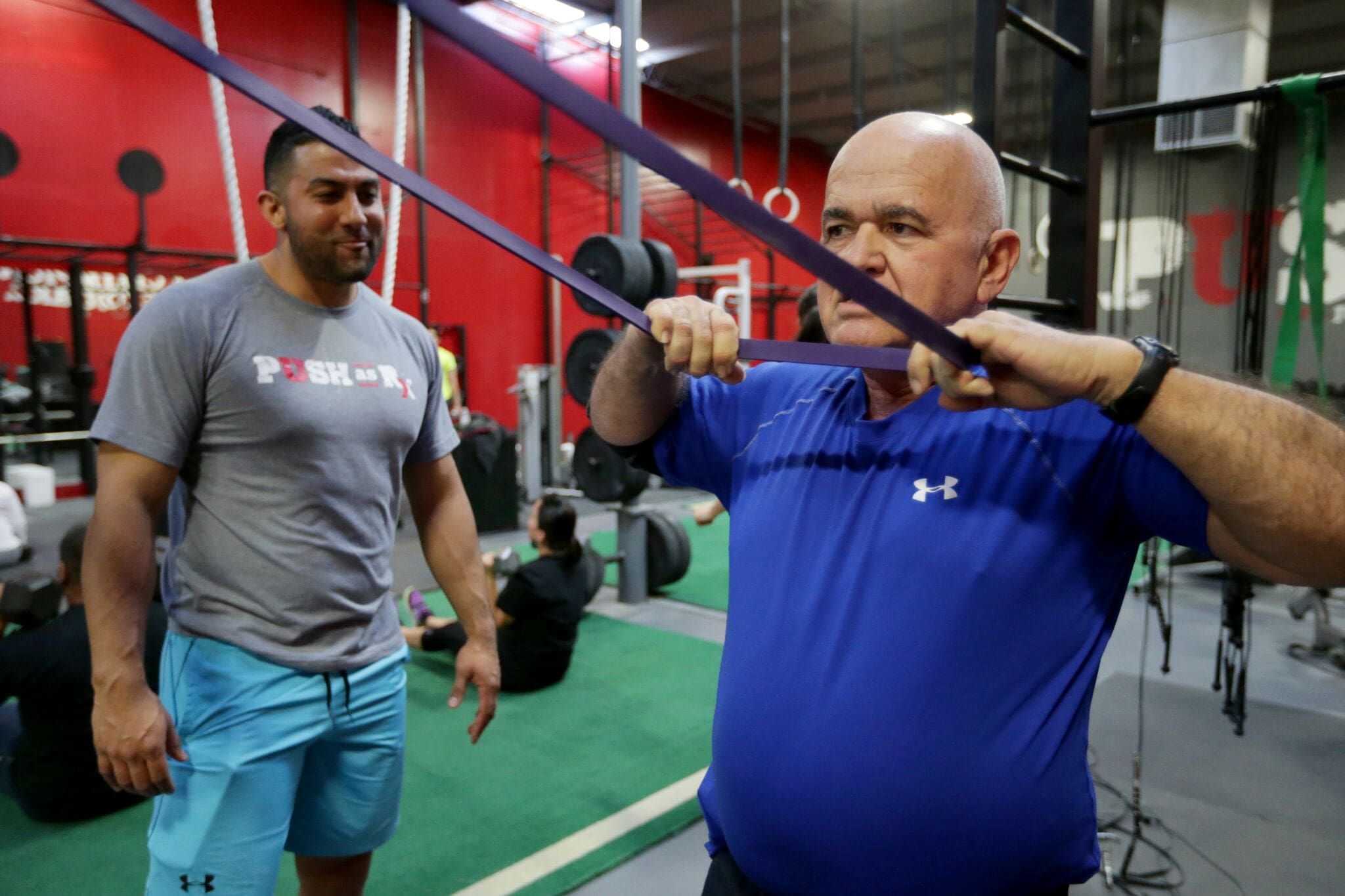


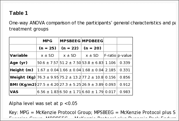
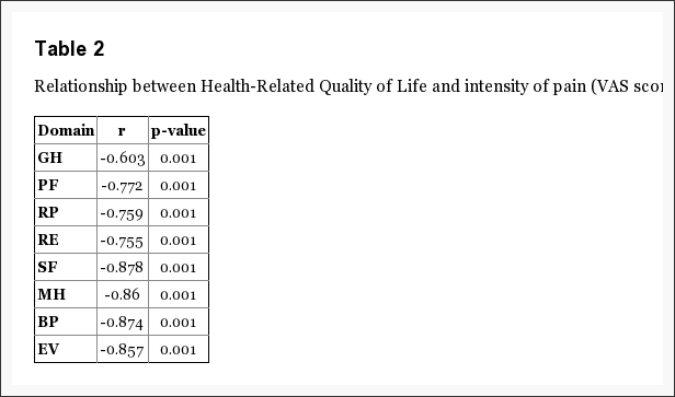
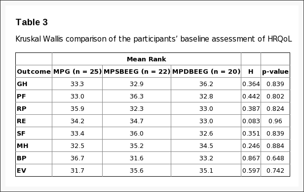
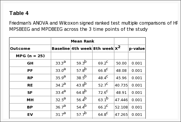
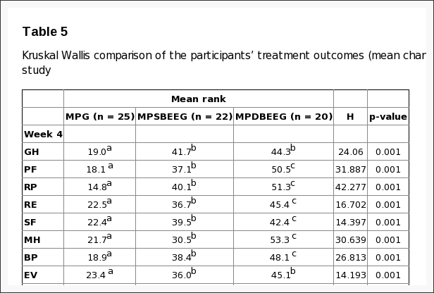


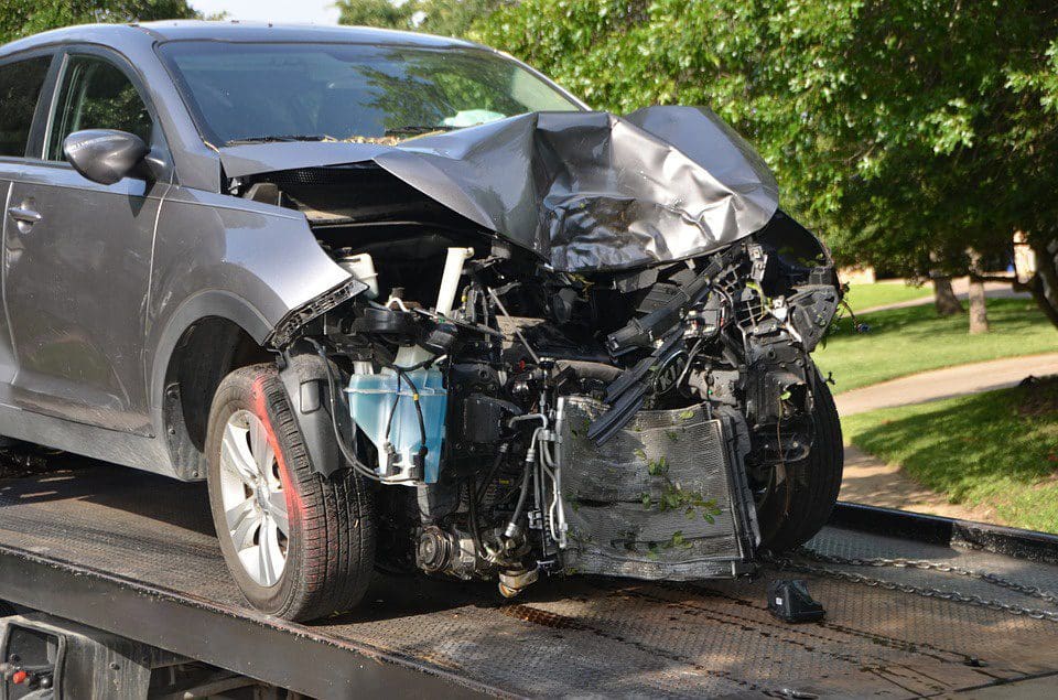
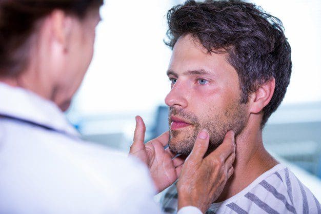
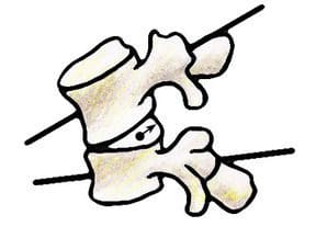
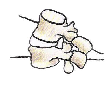
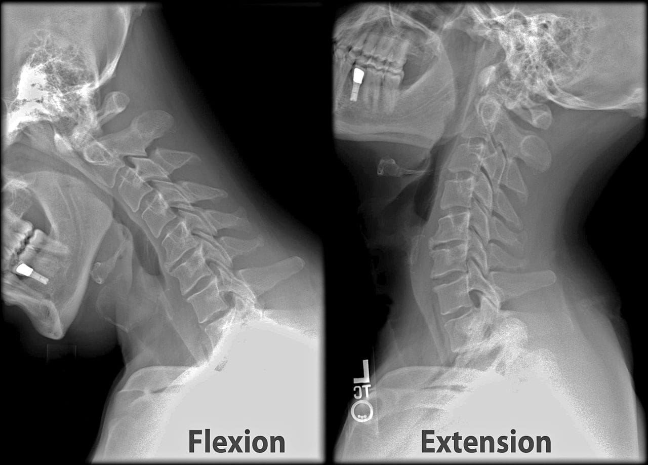
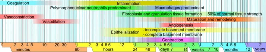


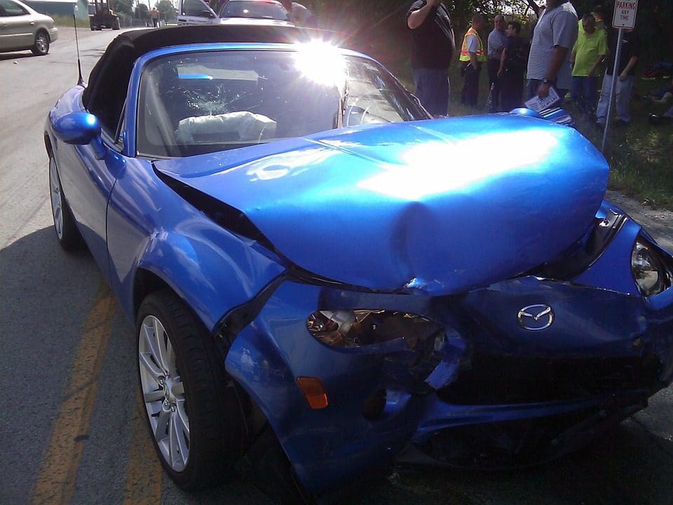

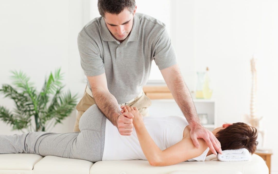
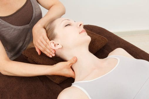

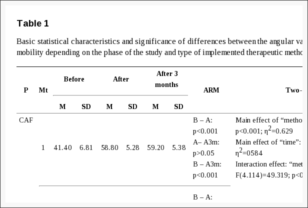
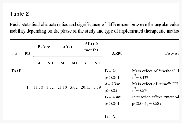
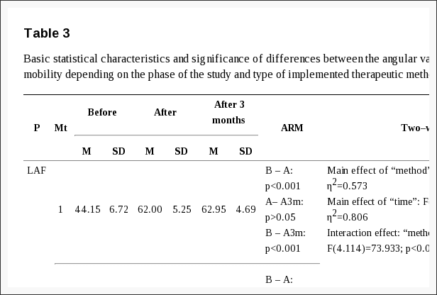
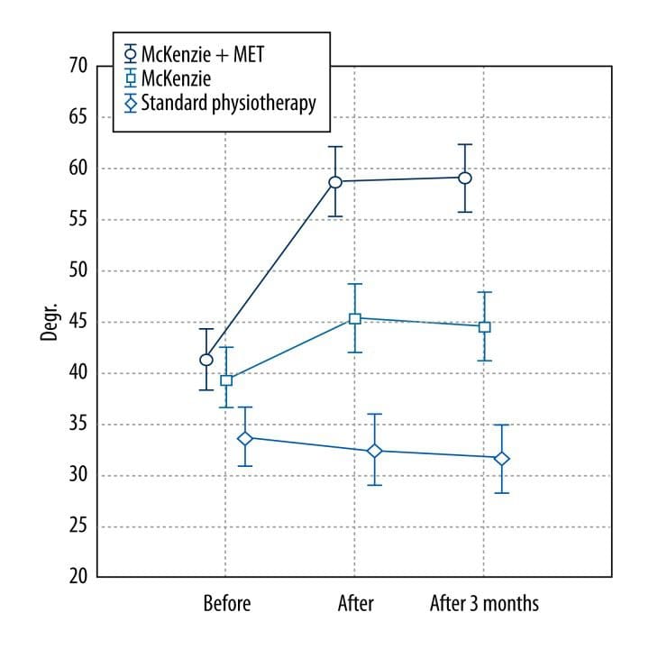
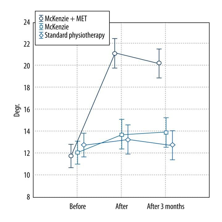
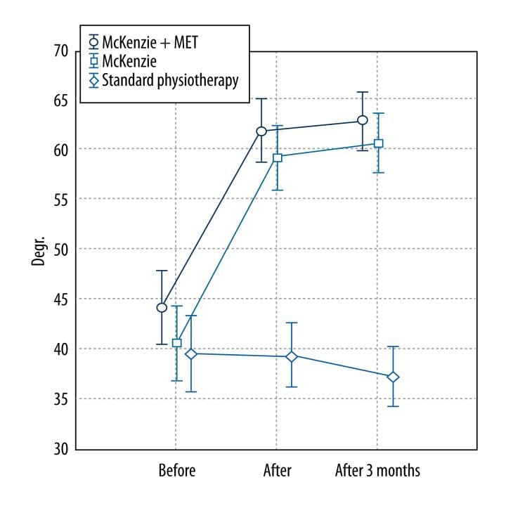
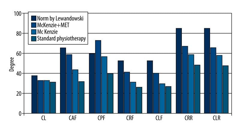
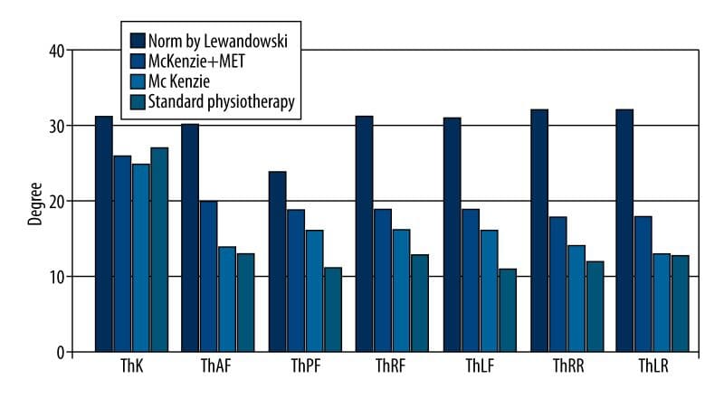
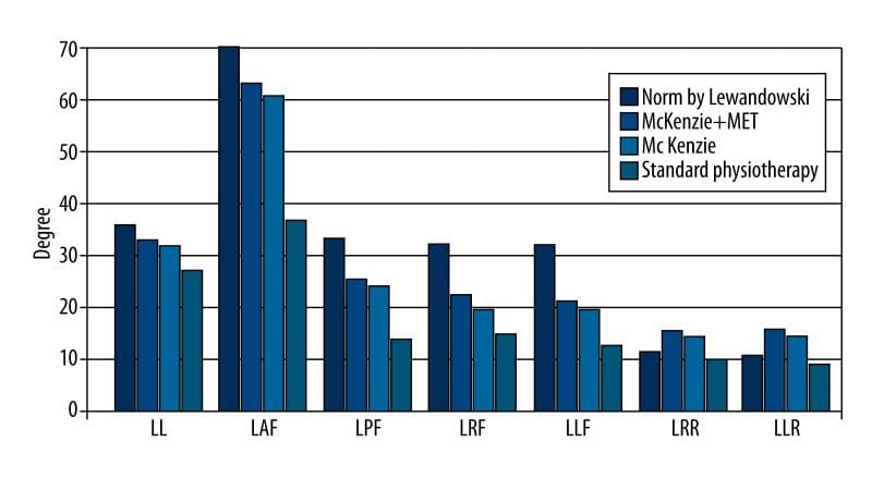
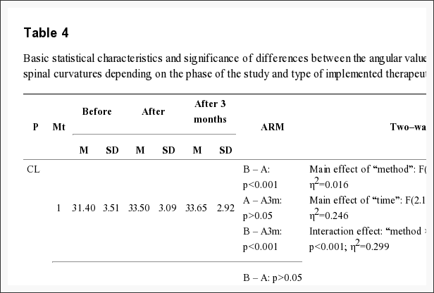
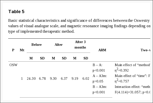
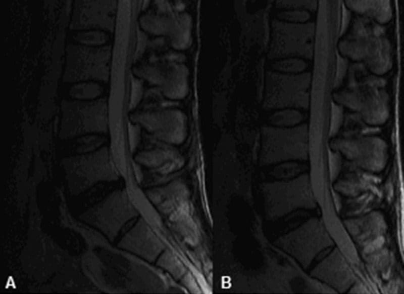
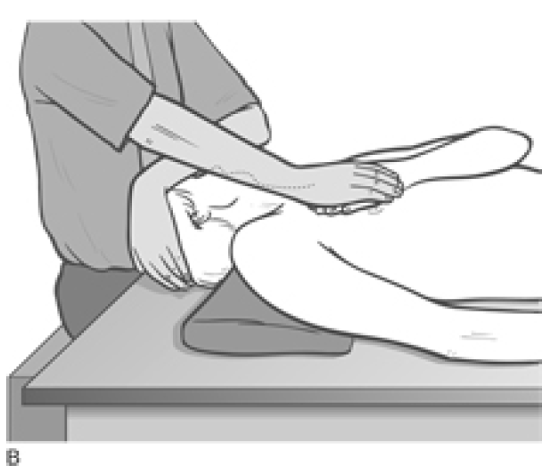
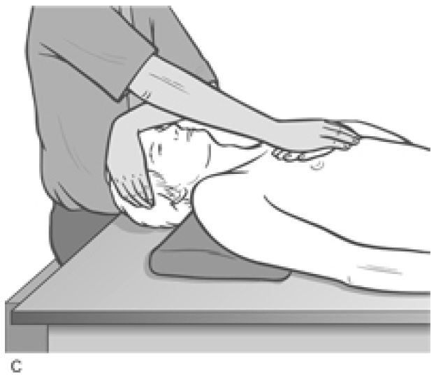
 MRI may be requested for: