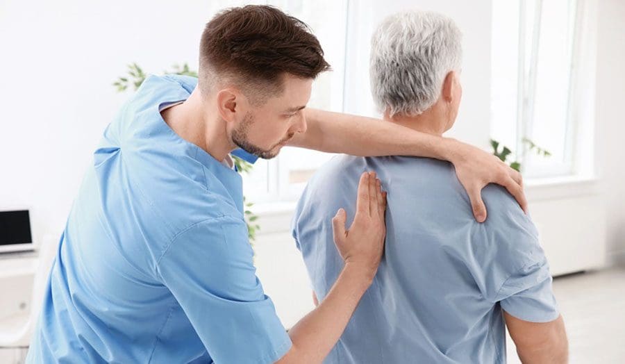
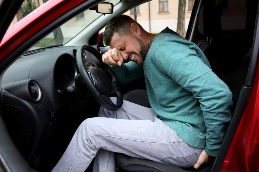
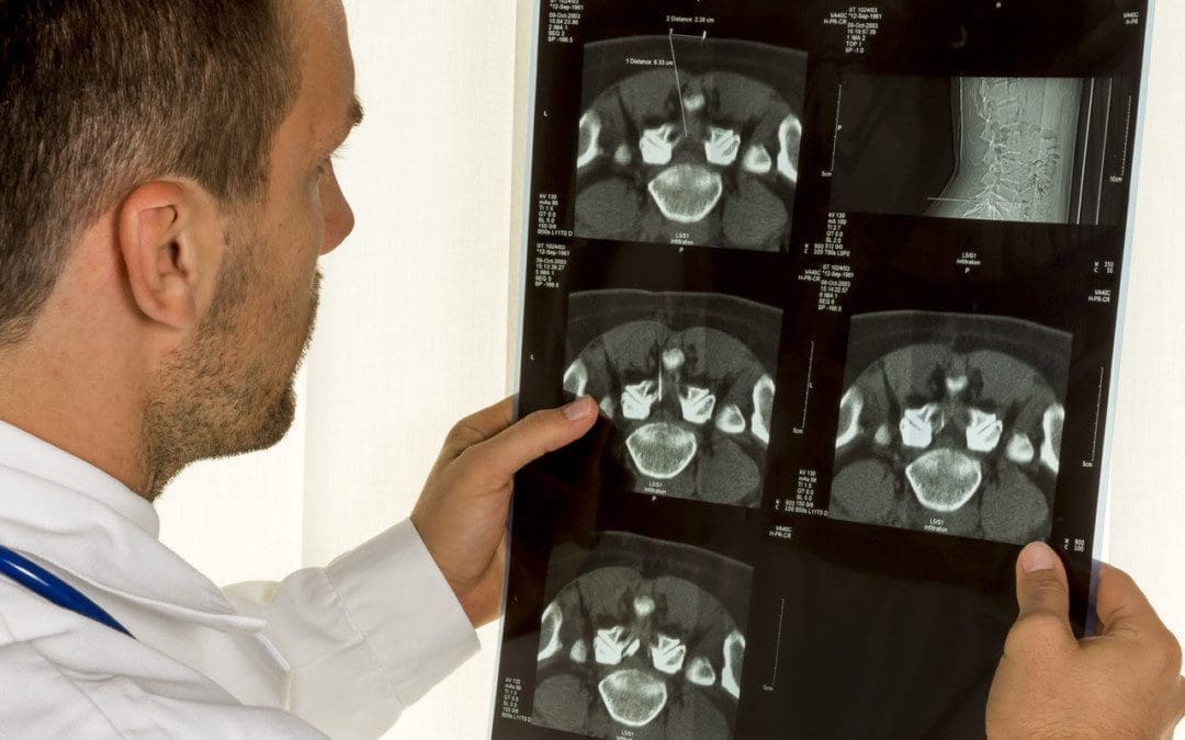
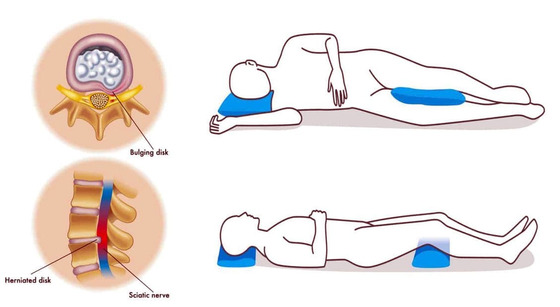
Non-surgical spinal decompression relieves pain related to spinal conditions, injuries, and disorders that provides a comfortable, affordable alternative treatment option to costly and invasive surgical procedures. Non-surgical spinal decompression can also relieve pain associated with post-surgical rehabilitation. A key to successful outcomes is including added components of the treatment that includes spinal decompression nutrition.
Spinal Decompression Nutrition
Proper nutrition and a balanced diet are essential elements of overall health. Individuals suffering from herniated discs, bulging discs, degenerated discs, sciatica, and chronic low back and neck pain are often deficient in vitamins and minerals, leading to inflammation and pain. These deficiencies can cause or exacerbate the pain and prevent or slow down healing. The bones, muscles, and other structures in the spine need proper nutrition to be strong enough to support the body and perform functions optimally. A health coach and nutritionist can recommend the proper diet and supplements to expedite healing, depending on the patient, the circumstances, and the individual situation. A non-inflammatory diet can make a big difference in patients’ symptoms and the effectiveness of decompression therapy.
The Right Foods
Eating a balanced diet with the right amount and variety of vitamins and nutrients can reduce back problems by nourishing the spine’s bones, muscles, discs, and other structures. While a healthy diet calls for various vitamins and nutrients, several healthy choices can directly benefit the spine. First and foremost is:
Sugar and Nitrate Reduction
High sugar diets lack the nutrients needed to prevent the release of inflammatory mediators.
The average individual consumes around 100 lbs of sugar per year.
Dessert foods are high infatty acids, which increase inflammation.
Any foods containing high fructose corn syrup like salad dressings and sodas.
Processed foods high in nitrates like hot dogs, sausage, and lunch meats.
Super Foods
Increase healing of the spine at the cellular level with superfoods that include:
Water – maintaining hydration is important for re-hydrating degenerated, dried-out discs.
Pre Spinal Decompression Nutrition
The human body was created to heal itself; however, getting the proper nutrition can be difficult as circulation is impeded/blocked when going through a back injury or spinal condition. Eating and/or supplementing with essential nutrients and minerals could be recommended to encourage and engage the healing process. Using nutrients to improve recovery and healing is known as immunonutrition. Pre-decompression allows the tissues to begin healing. Then the chiropractic health team can prepare the tissues for decompression through therapeutic massage, heat, low-level laser therapy, and ultrasound.
DRX9000 Explained by a Neurosurgeon
References
Calder, Philip C. “Fatty acids and inflammation: the cutting edge between food and pharma.” European journal of pharmacology vol. 668 Suppl 1 (2011): S50-8. doi:10.1016/j.ejphar.2011.05.085
Gay R. “All About Spinal Decompression Therapy.” Spine-health. http://www.spine-health.com/treatment/chiropractic/all-about-spinal-decompression-therapy. Published September 2013. Accessed April 2015.
InformedHealth.org [Internet]. Cologne, Germany: Institute for Quality and Efficiency in Health Care (IQWiG); 2006-. What is inflammation? 2010 Nov 23 [Updated 2018 Feb 22]. Available from: https://www.ncbi.nlm.nih.gov/books/NBK279298/
Innes, Jacqueline K, and Philip C Calder. “Omega-6 fatty acids and inflammation.” Prostaglandins, leukotrienes, and essential fatty acids vol. 132 (2018): 41-48. doi:10.1016/j.plefa.2018.03.004
Whenever stepping out onto a playing field or gym, there is a risk of suffering sports back injuries. Back pulls, strain and sprain injuries are the most common. Low back pain is one of the most prevalent complaints at all levels of competition. 90% of these acute back injuries will heal on their own, usually in about three months. However, sometimes these injuries can be more severe and require professional medical care. Treatment options for different groups of athletes include nonsurgical motorized spinal decompression.
Sports Back Injuries
Injury mechanisms vary from sport to sport, but there are recommendations regarding spinal decompression treatment for these injuries and return to play. Chiropractic healthcare specialists understand the sport-specific injury patterns and treatment guidelines for athletes following a back injury. Spinal decompression treatments are beneficial and result in higher rates of return to play depending on the specific sport of the injured athlete. A chiropractor will create a personalized spinal decompression treatment plan for the sport-specific context to meet the athlete’s short and long-term needs.
An estimated 10–15% of athletes will experience low back pain.
All types of sports place increased stress on the lumbar spine through physically demanding and repetitive movements/motions.
The repetitive shifting, bending, twisting, jumping, flexion, extension, and spinal axial loadingmotions contribute to low back pain even though the athletes are in top shape with increased strength and flexibility.
Injury patterns demonstrate the increased stresses that athletes place on the lumbar spine.
Common Spine Sports Injuries
Cervical Neck Injuries
Stingers are a type of neck injury.
A stinger is also known as a burner is an injury that happens when the head or neck gets hit to one side, causing the shoulder to be pulled in the opposite direction.
These injuries manifest as numbness or tingling in the shoulder from stretching or compressing the cervical nerve roots.
Lumbar Lower Back Sprains and Strains
When trying to lift too much weight or using an improper lifting technique when working out with weights.
Fast running, quick stopping, and shifting can cause the low back and hip muscles to get overly pulled/stretched.
Staying low to the ground and springing/jumping up can cause abnormal stretching or tearing of the muscle fibers.
Fractures and Injuries to the Supporting Spinal Structures
In sports that involve repetitive extension movements, spinal stress fractures are relatively common.
Excessive and repeated strain to the spinal column area leads to low back pain and injury.
Nonsurgical Spinal Decompression
Nonsurgical spinal decompression is motorized traction that is used to relieve compression pressure, restore spinal disc height, and relieve back pain.
Spinal decompression works to gently stretch the spine changing the force and position of the spine.
The gel-like cushions between the vertebrae are pulled to open up the spacing taking pressure off nerves and other structures.
This allows bulging or herniated discs to return to their normal position and promotes optimal circulation of blood, water, oxygen, and nutrient-rich fluids into the discs to heal, as well as, injured or diseased spinal nerve roots.
DRX 9000 Decompression
References
Ball, Jacob R et al. “Lumbar Spine Injuries in Sports: Review of the Literature and Current Treatment Recommendations.” Sports medicine – open vol. 5,1 26. 24 Jun. 2019, doi:10.1186/s40798-019-0199-7
Jonasson, Pall et al. “Prevalence of joint-related pain in the extremities and spine in five groups of top athletes.” Knee surgery, sports traumatology, arthroscopy: official journal of the ESSKA vol. 19,9 (2011): 1540-6. doi:10.1007/s00167-011-1539-4
Lawrence, James P et al. “Back pain in athletes.” The Journal of the American Academy of Orthopaedic Surgeons vol. 14,13 (2006): 726-35. doi:10.5435/00124635-200612000-00004
Petering, Ryan C, and Charles Webb. “Treatment options for low back pain in athletes.” Sports health vol. 3,6 (2011): 550-5. doi:10.1177/1941738111416446
Sanchez, Anthony R 2nd et al. “Field-side and prehospital management of the spine-injured athlete.” Current sports medicine reports vol. 4,1 (2005): 50-5. doi:10.1097/01.csmr.0000306072.44520.22
Individuals with chronic back and/or leg pain are encouraged to try spinal decompression. Non-surgical spinal decompression is a treatment option therapy that has been proven to be safe, gentle, and successful. This therapy is motorized traction that takes the pressure off the spinal discs and stretches out the spine to its correct position. It is highly effective, comfortable, affordable, and a safe alternative to surgery. At Injury Medical Chiropractic and Functional Medicine Clinic, our spinal decompression team/tables effectively treat:
Neck pain
Chronic back pain
Sciatica
Bulging discs
Herniated discs
Degenerated discs
Whiplash
Try Spinal Decompression
The vertebral bones protect the spinal cord. Everyday wear-and-tear, improper posture and injury can cause parts of the vertebrae to compress the spinal cord’s nerves, leading to pain, numbness, or tingling. Non-surgical spinal decompression therapy is also known as NSSD or SDT/Spinal Decompression Therapy. The goal of the treatment is to restore optimal health to the spine. Pain-causing conditions can be reversed or healed, and discs can be normalized through the decompression process as it encourages spinal repositioning to promote optimal healing.
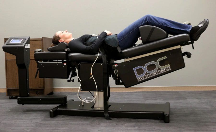
Decompression Table
The spinal decompression table may consist of a manually operated cable and pulley system or a computerized table segmented by the upper and lower body.
The angle and pressure applied depend on the type of injury and the individual’s needs.
Each procedure is carefully calculated to reposition the spinal discs and disc material to alleviate pain.
How It Works
Spinal decompression is a mechanized version of a chiropractic adjustment. By gently stretching and moving the spine, the vertebrae have proper alignment restored, restoring range of motion, decreasing or eliminating pain, and improving mobility and function.
The individual is strapped to the machine with a harness that helps position the back for optimal decompression.
Depending on the condition and severity, the therapist will choose from a list of decompression programs.
Slowly, the spine is stretched and lengthened, relieving pressure.
The spine’s stretching and repositioning are different from standard physical therapy and manual manipulation treatment.
It is a gradual process to prevent the body from muscle guarding as the natural response to avoid injury.
Treatment Benefits
An examination is required to see if an individual meets the criteria. Non-surgical spinal decompression therapy has been shown to:
Reduce or eliminate pain.
Rehydrate spinal discs.
Reduce disc bulging/herniation.
Improve functional abilities.
Decrease the need for surgery.
DRX9000
References
Apfel, Christian C et al. “Restoration of disk height through non-surgical spinal decompression is associated with decreased discogenic low back pain: a retrospective cohort study.” BMC musculoskeletal disorders vol. 11 155. 8 Jul. 2010, doi:10.1186/1471-2474-11-155
Koçak, Fatmanur Aybala et al. “Comparison of the short-term effects of the conventional motorized traction with non-surgical spinal decompression performed with a DRX9000 device on pain, functionality, depression, and quality of life in patients with low back pain associated with lumbar disc herniation: A single-blind randomized controlled trial.” Turkish Journal of physical medicine and rehabilitation vol. 64,1 17-27. 16 Feb. 2017, doi:10.5606/tftrd.2017.154
Macario, Alex, and Joseph V Pergolizzi. “Systematic literature review of spinal decompression via motorized traction for chronic discogenic low back pain.” Pain practice: the Official Journal of World Institute of Pain vol. 6,3 (2006): 171-8. doi:10.1111/j.1533-2500.2006.00082.x
Any vehicle crash, collision, or accident can cause various injuries, with back pain issues as a primary injury or a side effect from other injuries. Usually, injury symptoms begin right after the collision, but in other cases, individuals may not start experiencing symptoms until hours, days, or even weeks later. This is from the adrenaline that rushes throughout the body during the collision/fight or flight response delaying the injury symptoms. There are reports of individuals who walk away from an accident unscathed but require urgent medical treatment a short while later. Chiropractic care can provide manual and spinal motorized decompression benefits.
Decompression Benefits
Head Injuries
Head injuries occur when drivers and/or passengers hit their heads on the steering wheel, windows, dashboard, metal frame, and sometimes each other.
A head injury is considered a severe condition that can cause concussions, skull fractures, comas, hearing loss, cognitive and memory issues, and vision problems.
A significant head injury can cause extensive and costly medical treatment with the possibility of long-term medical care.
Neck Injuries
Neck injuries are common in vehicle collisions.
The most common is whiplash, with the head and neck-snapping from indirect blunt force, like being rear-ended.
Whiplash can cause significant damage to the ligaments and muscles, like swelling and neck pain, and temporary paralysis of the vocal cords.
Injury patterns of whiplash can differ depending on the speed, force, and overall health of the individual involved.
Back Injuries
Back injuries can range in severity from sprains to significant damage involving the nerves and/or the spinal cord.
If the damage is severe, it can lead to loss of sensation in the body, loss of limb control, or permanent paralysis.
Disc herniation/s can lead to disability, muscle weakness, tingling and numbness in the limbs, and radiating body pain.
Chest and Torso Injuries
Vehicle collision forces can result in severe chest injuries that include broken ribs.
Broken ribs might not sound dangerous by themselves; they can puncture the lungs leading to other injuries and internal bleeding.
The legs, feet, arms, and hands are frequently injured, broken, and sometimes dislocated.
Motorcyclists are also at a higher risk for significant injury that includes:
Multiple fractures, internal injury, head injuries, and severe ligament damage.
Pedestrians struck by a vehicle have an increased risk for a combination of all injuries at once.
Non-Surgical Decompression Benefits
Chiropractors are trained to identify and treat injuries from vehicle collisions.
Non-surgical spinal decompression gently stretches the spine using a motorized traction device to help reposition the spine and remove the pressure.
As the pressure is taken off, the spinal discs regain their natural height, relieving the pressure on the nerves and other spinal structures.
Optimal healing is promoted by an improved circulation of nutrients, water, and oxygen to the injury site.
Decompression helps to strengthen the muscles in the affected area.
It provides positive spinal structural changes.
Improves nervous system function.
Non-surgical decompression is a tool for correcting injuries and relieving pain, allowing optimal health for the individual.
DOC Decompression Table
References
Apfel, Christian C et al. “Restoration of disk height through non-surgical spinal decompression is associated with decreased discogenic low back pain: a retrospective cohort study.” BMC musculoskeletal disorders vol. 11 155. 8 Jul. 2010, doi:10.1186/1471-2474-11-155
Koçak, Fatmanur Aybala et al. “Comparison of the short-term effects of the conventional motorized traction with non-surgical spinal decompression performed with a DRX9000 device on pain, functionality, depression, and quality of life in patients with low back pain associated with lumbar disc herniation: A single-blind randomized controlled trial.” Turkish Journal of physical medicine and rehabilitation vol. 64,1 17-27. 16 Feb. 2017, doi:10.5606/tftrd.2017.154
Macario, Alex, and Joseph V Pergolizzi. “Systematic literature review of spinal decompression via motorized traction for chronic discogenic low back pain.” Pain practice: the Official Journal of World Institute of Pain vol. 6,3 (2006): 171-8. doi:10.1111/j.1533-2500.2006.00082.x
Tremors are extremely rare, but they can result from spinal compression and not necessarily a brain condition like Parkinson’s disease. Tremors are abnormal, involuntary body movements with various causes, most of which are connected to the brain and not the spine. A study reports that more than 75% of individuals with Parkinson’s experienced a resting tremor, and about 60% experience tremors while moving. Sometimes the spine is the contributor caused by compression of the spinal cord.
Spinal Compression Study
A 90-year-old man was hospitalized after having tremors, with Parkinson’s being the initial diagnosis. The tremors progressed to the point where the man could not feed himself or walk without support. The case became the focus of a medical report published by physicians in the Department of Orthopaedic Surgery, Division of the Spine, Singapore Tan Tock Seng Hospital. Along with the tremors, symptoms progressed to:
Difficulty with fine motor skills like buttoning a shirt.
However, it was ruled out because the patient was not presenting with other Parkinson’s symptoms.
For individuals with cervical spondylotic myelopathy tremors, surgery can be used to help the condition. However, with cervical myelopathy, there is often some permanent damage. Individuals have shown that post-surgery and decompression, symptoms still present, maybe not as much, but there will be a need for a symptom management plan.
Prevention
The best way to prevent tremors associated with cervical spondylotic myelopathy is to minimize the strain on the spine that can lead to herniated discs and/or other spinal injuries. The discs in the spine degenerate, dry out and start cracking with age, increasing the risk of rupture. If a tremor develops, contact a doctor, spine specialist, or chiropractor to help diagnose the condition. These doctors can perform physical and neurological tests to determine the cause and treatment options.
Body Composition
Aging Health
Steady weight gain throughout life can lead to adult-onset diabetes. This is partly caused by having more body fat and progressive muscle loss. Loss of skeletal muscle mass is linked to insulin resistance that involves:
The less muscle is available, the less insulin sensitive the body becomes.
As insulin sensitivity decreases, the body becomes more resistant, increasing risk factors for type II diabetes.
This can lead to osteoporosis, where the old bone is reabsorbed more and less new bone is created.
Both men and women can experience decreased muscle mass that can lead to:
Thinner bones
Weaker bones
Increased risk of osteoporosis and severe injury from falls.
To help prevent these issues, it is recommended to:
It is recommended to space out protein intake across meals rather than consuming it all at once. This helps to ensure the proper amount is acquired.
Monitoring body composition regularly can help minimize muscle mass loss and fat mass gain as the body ages.
A regular strength training routine will help strengthen bones muscles and maintain optimal circulation.
References
Heusinkveld, Lauren E et al. “Impact of Tremor on Patients With Early Stage Parkinson’s Disease.” Frontiers in neurology vol. 9 628. 3 Aug. 2018, doi:10.3389/fneur.2018.00628
Jancso, Z et al. “Differences in weight gain in hypertensive and diabetic elderly patients primary care study.” The Journal of nutrition, health & aging vol. 16,6 (2012): 592-6. doi:10.1007/s12603-011-0360-6
Srikanthan, Preethi, and Arun S Karlamangla. “Relative muscle mass is inversely associated with insulin resistance and prediabetes. Findings from the third National Health and Nutrition Examination Survey.” The Journal of clinical endocrinology and metabolism vol. 96,9 (2011): 2898-903. doi:10.1210/jc.2011-0435
Tapia Perez, Jorge Humberto et al. “Treatment of Spinal Myoclonus Due to Degenerative Compression Myelopathy with Cervical Spinal Cord Stimulation: A Report of 2 Cases.” World neurosurgery vol. 136 (2020): 44-48. doi:10.1016/j.wneu.2019.12.170
Sciatica motor vehicle crash. After an automobile crash/accident, symptoms of pain and discomfort can immediately follow the force of impact, indicating an injury. Many injuries and symptoms appear right away, like:
Pain from high-impact trauma and cuts.
Bone fractures.
Dislocations.
Neck whiplash.
Back pain.
The sciatic nerve is the largest in the body, and any damage can cause pain in one or both sides of the body. Pressure and compression on the nerves, ligaments, and muscles accompanied by weakness or numbness in the lower back, legs, or feet could cause delayed sciatica symptoms hours, days, even weeks later. It is critical to meet with a doctor and auto accident chiropractor after any type of accident, large or small, to develop a thorough personalized treatment plan.
Sciatica Motor Vehicle Crash
Sciatica can be brought on by a pinched nerve, which is often the result of the spine shifting out of place, causing herniation and compression on the sciatic nerve. The trauma from a motor vehicle accident can cause the spinal discs to be knocked out of place, rupture, and leak out, irritating the surrounding tissue and nerve endings. Back injuries are among the most common forms of damage/injury resulting from a motor vehicle accident/crash that can lead to sciatica. Broken and/or fractured vertebral, hip, or pelvis bone fragments can compress the sciatic nerve. Even when the initial result of the impact does not result in sciatica, over time, an untreated back injury could lead to sciatica symptoms.
Symptoms
Motor vehicle crashes often activate or aggravate pre-existing conditions like asymptomatic degenerative disc disease, affecting the sciatic nerve causing discomfort and pain. Common symptoms include:
Mild discomfort or achiness.
Tingling sensations from the lower back and down the back of the leg.
Weakness, numbness, or difficulty moving the leg and foot.
Inability to bend the foot upward at the ankle- known as foot drop.
Constant pain in one side of the buttocks or leg.
Sharp pain that makes it difficult to stand up and walk
Difficulty sitting.
Burning or tingling in one leg can worsen when sitting.
Intense pain.
Sharp burning and/or what feels like shooting electricity pain.
Diagnosis
A spine doctor and chiropractor will use diagnostic imaging tools like X-rays, and CT scans to see the scope of the injured area.
An X-ray will show a detailed image of the spine and affected bones in the area.
A CT scan will include a 3D image that shows the surrounding musculature, tissues, and nerves that could be damaged/injured.
Treatment
The doctor and chiropractor will then develop an appropriate and personalized treatment plan that could utilize various methods and techniques.
Chiropractic is commonly the first treatment to realign the spine and relieve pressure on the nerve.
A pain management specialist or physical therapist will be brought in for rehabilitation/recovery as adjustments progress.
Orthopedists and neurologists may be brought in for less conservative treatment in more severe cases, including surgical options.
Other treatments can include steroid injections or anti-inflammatory medicines to relieve nerve pressure.
Body Composition
Injury Rehabilitation Phase
Current in-clinic methods of measuring the composition of an injured body are indirect, while medically advanced techniques limit the frequency of testing. InBody provides cost-effective, comprehensive, and timely measurements that identify areas of weakness from damage, injury, or recent surgery and develop a customized rehabilitation program to improve functional status.
During the rehabilitation phase, increased sedentary behavior and/or immobilization causes muscle loss in the injured or operated region. By independently evaluating lean mass in each segment of the arms, legs, and torso, a chiropractor or physical therapist gathers baseline information on the body segments with restricted mobility.
InBody can help provide further insight into an individual’s body composition to analyze long-term health risks and develop a personalized exercise intervention to improve overall health and reduce health risks. This provides beneficial information for identifying potential imbalances related to muscle loss post-injury/surgery that can be targeted and improved. Identifying these imbalances allows the therapists to increase functional fitness and mobility, helping the individual reduce the risk of re-injury or new injuries.
References
Defouilloux, B et al. “A propos de trois observations chez des polytraumatisées de la route présentag une fracture du bassin associée à des signes neurologiques” [Apropos of 3 cases of multiple traffic injuries presenting pelvic fractures associated with neurologic signs]. Journal de radiologie, d’electrologie, et de medecine nucleaire vol. 48,8 (1967): 505-6.
Noble, J et al. “Analysis of upper and lower extremity peripheral nerve injuries in a population of patients with multiple injuries.” The Journal of trauma vol. 45,1 (1998): 116-22. doi:10.1097/00005373-199807000-00025
Walsh, K et al. “Risk of low back pain in people admitted to hospital for traffic accidents and falls.” Journal of epidemiology and community health vol. 46,3 (1992): 231-3. doi:10.1136/jech.46.3.231
Sleeping with a bulging disc can be challenging for the body to achieve the proper rest. And sleeping in an awkward position can add stress to the spine, making the bulge worse, which can cause tingling, numbness, pain, and digestive problems. This can disrupt the sleep cycle and prevent proper healing of the spinal injury.
Sleeping With a Bulging Disc
When sleeping, most back pain occurs in the lumbar or lower back, in one of two places where the spine meets the pelvis. Around 95% of lower back herniations happen in the L4-L5 spinal segment or the L5-S1 Lumbosacral joint. Any back pain can turn into a vicious cycle of:
Sleeping with a bulging disc requires maintaining the ears, shoulders, and hips aligned to keep the spine aligned.
Sleeping on The Back
Back sleeping done correctly is the best way to sleep for the spine’s health. The important thing is to ensure the entire back is supported when sleeping. If there is a gap or space between the mattress and the back, the weight and gravity force the spine to lower in an unnatural way to fill the space. This can cause back muscle soreness, injury, and sciatica. A thin pillow, blanket, or towel can be used to fill the space, giving the spine the support it needs. Back sleepers can also benefit from a pillow or two under the knees to elevate the legs and help maintain the natural curve of the pine.
Sleeping on The Side
Side sleepers can try pulling the legs up toward the chest, and placing a pillow between the knees can provide relief when sleeping with a bulging disc. Pulling the legs up in the fetal position can relieve pressure on the discs. It is recommended to switch sides to keep the spine balanced. This helps maintain hip alignment, which helps keep the spine in a neutral position.
Sleeping On The Stomach
It is recommended to avoid sleeping on the stomach. This pulls the spine down into an unnatural curve that can cause and exacerbate back pain. For individuals that naturally stomach sleep, it is recommended to place a pillow under the hips and lower abdomen to prevent unnatural spinal positioning.
Chiropractic Relief
Utilizing the right sleeping position can provide pain relief and thorough rest. However, sleeping with a herniated disc is far from what is needed to get back to a normal healthy sleeping pattern. This depends on the location of the bulging disc, severity, and cause. A chiropractor can:
Help the individual develop an optimal sleeping routine and positioning.
Body Composition
Sleep and Growth Hormone In Children
Growth, in all ages, is primarily controlled by growth hormone. The hormone is regulated by the hypothalamus and pituitarygland which plays an important role in sleep. Growth hormone has been found:
It peaks during the beginning of deep sleep.
There are multiple smaller peaks during the other stages of sleep.
Those who have a delay at the beginning of deep sleep have delayed rises in growth hormone levels.
For children to grow they need to have proper levels of growth hormone. This means they need to have the proper amount of sleep for proper body composition. Research has found that increased levels of sleep resulted in less overall fat mass and a reduced percentage of body fat allowing their bodies to grow.
References
Al Qaraghli MI, De Jesus O. Lumbar Disc Herniation. [Updated 2021 Aug 30]. In: StatPearls [Internet]. Treasure Island (FL): StatPearls Publishing; 2022 Jan-. Available from: https://www.ncbi.nlm.nih.gov/books/NBK560878/
Desouzart, Gustavo et al. ‘Effects of Sleeping Position on Back Pain in Physically Active Seniors: A Controlled Pilot Study. 1 Jan. 2016: 235 – 240.
Kose, Gulsah et al. “The Effect of Low Back Pain on Daily Activities and Sleep Quality in Patients With Lumbar Disc Herniation: A Pilot Study.” The Journal of neuroscience nursing: Journal of the American Association of Neuroscience Nurses vol. 51,4 (2019): 184-189. doi:10.1097/JNN.0000000000000446
Sener, Sevgi, and Ozkan Guler. “Self-reported data on sleep quality and psychologic characteristics in patients with myofascial pain and disc displacement versus asymptomatic controls.” The International Journal of prosthodontics vol. 25,4 (2012): 348-52.
IFM's Find A Practitioner tool is the largest referral network in Functional Medicine, created to help patients locate Functional Medicine practitioners anywhere in the world. IFM Certified Practitioners are listed first in the search results, given their extensive education in Functional Medicine