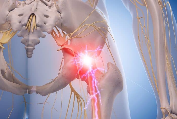
Pain along the pelvis and groin region is known as osteitis pubis. Osteitis pubis develops through the inflammation of the pubic symphysis, or the joints of the major pelvic bones found at the front of the pelvis.
The pubic symphysis is a thin joint which generally provides very minimal motion. The joint retains the two sides of the pelvis together in the front, where they connect�at the sacrum in the rear side of the pelvis.
Osteitis Pubis Symptoms
Osteitis pubis is commonly characterized by pain in the front of the pelvis. Other causes of pelvic pain, such as a strain or a sprain, are frequently confused and diagnosed as osteitis pubis. While many patients report painful symptoms on one side, the�pain�typically occurs in the middle of the pelvis. Other symptoms of osteitis pubis include limping and weakness.
Osteitis Pubis Causes
For some patients, the pubic symphysis itself can become irritated and inflamed, causing the well-known symptoms of osteitis pubis. Other common causes of osteitis pubis comprise of: sports injuries, particularly from football, hockey, and soccer; pregnancy; gynecologic or abdominal surgical interventions; and trauma or injury from accidents.
�
Osteitis pubis is known as the inflammation of the pubis symphysis which causes various degrees of lower abdominal, pelvic, and groin pain. Symptoms of osteitis pubis include pain and discomfort in the region of the pelvis when engaging in physical activities, and loss of flexibility. A variety of causes, including sports injuries, can cause osteitis pubis. Fortunately, rest alone can help treat this painful health issue.
Dr. Alex Jimenez D.C., C.C.S.T.
Osteitis Pubis Diagnosis
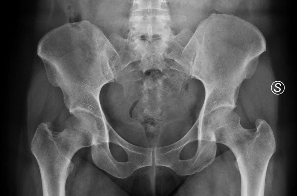
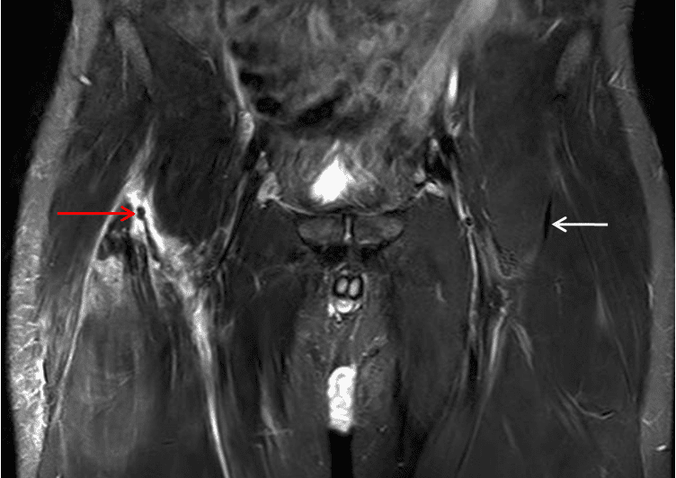
Diagnosis of osteitis pubis generally involves x-rays which demonstrate an irregular pubic symphysis with sclerotic, or thick, bone borders as a result of chronic inflammation. An MRI test is generally not required, however, it will help demonstrate the inflammation of the bone and the joint.
Additional tests may be performed to ensure there’s no infection in the bone which could also be causing symptoms similar to osteitis pubis. This complication is more of a concern for those patients who have had recent surgery or for those who are more prone to suffer from infections.
Osteitis Pubis Management
The most recommended treatment for osteitis pubis is rest. Since inflammation is the problem, the human body often only requires the joint to rest in order to heal correctly. Other treatment, however, consists of:
Rest
An essential treatment for osteitis pubis is rest as this will permit the intense inflammation in the pelvis and groin to subside. For many patients, rest alone is the only treatment necessary for their�osteitis pubis. If the pain is severe, crutches or a cane may provide additional assistance.
Ice and Heat
Ice packs and heating pads are among the most commonly used remedies for inflammation. Make sure to follow the instructions of your healthcare professional before utilizing ice and heat for your osteitis pubis symptoms.
Chiropractic Care
Chiropractic care is a well-known, alternative treatment option for osteitis pubis. A doctor of chiropractic, or chiropractor, will utilize a variety of treatment methods and techniques, to help restore strength, mobility, and flexibility while rest is needed to subside the painful symptoms. Chiropractic care can also help correct any spinal misalignments which may be causing additional pain and discomfort for the patient.
Drugs and/or Medications
Nonsteroidal anti-inflammatory drugs and/or medications, commonly referred to as NSAIDs, are frequent prescriptions provided for patients with hip pain brought on by problems like arthritis, bursitis, and tendonitis.
Treatment of osteitis pubis may take some time to completely relieve the painful symptoms. The use of drugs and/or medications is demonstrated to be better than the other treatment options listed above, although attempts to heal osteitis pubis with cortisone injections have been tested.
Surgical interventions are generally not necessary for patients with osteitis pubis.�The scope of our information is limited to chiropractic as well as to spinal injuries and conditions. To discuss the subject matter, please feel free to ask Dr. Jimenez or contact us at�915-850-0900�.
Curated by Dr. Alex Jimenez
Additional Topics: Acute Back Pain
Back pain�is the most prevalent cause of disability worldwide and the second most common reason for doctor office visits, outnumbered only by upper-respiratory infections. Approximately 80 percent of the population will experience back pain at least once throughout their life. The spine is a complex structure made up of bones, joints, ligaments, and muscles, among other soft tissues. Because of this, injuries and/or aggravated conditions, such as�herniated discs, can eventually lead to symptoms of back pain. Sports injuries or automobile accident injuries are often the most frequent cause of back pain, however, sometimes the simplest of movements can have painful results. Fortunately, alternative treatment options, such as chiropractic care, can help ease back pain through the use of spinal adjustments and manual manipulations, ultimately improving pain relief.
Sciatica is a collection of symptoms in the low back, which radiate down one or both legs. Sciatica is generally caused by the compression or irritation of the sciatic nerve, the largest nerve in the human body. One of the most common health issues that cause sciatic nerve pain is called piriformis syndrome. The piriformis muscle stretches from the front of the sacrum, the triangle-shaped bone between the hipbones on the pelvis.
The piriformis muscle extends to the top of the femur around the sciatic nerve. The femur, as previously mentioned, is the large bone in the upper leg. The piriformis muscle functions by helping the thigh move from side to side. A piriformis muscle spasm, or any other type of injury and/or condition along the piriformis muscle, can place pressure on the sciatic nerve and cause pain and discomfort. The result is piriformis�syndrome.
Piriformis Syndrome Causes and Symptoms
Sciatic nerve pain,�or sciatica, is one of the most prevalent�symptoms of piriformis syndrome. The pain and discomfort, however, may be felt in another part of the body. This is known as referred pain. Other common symptoms of piriformis syndrome include tingling sensations and numbness; tenderness;�difficulty sitting along with�pain while sitting and pain in the buttocks and thighs with physical activities.
The piriformis muscle can easily become damaged or injured from periods of inactivity or an excessive amount of exercise. Some common causes of piriformis syndrome include overuse; repetitive movements involving the legs; sitting for lengthy periods of time; lifting heavy objects; and extensive stair climbing. Sports injuries or automobile accident injuries can also harm the piriformis muscle and cause it to compress the sciatic nerve.�
�
Piriformis Syndrome Diagnosis
A doctor appointment for diagnosis of piriformis syndrome may include a review of the patient’s health history, their symptoms, and other probable causes of their pain and discomfort. If you recall straining a muscle during physical activity, be sure to share that information with your doctor. The�doctor may also perform a physical exam. The patient will participate in a series of range of movements to determine the cause of symptoms.
Some imaging tests may also be essential to help rule out other causes of piriformis syndrome. A CT scan or an MRI scan may help the healthcare professional determine whether even a herniated disc or arthritis is causing the patient’s pain and discomfort. An ultrasound of the piriformis muscle may also be helpful in diagnosing the problem if it seems that piriformis syndrome is causing the patient’s overall symptoms.
�
Piriformis syndrome is a health issue associated with the compression or impingement of the sciatic nerve around the piriformis muscle. Symptoms may include pain and discomfort, tingling sensations and numbness along the low back, or sciatica. Chiropractic care is a well-known alternative treatment option which can help reduce the compression of the sciatic nerve and improve piriformis syndrome.
Dr. Alex Jimenez D.C., C.C.S.T.
Piriformis Syndrome Treatment
Piriformis syndrome may often not need any treatment to�relieve its symptoms. Just avoiding the physical activities which caused the pain and discomfort to manifest and rest can help improve the health issue. If symptoms do persist, however, alternating between ice and heat can help decrease pain. Apply ice for 15 to 20 minutes then use a heating pad on the affected area. Try that every couple of hours to help relieve symptoms.
Over-the-counter painkillers�may also help decrease pain and discomfort. The symptoms associated with piriformis syndrome can go away with no additional treatment, however, if it doesn’t, the patient might benefit from alternative treatment options, such as chiropractic care or physical therapy. Chiropractic care is a treatment approach which utilizes spinal adjustments and manual manipulations to treat a variety of injuries and/or conditions.
A chiropractor,�or doctor of chiropractic, may also provide piriformis syndrome relief through the use of transcutaneous electrical nerve stimulator, or TENS, treatment. A TENS device is a handheld unit which sends electrical charges directly to the affected region of the piriformis muscle. The nerves are then stimulated by the electric energy, which interferes with pain signals being transmitted to the brain.
The chiropractor or physical therapist may also recommend a series of lifestyle modifications, including physical activity guidance and nutritional advice. Various stretches and exercises can help improve the strength, flexibility, and mobility of the�piriformis muscle. In severe cases of piriformis syndrome, corticosteroid injections or even surgical interventions may be required to help alleviate the symptoms.�The scope of our information is limited to chiropractic as well as to spinal injuries and conditions. To discuss the subject matter, please feel free to ask Dr. Jimenez or contact us at�915-850-0900�.
Curated by Dr. Alex Jimenez
Additional Topics: Chiropractic for Athletes with Back Pain
Back pain�is one of the most prevalent causes of disability and missed days at work worldwide. Back pain is the second most common reason for doctor office visits, outnumbered only by upper-respiratory infections. Approximately 80 percent of the population will experience back pain at least once throughout their life. The spine is a complex structure made up of bones, joints, ligaments, and muscles, among other soft tissues. Because of this, injuries and/or aggravated conditions, such as�herniated discs, can eventually lead to symptoms of back pain. Sports injuries or automobile accident injuries are often the most frequent cause of back pain, however, sometimes the simplest of movements can have painful results. Fortunately, alternative treatment options, such as chiropractic care, can help ease back pain through the use of spinal adjustments and manual manipulations, ultimately improving pain relief.
Athletic pubalgia is a debilitating health issue which affects the groin. The injury commonly happens through sports that use sudden changes of direction or intense twisting motions. Also referred to as a sports hernia, athletic pubalgia is characterized as a tear or strain in any soft tissue (muscle, tendon, ligament) of the abdominal or lower abdomen region.
Physiology of Athletic Pubalgia
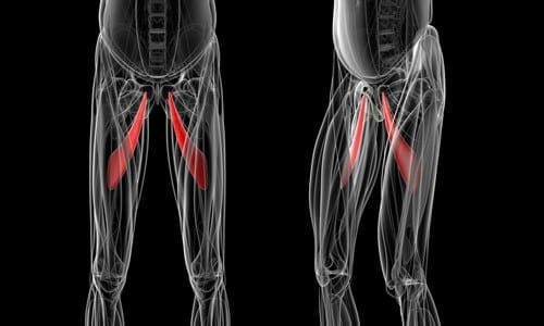
The soft tissues most often affected by athletic pubalgia are the oblique muscles found in the lower abdomen, especially in the tendons that attach the oblique muscles to the pubic bone. In many instances, the joints that connect the thigh muscles to the pubic bone,�known as the adductor muscles, are also stretched or torn as a result of athletic pubalgia.
Physical activities which involve planting the feet and twisting with maximum exertion can cause athletic pubalgia. A sports hernia is most prevalent in vigorous sports, such as hockey, soccer, wrestling, and football. Athletic pubalgia�causes pain and discomfort in the groin region which typically gets better with rest but comes back with physical activity.
A sports�hernia does not result in a visible bulge in the groin, such as the well-known inguinal hernia does. As time passes, athletic pubalgia can lead to an inguinal hernia, and abdominal organs can push against the diminished cells to form a visible bulge. Without treatment, this sports injury could lead to chronic, disabling pain and other symptoms.
Healthcare Professional Diagnosis
During the first consultation, a doctor will discuss the individual’s symptoms and how the injury happened. To�diagnose athletic pubalgia, the healthcare professional will look for tenderness in the groin or above the pubis. Although a sports hernia may be related to an inguinal hernia, the doctor may not find any hernias during a physical examination.
Furthermore, to help determine the presence of athletic pubalgia, the healthcare professional will probably ask the patient to perform a sit-up or to�bend the trunk against resistance. If you have a sports hernia, these tests will be painful. The doctor may also require�x-rays or magnetic resonance imaging (MRI) to help determine whether you have athletic pubalgia.�The scope of our information is limited to chiropractic as well as to spinal injuries and conditions. To discuss the subject matter, please feel free to ask Dr. Jimenez or contact us at�915-850-0900�.
Curated by Dr. Alex Jimenez
Additional Topics: Acute Back Pain
Back pain�is one of the most prevalent causes of disability and missed days at work worldwide. Back pain attributes to the second most common reason for doctor office visits, outnumbered only by upper-respiratory infections. Approximately 80 percent of the population will experience back pain at least once throughout their life. The spine is a complex structure made up of bones, joints, ligaments, and muscles, among other soft tissues. Because of this, injuries and/or aggravated conditions, such as�herniated discs, can eventually lead to symptoms of back pain. Sports injuries or automobile accident injuries are often the most frequent cause of back pain, however, sometimes the simplest of movements can have painful results. Fortunately, alternative treatment options, such as chiropractic care, can help ease back pain through the use of spinal adjustments and manual manipulations, ultimately improving pain relief.
Athletic pubalgia, also known as a hockey hernia,�hockey groin, Gilmore’s Groin,�sports hernia, or groin disruption, is a health issue of the pubic joint. It is a condition characterized by chronic groin pain in athletes and identified by a dilated ring of the inguinal canal. Soccer and ice hockey players are the athletes most commonly affected by athletic pubalgia, and both recreational and professional athletes can be impacted.
Athletic Pubalgia Symptoms
Symptoms of athletic pubalgia�generally manifest as pain following physical activity, most frequently through hip extension, and twisting and turning movements. The painful symptoms usually radiate into the adductor muscle region and the testicles, although it is often difficult for the individual to pinpoint the exact location of the�symptoms. Athletes with athletic pubalgia�experience soreness and stiffness after physical activity.
Any exertion which increases intra-abdominal pressure, such as sneezing or�coughing, as well as physical activity, can lead to pain. While pain in the stomach and pelvis can occur due to a variety of health issues, including injuries to the low back, or lumbar spine, the hip joint, the sacroiliac joint, and the abdomen, along with the genito-urinary system, diagnosis of athletic pubalgia demands skillful differentiation and evaluation.
Clinical Presentation of Athletic Pubalgia
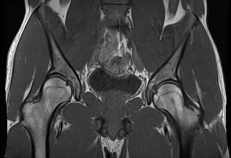
The diagnosis of athletic pubalgia is based on the patient’s history, where healthcare professionals may also depend on the use�of magnetic resonance imaging,�or MRI. Symptoms can frequently be reproduced by certain movements, such as performing crunches or sit-ups. Pain associated with athletic pubalgia may also be elicited with the patient in a “frog posture,” in which the individual is supine with knees bent and heels together.
Many athletes experience concomitant fatigue or tearing of the�adductor muscles or labral tears of the hip. If there is stiffness in the adductor muscles post-injury, painful symptoms can manifest. Alternative treatment options should be to restore normal movement after the adductor has begun to heal, normally 6 to 8 weeks post-injury. Moreover, sleeping in a prone position with the hip on the affected side flexed and externally rotated can offer relief to some athletes with athletic pubalgia.
The precise prevalence of this health issue is unknown. Conservative therapies,�such as gentle stretching, may temporarily alleviate painful symptoms, however, definitive treatment options should be considered for long-term relief.�The scope of our information is limited to chiropractic as well as to spinal injuries and conditions. To discuss the subject matter, please feel free to ask Dr. Jimenez or contact us at�915-850-0900�.
Curated by Dr. Alex Jimenez
Additional Topics: Acute Back Pain
Back pain�is one of the most prevalent causes of disability and missed days at work worldwide. Back pain attributes to the second most common reason for doctor office visits, outnumbered only by upper-respiratory infections. Approximately 80 percent of the population will experience back pain at least once throughout their life. The spine is a complex structure made up of bones, joints, ligaments, and muscles, among other soft tissues. Because of this, injuries and/or aggravated conditions, such as�herniated discs, can eventually lead to symptoms of back pain. Sports injuries or automobile accident injuries are often the most frequent cause of back pain, however, sometimes the simplest of movements can have painful results. Fortunately, alternative treatment options, such as chiropractic care, can help ease back pain through the use of spinal adjustments and manual manipulations, ultimately improving pain relief.
The rectus femoris muscle attaches to the pelvis and just below the knee as it is one of four muscles found at the front part of the thigh. It functions by extending the knee and flexing the hip. The rectus femoris muscle is made up of�fibers which adapt to quick action. Rectus femoris muscle strain is caused by forceful movements, such as kicking a ball or when beginning to sprint, and it is particularly vulnerable to stress and pressure.
Painful symptoms generally manifest at the top of the thigh after the rectus femoris muscle suffers a strain or tear. In severe cases, the health issue may even become noticeable if the tissue is completely ruptured. Fortunately, complete tears are rare. Healthcare professionals will commonly use an MRI scan to diagnose the extent of the sports injury. Proper diagnosis and treatment�are�essential. A rectus femoris muscle strain should not be rushed, as individuals who return-to-sport too soon may suffer re-injury.
Treatment for Rectus Femoris Strain
According to many healthcare professionals, when it comes to sports injuries to the rectus femoris muscle, it’s crucial to immediately apply the RICE principle (Rest, Ice, Compression, and Elevation) to the affected thigh. This treatment aims to decrease bleeding and inflammation to the muscle. Also, it will help reduce painful symptoms after the injury. Based on how much pain has been experienced, simple painkillers might be utilized, although it’s best to attempt to prevent the use of these.
Once movement is restored enough to allow the individual to walk using their regular range of motion, and once the swelling has gone down, then you will have recovered from the acute phase of the injury. It would then be an excellent time to engage in physical activity, without inflicting damage or stress to the quadriceps muscles. This can be performed on an exercise bicycle or through swimming, where the weight is kept�off the limb. Stretches and gentle resistance exercises are crucial, as this will help to align the scar tissue that has formed during the healing process.
Recovery must be monitored so that improvements can be noted and the treatment shifted to help the rehabilitation process. It is hard to measure the length of time to complete recovery. It can take from six to eight weeks or even longer, although some people will commonly recover within one to four weeks.�The scope of our information is limited to chiropractic as well as to spinal injuries and conditions. To discuss the subject matter, please feel free to ask Dr. Jimenez or contact us at�915-850-0900�.
Curated by Dr. Alex Jimenez
Additional Topics: Acute Back Pain
Back pain�is one of the most prevalent causes of disability and missed days at work worldwide. Back pain attributes to the second most common reason for doctor office visits, outnumbered only by upper-respiratory infections. Approximately 80 percent of the population will experience back pain at least once throughout their life. The spine is a complex structure made up of bones, joints, ligaments, and muscles, among other soft tissues. Because of this, injuries and/or aggravated conditions, such as�herniated discs, can eventually lead to symptoms of back pain. Sports injuries or automobile accident injuries are often the most frequent cause of back pain, however, sometimes the simplest of movements can have painful results. Fortunately, alternative treatment options, such as chiropractic care, can help ease back pain through the use of spinal adjustments and manual manipulations, ultimately improving pain relief.
El Paso, TX. Massage therapist, Sandra Rubio has worked with Dr. Alex Jimenez for about six years. Sandra discovered how useful and essential chiropractic care can be by caring for individuals on a routine basis. Sandra explains how Dr. Alex Jimenez provides patients with having a better means of healing themselves, without the use of drugs and medications or even through an operation. The trust between Dr. Jimenez and the patient establishes a favorable treatment outcome for many athletes with sports injuries as well as patients with other types of injuries or conditions via chiropractic care. Sandra Rubio recommends Dr. Alex Jimenez as the non-surgical selection for sports accidents.
El Paso, TX. Sports Injury Chiropractic Treatment
Sprains and strains are some of the most common kind of non-surgical injuries frequently reported by an athlete. Sprains are injuries which affect demanding bands that connect the joints and bones, the ligaments. They can be deformed or tear by the stretching of these outside their range. Strains are injuries that affect the muscle fibers or tendons, which operate by anchoring bones. Seeking prompt medical attention can help these cure faster to allow the athlete to be able to heal more quickly. Although sports injuries are mild or moderate, some treatment options can help treat sports injuries.
We are blessed to present to you�El Paso�s Premier Wellness & Injury Care Clinic.
As El Paso�s Chiropractic Rehabilitation Clinic & Integrated Medicine Center,�we passionately are focused on treating patients after frustrating injuries and chronic pain syndromes. We focus on improving your ability through flexibility, mobility and agility programs tailored for all age groups and disabilities.
If you have enjoyed this video and we have helped you in any way, please feel free to subscribe and recommend�us.
Recommend: Dr. Alex Jimenez � El Paso, TX. Chiropractor
Of course, I’m prone to injuries and Dr. Alex Jimenez has been helping me. I’ve known him for about six years and every time something comes up, either it’s a small injury or a major one, he’s always been there and he’s always helped me get back to my feet to start playing sports again really fast.
Madison Hill
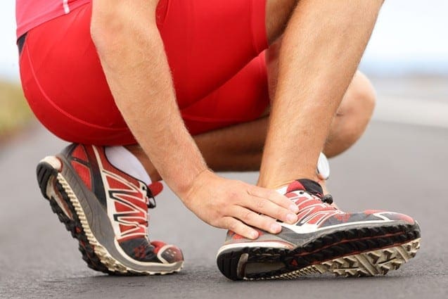
Ankle pain refers to any type of pain or discomfort in the ankle. This pain could generally be due to an injury, such as a sprain, or due to another health issue. As stated by the National University of Health Sciences, or NUHS, an ankle sprain is one of the most frequent causes of foot pain, making up 85 percent of all ankle injuries. A sprain occurs when the ligaments tear or are overstretched.
Most ankle sprains are lateral sprains, which occur when the foot rolls, causing the ankle to twist toward the ground. This action rips or stretches the ligaments, which connect two bones or cartilages and holds a joint together. A sprained ankle often swells and lumps for a temporary amount of time. However, it might take a couple of weeks to get a severe injury like this to�heal completely.
Once healed, the sprained ankle is occasionally permanently weaker and less stable compared to the other ankle. According to a paper released by the American Academy of Family Physicians, or AAFP, the highest risk for ankle sprains includes a previous ankle sprain. Although, ankle sprains are not the only cause of foot pain. Below, we will discuss several common causes of foot and ankle pain as well as their treatment.
Causes of Ankle and Foot Pain
The ankle is a hinge joint formed by the assembly of three bones: the tibia, the fibula, and the talus. The bony knobs on both sides are called the malleoli. Overall, the ankle is an intricate structure. These constructions provide support for walking and standing. Also, stability is provided by the ligaments on the surface of the ankle. Additionally, some tendons also attach to the muscles of the ankle.
Ankle pain may be brought on by various ailments, such as sprain, strain, arthritis, gout, and tendinitis, among others. These kinds of injuries can occur on both sides of the joint. There can be pain and discomfort as well as swelling. A sprain is considered to be the most frequent cause of foot pain. As�mentioned above, a sprain is generally caused when the ankle rolls or twists so the ankle moves toward the ground, tearing or overstretching the ligaments of the ankle that hold the bones together.
An x-ray is typically done to rule out a fracture. The remedy for an ankle strain or sprain generally includes restricting the total amount of weight-bearing on the ankle, getting rest and applying ice. Drugs and/or medications can reduce symptoms. Chiropractic care can also help diagnose and treat ankle sprains and strains. Ankle and foot pain may also be due to:
Arthritis, specifically osteoarthritis,
Gout
Tendinitis
Nerve injury or disease, such as sciatica
Blocked blood vessels
Infection from the joint
While ankle strains and sprains are the most common form of foot pain, arthritis can also frequently lead to ankle pain. Arthritis is the inflammation of the joints, although multiple kinds of arthritis may lead to pain in the joints. Foot pain can be caused by three common forms of arthritis: osteoarthritis, rheumatoid arthritis, and post-traumatic arthritis.
Osteoarthritis is a degenerative condition where the cartilage slowly begins to wear away. Osteoarthritis�causes the natural wear and tear of the joints associated with age. Older adults are more inclined to develop osteoarthritis. In most cases, an individual’s pain and discomfort, including swelling and�stiffness, among other symptoms may worsen over time.
Rheumatoid arthritis is a chronic autoimmune disease. This health issue may severely impact the foot and ankle joints. With rheumatoid arthritis, the human body’s immune cells attack the synovium covering the foot joints. Joint deformity is common with rheumatoid arthritis. A fungal or bacterial infection causes septic arthritis. If the septic arthritis is among the ankle regions, this may result in foot pain.
Following an injury, post-traumatic arthritis can develop from trauma or damage to the ankle or foot. Previous fractures and dislocations are the most common ailments that may lead to post-traumatic arthritis. Like gout, which we will discuss further below, the joints begin to wear away, although it may take several years for this to happen after the injury.
Gout occurs when uric acid accumulates in the human body. This higher than average concentration of uric acid, which is generally a by-product of the human body’s normal breakdown of older cells, can deposit crystals in the joints, causing sharp pain. Pseudogout is a similar illness where calcium deposits build up in the joints. Indicators of gout and pseudogout include soreness, swelling, and redness.
Tendinitis is a swelling of the tendon. In the ankle, it may frequently involve the anterior tibial tendon or the Achilles tendon. Tendinitis can result from an overuse injury or disorders like rheumatoid arthritis and ankylosing spondylitis. All types of tendinitis trigger pain, inflammation, and tenderness. Drugs and/or medications, applying ice and immobilizing the region are often the first line of treatment for tendinitis. Chiropractic care can also be helpful in the treatment of tendinitis. Casting may be required if the patient’s tendinitis is severe or advanced.
�
Foot pain can commonly occur due to ankle injuries. In the United States alone, approximately 2 million acute ankle sprains occur every year, one of the most prevalent causes of ankle pain. Chiropractic care is a popular alternative treatment option which can help treat a variety of health issues, including foot and ankle pain.
Dr. Alex Jimenez D.C., C.C.S.T.
Chiropractic Care for Foot and Ankle Pain
Chiropractors utilize a mixture of treatment techniques and methods to ease ankle and foot pain. Chiropractic care is a safe and effective, alternative treatment option which focuses on the diagnosis, treatment, and prevention of a variety of injuries and conditions associated with the musculoskeletal and nervous system, including foot and ankle pain.
Soft tissue and joint mobilizations are done to restore proper mechanics and muscle activation. Manual therapy may be used to improve the mobility of the ankle and foot along with reducing pain. Furthermore, a chiropractor may recommend a series of lifestyle modifications to help promote a faster recovery process. Exercises are targeted to the areas that were affected. Balance training might also be implemented.
Some treatment modalities that chiropractors utilize to treat injuries to the foot and ankle include ultrasound, electrical stimulation, heat and ice treatment, and massage. These treatment methods increase circulation to enhance recovery, decrease inflammation, reduce pain and improve mobility. When you visit a healthcare professional, a full evaluation is done, goals are discussed along with an individualized treatment program which is intended to target your specific treatment requirements.
Home Treatment for Ankle and Foot Pain
For immediate at-home treatment of foot and ankle pain, the RICE system is generally recommended. The RICE treatment includes:
Rest: Avoid putting weight on the ankle. Try to move as little as possible for the first couple of days. If you have to walk or run, consider using a cane or crutches.
Ice: Begin by putting a bag of ice in your ankle for a minimum of 20 minutes at a time. Repeat this three to five times every day for three days. This�treatment helps decrease pain. Give yourself about 90 minutes between sessions.
Compression: Wrap your injured foot with an elastic bandage, such as an ACE bandage. Don’t wrap it too tightly to where your feet turn blue or your ankle becomes numb.
Elevation: Whenever possible, keep your ankle raised over heart level on a pile of pillows or another type of support arrangement to promote healing.
It’s possible to take over-the-counter drugs and/or medications, such as acetaminophen or ibuprofen, to relieve swelling and pain, however, these are often only offer temporary relief from the symptoms. Make sure to talk to a certified and qualified healthcare professional regarding any home treatment options to prevent further injury and symptoms.
If�you are suffering from foot pain or ankle pain, do not delay anymore. Chiropractors can help patients who suffer from foot, and ankle pain and they can help you, too. The scope of our information is limited to chiropractic as well as to spinal injuries and conditions. To discuss the subject matter, please feel free to ask Dr. Jimenez or contact us at�915-850-0900�.
Curated by Dr. Alex Jimenez
Additional Topics: Acute Back Pain
Back pain�is one of the most prevalent causes of disability and missed days at work worldwide. Back pain is attributed�to the second most common reason for doctor office visits, outnumbered only by upper-respiratory infections. Approximately 80 percent of the population will experience back pain at least once throughout their life. The spine is a complex structure made up of bones, joints, ligaments, and muscles, among other soft tissues. Because of this, injuries and/or aggravated conditions, such as�herniated discs, can eventually lead to symptoms of back pain. Sports injuries or automobile accident injuries are often the most frequent cause of back pain, however, sometimes the simplest of movements can have painful results. Fortunately, alternative treatment options, such as chiropractic care, can help ease back pain through the use of spinal adjustments and manual manipulations, ultimately improving pain relief.
IFM's Find A Practitioner tool is the largest referral network in Functional Medicine, created to help patients locate Functional Medicine practitioners anywhere in the world. IFM Certified Practitioners are listed first in the search results, given their extensive education in Functional Medicine