ClickCease
+1-915-850-0900 spinedoctors@gmail.com
Select Page

Research Studies

Back Clinic Research Studies. Dr. Alex Jimenez has compiled study and research projects that are pertinent to the science and art of chiropractic medicine. The subsets can be classified as following: Case Study, Case Series, Cross-Sectional, Cohort, Case-Control, and Randomized Control Trials. Each subset of study profiles has its merits and scientific significance.

It is our intention to bring clarity to present-day research models. We will discuss and present significant clinical interpretations that may serve outpatients well. Great care in selecting appropriate and well-documented models has been enforced in our blog. We gladly will listen and heed comments on the discussed subject matters presented. For answers to any questions you may have please call Dr. Jimenez at 915-850-0900


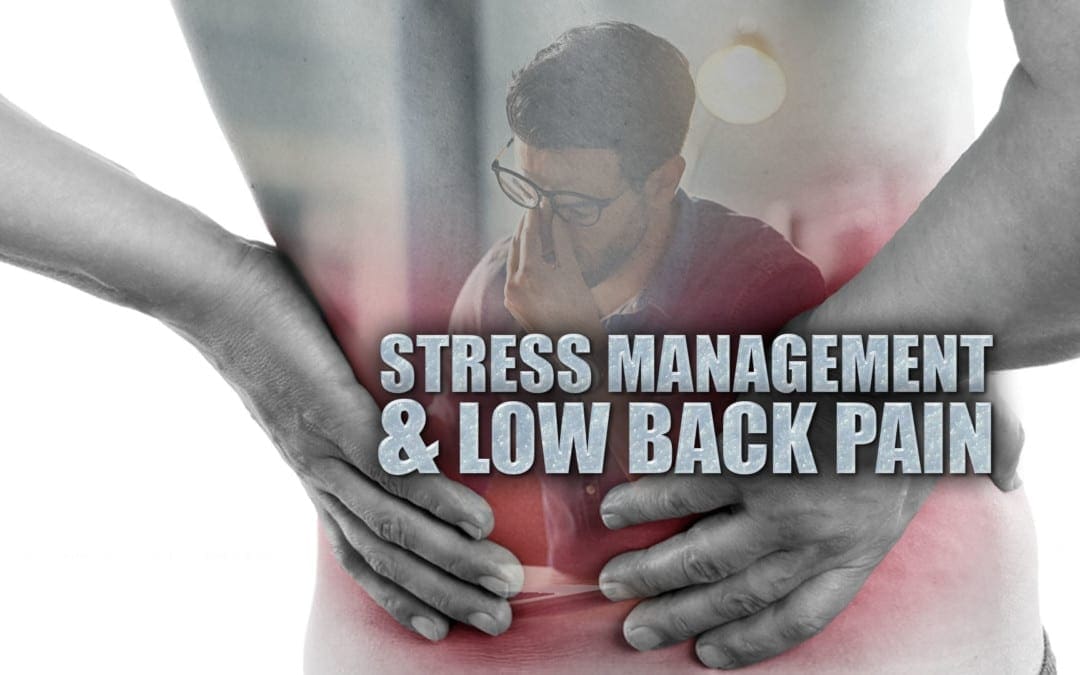
Stress Management & Low Back Pain in El Paso, TX

Stress Management & Low Back Pain in El Paso, TX

People experience stress on a regular basis. From worries about finances or employment to problems with your kids or significant other, even concerns about the condition of the world, can register as stressors for many individuals. Stress causes both acute (immediate) and chronic (long-term) health issues, including low back pain, a common symptom frequently reported by many patients who suffer from constant stress. Fortunately, several holistic treatment approaches, including chiropractic care, can help alleviate both the feelings and effect of stress, ultimately guiding people through proper stress management methods.


Symptoms of Stress


Stress triggers the body’s fight or flight response. The adrenaline surge you experience after hearing a loud sound is simply one of the remaining characteristics of our ancestors, afraid that that loud noise came from something which wanted to eat them.


Stress causes a number of physical changes in the body, starting with the brain. The heart rate increases and starts directing blood to the other extremities. Hearing and eyesight become more acute. And the adrenal glands begin secreting adrenaline as a means of preparing the body for physical exertion. This is exactly what the “flight or fight response” really means.


If you are walking alone at night and hear footsteps behind you, the fight of flight response can be incredibly effective towards your safety. However, if you experience prolonged stress, this sort of physical reaction contributes to a variety of health issues, such as high blood pressure, diabetes, a compromised immune system and muscle tissue damage. That’s because your body doesn’t recognize that there are different kinds of stress; it only knows that stress represents danger and it reacts accordingly.


Stress Management with Chiropractic Care


Chiropractic care can help improve as well as manage many symptoms of stress. This is because the spine is the root of the nervous system. Spinal adjustments and manual manipulations calm the fight or flight response by activating the parasympathetic system. Additionally, chiropractic can relieve pain and muscular tension, improve circulation, and correct spinal misalignments. These benefits all combine to ease the symptoms of stress, which reduces how stressed the patient feels.


A Well-Rounded Strategy


Chiropractors guide their patients through an assortment of stress management procedures, including dietary changes, exercise, meditation, and relaxation methods. A healthy diet can help the body handle an assortment of issues, including stress. Following a diet rich in fruits and vegetables, lean proteins, and complex carbohydrates, with minimal processed and prepackaged foods, can significantly improve overall health and wellness. Exercise is an effective stress reliever. The energy you expend through exercise relieves tension as well as the energy of stress. It also releases endorphins, which help elevate mood. Yoga is an especially effective kind of physical activity for relieving stress.


Meditation can be performed in a variety of ways and it can be practiced by various healthcare professionals. For some, writing in a journal is a kind of meditation, while others are more conventional in their strategy. Many relaxation techniques are closely linked to meditation, such as breathing exercises, releasing muscle tension, and listening to calming music or nature sounds.


  • Breathing exercises are simple and offer immediate stress relief. Begin with inhaling slowly and deeply through your nose, while counting to six and extending your stomach. Hold your breath for a count of four, then release the breath through your mouth, counting to six again. Repeat the cycle for three to five occasions.
  • Release muscle tension through a technique known as “progressive muscle relaxation”. Find a comfortable position, either sitting with your feet on the ground, or lying on your back. Work your way through each muscle group, beginning at your toes or your head, tensing the muscle for a count of five, and then releasing. Wait 30 minutes and then proceed to the next muscle group. Wondering how to tense the muscles of your face? For the face, raise your eyebrows as large as you can and feel the tension in your forehead and scalp. For the central portion of your own face, squint your eyes and wrinkle your nose and mouth. Finally, for the lower face, clench your teeth and pull back the corners of your mouth.
  • Soothing sounds like instrumental music or nature sounds help relax the body and the brain.


Maintaining a balanced lifestyle while also incorporating chiropractic care as a stress management strategy is an effective way to help improve and cope with the symptoms of stress. Reducing stress can ultimately help maintain your overall well-being.


Mindfulness-Based Stress Reduction and Cognitive-Behavioral Therapy for Chronic Low Back Pain: Similar Effects on Mindfulness, Catastrophizing, Self-Efficacy and Acceptance in a Randomized Controlled Trial


Abstract


Cognitive-behavioral therapy (CBT) is believed to improve chronic pain problems by decreasing patient catastrophizing and increasing patient self-efficacy for managing pain. Mindfulness-based stress reduction (MBSR) is believed to benefit chronic pain patients by increasing mindfulness and pain acceptance. However, little is known about how these therapeutic mechanism variables relate to each other or whether they are differentially impacted by MBSR versus CBT. In a randomized controlled trial comparing MBSR, CBT, and usual care (UC) for adults aged 20-70 years with chronic low back pain (CLBP) (N = 342), we examined (1) baseline relationships among measures of catastrophizing, self-efficacy, acceptance, and mindfulness; and (2) changes on these measures in the 3 treatment groups. At baseline, catastrophizing was associated negatively with self-efficacy, acceptance, and 3 aspects of mindfulness (non-reactivity, non-judging, and acting with awareness; all P-values <0.01). Acceptance was associated positively with self-efficacy (P < 0.01) and mindfulness (P-values < 0.05) measures. Catastrophizing decreased slightly more post-treatment with MBSR than with CBT or UC (omnibus P = 0.002). Both treatments were effective compared with UC in decreasing catastrophizing at 52 weeks (omnibus P = 0.001). In both the entire randomized sample and the sub-sample of participants who attended ?6 of the 8 MBSR or CBT sessions, differences between MBSR and CBT at up to 52 weeks were few, small in size, and of questionable clinical meaningfulness. The results indicate overlap across measures of catastrophizing, self-efficacy, acceptance, and mindfulness, and similar effects of MBSR and CBT on these measures among individuals with CLBP.


Keywords: chronic back pain, self-efficacy, mindfulness, acceptance, catastrophizing, CBT, MBSR


Introduction


Cognitive-behavioral therapy (CBT) has been demonstrated effective, and is widely recommended, for chronic pain problems.[20] Mindfulness-based interventions (MBIs) also show promise for patients with chronic pain[12,14,25,44,65] and their use by this population is increasing. Understanding the mechanisms of action of psychosocial treatments for chronic pain and commonalities in these mechanisms across different therapies is critical to improving the effectiveness and efficiency of these treatments.[27,52] Key mechanisms of action of CBT for chronic pain include decreased catastrophizing and increased self-efficacy for managing pain.[6-8,56] Increased mindfulness is considered a central mechanism of change in MBIs,[14,26,30] which also increase pain acceptance.[16,21,27,38,59] However, little is known about the associations among pain catastrophizing, self-efficacy, acceptance, and mindfulness prior to psychosocial treatment or about differences in effects of CBT versus MBIs on these variables.


There is some evidence suggesting significant associations among these therapeutic mechanism variables. Evidence regarding relationships between catastrophizing and mindfulness is mixed. Some studies[10,18,46] have found negative associations between measures of pain catastrophizing and mindfulness. However, others found no significant relationship[19] or associations (inverse) between catastrophizing and some aspects of mindfulness (non-judging, non-reactivity, and acting with awareness) but not others (e.g., observing).[18] Catastrophizing has also been reported to be associated negatively with pain acceptance.[15,22,60] In a pain clinic sample, general acceptance of psychological experiences was associated negatively with catastrophizing and positively with mindfulness.[19] Pain self-efficacy has been observed to be correlated positively with acceptance and negatively with catastrophizing.[22]


Further suggesting overlap across mechanisms of different psychosocial treatments for chronic pain, increases in mindfulness[10] and acceptance[1,64] have been found after cognitive-behavioral pain treatments, and reductions in catastrophizing have been observed after mindfulness-based pain management programs.[17,24,37] Little research has examined effects of MBIs for chronic pain on self-efficacy, although a small pilot study of migraine patients found greater increases in self-efficacy with Mindfulness-Based Stress Reduction (MBSR) training than with usual care.[63] We were unable to identify any studies of the relationships among all these therapeutic mechanism variables or of changes in all these variables with CBT versus an MBI for chronic pain.


The aim of this study was to replicate and extend prior research by using data from a randomized controlled trial (RCT) comparing MBSR, CBT, and usual care (UC) for chronic low back pain (CLBP)[12] to examine: (1) baseline relationships among measures of mindfulness and pain catastrophizing, self-efficacy, and acceptance; and (2) short- and long-term changes on these measures in the 3 treatment groups. Based on theory and previous research, we hypothesized that: (1) at baseline, catastrophizing would be inversely related to acceptance, self-efficacy, and 3 dimensions of mindfulness (non-reactivity, non-judging, acting with awareness), but not associated with the observing dimension of mindfulness; (2) at baseline, acceptance would be associated positively with self-efficacy; and (3) from baseline to 26 and 52 weeks, acceptance and mindfulness would increase more with MBSR than with CBT and UC, and catastrophizing would decrease more and self-efficacy would increase more with CBT than with MBSR and UC.


Methods


Setting, Participants and Procedures


Study participants were enrolled in an RCT comparing group MBSR, group CBT, and UC for non-specific chronic back pain between September 2012 and April 2014. We previously reported details of the study methods,[13] Consolidated Standards of Reporting Trials (CONSORT) flow diagram,[12] and outcomes.[12] In brief, participants were recruited from Group Health, an integrated healthcare system in Washington State, and from mailings to residents of communities served by Group Health. Eligibility criteria included age 20 – 70 years, back pain for at least 3 months, patient-rated bothersomeness of pain during the previous week ?4 (0 – 10 scale), and patient-rated pain interference with activities during the previous week ?3 (0 – 10 scale). We used International Classification of Diseases, Ninth Revision, Clinical Modification (ICD-9-CM)43 diagnostic codes from electronic medical records (EMR) of visits in the previous year and telephone screening to exclude patients with specific causes of low back pain. Exclusion criteria also included pregnancy, spine surgery in the previous 2 years, disability compensation or litigation, fibromyalgia or cancer diagnosis, other major medical condition, plans to see a medical specialist for back pain, inability to read or speak English, and participation in a �mind-body� treatment for back pain in the past year. Potential participants were told that they would be randomized to one of �two different widely-used pain self-management programs that have been found helpful for reducing pain and making it easier to carry out daily activities� or to continued usual care. Those assigned to MBSR or CBT were unaware of the specific treatment they would receive until the first intervention session. The study was approved by the Group Health institutional review board and all participants provided informed consent.


Participants were randomized to the MBSR, CBT, or UC conditions. Randomization was stratified based on the baseline value of the primary outcome, a modified version of the Roland Disability Questionnaire (RDQ),[42] into 2 back pain-related physical limitation stratification groups: moderate (RDQ score ?12 on the 0 – 23 scale) and high (RDQ scores ?13). To mitigate possible disappointment with not being randomized to CBT or MBSR, participants randomized to UC received $50 compensation. Data were collected from participants in computer-assisted telephone interviews by trained survey staff. All participants were paid $20 for each interview completed.


Measures


Participants provided descriptive information at the screening and baseline interviews, and completed the study measures at baseline (before randomization) and 8 (post-treatment), 26 (the primary study endpoint), and 52 weeks post-randomization. Participants also completed a subset of the measures at 4 weeks, but these data were not examined for the current report.


Descriptive Measures and Covariates


The screening and baseline interviews assessed, among other variables not analyzed for the present study, sociodemographic characteristics (age, gender, race, ethnicity, education, work status); pain duration (defined as length of time since a period of 1 or more weeks without low back pain); and number of days with back pain in the past 6 months. In this report, we describe the sample at baseline on these measures and on the primary outcome measures in the RCT: the modified Roland-Morris Disability Questionnaire (RDQ)[42] and a numerical rating of back pain bothersomeness. The RDQ, a widely-used measure of back pain-related functional limitations, asks whether 24 specific activities are limited today by back pain (yes or no).[45] We used a modified version that included 23 items[42] and asked about the previous week rather than today only. Back pain bothersomeness was measured by participants� ratings of how bothersome their back pain was during the previous week on a 0 to 10 numerical rating scale (0 = �not at all bothersome� and 10 = �extremely bothersome�). The covariates for the current report were the same as those in our prior analyses of the interventions� effects on the outcomes:[12] age, gender, education, and pain duration (less than one year versus at least one year since experiencing 1 week without low back pain). We decided a priori to control for these variables because of their potential to affect the therapeutic mechanism measures, participant response to treatment, and/or likelihood of obtaining follow-up information.


Measures of Potential Therapeutic Mechanisms


Mindfulness. Mindfulness has been defined as the awareness that emerges through purposeful, non-judgmental attention to the present moment.[29] We administered 4 subscales of the Five Facet Mindfulness Questionnaire-Short Form (FFMQ-SF):[5] Observing (noticing internal and external experiences; 4 items); Acting with Awareness (attending to present moment activities, as contrasted to behaving automatically while attention is focused elsewhere; 5 items); Non-reactivity (non-reactivity to inner experiences: allowing thoughts and feelings to arise and pass away without attachment or aversion; 5 items); and Non-judging (non-judging of inner experiences: engaging in a non-evaluative stance towards thoughts, emotions, and feelings; 5-item scale; however, one question [�I make judgments about whether my thoughts are good or bad�] inadvertently was not asked.). The FFMQ-SF has been demonstrated to be reliable, valid, and sensitive to change.[5] Participants rated their opinion of what generally is true for them in terms of their tendency to be mindful in their daily lives (scale from 1 = �never or very rarely true� to 5 = �very often or always true�). For each scale, the score was calculated as the mean of the answered items and thus the possible range was 1-5, with higher scores indicating higher levels of the mindfulness dimension. Prior studies have used sum scores rather than means, but we elected to use mean scores given the greater ease of interpretation.


Pain catastrophizing. The Pain Catastrophizing Scale (PCS) is a 13-item measure assessing pain-related catastrophizing, including rumination, magnification, and helplessness.[50] Participants rated the degree to which they had certain thoughts and feelings when experiencing pain (scale from 0 = �not at all� to 4 = �all the time�). Item responses were summed to yield a total score (possible range = 0-52). Higher scores indicate greater endorsement of catastrophic thinking in response to pain.


Pain acceptance. The Chronic Pain Acceptance Questionnaire-8 (CPAQ-8), an 8-item version of the 20-item Chronic Pain Acceptance Questionnaire (CPAQ), has been shown to be reliable and valid.[22,23] It has 2 scales: Activity Engagement (AE; engagement in life activities in a normal manner even while pain is being experienced) and Pain Willingness (PW; disengagement from attempts to control or avoid pain). Participants rated items on a scale from 0 (�never true�) to 6 (�always true�). Item responses were summed to create scores for each subscale (possible range 0-24) and the overall questionnaire (possible range 0-48). Higher scores indicate greater activity engagement/pain willingness/pain acceptance. Prior research suggests that the 2 subscales are moderately correlated and that each makes an independent contribution to the prediction of adjustment in people with chronic pain.[22]


Pain self-efficacy. The Pain Self-efficacy Questionnaire (PSEQ) consists of 10 items assessing individuals� confidence in their ability to cope with their pain and engage in activities despite their pain, each rated on a scale from 0 = �not at all confident� to 6 = �completely confident.�[39] The questionnaire has been demonstrated to be valid, reliable, and sensitive to change.[39] Item scores are summed to yield a total score (possible range 0-60); higher scores indicate greater self-efficacy.


Interventions


The 2 interventions were comparable in format (group), duration, frequency, and number of participants per group cohort. Both the MBSR and CBT interventions consisted of 8 weekly 2-hour sessions supplemented by home activities. For each intervention, we developed a therapist/instructor’s manual and participant’s workbook, both with structured and detailed content for each session. In each intervention, participants were assigned home activities and there was emphasis on incorporating intervention content in their daily lives. Participants were given materials to read at home and CDs with relevant content for home practice (e.g., meditation, body scan, and yoga in MBSR; relaxation and imagery exercises in CBT). We previously published detailed descriptions of both interventions,[12,13] but describe them briefly here.


MBSR


The MBSR intervention was modeled closely after the original program developed by Kabat-Zinn[28] and based on the 2009 MBSR instructor’s manual.[4] It consisted of 8 weekly sessions and an optional 6-hour retreat between the 6th and 7th sessions. The protocol included experiential training in mindfulness meditation and mindful yoga. All sessions included mindfulness exercises (e.g., body scan, sitting meditation) and mindful movement (most commonly, yoga).


CBT


The group CBT protocol included the techniques most commonly applied in CBT for CLBP[20,58] and used in prior studies.[11,33,41,51,53-55,57,61] The intervention included: (1) education about (a) chronic pain, (b) maladaptive thoughts (including catastrophizing) and beliefs (e.g., inability to control pain, hurt equals harm) common among individuals with chronic pain, (c) the relationships between thoughts and emotional and physical reactions, (d) sleep hygiene, and (e) relapse prevention and maintenance of gains; and (2) instruction and practice in identifying and challenging unhelpful thoughts, generating alternative appraisals that are more accurate and helpful, setting and working towards behavioral goals, abdominal breathing and progressive muscle relaxation techniques, activity pacing, thought-stopping and distraction techniques, positive coping self-statements, and coping with pain flare-ups. None of these techniques were included in the MBSR intervention, and mindfulness, meditation, and yoga techniques were not included in CBT. CBT participants were also given a book (The Pain Survival Guide[53]) and asked to read specific chapters between sessions. During each session, participants completed a personal action plan for activities to do between sessions.


Usual Care


Patients assigned to UC received no MBSR training or CBT as part of the study and received whatever health care they would customarily receive during the study period.


Instructors/Therapists and Treatment Fidelity Monitoring


As previously reported,[12] all 8 MBSR instructors received formal training in teaching MBSR from the Center for Mindfulness at the University of Massachusetts or equivalent training and had extensive previous experience teaching MBSR. The CBT intervention was conducted by 4 Ph.D.-level licensed psychologists with previous experience providing individual and group CBT to patients with chronic pain. Details of instructor training and supervision and treatment fidelity monitoring were provided previously.[12]


Statistical Analyses


We used descriptive statistics to summarize the observed baseline characteristics by randomization group, separately for the entire randomized sample and the subsample of participants who attended 6 or more of the 8 intervention classes (MBSR and CBT groups only). To examine the associations between the therapeutic mechanism measures at baseline, we calculated Spearman rho correlations for each pair of measures.


To estimate changes over time in the therapeutic mechanism variables, we constructed linear regression models with the change from baseline as the dependent variable, and included all post-treatment time points (8, 26, and 52 weeks) in the same model. A separate model was estimated for each therapeutic mechanism measure. Consistent with our approach for analyzing outcomes in the RCT,[12] we adjusted for age, gender, education, and baseline values of pain duration, pain bothersomeness, the modified RDQ, and the therapeutic mechanism measure of interest in that model. To estimate the treatment effect (difference between groups in change on the therapeutic mechanism measure) at each time point, the models included main effects for treatment group (CBT, MBSR, and UC) and time point (8, 26, and 52 weeks), and terms for the interactions between these variables. We used generalized estimating equations (GEE)[67] to fit the regression models, accounting for possible correlation between repeated measures from individual participants. To account for potential bias caused by differential attrition across treatment groups, our primary analysis used a 2-step GEE modeling approach to impute missing data on the therapeutic mechanism measures. This approach uses a pattern mixture model framework for non-ignorable non-response and adjusts the variance estimates in the final outcome model parameters to account for using imputed data.[62] We also, as a sensitivity analysis, conducted the regression analyses again with observed rather than imputed data to evaluate whether using imputed data had a substantial effect on the results and to allow direct comparison to other published studies.


The primary analysis included all randomized participants, using an intent-to-treat (ITT) approach. We repeated the regression analyses using the subsample of participants who were randomized to MBSR or CBT and who attended at least 6 of the 8 sessions of their assigned treatment (�as-treated� or �per protocol� analysis). For descriptive purposes, using regression models for the ITT sample with imputed data, we estimated mean scores (and their 95% confidence intervals [CI]) on the therapeutic mechanism variables at each time point adjusted for age, gender, education, and baseline values of pain duration, pain bothersomeness, and the modified RDQ.


To provide context for interpreting the results, we used t-tests and chi-square tests to compare the baseline characteristics of participants who did versus did not complete at least 6 of the 8 intervention sessions (MBSR and CBT groups combined). We compared intervention participation by group, using a chi-square test to compare the proportions of participants randomized to MBSR versus CBT who completed at least 6 of the 8 sessions.


Dr. Alex Jimenez’s Insight

Stress is primarily a part of the “fight or flight” response which helps the body effectively prepare for danger. When the body enters�a state of mental or emotional strain or tension due to adverse or very demanding circumstances,�a complex mix of hormones and chemicals, such as adrenaline, cortisol and norepinephrine, are secreted in order to prepare the body for physical and psychological action.�While short-term stress provides us with the necessary amount of edge required to improve our overall performance, long-term stress has been associated to a variety of health issues, including low back pain and sciatica. Stress management methods and techniques, including meditation and chiropractic care, have been demonstrated to help improve treatment outcomes of low back pain and sciatica. The following article discusses several types of stress management treatments and describes their effect on overall health and wellness.


Results


Characteristics of the Study Sample


As previously reported,[12] among 1,767 individuals who expressed interest in the study and were screened for eligibility, 1,425 were excluded (most commonly due to pain not present for more than 3 months and inability to attend the intervention sessions). The remaining 342 individuals enrolled and were randomized. Among the 342 individuals randomized, 298 (87.1%), 294 (86.0%), and 290 (84.8%) completed the 8-, 26-, and 52-week assessments, respectively.


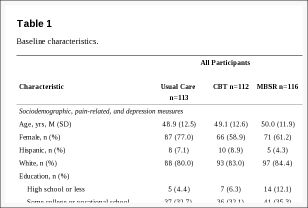
Table 1 shows the characteristics of the sample at baseline. Among all participants, the mean age was 49 years, 66% were female, and 79% reported having had back pain for at least one year without a pain-free week. On average, PHQ-8 scores were at the threshold for mild depressive symptom severity.[32] Mean scores on the Pain Catastrophizing Scale (16-18) were below the various cut-points suggested for clinically relevant catastrophizing (e.g., 24,47 3049). Pain Self-Efficacy Scale scores were somewhat higher on average (about 5 points on the 0-60 scale) in our sample as compared with the primary care patients with low back pain enrolled in an RCT evaluating group CBT in England,[33] and about 15 points higher than among individuals with chronic pain attending a mindfulness-based pain management program in England.[17]


Table 1 Baseline Characteristics


About half of participants randomized to MBSR (50.9%) or CBT (56.3%) attended at least 6 sessions of their assigned treatment; the difference between treatments was not statistically significant (chi-square test, P = 0.42). At baseline, those randomized to MBSR and CBT who completed at least 6 sessions, as compared to those who did not, were significantly older (mean [SD] = 52.2 [10.9] versus 46.5 [13.0] years) and reported significantly lower levels of pain bothersomeness (mean [SD] = 5.7 [1.3] versus 6.4 [1.7]), disability (mean [SD] RDQ = 10.8 [4.5] versus 12.7 [5.0]), depression (mean [SD] PHQ-8 = 5.2 [4.1] versus 6.3 [4.3]), and catastrophizing (mean [SD] PCS = 15.9 [10.3] versus 18.9 [9.8]), and significantly greater pain self-efficacy (mean [SD] PSEQ = 47.8 [8.3] versus 43.2 [10.3]) and pain acceptance (CPAQ-8 total score mean [SD] = 31.3 [6.2] versus 29.0 [6.7]; CPAQ-8 Pain Willingness mean [SD] = 12.3 [4.1] versus 10.9 [4.8]) (all P-values < 0.05). They did not differ significantly on any other variable shown in Table 1.


Baseline Associations Between Therapeutic Mechanism Measures


Table 2 shows the Spearman correlations between the therapeutic mechanism measures at baseline. Our hypotheses about the baseline relationships among these measures were confirmed. Catastrophizing was correlated negatively with 3 dimensions of mindfulness (non-reactivity rho = ?0.23, non-judging rho = ?0.30, and acting with awareness rho = ?0.21; all P-values < 0.01), but not associated with the observing dimension of mindfulness (rho = ?0.01). Catastrophizing was also correlated negatively with acceptance (total CPAQ-8 score rho = ?0.55, Pain Willingness subscale rho = ?0.47, Activity Engagement subscale rho = ?0.40) and pain self-efficacy (rho = ?0.57) (all P-values < 0.01). Finally, pain self-efficacy was correlated positively with pain acceptance (total CPAQ-8 score rho = 0.65, Pain Willingness subscale rho = 0.46, Activity Engagement subscale rho = 0.58; all P-values < 0.01).


Table 2 Spearman rho Correlations


Treatment Group Differences in Changes on Therapeutic Mechanism Measures Among all Randomized Participants


Table 3 shows the adjusted mean changes from baseline in each study group and the adjusted mean differences between treatment groups on the therapeutic mechanism measures at each follow-up in the entire randomized sample. Figure 1 shows the adjusted mean PCS scores for each group at each time point. Contrary to our hypothesis that catastrophizing would decrease more with CBT than with MBSR, catastrophizing (PCS score) decreased significantly more from pre- to post-treatment in the MBSR group than in the CBT group (MBSR versus CBT adjusted mean [95% CI] difference in change = ?1.81 [?3.60, ?0.01]). Catastrophizing also decreased significantly more in MBSR than in UC (MBSR versus UC adjusted mean [95% CI] difference in change = ?3.30 [?5.11, ?1.50]), whereas the difference between CBT and UC was not significant. At 26 weeks, the treatment groups did not differ significantly in change in catastrophizing from baseline. However, at 52 weeks, both the MBSR and the CBT groups showed significantly greater decreases than did the UC group, and there was no significant difference between MBSR and CBT.
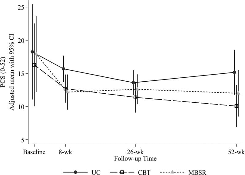
 

Figure 1 Adjusted Mean PCS Scores

Figure 1: Adjusted mean Pain Catastrophizing Scale (PCS) scores (and 95% confidence intervals) at baseline (pre-randomization), 8 weeks (post-treatment), 26 weeks, and 52 weeks for participants randomized to CBT, MBSR, and UC. Estimated means are adjusted for participant age, gender, education, whether or not at least 1 year since week without pain, and baseline RDQ and pain bothersomeness.


Table 3 Adjusted Mean Change from Baseline and Adjusted Mean Differences


Figure 2 shows the adjusted mean PSEQ scores for each group at each time point. Our hypothesis that self-efficacy would increase more with CBT than with MBSR and with UC was only partially confirmed. Self-efficacy (PSEQ scores) did increase significantly more from pre- to post-treatment with CBT than with UC, but not with CBT relative to the MBSR group, which also increased significantly more than did the UC group (adjusted mean [95% CI] difference in change on PSEQ from baseline for CBT versus UC = 2.69 [0.96, 4.42]; CBT versus MBSR = 0.34 [?1.43, 2.10]; MBSR versus UC = 3.03 [1.23, 4.82]) (Table 3). The omnibus test for differences across groups in self-efficacy change was not significant at 26 or 52 weeks.
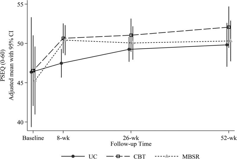
 

Figure 2 Adjusted Mean PSEQ Scores

Figure 2: Adjusted mean Pain Self-Efficacy Questionnaire (PSEQ) scores (and 95% confidence intervals) at baseline (pre-randomization), 8 weeks (post-treatment), 26 weeks, and 52 weeks for participants randomized to CBT, MBSR, and UC. Estimated means are adjusted for participant age, gender, education, whether or not at least 1 year since week without pain, and baseline RDQ and pain bothersomeness.


Our hypothesis that acceptance would increase more with MBSR than with CBT and with UC was generally not confirmed. The omnibus test for differences across groups was not significant for the total CPAQ-8 or the Activity Engagement subscale at any time point (Table 3). The test for the Pain Willingness subscale was significant at 52 weeks only, when both the MBSR and CBT groups showed greater increases as compared with UC, but not as compared with each other (adjusted mean [95% CI] difference in change for MBSR versus UC = 1.15 [0.05, 2.24]; CBT versus UC = 1.23 [0.16, 2.30]).


Our hypothesis that mindfulness would increase more with MBSR than with CBT was partially confirmed. Both the MBSR and CBT groups showed greater increases as compared with UC on the FFMQ-SF Non-reactivity scale at 8 weeks (MBSR versus UC = 0.18 [0.01, 0.36]; CBT versus UC = 0.28 [0.10, 0.46]), but differences at later follow-ups were not statistically significant (Table 3, Figure 3). There was a significantly greater increase on the Non-judging scale with MBSR versus CBT (adjusted mean [95% CI] difference in change = 0.29 [0.12, 0.46]) as well as between MBSR and UC (0.32 [0.13, 0.50]) at 8 weeks, but no significant difference between groups at later time points (Figure 4). The omnibus test for differences among groups was not significant for the Acting with Awareness or Observing scales at any time point.
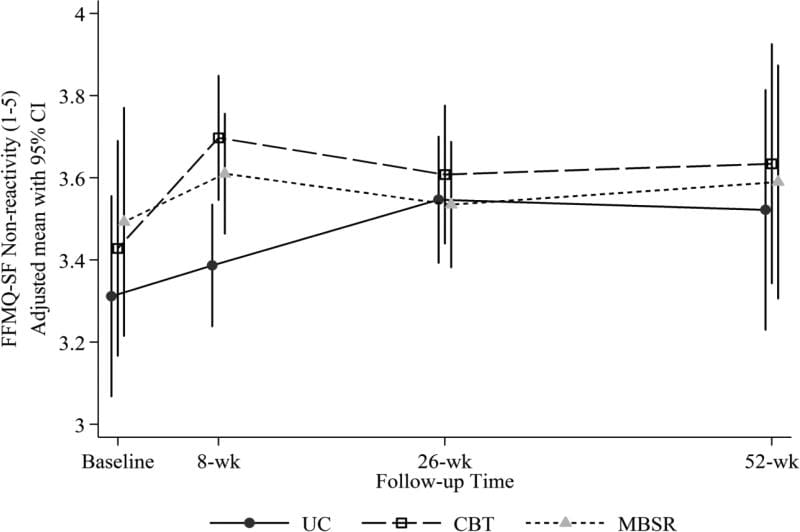
 

Figure 3 Adjusted Mean FFMQ-SF Non Reactivity Scores

Figure 3: Adjusted mean Five Facet Mindfulness Questionnaire-Short Form (FFMQ-SF) Non-reactivity scores (and 95% confidence intervals) at baseline (pre-randomization), 8 weeks (post-treatment), 26 weeks, and 52 weeks for participants randomized to CBT, MBSR, and UC. Estimated means are adjusted for participant age, gender, education, whether or not at least 1 year since week without pain, and baseline RDQ and pain bothersomeness.
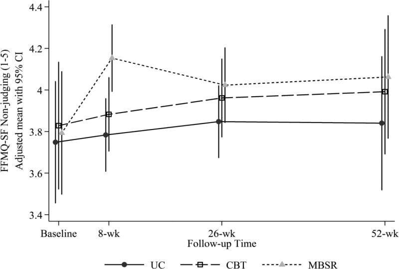
 

Figure 4 Adjusted Mean FFMQ-SF Non Judging Scores

Figure 4: Adjusted mean Five Facet Mindfulness Questionnaire-Short Form (FFMQ-SF) Non-judging scores (and 95% confidence intervals) at baseline (pre-randomization), 8 weeks (post-treatment), 26 weeks, and 52 weeks for participants randomized to CBT, MBSR, and UC. Estimated means are adjusted for participant age, gender, education, whether or not at least 1 year since week without pain, and baseline RDQ and pain bothersomeness.


The sensitivity analyses using observed rather than imputed data yielded almost identical results, with 2 minor exceptions. The difference between MBSR and CBT in change in catastrophizing at 8 weeks, although similar in magnitude, was no longer statistically significant due to slight confidence interval changes. Second, the omnibus test for the CPAQ-8 Pain Willingness scale at 52 weeks was no longer statistically significant (P = 0.07).


Treatment Group Differences in Changes on Therapeutic Mechanism Measures Among Participants Randomized to CBT or MBSR Who Completed at Least 6 Sessions


Table 4 shows the adjusted mean change from baseline and adjusted mean between-group differences on the therapeutic mechanism measures at 8, 26, and 52 weeks for participants who were randomized to MBSR or CBT and completed 6 or more sessions of their assigned treatment. The differences between MBSR and CBT were similar in size to those in the ITT sample. There were only a few differences in statistical significance of the comparisons. In contrast to the results using the ITT sample, the difference between MBSR and CBT in catastrophizing (PCS) at 8 weeks was no longer statistically significant and at 52 weeks, the CBT group increased significantly more than did the MBSR group on the FFMQ-SF Observing scale (adjusted mean difference in change from baseline for MBSR versus CBT = ?0.30 [?0.53, ?0.07]). The sensitivity analyses using observed rather than imputed data yielded no meaningful differences in results.


Table 4 Adjusted Mean Change from Baseline and Adjusted Mean Differences


Discussion


In this analysis of data from an RCT comparing MBSR, CBT, and UC for CLBP, our hypotheses that MBSR and CBT would differentially affect measures of constructs believed to be therapeutic mechanisms generally were not confirmed. For example, our hypothesis that mindfulness would increase more with MBSR than with CBT was confirmed for only 1 of 4 measured facets of mindfulness (non-judging). Another facet, acting with awareness, increased more with CBT than with MBSR at 26 weeks. Both differences were small. Increased mindfulness after a CBT-based multidisciplinary pain program[10] was reported previously; our findings further support a view that both MBSR and CBT increase mindfulness in the short-term. We found no long-term effects of either treatment relative to UC on mindfulness.


Also contrary to hypothesis, catastrophizing decreased more post-treatment with MBSR than with CBT. However, the difference between treatments was small and not statistically significant at later follow-ups. Both treatments were effective compared with UC in decreasing catastrophizing at 52 weeks. Although previous studies demonstrated reductions in catastrophizing after both CBT[35,48,56,57] and mindfulness-based pain management programs,[17,24,37] ours is the first to demonstrate similar decreases for both treatments, with effects up to 1 year.


Increased self-efficacy has been shown to be associated with improvements in pain intensity and functioning,[6] and an important mediator of CBT benefits.[56] However, contrary to our hypothesis, pain self-efficacy did not increase more with CBT than with MBSR at any time point. Compared with UC, there were significantly greater increases in self-efficacy with both MBSR and CBT post-treatment. These results mirror previous findings of positive effects of CBT, including group CBT for back pain,[33] on self-efficacy.[3,56,57] Little research has examined self-efficacy changes after MBIs for chronic pain, although self-efficacy increased more with MBSR than with usual care for patients with migraines in a pilot study[63] and more with MBSR than with health education for CLBP in an RCT.[37] Our findings add to knowledge in this area by indicating that MBSR has short-term benefits for pain self-efficacy similar to those of CBT.


Prior uncontrolled studies found equivalent increases in pain acceptance after group CBT and Acceptance and Commitment Therapy64 (which, unlike traditional CBT, specifically fosters pain acceptance), and increased acceptance after CBT-based multidisciplinary pain treatment.[1,2] In our RCT, acceptance increased in all groups over time, with only 1 statistically significant difference among the 3 groups across the 3 acceptance measures and 3 follow-up time points (a greater increase with both MBSR and CBT than with UC on the Pain Willingness subscale at 52 weeks). This suggests that acceptance may increase over time regardless of treatment, although this needs to be confirmed in additional research.


Two possibilities could explain our previously-reported findings of generally similar effectiveness of MBSR and CBT for CLBP:[12] (1) the treatment effects on outcomes were due to different, but equally effective, therapeutic mechanisms, or (2) the treatments had similar effects on the same therapeutic mechanisms. Our current findings support the latter view. Both treatments may improve pain, function, and other outcomes through different strategies that decrease individuals� views of their pain as threatening and disruptive and encourage activity participation despite pain. MBSR and CBT differ in content, but both include relaxation techniques (e.g., progressive muscle relaxation in CBT, meditation in MBSR, breathing techniques in both) and strategies to decrease the threat value of pain (education and cognitive restructuring in CBT, accepting experiences without reactivity or judgment in MBSR). Thus, although CBT emphasizes learning skills for managing pain and decreasing negative emotional responses, and MBSR emphasizes mindfulness and meditation, both treatments may help patients relax, react less negatively to pain, and view thoughts as mental processes rather than as accurate representations of reality, thereby resulting in decreased emotional distress, activity avoidance, and pain bothersomeness.


Our analyses also revealed overlap among measures of different constructs believed to mediate the effects of MBSR and CBT on chronic pain outcomes. As hypothesized, prior to treatment, pain catastrophizing was associated negatively with pain self-efficacy, pain acceptance, and 3 dimensions of mindfulness (non-reactivity, non-judging, and acting with awareness), and pain acceptance was associated positively with pain self-efficacy. Pain acceptance and self-efficacy were also associated positively with measures of mindfulness. Our results are consistent with prior observations of negative associations between measures of catastrophizing and acceptance,[15,19,60] negative correlations between measures of catastrophizing and mindfulness,[10,46,18] and positive associations between measures of pain acceptance and mindfulness.[19]


As a group, to the extent that these measures reflect their intended constructs, these findings support a view of catastrophizing as inversely associated with two related constructs that reflect participation in customary activities despite pain but differ in emphasis on disengagement from attempts to control pain: pain acceptance (disengagement from attempts to control pain and participation in activities despite pain) and self-efficacy (confidence in ability to manage pain and participate in customary activities). The similarity of some questionnaire items further supports this view and likely contributes to the observed associations. For example, both the CPAQ-8 and the PSEQ contain items about doing normal activities despite pain. Furthermore, there is an empirical and conceptual basis for a view of catastrophizing (focus on pain with highly negative cognitive and affective responses) as also inversely associated with mindfulness (i.e., awareness of stimuli without judgment or reactivity), and for viewing mindfulness as consistent with, but distinct from, acceptance and self-efficacy. Further work is needed to clarify the relationships between these theoretical constructs and the extent to which their measures assess (a) constructs that are related but theoretically and clinically distinct versus (b) different aspects of an overarching theoretical construct.


It remains possible that MBSR and CBT differentially affect important mediators not assessed in this study. Our results highlight the need for further research to more definitively identify the mediators of the effects of MBSR and CBT on different pain outcomes, develop measures that assess these mediators most comprehensively and efficiently, better understand the relationships among therapeutic mechanism variables in affecting outcomes (e.g., decreased catastrophizing may mediate the effect of mindfulness on disability[10]), and refine psychosocial treatments to more effectively and efficiently impact these mediators. Research is also needed to identify patient characteristics associated with response to different psychosocial interventions for chronic pain.


Several study limitations warrant discussion. Participants had low baseline levels of psychosocial distress (e.g., catastrophizing, depression) and we studied group CBT, which has demonstrated efficacy,[33,40,55] resource-efficiency, and potential social benefits, but which may be less effective than individual CBT.[36,66] The results may not generalize to more distressed populations (e.g., pain clinic patients), which would have more room to improve on measures of maladaptive functioning and greater potential for treatments to differentially affect these measures, or to comparisons of MBSR with individual CBT.


Only somewhat over half of the participants randomized to MBSR or CBT attended at least 6 of the 8 sessions. Results could differ in studies with higher rates of treatment adherence; however, our results in �as-treated� analyses generally mirrored those of ITT analyses. Treatment adherence has been shown to be associated with benefits from both CBT for chronic back pain[31] and MBSR.[9] Research is needed to identify ways to increase MBSR and CBT session attendance, and to determine whether treatment effects on therapeutic mechanism and outcome variables are strengthened with greater adherence and practice.


Finally, our measures may not have adequately captured the intended constructs. For example, our mindfulness and pain acceptance measures were short forms of original measures; although these short forms have demonstrated reliability and validity, the original measures or other measures of these constructs might perform differently. Lauwerier et al.[34] note several problems with the CPAQ-8 Pain Willingness scale, including under-representation of pain willingness items. Furthermore, pain acceptance is measured differently across different pain acceptance measures, possibly reflecting differences in definitions.[34]


In sum, this is the first study to examine relationships among measures of key hypothesized mechanisms of MBSR and CBT for chronic pain – mindfulness and pain catastrophizing, self-efficacy, and acceptance – and to examine changes in these measures among participants in an RCT comparing MBSR and CBT for chronic pain. The catastrophizing measure was inversely associated with moderately inter-related measures of acceptance, self-efficacy, and mindfulness. In this sample of individuals with generally low levels of psychosocial distress at baseline, MBSR and CBT had similar short- and long-term effects on these measures. Measures of catastrophizing, acceptance, self-efficacy, and mindfulness may tap different aspects of a continuum of cognitive, affective, and behavioral responses to pain, with catastrophizing and activity avoidance at one end of the continuum and continued participation in usual activities and lack of negative cognitive and affective reactivity to pain at the other. Both MBSR and CBT may have therapeutic benefits by helping individuals with chronic pain shift from the former to the latter. Our results suggest the potential value of refining both measures and models of mechanisms of psychosocial pain treatments to more comprehensively and efficiently capture key constructs important in adaptation to chronic pain.


Summary


MBSR and CBT had similar short- and long-term effects on measures of mindfulness and pain catastrophizing, self-efficacy, and acceptance.


Acknowledgements


Research reported in this publication was supported by the National Center for Complementary & Integrative Health of the National Institutes of Health under Award Number R01AT006226. Preliminary results related to this study were presented in a poster at the 34th annual meeting of the American Pain Society, Palm Springs, May 2015 (Turner, J., Sherman, K., Anderson, M., Balderson, B., Cook, A., and Cherkin, D.: Catastrophizing, pain self-efficacy, mindfulness, and acceptance: Relationships and changes among individuals receiving CBT, MBSR, or usual care for chronic back pain).


Footnotes


Conflict of interest statement: Judith Turner receives royalties from PAR, Inc. on sales of the Chronic Pain Coping Inventory (CPCI) and CPCI/Survey of Pain Attitudes (SOPA) score report software. The other authors report no conflicts of interest.


In conclusion, stress is part of an essential response necessary to keep our body’s on edge in the case of danger, however, constant stress when there’s no real danger can become a real issue for many individuals, especially when symptoms of low back pain, among others begin to manifest. The purpose of the article above was to determine the effectiveness of stress management in the treatment of low back pain. Ultimately, stress management was concluded to help with treatment. Information referenced from the National Center for Biotechnology Information (NCBI). The scope of our information is limited to chiropractic as well as to spinal injuries and conditions. To discuss the subject matter, please feel free to ask Dr. Jimenez or contact us at 915-850-0900 .


Curated by Dr. Alex Jimenez


Green-Call-Now-Button-24H-150x150-2-3.png


Additional Topics: Back Pain


According to statistics, approximately 80% of people will experience symptoms of back pain at least once throughout their lifetimes. Back pain is a common complaint which can result due to a variety of injuries and/or conditions. Often times, the natural degeneration of the spine with age can cause back pain. Herniated discs occur when the soft, gel-like center of an intervertebral disc pushes through a tear in its surrounding, outer ring of cartilage, compressing and irritating the nerve roots. Disc herniations most commonly occur along the lower back, or lumbar spine, but they may also occur along the cervical spine, or neck. The impingement of the nerves found in the low back due to injury and/or an aggravated condition can lead to symptoms of sciatica.


blog picture of cartoon paperboy big news


IMPORTANT TOPIC: EXTRA EXTRA: A Healthier You!


OTHER IMPORTANT TOPICS: EXTRA: Sports Injuries? | Vincent Garcia | Patient | El Paso, TX Chiropractor

 

Blank
References
1.��kerblom S, Perrin S, Rivano Fischer M, McCracken LM. The mediating role of acceptance in multidisciplinary cognitive-behavioral therapy for chronic pain.�J Pain.�16(7):606�615.�[PubMed]
2.�Baranoff J, Hanrahan SJ, Kapur D, Connor JP. Acceptance as a process variable in relation to catastrophizing in multidisciplinary pain treatment.�Eur J Pain.�2013;17(1):101�110.�[PubMed]
3.�Bernardy K, Fuber N, Kollner V, Hauser W. Efficacy of cognitive-behavioral therapies in fibromyalgia syndrome � a systematic review and metaanalysis of randomized controlled trials.�J Rheumatol.�2010;37(10):1991�2005.�[PubMed]
4.�Blacker M, Meleo-Meyer F, Kabat-Zinn J, Santorelli SF.�Stress Reduction Clinic Mindfulness-Based Stress Reduction (MBSR) Curriculum Guide.�Center for Mindfulness in Medicine, Health Care, and Society, Division of Preventive and Behavioral Medicine, Department of Medicine, University of Massachusetts Medical School; Worcester, MA: 2009.
5.�Bohlmeijer E, ten Klooster P, Fledderus M, Veehof M, Baer R. Psychometric properties of the five facet mindfulness questionnaire in depressed adults and development of a short form.�Assessment.�2011;18:308�320.�[PubMed]
6.�Brister H, Turner JA, Aaron LA, Mancl L. Self-efficacy is associated with pain, functioning, and coping among patients with chronic temporomandibular disorder pain.�J Orofac Pain.�2006;20:115�124.�[PubMed]
7.�Burns JW, Glenn B, Bruehl S, Harden RN, Lofland K. Cognitive factors influence outcome following multidisciplinary chronic pain treatment: a replication and extension of a cross-lagged panel analysis.�Behav Res Ther.�2003;41:1163�1182.�[PubMed]
8.�Burns JW, Kubilus A, Bruehl S, Harden RN, Lofland K. Do changes in cognitive factors influence outcome following multidisciplinary treatment for chronic pain? A cross-lagged panel analysis.�J Consult Clin Psychol.�2003;71:81�91.�[PubMed]
9.�Carmody J, Baer R. Relationships between mindfulness practice and levels of mindfulness, medical and psychological symptoms and well-being in a mindfulness-based stress reduction program.�J Behav Med.�2008;31:23�33.�[PubMed]
10.�Cassidy EL, Atherton RJ, Robertson N, Walsh DA, Gillett R. Mindfulness, functioning and catastrophizing after multidisciplinary pain management for chronic low back pain.�Pain.�2012;153(3):644�650.�[PubMed]
11.�Caudill M.�Managing Pain Before It Manages You.�Guilford Press; New York: 1994.
12.�Cherkin DC, Sherman KJ, Balderson BH, Cook AJ, Anderson ML, Hawkes RJ, Hansen KE, Turner JA. Effect of mindfulness-based stress reduction vs cognitive behavioral therapy or usual care on back pain and functional limitations in adults with chronic low back pain: a randomized clinical trial.�JAMA.�2016;315(12):1240�1249.�[PMC free article][PubMed]
13.�Cherkin DC, Sherman KJ, Balderson BH, Turner JA, Cook AJ, Stoelb B, Herman PM, Deyo RA, Hawkes RJ. Comparison of complementary and alternative medicine with conventional mind�body therapies for chronic back pain: protocol for the Mind�body Approaches to Pain (MAP) randomized controlled trial.�Trials.�2014;15:211�211.�[PMC free article][PubMed]
14.�Chiesa A, Serretti A. Mindfulness-based interventions for chronic pain: a systematic review of the evidence.�J Altern Complement Med.�2011;17:83�93.�[PubMed]
15.�Chiros C, O’Brien W. Acceptance, appraisals, and coping in relation to migraine headache: an evaluation of interrelationships using daily diary methods.�J Behav Med.�2011;34(4):307�320.�[PubMed]
16.�Cramer H, Haller H, Lauche R, Dobos G. Mindfulness-based stress reduction for low back pain. A systematic review.�BMC Complement Altern Med.�2012;12(1):162.�[PMC free article][PubMed]
17.�Cusens B, Duggan GB, Thorne K, Burch V. Evaluation of the Breathworks mindfulness-based pain management programme: effects on well-being and multiple measures of mindfulness.�Clin Psychol Psychother.�2010;17(1):63�78.�[PubMed]
18.�Day MA, Smitherman A, Ward LC, Thorn BE. An investigation of the associations between measures of mindfulness and pain catastrophizing.�Clin J Pain.�2015;31(3):222�228.�[PubMed]
19.�de Boer MJ, Steinhagen HE, Versteegen GJ, Struys MMRF, Sanderman R. Mindfulness, acceptance and catastrophizing in chronic pain.�PLoS ONE.�2014;9(1):e87445.�[PMC free article][PubMed]
20.�Ehde DM, Dillworth TM, Turner JA. Cognitive-behavioral therapy for individuals with chronic pain.�Am Psychol.�2014;69(2):153�166.�[PubMed]
21.�Esmer G, Blum J, Rulf J, Pier J. Mindfulness-based stress reduction for failed back surgery syndrome: a randomized controlled trial.�JAOA.�2010;110(11):646�652.�[PubMed]
22.�Fish RA, Hogan MJ, Morrison TG, Stewart I, McGuire BE. Willing and able: a closer look at pain willingness and activity engagement on the chronic pain acceptance questionnaire (CPAQ-8).�J Pain.�2013;14(3):233�245.�[PubMed]
23.�Fish RA, McGuire B, Hogan M, Morrison TG, Stewart I. Validation of the Chronic Pain Acceptance Questionnaire (CPAQ) in an Internet sample and development and preliminary validation of the CPAQ-8.�Pain.�2010;149(3):435�443.�[PubMed]
24.�Gardner-Nix J, Backman S, Barbati J, Grummitt J. Evaluating distance education of a mindfulness-based meditation programme for chronic pain management.�J Telemed Telecare.�2008;14(2):88�92.[PubMed]
25.�Grossman P, Tiefenthaler-Gilmer U, Raysz A, Kesper U. Mindfulness training as an intervention for fibromyalgia: evidence of postintervention and 3-year follow-up benefits in well-being.�Psychother Psychosom.�2007;76:226�233.�[PubMed]
26.�Gu J, Strauss C, Bond R, Cavanagh K. How do mindfulness-based cognitive therapy and mindfulness-based stress reduction improve mental health and wellbeing? A systematic review and meta-analysis of mediation studies.�Clin Psychol Rev.�2015;37:1�12.�[PubMed]
27.�Jensen MP. Psychosocial approaches to pain management: An organizational framework.�PAIN.�2011;152(4):717�725.�[PubMed]
28.�Kabat-Zinn J. An outpatient program in behavioral medicine for chronic pain patients based on the practice of mindfulness meditation: theoretical considerations and preliminary results.�Gen Hosp Psychiatry.�1982;4(1):33�47.�[PubMed]
29.�Kabat-Zinn J. Mindfulness-Based Interventions in context: past, present, and future.�Clin Psychol.�2003;10(2):144�156.
30.�Keng S, Smoski MJ, Robins CJ, Ekblad AG, Brantley JG. Mechanisms of change in mindfulness-based stress reduction: self-compassion and mindfulness as mediators of intervention outcomes.�J Cogn Psychother.�2012;26:270�280.
31.�Kerns RD, Burns JW, Shulman M, Jensen MP, Nielson WR, Czlapinski R, Dallas MI, Chatkoff D, Sellinger J, Heapy A, Rosenberger P. Can we improve cognitive�behavioral therapy for chronic back pain treatment engagement and adherence? A controlled trial of tailored versus standard therapy.�Health Psychol.�2014;33(9):938�947.�[PubMed]
32.�Kroenke K, Spitzer RL, Williams JBW, L�we B. The Patient Health Questionnaire Somatic, Anxiety, and Depressive Symptom Scales: a systematic review.�Gen Hosp Psychiatry.�2010;32(4):345�359.[PubMed]
33.�Lamb SE, Hansen Z, Lall R, Castelnuovo E, Withers EJ, Nichols V, Potter R, Underwood MR. Group cognitive behavioural treatment for low-back pain in primary care: a randomised controlled trial and cost-effectiveness analysis.�Lancet.�2010;375(9718):916�923.�[PubMed]
34.�Lauwerier E, Caes L, Van Damme S, Goubert L, Rosseel Y, Crombez G. Acceptance: What’s in a name? A content analysis of acceptance instruments in individuals with chronic pain.�J Pain.�2015;16:306�317.�[PubMed]
35.�Litt MD, Shafer DM, Ibanez CR, Kreutzer DL, Tawfik-Yonkers Z. Momentary pain and coping in temporomandibular disorder pain: Exploring mechanisms of cognitive behavioral treatment for chronic pain.�PAIN.�2009;145(1-2):160�168.�[PMC free article][PubMed]
36.�Moreno S, Gili M, Magall�n R, Bauz� N, Roca M, del Hoyo YL, Garcia-Campayo J. Effectiveness of group versus individual cognitive-behavioral therapy in patients with abridged somatization disorder: a randomized controlled trial.�Psychosom Med.�2013;75(6):600�608.�[PubMed]
37.�Morone NE, Greco CM, Moore CG, Rollman BL, Lane B, Morrow LA, Glynn NW, Weiner DK. A mind-body program for older adults with chronic low back pain: a randomized clinical trial.�JAMA Internal Medicine.�2016;176:329�337.�[PubMed]
38.�Morone NE, Greco CM, Weiner DK. Mindfulness meditation for the treatment of chronic low back pain in older adults: A randomized controlled pilot study.�Pain.�2008;134(3):310�319.�[PMC free article][PubMed]
39.�Nicholas MK. The pain self-efficacy questionnaire: Taking pain into account.�Eur J Pain.�2007;11(2):153�163.�[PubMed]
40.�Nicholas MK, Asghari A, Blyth FM, Wood BM, Murray R, McCabe R, Brnabic A, Beeston L, Corbett M, Sherrington C, Overton S. Self-management intervention for chronic pain in older adults: a randomised controlled trial.�PAIN.�2013;154:824�835.�[PubMed]
41.�Otis JD.�Managing chronic pain – a cognitive-behavioral therapy approach: therapist guide.�Oxford University Press; New York: 2007.
42.�Patrick DL, Deyo RA, Atlas SJ, Singer DE, Chapin A, Keller RB. Assessing health-related quality of life in patients with sciatica.�Spine.�1995;20(17):1899�1909.�[PubMed]
43.�Public Health Service and Health Care Financing Administration . Public Health Service; Washington, D. C.: International classification of diseases, 9th revision, clinical modification.. 1980.
44.�Reiner K, Tibi L, Lipsitz JD. Do mindfulness-based interventions reduce pain intensity? A critical review of the literature.�Pain Med.�2013;14(2):230�242.�[PubMed]
45.�Roland M, Morris R. A study of the natural history of back pain. Part 1: Development of a reliable and sensitive measure of disability in low-back pain.�Spine.�1983;8(2):141�144.�[PubMed]
46.�Sch�tze R, Rees C, Preece M, Sch�tze M. Low mindfulness predicts pain catastrophizing in a fear-avoidance model of chronic pain.�Pain.�2010;148(1):120�127.�[PubMed]
47.�Scott W, Wideman T, Sullivan M. Clinically meaningful scores on pain catastrophizing before and after multidisciplinary rehabilitation: a prospective study of individuals with subacute pain after whiplash injury.�Clin J Pain.�2014;30:183�190.�[PubMed]
48.�Smeets RJEM Vlaeyen JWS, Kester ADM Knottnerus JA. Reduction of pain catastrophizing mediates the outcome of both physical and cognitive-behavioral treatment in chronic low back pain.�J Pain.�2006;7:261�271.�[PubMed]
49.�Sullivan M.�The pain catastrophizing scale user manual.�2009�sullivan-painresearch.mcgill.ca/pdf/pcs/PCSManual_English.pdf.
50.�Sullivan MJL, Bishop SR, Pivik J. The pain catastrophizing scale: development and validation.�Psychol Assess.�1995;7(4):524�532.
51.�Thorn BE.�Cognitive therapy for chronic pain: a step-by-step guide.�The Guilford Press; New York: 2004.
52.�Thorn BE, Burns JW. Common and specific treatment mechanisms in psychosocial pain interventions: the need for a new research agenda.�PAIN.�2011;152:705�706.�[PubMed]
53.�Turk D, Winter F.�The Pain Survival Guide: How to Reclaim Your life.�American Psychological Association; Washington, D.C.: 2005.
54.�Turner JA. Comparison of group progressive-relaxation training and cognitive-behavioral group therapy for chronic low back pain.�J Consult Clin Psychol.�1982;50:757�765.�[PubMed]
55.�Turner JA, Clancy S. Comparison of operant behavioral and cognitive-behavioral group treatment for chronic low back pain.�J Consult Clin Psychol.�1988;56:261�266.�[PubMed]
56.�Turner JA, Holtzman S, Mancl L. Mediators, moderators, and predictors of therapeutic change in cognitive-behavioral therapy for chronic pain.�Pain.�2007;127:276�286.�[PubMed]
57.�Turner JA, Mancl L, Aaron LA. Short- and long-term efficacy of brief cognitive-behavioral therapy for patients with chronic temporomandibular disorder pain: a randomized, controlled trial.�Pain.�2006;121:181�194.�[PubMed]
58.�Turner JA, Romano JM. Cognitive-behavioral therapy for chronic pain. In: Loeser JD, editor.�Bonica’s Management of Pain.�Lippincott Williams & Wilkins; Philadelphia: 2001. pp. 1751�1758.
59.�Veehof MM, Oskam M-J, Schreurs KMG, Bohlmeijer ET. Acceptance-based interventions for the treatment of chronic pain: A systematic review and meta-analysis.�PAIN��2011;152(3):533�542.[PubMed]
60.�Viane I, Crombez G, Eccleston C, Poppe C, Devulder J, Van Houdenhove B, De Corte W. Acceptance of pain is an independent predictor of mental well-being in patients with chronic pain: empirical evidence and reappraisal.�Pain.�2003;106(1�2):65�72.�[PubMed]
61.�Vitiello M, McCurry S, Shortreed SM, Balderson BH, Baker L, Keefe FJ, Rybarczyk BD, Von Korff M. Cognitive-behavioral treatment for comorbid insomnia and osteoarthritis pain in primary care: the lifestyles randomized controlled trial.�JAGS.�2013;61:947�956.�[PMC free article][PubMed]
62.�Wang M, Fitzmaurice GM. A simple imputation method for longitudinal studies with non ignorable non-responses.�Biom J.�2006;48:302�318.�[PubMed]
63.�Wells RE, Burch R, Paulsen RH, Wayne PM, Houle TT, Loder E. Meditation for migraines: a pilot randomized controlled trial.�Headache.�2014;54(9):1484�1495.�[PubMed]
64.�Wetherell JL, Afari N, Rutledge T, Sorrell JT, Stoddard JA, Petkus AJ, Solomon BC, Lehman DH, Liu L, Lang AJ, Hampton Atkinson J. A randomized, controlled trial of acceptance and commitment therapy and cognitive-behavioral therapy for chronic pain.�Pain.�2011;152(9):2098�2107.�[PubMed]
65.�Wong SY-S, Chan FW-K, Wong RL-P, Chu M-C, Kitty Lam Y-Y, Mercer SW, Ma SH. Comparing the effectiveness of mindfulness-based stress reduction and multidisciplinary intervention programs for chronic pain: a randomized comparative trial.�Clin J Pain.�2011;27(8):724�734.�[PubMed]
66.�Yamadera W, Sato M, Harada D, Iwashita M, Aoki R, Obuchi K, Ozone M, Itoh H, Nakayama K. Comparisons of short-term efficacy between individual and group cognitive behavioral therapy for primary insomnia.�Sleep Biol Rhythms.�2013;11(3):176�184.�[PMC free article][PubMed]
67.�Zeger SL, Liang JK-Y. Longitudinal data analysis for discrete and continuous outcomes.�Biometrics.�1986;42:121�130.�[PubMed]
Close Accordion
Herniated Disc & Sciatica Nonoperative Treatment in El Paso, TX

Herniated Disc & Sciatica Nonoperative Treatment in El Paso, TX

A herniated disc, also known as a slipped or ruptured disc, is a healthcare condition which occurs when a tear in the outer, fibrous ring of an intervertebral disc causes its soft, central portion to bulge out from the damaged, surrounding cartilage. Disc herniations are generally due to the degeneration of the outer ring of an intervertebral disc, known as the anulus fibrosus. Trauma, lifting injuries or straining may also cause a herniated disc. A tear in the intervertebral disc may result in the release of chemicals which may cause irritation and ultimately become the direct cause of severe back pain, even without nerve root compression.


Disc herniations also commonly develop following a previously existing disc protrusion, a healthcare condition in which the outermost layers of the anulus fibrosus remain intact, however, these can bulge if the disc is placed under pressure. Unlike a disc herniation, none of the gel-like section escapes the intervertebral disc. Herniated discs often heal on their own within several weeks. Severe disc herniations may require surgery, however, a variety of research studies have demonstrated that nonoperative treatment may help improve and manage the recovery process of a herniated disc without the need for surgical interventions.


Surgical vs Nonoperative Treatment for Lumbar Disk Herniation Using The Spine Patient Outcomes Research Trial (SPORT): A Randomized Trial


Abstract


  • Context: Lumbar diskectomy is the most common surgical procedure performed for back and leg symptoms in US patients, but the efficacy of the procedure relative to nonoperative care remains controversial.
  • Objective: To assess the efficacy of surgery for lumbar intervertebral disk herniation.
  • Design, Setting, and Patients: The Spine Patient Outcomes Research Trial, a randomized clinical trial enrolling patients between March 2000 and November 2004 from 13 multidisciplinary spine clinics in 11 US states. Patients were 501 surgical candidates (mean age, 42 years; 42% women) with imaging-confirmed lumbar intervertebral disk herniation and persistent signs and symptoms of radiculopathy for at least 6 weeks.
  • Interventions: Standard open diskectomy vs nonoperative treatment individualized to the patient.
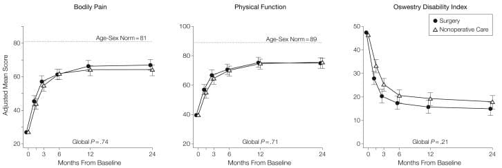
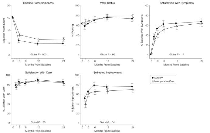
  • Main Outcome Measures: Primary outcomes were changes from baseline for the Medical Outcomes Study 36-item Short-Form Health Survey bodily pain and physical function scales and the modified Oswestry Disability Index (American Academy of Orthopaedic Surgeons MODEMS version) at 6 weeks, 3 months, 6 months, and 1 and 2 years from enrollment. Secondary outcomes included sciatica severity as measured by the Sciatica Bothersomeness Index, satisfaction with symptoms, self-reported improvement, and employment status.
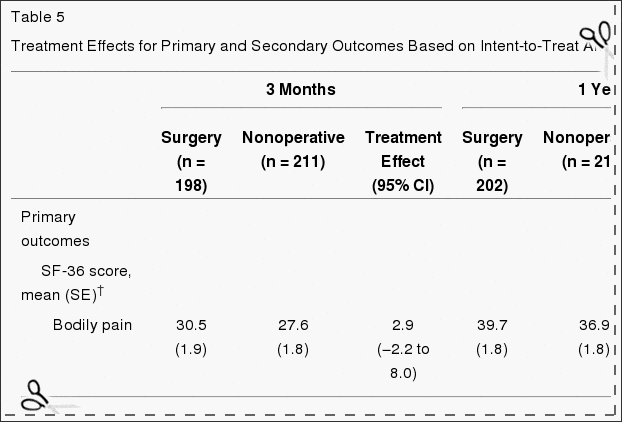
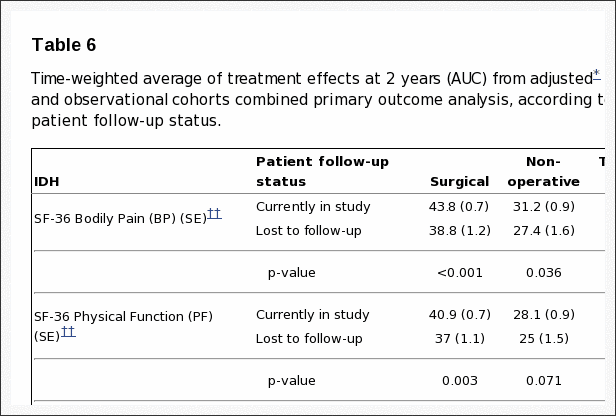
  • Results: Adherence to assigned treatment was limited: 50% of patients assigned to surgery received surgery within 3 months of enrollment, while 30% of those assigned to nonoperative treatment received surgery in the same period. Intent-to-treat analyses demonstrated substantial improvements for all primary and secondary outcomes in both treatment groups. Between-group differences in improvements were consistently in favor of surgery for all periods but were small and not statistically significant for the primary outcomes.
  • Conclusions: Patients in both the surgery and the nonoperative treatment groups improved substantially over a 2-year period. Because of the large numbers of patients who crossed over in both directions, conclusions about the superiority or equivalence of the treatments are not warranted based on the intent-to-treat analysis.
  • Trial Registration: clinicaltrials.gov Identifier: NCT00000410


Lumbar diskectomy is the most common surgical procedure performed in the United States for patients having back and leg symptoms; the vast majority of the procedures are elective. However, lumbar disk herniation is often seen on imaging studies in the absence of symptoms[1,2] and can regress over time without surgery.[3] Up to 15-fold variation in regional diskectomy rates in the United States[4] and lower rates internationally raise questions regarding the appropriateness of some of these surgeries.[5,6]


Several studies have compared surgical and nonoperative treatment of patients with herniated disk, but baseline differences between treatment groups, small sample sizes, or lack of validated outcome measures in these studies limit evidence-based conclusions regarding optimal treatment.[7-12] The Spine Patient Outcomes Research Trial (SPORT) was initiated in March 2000 to compare the outcomes of surgical and nonoperative treatment for lumbar intervertebral disk herniation, spinal stenosis, or degenerative spondylolisthesis.[13] The trial included both a randomized cohort and an observational cohort who declined to be randomized in favor of designating their own treatment but otherwise met all the other criteria for inclusion and who agreed to undergo follow-up according to the same protocol. This article reports intent-to-treat results through 2 years for the randomized cohort.


Methods


Study Design


SPORT was conducted at 13 multidisciplinary spine practices in 11 US states (California, Georgia, Illinois, Maine, Michigan, Missouri, Nebraska, New York, New Hampshire, Ohio, Pennsylvania). The human subjects committee of each participating institution approved a standardized protocol. All patients provided written informed consent. An independent data and safety monitoring board monitored the study at 6-month intervals.[13]


Patient Population


Patients were considered for inclusion if they were 18 years and older and diagnosed by participating physicians during the study enrollment period as having intervertebral disk herniation and persistent symptoms despite some nonoperative treatment for at least 6 weeks. The content of preenrollment nonoperative care was not prespecified in the protocol but included education/counseling (71%), physical therapy (67%), epidural injections (42%), chiropractic therapy (32%), anti-inflammatory medications (61%), and opioid analgesics (40%).


Specific inclusion criteria at enrollment were radicular pain (below the knee for lower lumbar herniations, into the anterior thigh for upper lumbar herniations) and evidence of nerve-root irritation with a positive nerve-root tension sign (straight leg raise�positive between 30� and 70� or positive femoral tension sign) or a corresponding neurologic deficit (asymmetrical depressed reflex, decreased sensation in a dermatomal distribution, or weakness in a myotomal distribution). Additionally, all participants were surgical candidates who had undergone advanced vertebral imaging (97% magnetic resonance imaging, 3% computed tomography) showing disk herniation (protrusion, extrusion, or sequestered fragment)[14] at a level and side corresponding to the clinical symptoms. Patients with multiple herniations were included if only one of the herniations was considered symptomatic (ie, if only one was planned to be operated on).


Exclusion criteria included prior lumbar surgery, cauda equina syndrome, scoliosis greater than 15�, segmental instability (>10� angular motion or >4-mm translation), vertebral fractures, spine infection or tumor, inflammatory spondyloarthropathy, pregnancy, comorbid conditions contraindicating surgery, or inability/unwillingness to have surgery within 6 months.


Study Interventions


The surgery was a standard open diskectomy with examination of the involved nerve root.[15,16] The procedure agreed on by all participating centers was performed under general or local anesthesia, with patients in the prone or knee-chest position. Surgeons were encouraged to use loupe magnification or a microscope. Using a midline incision reflecting the paraspinous muscles, the interlaminar space was entered as described by Delamarter and McCullough.[15] In some cases the medial border of the superior facet was removed to provide a clear view of the involved nerve root. Using a small annular incision, the fragment of disk was removed as described by Spengler.[16] The canal was inspected and the foramen probed for residual disk or bony pathology. The nerve root was decompressed, leaving it freely mobile.


The nonoperative treatment group received �usual care,� with the study protocol recommending that the minimum nonsurgical treatment include at least active physical therapy, education/counseling with home exercise instruction, and nonsteroidal anti-inflammatory drugs, if tolerated. Other nonoperative treatments were listed, and physicians were encouraged to individualize treatment to the patient; all nonoperative treatments were tracked prospectively.[13,17]


Study Measures


The primary measures were the Medical Outcomes Study 36-Item Short-Form Health Survey (SF-36) bodily pain and physical function scales[18-21] and the American Academy of Orthopaedic Surgeons MODEMS version of the Oswestry Disability Index (ODI).[22] As specified in the trial protocol, the primary outcomes were changes from baseline in these scales at 6 weeks, 3 months, 6 months, and 1 and 2 years from enrollment.


Secondary measures included patient self-reported improvement, work status, and satisfaction with current symptoms and with care.[23] Symptom severity was measured by the Sciatica Bothersomeness Index (range, 0-24; higher scores represent worse symptoms).[24,25]


Recruitment, Enrollment, and Randomization


A research nurse at each site identified potential participants and verified eligibility. For recruitment and informed consent, evidence-based videotapes described the surgical and non-operative treatments and the expected benefits, risks, and uncertainties.[26,27] Participants were offered enrollment in either the randomized trial or a concurrent observational cohort, the results of which are reported in a companion article.


Enrollment began in March 2000 and ended in November 2004. Baseline variables were collected prior to randomization. Patients self-reported race and ethnicity using National Institutes of Health categories.


Computer-generated random treatment assignment based on permuted blocks (randomly generated blocks of 6, 8, 10, and 12)[28] within sites occurred immediately after enrollment via an automated system at each site, ensuring proper allocation concealment. Study measures were collected at baseline and at regularly scheduled follow-up visits. Short-term follow-up visits occurred at 6 weeks and 3 months. If surgery was delayed beyond 6 weeks, additional follow-up data were obtained 6 weeks and 3 months postoperatively. Longer-term follow-up visits occurred at 6 months, 1 year from enrollment, and annually thereafter.


Statistical Analyses


We originally determined a sample size of 250 patients in each treatment group to be sufficient (with a 2-sided significance level of .05 and 85% power) to detect a 10-point difference in the SF-36 bodily pain and physical functioning scales or a similar effect size in the ODI. This difference corresponded to patients’ reports of being �a little better� in the Maine Lumbar Spine Study (MLSS).[29] The sample size calculation allowed for up to 20% missing data but did not account for any specific levels of nonadherence.


The analyses for the primary and secondary outcomes used all available data for each period on an intent-to-treat basis. Predetermined end points for the study included results at each of 6 weeks, 3 months, 6 months, 1 year, and 2 years. To adjust for the possible effect of missing data on the study results, the analysis of mean changes for continuous outcomes was performed using maximum likelihood estimation for longitudinal mixed-effects models under �missing at random� assumptions and including a term for treatment center. Comparative analyses were performed using the single imputation methods of baseline value carried forward and last value carried forward, as well as a longitudinal mixed model controlling for covariates associated with missed visits.[30]


For binary secondary outcomes, longitudinal logistic regression models were fitted using generalized estimating equations[31] as implemented in the PROC GENMOD program of SAS version 9.1 (SAS Institute Inc, Cary, NC). Treatment effects were estimated as differences in the estimated proportions in the 2 treatment groups.


P<.05 (2-sided) was used to establish statistical significance. For the primary outcomes, 95% confidence intervals (CIs) for mean treatment effects were calculated at each designated time point. Global tests of the joint hypothesis of no treatment effect at any of the designated periods were performed using Wald tests[32] as implemented in SAS. These tests account for the intraindividual correlation due to repeated measurements over time.[32]


Nonadherence to randomly assigned treatment may mean that the intention-to-treat analysis underestimates the real benefit of the treatment.[33,34] As a preplanned sensitivity analysis, we also estimated an �as-treated� longitudinal analysis based on comparisons of those actually treated surgically and nonoperatively. Repeated measures of outcomes were used as the dependent variables, and treatment received was included as a time-varying covariate. Adjustments were made for the time of surgery with respect to the original enrollment date to approximate the designated follow-up times. Baseline variables that were individually found to predict missing data or treatment received at 1 year were included to adjust for possible confounding.


Results


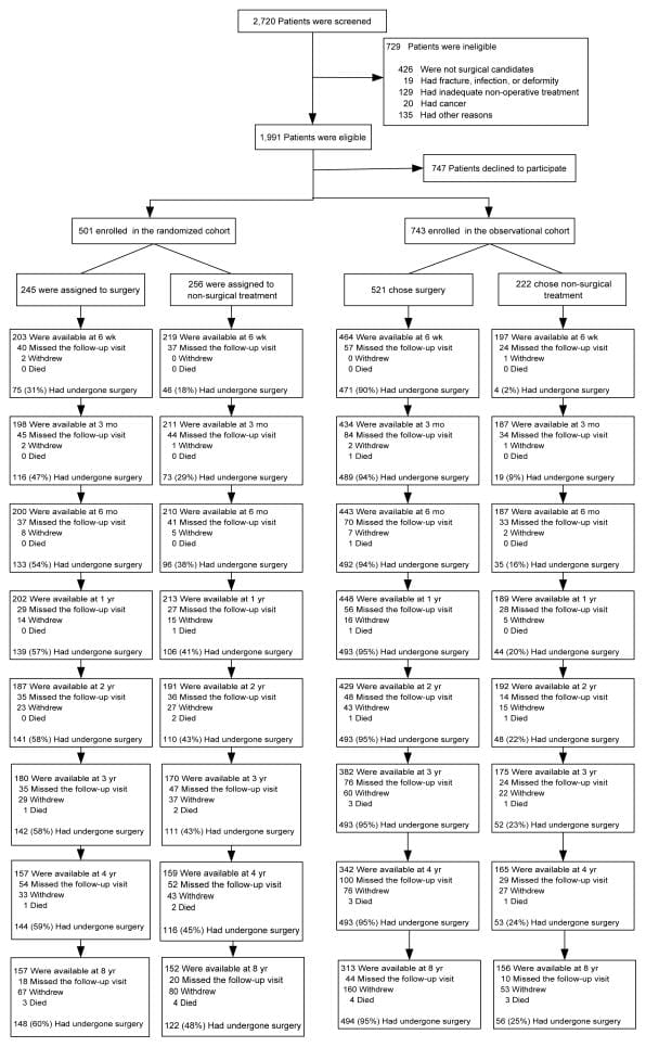
SPORT achieved full enrollment, with 501 (25%) of 1991 eligible patients enrolled in the randomized trial. A total of 472 participants (94%) completed at least 1 follow-up visit and were included in the analysis. Data were available for between 86% and 73% of patients at each of the designated follow-up times (Figure 1).


Figure 1 Flow Diagram of the SPORT RCT of Disc Herniation

Figure 1: Flow Diagram of the SPORT Randomized Controlled Trial of Disk Herniation: Exclusion, Enrollment, Randomization, and Follow-up.


Patient Characteristics


Baseline patient characteristics are shown in Table 1. Overall, the study population had a mean age of 42 years, with majorities being male, white, employed, and having attended at least some college; 16% were receiving disability compensation. All patients had radicular leg pain, 97% in a classic dermatomal distribution. Most of the herniations were at L5-S1, posterolateral, and were extrusions by imaging criteria.[14] The 2 randomized groups were similar at baseline.


Table 1 Patient Baseline Demographics


Nonoperative Treatments


A variety of nonoperative treatments were used during the study (Table 2). Most patients received education/counseling (93%) and anti-inflammatory medications (61%) (nonsteroidal anti-inflammatory drugs, cyclooxygenase 2 inhibitors, or oral steroids); 46% received opiates; more than 50% received injections (eg, epidural steroids); and 29% were prescribed activity restriction. Forty-four percent received active physical therapy during the trial; however, 67% had received it prior to enrollment.


Table 2 Nonoperative Treatments


Surgical Treatment and Complications


Table 3 gives the characteristics of surgical treatment and complications. The median surgical time was 75 minutes (interquartile range, 58-90), with a median blood loss of 49.5 mL (interquar-tile range, 25-75). Only 2% required transfusions. There were no perioperative deaths; 1 patient died from complications of childbirth 11 months after enrollment. The most common intraoperative complication was dural tear (4%). There were no postoperative complications in 95% of patients. Reoperation occurred in 4% of patients within 1 year of the initial surgery; more than 50% of the reoperations were for recurrent herniations at the same level.


Table 3 Operative Treatments, Complications and Events


Nonadherence


Nonadherence to treatment assignment affected both groups, ie, some patients in the surgery group chose to delay or decline surgery, and some in the nonoperative treatment group crossed over to receive surgery (Figure 1). The characteristics of crossover patients that were statistically different from patients who did not cross over are shown in Table 4. Those more likely to cross over to receive surgery tended to have lower incomes, worse baseline symptoms, more baseline disability on the ODI, and were more likely to rate their symptoms as getting worse at enrollment than the other patients receiving nonoperative treatment. Those more likely to cross over to receive nonoperative care were older, had higher incomes, were more likely to have an upper lumbar disk herniation, less likely to have a positive straight leg�raising test result, had less pain, better physical function, less disability on the ODI, and were more likely to rate their symptoms as getting better at enrollment than the other surgery patients.


Table 4 Statistically Significant Baseline Demographics


Missing Data


The rates of missing data were equivalent between the groups at each time point, with no evidence of differential dropout according to assigned treatment. Characteristics of patients with missed visits were very similar to those of the rest of the cohort except that patients with missing data were less likely to be married, more likely to be receiving disability compensation, more likely to smoke, more likely to display baseline motor weakness, and had lower baseline mental component summary scores on the SF-36.


Intent-to-Treat Analyses


Table 5 shows estimated mean changes from baseline and the treatment effects (differences in changes from baseline between treatment groups) for 3 months, 1 year, and 2 years. For each measure and at each point, the treatment effect favors surgery. The treatment effects for the primary outcomes were small and not statistically significant at any of the points. As shown in Figure 2, both treatment groups showed strong improvements at each of the designated follow-up times, with small advantages for surgery. However, for each primary outcome the combined global test for any difference at any period was not statistically significant. This test accounts for intraindividual correlations as described in the �Methods� section.


Figure 2 Mean Scores Over Time

Figure 2: Mean Scores Over Time for SF-36 Bodily Pain and Physical Function Scales and Oswestry Disability Index.


Table 5 Treatment Effects for Primary and Secondary Outcomes

Table 5: Treatment Effects for Primary and Secondary Outcomes Based on Intent-to-Treat Analyses*


For the secondary outcome of sciatica bothersomeness, Table 5 and Figure 3 show that there were greater improvements in the Sciatica Bothersomeness Index in the surgery group at all designated follow-up times: 3 months (treatment effect, ?2.1; 95% CI, ?3.4 to ?0.9), 1 year (treatment effect, ?1.6; 95% CI, ?2.9 to ?0.4), and 2 years (treatment effect, ?1.6; 95% CI, ?2.9 to ?0.3), with results of the global hypothesis test being statistically significant (P=.003). Patient satisfaction with symptoms and treatment showed small effects in favor of surgery while employment status showed small effects in favor of nonoperative care, but none of these changes was statistically significant. Self-rated progress showed a small statistically significant advantage for surgery (P=.04).


Figure 3 Measures Over Time

Figure 3: Measures Over Time for Sciatica Bothersomeness Index, Employment Status, Satisfaction With Symptoms, Satisfaction With Care, and Self-rated Improvement.


As-treated analyses based on treatment received were performed with adjustments for the time of surgery and factors affecting treatment crossover and missing data. These yielded far different results than the intent-to-treat analysis, with strong, statistically significant advantages seen for surgery at all follow-up times through 2 years. For example, at 1 year the estimated treatment effects for the SF-36 bodily pain and physical function scales, the ODI, and the sciatica measures were 15.0 (95% CI, 10.9 to 19.2), 17.5 (95% CI, 13.6 to 21.5), ?15.0 (95% CI, ?18.3 to ?11.7), and ?3.2 (95% CI, ?4.3 to ?2.1), respectively.


Sensitivity analysis was performed for 4 different analytic methods of dealing with the missing data. One method was based on simple mean changes for all patients with data at a given time point with no special adjustment for missing data. Two methods used single imputation methods�baseline value carried forward and last value carried forward.[32] The latter method used the same mixed-models approach for estimating mean changes as given in Table 5 but also adjusted for factors affecting the likelihood of missing data. Treatment effect estimates at 1 year ranged from 1.6 to 2.9 for the SF-36 bodily pain scale, 0.74 to 1.4 for the physical function scale, ?2.2 to ?3.3 for the ODI, and ?1.1 to ?1.6 for the sciatica measures. Given these ranges, there appear to be no substantial differences between any of these methods.


Dr Jimenez White Coat

Dr. Alex Jimenez’s Insight

Herniated disc symptoms vary on the location of the condition and on the surrounding soft tissues affected along the spine. Lumbar disc herniations, one of the most common area for herniated discs to occur, are characterized by the compression of the nerve roots along the lower back and can generally cause symptoms of sciatica. Surgery is commonly recommended to treat disc herniations, however, numerous treatment methods can help manage the condition without the need of surgical interventions. A research study conducted on sciatica caused by herniated discs determined that about 73 percent of participants experienced an improvement in symptoms with nonoperative treatment. The results of this article concluded that nonoperative treatment can be as effective as surgery in the treatment of herniated discs.


Comment


Both operated and nonoperated patients with intervertebral disk herniation improved substantially over a 2-year period. The intent-to-treat analysis in this trial showed no statistically significant treatment effects for the primary outcomes; the secondary measures of sciatica severity and self-reported progress did show statistically significant advantages for surgery. These results must be viewed in the context of the substantial rates of nonadherence to assigned treatment. The pattern of nonadherence is striking because, unlike many surgical studies, both the surgical and nonoperative treatment groups were affected.[35] The most comparable previous trial[8] had 26% crossover into surgery at 1 year, but only 2% crossover out of surgery. The mixing of treatments due to crossover can be expected to create a bias toward the null.[34] The large effects seen in the as-treated analysis and the characteristics of the crossover patients suggest that the intent-to-treat analysis underestimates the true effect of surgery.


SPORT findings are consistent with clinical experience in that relief of leg pain was the most striking and consistent improvement with surgery. Importantly, all patients in this trial had leg pain with physical examination and imaging findings that confirmed a disk herniation. There was little evidence of harm from either treatment. No patients in either group developed cauda equina syndrome; 95% of surgical patients had no intraoperative complications. The most common complication, dural tear, occurred in 4% of patients, similar to the 2% to 7% noted in the meta-analysis by Hoffman et al,7 2.2% seen in the MLSS,[29] and 4% in the recent series from Stanford.[36]


One limitation is the potential lack of representativeness of patients agreeing to be randomized to surgery or nonoperative care; however, the characteristics of patients agreeing to participate in SPORT were very similar to those in other studies.[29,36] The mean age of 42 years was similar to the mean ages in the MLSS,[29] the series of Spangfort,[37] and the randomized trial by Weber,[8] and only slightly older than those in the recent series from Stanford (37.5 years).[36] The proportion of patients receiving workers’ compensation in SPORT (16%) was similar to the proportion in the Stanford population (19%) but lower than that in the MLSS population (35%), which specifically oversampled patients receiving compensation. Baseline functional status was also similar, with a mean baseline ODI of 46.9 in SPORT vs 47.2 in the Stanford series, and a mean baseline SF-36 physical function score of 39 in SPORT vs 37 in the MLSS.


The strict eligibility criteria, however, may limit the generalizability of these results. Patients unable to tolerate symptoms for 6 weeks and demanding earlier surgical intervention were not included, nor were patients without clear signs and symptoms of radiculopathy with confirmatory imaging. We can draw no conclusions regarding the efficacy of surgery in these other groups. However, our entry criteria followed published guidelines for patient selection for elective diskectomy, and our results should apply to the majority of patients facing a surgical decision.[38,39]


To fully understand the treatment effect of surgery compared with nonoperative treatment, it is worth noting how each group fared. The improvements with surgery in SPORT were similar to those of prior series at 1 year: for the ODI, 31 points vs 34 points in the Stanford series; for the bodily pain scale, 40 points vs 44 in the MLSS; and for sciatica bothersomeness, 10 points vs 11 in the MLSS. Similarly, Weber[8] reported 66% �good� results in the surgery group, compared with the 76% reporting �major improvement� and 65% satisfied with their symptoms in SPORT.


The observed improvements with nonoperative treatment in SPORT were greater than those in the MLSS, resulting in the small estimated treatment effect. The nonoperative improvement of 37, 35, and 9 points in bodily pain, physical function, and sciatica bothersomeness, respectively, were much greater than the improvements of 20, 18, and 3 points reported in the MLSS. The greater improvement with nonoperative treatment in SPORT may be related to the large proportion of patients (43%) who underwent surgery in this group.


The major limitation of SPORT is the degree of nonadherence with randomized treatment. Given this degree of crossover, it is unlikely that the intent-to-treat analysis can form the basis of a valid estimate of the true treatment effect of surgery. The �as-treated� analysis with adjustments for possible confounders showed much larger effects in favor of surgical treatment. However, this approach does not have the strong protection against confounding that is afforded by randomization. We cannot exclude the possibility that baseline differences between the as-treated groups, or the selective choice of some but not other patients to cross over into surgery, may have affected these results, even after controlling for important covariates. Due to practical and ethical constraints, this study was not masked through the use of sham procedures. Therefore, any improvements seen with surgery may include some degree of �placebo effect.�


Another potential limitation is that the choice of nonoperative treatments was at the discretion of the treating physician and patient. However, given the limited evidence regarding efficacy for most nonoperative treatments for lumbar disk herniation and individual variability in response, creating a limited, fixed protocol for nonoperative treatment was neither clinically feasible nor generalizable. The nonoperative treatments used were consistent with published guidelines.[17,38,39] Compared with the MLSS, SPORT had lower use of activity restriction, spinal manipulation, transcutaneous electrical nerve stimulation, and braces and corsets, and higher rates of epidural steroid injections and use of narcotic analgesics. This flexible nonoperative protocol had the advantages of individualization that considered patient preferences in the choice of nonoperative treatment and of reflecting current practice among multidisciplinary spine practices. However, we cannot make any conclusion regarding the effect of surgery vs any specific nonoperative treatment. Similarly, we cannot adequately assess the relative efficacy of any differences in surgical technique.


Conclusion


Patients in both the surgery and nonoperative treatment groups improved substantially over the first 2 years. Between-group differences in improvements were consistently in favor of surgery for all outcomes and at all time periods but were small and not statistically significant except for the secondary measures of sciatica severity and self-rated improvement. Because of the high numbers of patients who crossed over in both directions, conclusions about the superiority or equivalence of the treatments are not warranted based on the intent-to-treat analysis alone.


Acknowledgments & Footnotes


Ncbi.nlm.nih.gov/pmc/articles/PMC2553805/


Manipulation or Microdiskectomy for Sciatica? A Prospective Randomized Clinical Study


Abstract


Objective: The purpose of this study was to compare the clinical efficacy of spinal manipulation against microdiskectomy in patients with sciatica secondary to lumbar disk herniation (LDH).

Methods: One hundred twenty patients presenting through elective referral by primary care physicians to neurosurgical spine surgeons were consecutively screened for symptoms of unilateral lumbar radiculopathy secondary to LDH at L3-4, L4-5, or L5-S1. Forty consecutive consenting patients who met inclusion criteria (patients must have failed at least 3 months of nonoperative management including treatment with analgesics, lifestyle modification, physiotherapy, massage therapy, and/or acupuncture) were randomized to either surgical microdiskectomy or standardized chiropractic spinal manipulation. Crossover to the alternate treatment was allowed after 3 months.

Results: Significant improvement in both treatment groups compared to baseline scores over time was observed in all outcome measures. After 1 year, follow-up intent-to-treat analysis did not reveal a difference in outcome based on the original treatment received. However, 3 patients crossed over from surgery to spinal manipulation and failed to gain further improvement. Eight patients crossed from spinal manipulation to surgery and improved to the same degree as their primary surgical counterparts.

Conclusions: Sixty percent of patients with sciatica who had failed other medical management benefited from spinal manipulation to the same degree as if they underwent surgical intervention. Of 40% left unsatisfied, subsequent surgical intervention confers excellent outcome. Patients with symptomatic LDH failing medical management should consider spinal manipulation followed by surgery if warranted.


In conclusion, a herniated disc causes the soft, central portion of an intervertebral disc to bulge out a tear in its outer, fibrous ring as a result of degeneration, trauma, lifting injuries or straining. Most disc herniations can heal on their own but those considered to be severe may require surgical interventions to treat them. Research studies, such as the one above, have demonstrated that nonoperative treatment may help the recovery of a herniated disc without the need for surgery. Information referenced from the National Center for Biotechnology Information (NCBI). The scope of our information is limited to chiropractic as well as to spinal injuries and conditions. To discuss the subject matter, please feel free to ask Dr. Jimenez or contact us at 915-850-0900 .


Curated by Dr. Alex Jimenez


Green-Call-Now-Button-24H-150x150-2-3.png


Additional Topics: Back Pain


According to statistics, approximately 80% of people will experience symptoms of back pain at least once throughout their lifetimes. Back pain is a common complaint which can result due to a variety of injuries and/or conditions. Often times, the natural degeneration of the spine with age can cause back pain. Herniated discs occur when the soft, gel-like center of an intervertebral disc pushes through a tear in its surrounding, outer ring of cartilage, compressing and irritating the nerve roots. Disc herniations most commonly occur along the lower back, or lumbar spine, but they may also occur along the cervical spine, or neck. The impingement of the nerves found in the low back due to injury and/or an aggravated condition can lead to symptoms of sciatica.


blog picture of cartoon paperboy big news


IMPORTANT TOPIC: EXTRA EXTRA: A Healthier You!


OTHER IMPORTANT TOPICS: EXTRA: Sports Injuries? | Vincent Garcia | Patient | El Paso, TX Chiropractor
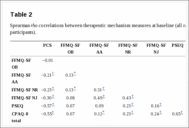
 

Blank
References
1.�Boden SD, Davis DO, Dina TS, Patronas NJ, Wiesel SW. Abnormal magnetic-resonance scans of the lumbar spine in asymptomatic subjects: a prospective investigation.�J Bone Joint Surg Am.�1990;72:403�408.�[PubMed]
2.�Jensen MC, Brant-Zawadzki MN, Obuchowski N, Modic MT, Malkasian D, Ross JS. Magnetic resonance imaging of the lumbar spine in people without back pain.�N Engl J Med.�1994;331:69�73.[PubMed]
3.�Saal JA, Saal JS. Nonoperative treatment of herniated lumbar intervertebral disc with radiculopathy.�Spine.�1989;14:431�437.�[PubMed]
4.�Weinstein JN, Dartmouth Atlas Working Group .�Dartmouth Atlas of Musculoskeletal Health Care.American Hospital Association Press; Chicago, Ill: 2000.
5.�Deyo RA, Weinstein JN. Low back pain.�N Engl J Med.�2001;344:363�370.�[PubMed]
6.�Weinstein JN, Bronner KK, Morgan TS, Wennberg JE. Trends and geographic variations in major surgery for degenerative diseases of the hip, knee, and spine.�Health Aff (Millwood)�2004;(suppl Web exclusive):var81�89.�[PubMed]
7.�Hoffman RM, Wheeler KJ, Deyo RA. Surgery for herniated lumbar discs: a literature synthesis.�J Gen Intern Med.�1993;8:487�496.�[PubMed]
8.�Weber H. Lumbar disc herniation: a controlled, prospective study with ten years of observation.�Spine.�1983;8:131�140.�[PubMed]
9.�Buttermann GR. Treatment of lumbar disc herniation: epidural steroid injection compared with discectomy: a prospective, randomized study.�J Bone Joint Surg Am.�2004;86:670�679.�[PubMed]
10.�Gibson JN, Grant IC, Waddell G. The Cochrane review of surgery for lumbar disc prolapse and degenerative lumbar spondylosis.�Spine.�1999;24:1820�1832.�[PubMed]
11.�Gibson JN, Grant IC, Waddell G. Surgery for lumbar disc prolapse.�Cochrane Database Syst Rev.�2000;(3):CD001350.�[PubMed]
12.�Jordan J, Shawver Morgan T, Weinstein J, Konstantinou K. Herniated lumbar disc.�Clin Evid.�2003 June;:1203�1215.
13.�Birkmeyer NJ, Weinstein JN, Tosteson AN, et al. Design of the Spine Patient Outcomes Research Trial (SPORT)�Spine.�2002;27:1361�1372.�[PMC free article][PubMed]
14.�Fardon DF, Milette PC. Nomenclature and classification of lumbar disc pathology: recommendations of the Combined Task Forces of the North American Spine Society, American Society of Spine Radiology, and American Society of Neuroradiology.�Spine.�2001;26:E93�E113.�[PubMed]
15.�Delamarter R, McCullough J. Microdiscectomy and microsurgical laminotomies. In: Frymoyer J, editor.�The Adult Spine: Principles and Practice.�2nd ed. Lippincott-Raven Publishers; Philadelphia, Pa: 1996.
16.�Spengler DM. Lumbar discectomy: results with limited disc excision and selective foraminotomy.�Spine.�1982;7:604�607.�[PubMed]
17.�Cummins J, Lurie JD, Tosteson T, et al. Descriptive epidemiology and prior healthcare utilization of patients in the Spine Patient Outcomes Research Trial’s (SPORT) three observational cohorts: disc herniation, spinal stenosis, and degenerative spondylolisthesis.�Spine.�2006;31:806�814.�[PMC free article][PubMed]
18.�Ware JE, Jr, Sherbourne D. The MOS 36-item short-form health survey (SF-36), I: conceptual framework and item selection.�Med Care.�1992;30:473�483.�[PubMed]
19.�Ware JE., Jr .�SF-36 Health Survey: Manual and Interpretation Guide.�Nimrod Press; Boston, Mass: 1993.
20.�McHorney CA, Ware JE, Jr, Lu JF, Sherbourne CD. The MOS 36-item Short-Form Health Survey (SF-36), III: tests of data quality, scaling assumptions, and reliability across diverse patient groups.�Med Care.�1994;32:40�66.�[PubMed]
21.�Stewart AL, Greenfield S, Hays RD, et al. Functional status and well-being of patients with chronic conditions: results from the Medical Outcomes Study.�JAMA.�1989;262:907�913.�[PubMed]
22.�Daltroy LH, Cats-Baril WL, Katz JN, Fossel AH, Liang MH. The North American Spine Society lumbar spine outcome assessment instrument: reliability and validity tests.�Spine.�1996;21:741�749.[PubMed]
23.�Deyo RA, Diehl AK. Patient satisfaction with medical care for low-back pain.�Spine.�1986;11:28�30.[PubMed]
24.�Atlas SJ, Deyo RA, Patrick DL, Convery K, Keller RB, Singer DE. The Quebec Task Force classification for spinal disorders and the severity, treatment, and outcomes of sciatica and lumbar spinal stenosis.�Spine.�1996;21:2885�2892.�[PubMed]
25.�Patrick DL, Deyo RA, Atlas SJ, Singer DE, Chapin A, Keller RB. Assessing health-related quality of life in patients with sciatica.�Spine.�1995;20:1899�1908.�[PubMed]
26.�Phelan EA, Deyo RA, Cherkin DC, et al. Helping patients decide about back surgery: a randomized trial of an interactive video program.�Spine.�2001;26:206�211.�[PubMed]
27.�Weinstein JN. Partnership: doctor and patient: advocacy for informed choice vs. informed consent.�Spine.�2005;30:269�272.�[PubMed]
28.�Friedman L, Furberg C, DeMets D.�Fundamentals of Clinical Trials.�3rd ed. Springer-Verlag; Cambridge, Mass: 1998. The randomization process; pp. 61�81.
29.�Atlas SJ, Deyo RA, Keller RB, et al. The Maine Lumbar Spine Study, II: 1-year outcomes of surgical and nonsurgical management of sciatica.�Spine.�1996;21:1777�1786.�[PubMed]
30.�Little R, Rubin D.�Statistical Analysis With Missing Data.�2nd ed. John Wiley & Sons; Philadelphia, Pa: 2002.
31.�Diggle P, Haeagery P, Liang K, Zeger S.�The Analysis of Longitudinal Data.�2nd ed. Oxford University Press; Oxford, England: 2002.
32.�Fitzmaurice G, Laird N, Ware J.�Applied Longitudinal Analysis.�John Wiley & Sons; Philadelphia, Pa: 2004.
33.�Altman DG, Schulz KF, Moher D, et al. The revised CONSORT statement for reporting randomized trials: explanation and elaboration.�Ann Intern Med.�2001;134:663�694.�[PubMed]
34.�Meinert CL.�Clinical Trials: Design, Conduct, and Analysis.�Oxford University Press; New York, NY: 1986.
35.�Kuppermann M, Varner RE, Summitt RL, Jr, et al. Effect of hysterectomy vs medical treatment on health-related quality of life and sexual functioning: the medicine or surgery (Ms) randomized trial.�JAMA.�2004;291:1447�1455.�[PubMed]
36.�Carragee EJ, Han MY, Suen PW, Kim D. Clinical outcomes after lumbar discectomy for sciatica: the effects of fragment type and anular competence.�J Bone Joint Surg Am.�2003;85:102�108.�[PubMed]
37.�Spangfort EV. The lumbar disc herniation: a computer-aided analysis of 2,504 operations.�Acta Orthop Scand Suppl.�1972;142:1�95.�[PubMed]
38.�Agency for Health Care Policy and Research .�Acute Low Back Problems in Adults.�US Dept of Health & Human Services; Bethesda, Md: 1994.
39.�North American Spine Society .�North American Spine Society Phase III Clinical Guidelines for Multidisciplinary Spine Care Specialists.�NASS; LaGrange, Ill: 2000. Herniated disc.
Close Accordion
Rapid Pain Relief for Herniated Discs in El Paso, TX

Rapid Pain Relief for Herniated Discs in El Paso, TX

Herniated discs are a debilitating condition characterized by pain, numbness and weakness in one or more limbs. While some people may experience no pain at all, those that do may often wish for fast pain relief to avoid long periods of sick leave from their jobs. Many healthcare professionals recommend surgery for patients with persistent and/or worsening herniated disc symptoms but other non-operative treatment options can help treat disc herniations. The purpose of the following article is to demonstrate how a�structured physiotherapy treatment model can provide rapid relief to patients who qualify for lumbar disc surgery.


A Structured Physiotherapy Treatment Model Can Provide Rapid Relief to Patients Who Qualify for Lumbar Disc Surgery: A Prospective Cohort Study


Abstract


  • Objective: To evaluate a structured physiotherapy treatment model in patients who qualify for lumbar disc surgery.
  • Design: A prospective cohort study.
  • Patients: Forty-one patients with lumbar disc herniation, diagnosed by clinical assessments and magnetic resonance imaging.
  • Methods: Patients followed a structured physiotherapy treatment model, including Mechanical Diagnosis and Therapy (MDT), together with graded trunk stabilization training. Study outcome measures were the Oswestry Disability Index, a visual analogue scale for leg and back pain, the Tampa Scale for Kinesiophobia, the European Quality of Life in 5 Dimensions Questionnaires, the Zung Self-Rating Depression Scale, the Self-Efficacy Scale, work status, and patient satisfaction with treatment. Questionnaires were distributed before treatment and at 3-, 12- and 24-month follow-ups.
  • Results: The patients had already improved significantly (p<0.001) 3 months after the structured physiotherapy treatment model in all assessments: disability, leg and back pain, kinesiophobia, health-related quality of life, depression and self-efficacy. The improvement could still be seen at the 2-year follow-up.
  • Conclusion: This study recommends adopting the structured physiotherapy treatment model before considering surgery for patients with symptoms such as pain and disability due to lumbar disc herniation.
  • Keywords: intervertebral disc displacement; rehabilitation; physical therapy modalities.


Introduction


Symptoms of lumbar disc herniation are relatively common in the general population, although the prevalence rates vary widely between different studies (1). Symptom severity also varies and, in many patients, pain and loss of function may lead to disability and long periods of sick leave (2). Spontaneous resolution of symptoms after a lumbar disc herniation is regarded as common, which makes it difficult to evaluate the effects of treatment. Furthermore, in studies evaluating spontaneous healing, different physiotherapy treatments are often included, together with pain medication (3�5), which makes it difficult to determine the extent of natural healing. On the other hand, in patients with sciatica, but without confirmed disc herniation on magnetic resonance imaging (MRI), approximately one-third of subjects recover 2 weeks after the onset of sciatica and approximately three-quarters recover after 3 months (6).


In contrast to evaluating spontaneous healing, surgery for lumbar disc herniation has been investigated in numerous studies. Surgery has been compared with a variety of treatments, such as education, chiropractic, unspecified physiotherapy, acupuncture, injections and medication (7�10). The non-surgical treatments have, however, been described only in vague terms, and variations in treatments have been used. Previous studies have reported favourable short-term (after 1 year) outcomes for surgery, but no major differences between surgical and other treatments have been demonstrated in the long term (over 2 years) (7, 10, 11). The conclusions that are drawn from the comparison between surgery and non-systematic non-surgical treatments may thus be misleading. This has been confirmed in a systematic review, which concluded that there is conflicting evidence as to whether surgery is more beneficial than nonsurgical care for both short- and long-term follow-up (12).


Kinesiophobia has been evaluated in patients after lumbar disc surgery, and almost 50% of patients were classified as having kinesiophobia (13). To our knowledge kinesiophobia has not been evaluated in patients with lumbar disc herniation treated with a structured physiotherapy treatment.


There are many different non-surgical treatment methods for patients with low-back pain and sciatica. One common management method is Mechanical Diagnosis and Therapy (MDT), also known as the McKenzie method, which aims to eliminate or minimize pain (14). A systematic review from 2004 of the efficacy of MDT showed that patients with low-back pain treated�with MDT reported a greater, more rapid reduction in pain and disability compared with non-steroidal anti-inflammatory drugs (NSAIDs), educational booklets, back massage and back care advice, strength training, spinal mobilization and general exercises (15). In a randomized controlled trial with a 1-year follow-up from 2008, Paatelma and co-workers (16) found that the McKenzie method was only marginally more effective compared with only giving advice to patients with low-back pain. For patients with low-back pain, sciatica and a verified lumbar disc herniation, it has, however, been shown that a selected group of patients who responded to MDT after 5 days of treatment also reported that they were satisfied after 55 weeks (17). The patients started treatment just 12 days after the onset of symptoms and the effects of spontaneous healing cannot therefore be excluded. Taken together, the treatment effects of MDT for patients with a verified lumbar disc herniation appear to require further evaluation.


Trunk stabilization exercises, which aim to restore deep trunk muscle control, have been used for the prevention and rehabilitation of low-back pain (18). A randomized controlled trial revealed a reduction in the recurrence of low-back pain episodes after specific trunk stabilization exercises compared with a control group receiving advice and the use of medication (19). Dynamic lumbar stabilization exercises have been found to relieve pain and improve function in patients who have undergone microdiscectomy (20). The effects of trunk stabilization exercises combined with MDT have, however, not been studied in patients with non-operated lumbar disc herniation. MDT is seldom recommended for patients with MRI verified lumbar disc herniation with a broken outer annulus. At our hospital, however, we have several years of good clinical experience of a combination of MDT and trunk stabilization exercises for this category of patients. To our knowledge, no previous study has investigated whether patients with a lumbar disc herniation verified by MRI, symptoms for at least 6 weeks (minimizing effects of spontaneous healing) and who qualified for disc surgery could improve with a structured physiotherapy treatment model including MDT and gradually progressive trunk stabilization exercises. The aim of this study was therefore to�evaluate a structured physiotherapy treatment model in patients who qualified for lumbar disc surgery.


Material and Methods


During the study inclusion period, 150 patients, who were referred to the orthopaedic clinic at Sahlgrenska University Hospital, Gothenburg, from November 2003 to January 2008, were identified as potential participants since disc herniation was confirmed with MRI. Inclusion criteria were: 18�65 years of age; MRI confirming disc herniation explaining the clinical findings; symptoms for at least 6 weeks (minimizing the effects of spontaneous healing) and pain distribution with concomitant neurological disturbances correlated to the affected nerve root. Exclusion criteria were: cauda equina syndrome, previous spinal surgery, other spinal diseases, such as spinal stenosis and spondylolisthesis, and inadequate command of Swedish. However, 70 patients were excluded because of spontaneous resolution of pain and symptoms. The remaining 80 patients met the inclusion criteria and qualified for surgery. Orthopaedic surgeons determined whether the patients qualified for lumbar disc surgery after MRI and physical examination according to the recommendations of the American Academy of Orthopaedic Surgeons for patients with lumbar disc herniation (21).


Figure 1 Study Flowchart

Initially, the study was planned as a randomized controlled trial (RCT) between a structured physiotherapy treatment model and surgery, but the number of patients was not sufficient to obtain acceptable power. Eighteen of the 80 patients were initially randomized to physiotherapy, 17 patients were randomized to surgery and 45 patients did not agree to undergo randomization. Twenty-seven of the 45 patients who did not agree to randomization agreed to take part in the structured physiotherapy treatment and 18 patients agreed to undergo surgery. A decision was therefore made solely to present a cohort of 45 patients treated according to the structured physiotherapytreatment protocol (Fig. 1). Patients were given verbal and written information and informed consent was obtained. The study was approved by the Regional Ethical Review Board.


Before structured physiotherapy treatment began, 4 patients recovered to the extent that they could no longer be accepted as surgical candidates and they were therefore excluded from the study. The remaining 41 patients treated according to the structured physiotherapy model are presented in this paper.


A Structured Physiotherapy Treatment Model


Six physiotherapists with credentialed examinations in MDT, which is an examination within the MDT concept after completing 4 courses of 4 days each for evaluating and treating patients with spinal problems. Following completion of these courses, an extensive literature study and practice in evaluating and treating patients is required before the examination can be completed. The physiotherapists involved in the study had 5�20 years of clinical experience of treating patients with back problems and herniated lumbar disc. The inter-examiner reliability of the MDT assessment has been shown to be good if the examiner is trained in the MDT method (22). The physiotherapists examined and treated the patients during a 9-week period (Table I). For the first 2 weeks of treatment, an MDT protocol was followed, based on clinical examinations of individual mechanical and symptomatic responses to positions and movements, with the aim of minimizing pain and with the emphasis on self-management (14). During the third week of treatment, graded trunk stabilization exercises were added to the MDT protocol. The purpose of graded trunk stabilization exercises was to improve muscle control (23). The low-load muscular endurance exercises were gradually increased in intensity on an individual�basis with respect to the patients� reported leg pain and the observed movement control and quality. During treatment, the patients were encouraged to continue exercising on their own at a gym, or to perform some other type of physical training of their own choice after the structured physiotherapy treatment was concluded. Four weeks after the completion of the 9-week physiotherapy treatment period, the patients attended a follow-up visit with the physiotherapist who had treated them. The aim of this visit was to encourage a high level of compliance with respect to continued trunk stabilization exercises and MDT practice (Table I).


Table 1 Treatment Procedures


Study Outcome Measures


The patients were given a battery of questionnaires to complete. Independent examiners, who were not involved in the treatment, distributed the questionnaires before treatment (baseline) and at the 3-, 12- and 24-month follow-ups.


The primary outcome measures were pain intensity in the leg, rated using a visual analogue scale (VAS) 0�100 mm (24) and the Oswestry Disability Index (ODI) 0�100 % (25). A score of 0�10 mm on the VAS was defined as no pain according to �berg et al. (26). An ODI score of 0�20% was defined as minimal or no disability, and a score of over 40% was defined as severe disability (25). These primary outcome measures are commonly used in evaluations after surgery for lowback pain and for assessing patients with lumbar disc herniation (27).


Secondary outcome measures included pain intensity in the back rated using a VAS and the degree of kinesiophobia using the Tampa Scale for Kinesiophobia (TSK). The TSK score varies between 17 and 68 and a cut-off more than 37 was defined as a high degree of kinesiophobia (28). Health-Related Quality of Life (HRQoL) in the European Quality of Life in 5 Dimensions Questionnaires (EQ-5D) was used. The EQ-5D includes 2 parts, EQ-5Dindex ranges from 0 to 1.0, where 1.0 is optimal health and EQ-5DVAS is a vertical visual analogue scale ranging from 0 (worst possible health state) to 100 (best possible health state) (29). The Zung Self-Rating Depression Scale (ZDS) ranges from 20�80 and the more depressed the patient is, the higher score (30). The Self-Efficacy Scale (SES) ranges from 8 to 64, with higher scores indicating more positive beliefs (31) was also used. Work status was measured using a 3-grade Likert scale: working full time, full-time sick leave and part-time sick leave. Likewise, patient�satisfaction with treatment was measured on a 3-grade Likert scale; satisfied, less satisfied and dissatisfied (32). These secondary outcome measures evaluate bio-psychosocial factors described as important in connection with lumbar disc surgery (33).


Table 2 Baseline Characteristics for the 41 Patients


Statistical Analyses


The results are presented as median values and interquartile range (IQR), except for age, which is presented as the mean and standard deviation (SD). Changes over time within the group were analysed with the Wilcoxon signed-rank test. Statistical significance was set at an alpha level of 0.05.


Results


The baseline characteristics are shown in Table II. No patient had undergone surgery at the 3-month follow-up. At the 12-month follow-up, 3 patients had undergone surgery and, at the 24-month follow-up, 1 additional patient had been operated on. After surgery, these 4 patients were excluded from further follow-ups (Fig. 1).


Change Over Time in Primary Outcome Measures


Disability. The patients showed significant improvements (p < 0.001) in ODI at the 3-month follow-up compared with baseline. The median (IQR) score decreased from 42 (27�53) to 14 (8�33). This improvement could still be seen at 12 and 24 months (Table III and Fig. 2). At baseline, 22 patients reported�severe disability (54%) and 3 patients reported no disability. The degree of disability decreased at the 3-month follow-up, as only 9 patients (22%) reported severe disability and 26 (64%) reported no disability. At 12- and 24-month follow-ups only 2 patients (5%) reported severe disability. At 12-month followup 26 patients still reported no disability, and at 24-month follow-up 27 patients reported no disability.


Figure 2 Visual Analogue Scale Leg Pain and Oswestry Disability Index


Leg pain. A significant reduction in patients� leg pain was found at the 3-month follow-up (p < 0.001) on the VAS compared with baseline. The median (IQR) on the VAS decreased from 60 (40�75) to 9 (2�27). This improvement could still be seen at the 12- and 24-month follow-ups (Table III and Fig. 2). Before treatment, all patients reported leg pain. Three months after treatment, the median on the VAS was 9 mm, i.e. classified as no leg pain (26). Twenty-three patients (56%) reported no leg pain at the 3-month follow-up. At the 12-month follow-up 22 patients reported no leg pain, and after 24 months 24 patients reported no leg pain.


Table 3 Changes Over Time in Primary and Secondary Outcome Measures


Change in Secondary Outcome Measures Over Time


Back pain. A significant improvement in back pain was found at the 3-month follow-up (p < 0.001) on the VAS compared with baseline. This improvement could still be seen at 12 and 24 months (Table III). At baseline, 6 patients (15%) reported no back pain. Three months after treatment began, 20 patients (49%) reported no back pain.


Figure 3 Number of Patients Classified with Kinesiophobia at Baseline


Kinesiophobia. The degree of kinesiophobia showed a significant improvement at the 3-month follow-up (p < 0.001) and the improvement could be seen throughout the follow-up period (Table III). Before treatment, 25 patients (61%) were classified as having kinesiophobia and 15 patients (37%) had no kinesiophobia, while data for 1 patient was missing. After 3 months, 15 patients (37%) had kinesiophobia and 26 (63%) had no kinesiophobia. At the 12-month follow-up, the number of patients with kinesiophobia had reduced to 4 (11%) (Fig. 3).


Health-related quality of life, depression and self-efficacy. All 4 assessments (EQ-5Dindex, EQ-5DVAS, ZDS and SES) showed significant improvements at the 3-month follow-up (p < 0.001). This improvement could still be seen at 12 and 24 months (Table III).


Sick leave. At baseline, 22 patients (54%) were on full-time sick leave (Table IV), compared with 9 (22%) patients at�the 3-month follow-up. At baseline, 14 patients (34%) were working full time, compared with 22 (54%) at the 3-month follow-up.


Table 4 Number of Patients on Sick Leave at Each Follow Up


Satisfaction with Treatment


At the 3-month follow-up, 32 (78%) of 41 patients were satisfied with the structured physiotherapy treatment. Seven patients were less satisfied and 2 patients were dissatisfied. Both of the dissatisfied patients were later operated. At the 2-year follow-up, the number of satisfied patients was 29 (80%) of 36. Seven patients were less satisfied, but none dissatisfied after structured physiotherapy treatment.


Dr Jimenez White Coat

Dr. Alex Jimenez’s Insight

A disc herniation in the lumbar spine can cause pain, numbness and weakness in the lower back. Because of the severity of the symptoms, many patients seeking fast pain relief consider surgery. However, many non-operative treatment options can help improve as well as manage lumbar herniated disc symptoms.�A structured physiotherapy treatment model can provide rapid pain relief to patients who would otherwise qualify for lumbar disc surgery, according to the following article. Patients looking to avoid taking long periods of sick leave from work due to their symptoms may benefit from a structured physiotherapy treatment model. As with any type of injury and/or condition, the use of other treatment options should be properly considered before turning to surgical interventions for fast pain relief.


Discussion


The principal finding of this study was that patients who qualified for lumbar disc surgery improved to a statistically significant and clinically substantial degree just 3 months after the start of the structured physiotherapy treatment in all assessments: disability, leg and back pain, kinesiophobia, health-related quality of life, depression and self-efficacy. The improvements could still be seen at the 2-year follow-up.


The natural course of healing must be considered carefully, especially when evaluating treatment effects in patients with disc herniation. The symptoms often vary over time and many discs heal spontaneously and the symptoms cease. Approximately 75% of patients with sciatica, without an MRI-verified disc herniation, recover within 3 months, and approximately one-third of patients recover within 2 weeks after the onset of sciatica (6). The natural course of sciatica was evaluated in a randomized controlled trial (34), which compared NSAIDs with placebo. The patients were, however, examined within 14 days after the onset of radiating leg pain. After 3 months, 60% of the patients had recovered and, after 12 months, 70% had recovered. In order to minimize the influence of spontaneous healing in the present study, the patients were therefore included only if they had had persistent pain and disability for more than 6 weeks. In fact, the majority of the patients had had pain and disability for more than 3 months. It is therefore most likely that the effects of treatment seen in the present study are, in the majority of patients, an effect of the structured physiotherapy treatment model and not a result of spontaneous healing.


In the study by Weber et al. (34), the VAS leg pain mean score was reduced from 54 mm at baseline to 19 mm within 4 weeks for all 183 patients, regardless of treatment. After 1 year, the VAS leg pain mean score was 17 mm. The patients in the present study who were a little worse at baseline (60 mm) reported 9 mm on the VAS leg pain just 3 months after treatment. Consequently, in the present study, the median VAS level had already been reduced to under the no-pain score, defined as 0�10 on the VAS (26), at the 3-month follow-up and this was maintained to the 12- and 24-month follow-ups.


Physiotherapy treatment for patients with lumbar disc herniation can lead to improvements. Br�tz et al. (17) included a selected group of patients who responded with the centralization of pain after the first 5 daily sessions of treatment according to the MDT method. Centralization of pain is defined as a clinically induced change in the location of pain referred from the spine, that moves from the most distal position toward the lumbar midline (35). However, the patients� medium duration of symptoms before treatment was only 12 days and the possibility that patients recovered naturally cannot therefore be excluded (17).


In a retrospective study, 95 patients were treated with a functional restoration programme (36). The patients achieved significant improvements after a mean treatment period of 8.7 months. The evaluation was performed at discharge only. With a treatment period of this length, it is, however, difficult to differentiate between the effects of treatment and the natural healing process. In the present study, a shorter treatment period was adopted, and large and significant improvements were found after just 3 months and were still present at the 24-month follow-up. It is therefore not likely that the natural healing process was responsible for the positive results in the present study.


In a prospective study of 82 consecutive patients with acute severe sciatica, included for conservative management, only a minority of the patients had made a full recovery after 12 months (37). Twenty-five percent of the patients underwent surgery within 4 months and one-third had surgery within 1 year. In spite of the fact that the inclusion criteria in the present study followed the recommendations for surgery (21, 38), no patient required surgery at the 3-month follow-up and, after 12 months, only 3 patients (7%) had undergone surgery. The interpretation of the divergence could be that the structured physiotherapy treatment model used in the present study appeared to influence patients with lumbar disc herniation in a very positive direction. One recommendation is therefore to follow the structured physiotherapy treatment model before considering surgery.


In this study, MRI verification of disc herniation was an inclusion criterion. In clinical practice, MRI verification is not mandatory, as it is in surgical treatment, before introducing structured physiotherapy treatment to patients with symptoms from a disc herniation. Consequently, treatment according to the structured physiotherapy treatment model can start early after the commencement of symptoms, as it is not necessary to wait for an MRI. It is possible to speculate that, if treatment with a structured physiotherapy model starts earlier than in the present study, the improvements would be even better, further reducing the risk of persistent pain and accompanying problems. Moreover, the need for MRI is likely to diminish; this, however, should be further evaluated in future studies.


One explanation for the good results of this study could be that the patients followed a structured physiotherapy treatment model, comprising MDT and trunk stabilization exercises, allowing for an individual design and progression of the treatment. Similar results were described in a retrospective cohort study (39) using several treatment methods for pain control as well as for exercise training for patients with lumbar disc herniation. The evaluation was not carried out until approximately 31 months after treatment. The results of Saal et al. (39) and of the present study are in agreement, in that structured physiotherapy treatment can reduce symptoms, but symptoms were relieved much more rapidly in the present study.


In a multicentre study comprising 501 patients, randomized to surgery or non-operative care, 18% of the patients assigned to non-operative treatment underwent surgery within 6 weeks and 30% had surgery at approximately 3 months (7). The nonoperative treatment group received non-specified �usual care�, which could include a variety of different treatment methods. In contrast, the patients in the present study were offered a structured physiotherapy treatment model that included both bio-psychological and social components, as described in the International Classification of Functioning, Disability and Health (40).


There are many possible explanations for the positive effects seen in this present study, and 5 of these will now be discussed. Firstly, the patients were well informed about the design of the structured physiotherapy treatment model, including the timetable for different phases of the treatment and when the treatment was planned to end. This information enhanced the patients� opportunity for self-management and gave them an active role in treatment decision-making.


Secondly, the patients acquired strategies to deal with their pain by using the different activities and movements in order to reduce pain according to the MDT method (14). The MDT method aims to enhance the patients� ability to cope with the symptoms, motivate the patient to comply with the treatment and empower them to achieve independence. Leijon et al. (41) have shown that low levels of motivation plus pain are important factors that enhance non-adherence to physical activity. It therefore appears important to reduce pain and increase motivation as early as possible. It is reasonable to believe that, when the patients participated in the evaluation of different activities and exercises, this augmented their opportunity to discover the connection between activities and the following reduction or increase in symptoms. This could have led to the increased self-efficacy and empowerment of the patients. The use of empowerment in physiotherapy has been recommended in a review by Perrault (42), who argues that empowerment improves the intervention.


Thirdly, the intensity of exercises was gradually increased on an individual basis with respect to the patients� reported pain. The objective was to strengthen the patients� self-efficacy, which also improved significantly in the present study. Fourthly, the trunk stabilization exercises were conducted with the aim of increasing deep trunk muscle control (23). It can be speculated that the physiological effects of training may also have led to reduced pain through increased blood circulation, muscle relaxation and the release of pain-reducing substances, such as endorphins.


Finally, one reason for the improvements could be that the physiotherapists were experienced and well educated in the MDT method. Subsequently, the physiotherapists were able to guide the patients during the rehabilitation process. It is, however, not possible to determine whether and how much each of the reasons discussed above contributed to the improvements. It seems reasonable to assume that all 5 factors were operating.


In this study, the majority of patients experienced kinesiophobia before treatment started. As early as 3 months after the structured physiotherapy treatment started, the number of patients with kinesiophobia fell dramatically and the majority of patients no longer experienced kinesiophobia. These results are in agreement with those of a study of patients with chronic pain and high kinesiophobia who increased their physical activity level after a pain management programme designed to enable the patients to regain overall function (43).


There are some limitations to this study. It is not possible to exclude the possibility that some patients may have improved spontaneously without treatment. Measures were taken to limit this risk by using symptoms for at least 6 weeks as an inclusion criterion. Again, the majority of patients had symptoms for more than 3 months. Another limitation might relate to whether the patients were selected accurately for the study. Clinically experienced orthopaedic surgeons evaluated the clinical findings and the MRI scans and classified the patients as surgical candidates based on recommendations from the American Academy of Orthopaedic Surgeons for intervention for disc herniation published in 1993 (21). The patients included in the present study also fulfilled the recommendations as presented by Bono and co-workers in 2006 (38). The patients can therefore be regarded as serving as their own controls, and comparisons can be made with baseline symptoms and with patients from other studies. An RCT would have been the best way to explore different treatment options; however, we did not reach the number of patients required for an RCT. As the treatment model used in the present study has not been evaluated previously in a group of patients with long-standing pain, with the majority of the patients having pain for more than 3 months due to disc herniation, and, as the results are clinically interesting, it was decided to present the results as a cohort study.


In conclusion, this study shows that patients eligible for lumbar disc surgery improved significantly after treatment with the structured physiotherapy model, as early as 3 months after treatment, and the results could still be seen at the 24-month follow-up. Consequently, these patients did not qualify for lumbar disc surgery 3 months after the physiotherapy treatment started. Moreover, the majority of patients had symptoms for more than 3 months at the start of treatment and, for this reason, most of the spontaneous healing ought to have occurred before this study started. This study therefore recommends adoption of the structured physiotherapy treatment model before considering surgery when patients report symptoms such as pain and disability due to lumbar disc herniation.


Acknowledgements


The authors would like to thank physiotherapists Patrik Drevander, Christina Grund�n, Sofia Frid�n and Eva Fahlgren for treating the patients and Valter Sundh for statistical support. This study was supported by grants from the Health & Medical Care Committee of the V�stra G�taland Region, Ren�e Eander�s Foundation and Wilhelm & Martina Lundgren�s Foundation of Science.


Herniated discs can cause pain, numbness and weakness, a variety of symptoms which may often become so severe, that surgery might seem like the only option for fast relief. However, a�structured physiotherapy treatment model can provide rapid relief to patients who qualify for lumbar disc surgery, according to the results of the research study. Information referenced from the National Center for Biotechnology Information (NCBI). The scope of our information is limited to chiropractic as well as to spinal injuries and conditions. To discuss the subject matter, please feel free to ask Dr. Jimenez or contact us at 915-850-0900 .


Curated by Dr. Alex Jimenez


Green-Call-Now-Button-24H-150x150-2-3.png


Additional Topics: Neck Pain


Neck pain is a common complaint which can result due to a variety of injuries and/or conditions. According to statistics, automobile accident injuries and whiplash injuries are some of the most prevalent causes for neck pain among the general population. During an auto accident, the sudden impact from the incident can cause the head and neck to jolt abruptly back-and-forth in any direction, damaging the complex structures surrounding the cervical spine. Trauma to the tendons and ligaments, as well as that of other tissues in the neck, can cause neck pain and radiating symptoms throughout the human body.


blog picture of cartoon paperboy big news


IMPORTANT TOPIC: EXTRA EXTRA: A Healthier You!


OTHER IMPORTANT TOPICS: EXTRA: Sports Injuries? | Vincent Garcia | Patient | El Paso, TX Chiropractor
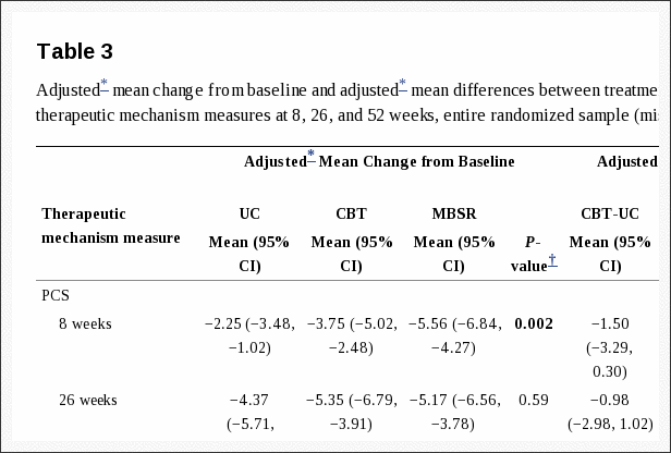
 

Blank
References

1. Konstantinou K, Dunn KM. Sciatica: review of epidemiological
studies and prevalence estimates. Spine (Phila Pa 1976) 2008;
33: 2464�2472.
2. Nygaard OP, Kloster R, Solberg T. Duration of leg pain as a
predictor of outcome after surgery for lumbar disc herniation:
a prospective cohort study with 1-year follow up. J Neurosurg
2000; 92: 131�134.
3. Orief T, Orz Y, Attia W, Almusrea K. Spontaneous resorption
of sequestrated intervertebral disc herniation. World Neurosurg
2012; 77: 146�152.
4. Maigne JY, Rime B, Deligne B. Computed tomographic follow-up
study of forty-eight cases of nonoperatively treated lumbar intervertebral
disc herniation. Spine (Phila Pa 1976) 1992; 17: 1071�1074.
5. Takada E, Takahashi M, Shimada K. Natural history of lumbar disc
hernia with radicular leg pain: spontaneous MRI changes of the
herniated mass and correlation with clinical outcome. J Orthopaed
Surg (Hong Kong) 2001; 9: 1�7.
6. Vroomen PC, de Krom MC, Knottnerus JA. Predicting the outcome
of sciatica at short-term follow-up. Br J Gen Pract 2002;
52: 119�123.
7. Weinstein JN, Tosteson TD, Lurie JD, Tosteson AN, Hanscom
B, Skinner JS, et al. Surgical vs nonoperative treatment for lumbar
disk herniation: the Spine Patient Outcomes Research Trial
(SPORT): a randomized trial. JAMA 2006; 296: 2441�2450.
8. Peul WC, van den Hout WB, Brand R, Thomeer RT, Koes BW.
Prolonged conservative care versus early surgery in patients with
sciatica caused by lumbar disc herniation: two year results of a
randomised controlled trial. BMJ 2008; 336: 1355�1358.
9. Atlas SJ, Keller RB, Wu YA, Deyo RA, Singer DE. Long-term
outcomes of surgical and nonsurgical management of sciatica secondary
to a lumbar disc herniation: 10 year results from the maine
lumbar spine study. Spine (Phila Pa 1976) 2005; 30: 927�935.
10. Weber H. Lumbar disc herniation. A controlled, prospective
study with ten years of observation. Spine (Phila Pa 1976) 1983;
8: 131�140.
11. Osterman H, Seitsalo S, Karppinen J, Malmivaara A. Effectiveness of microdiscectomy for lumbar disc herniation: a randomized
controlled trial with 2 years of follow-up. Spine (Phila Pa 1976)
2006; 31: 2409�2414.
12. Jacobs WC , van Tulder M, Arts M, Rubinstein SM, van Middelkoop
M, Ostelo R, et al. Surgery versus conservative management of
sciatica due to a lumbar herniated disc: a systematic review. Eur
Spine J 2011; 20: 513�522.
13. Svensson GL, Lundberg M, �stgaard HC, Wendt GK. High degree
of kinesiophobia after lumbar disc herniation surgery: a crosssectional
study of 84 patients. Acta Orthop 2011; 82: 732�736.
14. McKenzie R, May S. The lumbar spine: mechanical diagnosis
& therapy. 2nd ed. Spinal Publications New Zealand Limited:
Wellington; 2003.
15. Clare HA, Adams R, Maher CG. A systematic review of efficacy
of McKenzie therapy for spinal pain. Aust J Physiother 2004;
50: 209�216.
16. Paatelma M, Kilpikoski S, Simonen R, Heinonen A, Alen M, Videman
T. Orthopaedic manual therapy, McKenzie method or advice
only for low back pain in working adults: a randomized controlled
trial with one year follow-up. J Rehabil Med 2008; 40: 858�863.
17. Br�tz D, Kuker W, Maschke E, Wick W, Dichgans J, Weller M.
A prospective trial of mechanical physiotherapy for lumbar disk
prolapse. J Neurol 2003; 250: 746�749.
18. Hodges PW, Moseley GL. Pain and motor control of the lumbopelvic
region: effect and possible mechanisms. J Electromyogr
Kinesiol 2003; 13: 361�370.
19. Hides JA, Jull GA, Richardson CA. Long-term effects of specific
stabilizing exercises for first-episode low back pain. Spine (Phila
Pa 1976) 2001; 26: E243�E248.
20. Yilmaz F, Yilmaz A, Merdol F, Parlar D, Sahin F, Kuran B. Efficacy
of dynamic lumbar stabilization exercise in lumbar microdiscectomy.
J Rehabil Med 2003; 35: 163�167.
21. Nachemson AL. Lumbar disc herniation � conclusions. Acta Orthop
Scand Suppl 1993; 251: 49�50.
22. Kilpikoski S, Airaksinen O, Kankaanpaa M, Leminen P, Videman
T, Alen M. Interexaminer reliability of low back pain assessment
using the McKenzie method. Spine (Phila Pa 1976) 2002; 27:
E207�E214.
23. Richardson CA, Jull GA. Muscle control-pain control. What exercises
would you prescribe? Man Ther 1995; 1: 2�10.
24. Scott J, Huskisson EC. Graphic representation of pain. Pain 1976;
2: 175�184.
25. Fairbank JC, Couper J, Davies JB, O�Brien JP. The Oswestry
low back pain disability questionnaire. Physiotherapy 1980; 66:
271�273.
26. �berg B, Enthoven P, Kjellman G, Skargren E. Back pain in
primary care: a prospective cohort study of clinical outcome and
healthcare consumption. Adv Physiother 2003; 5: 98.
27. Bombardier C. Outcome assessments in the evaluation of treatment
of spinal disorders: summary and general recommendations. Spine
2000; 25: 3100�3103.
28. Vlaeyen JW, Kole-Snijders AM, Boeren RG, van Eek H. Fear of
movement/(re)injury in chronic low back pain and its relation to
behavioral performance. Pain 1995; 62: 363�372.
29. EuroQol � a new facility for the measurement of health-related quality
of life. The EuroQol Group. Health Policy 1990; 16: 199�208.
30. Zung WW. A self-rating depression scale. Arch Gen Psychiatry
1965; 12: 63�70.
31. Estlander AM, Vanharanta H, Moneta GB, Kaivanto K. Anthropometric
variables, self-efficacy beliefs, and pain and disability
ratings on the isokinetic performance of low back pain patients.
Spine 1994; 19: 941�947.
32. Str�mqvist B, J�nsson B, Fritzell P, H�gg O, Larsson BE, Lind B.
The Swedish National Register for lumbar spine surgery: Swedish
Society for Spinal Surgery. Acta Orthop Scand 2001; 72: 99�106.
33. den Boer JJ, Oostendorp RA, Beems T, Munneke M, Oerlemans
M, Evers AW. A systematic review of bio-psychosocial risk factors
for an unfavourable outcome after lumbar disc surgery. Eur Spine
J 2006; 15: 527�536.
34. Weber H, Holme I, Amlie E. The natural course of acute sciatica
with nerve root symptoms in a double-blind placebo-controlled
trial evaluating the effect of piroxicam. Spine (Phila Pa 1976)
1993; 18: 1433�1438.
35. Werneke M, Hart DL, Cook D. A descriptive study of the centralization
phenomenon. A prospective analysis. Spine (Phila Pa
1976) 1999; 24: 676�683.
36. Hahne AJ, Ford JJ, Hinman RS, Taylor NF, Surkitt LD, Walters
AG, et al. Outcomes and adverse events from physiotherapy
functional restoration for lumbar disc herniation with associated
radiculopathy. Disabil Rehabil 2011; 33: 1537�1547.
37. Balague F, Nordin M, Sheikhzadeh A, Echegoyen AC, Brisby H,
Hoogewoud HM, et al. Recovery of severe sciatica. Spine (Phila
Pa 1976) 1999; 24: 2516�2524.
38. Bono CM, Wisneski R, Garfin SR. Lumbar disc herniations. In:
Herkowitz HN, Garfin SR, Eismont FJ, Bell GR, Balderston RA,
editors. Rothman-Simeone the spine. 5th ed. Saunders Elsevier:
Philadelphia; 2006: p. 979�980.
39. Saal JA, Saal JS. Nonoperative treatment of herniated lumbar
intervertebral disc with radiculopathy. An outcome study. Spine
(Phila Pa 1976) 1989; 14: 431�437.
40. World Health Organisation. International Classification of Functioning,
Disability and Health (ICF). 2001 [cited 2012 Oct 9].
Available from: www.who.int/classifications/icf/en/.
41. Leijon ME, Faskunger J, Bendtsen P, Festin K, Nilsen P. Who is
not adhering to physical activity referrals, and why? Scand J Prim
Health Care 2011; 29: 234�240.
42. Perreault K. Linking health promotion with physiotherapy for low
back pain: a review. J Rehabil Med 2008; 40: 401�409.
43. Koho P, Orenius T, Kautiainen H, Haanpaa M, Pohjolainen T, Hurri
H. Association of fear of movement and leisure-time physical
activity among patients with chronic pain. J Rehabil Med 2011;
43: 794�799.

Close Accordion
Vertebrogenic Autonomic Dysfunction Subjective Symptoms: A Prospective Study

Vertebrogenic Autonomic Dysfunction Subjective Symptoms: A Prospective Study

The autonomic concomitants of cervical pathomechanics?(the posterior cervical sympathetic syndrome) have been?widely reported. The literature documenting the autonomic? manifestations of thoracic and lumbar articular dysfunctions? has not been as extensive. The present study? attempts to determine the incidence and nosography of ?vertebrogenic autonomic dysfunction (V.A.D.) in a sample?of 250 consecutive back pain subjects. Thirty-nine per cent?of all back pain subjects exhibited probable evidence of ?V.A.D. The incidence of V.A.D. was distributed as follows:?cervicogenic cephalalgia – 60%76 (i.e., disturbed?vision, dysequilibrium, gastrointestinal upset); thoracalgia?- 54% (ie., nausea, flatus); and lumbalgia – 31% (i e.,?constipation, urinary frequency, menstrual disturbances).

It is a well known clinical fact that diseases of the internal? organs may produce functional changes and symptoms ?or signs in the musculoskeletal system of the body. These ?somatic manifestations of visceral disease are fairly commonplace ?and are considered to be of importance in the?diagnosis and localization of internal disease. All clinicians ?have observed the conspicuous costovertebral angle muscle?spasm resulting from acute renal disorders (1). Abdominal?wall tension and tenderness are characteristic of certain?intra-abdominal and intrapelvic diseases (2-7). Coronary? disease is frequently accompanied by painful trigger areas?in the muscles of the chest and shoulder (8-9).?The premise that disorders of the musculoskeletal?system may reflexly cause autonomic dysfunction and?symptoms attributable to visceral malfunction is common?to both the chiropractic (10) and osteopathic schools (11),?but is not widely accepted by the medical profession. Physiologic ?research, based primarily on animal experimentation,?has demonstrated that somatovisceral reflex activity?is indeed a physiologic fact (12-19); however, much remains?to be done to understand the importance of these?reflexes in normal and abnormal human physiology. The ?literature of the osteopathic and chiropractic disciplines?has historically contained an abundance of anecdotal and?conceptual data in support of the somatovisceral reflex?hypothesis as a mechanism for symptom production in?man. Very little controlled clinical data has been presented?to support this hypothesis.

Palmer in 1895 (20) was probably one of the first to?report a simple cause and effect relationship regarding? somatovisceral symptom production in a patient. His subject?had apparently been working in a cramped position?and felt something “give in his back.” He claimed that he?simultaneously became deaf. Palmer examined the?patient’s spine and found a “displaced fourth dorsal vertebra”?(sympathetic vasomotor nerves to the cranium) and?corrected it with a manipulation. The patient’s hearing?was restored. Palmer thus deduced that the hearing loss?had been vertebrogenic in origin.

Three decades later, the allopathic observers, Barre in?1925 and Lieou in 1928, reported similar series of cases in?which disorders of the cervical spine were accompanied by?dysfunction of the organs of the head (21,22). In addition?to hearing loss, the following cervicogenic symptoms have?been observed: vertigo, dysequilibrium, tinnitus, scotomata,?decreased vision, dysphagia, dysphonia, cough, anxiety?and asthenia. (23)

The Barr6-Lieou syndrome (posterior cervical sympathetic ?syndrome) has been repeatedly recorded in the literature?since 1928. This syndrome represents a generally?accepted classic example of somatovisceral reflex?pathology and is to be found in much of the authoritative?orthopaedic literature of today (24).

Reports of somatically-induced visceral dysfunction have? not been confined to the cervical region, nor have they?been restricted to the literature of the chiropractic and?osteopathic schools. This is noteworthy, insofar as allopathic?training does not usually emphasize (or even include)?studies of somatovisceral reflex physiology. One?would therefore expect that allopathic observers would?approach their clinical observations with an absence of?bias and a low index of suspicion for somatovisceral?pathology. Nevertheless, such disorders have been obvious?enough to be noted and reported. Table 1 summarizes allopathic?observations in this regard as reported by Wills?(25), Ussher (26), Travell (27,28,29), Jackson (24), Cooper?(30), Lewit (31), Ushio et al (32), Love (33) and Ver Brugghen?(34).

The literature cited here would tend to indicate that ?somatically induced visceral dysfunction and symptom?production is indeed a clinical problem. The prevalence of?the problem is not known. The investigators reviewed in ?Table 1 have for the most part, not attempted to discover?the percentile incidence of autonomic symptoms in musculoskeletal?disease, nor have they made it clear whether?their data was based on consecutive groups of first-contact?patients or on treatment-resistant subjects who were referred?to their departments for special care.

vertebrogenic El Paso, TX

The purpose of this investigation was to determine the frequency of symptoms attributable to vertebrogenic autonomic dysfunction in a consecutive group of first-contact chiropractic patients.

Method

A pilot study, of the prospective-descriptive format, was designed. The focus of the study was on the anamnesis, with particular attention given to the sequence of clinical events. (For example, do patients with back pain simultaneously develop autonomic symptoms? If so, how often? 52 Do the autonomic symptoms disappear upon recovery from the spinal pain?) The emphasis then, was on seeking a parallelism between the development and disappearance of back pain with the development and disappearance of visceral symptoms.

Inclusion/Exclusion and Data Gathering:

The following guidelines for data gathering were adhered to throughout the study:

1. Two hundred and fifty consecutive first-contact patients who presented with back pain (cervical, thoracic or lumbar) comprised the sample for analysis.

2. After a thorough case-history was obtained from each subject, careful notation was made of associated symptoms. The phrasing of this latter aspect of the interview was as follows: “Have you, since the onset of your back pain, developed any other seemingly unrelated symptoms for example, have you felt generally ill? Have you been constipated? Have you had to empty your bladder more frequently – or less frequently?” Usually, four or five examples of known vertebrogenic symptoms were mentioned. See Table 1. A systems review followed. As additional symptoms were uncovered, it was determined whether or not their onset in any way paralleled the onset of back pain.

3. The patient was not given a r6sume prior to the beginning of treatment for his back pain. No further comment was made regarding the subject’s visceral symptoms, if present. No mention was made of any relationship between the spinal pain and the visceral symptoms. A chart of the autonomic nervous system was removed from the office. The patient was not told that analytical data was being gathered. The attempt here was to limit, as much as possible, the introduction of accentuated placebogenic and/or Hawthorne effects (35) into the study.

4. The subjects who were accepted as chiropractic patients were treated with spinal manipulative therapy (primarily chiropractic high velocity, controlled amplitude adjustment). In some cases, ancillary physical therapeutic methods were utilized as indicated. Patients who were referred to other health disciplines for care (i.e. surgery) remained in the study providing that follow-up data were available. (This study was not designed to test the effectiveness of manipulative therapy its sole purpose was to investigate spinovisceral symptom parallelism see illustrative case 4.)

5. The subjects were re-evaluated when they became asymptomatic with respect to their back pain or were much improved (at least 80%o by mutual patient-examiner agreement). The patients were at this time questioned regarding the status of any associated visceral symptoms that had been previously reported. The observation that recovery from visceral symptoms paralleled recovery from the spinal pain was taken to be confirmatory evidence of vertebrogenic autonomic dysfunction in that subject.

Subjects were excluded from the study for the following reasons: non-acceptance as a patient because of organic pathology or other contraindications, non-compliance, self-discharge from care, unsatisfactory recovery from the spinal complaint and incomplete admission or follow-up records. When a patient was dropped from the study, the next consecutive patient history was admitted to the data pool so that the target number of 250 subjects could be met.

A survey of drug usage by patients in this study revealed the following: 52% of patients did not use drugs at all; 32% has self-administered approximately 1 to 6 non-prescription analgesic tablets (i.e. aspirin, codeine) and had subsequently sought professional care; 16% were on regular therapeutic dosages of physician prescribed or self-administered pharmaceuticals either for spinal pain or for other unrelated conditions. Most of the subjects who had used pharmaceuticals remained in the study. They were included or excluded after careful analysis of the individual data (see data analysis guidelines).

Vertebrogenic Data Analysis:

The presence or absence of vertebrogenic autonomic dysfunction (V.A.D.) was determined from the admission and follow-up records on each subject. The likelihood of vertebrogenicity was determined on the strength of the gathered data. The information on each case was then categorized as yielding probable, possible or negative evidence of vertebrogenic autonomic dysfunction. The guidelines for categorization were as follows:

1. Probable: Patients who exhibited an unequivocal parallelism between the onset and abatement of back pain with the onset and abatement of visceral symptoms were considered to have evidence of probable vertebrogenic autonomic dysfunction.

2. Possible: If there was any doubt regarding the accuracy of the gathered data (e.g. inconsistency of the subject’s responses to questions) the findings were categorized as “possible V.A.D.” If any discrepancies were noted in spinovisceral symptom parallelism, the data was considered to yield evidence of possible vertebrogenic autonomic dysfunction. For example, patients who fully recovered from spinal pains, but were left with vestiges of autonomic symptoms or patients who recovered from autonomic symptoms but were left with some spinal pain, were placed in the V.A.D. possible category.

3. Negative: Subjects who developed back pain but did not experience any symptoms attributable to autonomic dysfunction were classified as “V.A.D. negative”.

As stated in the previous section, data from patients who had used pharmaceuticals were individually scrutinized for inclusion or exclusion, and categorization. This analytical process would best be illustrated by the following examples:

Case 1: This patient developed acute lumbalgia. He self-administered four 222 tablets� (total 32 mgs. codeine) on day 1 and presented here the next morning. He reported constipation as an associated symptom. The constipation resolved on day 3 although the lumbalgia continued. He remained in the study and was classified as “negative V.A.D.”

Case 2: This patient developed cervicalgia and self-administered 6 aspirin over day 1. She presented on day 2 and reported dizziness, gastric upset and flatus as associated symptoms. She discontinued her aspirin, but the autonomic symptoms persisted throughout the course of her treatment. All spinal and associated symptoms abated during the 3rd week. She remained in the study and was classified as “possible V.A.D.”

Case 3: This patient developed a severe cervical-brachial neuralgia. Her family physician prescribed Tylenol� and diazepam. She presented on day 10. She reported anorexia, nausea, flatus, blurred vision, dizziness and mental fogging as associated symptoms. On day 24 her cervicalgia and all associated symptoms had cleared, yet she remained on the prescribed medication. She was classified as “probable V.A.D.”

Results

Of the original 250 subjects who met the criteria for admission to the study, 22 were subsequently lost (20 with self-discharges and/or poor results, 2 with organic disease found on continuing examination) and replaced by next consecutive patients. Ninety-eight (39%) of all subjects exhibited “probable” and thirty-three (13%) exhibited “possible” autonomic dysfunction. Also noteworthy was the observation that four (2%) of the subjects experienced reactivation (relapse) of previously quiescent visceral disease during their back pain episodes (2 duodenal ulcers, 2 lower genitourinary infections).

The levels of spinal complaint fell into the following four categories:

1. cervicalgia with cephalalgia,

2. cervicalgia (with or without upper extremity pain),

3. thoracalgia, and

4. lumbalgia (with or without lower extremity pain).

The frequency of autonomic dysfunction for each category is given in Table 2. The frequency distributions of the various autonomic manifestations observed are given in Tables 3 – 6.

While Tables 3 – 6 give the frequency of symptoms observed in the present study, they do not give an accurate picture of vertebrogenic autonomic dysfunction as actually seen in clinical practice. Many of the subjects had multiple autonomic symptoms and these symptom complexes are not accurately portrayed by tables. The following case studies are instructive:

vertebrogenic El Paso, TXvertebrogenic El Paso, TX

Case 4: Mrs. F. presented with neck pain and headaches. She had sustained a whiplash injury 2 years previously and her symptoms had cleared with chiropractic therapy. Her present symptoms had recurred 2 months prior to her consultation here. She complained of a constant, moderate to severe upper cervical ache which radiated into the occipital-frontal regions. The cephalalgia occurred daily and intensified in the mid afternoon. She reported associated dizziness and difficulty in focusing her eyes. She could not clearly focus on close objects and stated that while driving, the windshield seemed to move back and forth in relation to her eyes. She was constantly squinting in an effort to see clearly. She had been seen by an optometrist 3 weeks previously with negative findings. On examination, cervical extension, right side-bending and left rotation were painful and limited. There was palpable paravertebral muscle spasm and restriction of motion in the upper cervical articulations. Trigger points were found here which reproduced the cephalalgia.

Cervicogenic autonomic dysfunction was objectivated with the following manoeuvres:

1. Triggers: Firm pressure over the right lamina of C2 precipitated an immediate bout of blurred vision and diplopia.

2. Resisted Motion: Resisted cervical extension (with the head fixed so as to exclude vestibular motion) resulted in an episode of “swooning” and vertigo.

3. Cervical Torsion: The patient’s head was fixed by an assistant (so as to exclude vestibular motion) and the torso was rotated, flexed, extended, side-bent and circumducted under the immobilized cranium. These maneuvers precipitated vertigo.

Cervicogenic autonomic dysfunction was further confirmed by the therapeutic test. The patient was treated with the manual adjustment of the upper cervical spine. The results were as follows:

1. The initial manipulation caused a transitory aggravation of the dizziness and the visual symptoms.

2.�This was quickly relieved by manual traction.

3.�The patient was fully recovered from all articular (pain) and non-articular (autonomic) symptoms by the tenth office visit. She remained asymptomatic over a 4 month follow-up period.

Case 5: Mrs. J. presented with low cervical, right scapular and right mid-thoracic pain of several months duration. There was associated dizziness, staggering and blurred vision. She had been referred to an ENT specialist who was non-commital in regards to a diagnosis. On examination, a trigger point was located on the antero-lateral aspect of C6-7 which reproduced her pain. The cervical torsion test was strongly positive the patient requiring support because of the precipitated vertigo. The lower cervical segments were adjusted and all symptoms abated after 5 visits. The patient has had several relapses over the ensuing months. Dysequilibrium has been a constant concomitant of each attack.

Case 6: Mr. R. Complained of a constant, dull mid-thoracic ache of 3 weeks duration. The symptoms had occurred after the patient spent several days working in the stooped forward position. The patient also complained of nausea and a feeling of a “lump” in the epigastric region. He stated that his food remained in his stomach for several hours after ingestion. He regurgitated frequently and was troubled with belching and flatus. He occasionally had crampy abdominal pains. On examination, the spinous processes and right costotransverse articulations of T4 to T7 were exquisitely tender. There was rhomboid. spasm on the right and mid-thoracic motion was�restricted. A single adjustment to this level was followed by immediate pain relief together with several minutes of massive gaseous eructation. All other symptoms cleared over the next few hours and did not recur over a 3 month follow-up period.

vertebrogenic El Paso, TXvertebrogenic El Paso, TXvertebrogenic El Paso, TXCase 7: Mr. V. presented with acute lumbo-sciatica of one week’s duration. The pain had occurred after a straight-legged lift. Mr. V. reported that since the occurrence of his injury he had been troubled with alternating bouts of constipation and diarrhea, urinary frequency, nocturia, partial urinary retention, impotence and “retraction” of the testes. He exhibited sciatic kyphoscoliosis. All trunk movements were limited and painful. Straight-leg raising, foot dorsiflexion and Valsalva maneuvers were positive. Deep springing of the L4 vertebra aggravated his pain. Sphincter tone was normal. A trial of manipulation afforded only temporary (hours-days) relief. The left Achilles reflex became sluggish and the patient was referred for neurosurgery. An L4 discotomy resulted in complete recovery from all spinal and visceral symptoms.

Case 8: Mrs. R. presented with an acute lumbalgia which referred to the left groin. The pain had occurred 4 days previously and since that time she had been troubled with constipation, flatus, urinary frequency and a burning dysuria. Her urinalysis was negative for abnormality. Lower lumbar and left sacroiliac dysfunctions were corrected with manipulation. All symptoms had cleared by the fourth office visit.

At the time of discharge from care, Mrs. R. asked if her bowel and bladder symptoms could have anything to do with the lower back. She stated that every time she had a lower back episode she developed the same pelvic symptoms. She, had had several unremarkable bowel, gall bladder and urinary investigations in regards to these symptoms.

Discussion:�Vertebrogenic Autonomic Dysfunction

The exact mechanism of spinovisceral symptom production is not conclusively known. Several pathophysiologic hypotheses could be advanced to explain these clinical phenomena. It is also possible that different pathological processes are operant in different individuals. Postulated mechanisms of spinovisceral symptom production are summarized below:

Pain: The autonomic concomitants of severe pain are well known. They are due to mass sympathetic stimulation and include such symptoms as agitation, hyperhidrosis, pupillary dilation and vomiting.

Stress-Endocrine: Selye (36) has pointed out that many disease states are accompanied by two groups of symptoms – the symptoms produced by the stressor and another group of symptoms resulting from the body’s endocrinal defense responses to the stressor. Using the eosinophil count as a stress indicator, I presented evidence which would suggest that 54% of severe lumbalgic episodes are accompanied by an alarm-endocrine reaction (37). It is possible that some of the symptoms observed in this study have an endocrine-chemical basis.

Somato-Psycho-Visceral Reflexes: It is well known that certain patients look at their pains through a psychological magnifying glass and develop all manner of unrelated symptoms. Psychogenic symptom production, no doubt, introduces an artifact into this study. It is, however, very interesting to note that most of the symptoms observed here, bear a segmental relationship to the level of spinal lesion (only one patient in this series had any knowledge of the anatomy of the autonomic nervous system). These segmental spino-psycho-visceral relationships may be explained by osteopathic research. In a brilliant experiment, Korr et al have shown that when the psyche is stimulated, maximum sympathetic outflow occurs at hyperirritable cord levels which have been previously sensitized (facilitated) by proprioceptive input from preexisting spinal joint lesions (38).

Somato-Visceral Reflexes: Somato-autonomic reflexes in the laboratory animal are a physiologic fact. They are observed under certain conditions in man. The postulated modus operandi in spinal lesions is as follows: the lesioned spinal joints trigger an increased afferent input (pain, proprioception) into the related cord segments. The lateral horn cells are facilitated via the internuncial neurons and impulses spill over into the sympathetic efferents causing activation (motor, vasomotor, secretory) of the target 56 viscera. Such sustained pathophysiologic activity could well be responsible for spinovisceral symptoms observed in back pain subjects.

Nerve Compression: It is well known that discal lesions, vertebral exostoses and stenosis may compress nervous tissue and cause autonomic dysfunction or paresis. It is also possible that root compression may interfere with axoplasmic flow. If, for the purpose of survey, one considers paresthesia/anesthesia to be indicative of nerve-tissue compromise, then 30% of first-contact patients presenting at this office have nerve compression syndromes. This could be responsible for autonomic dysfunction in selected patients.

Vascular Compromise: Cervical degenerative lesions may compromise the vertebral arteries and cause cranial symptoms. This mechanism could have been operative in aged patients; however, the slowly progressive cerebral dysfunction (39), which allegedly occurs with sustained neck torsion in this condition, was not observed in any of the subjects.

Proprioceptive Cross-Talk: It is possible that unequal cervical muscle tensions might feed the central nervous system with confusing proprioceptive information so as to disturb normal righting reflexes. This mechanism could explain the high incidence of dysequilibrium in subjects with neck injuries.

Other: The constipation observed in severe lumbalgic patients may be antalgic or due to lack of exercise.

The spinovisceral syndromes described here are typical of the symptom complexes observed in the everyday clinical practices of all manipulative practitioners. Further studies, to more precisely explore these common clinical problems, are warranted. The present study represents a first-attempt prospective investigation and the presented statistics may be considered to be “ball-park” figures only. A much larger subject population is required. Analysis of the gathered data in a preliminary study such as this, almost invariably exposes deficiencies in the original prospective design which could be corrected to improve future investigations (40). The following recommendations should be considered:

Investigator Artifact: Solo investigators who study their own patient populations may be subject to impaired objectivity. Future studies should utilize a multi-disciplinary team of investigators to design and carry out the project.

Data Artifacts: All data gathering methods (verbal interviews and questionnaires) are subject to error (40,41). The interview method was used in the present study. Future studies should include both verbal interviews and written questionnaires to facilitate cross-checking of the accuracy of the gathered data.

Psychologic Artifacts: Clinical studies are frequently criticized for failing to establish psychological base-lines for the studied subjects. Written questionnaires could incorporate standard psychological testing proforma.

Pharmacologic Artifacts: Investigator judgement (bias) was used in categorizing data on drug users. Future studies should exclude these subjects, or at least analyze their data in a separate category.

Statistical Artifacts: A much larger study population is required in order to establish accurate prevalence rates for vertebrogenic autonomic dysfunction. Formal statistical analysis is also required.

Follow-Up: The foregoing study was designed to be an acute investigation of a common clinical syndrome. It asked simple, straight-forward questions regarding the sequence of events during back pain episodes. While many of the subjects were followed for months, and even years, it is debatable if long term follow-up would bring forth any additional answers to the questions asked. It is important to note, however, that historical chiropractic and osteopathic theory, in common with the psychosomatic school, hypothesizes that long standing functional disorders may eventually lead to organic pathology. Follow-up of patients with recurrent spinal lesions, over many years, might constitute one way of testing this hypothesis.

Conclusions:�Vertebrogenic Autonomic Dysfunction

The autonomic manifestations of back pain have been explored. This preliminary study indicates that 39%o of all back pain subjects have symptoms attributable to irritation of the- autonomic nervous system by spinal lesions. Further studies, which incorporate more rigid protocol, are required to accurately describe the nosographic features of these common clinical syndromes.

blank
References:

1. Guyton A. Textbook of medical physiology. Fifth edition. Philadelphia:
W B Saunders, 1976.
2. Brobeck JR. Physiologic basis ofLmedical practice. Tenth edition.
Baltimore: Williams and Wilkins, 1979.
3. Guyton A. Basis human physiology. Second edition. Philadelphia:
W B Saunders, 1977.
4. Beeson P, McDermott W. Textbook of medicine. Fourteenth edition.
Philadelphia: W B Saunders, 1975.
5. Cecil R, Loeb R. Textbook of medicine. Ninth edition. Philadelphia:.
W B Saunders, 1955.
6. Adams J. Outline of orthopaedics. Sixth edition. Edinburgh: Livingston,
1967.
7. Pottenger F. Symptoms of visceral disease. Seventh edition. St Louis:
CV Mosby, 1953.
8. Pinzler S, Travell J. Therapy directed at the somatic component of
cardiac pain. Am H J 1948; 35: 248-268.
9. Travell J, Pinzler S. The myofascial genesis of pain. Postgraduate
Medicine 1952; 11: 425-430.
10. Verner J. The science and logic of chiropractic. Englewood: J Verner,
1941.
11. MacDonald G, Hargrave-Wilson W. The osteopathic lesion. London:
Heinemann, 1935.
12. Sato A, Schmidt R. Somato sympathetic reflexes: afferent fibers,
central pathways, discharge characteristics. Physiological Reviews
1973; 53: 916-947.
13. Sato A, Sato Y, Shimada F, Torigata Y. Changes in vesical function
produced by cutaneous stimulation in rats. Brain Research 1975; 94:
465-474.
14. Sato A, Sato Y, Shimada F, Torigata Y. Changes in gastric motility
produced by nociceptive stimulation of the skin in rats. Brain Research
1975; 87: 151-159.
15. Sato A, Sato Y, Shimada F, Torigata Y. Varying changes in heart
rate produced by nociciptive stimulation of the skin in rats at different
temperatures. Brain Research 1976; 110: 301-311.
16. Haldeman S. Interactions between the somatic and visceral nervous
systems. JCCA 1971; 15(3): 20-25.
17. Sato A. The importance of somato-autonomic reflexes in the regulation
of visceral organ function. JCCA 1976; 20(4): 32-38.
18. Coote J. Somatic sources of afferent input as factors in aberrant
autonomic, sensory and motor function. In: Korr IM, ed. The neurobiologic
mechanisms in manipulative therapy. New York: Plenum
Press, 1978: 91-127.
19. Appenzeller 0. Somatoautonomic reflexology – normal and abnormal.
In:- Korr IM, ed. The neurobiologic mechanisms in nmanipulative
therapy. New York: Plenum Press, 1978: 179-217.
20. Palmer D. The science, art and philosophy of chiropractic. Portland:
Portland Printing House, 1910: 18.
21. Barre J. Rev Neurol 1926; 33: 1246
22. Lieou Y. Syndrome sympathique cervical posterieur et arthrite cervicale
chronique. These de Strasbourg, 1928. (Fre)
23. Gayral L, Neuwirth E. Oto-neuro-ophthalmologic manifestations of
cervical origin. NY State J Med 1954; 54: 1920-1926.
24. Jackson R. The cervical syndrome. Springfield: Charles C Thomas,
1966: l31-144.
25. Wills 1, Atsatt R. The viscerospinal syndrome: a confusing factor in
surgical diagnosis. Arch Surg 1934; 29: 661-668.
26. Ussher N. The viscerospinal syndrome – a new concept of visceromotor
and sensory changes in relation to deranged spinal structures.
Ann Int Med 1940; 13(2): 2057-2090.
27. Travell J, Bigelow N. Role of somatic trigger areas in the patterns of
hysteria. Psychsom Med 1947; 2: 353-363.
28. Travell J. Referred pain from skeletal muscle. NY State J Med; 1955
Feb: 331-340.
29. Travell J. Mechanical headache. Headache 1967 Feb: 23-29.
30. Cooper A. Trigger-point injection: its place in physical medicine.
Arch Phys Med Rehab 1961; 704-709.
31. Lewit K. Menieres disease and the cervical spine. Rev Czechoslovak
Med 1961; 7(2): 129-139.
32. Ushio N, Hinoki M, Hine S, Okada S, Ishida Y, Koike S, Shizuba S.
Studies on ataxia of lumbar origin in cases of vertigo due to whiplash
injury. Agressologie 1973; 14(D): 73-82.
33. Love J, Schorn V. Thoracic disc protrusions. JAMA 1%5;
43-62.
34. Ver Brugghen A. Massive extrusions of lumbar intervertebral discs.
Surg Gynecol Obstet 1945; 81: 269.
35. Treece E, Treece J. Elements of research in nursing. First edition.
Saint Louis: CV Mosby, 1973.
36. Selye H. The stress of life. New York: McGraw-Hill, 1956.
37. Johnston R. Vertebrogenic stress eosinopenia. JCCA 1974; 18(4):
14-20.
38. Korr l, Thomas P, Wright R. Symposium on the functional implications
of segmental faciliation. JAOA 1955; 54: 173.
39. Houle J. Assessing hemodynamics of the vertebro-basilar complex
through angiothlipsis. JCCA 1972 June: 35-36, 41.
40. Friedman G. Primer of epidemiology. First edition. New York:
McGraw-Hill, 1974.
41. Koran L. The reliability of clinical- methods, data and judgments.
New Engl J Med 1975; 293: 642-646.

Close Accordion
Amazing Results from Herniated Disc Early Treatment | El Paso, TX

Amazing Results from Herniated Disc Early Treatment | El Paso, TX

A herniated disc is typically a very painful condition, especially if the inner gel-like substance of the intervertebral disc, known as the nucleus pulposus, pushes through the thick, outer ring of cartilage and puts pressure on the sensitive nerves of the spine. Discs are soft, rubbery pads found between each vertebrae of the spine that act as shock-absorbers, allowing the spine to bend and/or flex. An intervertebral disc may begin to rupture as a result of wear-and-tear or due to a sudden injury. Fortunately, most individuals who’ve suffered a herniated disc can find relief from a variety of non-operative treatments before considering surgery. The following article highlights the impact of early treatment for herniated discs in the lumbar spine, or low back.


The Impact of Early Recovery on Long-Term Outcomes in a Cohort of Patients Undergoing Prolonged Non-Operative Treatment for Lumbar Disc Herniation: Clinical Article


Abstract


Object


The authors comprehensively studied the recovery of individual patients undergoing treatment for lumbar disc herniation. The primary goal was to gain insight into the variability of individual patient utility scores within a treatment cohort. The secondary goal was to determine how the rates and variability of patient recovery over time, represented by improvement in utility scores, affected long-term patient outcomes.


Methods


EuroQol Group�5 Dimension (EQ-5D) scores were obtained at baseline and at 2, 4, 8, 12, 26, 38, and 52 weeks for 93 patients treated under a prolonged conservative care protocol for lumbar disc herniation. Gaussian kernel densities were used to estimate the distribution of utility scores at each time point. Logistic regression and multistate Markov models were used to characterize individual patient improvement over time. Fisher exact tests were used to compare the distribution of EQ-5D domain scores.


Results


The distribution of utility scores was bimodal at 1 year and effectively sorted patients into a �higher� utility group (EQ-5D = 1; 43% of cohort) and a �lower� utility group (EQ-5D ? 0.86; 57% of cohort). Fisher exact tests revealed that pain/discomfort, mobility, and usual activities significantly differed between the 2 utility groups (p ? 0.001). The utility groups emerged at 8 weeks and were stable for the remainder of the treatment period. Using utility scores from 8 weeks, regression models predicted 1-year outcomes with 62% accuracy.


Conclusions


This study is the first to comprehensively consider the utility recovery of individual patients within a treatment cohort for lumbar disc herniation. The results suggest that most utility is recovered during the early treatment period. Moreover, the findings suggest that initial improvement is critical to a patient’s long-term outcome: patients who do not experience significant initial recovery appear unlikely to do so at a later time under the same treatment protocol.


Abbreviations used in this paper: AUC = area under a receiver-operating curve; EQ-5D = EuroQol Group�5 Dimension. Address correspondence to: Matthew C. Cowperthwaite, Ph.D., The University of Texas at Austin, Texas Advanced Computing Center, J.J. Pickle Research Campus, ROC 1.101, 10100 Burnet Rd., Austin, TX 78758. email: mattccowp@mac.com.


Plublished online June 28, 2013; DOI: 10.3171/2013.5.SPINE12992.


Introduction


Lumbar disc herniation is one of the most common causes of low-back pain and radiculopathy.[4] Treatment for patients with a herniated lumbar disc usually begins with conservative care such as analgesics, epidural steroid injections, and physical therapy,[1,5] with surgery reserved for patients with severe nerve root or cauda equina dysfunction or if conservative therapy is unsuccessful in controlling the symptoms.


Several recent studies have compared the effectiveness of conservative care and surgical treatment protocols for treating herniated lumbar discs, and have arrived at varying conclusions.[2,3,9,10,15�18] However, these studies have generally considered outcomes over a period of years, which is a significant length of time for patients who are waiting for their quality of life to improve. In clinical practice, this often leads to the following dilemma: most patients, particularly those with moderate symptoms, would prefer to avoid surgery, but are unwilling to wait an indefinite period of time for their symptoms to resolve. Unsurprisingly, lumbar discectomy is the most frequently performed surgical procedure in the US.[17,18]


Moreover, the above-mentioned studies have typically compared the average difference between treatment groups, without regard for individual recovery within the cohort. Additionally, this approach assumes that recovery in the protocols being compared proceeded similarly between observation intervals. To better understand the treatment responses of individual patients and the time frames of their responses, we comprehensively analyzed a cohort of patients undergoing a prolonged conservative care treatment protocol to gain insight into the dynamics of individual patient recovery over time, and whether these recovery dynamics influence long-term outcomes.


Methods


Study Data Set


The data set contained 142 patients randomized to a protocol of prolonged conservative care as part of the Leiden�The Hague Spine Intervention Prognostic Study.[10,15] The Sciatica Trial was reviewed and approved by the Medical Ethics Committee of Leiden University Medical Center.[11] Patients were enrolled into the Sciatica Trial entirely in the Netherlands.


In the Sciatica Trial, all patients aged 18 to 65 years, with persistent radicular pain in the L-4, L-5, or S-1 dermatome (with or without mild neurological deficit), severe disabling leg pain (lumbosacral radicular syndrome) lasting 6�12 weeks, and radiologically (MRI) confirmed disc herniation were considered eligible to enroll in the trial. Cauda equina syndrome or severe paresis, prior complaints of lumbosacral radicular syndrome in the previous 12 months, history of same-level unilateral disc surgery, spinal canal stenosis, and degenerative or lytic spondylolisthesis were all exclusion criteria. Cohort demographics and baseline characteristics were previously described; all patients reported both back and leg pain, but leg pain was generally more severe (mean leg pain 67.2 � 27.7 vs back pain 33.8 � 29.6, measured on a 100-point, horizontal visual analog scale).[15]


The Sciatica Trial used a pragmatic study design: conservative-care management was influenced as little as possible and was supervised by each patient’s general practitioner. Use of analgesics and physical therapy was determined by the treating physician. In this cohort, 46 patients (32%) elected to have surgery before the end of the 1st year; the mean timing of surgery was 12.6 weeks after the start of treatment. The surgical patients and 3 additional subjects with more than 2 missing utility measures were removed from the sample, resulting in a cohort of 93 patients considered in the present study; the crossover patients will be discussed in a separate study (manuscript in preparation). Our results were qualitatively unchanged when the excluded patients were retained in the analyses (data not shown).


In the Leiden�The Hague Spine Intervention Prognostic Study the EQ-5D instrument was used to measure patient utility at baseline and at 2, 4, 8, 12, 26, 38, and 52 weeks after enrollment into the study. The average duration of sciatica prior to enrollment was 9.5 weeks.[10,15] Utility is a valuation of a patient’s quality of life on a scale between 0 (as bad as dead) and 1 (perfect health). To estimate utility, the EQ-5D assesses a patient’s functional impairment in 5 domains: mobility, self-care, usual activities, pain, and anxiety.[6] For each domain, patients self-report the scores of 1 (no problems), 2 (some problems), or 3 (extreme problems). Utility scores were computed using the US valuation model,[12] which clearly distinguishes patients reporting no health problems (EQ-5D = 1) from those reporting at least some health problems (EQ-5D ? 0.86). Our results are independent of the particular valuation model (not shown). Completeness of the EQ-5D measures during follow-up ranged from 98% at 2 weeks to 90% at 38 weeks.


Statistical Analysis


All statistical analyses were conducted using the R statistical environment (version 2.9.2; www.rproject.org/) with the additional �msm,�[8] �ROCR,�[14] and �rms�[7] packages (all freely available from cran.rproject.org). Continuous variables are presented as means (� SEM) and were compared using 2-tailed Student t-tests. Significance was assessed at an ? ? 0.05 significance level, unless otherwise indicated. Missing EQ-5D measures were imputed using the mean of the measures at adjacent time points; our results are qualitatively similar under forward or backward imputation schemes (not shown).


Gaussian kernel density estimates were computed to estimate the distribution of utility scores. The kernel density estimates were estimated using a Silverman’s �rule-of-thumb� bandwidth and a Gaussian smoothing kernel.[13] The left- and right-most points were set to the theoretical minimum and maximum EQ-5D values, respectively, so that the area under the density curve summed to 1.


To determine whether specific EQ-5D domains differed between utility groups, Fisher-exact tests were conducted on contingency tables of the number of patients in each utility group that reported scores of 1, 2, or 3. Significance was assessed using a Bonferroni-corrected p value of 0.01.


Two-state, continuous-time Markov models were used to study the patterns and probabilities of patients transitioning between a �lower� utility (EQ-5D ? 0.86) and a �higher� utility group (EQ-5D = 1). The threshold utility value defining the groups remained fixed over time and was used to assign each patient to a utility group at each observation time. The models were fitted using the �msm� package[14] with piecewise-constant transition intensity matrices (Qt) estimated for each time interval between the points t = 0, 4, 8, 12, 26, 38, 52 (t = 2 was omitted because there were insufficient transitions to yield a robust model). Transition intensities were permitted to change between subsequent observation intervals, but remained homogeneous within each observation interval. The starting transition intensities were based on the observed frequencies of transitions in the data set and were calculated using the formula


Article-Formula.jpg


in which nij is the observed number of transitions from Group i to Group j over the duration of the study period (T), and nj is the initial number of patients in Group j. The fitted models were robust to the choice of starting transition intensities and yielded qualitatively similar parameter estimates over a range of starting parameters (not shown). The likelihood function was maximized using a Nelder-Mead algorithm, and convergence was visually verified and typically occurred well short of the maximum number of iterations.


Logistic regression models were used to test whether utility measurements from earlier time points could predict long-term outcomes. These models only included utility values up to a particular time point as predictors, with the response variable being the patient’s 1-year outcome (higher or lower utility group) modeled as a dichotomous variable; no additional clinical or demographic covariates were included in the models. The models were fitted using the �rms� package[7] and the fit was assessed using chi-square tests (? ? 0.05). Separate regression models were created for all utility measurements up to and including those for 2, 4, 8, 12, and 26 weeks; for example, the 8-week model would include utility measurements at 0, 2, 4, and 8 weeks. The AUC statistic was used to assess the performance of the models and was calculated using the ROCR package.[14]


Results


Delineation of Higher and Lower Utility Groups


The distributions of patient utility scores markedly changed over the course of 1 year of conservative care (Fig. 1). At baseline, the majority of patients reported a relatively poor quality of life; the mean EQ-5D score was 0.55 (median 0.60). Two distinct utility groups were found to be present at baseline: a �lower� utility group (EQ-5D ? 0.86) and a �higher� utility group (EQ-5D = 1). At 6 months, the lower utility group (n = 62, 67%) was larger than the higher utility group (n = 31, 33%); at 1 year, the lower utility group (n = 53, 57%) had declined, but remained larger than the higher utility group (n = 40, 43%).


Figure 1 Distribution of EQ-5D Patient Utilities | El Paso, TX Chiropractor

Figure 1: Distribution of EQ-5D patient utilities at baseline, 6 months, and 1 year. The solid lines depict Gaussian kernel density estimates (right axis) of each distribution. The gray lines outline the histogram with the height of each bar representing the frequency of patients (left axis) in the equal-width bins (0.05) with utility greater than the lower bound and less than or equal to the upper bound. The bounds of both distributions are set to the theoretical minimum and maximum of the EQ-5D utility instrument.


EQ-5D Domain Scores Between Groups


The average scores in each domain of the EQ-5D (Table 1) suggested that the pain/discomfort (low score = 1.9, high score = 1.0), mobility (low score = 1.4, high score = 1.0), and usual activities (low score = 1.5, high score = 1.0) domains differed most significantly between the high and low utility groups (p ? 0.001). The anxiety (low score = 1.2, high score = 1.0) and self-care (low score = 1.1, high score = 1.0) domains differed much less between the 2 utility groups, although they were also significant (p < 0.01).


Table 1 Distribution of Scores in Each EQ-5D Domain | El Paso, TX Chiropractor


Trajectory of Patient Utility Over Time


The series of patient utility scores measured over the study period are referred to as utility �trajectories,� which were studied to understand how patients recovered over the study period. In the study cohort, all patients experienced improvement during at least 1 observation period; only 19.3% (n = 18) never experienced a decline during their recovery. Recovery was variable: 49.5% of the patients (n = 46) experienced at least 2 reversals, which were defined as improvements (declines) immediately followed by declines (improvements) at the next observation. Furthermore, only 29% of patients (n = 27) had stable trajectories with no reversals. Overall, increases in utility were 4 times more common than decreases in utility.


The utility of the entire cohort increased by 0.296 (51.8% above baseline; p ? 0.001, Wilcoxon Mann-Whitney test) over the year (Fig. 2), but was markedly faster during the first 2 months (0.022/week) than the final 3 months (0.005/week). Over the same time frames, utility scores improved by 0.178 (35.2% above the baseline average) over the first 2 months and by 0.063 (1.3% above the 9-month average) during the final 3 months. The mean utility scores significantly differed between the 2 final utility groups at 8 weeks and remained significant for the rest of the year (p < 0.01, Student t-test; Fig. 2).


Figure 2 Graph of Mean Patient Utilities | El Paso, TX Chiropractor

Figure 2: Graph of mean patient utilities at each measurement time point. Error bars represent 95% CIs about the mean. High and low utility group refers to the final group in which the patient belongs at the 1-year time point.


Modeling Patient Recovery


Given that 2 utility groups were present over the study period, Markov models were used to study the robustness of these groups by estimating the likelihood of patients switching between the groups. The models suggested that the average probability of a patient remaining within their utility group was 97.9% and 97.6% for patients currently in the low and high utility groups, respectively (Fig. 3). The probability of a patient transitioning from the low to the high utility group was 2.1%; the corresponding probability for transitions from the high to the low utility group was 2.3%.


Figure 3 Graphs of the Markov Transition Probabilities | El Paso, TX Chiropractor

Figure 3: Graphs of the Markov transition probabilities (per week) for transitions within (lower) and between (upper) utility groups. Each point is centered at the middle of each time interval and represents the maximum-likelihood estimate of the per-week transition probability during the entire interval. Error bars (mean width of the 95% CI was 1.8) were omitted for clarity because the differences were not significant.


The models also suggested that the likelihood of a patient transitioning to another utility group declined over the study period. During the first 8 weeks, 2.8% and 3.5% of patients experienced low-to-high and high-to-low transitions, respectively; over the last 3 months, 1.6% and 1.3% of patients experienced low-to-high and high-to-low group transitions, respectively.


Predicting Individual Patient Outcome


At 8 weeks, logistic regression models could predict a patient’s outcome (final utility group) with modest accuracy (AUC = 0.62, or 62%). The accuracy of the models steadily increased as data from later time points were included; the 26-week model performance was good with an AUC of 0.78 (Fig. 4). The amount of improvement in utility scores from baseline to 8 weeks was also investigated as a predictor of good outcome (higher utility group). Patients with EQ-5D scores that improved by at least 0.30 during the first 8 weeks of treatment were 60% more likely to have a good outcome.


Figure 4 Graph Showing the Accuracy of Classifiers Based on Patient Utilities | El Paso, TX Chiropractor

Figure 4: Graph showing the accuracy of classifiers based on patient utilities. The horizontal line is drawn at 0.50, above which models would perform better than randomly assigning patients to utility groups.


Dr Jimenez White Coat

Dr. Alex Jimenez’s Insight

Herniated disc commonly develop in the lumbar spine, or lower back. Also referred to as a slipped disc or a ruptured disc, a herniated disc occurs when the soft, gel-like center of an intervertebral disc pushes through a tear in its surrounding outer ring, known as the annulus fibrosus. The symptoms of a herniated disc are generally specific to the exact level of the spine where the disc herniation occurs and whether or not the nerve tissue has been irritated by the intervertebral disc material leaking out of the inside of the disc. The most common symptoms of a disc herniation include pain, numbness, weakness and tingling sensations as well as causing radiating symptoms along the upper or lower extremities. Depending on the severity of the symptoms, herniated disc treatment can include, drugs and/or medications, epidural injections, physical therapy, chiropractic, and surgery, among others. According to the following article, early treatment can help promote and manage a faster herniated disc recovery from prolonged non-operative treatment methods.


Discussion


Several studies have sought to compare the relative effectiveness of surgery and conservative care for treatment of a lumbar disc herniation.[4�9,11] Generally, these studies have compared �average� differences between the study cohorts, while the individual trajectories by which patient utility changes over time have received less attention. To our knowledge, this study provides the first comprehensive statistical analysis of individual patient-level utility data from a large cohort of patients randomized to a prolonged conservative-care treatment protocol for lumbar disc herniation.[9]


The decision to proceed with surgery is straightforward in patients with severe, disabling symptoms or neurological deficits. Likewise, the decision to continue conservative care is simple for patients with mild symptoms or those who are content to live with their symptoms indefinitely. However, patients with moderate symptoms often present a greater challenge because most patients would prefer to avoid surgery if possible, but are also not content to wait indefinitely for their pain to resolve. These patients often ask for more than just the overall probability they will improve eventually; they usually want to know when they will recover. Moreover, they are usually interested in whether their current symptoms and progress affect the probability and extent of their future improvement.


For patients with moderate symptoms, the following observations from our study may be useful. First, the utility scores for individual patients diverged sharply at 8 weeks and were thereafter easily classified as either those reporting no health problems (higher utility, EQ-5D = 1) or those reporting at least some health problems (lower utility, EQ-5D ? 0.86). Among the lower utility group, the �pain/discomfort,� �mobility,� and �usual activities� domains of the EQ-5D differed most significantly from the higher utility group, which could potentially represent incompletely treated radiculopathy. Second, most improvement occurred early: almost one-third of the overall improvement in utility came in the first 2 months, while only 1% occurred in the last 3 months. Third, recovery is variable, with most patients (80%) experiencing at least 1 interval of deterioration and only 19% continuously improving without any setbacks. This may provide some reassurance to patients with generally good recovery to �stay the course� without resorting to more invasive measures such as surgery simply because of what may be a brief transient decrease in quality of life. Lastly, the probability of moving into another group was quite low (2%), which may be considered when counseling a patient who is not improving with his or her current treatment regimen.


We note the following limitations inherent in this cohort study. First, this is an observational study, and therefore we cannot infer causality for the emergence of the 2 utility groups, and because the individual treatment plans were unknown to us, we cannot comment on any specific type of conservative therapy. However, even if one considers the patients in the low utility group as nonresponders to conservative therapy (which is likely at least partly incorrect), the study does not imply that surgery would necessarily be beneficial in these patients. Second, the EQ-5D scores a patient’s overall health, and therefore unknown comorbid conditions likely account for at least some of the patients residing in the lower utility group and for part of the utility fluctuations. However, in the clinical setting, it should be obvious as to whether a patient’s symptoms are resulting from unresolved radiculopathy or from preexisting comorbidities. Lastly, we excluded crossover patients from our analysis. Crossover patients are likely those with the most severe symptoms and thus our results may be limited to patients with mild to moderate symptoms. However, we believe this exclusion is appropriate because, as mentioned above, the decision to operate is fairly straightforward when a patient has severe symptoms. From a clinical standpoint, patients with moderate symptoms and without neurological deficits after 8 weeks need the most information about the potential time course and extent of their nonoperative recovery to make an informed treatment decision.


The focus of the present study is individual utility recovery within a patient cohort rather than comparing average response to different treatment protocols. The goal was to gain insight into the dynamics of utility recovery among individual patients treated conservatively, but our approach could be applied to almost any treatment protocol. Studies of the changes (improvements or declines) in individual utility over time are useful because they may provide insights into a patient’s perception of their current treatment protocol (for example, patients in the low utility group would likely report a poor response to treatment), and also to identify a point at which continuing the same treatment is unlikely to improve a patient’s quality of life. Patients entering a conservative-care treatment protocol are likely to experience an initial period of rapid recovery, followed by a longer phase of more modest recovery. Our results suggest that, once the long-term recovery phase begins, patients are unlikely to spontaneously change their recovery for better or worse under the same treatment protocol. Lastly, patient utility scores early in the treatment process were reasonable predictors of long-term outcomes. This study is a comprehensive characterization of individual patients’ recovery of health utility from a lumbar disc herniation, and provides a unique picture for clinicians taking care of these patients. Our findings suggest that most recovery occurs early during treatment, and this early recovery period is important to long-term outcomes.


Conclusions


In a cohort of patients undergoing prolonged conservative care for treatment of lumbar disc herniation, 57% of the patients had lingering health problems at 1 year. Utility was recovered most rapidly early in the treatment process, and the majority of utility was also recovered in the initial treatment period. After the initial recovery period, we could identify with reasonable accuracy those patients who would fully recover and those who would not. Over the course of the year, recovery was observed to be highly variable, although most fluctuations were relatively small and only transient. These findings suggest that patients not initially responding to their treatment protocol should consider other options because they are unlikely to respond at a later time. However, patients and clinicians should also be mindful of transient decreases in quality of life, and carefully consider any changes in their treatment plan.


Disclosure


This work was partially supported by a charitable grant from the St. David’s Foundation Impact Fund to Dr. Cowperthwaite, and does not necessarily represent the views of the Impact Fund or the St. David’s Foundation.


Author contributions to the study and manuscript preparation include the following. Conception and design: all authors. Acquisition of data: Cowperthwaite, van den Hout. Analysis and interpretation of data: all authors. Drafting the article: Cowperthwaite. Critically revising the article: all authors. Reviewed submitted version of manuscript: all authors. Approved the final version of the manuscript on behalf of all authors: Cowperthwaite. Statistical analysis: Cowperthwaite, van den Hout. Administrative/technical/material support: Cowperthwaite. Study supervision: Cowperthwaite.


In conclusion, early non-operative treatment of lumbar herniated disc can effectively improve as well as manage recovery outcomes in patients with the condition. It’s important for patients with disc herniations in the lumbar spine to comprehend the source of their issue before receiving appropriate treatment for their symptoms. Furthermore, non-operative treatment is effective in most patients, surgical interventions may be considered according to the individual’s recovery outcome. Information referenced from the National Center for Biotechnology Information (NCBI). The scope of our information is limited to chiropractic as well as to spinal injuries and conditions. To discuss the subject matter, please feel free to ask Dr. Jimenez or contact us at 915-850-0900 .


Curated by Dr. Alex Jimenez


Green-Call-Now-Button-24H-150x150-2-3.png


Additional Topics: Neck Pain


Neck pain is a common complaint which can result due to a variety of injuries and/or conditions. According to statistics, automobile accident injuries and whiplash injuries are some of the most prevalent causes for neck pain among the general population. During an auto accident, the sudden impact from the incident can cause the head and neck to jolt abruptly back-and-forth in any direction, damaging the complex structures surrounding the cervical spine. Trauma to the tendons and ligaments, as well as that of other tissues in the neck, can cause neck pain and radiating symptoms throughout the human body.


blog picture of cartoon paperboy big news


IMPORTANT TOPIC: EXTRA EXTRA: A Healthier You!


OTHER IMPORTANT TOPICS: EXTRA: Sports Injuries? | Vincent Garcia | Patient | El Paso, TX Chiropractor
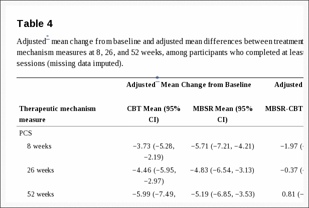


Blank
References

1. Andersson GB, Brown MD, Dvorak J, Herzog RJ, Kambin P, Malter A, et al.: Consensus summary of the diagnosis and treatment of lumbar disc herniation. Spine (Phila Pa 1976) 21:24 Suppl75S�78S, 1996 Medline
2. Atlas SJ, Deyo RA, Keller RB, Chapin AM, Patrick DL, Long JM, et al.: The Maine Lumbar Spine Study, Part II. 1-year outcomes of surgical and nonsurgical management of sciatica. Spine (Phila Pa 1976) 21:1777�1786, 1996 Crossref, Medline
3. Atlas SJ, Deyo RA, Keller RB, Chapin AM, Patrick DL, Long JM, et al.: The Maine Lumbar Spine Study, Part III. 1-year outcomes of surgical and nonsurgical management of lumbar spinal stenosis. Spine (Phila Pa 1976) 21:1787�1795, 1996 Crossref, Medline
4. Baldwin NG: Lumbar disc disease: the natural history. Neurosurg Focus 13:2E2, 2002
5. Dawson E, Bernbeck J: The surgical treatment of low back pain. Phys Med Rehabil Clin N Am 9:489�495, x, 1998
6. EuroQol Group: EuroQol�a new facility for the measurement of health-related quality of life. The EuroQol Group Health Policy 16:199�208, 1990 Crossref, Medline
7. Harrell FE: Regression Modeling Strategies: With Applications to Linear Models, Logistic Regression and Survival Analysis New York, Springer, 2001
8. Jackson CH, Sharples LD, Thompson SG, Duffy SW, Couto E: Multistate Markov models for disease progression with classification error. The Statistician 52:193�209, 2003
9. Keller RB, Atlas SJ, Singer DE, Chapin AM, Mooney NA, Patrick DL, et al.: The Maine Lumbar Spine Study, Part I. Background and concepts. Spine (Phila Pa 1976) 21:1769�1776, 1996 Crossref, Medline
10. Peul WC, van den Hout WB, Brand R, Thomeer RTWM, Koes BW: Prolonged conservative care versus early surgery in patients with sciatica caused by lumbar disc herniation: two year results of a randomised controlled trial. BMJ 336:1355�1358, 2008 Crossref, Medline
11. Peul WC, van Houwelingen HC, van der Hout WB, Brand R, Eekhof JA, Tans JT, et al.: Prolonged conservative treatment or �early� surgery in sciatica caused by a lumbar disc herniation: rationale and design of a randomized trial [ISRCT 26872154]. BMC Musculoskelet Disord 6:8, 2005 Crossref, Medline
12. Shaw JW, Johnson JA, Coons SJ: US valuation of the EQ-5D health states: development and testing of the D1 valuation model. Med Care 43:203�220, 2005 Crossref, Medline
13. Silverman BW: Density Estimation for Statistics and Data Analysis London, Chapman & Hall, 1986
14. Sing T, Sander O, Beerenwinkel N, Lengauer T: ROCR: visualizing classifier performance in R. Bioinformatics 21:3940�3941, 2005
15. van den Hout WB, Peul WC, Koes BW, Brand R, Kievit J, Thomeer RTWM, et al.: Prolonged conservative care versus early surgery in patients with sciatica from lumbar disc herniation: cost utility analysis alongside a randomised controlled trial. BMJ 336:1351�1354, 2008 Crossref, Medline
16. Weber H: Lumbar disc herniation. A controlled, prospective study with ten years of observation. Spine (Phila Pa 1976) 8:131�140, 1983 Crossref, Medline
17. Weinstein JN, Lurie JD, Tosteson TD, Skinner JS, Hanscom B, Tosteson ANA, et al.: Surgical vs nonoperative treatment for lumbar disk herniation: the Spine Patient Outcomes Research Trial (SPORT) observational cohort. JAMA 296:2451�2459, 2006 Crossref, Medline
18. Weinstein JN, Tosteson TD, Lurie JD, Tosteson ANA, Hanscom B, Skinner JS, et al.: Surgical vs nonoperative treatment for lumbar disk herniation: the Spine Patient Outcomes Research Trial (SPORT): a randomized trial. JAMA 296:2441�2450, 2006 Crossref, Medline

Close Accordion
Blank
Cited By

1. Anurekha Ramakrishnan, MS, K. Michael Webb, MD, and Matthew C. Cowperthwaite, PhD. (2017) One-year outcomes of early-crossover patients in a cohort receiving nonoperative care for lumbar disc herniation. Journal of Neurosurgery: Spine 27:4, 391-396. . Online publication date: 1-Oct-2017. Abstract | Full Text | PDF (2037 KB)
2. Kimberly A Plomp, Una Strand Vi�arsd�ttir, Darlene A Weston, Keith Dobney, Mark Collard. (2015) The ancestral shape hypothesis: an evolutionary explanation for the occurrence of intervertebral disc herniation in humans. BMC Evolutionary Biology 15:1. . Online publication date: 1-Dec-2015. [Crossref]

Close Accordion
Migraine Pain & Lumbar Herniated Disc Treatment in El Paso, TX

Migraine Pain & Lumbar Herniated Disc Treatment in El Paso, TX

One of the most prevalent causes of lower back pain and sciatica may be due to the compression of the nerve roots in the low back from a lumbar herniated disc, or a ruptured disc in the lumbar spine. Common symptoms of lumbar herniated discs include varying intensities of pain, muscle spasms or cramping, sciatica and leg weakness as well as loss of proper leg function. While these may not appear to be closely associated with each other, a lumbar herniated disc may also affect the cervical spine, manifesting symptoms of migraine and headache. The purpose of the following articles is to educate patients and demonstrate the relation between migraine pain and lumbar herniated disc, further discussing the treatment of these two common conditions.


A Critical Review of Manual Therapy Use for Headache Disorders: Prevalence, Profiles, Motivations, Communication and Self-Reported Effectiveness


Abstract


Background


Despite the expansion of conventional medical treatments for headache, many sufferers of common recurrent headache disorders seek help outside of medical settings. The aim of this paper is to evaluate research studies on the prevalence of patient use of manual therapies for the treatment of headache and the key factors associated with this patient population.


Methods


This critical review of the peer-reviewed literature identified 35 papers reporting findings from new empirical research regarding the prevalence, profiles, motivations, communication and self-reported effectiveness of manual therapy use amongst those with headache disorders.


Results


While available data was limited and studies had considerable methodological limitations, the use of manual therapy appears to be the most common non-medical treatment utilized for the management of common recurrent headaches. The most common reason for choosing this type of treatment was seeking pain relief. While a high percentage of these patients likely continue with concurrent medical care, around half may not be disclosing the use of this treatment to their medical doctor.


Conclusions


There is a need for more rigorous public health and health services research in order to assess the role, safety, utilization and financial costs associated with manual therapy treatment for headache. Primary healthcare providers should be mindful of the use of this highly popular approach to headache management in order to help facilitate safe, effective and coordinated care.


Keywords: Headache, Migraine, Tension headache, Cervicogenic headache, Manual therapy, Physical therapy, Chiropractic, Osteopathy, Massage


Background


The co-occurrence of tension headache and migraine is very high [1]. Respectively, they are the second and third most common disorders worldwide with migraine ranking as the seventh highest specific cause of disability globally [2] and the sixteenth most commonly diagnosed condition in the US [3]. These common recurrent headache disorders place a considerable burden upon the personal health, finances and work productivity of sufferers [3�5] with migraine further complicated by an association with cardiovascular and psychiatric co-morbidities [6, 7].


Preventative migraine drug treatments include analgesics, anticonvulsants, antidepressants and beta-blockers. Preventative drug treatments for tension-type headaches can include analgesics, NSAIDs, muscle relaxants and botulinum toxin as well as anticonvulsants and antidepressants. While preventative drug treatments are successful for a significant proportion of sufferers, headache disorders are still reported as under-diagnosed and under-treated within medical settings [8�16] with other studies reporting sufferers can cease continuing with preventative headache medications long-term [9, 17].


There is a number of non-drug approaches also utilized for the prevention of headaches. These include psychological therapies such as cognitive behavioral therapy, relaxation training and EMG (electromyography) biofeedback. In addition, there is acupuncture, nutritional supplementation (including magnesium, B12, B6, and Coenzyme Q10) and physical therapies. The use of physical therapies is significant, with one recent global survey reporting physical therapy as the most frequently used �alternative or complementary treatment� for headache disorders across many countries [18]. One of the most common physical therapy interventions for headache management is manual therapy (MT), [19�21] which we define here as treatments including �spinal manipulation (as commonly performed by chiropractors, osteopaths, and physical therapists), joint and spinal mobilization, therapeutic massage, and other manipulative and body-based therapies� [22].


Positive results have been reported in many clinical trials comparing MT to controls [23�27], other physical therapies [28�30] and aspects of medical care [31�34]. More high quality research is needed however to assess the efficacy of MT as a treatment for common recurrent headaches. Recent systematic reviews of randomized clinical trials of MT for the prevention of migraine report a number of significant methodological short-comings and the need for more high quality research before any firm conclusions can be made [35, 36]. Recent reviews of MT trials for tension-type headache and cervicogenic headache are cautious in reporting positive outcomes and the strong need for further robust research [37�41]. Despite the limited clinical evidence there has been no critical review of the significant use of MT by headache populations.


Methods


The aim of this study is to report from the peer-reviewed literature; 1) the prevalence of MT use for the treatment of common recurrent headaches and 2) factors associated with this use across several key themes. The review further identifies key areas worthy of further research in order to better inform clinical practice, educators and healthcare policy within this area.


Design


A comprehensive search of peer-reviewed articles published in English between 2000 and 2015 reporting new empirical research findings of key aspects of MT use among patients with migraine and non-migraine headache disorders was undertaken. Databases searched were MEDLINE, AMED, CINAHL, EMBASE and EBSCO. The key words and phrases used were: �headache�, �migraine�, �primary headache�, �cephalgia�, �chronic headache� AND �manual therapy�, �spinal manipulation�, �manipulative therapy�, �spinal mobilization�, �chiropractic�, �osteopathy�, �massage�, �physical therapy� or �physiotherapy� AND then �prevalence�, �utilization� or �profile� was used for additional searches against the previous terms. The database search was accompanied by a hand search of prominent peer-reviewed journals. All authors accessed the reviewed literature (data) and provided input to analysis.


Due to the focus of the review, literature reporting randomized control trials and similar clinical research designs were excluded as were articles identified as letters, correspondence, editorials, case reports and commentaries. Further searches were undertaken of the bibliographies in the identified publications. All identified articles were screened and only those reporting new empirical findings on MT use for headache in adults were included in the review. Articles identified and selected for the review were research manuscripts mostly within epidemiological and health economics studies. The review includes papers reporting MT use pooled with the use of other therapies, but only where MT patients comprised a large proportion (as stated) of the included study population. Results were imported into Endnote X7 and duplicates removed.


Search Outcomes, Analyses and Quality Appraisal


Figure 1 outlines the literature search process. The initial search identified 3286 articles, 35 of which met the inclusion criteria. Information from each article was organized into a review table (Table 1) to summarise the findings of the included papers. Information is reported under two selected headache groups and within each individual MT profession – chiropractic, physiotherapy, osteopathy and massage therapy � where sufficient detail was available.


Figure 1 Flow Chart of Study Selection

Figure 1: Flow Chart of Study Selection.


Table 1 Research Based Studies of Manual Therapy Use

Table 1: Research-based studies of manual therapy use for headache disorders.


An appraisal of the quality of the articles identified for review was conducted using a quality scoring system (Table 2) developed for the critical appraisal of health literature used for prevalence and incidence of health problems [42] adapted from similar studies [43�45]. This scoring system was applicable to the majority of study designs involving surveys and survey-based structured interviews (29 of the 35 papers) but was not applicable to a small number of included studies based upon clinical records, secondary analysis or practitioner characteristics.


Table 2 Description of Quality Criteria and Scoring


Two separate authors (CM and JA) independently searched and scored the articles. Score results were compared and any differences were further discussed and resolved by all the authors. The quality score of each relevant article is reported in Table 3.


Table 3 Quality Score for Selected Studies


Results


The key findings of the 35 articles were grouped and evaluated using a critical review approach adapted from previous research [46, 47]. Based on the limited information available for other headache types, prevalence findings are reported within one of two categories – either as �migraine� for papers reporting studies where the population was predominately or entirely made up of migraine patients or as �headache� for papers where the study population was predominately other headache types (including tension-type headaches, cluster headaches, cervicogenic headache) and/or where the headache type was not clearly stated. Ten papers reported findings examining prevalence rates for the �migraine� category alone, 18 papers reported findings examining prevalence for the �headache� category alone and 3 papers reported findings for both categories. Based on the nature of the information available, prevalence use was categorised by manual therapy providers. The extracted data was then analysed and synthesized into four thematic categories: prevalence; profile and motivations for MT use; concurrent use and order of use of headache providers; and self-reported evaluation of MT treatment outcomes.


Prevalence of MT Use


Thirty-one of the reviewed articles with a minimum sample size (>100) reported findings regarding prevalence of MT use. The prevalence of chiropractic use for those with migraine ranged from 1.0 to 36.2% (mean: 14.4%) within the general population [19�21, 48�52] and from 8.9 to 27.1% (mean: 18.0%) within headache-clinic patient populations [53, 54]. The prevalence of chiropractic use for those reported as headache ranged from 4 to 28.0% (mean: 12.9%) within the general population [20, 48, 51, 55�57]; ranged from 12.0 to 22.0% (mean: 18.6%) within headache/pain clinic patient populations [58�60] and from 1.9 to 45.5% (mean: 9.8%) within chiropractic patient populations [61�69].


The prevalence use of physiotherapy for those with migraine ranged from 9.0 to 57.0% (mean: 24.7%) within the general population [19, 20, 48, 52] and from 4.9 to 18.7% (mean: 11.8%) within headache-clinic patient populations [54, 70]. The prevalence use of physiotherapy for those reported as headache ranged from 12.2 to 52.0% (mean: 32.1%) within the general population [20, 48] and from 27.8 to 35.0%% (mean: 31.4%) within headache/pain clinic populations [60, 70].


Massage therapy use for those with migraine ranged from 2.0 to 29.7% (mean: 15.6%) within the general population [49, 50, 71] and from 10.1 to 56.4% (mean: 33.9%) within headache-clinic populations [53, 54, 72, 73]. Massage/acupressure use for those reported as headache within headache/pain clinic patient populations ranged from 12.0 to 54.0% (mean: 32.5%) [58�60, 70].


Osteopathy use for those with migraine was reported as 1% within the general population [49]; as 2.7% within a headache-clinic patient population [53] and as 1.7% within an osteopathy patient population [74]. For headache the prevalence was 9% within a headache/pain clinic population [60] and ranged from 2.7 to 10.0% (mean: 6.4%) within osteopathy patient populations [74, 75].


The combined prevalence rate of MT use across all MT professions for those with migraine ranged from 1.0 to 57.0% (mean: 15.9%) within the general population; ranged from 2.7 to 56.4% (mean: 18.4%) within headache-clinic patient populations and was reported as 1.7% in one MT patient population. The combined prevalence rate of MT use across all MT professions for those reported as headache ranged from 4.0 to 52.0% (mean: 17.7%) within the general population; ranged from 9.0 to 54.0% (mean: 32.3%) within headache-clinic patient populations and from 1.9 to 45.5% (mean: 9.25%) within MT patient populations.


Profile and Motivations for MT Use


While patient socio-demographic profiles were not reported within headache populations that were exclusively using MT, several studies report these findings where MT users made up a significant percentage of the non-medical headache treatments utilized by the study population (range 40% � 86%: mean 63%). While findings varied for level of income [58, 70] and level of education, [70, 72, 73] this patient group were more likely to be older [70, 72], female [20], have a higher rate of comorbid conditions [58, 70, 76] and a higher rate of previous medical visits [20, 58, 70] when compared to the non-user group. Overall, this group were reported to have a higher level of headache chronicity or headache disability than non-users [20, 54, 58, 70, 72, 77].


Several studies within headache-clinic populations report patient motivations for the use of complementary and alternative headache treatments where MT users made up a significant proportion of the study population (range 40% � 86%: mean 63%) [58, 70, 72, 78]. From these studies the most common motivation reported by study patients was �seeking pain relief� for headache which accounted for 45.4% � 84.0% (mean: 60.5%) of responses. The second most common motivation was patient concerns regarding the �safety or side effects� of medical headache treatment, accounting for 27.2% � 53.0% (mean: 43.8%) of responses [58, 70, 72]. �Dissatisfaction with medical care� accounted for 9.2% � 35.0% (mean: 26.1%) of responses [58, 70, 72].


A limited number of reviewed papers (all from Italy) report on the source of either the referral or recommendation to MT for headache treatment [53, 58, 59]. From these studies, referral from a GP to a chiropractor ranged from 50.0 to 60.8% (mean: 55.7%), while referral from friends/relatives ranged from 33.0 to 43.8% (mean: 38.7%) and self-recommendation ranged from 0 to 16.7% (mean: 5.6%). For massage therapy, referral from a GP ranged from 23.2 to 50.0% (mean: 36.6%), while referral from friends/relatives ranged from 38.4 to 42.3% (mean: 40.4%) and self-recommendation ranged from 7.7 to 38.4% (mean: 23.1%). For acupressure, referral from a GP ranged from 33.0 to 50.0% (mean: 41.5%), while referral from friends/relatives was reported as 50% and self-recommendation ranged from 0 to 16.6% (mean: 8.3%). One study reported findings for osteopathy where referral from both GP�s and friends/relatives was reported as 42.8% and self-recommendation was reported as 14.4%. Overall, the highest proportion of referrals within these studies was from GPs to chiropractors for chronic tension-type headache (56.2%), cluster headache (50%) and migraine (60.8%).


Concurrent Use and Order of Use of Headache Providers and Related Communication of MT Users


Several studies report on the concurrent use of medical headache management with complementary and alternative therapies. In those studies where the largest percentage of the patient population were users of MT�s (range 57.0% � 86.4%: mean 62.8%), [58, 70, 78] concurrent use of medical care ranged between 29.5% and 79.0% (mean: 60.0%) of the headache patient population.


These studies further report on the level of patient non-disclosure to medical providers regarding the use of MT for headache. Non-disclosure ranged between 25.5 and 72.0% (mean: 52.6%) of the patient population, with the most common reason for non-disclosure reported as the doctor �never asking�, ranging from 37.0 to 80.0% (mean: 58.5%). This was followed by a patient belief that �it was not important for the doctor to know� or �none of the doctor�s business�, ranging from 10.0 to 49.8% (mean: 30.0%). This was followed by a belief that either �the doctor would not understand� or �would discourage� these treatments, ranging from 10.0 to 13.0% (mean: 11.5%) [53, 77].


One large international study reported the ordering of the typical provider of headache care by comparing findings between several countries for migraine patients [21]. Primary care providers followed by neurologists were reported as the first and second providers for migraine treatment for nearly all countries examined. The only exception was Australia, where those with chronic migraine selected chiropractors as typical providers at equal frequency to neurologists (14% for both) while those with episodic migraine selected chiropractors at a greater frequency to neurologists (13% versus 5%). Comparatively, chiropractors were selected as the typical provider for those with chronic migraine by 10% in USA and Canada, 1% in Germany and 0% for UK and France. Chiropractors were selected as the typical provider for those with episodic migraine by 7% in USA, 6% in Germany, 4% in Canada and by 1% in both the UK and France.


Self-Reported Effectiveness of MT Treatment Outcomes


Several headache and pain-clinic population studies provide findings for the self-reported effectiveness of MT headache treatment. For chiropractic, patient self-reporting of partially effective or fully effective headache relief ranged from 27.0 to 82.0% (mean: 45.0%) [53, 58�60, 78]. For massage therapy, patient self-reporting of partially effective or fully effective headache relief ranged from 33.0 to 64.5% (mean: 45.2%)[53, 58, 60, 73, 78], and for acupressure this ranged from 33.4 to 50.0% (mean: 44.5%) [53, 58, 59]. For osteopathy and physiotherapy, one study reported effectiveness as 17 and 36% respectively [60].

When results are combined across all MT professions the reporting of MT as either partially or fully effective ranged from 17.0 to 82.0% (mean 42.5%) [53, 58�60, 73, 78]. In addition, one general population study provides findings for the self-reported effectiveness for chiropractic and physiotherapy at 25.6 and 25.1% respectively for those with primary chronic headache and 38 and 38% respectively for those with secondary chronic headache [79].


Discussion


This paper provides the first critical integrative review on the prevalence and key factors associated with the use of MT treatment for headaches within the peer-reviewed literature. While study methodological limitations and lack of data prevent making strong conclusions, these findings raise awareness of issues of importance to policy-makers, educators, headache providers and future research.


Our review found that MT use was generally higher within medical headache-clinic populations when compared to general populations. However, the use of individual MT providers does vary between different regions and this is likely due to a number of factors including variation in public access, healthcare funding and availability of MT providers. For example, the use of physiotherapy for some headache types may be relatively higher in parts of Europe [20, 60] while the use of chiropractors for some headache types may be relatively higher in Australia and the USA [19, 21]. Overall, the prevalence use of MT for headache appears to be substantial and likely to be the most common type of physical therapy utilized for headache in many countries [19�21, 49]. More high quality epidemiological studies are needed to measure the prevalence of MT use across different headache types and sub-types, both within the general population and clinical populations.


Beyond prevalence, data is more limited regarding who, how and why headache patients seek MT. From the information available however, the healthcare needs of MT headache patients may be more complex and multi-disciplinary in nature compared to those under usual medical care alone. Socio-demographic findings suggest that users of MT and other complementary and alternative therapies have a higher level of headache disability and chronicity compared to non-users. This finding may correlate with the higher prevalence of MT users within headache-clinic populations and a history of more medical appointments. This may also have implications for future MT trial designs both in terms of the selection of trial subjects from inside versus outside MT clinical settings and the decision to test singular MT interventions versus MT in combination with other interventions.


Limited information suggests that a pluralistic approach toward the use of medical and non-medical headache treatments such as MT is common. While findings suggest MT is sought most often for reasons of seeking headache relief, the evidence to support the efficacy of MT for headache relief is still limited. MT providers must remain mindful of the quality of the evidence for a given intervention for a given headache disorder and to inform patients where more effective or safer treatment interventions are available. More research is needed to assess these therapies individually and through multimodal approaches and for studies to include long-term follow-up.


Information limited to Italy, suggests referral from GPs for MT headache treatment can be common in some regions, while this is less likely to widespread given the issue of patient non-disclosure to medical doctors regarding the use of this treatment in other studies. High quality healthcare requires open and transparent communication between patients and providers and between the providers themselves. Non-disclosure may adversely influence medical management should unresponsive patients require further diagnostic investigations [80] or the implementation of more effective approaches to headache management [81] or prevents discussion in circumstances where MT may be contraindicated [82]. Primary headache providers may benefit from paying particular attention to the possibility of non-disclosure of non-medical headache treatments. Open discussion between providers and patients about the use of MT for headache and the associated outcomes may improve overall patient care.


Future Research


Despite the strong need for more high quality research to assess the efficacy of MT as a treatment for headache, the substantial use of MT brings attention to the need for more public health and health services research within this area of headache management. The need for this type of research was identified in a recent global report on the use of headache-related healthcare resources [18]. Furthering this information can lead to improvements in healthcare policy and the delivery of healthcare services.


The substantial use of physical therapies such as MT has been under-reported within many of the national surveys reporting headache-related healthcare utilization [3, 5, 83�85]. Regardless, the role of physical therapies in headache management continues to be assessed, often within mainstream and integrated headache management settings [86�89]. Continuing this research may further our understanding of the efficacy and outcomes associated with a more multidisciplinary approach to headache management.


Further to this is the need for more research to understand the healthcare utilization pathways associated with those patients who use MT in their headache management. Little is known about the sociodemographic background, types of headaches, level of headache disability and comorbidities more common to this patient population. In turn, such information can provide insights that may be valuable to provider clinical decision-making and provider education.


Limitations


The design and findings of our review has a number of limitations. The design of the review was limited by a search within English language journals only. As a result, some research on this topic may have been missed. While the quality scoring system adopted for this review requires further validation, the data we collected was limited by the low to moderate quality of available papers which averaged 6.4 out of 10 points (Table 3). The low scoring was largely due to significant methodological issues and the small sample size associated with much of the collected papers. Much of the data on this topic was heterogeneous in nature (telephone, postal surveys and face-to-face interviews). There was a lack of validated practitioner and patient questionnaires to report findings, such as for questions on prevalence, where the time frames utilized varied between �currently�, �last 12 months� and �ever�.


Data on the prevalence of MT use for headache was limited particularly within individual MT provider populations when compared to data found within the general population and headache-clinic populations. Many studies assessed the use of MT for headache without identifying headache types. Only one study inside an MT population had reported the percentage of patients attending for reasons of migraine alone (osteopathy). The prevalence of MT use for headache was reported most within chiropractic patient population studies, however information was limited on the types of headache. We found no studies reporting the prevalence of headache patients within physiotherapy or massage therapy patient populations using our search terms.


A lack of data for some themes necessitated providing findings pooled with users of other non-medical headache providers. Data within many geographical regions was very limited with the most limited data was on the source of referral to MT headache providers (three papers from Italy only). These limitations support the call for more research to be focused exclusively within MT populations and different regional areas before stronger conclusions can be drawn.


Conclusion


The needs of those with headache disorders can be complex and multi-disciplinary in nature. Beyond clinical research, more high quality public health and health services research is needed to measure and examine a number of issues of significance to the delivery and use of MT�s within headache management. With unmet needs still remaining for many who suffer recurrent headaches, clinicians should remain cognizant of the use of MT�s and remain open to discussing this approach to headache management in order to ensure greater safety, effectiveness and coordination of headache care.


Acknowledgements


Not applicable.


Funding


This research received no specific grant from any funding agency in the public, commercial or not-for-profit sectors while the first author on this paper receives a PhD scholarship made available by the Australian Chiropractors� Association.


Availability of Data and Materials


Not applicable (all data is reported in article).


Authors’ Contributions


CM, JA and DS designed the paper. CM carried out the literature search, data collection and selection. CM and DS provided the analysis and interpretation. CM and JA wrote the drafts. All authors contributed to the critical review and intellectual content. All authors read and approved the final manuscript.


Competing Interests


The authors declare that they have no competing interests.


Consent for Publication


Not applicable.


Ethics Approval and Consent to Participate


Not applicable.


Publisher�s Note


Springer Nature remains neutral with regard to jurisdictional claims in published maps and institutional affiliations.


Abbreviations


  • MT Manual therapy
  • EMG Electromyography


Contributor Information


Ncbi.nlm.nih.gov/pmc/articles/PMC5364599/


Dr Jimenez White Coat

Dr. Alex Jimenez’s Insight

A staggering 15% of the population suffers from migraines, a debilitating condition which affects an individual’s ability to engage in everyday activities. Although widely misunderstood by researches today, I believe that migraine pain can be a symptom of a much bigger underlying health issue. Lumbar herniated discs, or ruptured discs in the lumbar spine, are a common cause of lower back pain and sciatica. When the soft, gel-like center of a lumbar herniated disc compresses the nerve roots of the low back, it can result in symptoms of pain and discomfort, numbness and weakness in the lower extremities. What’s more, a lumbar herniated disc can unbalance the structure and function of the entire spine, eliciting symptoms along the cervical spine that could ultimately trigger migraines. People who constantly experience migraine pain often have to carefully go about their day in hopes of avoiding the blaze of another painful episode. Fortunately, many migraine pain and lumbar herniated disc treatment methods are available to help improve as well as manage the symptoms. Other treatment options can also be considered before surgical interventions.


Surgical versus Non-Operative Treatment for Lumbar Disc Herniation: Eight-Year Results for the Spine Patient Outcomes Research Trial (SPORT)


Abstract


Study Design


Concurrent prospective randomized and observational cohort studies.


Objective


To assess the 8-year outcomes of surgery vs. non-operative care.


Summary of Background Data


Although randomized trials have demonstrated small short-term differences in favor of surgery, long-term outcomes comparing surgical to non-operative treatment remain controversial.


Methods


Surgical candidates with imaging-confirmed lumbar intervertebral disc herniation meeting SPORT eligibility criteria enrolled into prospective randomized (501 participants) and observational cohorts (743 participants) at 13 spine clinics in 11 US states. Interventions were standard open discectomy versus usual non-operative care. Main outcome measures were changes from baseline in the SF-36 Bodily Pain (BP) and Physical Function (PF) scales and the modified Oswestry Disability Index (ODI – AAOS/Modems version) assessed at 6 weeks, 3 and 6 months, and annually thereafter.


Results


Advantages were seen for surgery in intent-to-treat analyses for the randomized cohort for all primary and secondary outcomes other than work status; however, with extensive non-adherence to treatment assignment (49% patients assigned to non-operative therapy receiving surgery versus 60% of patients assigned to surgery) these observed effects were relatively small and not statistically significant for primary outcomes (BP, PF, ODI). Importantly, the overall comparison of secondary outcomes was significantly greater with surgery in the intent-to-treat analysis (sciatica bothersomeness [p > 0.005], satisfaction with symptoms [p > 0.013], and self-rated improvement [p > 0.013]) in long-term follow-up. An as-treated analysis showed clinically meaningful surgical treatment effects for primary outcome measures (mean change Surgery vs. Non-operative; treatment effect; 95% CI): BP (45.3 vs. 34.4; 10.9; 7.7 to 14); PF (42.2 vs. 31.5; 10.6; 7.7 to 13.5) and ODI (?36.2 vs. ?24.8; ?11.2; ?13.6 to ?9.1).


Conclusion


Carefully selected patients who underwent surgery for a lumbar disc herniation achieved greater improvement than non-operatively treated patients; there was little to no degradation of outcomes in either group (operative and non-operative) from 4 to 8 years.


Keywords: SPORT, intervertebral disc herniation, surgery, non-operative care, outcomes


Introduction


Lumbar discectomy for relief of sciatica in patients with intervertebral disc herniation (IDH) is a well-researched and common indication for spine surgery, yet rates of this surgery exhibit considerable geographic variation.[1] Several randomized trials and large prospective cohorts have demonstrated that surgery provides faster pain relief and perceived recovery in patients with herniated disc.[2�6] The effect of surgery on longer term outcomes remains less clear.


In a classic RCT evaluating surgery versus non-operative treatment for lumbar IDH, Weber et al. showed a greater improvement in the surgery group at 1 year that was statistically significant; there was also greater improvement for surgery at 4 years, although not statistically significant, but no apparent difference in outcomes at 10 years.[2] However, a number of patients in the non-operative group eventually underwent surgery over that time, complicating the interpretation of the long-term results. The Maine Lumbar Spine Study, a prospective observational cohort, found greater improvement at one year in the surgery group that narrowed over time, but remained significantly greater in the surgical group for sciatica bothersomeness, physical function, and satisfaction, but no different for work or disability outcomes.[3] This paper reports 8-year results from the Spine Patient Outcomes Research Trial (SPORT) based on the continued follow-up of the herniated disc randomized and observational cohorts.


Methods


Study Design


SPORT is a randomized trial with a concurrent observation cohort conducted in 11 US states at 13 medical centers with multidisciplinary spine practices. The human subjects committees at each participating institution approved a standardized protocol for both the observational and the randomized cohorts. Patient inclusion and exclusion criteria, study interventions, outcome measures, and follow-up procedures have been reported previously.[5�8]


Patient Population


Men and women were eligible if they had symptoms and confirmatory signs of lumbar radiculopathy persisting for at least six weeks, disc herniation at a corresponding level and side on imaging, and were considered surgical candidates. The content of pre-enrollment non-operative care was not pre-specified in the protocol.[5�7] Specific enrollment and exclusion criteria are reported elsewhere.[6,7]


A research nurse at each site identified potential participants, verified eligibility and used a shared decision making video for uniformity of enrollment. Participants were offered enrollment in either the randomized trial or the observational cohort. Enrollment began in March of 2000 and ended in November of 2004.


Study Interventions


The surgery was a standard open discectomy with examination of the involved nerve root.[7,9] The non-operative protocol was �usual care� recommended to include at least: active physical therapy, education/counseling with home exercise instruction, and non-steroidal anti-inflammatory drugs if tolerated. Non-operative treatments were individualized for each patient and tracked prospectively.[5�8]


Study Measures


Primary endpoints were the Bodily Pain (BP) and Physical Function (PF) scales of the SF-36 Health Survey[10] and the AAOS/Modems version of the Oswestry Disability Index (ODI)[11] as measured at 6 weeks, 3 and 6 months, and annually thereafter. If surgery was delayed beyond six weeks, additional follow-up data was obtained 6 weeks and 3 months post-operatively. Secondary outcomes included patient self-reported improvement; work status; satisfaction with current symptoms and care;[12] and sciatica severity as measured by the sciatica bothersomeness index.[13,14] Treatment effect was defined as the difference in the mean changes from baseline between the surgical and non-operative groups.


Statistical Considerations


Initial analyses compared means and proportions for baseline patient characteristics between the randomized and observational cohorts and between the initial treatment arms of the individual and combined cohorts. The extent of missing data and the percentage of patients undergoing surgery were calculated by treatment arm for each scheduled follow-up. Baseline predictors of time until surgical treatment (including treatment crossovers) in both cohorts were determined via a stepwise proportional hazards regression model with an inclusion criterion of p < 0.1 to enter and p > 0.05 to exit. Predictors of missing follow-up visits at yearly intervals up to 8 years were separately determined via stepwise logistic regression. Baseline characteristics that predicted surgery or a missed visit at any time-point were then entered into longitudinal models of primary outcomes. Those that remained significant in the longitudinal models of outcome were included as adjusting covariates in all subsequent longitudinal regression models to adjust for potential confounding due to treatment selection bias and missing data patterns.[15] In addition, baseline outcome, center, age and gender were included in all longitudinal outcome models.


Primary analyses compared surgical and non-operative treatments using changes from baseline at each follow-up, with a mixed effects longitudinal regression model including a random individual effect to account for correlation between repeated measurements within individuals. The randomized cohort was initially analyzed on an intent-to-treat basis.[6] Because of cross-over, additional analyses were performed based on treatments actually received. In these as-treated analyses, the treatment indicator was a time-varying covariate, allowing for variable times of surgery. Follow-up times were measured from enrollment for the intent-to-treat analyses, whereas for the as-treated analysis the follow-up times were measured from the beginning of treatment (i.e. the time of surgery for the surgical group and the time of enrollment for the non-operative group), and baseline covariates were updated to the follow-up immediately preceding the time of surgery. This procedure has the effect of including all changes from baseline prior to surgery in the estimates of the non-operative treatment effect and all changes after surgery in the estimates of the surgical effect. The six-point sciatica scales and binary outcomes were analyzed via longitudinal models based on generalized estimating equations[16] with linear and logit link functions respectively, using the same intent-to-treat and adjusted as-treated analysis definitions as the primary outcomes. The randomized and observational cohorts were each analyzed to produce separate as-treated estimates of treatment effect. These results were compared using a Wald test to simultaneously test all follow-up visit times for differences in estimated treatment effects between the two cohorts.[15] Final analyses combined the cohorts.


To evaluate the two treatment arms across all time-periods, the time-weighted average of the outcomes (area under the curve) for each treatment group was computed using the estimates at each time period from the longitudinal regression models and compared using a Wald test.[15]


Kaplan-Meier estimates of re-operation rates at 8 years were computed for the randomized and observational cohorts and compared via the log-rank test.[17,18]


Computations were done using SAS procedures PROC MIXED for continuous data and PROC GENMOD for binary and non-normal secondary outcomes (SAS version 9.1 Windows XP Pro, Cary, NC). Statistical significance was defined as p < 0.05 based on a two-sided hypothesis test with no adjustments made for multiple comparisons. Data for these analyses were collected through February 4, 2013.


Results


Overall, 1,244 SPORT participants with lumbar intervertebral disc herniation were enrolled (501 in the randomized cohort, and 743 in the observational cohort) (Figure 1). In the randomized cohort, 245 were assigned to surgical treatment and 256 to non-operative treatment. Of those randomized to surgery, 57% had surgery by 1 year and 60% by 8 years. In the group randomized to non-operative care, 41% of patients had surgery by 1 year and 48% by 8 years. In the observational cohort, 521 patients initially chose surgery and 222 patients initially chose non-operative care. Of those initially choosing surgery, 95% received surgery by 1 year; at 8 years 12 additional patients had undergone primary surgery. Of those choosing non-operative treatment, 20% had surgery by 1 year and 25% by 8 years. In both cohorts combined, 820 patients received surgery at some point during the first 8 years; 424 (34%) remained non-operative. Over the 8 years, 1,192 (96%) of the original enrollees completed at least 1 follow-up visit and were included in the analysis (randomized cohort: 94% and observational cohort 97%); 63% of initial enrollees supplied data at 8 years with losses due to dropouts, missed visits, or deaths (Figure 1).


Figure-1-Exclusion-Enrollment-Randomization-and-Follow-Up

Figure 1: Exclusion, enrollment, randomization and follow-up of trial participants.


Patient Characteristics


Baseline characteristics have been previously reported and are summarized in Table 1.[5,6,8] The combined cohorts had an overall mean age of 41.7 with slightly more men than women. Overall, the randomized and observational cohorts were similar. However, patients in the observational cohort had more baseline disability (higher ODI scores), were more likely to prefer surgery, more often rated their problem as worsening, and were slightly more likely to have a sensory deficit. Subjects receiving surgery over the course of the study were: younger; less likely to be working; more likely to report being on worker�s compensation; had more severe baseline pain and functional limitations; fewer joint and other co-morbidities; greater dissatisfaction with their symptoms; more often rated their condition as getting worse at enrollment; and were more likely to prefer surgery. Subjects receiving surgery were also more likely to have a positive straight leg test, as well as more frequent neurologic, sensory, and motor deficits. Radiographically, their herniations were more likely to be at the L4�5 and L5-S1 levels and to be posterolateral in location.


Table 1 Patient Baseline Demographic Characteristics, Comorbidities and Health Status Measures

Table 1: Patient baseline demographic characteristics, comorbidities and health status measures according to study cohort and treatment received.


Surgical Treatment and Complications


Overall surgical treatment and complications were similar between the two cohorts (Table 2). The average surgical time was slightly longer in the randomized cohort (80.5 minutes randomized vs. 74.9 minutes observational, p=0.049). The average blood loss was 75.3cc in the randomized cohort vs. 63.2cc in the observational, p=0.13. Only 6 patients total required intra-operative transfusions. There were no perioperative mortalities. The most common surgical complication was dural tear (combined 3% of cases). Re-operation occurred in a combined 11% of cases by 5 years, 12% by 6 years, 14% by 7 years, and 15% by 8 years post-surgery. The rates of reoperation were not significantly different between the randomized and observational cohorts. Eighty-seven of the 119 re-operations noted the type of re-operation; approximately 85% of these (74/87) were listed as recurrent herniations at the same level. One death occurred within 90 days post-surgery related to heart surgery at another institution; the death was judged to be unrelated and was reported to the Institutional Review Board and the Data and Safety Monitoring Board.


Table 2 Operative Treatments, Complications and Events

Cross-Over


Non-adherence to treatment assignment affected both treatment arms: patients chose to delay or decline surgery in the surgical arm and crossed over to surgery in the non-operative arm. (Figure 1) Statistically significant differences of patients crossing over to non-operative care within 8 years of enrollment were that they were older, had higher incomes, less dissatisfaction with their symptoms, more likely to have a disc herniation at an upper lumbar level, more likely to express a baseline preference for non-operative care, less likely to perceive their symptoms as getting worse at baseline, and had less baseline pain and disability (Table 3). Patients crossing over to surgery within 8 years were more dissatisfied with their symptoms at baseline; were more likely to perceive they were getting worse at baseline; more likely to express a baseline preference for surgery; and had worse baseline physical function and more self-rated disability.


Table 3 Statistically Significant Predictors of Adherence to Treatment

Table 3: Statistically significant predictors of adherence to treatment among RCT patients.


Main Treatment Effects


Intent-to-Treat Analysis In the intention-to-treat analysis of the randomized cohort, all measures over 8 years favored surgery but there were no statistically significant treatment effects in the primary outcome measures (Table 4 and Figure 2). In the overall intention-to-treat comparison between the two treatment groups over time (area-under the curve), secondary outcomes were significantly greater with surgery in the intention-to-treat analysis (sciatica bothersomeness (p=0.005), satisfaction with symptoms (p=0.013), and self-rated improvement (p=0.013)) (Figure 3) Improvement in sciatica bothersomeness index was also statistically significant in favor of surgery at most individual time point comparisons (although non-significant in years 6 and 7) (Table 4).


Figure-2-Primary-Outcomes-in-the-Randomized-and-Observational-Cohorts

Figure 2: Primary outcomes (SF-36 Bodily Pain and Physical Function, and Oswestry Disability Index) in the randomized and observational cohorts during 8 years of follow-up.


Figure-3-Secondary-Outcomes-in-the-Randomized-and-Observational-Cohorts.

Figure 3: secondary outcomes (Sciatica Bothersomeness, Satisfaction with Symptoms, and Self-rated Global Improvement) in the randomized and observational cohorts during 8 years of follow-up.


Table 4 Primary Analysis Results for Years 1 to 8

Table 4: Primary analysis results for years 1 to 8. Intent-to-treat for the randomized cohort and adjusted* analyses according to treatment received for the randomized and observational cohorts combined.


As-Treated Analysis The adjusted as-treated effects seen in the randomized and observational were similar. Accordingly, the cohorts were combined for the final analyses. Treatment effects for the primary outcomes in the combined as-treated analysis were clinically meaningful and statistically significant out to 8 years: SF-36 BP 10.9 p < 0.001 (95% CI 7.7 to 14); SF-36 PF 10.6 p<0.001 (95% CI 7.7 to 13.5); ODI ?11.3 p<0.001 (95% CI ?13.6 to ?9.1) (Table 4). The footnote for Table 4 describes the adjusting covariates selected for the final model.


Results from the intent-to-treat and as-treated analyses of the two cohorts are compared in Figure 2. In the combined analysis, treatment effects were statistically significant in favor of surgery for all primary and secondary outcome measures (with the exception of work status which did not differ between treatment groups) at each time point (Table 4 and Figure 3).


Loss-to-Follow-Up


At the 8-year follow-up, 63% of initial enrollees supplied data, with losses due to dropouts, missed visits, or deaths. Table 5 summarized the baseline characteristics of those lost to follow-up compared to those retained in the study at 8-years. Those who remained in the study at 8 years were – somewhat older; more likely to be female, white, college educated, and working at baseline; less likely to be disabled, receiving compensation, or a smoker; less symptomatic at baseline with somewhat less bodily pain, better physical function, less disability on the ODI, better mental health, and less sciatica bothersomeness. These differences were small but statistically significant. Table 6 summarizes the short-term outcomes during the first 2 years for those retained in the study at 8 years compared to those lost to follow-up. Those lost to follow-up had worse outcomes on average; however this was true in both the surgical and non-operative groups with non-significant differences in treatment effects. The long-term outcomes are therefore likely to be somewhat over-optimistic on average in both groups, but the comparison between surgical and non-operative outcomes appear likely to be un-biased despite the long-term loss to follow-up.


Table 5 Patient Baseline Demographic Characteristics, Comorbidities and Health Status Measures

Table 5: Patient baseline demographic characteristics, comorbidities, and health status measures according to patient follow-up status as of 02/01/2013 when the IDH8yr data were pulled.


Table 6 Time Weighted Average of Treatment Effects

Table 6: Time-weighted average of treatment effects at 2 years (AUC) from adjusted* as-treated randomized and observational cohorts combined primary outcome analysis, according to treatment received and patient follow-up status.


Discussion


In patients with a herniated disc confirmed by imaging and leg symptoms persisting for at least 6 weeks, surgery was superior to non-operative treatment in relieving symptoms and improving function. In the as-treated analysis, the treatment effect for surgery was seen as early as 6 weeks, appeared to reach a maximum by 6 months and persisted over 8 years; it is notable that the non-operative group also improved significantly and this improvement persisted with little to no degradation of outcomes in either group (operative and non-operative) between 4 and 8 years. In the longitudinal intention-to-treat analysis, all the outcomes showed small advantages for surgery, but only the secondary outcomes of sciatica bothersomeness, satisfaction with symptoms, and self-rated improvement were statistically significant. The persistent small benefit in the surgery group over time has made the overall intention-to-treat comparison more statistically significant over time despite high levels of cross-over. The large effects seen in the as-treated analysis after adjustments for characteristics of the crossover patients suggest that the intent-to-treat analysis may underestimate the true effect of surgery since the mixing of treatments due to crossover can be expected to create a bias toward the null in the intent-to-treat analyses.[4,19] Loss to follow-up among patients who were somewhat worse at baseline and with worse short-term outcomes probably leads to overly-optimistic estimated long-term outcomes in both surgery and non-operative groups but unbiased estimates of surgical treatment effects.


Comparisons to Other Studies


There are no other long-term randomized studies reporting the same primary outcome measures as SPORT. The results of SPORT primary outcomes at 2 years were quite similar to those of Peul et al but longer follow up for the Peul study is necessary for further comparison.[4,20] In contrast to the Weber study, the differences in the outcomes in SPORT between treatment groups remained relatively constant between 1 and 8 years of follow-up. One of the factors in this difference may be the sensitivity of the outcome measures � for example, sciatica bothersomeness, which was significantly different out to 8 years in the intention-to-treat, may be a more sensitive marker of treatment success than the general outcome measure used by Weber et al.[2]


The long-term results of SPORT are similar to the Maine Lumbar Spine Study (MLSS).[21] The MLSS reported statistically significantly greater improvements at 10 years in sciatica bothersomeness for the surgery group (?11.9) compared to the nonsurgical groups (?5.8) with a treatment effect of ?6.1 p=0.004; in SPORT the improvement in sciatica bothersomeness in the surgical group at 8 years was similar to the 10 year result in MLSS (?11) though the non-operative cohort in SPORT did better than their MLSS counterparts (?9.1) however the treatment effect in SPORT, while smaller, remained statistically significant (?1.5; p<0.001) due to the much larger sample size. Greater improvements in the non-operative cohorts between SPORT and MLSS may be related to differences in non-operative treatments over time, differences between the two cohorts since the MLSS and did not require imaging confirmation of IDH.


Over the 8 years there was little evidence of harm from either treatment. The 8-year rate of re-operation was 14.7%, which is lower than the 25% reported by MLSS at 10 years.[22]


Limitations


Although our results are adjusted for characteristics of cross over patients and control for important baseline covariates, the as-treated analyses presented do not share the strong protection from confounding that exists for an intent-to-treat analysis.[4�6] However, However, intent-to-treat analyses are known to be biased in the presence of noncompliance at the level observed in SPORT, and our adjusted as-treated analyses have been shown to produce accurate results under reasonable assumptions about the dependence of compliance on longitudinal outcomes.[23] Another potential limitation is the heterogeneity, of the non-operative treatment interventions, as discussed in our prior papers.[5,6,8] Finally, attrition in this long-term follow-up study meant that only 63% of initial enrollees supplied data at 8 years with losses due to dropouts, missed visits, or deaths; based on analyses at baseline and at short-term follow-up, this likely leads to somewhat overly-optimistic estimated long-term outcomes in both treatment groups but an unbiased estimation of surgical treatment effect.


Conclusions


In the intention-to-treat analysis, small, statistically insignificant surgical treatment effects were seen for the primary outcomes but statistically significant advantages for sciatica bothersomeness, satisfaction with symptoms, and self-rated improvement were seen out to 8 years despite high levels of treatment cross-over. The as-treated analysis combining the randomized and observational cohorts, which carefully controlled for potentially confounding baseline factors, showed significantly greater improvement in pain, function, satisfaction, and self-rated progress over 8 years compared to patients treated non-operatively. The non-operative group, however, also showed substantial improvements over time, with 54% reporting being satisfied with their symptoms and 73% satisfied with their care after 8 years.


Acknowledgments


The National Institute of Arthritis and Musculoskeletal and Skin Diseases (U01-AR45444; P60-AR062799) and the Office of Research on Women�s Health, the National Institutes of Health, and the National Institute of Occupational Safety and Health, the Centers for Disease Control and Prevention grant funds were received in support of this work. Relevant financial activities outside the submitted work: consultancy, grants, stocks.


This study is dedicated to the memories of Brieanna Weinstein and Harry Herkowitz, leaders in their own rights, who simply made the world a better place.


Footnotes


Other comorbidities include: stroke, diabetes, osteoporosis, cancer, fibromyalgia, cfs, PTSD, alcohol, drug dependency, heart, lung, liver, kidney, blood vessel, nervous system, hypertension, migraine, anxiety, stomach, bowel


In conclusion, individuals who suffer from migraine pain require the most effective type of treatment in order to help improve as well as manage their symptoms, particularly if their migraines were elicited from a lumbar herniated disc. The purpose of the following articles was to associate the two conditions with each other and demonstrate the results of the research above. Various treatment options can be considered before surgery for migraine pain and lumbar herniated disc treatment. Information referenced from the National Center for Biotechnology Information (NCBI). The scope of our information is limited to chiropractic as well as to spinal injuries and conditions. To discuss the subject matter, please feel free to ask Dr. Jimenez or contact us at 915-850-0900 .


Curated by Dr. Alex Jimenez


Green-Call-Now-Button-24H-150x150-2-3.png


Additional Topics: Neck Pain


Neck pain is a common complaint which can result due to a variety of injuries and/or conditions. According to statistics, automobile accident injuries and whiplash injuries are some of the most prevalent causes for neck pain among the general population. During an auto accident, the sudden impact from the incident can cause the head and neck to jolt abruptly back-and-forth in any direction, damaging the complex structures surrounding the cervical spine. Trauma to the tendons and ligaments, as well as that of other tissues in the neck, can cause neck pain and radiating symptoms throughout the human body.


blog picture of cartoon paperboy big news


IMPORTANT TOPIC: EXTRA EXTRA: A Healthier You!


OTHER IMPORTANT TOPICS: EXTRA: Sports Injuries? | Vincent Garcia | Patient | El Paso, TX Chiropractor

 

Blank
References
1. Lyngberg AC, Rasmussen BK, J�rgensen T, Jensen R. Has the prevalence of migraine and tension-type headache changed over a 12-year period? a Danish population survey. Eur J Epidemiol. 2005;20:243�9. doi: 10.1007/s10654-004-6519-2. [PubMed] [Cross Ref]
2. Vos T, Flaxman A, Naghavi M. Years lived with disability (YLDs) for 1160 sequelae of 289 diseases and injuries 1990�2010: a systematic analysis for the global burden of disease study 2010. Lancet. 2012;380:2163�96. doi: 10.1016/S0140-6736(12)61729-2. [PubMed] [Cross Ref]
3. Burch RC, Loder S, Loder E, Smitherman TA. The prevalence and burden of migraine and severe headache in the united states: updated statistics from government health surveillance studies. Headache. 2015;55:21�34. doi: 10.1111/head.12482. [PubMed] [Cross Ref]
4. Lanteri-Minet M. Economic burden and costs of chronic migraine. Curr Pain Headache Rep. 2014;18:385. doi: 10.1007/s11916-013-0385-0. [PubMed] [Cross Ref]
5. Bloudek L, Stokes M, Buse D, Wilcox T, Lipton R, Goadsby P, Varon S, Blumenfeld A, Katsarava Z, Pascual J, et al. Cost of healthcare for patients with migraine in five European countries: results from the international burden of migraine study (IBMS) J Headache Pain. 2012;13:361�78. doi: 10.1007/s10194-012-0460-7. [PMC free article] [PubMed] [Cross Ref]
6. Antonaci F, Nappi G, Galli F, Manzoni GC, Calabresi P, Costa A. Migraine and psychiatric comorbidity: a review of clinical findings. J Headache Pain. 2011;12:115�25. doi: 10.1007/s10194-010-0282-4. [PMC free article] [PubMed] [Cross Ref]
7. Kurth T, Chabriat H, Bousser M-G. Migraine and stroke: a complex association with clinical implications. Lancet Neurol. 2012;11:92�100. doi: 10.1016/S1474-4422(11)70266-6. [PubMed] [Cross Ref]
8. Lipton R, Goadsby P, Sawyer J, Blakeborough P, Stewart W. Migraine: diagnosis and assessment of disability. Rev Contemp Pharmaco. 2000;11:63�73.
9. Diamond S, Bigal ME, Silberstein S, Loder E, Reed M, Lipton RB. Patterns of diagnosis and acute and preventive treatment for migraine in the united states: results from the American migraine prevalence and prevention study. Headache. 2007;47:355�63. [PubMed]
10. Lipton RB, Bigal ME, Diamond M, Freitag F, Reed M, Stewart WF. Migraine prevalence, disease burden, and the need for preventive therapy. Neurology. 2007;68:343�9. doi: 10.1212/01.wnl.0000252808.97649.21. [PubMed] [Cross Ref]
11. Berger A, Bloudek LM, Varon SF, Oster G. Adherence with migraine prophylaxis in clinical practice. Pain Pract. 2012;12:541�9. doi: 10.1111/j.1533-2500.2012.00530.x. [PubMed] [Cross Ref]
12. Peres MFP, Silberstein S, Moreira F, Corchs F, Vieira DS, Abraham N, Gebeline-Myers C. Patients’ preference for migraine preventive therapy. Headache. 2007;47:540�5. doi: 10.1111/j.1526-4610.2007.00757.x. [PubMed] [Cross Ref]
13. Nicholson RA, Rooney M, Vo K, O’Laughlin E, Gordon M. Migraine care among different ethnicities: Do disparities exist? Headache. 2006;46:754�65. doi: 10.1111/j.1526-4610.2006.00453.x. [PMC free article] [PubMed] [Cross Ref]
14. Lafata JE, Tunceli O, Cerghet M, Sharma KP, Lipton RB. The use of migraine preventive medications among patients with and without migraine headaches. Cephalalgia. 2010;30:97�104. doi: 10.1111/j.1468-2982.2009.01909.x. [PubMed] [Cross Ref]
15. Cevoli S, D’Amico D, Martelletti P, Valguarnera F, Del Bene E, De Simone R, Sarchielli P, Narbone MC, Testa L, Genco S, et al. Underdiagnosis and undertreatment of migraine in Italy: a survey of patients attending for the first time 10 headache centres. Cephalalgia. 2009;29:1285�93. doi: 10.1111/j.1468-2982.2009.01874.x. [PubMed] [Cross Ref]
16. Stark RJ, Valenti L, Miller GC. Management of migraine in Australian general practice. Med J Aust. 2007;187:142. [PubMed]
17. Lipton RB, Buse DC, Serrano D, Holland S, Reed ML. Examination of unmet treatment needs among persons with episodic migraine: results of the American migraine prevalence and prevention (AMPP) study. Headache. 2013;53:1300�11. doi: 10.1111/head.12154. [PubMed] [Cross Ref]
18. WHO Lifting the Burden 2011: www.who.int/mental_health/management/who_atlas_headache_disorders.pdf?ua=1. Retrieved 8 August 2015
19. Bigal ME, Serrano D, Reed M, Lipton RB. Chronic migraine in the population Burden, diagnosis, and satisfaction with treatment. Neurology. 2008;71:559�66. doi: 10.1212/01.wnl.0000323925.29520.e7. [PubMed] [Cross Ref]
20. Kristoffersen ES, Grande RB, Aaseth K, Lundqvist C, Russell MB. Management of primary chronic headache in the general population: the Akershus study of chronic headache. J Headache Pain. 2012;13:113�20. doi: 10.1007/s10194-011-0391-8. [PMC free article] [PubMed] [Cross Ref]
21. Sanderson JC, Devine EB, Lipton RB, Bloudek LM, Varon SF, Blumenfeld AM, Goadsby PJ, Buse DC, Sullivan SD. Headache-related health resource utilisation in chronic and episodic migraine across six countries. J Neurol Neurosurg Psychiatry. 2013;84:1309�17. doi: 10.1136/jnnp-2013-305197. [PMC free article] [PubMed] [Cross Ref]
22. Biology of Manual Therapies (R21) National Institute of Health, 2014: grants.nih.gov/grants/guide/pa-files/PA-14-167.html Retrieved 11 August 2015
23. Marcus D, Scharff L, Mercer S, Turk D. Nonpharmacological treatment for migraine: incremental utility of physical therapy with relaxation and thermal biofeedback. Cephalalgia. 1998;18:266�72. doi: 10.1046/j.1468-2982.1998.1805266.x. [PubMed] [Cross Ref]
24. Lawler SP, Cameron LD. A randomized, controlled trial of massage therapy as a treatment for migraine. Ann Behav Med. 2006;32:50�9. doi: 10.1207/s15324796abm3201_6. [PubMed] [Cross Ref]
25. Tuchin PJ, Pollard H, Bonello R. A randomized controlled trial of chiropractic spinal manipulative therapy for migraine. J Manipulative Physiol Ther. 2000;23:91�5. doi: 10.1016/S0161-4754(00)90073-3. [PubMed] [Cross Ref]
26. Hoyt W, Shaffer F, Bard D, Benesler J, Blankenhorn G, Gray J, Hartman W, Hughes L. Osteopathic manipulation in the treatment of muscle-contraction headache. J Am Osteopath Assoc. 1979;78:322�5. [PubMed]
27. Jull G, Trott P, Potter H, Zito G, Niere K, Shirley D, Emberson J, Marschner I, Richardson C. A randomized controlled trial of exercise and manipulative therapy for cervicogenic headache. Spine (Phila Pa 1976) 2002;27:1835�43. doi: 10.1097/00007632-200209010-00004. [PubMed] [Cross Ref]
28. Haas M, Spegman A, Peterson D, Aickin M, Vavrek D. Dose-Response and Efficacy of Spinal Manipulation for Chronic Cervicogenic Headache: A Pilot Randomized Controlled Trial. Spine J. 2010;10:117�28. [PMC free article] [PubMed]
29. Bove G, Nilsson N. Spinal manipulation in the treatment of episodic tension-type headache: a randomized controlled trial. JAMA. 1998;280:1576�9. doi: 10.1001/jama.280.18.1576. [PubMed] [Cross Ref]
30. Parker GB, Pryor DS, Tupling H. Why does migraine improve during a clinical trial? Further results from a trial of cervical manipulation for migraine. Aust N Z J Med. 1980;10:192�8. doi: 10.1111/j.1445-5994.1980.tb03712.x. [PubMed] [Cross Ref]
31. Hsieh LL-C, Liou H-H, Lee L-H, Chen TH-H, Yen AM-F. Effect of acupressure and trigger points in treating headache: a randomized controlled trial. Am J Chin Med. 2010;38:1�14. doi: 10.1142/S0192415X10007634. [PubMed] [Cross Ref]
32. Boline P, Kassack K, Bronfort G, Nelson C, Anderson A. Spinal manipulation vs. amitriptyline for the treatment of chronic tension-type headaches: a randomized clinical trial. J Manipulative Physiol Ther. 1995;18:148�54. [PubMed]
33. Nelson CF, Bronfort G, Evans R, Boline P, Goldsmith C, Anderson AV. The efficacy of spinal manipulation, amitriptyline and the combination of both therapies for the prophylaxis of migraine headache. J Manipulative Physiol Ther. 1998;21:511�9. [PubMed]
34. Castien RF, Windt DA, Grooten A, Dekker J. Effectiveness of manual therapy for chronic tension-type headache: a pragmatic, randomised, clinical trial. Cephalalgia. 2011;31:133�43. doi: 10.1177/0333102410377362. [PubMed] [Cross Ref]
35. Chaibi A, Tuchin P, Russell M. Manual therapies for migraine: a systematic review. J Headache Pain. 2011;12:127�33. doi: 10.1007/s10194-011-0296-6. [PMC free article] [PubMed] [Cross Ref]
36. Posadzki P, Ernst E. Spinal manipulations for the treatment of migraine: a systematic review of randomized clinical trials. Cephalalgia. 2011;31:964�70. doi: 10.1177/0333102411405226. [PubMed] [Cross Ref]
37. Posadzki P, Ernst E. Spinal manipulations for tension-type headaches: a systematic review of randomized controlled trials. Complement Ther Med. 2012;20:232�9. doi: 10.1016/j.ctim.2011.12.001. [PubMed] [Cross Ref]
38. Racicki S, Gerwin S, DiClaudio S, Reinmann S, Donaldson M. Conservative physical therapy management for the treatment of cervicogenic headache: a systematic review. J Man Manip Ther. 2013;21:113�24. doi: 10.1179/2042618612Y.0000000025. [PMC free article] [PubMed] [Cross Ref]
39. Chaibi A, Russell MB. Manual therapies for cervicogenic headache: a systematic review. J Headache Pain. 2012;13:351�9. doi: 10.1007/s10194-012-0436-7. [PMC free article] [PubMed] [Cross Ref]
40. Chaibi A, Russell MB. Manual therapies for primary chronic headaches: a systematic review of randomized controlled trials. J Headache Pain. 2014;15:67. doi: 10.1186/1129-2377-15-67. [PMC free article] [PubMed] [Cross Ref]
41. Mesa-Jim�nez JA, Lozano-L�pez C, Angulo-D�az-Parre�o S, Rodr�guez-Fern�ndez �L, De-la-Hoz-Aizpurua JL, Fern�ndez-de-las-Pe�as C. Multimodal manual therapy vs. pharmacological care for management of tension type headache: A meta-analysis of randomized trials. Cephalalgia. 2015;35:1323�32. doi: 10.1177/0333102415576226. [PubMed] [Cross Ref]
42. Loney PL, Chambers LW, Bennett KJ, Roberts JG, Stratford PW. Critical appraisal of the health research literature prevalence or incidence of a health problem. Chronic Dis Inj Can. 1998;19:170. [PubMed]
43. Fejer R, Kyvik KO, Hartvigsen J. The Prevalence of neck pain in the world population: a systematic critical review of the literature. Eur Spine. 2006;15:834�48. doi: 10.1007/s00586-004-0864-4. [PMC free article] [PubMed] [Cross Ref]
44. Bishop F, Prescott P, Chan Y, Saville J, von Elm E, Lewith G. Complementary medicine use by men with prostate cancer: a systematic review of prevalence studies. Prostate Cancer Prostatic Dis. 2011;14:1�13. doi: 10.1038/pcan.2010.38. [PubMed] [Cross Ref]
45. Adams J, Barbery G, Lui C-W. Complementary and alternative medicine use for headache and migraine: a critical review of the literature. Headache. 2013;53:459�73. doi: 10.1111/j.1526-4610.2012.02271.x. [PubMed] [Cross Ref]
46. Adams J, Chi-Wai L, Sibbritt D, Broom A, Wardle J, Homer C. Attitudes and referral practices of maternity care professionals with regard to complementary and alternative medicine: an integrative review. J Adv Nurs. 2011;67:472�83. doi: 10.1111/j.1365-2648.2010.05510.x. [PubMed] [Cross Ref]
47. Solomon D, Adams J. The use of complementary and alternative medicine in adults with depressive disorders. A critical integrative review. J Affect Disord. 2015;179:101�13. doi: 10.1016/j.jad.2015.03.031. [PubMed] [Cross Ref]
48. Vukovi? V, Plavec D, Lovrenci? Huzjan A, Budisi? M, Demarin V. Treatment of migraine and tension-type headache in Croatia. J Headache Pain. 2010;11:227�34. doi: 10.1007/s10194-010-0200-9. [PMC free article] [PubMed] [Cross Ref]
49. Cooke LJ, Becker WJ. Migraine prevalence, treatment and impact: the canadian women and migraine study. Can J Neurol Sci. 2010;37:580�7. doi: 10.1017/S0317167100010738. [PubMed] [Cross Ref]
50. Wells RE, Bertisch SM, Buettner C, Phillips RS, McCarthy EP. Complementary and alternative medicine use among adults with migraines/severe headaches. Headache. 2011;51:1087�97. doi: 10.1111/j.1526-4610.2011.01917.x. [PMC free article] [PubMed] [Cross Ref]
51. Wells RE, Phillips RS, Schachter SC, McCarthy EP. Complementary and alternative medicine use among US adults with common neurological conditions. J Neurol. 2010;257:1822�31. doi: 10.1007/s00415-010-5616-2. [PMC free article] [PubMed] [Cross Ref]
52. Lyngberg AC, Rasmussen BK, J�rgensen T, Jensen R. Secular changes in health care utilization and work absence for migraine and tension-type headache: a population based study. Eur J Epidemiol. 2005;20:1007�14. doi: 10.1007/s10654-005-3778-5. [PubMed] [Cross Ref]
53. Rossi P, Di Lorenzo G, Malpezzi MG, Faroni J, Cesarino F, Di Lorenzo C, Nappi G. Prevalence, pattern and predictors of use of complementary and alternative medicine (CAM) in migraine patients attending a headache clinic in Italy. Cephalalgia. 2005;25:493�506. doi: 10.1111/j.1468-2982.2005.00898.x. [PubMed] [Cross Ref]
54. Minen MT, Seng EK, Holroyd KA. Influence of family psychiatric and headache history on migraine-related health care utilization. Headache. 2014;54:485�92. doi: 10.1111/head.12300. [PubMed] [Cross Ref]
55. Xue C, Zhang A, Lin V, Myers R, Polus B, Story D. Acupuncture, chiropractic and osteopathy use in Australia: a national population survey. BMC Public Health. 2008;8:105. doi: 10.1186/1471-2458-8-105. [PMC free article] [PubMed] [Cross Ref]
56. Gaumer G. Factors associated with patient satisfaction with chiropractic care: survey and review of the literature. J Manipulative Physiol Ther. 2006;29:455�62. doi: 10.1016/j.jmpt.2006.06.013. [PubMed] [Cross Ref]
57. Ndetan HT, Bae S, Evans MW, Jr, Rupert RL, Singh KP. Characterization of health status and modifiable risk behavior among United States adults using chiropractic care as compared with general medical care. J Manipulative Physiol Ther. 2009;32:414�22. doi: 10.1016/j.jmpt.2009.06.012. [PubMed] [Cross Ref]
58. Rossi P, Di Lorenzo G, Faroni J, Malpezzi MG, Cesarino F, Nappi G. Use of complementary and alternative medicine by patients with chronic tension-type headache: results of a headache clinic survey. Headache. 2006;46:622�31. doi: 10.1111/j.1526-4610.2006.00412.x. [PubMed] [Cross Ref]
59. Rossi P, Torelli P, Di Lorenzo C, Sances G, Manzoni GC, Tassorelli C, Nappi G. Use of complementary and alternative medicine by patients with cluster headache: results of a multi-centre headache clinic survey. Complement Ther Med. 2008;16:220�7. doi: 10.1016/j.ctim.2007.05.002. [PubMed] [Cross Ref]
60. Ossendorf A, Schulte E, Hermann K, Hagmeister H, Schenk M, Kopf A, Schuh-Hofer S, Willich SN, Bergh�fer A. Use of complementary medicine in patients with chronic pain. Eur J Integrative Med. 2009;1:93�8. doi: 10.1016/j.eujim.2009.05.002. [Cross Ref]
61. Brown BT, Bonello R, Fernandez-Caamano R, Eaton S, Graham PL, Green H. Consumer characteristics and perceptions of chiropractic and chiropractic services in Australia: results from a cross-sectional survey. J Manipulative Physiol Ther. 2014;37:219�29. doi: 10.1016/j.jmpt.2014.01.001. [PubMed] [Cross Ref]
62. Cherkin DC, Deyo RA, Sherman KJ, Hart LG, Street JH, Hrbek A, Davis RB, Cramer E, Milliman B, Booker J, et al. Characteristics of visits to licensed acupuncturists, chiropractors, massage therapists, and naturopathic physicians. J Am Board Fam Med. 2002;15:463�72. [PubMed]
63. Jackson P. Summary of the 2000 ACA professional survey on chiropractic practice. J Am Chiro Assn. 2001;38:27�30.
64. French S, Charity M, Forsdike K, Gunn J, Polus B, Walker B. Chiropractic Observation and Analysis Study (COAST): providing an understanding of current chiropractic practice. Med J Aust. 2013;10:687�91. [PubMed]
65. Ailliet L, Rubinstein SM, de Vet HCW. Characteristics of chiropractors and their patients in Belgium. J Manipulative Physiol Ther. 2010;33:618�25. doi: 10.1016/j.jmpt.2010.08.011. [PubMed] [Cross Ref]
66. Coulter I, Hurwitz E, Adams A, Genovese B, Hays R, Shekelle P. Patients using chiropractors in North America: who are they, and why are they in chiropractic care? Spine (Phila Pa 1976) 2002;27:291�8. doi: 10.1097/00007632-200202010-00018. [PubMed] [Cross Ref]
67. Rubinstein S, Pfeifle CE, van Tulder MW, Assendelft WJJ. Chiropractic patients in the Netherlands: A descriptive study. J Manipulative Physiol Ther. 2000;23:557�63. doi: 10.1067/mmt.2000.109675. [PubMed] [Cross Ref]
68. Hartvigsen J, Bolding-Jensen O, Hviid H, Grunnet-Nilsson N. Danish chiropractic patients then and now�a comparison between 1962 and 1999. J Manipulative Physiol Ther. 2003;26:65�9. doi: 10.1067/mmt.2003.14. [PubMed] [Cross Ref]
69. Brown B, Bonello R, Fernandez-Caamano R, Graham P, Eaton S, Green H. Chiropractic in Australia : a survey of the general public. Chiropractic J Aust. 2013;43:85�92.
70. Gaul C, Eismann R, Schmidt T, May A, Leinisch E, Wieser T, Evers S, Henkel K, Franz G, Zierz S. Use of complementary and alternative medicine in patients suffering from primary headache disorders. Cephalalgia. 2009;29:1069�78. doi: 10.1111/j.1468-2982.2009.01841.x. [PubMed] [Cross Ref]
71. Malone CD, Bhowmick A, Wachholtz AB. Migraine: treatments, comorbidities, and quality of life, in the USA. J Pain Res. 2015;8:537�47. doi: 10.2147/JPR.S88207. [PMC free article] [PubMed] [Cross Ref]
72. Gaul C, Schmidt T, Czaja E, Eismann R, Zierz S. Attitudes towards complementary and alternative medicine in chronic pain syndromes: a questionnaire-based comparison between primary headache and low back pain. BMC Complement Altern Med. 2011;11:1�8. doi: 10.1186/1472-6882-11-89. [PMC free article] [PubMed] [Cross Ref]
73. Karakurum Goksel B, Coskun O, Ucler S, Karatas M, Ozge A, Ozkan S. Use of complementary and alternative medicine by a sample of Turkish primary headache patients. Agri Dergisi. 2014;26:1�7. [PubMed]
74. Morin C, Aubin A. Primary reasons for osteopathic consultation: a prospective survey in quebec. PLoS One. 2014;9:e106259. doi: 10.1371/journal.pone.0106259. [PMC free article] [PubMed] [Cross Ref]
75. Orrock PJ. Profile of members of the Australian osteopathic association: part 2 � the patients. Int J Osteopath Med. 2009;12:128�39. doi: 10.1016/j.ijosm.2009.06.001. [Cross Ref]
76. Bethell C, Kemper KJ, Gombojav N, Koch TK. Complementary and conventional medicine use among youth with recurrent headaches. Pediatrics. 2013;132:e1173�e83. doi: 10.1542/peds.2013-1816. [PMC free article] [PubMed] [Cross Ref]
77. Lambert TD, Morrison KE, Edwards J, Clarke CE. The use of complementary and alternative medicine by patients attending a UK headache clinic. Complement Ther Med. 2010;18:128�34. doi: 10.1016/j.ctim.2010.05.035. [PubMed] [Cross Ref]
78. von Peter S, Ting W, Scrivani S, Korkin E, Okvat H, Gross M, Oz C, Balmaceda C. Survey on the use of complementary and alternative medicine among patients with headache syndromes. Cephalalgia. 2002;22:395�400. doi: 10.1046/j.1468-2982.2002.00376.x. [PubMed] [Cross Ref]
79. Kristoffersen ES, Aaseth K, Grande RB, Lundqvist C, Russell MB. Self-reported efficacy of complementary and alternative medicine: the Akershus study of chronic headache. J Headache Pain. 2013;13:113�20. doi: 10.1007/s10194-011-0391-8. [PMC free article] [PubMed] [Cross Ref]
80. Sobri M, Lamont A, Alias N, Win M. Red flags in patients presenting with headache: clinical indications for neuroimaging. Br J Radiol. 2014;76(908):532�35. [PubMed]
81. Carville S, Padhi S, Reason T, Underwood M, Group GD. Diagnosis and management of headaches in young people and adults: summary of NICE guidance. BMJ. 2012;345:e5765. doi: 10.1136/bmj.e5765. [PubMed] [Cross Ref]
82. Puentedura EJ, March J, Anders J, Perez A, Landers MR, Wallmann HW, Cleland JA. Safety of cervical spine manipulation: are adverse events preventable and are manipulations being performed appropriately? a review of 134 case reports. J Man Manip Ther. 2012;20:66�74. doi: 10.1179/2042618611Y.0000000022. [PMC free article] [PubMed] [Cross Ref]
83. Becker C, Brobert GP, Almqvist PM, Johansson S, Jick SS, Meier CR. Migraine incidence, comorbidity and health resource utilization in the UK. Cephalalgia (Wiley-Blackwell) 2008;28:57�64. doi: 10.1111/j.1468-2982.2007.01469.x. [PubMed] [Cross Ref]
84. Brandes JL. Global trends in migraine care: results from the MAZE survey. CNS Drugs. 2002;16:13�8. doi: 10.2165/00023210-200216001-00003. [PubMed] [Cross Ref]
85. Radtke A, Neuhauser H. Prevalence and burden of headache and migraine in Germany. Headache. 2009;49:79�89. doi: 10.1111/j.1526-4610.2008.01263.x. [PubMed] [Cross Ref]
86. Zeeberg P, Olesen J, Jensen R. Efficacy of multidisciplinary treatment in a tertiary referral headache centre. Cephalalgia (Wiley-Blackwell) 2005;25:1159�67. doi: 10.1111/j.1468-2982.2005.00980.x. [PubMed] [Cross Ref]
87. Wallasch T-M, Angeli A, Kropp P. Outcomes of a headache-specific cross-sectional multidisciplinary treatment program. Headache. 2012;52:1094�105. doi: 10.1111/j.1526-4610.2012.02189.x. [PubMed] [Cross Ref]
88. Wallasch T-M, Hermann C. Validation of criterion-based patient assignment and treatment effectiveness of a multidisciplinary modularized managed care program for headache. J Headache Pain. 2012;13:379�87. doi: 10.1007/s10194-012-0453-6. [PMC free article] [PubMed] [Cross Ref]
89. Gaul C, Visscher CM, Bhola R, Sorbi MJ, Galli F, Rasmussen AV, Jensen R. Team players against headache: multidisciplinary treatment of primary headaches and medication overuse headache. J Headache Pain. 2011;12:511�9. doi: 10.1007/s10194-011-0364-y. [PMC free article] [PubMed] [Cross Ref]
Close Accordion
Blank
References
1. Dartmouth Atlas Working Group. Dartmouth Atlas of Musculoskeletal Health Care. Chicago, IL: American Hospital Association Press; 2000.
2. Weber H. Lumbar disc herniation. A controlled, prospective study with ten years of observation. Spine. 1983;8:131�40. [PubMed]
3. Atlas SJ, Deyo RA, Keller RB, et al. The Maine Lumbar Spine Study, Part II. 1-year outcomes of surgical and nonsurgical management of sciatica. Spine. 1996;21:1777�86. [PubMed]
4. Peul WC, van Houwelingen HC, van den Hout WB, et al. Surgery versus prolonged conservative treatment for sciatica. N Engl J Med. 2007;356:2245�56. [PubMed]
5. Weinstein JN, Lurie JD, Tosteson TD, et al. Surgical vs nonoperative treatment for lumbar disk herniation: the Spine Patient Outcomes Research Trial (SPORT) observational cohort. Jama. 2006;296:2451�9. [PMC free article] [PubMed]
6. Weinstein JN, Tosteson TD, Lurie JD, et al. Surgical vs nonoperative treatment for lumbar disk herniation: the Spine Patient Outcomes Research Trial (SPORT): a randomized trial. Jama. 2006;296:2441�50. [PMC free article] [PubMed]
7. Birkmeyer NJ, Weinstein JN, Tosteson AN, et al. Design of the Spine Patient outcomes Research Trial (SPORT) Spine. 2002;27:1361�72. [PMC free article] [PubMed]
8. Weinstein JN, Lurie JD, Tosteson TD, et al. Surgical versus nonoperative treatment for lumbar disc herniation: four-year results for the Spine Patient Outcomes Research Trial (SPORT) Spine (Phila Pa 1976) 2008;33:2789�800. [PMC free article] [PubMed]
9. Delamarter R, McCullough J. Microdiscectomy & Microsurgical Laminotomies. In: Frymoyer J, editor. The Adult Spine: Principles and Practice. 2. Philadelphia: Lippincott-Raven Publishers; 1996.
10. McHorney CA, Ware JE, Jr, Lu JF, et al. The MOS 36-item Short-Form Health Survey (SF-36): III. Tests of data quality, scaling assumptions, and reliability across diverse patient groups. Med Care. 1994;32:40�66. [PubMed]
11. Daltroy LH, Cats-Baril WL, Katz JN, et al. The North American Spine Society lumbar spine outcome assessment Instrument: reliability and validity tests. Spine. 1996;21:741�9. [PubMed]
12. Deyo RA, Diehl AK. Patient satisfaction with medical care for low-back pain. Spine. 1986;11:28�30. [PubMed]
13. Atlas SJ, Deyo RA, Patrick DL, et al. The Quebec Task Force classification for Spinal Disorders and the severity, treatment, and outcomes of sciatica and lumbar spinal stenosis. Spine. 1996;21:2885�92. [PubMed]
14. Patrick DL, Deyo RA, Atlas SJ, et al. Assessing health-related quality of life in patients with sciatica. Spine. 1995;20:1899�908. discussion 909. [PubMed]
15. Fitzmaurice G, Laird N, Ware J. Applied Longitudinal Analysis. Philadelphia, PA: John Wiley & Sons; 2004.
16. Diggle PJ, Liang K-Y, Zeger SL. Analysis of Longitudinal Data. Oxford, England, UK: Oxford University Press; 1994.
17. Kaplan EL, Meier P. Nonparametric estimation from incomplete observations. Journal of the American Statistical Association. 1958;53:457�81.
18. Peto R, Peto J. Asymptotically Efficient Rank Invariant Test Procedures. Journal of the Royal Statistical Society Series a-General. 1972;135:185.
19. Meinert CL. Clinical Trials: Design, Conduct, and Analysis. New York, NY: Oxford University Press, Inc; 1986.
20. Peul WC, van den Hout WB, Brand R, et al. Prolonged conservative care versus early surgery in patients with sciatica caused by lumbar disc herniation: two year results of a randomised controlled trial. Bmj. 2008;336:1355�8. [PMC free article] [PubMed]
21. Atlas SJ, Keller RB, Chang Y, et al. Surgical and nonsurgical management of sciatica secondary to a lumbar disc herniation: five-year outcomes from the Maine Lumbar Spine Study. Spine. 2001;26:1179�87. [PubMed]
22. Atlas SJ, Keller RB, Wu YA, et al. Long-term outcomes of surgical and nonsurgical management of sciatica secondary to a lumbar disc herniation: 10 year results from the maine lumbar spine study. Spine. 2005;30:927�35. [PubMed]
23. Sitlani CM, Heagerty PJ, Blood EA, et al. Longitudinal structural mixed models for the analysis of surgical trials with noncompliance. Statistics in medicine. 2012;31:1738�60. [PMC free article] [PubMed]
Close Accordion
Migraine and Cervical Disc Herniation Treatment In El Paso, TX Chiropractor

Migraine and Cervical Disc Herniation Treatment In El Paso, TX Chiropractor

Migraine is a debilitating condition characterized by a headache of varying intensity, often accompanied by nausea and sensitivity to light and sound. While researchers today still don’t understand the true reason behind this primary headache disorder, many healthcare professionals believe a misalignment of the cervical spine can lead to migraine. However, new evidence-based research studies have determined that cervical disc herniation, a health issue associated with the intervertebral discs of the upper spine, may also cause head pain. The purpose of the following article is to educate patients and help them understand the source of their symptoms as well as to demonstrate several types of treatment effective for migraine and cervical disc herniation.


Manual Therapies for Primary Chronic Headaches: a Systematic Review of Randomized Controlled Trials


Abstract


This is to our knowledge the first systematic review regarding the efficacy of manual therapy randomized clinical trials (RCT) for primary chronic headaches. A comprehensive English literature search on CINHAL, Cochrane, Medline, Ovid and PubMed identified 6 RCTs all investigating chronic tension-type headache (CTTH). One study applied massage therapy and five studies applied physiotherapy. Four studies were considered to be of good methodological quality by the PEDro scale. All studies were pragmatic or used no treatment as a control group, and only two studies avoided co-intervention, which may lead to possible bias and makes interpretation of the results more difficult. The RCTs suggest that massage and physiotherapy are effective treatment options in the management of CTTH. One of the RCTs showed that physiotherapy reduced headache frequency and intensity statistical significant better than usual care by the general practitioner. The efficacy of physiotherapy at post-treatment and at 6 months follow-up equals the efficacy of tricyclic antidepressants. Effect size of physiotherapy was up to 0.62. Future manual therapy RCTs are requested addressing the efficacy in chronic migraine with and without medication overuse. Future RCTs on headache should adhere to the International Headache Society�s guidelines for clinical trials, i.e. frequency as primary end-point, while duration and intensity should be secondary end-point, avoid co-intervention, includes sufficient sample size and follow-up period for at least 6 months.


Keywords: Randomized clinical trials, Primary chronic headache, Manual therapies, Massage, Physiotherapy, Chiropractic


Introduction


Primary chronic headaches i.e. chronic migraine (CM), chronic tension-type headache (CTTH) and chronic cluster headache has significant health, economic and social costs. About 3% of the general population suffers from chronic headache with female predominance [1]. The International Classification of Headache Disorders III ? (ICDH-III ?) defines CM as ?15 headache days/month for at least 3 months with features of migraine in ?8 days/month, CTTH is defined as on average ?15 days/month with tension-type headache for at least 3 months, and chronic cluster headache as attacks at least every other day for more than 1 year without remission, or with remissions lasting <1 month [2].


About 80% consult their primary physician for primary chronic headache [3], and pharmacological management is considered first line of treatment. However, the risk is that it may cause overuse of acute headache medication due to frequent headache attacks. 47% of those with primary chronic headache in the general Norwegian population overused acute headache medication [1,4]. Considering the high use of acute medication, both prophylactic medication and non-pharmacological management should therefore be considered in the management [5,6]. Prophylactic medication is used only by 3% in the general Norwegian population, while 52% have tried physiotherapy and 28% have tried chiropractic spinal manipulative therapy [3]. Non-pharmacological management has furthermore the advantage of few and usually minor transient adverse events and no pharmacological interaction/adverse event [7].


Previous systematic reviews have focused on RCTs for tension-type headache, migraine and/or cervicogenic headache, but not on efficacy on primary chronic headache [5,6,8-11]. Manual therapy is a physical treatment used by physiotherapists, chiropractors, osteopaths and other practitioners to treat musculoskeletal pain and disability, and includes massage therapy, joint mobilization and manipulation [12].


This is to our knowledge the first systematic review assessing the efficacy of manual therapy randomized controlled trials (RCT) for primary chronic headache using headache frequency as primary end-point and headache duration and intensity as secondary end-points.


Review


Methods


The English literature search was done on CINHAL, Cochrane, Medline, Ovid and PubMed. Search words were; migraine, chronic migraine, tension-type headache, chronic tension-type headache, cluster headache, chronic cluster headache combined with the words; massage therapy, physiotherapy, spinal mobilization, manipulative therapy, spinal manipulative therapy, osteopathic treatment or chiropractic. We identified studies by a comprehensive computerized search. Relevant reviews were screened for additional relevant RCTs. The selection of articles was performed by the authors. All RCTs written in English using either of the manual therapies for CM, CTTH and/or chronic cluster headache were evaluated. Studies including combined headache types without specific results for CM, CTTH and/or chronic cluster headache were excluded. The review included manual therapy RCTs presenting at least one of the following efficacy parameters; headache frequency, duration and pain intensity for CM, CTTH and/or chronic cluster headache as recommended by the International Headache Society�s clinical trial guidelines [13,14]. Headache frequency is a primary end-point, while duration and pain intensity are secondary end-points. Headache diagnoses were preferentially classified according to the criteria of ICHD-III ? or previous editions [2,15-17]. The methodological quality of the included RCTs was evaluated using the PEDro scale, Table 1[18]. A RCT was considered to be of high quality if the PEDro score was ?6 of a maximum score of 10. The methodological quality of the RCTs was assessed by AC. The PRISMA 2009 checklist was applied for this systematic review. Effect size was calculated when possible. Effect size of 0.2 was regarded as small, 0.5 as medium and 0.8 as large [19].


Table-1-PEDro-Score-Yes-or-No-Items.png

Table 1: PEDro score yes or no items.


This systematic review was executed directly based on the ascertained RCTs available and has not been registered as a review protocol.


Results


The literature search identified six RCTs that met our inclusion criteria. One study applied massage therapy (MT) and five studies applied physiotherapy (PT) [20-25]. All studies assessed CTTH, while no studies assessed CM or chronic cluster headache.


Methodological quality Table 2 shows that the methodological PEDro score of the included RCTs ranged from 1 to 8 points. Four RCTs were considered of good methodological quality, while two RCTs had lower scores.


Table 2 The Methodological PEDro Score of the Included RCTs

Table 2: The methodological PEDro score of the included randomized controlled trials (RCTs).


Randomized controlled trials (RCT) Table 3 shows the study population, intervention and efficacy of the six RCTs.


Table 3 Results of Manual Therapy RCTs of CTTH

Table 3: Results of manual therapy randomized controlled trials (RCTs) of chronic tension-type headache (CTTH).


Massage therapy A Spanish physiotherapist conducted a 2-armed prospective crossover RCT with pairwise comparisons and blinded outcome measures [20]. The study included participants with CTTH diagnosed by a neurologist. The ICHD-II criteria for CTTH were slightly modified, i.e. pain intensity was defined as ?5 on a 0-10 numeric pain rating scale, and the accompanying symptoms photophobia, phonophobia or mild nausea was not allowed [16]. Primary and secondary end-points were not specified. Results are shown in Table 3.


Physiotherapy An American 3-armed retrospectively RCT had unblinded outcome measures [21]. The diagnostic criteria were ?25 headache days/month for >6 months without associated symptoms nausea, vomiting, photo- and phonophobia, but with tender muscles, i.e. CTTH with pericranial tenderness. Participants with cervicogenic headache or neurological findings were excluded. Primary and secondary end-points were not pre-specified, but headache index, defined here as headache frequency � severity, was the evaluated end-point.


A Turkish study conducted a 2-armed prospective RCT with unblinded outcome measures [22]. The participants were diagnosed with CTTH according to ICHD-I [15]. Participants with mixed headache, neurological and systemic aliment, or participants whom had received physiotherapy within 6 months prior to the study were excluded. Primary end-points was headache index defined as frequency � severity.


A Danish study conducted a 2-armed prospective RCT with blinded outcome measures [23]. Participants were diagnosed CTTH by a neurologist according to the criteria of ICHD-I [15]. Participants with other primary headaches, neuralgia, neurological, systemic or psychiatric disorders or medication overuse defined as >100 analgesic tablets or >2 doses of triptans and ergotamine per month were excluded. The primary end-point was headache frequency, and the secondary end-points were headache duration and intensity. The results shown in Table 3 were not influenced by pericranial muscles tenderness.


A Dutch study conducted a 2-armed prospective, multicentre RCT with blinded outcome measures [24]. Participants were diagnosed with CTTH by a physician according to ICHD-I [15]. Participants with multiple headache types or those whom had received physiotherapy within the last 6 months were excluded. Primary end-points were headache frequency while duration and intensity were secondary end-points.


The 2nd Dutch study conducted a 2-armed prospective pragmatic, multicentre RCT with self-reported primary and secondary end-points, i.e. headache frequency, duration and intensity [25]. Participants were diagnosed by a physician according to the criteria of ICHD-II [16]. Participants with rheumatoid arthritis, suspected malignancy, pregnancy, non-Dutch speaking, those whom had received physiotherapy within the last 2 months, triptan, ergotamine or opiods users were excluded.


Discussion


The current systematic review evaluating the efficacy of manual therapy in RCTs for primary chronic headaches only identified RCTs treating CTTH. Thus, the efficacy of CM and chronic cluster headache could not be evaluated in this review.


Methodological considerations The methodological quality of studies assessing manual therapies for headache disorders are frequently being criticised for being too low. Occasionally rightly so, but often do the methodological design prevent manual therapy studies from reaching what is considered gold standard in pharmacological RCTs. For instance, a placebo treatment is difficult to establish while the investigator cannot be blinded for its applied intervention. The average score of the included studies was 5.8 (SD 2.6) points and four studies were considered of good quality. All RCTs failed to include sample size ?50 in the smallest group. Sufficient sample size with power calculation prior is important to confine type 2 errors. Three studies did not state primary and secondary end-points, which confound effect-size calculation, and risk of type 2 errors inferred from multiple measures [20-22]. Conducting a manual therapy RCT is both time and cost consuming, while blinding often is difficult as there is no single validated standardized sham-treatment which can be used as a control group to this date. Thus, all of the included studies were pragmatic or used no treatment as a control group.


Apart from the participants in the retrospective study [21], all participants were diagnosed by a physician or neurologist. A diagnostic interview is the gold standard, while questionnaire and lay interviews are less precise diagnostic tools regarding headache disorders [26].


Co-intervention was only avoided in two studies [22,20]. Two studies performed intention-to-treat analysis which is recommended to protect against odd outcome values and preserve baseline comparability [24,25,27].


Results The massage therapy study included only 11 participants, but the massage group had significantly more reduction in their headache intensity than detuned ultrasound group [20].


54%, 82% and 85% of the participants in three of the physiotherapy RCTs had a ?50% reduction in headache frequency post-treatment [23-25], and the effect was maintained in the two studies that had a 6 months follow-up [24,25]. This is comparable with the 40-70% of participants whom have a similar effect using tricyclic antidepressants [28,29]. The effect of tricyclic also seems to improve over time, i.e. after more than 6 months treatment [29]. However, tricyclic antidepressants have a series of side effects in contrast to physiotherapy, while manual therapy requires more consultations. Two studies assessed headache index defined as headache frequency � intensity [21,22]. Both studies showed a significant improvement post-treatment and at 1 month and 6 months follow-up respectively.


Four of the studies reported 10.1 mean years with headache, thus, the effect observed is likely to be due to the therapeutic effect rather than spontaneous improvement or regression to the mean [21-23,25].


Acute headache medication is frequently used for primary headaches, and if the headache frequency increases, there is an increased risk for medication overuse headache. Increased use of prophylactic medication has thus been suggested in the management for primary chronic headaches [3]. Since manual therapies seems to have a beneficial effect that equals the effect of prophylactic medication [28,29], without the pharmacological side effects, manual therapies should be considered on an equal level as pharmacological management strategies.


Effect size could be calculated in three of the six RCTs. Effect size on headache frequency was up to 0.62, while it was less regarding duration and intensity, while headache index (frequency � intensity) was up to 0.37 (Table 3). Thus, a small to moderate effect size might however, be substantial to the individual, especially considering that nearly daily headache i.e. mean 12/14 days reduced to mean 3/14 days [25], which equals ?75% reduction in headache frequency. Usually a ?50% reduction is traditionally used in pain trails, but considering the fact that CTTH is difficult to treat, some investigators operate with ?30% improvement of primary efficacy parameter compared with placebo [30].


Limitations The present study might have possible biases. One of them being publication bias as the authors made no attempt to identify unpublished RCTs. Although we did perform a comprehensive search, we acknowledge it is possible to miss a single or few RCT, especially non-English RCT.


Conclusion


Manual therapy has an efficacy in the management of CTTH that equals prophylactic medication with tricyclic antidepressant. At present no manual therapy studies exist for chronic migraine or chronic cluster headache. Future manual therapy RCTs on primary chronic headache should adhere to the recommendation of the International Headache Society, i.e. primary end point is headache frequency and secondary end-points are duration and intensity. Future manual therapy studies on CM with and without medication overuse is also warranted, since such studies do not exist today.


Competing Interests


The authors declare that they have no competing interests.


Authors� Contributions


AC prepared the initial draft and performed the methodological assessment of the included studies. MBR had the original idea of the study, planned the overall design and revised the drafted manuscript. Both authors have read and approved the final manuscript.


Authors� Information


Aleksander Chaibi is a BPT, MChiro, PhD student and Michael Bj�rn Russell is a professor, MD, PhD, DrMedSci.


Acknowledgements


Akershus University Hospital, Norway, kindly provided research facilities.


Funding: The study received funding from Extrastiftelsen, the Norwegian Chiropractic Association in Norway and University of Oslo.


Dr Jimenez White Coat

Dr. Alex Jimenez’s Insight

Cervical disc herniation is a common condition which occurs when an intervertebral disc in the neck, or cervical spine, ruptures and its soft, gel-like center leaks out into the spinal canal, adding pressure to the nerve roots. Cervical herniated discs can cause symptoms of pain, numbness and weakness in the neck, shoulders, chest, arms and hands as well as radiating symptoms along the lower extremities. Migraine can also be a symptoms associated with herniated discs in the neck. As we age, the intervertebral discs naturally begin to degenerate, making them more susceptible to damage or injury. Common causes of cervical disc herniation include wear and tear, repetitive movements, improper lifting, injury, obesity and genetics.


Long Term Follow-Up of Cervical Intervertebral Disc Herniation in Patients Treated with Integrated Complementary and Alternative Medicine: a Prospective Case Series Observational Study


Abstract


Background


Symptomatic cervical intervertebral disc herniation (IDH) presenting as neck pain accompanied by arm pain is a common affliction whose prevalence continues to rise, and is a frequent reason for integrative inpatient care using complementary and alternative medicine (CAM) in Korea. However, studies on its long term effects are scarce.


Methods


A total 165 patients with cervical IDH admitted between January 2011 and September 2014 to a hospital that provides conventional and Korean medicine integrative treatment with CAM as the main modality were observed in a prospective observational study. Patients underwent CAM treatment administered by Korean medicine doctors (KMDs) in accordance with a predetermined protocol for the length of hospital stay, and additional conventional treatment by medical doctors (MDs) as referred by KMDs. Short term outcomes were assessed at discharge and long term follow-ups were conducted through phone interviews after discharge. Numeric rating scale (NRS) of neck and radiating arm pain, neck disability index (NDI), 5-point patient global impression of change (PGIC), and factors influencing long term satisfaction rates in PGIC were assessed.


Results


Of 165 patients who received inpatient treatment 20.8?�?11.2 days, 117 completed the long term follow-up up at 625.36?�?196.7 days post-admission. Difference in NRS between admission and discharge in the long term follow-up group (n?=?117) was 2.71 (95 % CI, 2.33, 3.09) for neck pain, 2.33 (95 % CI, 1.9, 2.77) for arm pain, and that of NDI 14.6 (95 % CI, 11.89, 17.32), and corresponding scores in the non-long term follow-up group (n?=?48) were 2.83 (95 % CI, 2.22, 3.45) for neck pain, 2.48 (95 % CI, 1.84, 3.12) for arm pain, and that of NDI was 14.86 (95 % CI, 10.41, 19.3). Difference in long term NRS of neck pain and arm pain from baseline was 3.15 (95 % CI, 2.67, 3.64), and 2.64 (95 % CI, 1.99, 3.29), respectively. PGIC was reported to be �satisfactory� or higher in 79.5 % of patients at long term follow-up.


Conclusions


Though the observational nature of this study limits us from drawing a more decisive conclusion, these results suggest that integrative treatment focused on CAM in cervical IDH inpatients may achieve favorable results in pain and functional improvement.


Trial Registration


ClinicalTrials.gov Identifier: NCT02257723. Registered October 2, 2014.


Keywords: Cervical intervertebral disc herniation, Complementary and alternative medicine, Integrative treatment, Inpatient treatment


Background


Neck pain is a common compliant whose point prevalence is estimated at 10�18 %, with lifetime prevalence reaching 30�50 %. Prevalence of neck pain in populations aged 40 or older is approximately 20 % [1, 2]. Neck pain is also related with restricted neck movement [3], and frequently accompanied by headache, dizziness, visual impairment, tinnitus, and autonomic nervous system dysfunction [4, 5]. Frequent concurrent symptoms include upper extremity pain and neurological disorders [6], and neck pain symptoms also persist in many cases leading to work loss due to discomfort [7]. Neck-related disability is generally more serious in patients with radiating pain than pain limited to the neck area [8, 9], and the main characteristic of cervical intervertebral disc herniation (IDH) is arm pain in the region innervated at the herniated disc level and/or compressed nerve root [10, 11].


The range of available treatments for cervical IDH is vast, spanning conservative treatments to various surgical modalities. Conservative treatments include NSAIDs, oral steroids, steroid injections, patient education, rest, Thomas collars, and physical therapy [12�14]. Surgical treatment may be considered when conservative treatment fails. Neuropathy from spinal cord compression is an absolute indication for surgery. Other indications include nerve root compression signs and related motor and sensory loss. Relative indications may involve decreased quality of life due to prolonged chronic pain [15]. While surgical treatment may benefit some patients suffering severe neurological symptoms, most studies on neuropathic pain of the spine state that the long term effects are not significant [16�20]. Although studies on the effect of conservative treatment in cervical IDH patients have occasionally been reported, whether it is effective is yet a matter of controversy, and there is a paucity of studies on the effect of complementary and alternative medicine (CAM) treatment.


According to Benefits by Frequency of Disease data from the 2013 Korean National Health Insurance Statistical Yearbook [21], 5585 patients received treatment for cervical disc disorders for 99,582 days in outpatient care, of which 100,205 days were covered by the National Health Insurance, and medical treatment expenses eligible for reimbursement surmounted to 5,370,217 Korean Won, with 4,004,731 Korean Won reimbursed. Cervical disc disorders was the 12th most frequent reason for admission to Korean medicine hospitals, showing that it is not uncommon to receive inpatient care for cervical IDH.


Such CAM treatments as acupuncture, pharmacopuncture, herbal medicine, and manual therapy are well-sought in Korea to the aim of securing a less invasive, non-surgical method of treatment. Jaseng Hospital of Korean medicine, a Korean medicine hospital accredited by the Korean Ministry of Health and Welfare to specialize in spine disorders, treats over 900,000 spinal disease outpatient cases per year. This hospital manages patients with an integrative system utilizing conventional and Korean medicine, where conventional doctors and Korean medicine doctors (KMDs) cooperate for optimal treatment results. Conventional doctors participate in diagnosis using imaging technology such as X-rays and MRIs, and in treatment by caring for a small percentage of patients potentially in need of more intensive care. KMDs supervise and manage the main treatment of all patients, and decide whether the patient requires additional diagnosis and treatment from a conventional doctor. Cervical IDH patients suffering neck pain or radiating pain unable to receive outpatient treatment are thus provided with concentrated non-surgical integrative treatment during admission.


Despite the widespread use of inpatient treatment for cervical IDH encompassing a number of treatment modalities, studies on its treatment effect in patients admitted for cervical IDH are scarce. An integrative inpatient treatment approach with focus on CAM may not be widely available to patients, and the objective of this study is to introduce and assess the feasibility and long term effect of this integrative treatment model in inpatients with cervical IDH using a practical study design.


Methods


Study Design


This study is a prospective observational study. We observed patients with a main complaint of neck pain or radiating arm pain diagnosed as cervical IDH and admitted from January 2011 to September 2014 at Jaseng Hospital of Korean medicine in Korea which provides integrated conventional and Korean medicine services with CAM as the main modality. The authors conducted a long term follow-up by phone interview during March 2015. Outcome measures covered 5 parts: numeric rating scale (NRS), neck disability index (NDI), patient global impression of change (PGIC), ever-surgery after discharge, and current treatment.


This study is a report on part of a registry collecting prospective data on integrated treatment for musculoskeletal disorder patients (ClinicalTrials.gov Identifier: NCT02257723). The study protocol was approved by the Institutional Review Boards of Jaseng Hospital of Korean medicine. All participants gave written informed consent prior to participation.


Participants


Patients meeting the following criteria were included.


  1. Admission for treatment of neck pain or radiating arm pain
  2. Cervical IDH confirmed on MRI
  3. Diagnosis by KMD that main cause of chief complaint (neck pain or radiating pain) is cervical IDH


Patients meeting the following criteria were excluded.


  1. Main complaint other than neck pain or radiating pain
  2. Concomitant musculoskeletal complaint (e.g. low back pain, knee pain)
  3. Cause of neck pain unrelated to cervical IDH (e.g. spinal tumor, pregnancy, rheumatoid arthritis)
  4. Refusal to participate in the study or nonagreement to collection and disclosure of personal information for study purposes


KMDs assessed the cause of current neck pain or arm pain symptoms with reference to neurological test results (sensory loss, motor weakness, and tendon reflex) and MRI readings by radiology specialists. Patients who met the proposed inclusion criteria were visited at the inpatient ward on the first day of admission for assessment by a KMD, and followed up using a similar interview and survey process upon discharge. If a patient was admitted multiple times during the study period, only the first admission record was appraised and included.


Interventions


Though the treatment protocol was comprised with most frequented treatments for cervical IDH patients, any and all treatment methods not included in the treatment protocol were allowed and available to all physicians and patients and use of these treatments (type and frequency) were recorded in electronic medical records pragmatically. Conventional treatments such as pain medications and epidural injections (using local anesthetics such as lidocaine, steroids, and anti-adhesion adjuvants) were administered by a conventional rehabilitation specialist through KMD referral. Only non-surgical treatments were allowed during admission.


Complementary and Alternative Medicine Treatment Protocol


Herbal medicine was taken 3 times/day in pill (2 g) and water-based decoction form (120 ml) (Ostericum koreanum, Eucommia ulmoides, Acanthopanax sessiliflorus, Achyranthes bidentata, Psoralea corylifolia, Saposhnikovia divaricata, Cibotium barometz, Lycium chinense, Boschniakia rossica, Cuscuta chinensis, Glycine max, and Atractylodes japonica). These herbs were carefully selected from herbs frequently prescribed for IDH treatment in Traditional Chinese Medicine and Korean Medicine [22] and the prescription was further developed through clinical practice [23]. The main ingredients of the herbal medicine used in this study (Acanthopanax sessiliflorus Seem, Achyranthes japonica Nakai, Saposhnikovia divaricata Schischk, Cibotium barometz J. Smith, Glycine max Merrill, and Eucommia ulmoides Oliver) have been studied in vivo and in vitro as GCSB-5 for their anti-inflammatory [24], and nerve [25] and joint protective effects [26], and clinically for non-inferiority in safety and efficacy compared to Celecoxib in treatment of osteoarthritis [27].


Acupuncture was administered 1�2 sessions/day at cervical Ah-shi points and acupuncture points pertaining to neck pain. Ah-shi point acupuncture refers to acupuncture needling of painful or pathological sites. Ah-shi points do not exactly match tender points or Buding, Tianying points, but generally correspond to points that induce relaxation or pain upon palpation [28].


The pharmacopuncture solution was prepared with ingredients similar to the orally administered herbal medicine (Ostericum koreanum, Eucommia ulmoides, Acanthopanax sessiliflorus, Achyranthes bidentata, Psoralea corylifolia, Saposhnikovia divaricata, Cibotium barometz, Lycium chinense, Boschniakia rossica, Cuscuta chinensis, Glycine max, and Atractylodes japonica) by decocting and freeze drying, then mixing the prepared powder with normal saline and adjusting for acidity and pH. Pharmacopuncture was administered 1 session/day at cervical Hyeopcheok (Huatuo Jiaji, EX B2) and Ah-shi points up to 1 cc using disposable injection needles (CPL, 1 cc, 26G x 1.5 syringe, Shinchang medical co. Korea).


Bee-venom pharmacopuncture was applied if the skin reaction test to bee-venom was negative. Diluted bee-venom solution (mixed with normal saline at a ratio of 1000:1) was injected at 4�5 cervical Hyeopcheok (Huatuo Jiaji, EX B2) and Ah-shi points at the physician�s discretion. Each point was injected with about 0.2 cc up to a total 0.5�1 cc using disposable injection needles (CPL, 1 cc, 26G x 1.5 syringe, Shinchang medical co. Korea)


Chuna spinal manipulation [29, 30], which is a Korean manipulation method that combines conventional manipulation techniques with high-velocity, low amplitude thrusts to joints slightly beyond the passive range of motion, and manual force within the passive range, was conducted 3�5 sessions/week.


Outcome Measures


All outcomes were assessed by KMDs who had received prior training and education. Demographic and health behavior characteristics (sex, age, occupation, smoking, alcohol consumption, and underlying disease) were collected on the first day of admission using short surveys on current pain levels and neurological exams. Follow-ups were conducted at 2 weeks post-admission or upon discharge and after discharge.


NRS [31] uses an 11-point scale to evaluate current neck pain and radiating pain where no pain is indicated by �0�, and the worst pain imaginable by �10�. NRS was assessed at admission, discharge, and long term follow-up. Due to lack of references on minimum clinically important difference (MCID) of neck pain or radiating pain for NRS, MCID for visual analogue scale (VAS) was used for further evaluation of NRS.


The NDI [32] is a 10-item survey that assesses the degree of disability from 0 to 5 in fulfilling daily activities. The total is divided by 50, then multiplied by 100. NDI was assessed at admission and discharge.


PGIC [33] was used to assess patient satisfaction rate of current state after admission. Satisfaction was rated with a 5-point scale ranging from very satisfactory, satisfactory, slightly satisfactory, dissatisfactory, and very dissatisfactory at discharge and long term follow-up.


Participants underwent physical and neurological examination at admission and discharge for objective motor and sensory evaluation of the cervical region. Range of motion (ROM) for neck flexion and extension, distraction, compression, Valsalva, Spurling, Adson�s, and swallowing tests, and upper extremity motor strength and sensory tests and deep tendon reflex tests were performed.


Safety Assessments


All potential adverse events regarding treatment, ranging from skin and local reactions to systemic reactions, and including change or aggravation in pain patterns were carefully observed, recorded and reported during admission. Adverse events associated with bee-venom therapy are known to range from skin reactions to severe immunological responses, and therefore adverse reactions including systemic immunological reactions requiring additional treatment (e.g. antihistaminic agents) were closely monitored. . Blood cell count, liver and renal function tests, and inflammatory activity tests were conducted in all patients at admission, and if there was an abnormal finding requiring follow-up as assessed by KMDs and conventional doctors, relevant markers were rechecked. A total 46 patients were judged to require follow-up at admission by KMDs and conventional doctors and were followed up accordingly during hospital stay, of which 9 patients showed abnormal findings in liver function at admission. Liver function was tracked in these nine patients. Presence of liver injury was also measured to assess possibility of drug-induced liver injury from herbal or conventional medicine intake using a definition of (a) ALT or DB increase of 2� or over the upper limit of normal (ULN) or (b) combined AST, ALP, and TB increase, provided one of them is above 2?�?ULN.


Statistical Methods


All analyses were conducted using statistical package SAS version 9.3 (SAS Institute, Cary, NC, USA), and p?<?0.05 was regarded to be statistically significant. Continuous data is presented as mean and standard deviation, and categorical data as frequency and percent (%). The mean difference in NRS of neck pain, NRS of radiating pain, and NDI between admission (baseline), discharge and long term follow-up was analyzed for significance with 95 % confidence intervals (CIs). Satisfaction rate assessed with a 5-point Likert scale at long term follow-up was recategorized into binary values of satisfactory (very satisfactory, or satisfactory) and dissatisfactory (slightly satisfactory, dissatisfactory, and very dissatisfactory). Multivariable logistic regression analysis was conducted to calculate odds ratios (ORs) and 95 % CIs, and estimate the influence of predictive factors on satisfaction rate. Baseline factors that met p?<?0.10 in univariate analysis were included in the final model with age and sex, and factors were selected using stepwise method (p?<?0.05).


Results


During the study period 784 patients with neck disorders were admitted, and of these, 234 patients were diagnosed with cervical IDH with no other major musculoskeletal complaints. Of the 234 cervical IDH patients, 175 patients had no missing values in NRS and NDI at admission and at 2 weeks post-admission or at discharge (short term follow-up). Ten patients were re-admissions and after inclusion of initial admission data if initial admission was during the study period, 165 patients remained. Long term follow-up assessments were conducted in 117 patients. In the non-long term follow-up group (n?=?48), 23 patients did not answer the phone, 10 refused to participate in the long term follow-up, and 15 had since changed number or had incoming calls barred (Fig. 1). Baseline characteristics by long term follow-up group and non-long term follow-up group are listed in Table 1. Though there were no other marked differences between the 2 groups, 29 patients in the long term follow-up group had been recommended surgery (24.8 %), while only 1 patient in the non-long term follow-up group (0.02 %) had been recommended.


Figure 1 Flow Diagram of the Study

Figure 1: Flow Diagram of the Study


Table 1 Baseline Demographic Characteristics

Table 1: Baseline demographic characteristics.


Average length of hospital stay was 20.8?�?11.2 days. The majority of participants received inpatient treatment focused on Korean medicine and CAM. Herbal medicine was taken in accordance with the treatment protocol in decoction form by 81.8 % of patients and in pill form in 86.1 %, and the other patients were prescribed other herbal medicines at the KMD�s discretion. In use of conventional treatments not specified in the CAM treatment protocol, 18.2 % patients took analgesic medications or intramuscular injections an average 2.7?�?2.3 times, and 4.8 % patients were administered 1.6?�?0.5 epidural injections during hospital stay (Table 2). We did not implement restrictions in pharmacological treatment for study purposes, and allowed conventional medicine physicians full freedom to assess and prescribe conventional medicine as the physician deemed necessary for the patient. NSAIDs, antidepressants, and muscle relaxants were the main medicines used, and opioids were administered in the short-term in only 2 patients.


Table 2 Length of Hospital Stay and Interventions Administered During Stay

Table 2: Length of hospital stay and interventions administered during stay.


NRS of neck pain, NRS of radiating pain, and NDI all decreased significantly at discharge and at long term follow-up compared to baseline (admission) (Table 3). The major site of pain of neck and radiating arm pain showed a decrease larger than MCID (NRS decrease of 2.5 or larger in neck pain or radiating pain), and NDI scores also improved over the MCID score of 7.5 [34, 35]. Difference in NRS at discharge in the long term follow-up group (n?=?117) was 2.71 (95 % CI, 2.33, 3.09) for neck pain, 2.33 (95 % CI, 1.9, 2.77) for arm pain, and that of NDI, 14.6 (95 % CI, 11.89, 17.32). Difference in NRS at long term follow-up for neck pain and arm pain from baseline was 3.15 (95 % CI, 2.67, 3.64) and 2.64 (95 % CI, 1.99, 3.29), respectively. Difference in NRS at discharge in the non-long term follow-up group (n?=?48) was 2.83 (95 % CI, 2.22, 3.45) for neck pain, 2.48 for arm pain (95 % CI, 1.84, 3.12), and that of NDI was 14.86 (95 % CI, 10.41, 19.3). The between-group difference in effect between admission and discharge in the long term follow-up and non-long term follow-up patients was not significant (NRS of neck pain : p-value?=?0.741; NRS of radiating arm pain: p-value?=?0.646; Neck disability index: p-value?=?0.775).


Table 3 Comparison of Numeric Rating Scale, Radiating Arm Pain and Neck Disability Index Score

Table 3: Comparison of numeric rating scale for neck and radiating arm pain and neck disability index score in long term follow-up group and non-long term follow-up group.


The average period from admission to long term follow-up was 625.36?�?196.7 days. All 165 patients answered the PGIC at discharge, and of these patients 84.2 % replied that their state was �satisfactory� or higher. A total 117 patients replied to PGIC at long term follow-up, and 79.5 % rated their current state to be �satisfactory� or higher. PGIC was reported to be very satisfactory in 48 patients (41.0 %), satisfactory in 45 (38.5 %), slightly satisfactory in 18 (15.4 %), and dissatisfactory in 6 (5.1 %). Nine patients had undergone surgery (7.6 %), while 21 patients replied that they were currently receiving treatment. Of patients currently under treatment, 10 patients (8.5 %) continued to receive CAM, 12 patients (10.3 %) had selected conventional treatment, and 1 patient was receiving both (Table 4).


Table 4 Period from Admission Date to Long Term Follow Up and Patient Global Impression of Change

Table 4: Period from admission date to long term follow-up, and patient global impression of change, ever-surgery and current treatment status in long term follow-up group.


Sex, age, and unilateral radiating pain satisfied p?<?0.10 in univariate analysis of baseline characteristics. Satisfaction rate increased with older age in multivariate analysis. Patients with unilateral radiating arm pain tended to be more satisfied with treatment that those without radiating pain. Also, patients receiving CAM treatment showed higher satisfaction rates than patients receiving no treatment (Table 5).


Table 5 Assessment of Predictive Baseline Factors

Table 5: Assessment of predictive baseline factors associated with satisfaction rate.


Liver function was measured in all patients at admission, and nine patients with liver enzyme abnormalities at admission received follow-up blood tests at discharge. Liver enzyme levels returned to normal in 6 patients at discharge, while 2 retained liver enzyme abnormalities, and 1 patient sustained liver injury and on further assessment was diagnosed with active hepatitis showing Hbs antigen positive and Hbs antibody negative. There were no cases of systemic immunological reactions to bee venom pharmacopuncture requiring additional treatment and no other adverse events were reported.


Discussion


These results show that inpatient treatment primarily focused on CAM maintains long term effects of pain relief and functional improvement in cervical IDH patients with neck pain or radiating arm pain. NRS and NDI scores at discharge and at long term follow-up all displayed significant decrease. Also, as statistical significance and clinical significance may differ, we checked for MCID and confirmed that both NRS and NDI scores improved over MCID. MCID has been reported at 2.5 in VAS for neck pain and radiating arm pain, and 7.5 in NDI scores [34, 35]. Average improvement in pain and functionality scales all exceeded MCID, and these results are likely to be reflected in patient satisfaction rate. Out of 165 patients, 128 patients (84.2 %) rated their current state as �satisfactory� or higher at discharge. At long term follow-up, 9 (7.6 %) out of 117 patients were confirmed to have received neck surgery, and most patients showed continued decrease in NRS and NDI. In addition, 96 patients (82.1 %) currently did not receive treatment for neck pain symptoms, and 93 patients (79.5 %) replied their state was �satisfactory� or higher. As comparison of between-group difference in the long term follow-up and non-long term follow-up patients was not designed a priori, this data may be regarded to be a post hoc data analysis. The between-group difference in effect between admission and discharge in the long term follow-up and non-long term follow-up patients was not significant, and in MCID, which could be considered a more clinical measure, the 2 groups produced comparable results.


Despite the fact that all patients underwent intensive Korean medicine treatment for the duration of hospital stay, no adverse events related to treatment were reported, demonstrating the safety of integrative medicine with focus on CAM. The authors had previously conducted a retrospective study to assess safety of herbal medicine and combined intake of herbal and conventional medicine in liver function test results of 6894 inpatients hospitalized at Korean medicine hospitals, and test results of the cervical disc herniation patients included in the present study were also described [36].


A major strength of this study is that it depicts clinical practice and the results reflect treatment as it is actually practiced in Korea in conventional and Korean medicine integrative treatment settings focused on CAM. Protocol treatment was standardized and comprised of interventions whose efficacy has been confirmed in pilot studies and frequently used in clinical practice, but the protocol also allowed for individual tailoring according to patient characteristics and symptoms as seen necessary by KMDs, and the percentage and frequency of these deviations were recorded. The satisfaction rate assessed at discharge not only reflects patient attitude toward treatment effect, but also increased medical costs entailed by inclusion of various treatments. Taking into account that the participants of this study were not patients recruited through advertisements, but patients visiting a Korean medicine hospital from personal choice receiving no economic compensation for study participation, the fact that most patients� satisfaction rate was high is particularly noteworthy. The results of this study contribute to an evidence base for superior efficacy of compositive treatment over individual treatment in patients diagnosed with cervical IDH, and verify feasibility of clinical implementation with consideration for increased compositive treatment costs.


The largest limitation of our study is probably the inherent quality of a prospective observational study lacking a control. We are unable to draw conclusions on whether the suggested CAM integrative treatment is superior to an active control (e.g. surgery, conventional non-surgical intervention) or the natural course of disease. Another limitation is the heterogeneity of the patient groups and treatment composition. Participants were cervical IDH patients of varying symptoms, severity and chronicity whose progress are generally known to differ, and interventions included conventional treatments such as epidural injections or pain medications in some cases. Therefore it would be more accurate to construe these results to be the effect of a conventional and Korean medicine integrative treatment system than that solely of CAM integrative treatment. The compliance rate of 74 % (n?=?175) at 2 weeks post-admission or discharge out of 234 admitted patients is low, especially considering the short follow-up period. This low compliance may be related to patient attitude toward study participation. As participants did not receive direct compensation for trial participation, they may have lacked incentive to continue participation, and the possibility that patients who refused follow-up assessment were dissatisfied with admission treatment should be considered. Long term assessment was conducted by phone interview in 117 patients (70 %) out of 165 baseline participants partly due to lapse in time, which limited the amount and quality of long term information that could be gathered and led to further patient loss from loss of contact.


Another limitation is that we failed to conduct more comprehensive medical evaluations. For example, although participants were diagnosed as disc herniation to be the main pathology based on MRI readings and neurological symptoms by KMDs, additional imaging information such as pathological disc level and severity of herniation were not collected. Also, data on subsequent recurrences, duration of all episodes and whether some were absolutely cured were not included in long term follow-up assessments, limiting multidimensional evaluation. In addition, while these cervical IDH patients required admission for severe neck and arm pain and consequent functional disability, the fact that this was the first attack of neck pain for many may have been cause for more favorable outcome.


However, the influence of long term follow-up compliance may not be confined to availability but potentially be associated with long term treatment effectiveness. As difference in characteristics of long term follow-up and non-long term follow-up patients may be reflected in short-term outcomes assessed at discharge and types and amount of additional conventional treatment, the fact that this study did not consider for these potential effects through additional analyses is a further limitation of this study.


Controversy still surrounds the efficacy of treatments for cervical IDH. While epidural steroid injections are the commonest modality of conservative treatment used in the United States [37] various systematic reviews show that effects are highly variable and not conclusive [38�44]. Two approaches are widely used in epidural injections: interlaminar and transforaminal approaches. The transforaminal approach has been criticized for safety risks [45�50], and though safer than the transforaminal approach, the interlaminar approach also holds potential risks [51�56]. Reports on the efficacy of conventional medicine for neuropathic pain show conflicting results [57�61], and study results on physical therapy are also inconsistent [62�64].


Gebremariam et al. [65] evaluated the efficacy of various cervical IDH treatments in a recent review, and concluded that though the single published study on conservative treatment versus surgery showed that surgery led to better results than conservative treatment, lacking intergroup analysis, there is no evidence supporting that one treatment is more superior. Despite recommendations for initial conservative treatment and management, some patients may select surgery for cervical IDH to the main aim of alleviating radiating pain in neuropathy and preventing progression of neurological damage in myelopathy [66]. Although the evidence base of conventional conservative and surgical treatments for cervical IDH weighing the benefits and harms is somewhat insufficient, the area has been extensively studied, while there is a distinct paucity of correlative studies on CAM.


Manchikanti et al. [67] stated in a 2 year follow-up study comparing epidural injection treatment with lidocaine and a mix of lidocaine and steroids for cervical IDH that NRS in the lidocaine group was 7.9?�?1.0 at baseline, and 3.8?�?1.6 at the 2 year follow-up, while NRS in the lidocaine and steroid group was 7.9?�?0.9 at baseline, and 3.8?�?1.7 at the 2 year follow-up. NDI in the lidocaine group was 29.6?�?5.3 at baseline, and 13.7?�?5.7 at the 2 year follow-up, and NDI in the lidocaine and steroid group was 29.2?�?6.1 at baseline, and 14.3?�?6.9 at the 2 year follow-up. When compared to our study, though improvement in NRS is slightly bigger in the study by Manchikanti et al., that of NDI is similar. The baseline NRS was higher at 7.9 in this previous study, and they did not differentiate between neck pain and radiating pain in NRS assessment.


The 1 year follow-up results comparing conservative treatment and plasma disc decompression (PDD) for contained cervical IDH show that VAS scores decreased 65.73, while NDI decreased 16.7 in the PDD group (n?=?61), and that VAS scores decreased 36.45, and NDI decreased 12.40 in the conservative treatment group (n?=?57) [68]. However, the study subject was limited to contained cervical IDH, the outcome measure for pain was VAS preventing direct comparison, and the follow-up period was shorter than our study.


The model of integrative treatment used at a Korean medicine hospital may be highly disparate from CAM treatment models used in Western countries. Although CAM treatment is gaining widespread popularity in the West, CAM is usually limited to �complementary� rather than �alternative� medicine, and is generally practiced by conventional practitioners as an adjunctive to conventional treatment after education on acupuncture/naturopathy/etc. or through referral to CAM specialists, of whom some do not hold individual practice rights. On the other hand, Korea adopts a dual medical system where KMDs hold practice rights equal to conventional practitioners, and she does not employ a primarily family practice-based medical system, allowing patients the freedom of primary treatment selection of conventional treatment or Korean medicine treatment. The participants of this study were patients visiting and admitted to a Korean medicine hospital for Korean medicine treatment of cervical IDH, and the integrative treatment model implemented at this Korean medicine hospital does not use CAM as a supplementary measure. Therefore, treatment comprised of CAM treatment such as acupuncture, herbal medicine, Chuna manipulation, and bee-venom pharmacopuncture in most patients, and conventional treatment was administered by conventional doctors through referral in a select few. A total 18.2 % of patients received analgesic medications prescriptions 2.7 times over an average admission period of 20.8 days, which is equivalent to 1�2 days worth�s prescription (calculated as 2 times/day), and epidural injections were administered to only 4.8 %, which is low considering that these patients required admission. It can be surmised that the main objective of admission in conservative treatment for most cervical IDH patients is alleviation of pain. The fact that many inpatients displayed significant pain and functional recovery in this study holds relevance for patients considering selecting a Korean medicine hospital for conservative treatment over surgery. Also, patients were confirmed to have maintained their improved state at long term follow-up, and only 9 received surgery out of the 117 patients assessed in the long term.


Patients were divided into 2 groups by satisfaction rate as evaluated at long term follow-up with PGIC, and multivariable logistic regression analysis was conducted on baseline characteristics to assess predictive factors for satisfaction and dissatisfaction. Older age was associated with higher satisfaction rate, and unilateral radiating pain was shown to be related with higher satisfaction rates than no radiating pain. In addition, patients receiving CAM treatment were associated with higher satisfaction rates compared to those not receiving treatment. This could be partly explained by the fact that more older patients may have higher levels of pain and be in more advanced stages of degeneration, resulting in more favorable and satisfactory treatment outcomes. Similarly, patients with unilateral radiating pain suffer neurological symptoms likely to be more severe than those with no radiating pain. In addition, patients continuing to receive CAM treatment may be more favorably predisposed toward CAM, resulting in higher satisfaction rates.


While numerous prospective long term studies have been conducted on injection treatment or surgical procedures, those on CAM treatment and inpatient treatment are few. The results of this study are comparable to the prospective long term results of injection treatment. Few studies have been conducted on admission treatment for patients with a main complaint of cervical IDH, which may be related with the difference in general healthcare systems.


Conclusions


In conclusion, although the observational nature of this study limits us from drawing more decisive conclusions lacking a control, 3 weeks� integrative inpatient treatment mainly comprised of CAM applied to actual clinical settings may result in satisfactory results and pain and functional improvement maintained in the long term in neck pain or radiating arm pain patients diagnosed with cervical IDH.


Acknowledgements


This work was supported by Jaseng Medical Foundation.


Abbreviations


  • IDH Intervertebral disc herniation
  • CAM Complementary and alternative medicine
  • KMD Korean medicine doctor
  • NRS Numeric rating scale
  • NDI Neck disability index
  • PGIC Patient global impression of change
  • MCID Minimum clinically important difference
  • VAS Visual analogue scale
  • ROM Range of motion
  • ULN Upper limit of normal
  • CI Confidence interval
  • OR Odds ratio
  • PDD Plasma disc decompression


Footnotes


Competing interests: The authors declare that they have no competing interests.


Authors� contributions: SHB, JWO, JSS, JHL and IHH conceived of the study and drafted the manuscript, and SHB, MRK and IHH wrote the final manuscript. SHB, JWO, YJA and ARC participated in data acquisition, and KBP performed the statistical analysis. YJL, MRK, YJA and IHH contributed to analysis and interpretation of data. SHB, JWO, JSS, JHL, YJL, MRK, YJA, ARC, KBP, BCS, MSL and IHH contributed to the study design and made critical revisions. All of the authors have read and approved the final manuscript.


Contributor information:Ncbi.nlm.nih.gov/pmc/articles/PMC4744400/


In conclusion, migraine and cervical disc herniation treatment such as manual therapy as well as integrated complementary and alternative medicine may be effective towards the improvement and management of their symptoms. Information referenced from the National Center for Biotechnology Information (NCBI). The above research studies utilized a variety of methods to conclude the final results. Although the findings were shown to be effective migraine and cervical disc herniation treatment, further research studies are required to determine their true efficacy. The scope of our information is limited to chiropractic as well as to spinal injuries and conditions. To discuss the subject matter, please feel free to ask Dr. Jimenez or contact us at 915-850-0900 .


Curated by Dr. Alex Jimenez


Green-Call-Now-Button-24H-150x150-2-3.png


Additional Topics: Neck Pain


Neck pain is a common complaint which can result due to a variety of injuries and/or conditions. According to statistics, automobile accident injuries and whiplash injuries are some of the most prevalent causes for neck pain among the general population. During an auto accident, the sudden impact from the incident can cause the head and neck to jolt abruptly back-and-forth in any direction, damaging the complex structures surrounding the cervical spine. Trauma to the tendons and ligaments, as well as that of other tissues in the neck, can cause neck pain and radiating symptoms throughout the human body.


blog picture of cartoon paperboy big news


IMPORTANT TOPIC: EXTRA EXTRA: A Healthier You!


OTHER IMPORTANT TOPICS: EXTRA: Sports Injuries? | Vincent Garcia | Patient | El Paso, TX Chiropractor
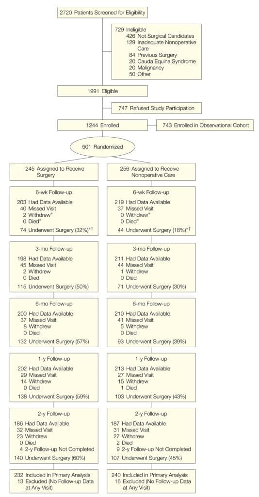
 

Blank
References

1. Grande RB, Aaseth K, Gulbrandsen P, Lundqvist C, Russell MB. Prevalence of primary chronic headache in a population-based sample of 30- to 44-year-old persons: the Akershus study of chronic headache. Neuroepidemiology. 2008;30(2):76�83. doi: 10.1159/000116244. [PubMed] [Cross Ref]
2. Headache Classification Committee of the International Headache Society. The International Classification of Headache Disorders, 3rd edition (beta version) Cephalalgia. 2013;33:629�808. [PubMed]
3. Kristoffersen ES, Grande RB, Aaseth K, Lundqvist C, Russell MB. Management of primary chronic headache in the general population: the Akershus study of chronic headache. J Headache Pain. 2012;13(2):113�120. doi: 10.1007/s10194-011-0391-8. [PMC free article] [PubMed] [Cross Ref]
4. Aaseth K, Grande RB, Kvaerner KJ, Gulbrandsen P, Lundqvist C, Russell MB. Prevalence of secondary chronic headaches in a population-based sample of 30-44-year-old persons: the Akershus study of chronic headache. Cephalalgia. 2008;28(7):705�713. doi: 10.1111/j.1468-2982.2008.01577.x. [PubMed] [Cross Ref]
5. Bronfort G, Nilsson N, Haas M, Evans R, Goldsmith CH, Assendelft WJ, Bouter LM. Non-invasive physical treatments for chronic/recurrent headache. Cochrane Database Syst Rev. 2004;3:1�69. [PubMed]
6. Chaibi A, Tuchin PJ, Russell MB. Manual therapies for migraine: a systematic review. J Headache Pain. 2011;12(2):127�133. doi: 10.1007/s10194-011-0296-6. [PMC free article] [PubMed] [Cross Ref]
7. Carnes D, Mars TS, Mullinger B, Froud R, Underwood M. Adverse events and manual therapy: a systematic review. Man Ther. 2010;15(4):355�363. doi: 10.1016/j.math.2009.12.006. [PubMed] [Cross Ref]
8. Lenssinck ML, Damen L, Verhagen AP, Berger MY, Passchier J, Koes BW. The effectiveness of physiotherapy and manipulation in patients with tension-type headache: a systematic review. Pain. 2004;112(3):381�388. doi: 10.1016/j.pain.2004.09.026. doi:10.1016/j.pain.2004.09.026. [PubMed] [Cross Ref]
9. Fernandez-de-Las-Penas C, Alonso-Blanco C, Cuadrado ML, Miangolarra JC, Barriga FJ, Pareja JA. Are manual therapies effective in reducing pain from tension-type headache: a systematic review. Clin J Pain. 2006;22(3):278�285. doi: 10.1097/01.ajp.0000173017.64741.86. doi:10.1097/01.ajp.0000173017.64741.86. [PubMed] [Cross Ref]
10. Chaibi A, Russell MB. Manual therapies for cervicogenic headache: a systematic review. J Headache Pain. 2012;13(5):351�359. doi: 10.1007/s10194-012-0436-7. [PMC free article] [PubMed] [Cross Ref]
11. Posadzki P, Ernst E. Spinal manipulations for tension-type headaches: a systematic review of randomized controlled trials. Complement Ther Med. 2012;20(4):232�239. doi: 10.1016/j.ctim.2011.12.001. doi:10.1016/j.ctim.2011.12.001. [PubMed] [Cross Ref]
12. French HP, Brennan A, White B, Cusack T. Manual therapy for osteoarthritis of the hip or knee � a systematic review. Man Ther. 2011;16(2):109�117. doi: 10.1016/j.math.2010.10.011. doi:10.1016/j.math.2010.10.011. [PubMed] [Cross Ref]
13. Tfelt-Hansen P, Block G, Dahlof C, Diener HC, Ferrari MD, Goadsby PJ, Guidetti V, Jones B, Lipton RB, Massiou H, Meinert C, Sandrini G, Steiner T, Winter PB. International Headache Society Clinical Trial Subcommittee. Guidelines for controlled trials of drugs in migraine: second edition. Cephalalgia. 2000;20(9):765�786. [PubMed]
14. Silberstein S, Tfelt-Hansen P, Dodick DW, Limmroth V, Lipton RB, Pascual J, Wang SJ. Task Force of the International Headache Society Clinical Trial Subcommittee. Guidelines for controlled trials of prophylactic treatment of chronic migraine in adults. Cephalalgia. 2008;28(5):484�495. doi: 10.1111/j.1468-2982.2008.01555.x. [PubMed] [Cross Ref]
15. Headache Classification Committee of the International Headache Society. Classification and diagnostic criteria for headache disorders, cranial neuralgias and facial pain: Headache Classification Committee of the International Headache Society. Cephalalgia. 1988;8(suppl 7):1�96. [PubMed]
16. Headache Classification Subcommittee of the International Society. The international classification of headache disorders: 2nd edition. Cephalalgia. 2004;24(Suppl 1):9�160. [PubMed]
17. Olesen J, Bousser MG, Diener HC, Dodick D, First M, Goadsby PJ, Gobel H, Lainez MJ, Lance JW, Lipton RB, Nappi G, Sakai F, Schoenen J, Silberstein SD, Steiner TJ. International Headache Society New appendix criteria open for a broader concept of chronic migraine. Cephalalgia. 2006;26(6):742�746. [PubMed]
18. Moseley AM, Herbert RD, Sherrington C, Maher CG. Evidence for physiotherapy practice: a survey of the Physiotherapy Evidence Database (PEDro) Aust J Physiother. 2002;48(1):43�49. doi: 10.1016/S0004-9514(14)60281-6. [PubMed] [Cross Ref]
19. Cohen J. Statistical Power Analysis for the Behavioral Sciences. 2. Routledge, USA; 1988.
20. Toro-Velasco C, Arroyo-Morales M, Fernandez-de-las-Penas C, Cleland JA, Barrero-Hernandez FJ. Short-term effects of manual therapy on heart rate variability, mood state, and pressure pain sensitivity in patients with chronic tension-type headache: a pilot study. J Manipulative Physiol Ther. 2009;32(7):527�535. doi: 10.1016/j.jmpt.2009.08.011. [PubMed] [Cross Ref]
21. Jay GW, Brunson J, Branson SJ. The effectiveness of physical therapy in the treatment of chronic daily headaches. Headache. 1989;29(3):156�162. doi: 10.1111/j.1526-4610.1989.hed2903156.x. [PubMed] [Cross Ref]
22. Demirturk F, Akarcali I, Akbayrak T, Citak I, Inan L. Results of two different manual therapy techniques in chronic tension-type headache. Pain Clin. 2002;14(2):121�128. doi: 10.1163/156856902760196333. [Cross Ref]
23. Torelli P, Jensen R, Olesen J. Physiotherapy for tension-type headache: a controlled study. Cephalalgia. 2004;24(1):29�36. doi: 10.1111/j.1468-2982.2004.00633.x. [PubMed] [Cross Ref]
24. Ettekoven VH, Lucas C. Efficacy of physiotherapy including a craniocervical training programme for tension-type headache; a randomized clinical trial. Cephalalgia. 2006;26(8):983�991. doi: 10.1111/j.1468-2982.2006.01163.x. [PubMed] [Cross Ref]
25. Castien RF, Van der Windt DA, Grooten A, Dekker J. Effectiveness of manual therapy for chronic tension-type headache: a pragmatic, randomised, clinical trial. Cephalalgia. 2011;31(2):133�143. doi: 10.1177/0333102410377362. [PubMed] [Cross Ref]
26. Rasmussen BK, Jensen R, Olesen J. Questionnaire versus clinical interview in the diagnosis of headache. Headache. 1991;31(5):290�295. doi: 10.1111/j.1526-4610.1991.hed3105290.x. [PubMed] [Cross Ref]
27. Moher D, Hopewell S, Schulz KF, Montori V, Gotzsche PC, Devereaux PJ, Elbourne D, Egger M, Altman DG. CONSORT 2010 explanation and elaboration: updated guidelines for reporting parallel group randomised trials. BMJ. 2010;340:c869. doi: 10.1136/bmj.c869. [PMC free article] [PubMed] [Cross Ref]
28. Bendtsen L, Jensen R, Olesen J. A non-selective (amitriptyline), but not a selective (citalopram), serotonin reuptake inhibitor is effective in the prophylactic treatment of chronic tension-type headache. J Neurol Neurosurg Psychiatry. 1996;61(3):285�290. doi: 10.1136/jnnp.61.3.285. [PMC free article] [PubMed] [Cross Ref]
29. Jackson JL, Shimeall W, Sessums L, Dezee KJ, Becher D, Diemer M, Berbano E, O�Malley PG. Tricyclic antidepressants and headaches: systematic review and meta-analysis. BMJ. 2010;341:c5222. doi: 10.1136/bmj.c5222. [PMC free article] [PubMed] [Cross Ref]
30. Bendtsen L, Bigal ME, Cerbo R, Diener HC, Holroyd K, Lampl C, Mitsikostas DD, Steiner TJ, Tfelt-Hansen P. Guidelines for controlled trials of drugs in tension-type headache: second edition. Cephalalgia. 2010;30(1):1�16. [PubMed]

Close Accordion
Blank
References

1. Bovim G, Schrader H, Sand T. Neck pain in the general population. Spine (Phila Pa 1976) 1994;19(12):1307�1309. doi: 10.1097/00007632-199406000-00001. [PubMed] [Cross Ref]
2. Brattberg G, Thorslund M, Wikman A. The prevalence of pain in a general population. The results of a postal survey in a county of Sweden. Pain. 1989;37(2):215�222. doi: 10.1016/0304-3959(89)90133-4. [PubMed] [Cross Ref]
3. Hagen KB, Harms-Ringdahl K, Enger NO, Hedenstad R, Morten H. Relationship between subjective neck disorders and cervical spine mobility and motion-related pain in male machine operators. Spine (Phila Pa 1976) 1997;22(13):1501�1507. doi: 10.1097/00007632-199707010-00015. [PubMed] [Cross Ref]
4. Fricton JR, Kroening R, Haley D, Siegert R. Myofascial pain syndrome of the head and neck: a review of clinical characteristics of 164 patients. Oral Surg Oral Med Oral Pathol. 1985;60(6):615�623. doi: 10.1016/0030-4220(85)90364-0. [PubMed] [Cross Ref]
5. Stovner LJ. The nosologic status of the whiplash syndrome: a critical review based on a methodological approach. Spine (Phila Pa 1976) 1996;21(23):2735�2746. doi: 10.1097/00007632-199612010-00006. [PubMed] [Cross Ref]
6. Frank AO, De Souza LH, Frank CA. Neck pain and disability: a cross-sectional survey of the demographic and clinical characteristics of neck pain seen in a rheumatology clinic. Int J Clin Pract. 2005;59(2):173�182. doi: 10.1111/j.1742-1241.2004.00237.x. [PubMed] [Cross Ref]
7. Andersson G. The epidemiology of spinal disorders. In: Frymoyer J, editor. The adult spine: principles and practice. Philadelphia: Lippincott Raven; 1997. pp. 130�141.
8. Rasmussen C, Leboeuf-Yde C, Hestbaek L, Manniche C. Poor outcome in patients with spine-related leg or arm pain who are involved in compensation claims: a prospective study of patients in the secondary care sector. Scand J Rheumatol. 2008;37(6):462�468. doi: 10.1080/03009740802241709. [PubMed] [Cross Ref]
9. Daffner SD, Hilibrand AS, Hanscom BS, Brislin BT, Vaccaro AR, Albert TJ. Impact of neck and arm pain on overall health status. Spine (Phila Pa 1976) 2003;28(17):2030�2035. doi: 10.1097/01.BRS.0000083325.27357.39. [PubMed] [Cross Ref]
10. Abbed KM, Coumans JV. Cervical radiculopathy: pathophysiology, presentation, and clinical evaluation. Neurosurgery. 2007;60(1 Supp1 1):S28�34. [PubMed]
11. Lauerman W, Scherping S, Wiesel S. The spine. In: Wiesel S, Delahay J, editors. Essentials of Orthopedic Surgery. 3. New York: Springer; 2007. pp. 276�332.
12. Carette S, Fehlings MG. Clinical practice. Cervical radiculopathy. N Engl J Med. 2005;353(4):392�399. doi: 10.1056/NEJMcp043887. [PubMed] [Cross Ref]
13. Hurwitz EL, Carragee EJ, van der Velde G, Carroll LJ, Nordin M, Guzman J, et al. Treatment of neck pain: noninvasive interventions: results of the Bone and Joint Decade 2000-2010 Task Force on Neck Pain and Its Associated Disorders. Spine (Phila Pa 1976) 2008;33(4 Suppl):S123�52. doi: 10.1097/BRS.0b013e3181644b1d. [PubMed] [Cross Ref]
14. Saal JS, Saal JA, Yurth EF. Nonoperative management of herniated cervical intervertebral disc with radiculopathy. Spine (Phila Pa 1976) 1996;21(16):1877�1883. doi: 10.1097/00007632-199608150-00008. [PubMed] [Cross Ref]
15. Clark C. The Cervical Spine. 4. Philadelphia: Lippincott Williams & Wilkins; 2005.
16. Engquist M, Lofgren H, Oberg B, Holtz A, Peolsson A, Soderlund A, et al. Surgery versus nonsurgical treatment of cervical radiculopathy: a prospective, randomized study comparing surgery plus physiotherapy with physiotherapy alone with a 2-year follow-up. Spine (Phila Pa 1976) 2013;38(20):1715�1722. [PubMed]
17. Nikolaidis I, Fouyas IP, Sandercock PA, Statham PF: Surgery for cervical radiculopathy or myelopathy. Cochrane Database Syst Rev 2010, (1):CD001466. doi(1):CD001466. [PubMed]
18. Weinstein JN, Tosteson TD, Lurie JD, Tosteson AN, Hanscom B, Skinner JS, et al. Surgical vs nonoperative treatment for lumbar disk herniation: the Spine Patient Outcomes Research Trial (SPORT): a randomized trial. JAMA. 2006;296(20):2441�2450. doi: 10.1001/jama.296.20.2441. [PMC free article] [PubMed] [Cross Ref]
19. Peul WC, van Houwelingen HC, van den Hout WB, Brand R, Eekhof JA, Tans JT, et al. Surgery versus prolonged conservative treatment for sciatica. N Engl J Med. 2007;356(22):2245�2256. doi: 10.1056/NEJMoa064039. [PubMed] [Cross Ref]
20. Weber H. Lumbar disc herniation. A controlled, prospective study with ten years of observation. Spine (Phila Pa 1976) 1983;8(2):131�140. doi: 10.1097/00007632-198303000-00003. [PubMed] [Cross Ref]
21. Kim JD, Son MS. 2013 National Health Insurance Statistical Yearbook. Seoul: Health Insurance Review and Assessment Service and National Health Insurance Service; 2014.
22. Lin XJ, Chen CY. Advances on study of treatment of lumbar disk herniation by Chinese medicinal herbs. Zhongguo Zhong Yao Za Zhi. 2007;32(3):186�191. [PubMed]
23. Stevens L, Duarte H, Park J. Promising implications for integrative medicine for back pain: a profile of a Korean hospital. J Altern Complement Med. 2007;13(5):481�484. doi: 10.1089/acm.2007.6263. [PubMed] [Cross Ref]
24. Chung HJ, Lee HS, Shin JS, Lee SH, Park BM, Youn YS, et al. Modulation of acute and chronic inflammatory processes by a traditional medicine preparation GCSB-5 both in vitro and in vivo animal models. J Ethnopharmacol. 2010;130(3):450�459. doi: 10.1016/j.jep.2010.05.020. [PubMed] [Cross Ref]
25. Kim TH, Yoon SJ, Lee WC, Kim JK, Shin J, Lee S, et al. Protective effect of GCSB-5, an herbal preparation, against peripheral nerve injury in rats. J Ethnopharmacol. 2011;136(2):297�304. doi: 10.1016/j.jep.2011.04.037. [PubMed] [Cross Ref]
26. Kim JK, Park SW, Kang JW, Kim YJ, Lee SY, Shin J, et al. Effect of GCSB-5, a Herbal Formulation, on Monosodium Iodoacetate-Induced Osteoarthritis in Rats. Evid Based Complement Alternat Med. 2012;2012:730907. [PMC free article] [PubMed]
27. Park YG, Ha CW, Han CD, Bin SI, Kim HC, Jung YB, et al. A prospective, randomized, double-blind, multicenter comparative study on the safety and efficacy of Celecoxib and GCSB-5, dried extracts of six herbs, for the treatment of osteoarthritis of knee joint. J Ethnopharmacol. 2013;149(3):816�824. doi: 10.1016/j.jep.2013.08.008. [PubMed] [Cross Ref]
28. Xu RD, Li H. Conception of Ashi points. Zhongguo Zhen Jiu. 2005;25(4):281�283. [PubMed]
29. Assendelft WJ, Morton SC, Yu EI, Suttorp MJ, Shekelle PG. Spinal manipulative therapy for low back pain. A meta-analysis of effectiveness relative to other therapies. Ann Intern Med. 2003;138(11):871�881. doi: 10.7326/0003-4819-138-11-200306030-00008. [PubMed] [Cross Ref]
30. Bronfort G, Haas M, Evans R, Kawchuk G, Dagenais S. Evidence-informed management of chronic low back pain with spinal manipulation and mobilization. Spine J. 2008;8(1):213�225. doi: 10.1016/j.spinee.2007.10.023. [PubMed] [Cross Ref]
31. Turk DC, Rudy TE, Sorkin BA. Neglected topics in chronic pain treatment outcome studies: determination of success. Pain. 1993;53(1):3�16. doi: 10.1016/0304-3959(93)90049-U. [PubMed] [Cross Ref]
32. Ponce de Leon S, Lara-Munoz C, Feinstein AR, Wells CK. A comparison of three rating scales for measuring subjective phenomena in clinical research. II. Use of experimentally controlled visual stimuli. Arch Med Res. 2004;35(2):157�162. doi: 10.1016/j.arcmed.2003.07.009. [PubMed] [Cross Ref]
33. Farrar JT, Young JP, Jr, LaMoreaux L, Werth JL, Poole RM. Clinical importance of changes in chronic pain intensity measured on an 11-point numerical pain rating scale. Pain. 2001;94(2):149�158. doi: 10.1016/S0304-3959(01)00349-9. [PubMed] [Cross Ref]
34. Carreon LY, Glassman SD, Campbell MJ, Anderson PA. Neck Disability Index, short form-36 physical component summary, and pain scales for neck and arm pain: the minimum clinically important difference and substantial clinical benefit after cervical spine fusion. Spine J. 2010;10(6):469�474. doi: 10.1016/j.spinee.2010.02.007. [PubMed] [Cross Ref]
35. Parker SL, Godil SS, Shau DN, Mendenhall SK, McGirt MJ. Assessment of the minimum clinically important difference in pain, disability, and quality of life after anterior cervical discectomy and fusion: clinical article. J Neurosurg Spine. 2013;18(2):154�160. doi: 10.3171/2012.10.SPINE12312. [PubMed] [Cross Ref]
36. Lee J, Shin JS, Kim MR, Byun JH, Lee SY, Shin YS, et al. Liver enzyme abnormalities in taking traditional herbal medicine in Korea: A retrospective large sample cohort study of musculoskeletal disorder patients. J Ethnopharmacol. 2015;169:407�412. doi: 10.1016/j.jep.2015.04.048. [PubMed] [Cross Ref]
37. Manchikanti L, Falco FJ, Singh V, Pampati V, Parr AT, Benyamin RM, et al. Utilization of interventional techniques in managing chronic pain in the Medicare population: analysis of growth patterns from 2000 to 2011. Pain Physician. 2012;15(6):E969�82. [PubMed]
38. Chou R, Atlas SJ, Stanos SP, Rosenquist RW. Nonsurgical interventional therapies for low back pain: a review of the evidence for an American Pain Society clinical practice guideline. Spine (Phila Pa 1976) 2009;34(10):1078�1093. doi: 10.1097/BRS.0b013e3181a103b1. [PubMed] [Cross Ref]
39. Airaksinen O, Brox JI, Cedraschi C, Hildebrandt J, Klaber-Moffett J, Kovacs F, et al. Chapter 4. European guidelines for the management of chronic nonspecific low back pain. Eur Spine J. 2006;15(Suppl 2):S192�300. doi: 10.1007/s00586-006-1072-1. [PMC free article] [PubMed] [Cross Ref]
40. Staal JB, de Bie RA, de Vet HC, Hildebrandt J, Nelemans P. Injection therapy for subacute and chronic low back pain: an updated Cochrane review. Spine (Phila Pa 1976) 2009;34(1):49�59. doi: 10.1097/BRS.0b013e3181909558. [PubMed] [Cross Ref]
41. Armon C, Argoff CE, Samuels J, Backonja MM, Therapeutics and Technology Assessment Subcommittee of the American Academy of Neurology Assessment: use of epidural steroid injections to treat radicular lumbosacral pain: report of the Therapeutics and Technology Assessment Subcommittee of the American Academy of Neurology. Neurology. 2007;68(10):723�729. doi: 10.1212/01.wnl.0000256734.34238.e7. [PubMed] [Cross Ref]
42. Parr AT, Diwan S, Abdi S. Lumbar interlaminar epidural injections in managing chronic low back and lower extremity pain: a systematic review. Pain Physician. 2009;12(1):163�188. [PubMed]
43. DePalma MJ, Slipman CW. Evidence-informed management of chronic low back pain with epidural steroid injections. Spine J. 2008;8(1):45�55. doi: 10.1016/j.spinee.2007.09.009. [PubMed] [Cross Ref]
44. Cohen SP, Bicket MC, Jamison D, Wilkinson I, Rathmell JP. Epidural steroids: a comprehensive, evidence-based review. Reg Anesth Pain Med. 2013;38(3):175�200. doi: 10.1097/AAP.0b013e31828ea086. [PubMed] [Cross Ref]
45. Scanlon GC, Moeller-Bertram T, Romanowsky SM, Wallace MS. Cervical transforaminal epidural steroid injections: more dangerous than we think? Spine (Phila Pa 1976) 2007;32(11):1249�1256. doi: 10.1097/BRS.0b013e318053ec50. [PubMed] [Cross Ref]
46. Rathmell JP, Benzon HT. Transforaminal injection of steroids: should we continue? Reg Anesth Pain Med. 2004;29(5):397�399. [PubMed]
47. Tiso RL, Cutler T, Catania JA, Whalen K. Adverse central nervous system sequelae after selective transforaminal block: the role of corticosteroids. Spine J. 2004;4(4):468�474. doi: 10.1016/j.spinee.2003.10.007. [PubMed] [Cross Ref]
48. Brouwers PJ, Kottink EJ, Simon MA, Prevo RL. A cervical anterior spinal artery syndrome after diagnostic blockade of the right C6-nerve root. Pain. 2001;91(3):397�399. doi: 10.1016/S0304-3959(00)00437-1. [PubMed] [Cross Ref]
49. Wallace MA, Fukui MB, Williams RL, Ku A, Baghai P. Complications of cervical selective nerve root blocks performed with fluoroscopic guidance. AJR Am J Roentgenol. 2007;188(5):1218�1221. doi: 10.2214/AJR.04.1541. [PubMed] [Cross Ref]
50. Rathmell JP, Aprill C, Bogduk N. Cervical transforaminal injection of steroids. Anesthesiology. 2004;100(6):1595�1600. doi: 10.1097/00000542-200406000-00035. [PubMed] [Cross Ref]
51. Manchikanti L, Malla Y, Wargo BW, Cash KA, Pampati V, Fellows B. A prospective evaluation of complications of 10,000 fluoroscopically directed epidural injections. Pain Physician. 2012;15(2):131�140. [PubMed]
52. Abbasi A, Malhotra G, Malanga G, Elovic EP, Kahn S. Complications of interlaminar cervical epidural steroid injections: a review of the literature. Spine (Phila Pa 1976) 2007;32(19):2144�2151. doi: 10.1097/BRS.0b013e318145a360. [PubMed] [Cross Ref]
53. Hodges SD, Castleberg RL, Miller T, Ward R, Thornburg C. Cervical epidural steroid injection with intrinsic spinal cord damage. Two case reports. Spine (Phila Pa 1976) 1998;23(19):2137�42. doi: 10.1097/00007632-199810010-00020. [PubMed] [Cross Ref]
54. Kaplan MS, Cunniff J, Cooke J, Collins JG. Intravascular uptake during fluoroscopically guided cervical interlaminar steroid injection at C6-7: a case report. Arch Phys Med Rehabil. 2008;89(3):553�558. doi: 10.1016/j.apmr.2007.08.165. [PubMed] [Cross Ref]
55. McGrath JM, Schaefer MP, Malkamaki DM. Incidence and characteristics of complications from epidural steroid injections. Pain Med. 2011;12(5):726�731. doi: 10.1111/j.1526-4637.2011.01077.x. [PubMed] [Cross Ref]
56. Shanthanna H, Park J. Acute epidural haematoma following epidural steroid injection in a patient with spinal stenosis. Anaesthesia. 2011;66(9):837�839. doi: 10.1111/j.1365-2044.2011.06770.x. [PubMed] [Cross Ref]
57. McCleane G. Does gabapentin have an analgesic effect on background, movement and referred pain? A randomized, double-blind, placebo controlled study. Pain Clinic. 2001;13:103�107. doi: 10.1163/156856901753420945. [Cross Ref]
58. Yildirim K, Sisecioglu M, Karatay S, Erdal A, Levent A, Ugur M, et al. The effectiveness of gabapentin in patients with chronic radiculopathy. Pain Clinic. 2003;15:213�218. doi: 10.1163/156856903767650718. [Cross Ref]
59. Khoromi S, Cui L, Nackers L, Max MB. Morphine, nortriptyline and their combination vs. placebo in patients with chronic lumbar root pain. Pain. 2007;130(1-2):66�75. doi: 10.1016/j.pain.2006.10.029. [PMC free article] [PubMed] [Cross Ref]
60. Khoromi S, Patsalides A, Parada S, Salehi V, Meegan JM, Max MB. Topiramate in chronic lumbar radicular pain. J Pain. 2005;6(12):829�836. doi: 10.1016/j.jpain.2005.08.002. [PubMed] [Cross Ref]
61. Baron R, Freynhagen R, Tolle TR, Cloutier C, Leon T, Murphy TK, et al. The efficacy and safety of pregabalin in the treatment of neuropathic pain associated with chronic lumbosacral radiculopathy. Pain. 2010;150(3):420�427. doi: 10.1016/j.pain.2010.04.013. [PubMed] [Cross Ref]
62. Hahne AJ, Ford JJ, McMeeken JM. Conservative management of lumbar disc herniation with associated radiculopathy: a systematic review. Spine (Phila Pa 1976) 2010;35(11):E488�504. [PubMed]
63. Salt E, Wright C, Kelly S, Dean A. A systematic literature review on the effectiveness of non-invasive therapy for cervicobrachial pain. Man Ther. 2011;16(1):53�65. doi: 10.1016/j.math.2010.09.005. [PubMed] [Cross Ref]
64. Kuijper B, Tans JT, Beelen A, Nollet F, de Visser M. Cervical collar or physiotherapy versus wait and see policy for recent onset cervical radiculopathy: randomised trial. BMJ. 2009;339:b3883. doi: 10.1136/bmj.b3883. [PMC free article] [PubMed] [Cross Ref]
65. Gebremariam L, Koes BW, Peul WC, Huisstede BM. Evaluation of treatment effectiveness for the herniated cervical disc: a systematic review. Spine (Phila Pa 1976) 2012;37(2):E109�18. doi: 10.1097/BRS.0b013e318221b5af. [PubMed] [Cross Ref]
66. Boselie TF, Willems PC, van Mameren H, de Bie RA, Benzel EC, van Santbrink H. Arthroplasty versus fusion in single-level cervical degenerative disc disease: a Cochrane review. Spine (Phila Pa 1976) 2013;38(17):E1096�107. doi: 10.1097/BRS.0b013e3182994a32. [PubMed] [Cross Ref]
67. Manchikanti L, Cash KA, Pampati V, Wargo BW, Malla Y. Cervical epidural injections in chronic discogenic neck pain without disc herniation or radiculitis: preliminary results of a randomized, double-blind, controlled trial. Pain Physician. 2010;13(4):E265�78. [PubMed]
68. Cesaroni A, Nardi PV. Plasma disc decompression for contained cervical disc herniation: a randomized, controlled trial. Eur Spine J. 2010;19(3):477�486. doi: 10.1007/s00586-009-1189-0. [PMC free article] [PubMed] [Cross Ref]

Close Accordion