Health & Immunity Series 1of 4 | El Paso, Tx (2020)
If you have enjoyed this video and/or we have helped you in any way
please feel free to subscribe and share us.
Thank You & God Bless.
Dr. Alex Jimenez RN, DC, MSACP, CCST
Subscribe: http://bit.ly/drjyt
Facebook Clinical Page: https://www.facebook.com/dralexjimenez/
Facebook Sports Page: https://www.facebook.com/pushasrx/
Facebook Injuries Page: https://www.facebook.com/elpasochiropractor/
Facebook Neuropathy Page: https://www.facebook.com/ElPasoNeuropathyCenter/
Facebook Fitness Center Page: https://www.facebook.com/PUSHftinessathletictraining/
Yelp: El Paso Rehabilitation Center: http://goo.gl/pwY2n2
Yelp: El Paso Clinical Center: Treatment: https://goo.gl/r2QPuZ
Clinical Testimonies: https://www.dralexjimenez.com/category/testimonies/
Information:
Clinical Site: https://www.dralexjimenez.com
Injury Site: https://personalinjurydoctorgroup.com
Sports Injury Site: https://chiropracticscientist.com
Back Injury Site: https://elpasobackclinic.com
Rehabilitation Center: https://www.pushasrx.com
Functional Medicine: https://wellnessdoctorrx.com
Fitness & Nutrition: http://www.push4fitness.com/team/
Twitter: https://twitter.com/dralexjimenez
Twitter: https://twitter.com/crossfitdoctor
The Functional Fitness Fellas | What is it? & Who Are They?
If you have enjoyed this video and/or we have helped you in any way
please feel free to subscribe and share us.
Thank You & God Bless.
Dr. Alex Jimenez RN, DC, MSACP, CCST
Subscribe: http://bit.ly/drjyt
Facebook Clinical Page: https://www.facebook.com/dralexjimenez/
Facebook Sports Page: https://www.facebook.com/pushasrx/
Facebook Injuries Page: https://www.facebook.com/elpasochiropractor/
Facebook Neuropathy Page: https://www.facebook.com/ElPasoNeuropathyCenter/
Facebook Fitness Center Page: https://www.facebook.com/PUSHftinessathletictraining/
Yelp: El Paso Rehabilitation Center: http://goo.gl/pwY2n2
Yelp: El Paso Clinical Center: Treatment: https://goo.gl/r2QPuZ
Clinical Testimonies: https://www.dralexjimenez.com/category/testimonies/
Information:
Clinical Site: https://www.dralexjimenez.com
Injury Site: https://personalinjurydoctorgroup.com
Sports Injury Site: https://chiropracticscientist.com
Back Injury Site: https://elpasobackclinic.com
Rehabilitation Center: https://www.pushasrx.com
Functional Medicine: https://wellnessdoctorrx.com
Fitness & Nutrition: http://www.push4fitness.com/team/
Twitter: https://twitter.com/dralexjimenez
Twitter: https://twitter.com/crossfitdoctor
What is the FASTING MIMICKING DIET & STRESS HORMONES – HEALTH | El Paso, Tx (2020)
If you have enjoyed this video and/or we have helped you in any way
please feel free to subscribe and share us.
Thank You & God Bless.
Dr. Alex Jimenez RN, DC, MSACP, CCST
Subscribe: http://bit.ly/drjyt
Facebook Clinical Page: https://www.facebook.com/dralexjimenez/
Facebook Sports Page: https://www.facebook.com/pushasrx/
Facebook Injuries Page: https://www.facebook.com/elpasochiropractor/
Facebook Neuropathy Page: https://www.facebook.com/ElPasoNeuropathyCenter/
Facebook Fitness Center Page: https://www.facebook.com/PUSHftinessathletictraining/
Yelp: El Paso Rehabilitation Center: http://goo.gl/pwY2n2
Yelp: El Paso Clinical Center: Treatment: https://goo.gl/r2QPuZ
Clinical Testimonies: https://www.dralexjimenez.com/category/testimonies/
Information:
Clinical Site: https://www.dralexjimenez.com
Injury Site: https://personalinjurydoctorgroup.com
Sports Injury Site: https://chiropracticscientist.com
Back Injury Site: https://elpasobackclinic.com
Rehabilitation Center: https://www.pushasrx.com
Functional Medicine: https://wellnessdoctorrx.com
Fitness & Nutrition: http://www.push4fitness.com/team/
Twitter: https://twitter.com/dralexjimenez
Twitter: https://twitter.com/crossfitdoctor
Treating Inflammation Naturally | El Paso, Tx (2020)
If you have enjoyed this video and/or we have helped you in any way
please feel free to subscribe and share us.
Thank You & God Bless.
Dr. Alex Jimenez RN, DC, MSACP, CCST
Subscribe: http://bit.ly/drjyt
Facebook Clinical Page: https://www.facebook.com/dralexjimenez/
Facebook Sports Page: https://www.facebook.com/pushasrx/
Facebook Injuries Page: https://www.facebook.com/elpasochiropractor/
Facebook Neuropathy Page: https://www.facebook.com/ElPasoNeuropathyCenter/
Facebook Fitness Center Page: https://www.facebook.com/PUSHftinessathletictraining/
Yelp: El Paso Rehabilitation Center: http://goo.gl/pwY2n2
Yelp: El Paso Clinical Center: Treatment: https://goo.gl/r2QPuZ
Clinical Testimonies: https://www.dralexjimenez.com/category/testimonies/
Information:
Clinical Site: https://www.dralexjimenez.com
Injury Site: https://personalinjurydoctorgroup.com
Sports Injury Site: https://chiropracticscientist.com
Back Injury Site: https://elpasobackclinic.com
Rehabilitation Center: https://www.pushasrx.com
Functional Medicine: https://wellnessdoctorrx.com
Fitness & Nutrition: http://www.push4fitness.com/team/
Twitter: https://twitter.com/dralexjimenez
Twitter: https://twitter.com/crossfitdoctor
Why Choose Functional Medicine – The “Why” Explained | El Paso, Tx (2020)
If you have enjoyed this video and/or we have helped you in any way please feel free to subscribe and share with us.
Thank You & God Bless.
Dr. Alex Jimenez RN, DC, MSACP, CCST
Subscribe: http://bit.ly/drjyt
Facebook Clinical Page: https://www.facebook.com/dralexjimenez/
Facebook Sports Page: https://www.facebook.com/pushasrx/
Facebook Injuries Page: https://www.facebook.com/elpasochiropractor/
Facebook Neuropathy Page: https://www.facebook.com/ElPasoNeuropathyCenter/
Facebook Fitness Center Page: https://www.facebook.com/PUSHftinessathletictraining/
Yelp: El Paso Rehabilitation Center: http://goo.gl/pwY2n2
Yelp: El Paso Clinical Center: Treatment: https://goo.gl/r2QPuZ
Clinical Testimonies: https://www.dralexjimenez.com/category/testimonies/
Information:
Clinical Site: https://www.dralexjimenez.com
Injury Site: https://personalinjurydoctorgroup.com
Sports Injury Site: https://chiropracticscientist.com
Back Injury Site: https://elpasobackclinic.com
Rehabilitation Center: https://www.pushasrx.com
Functional Medicine: https://wellnessdoctorrx.com
Fitness & Nutrition: http://www.push4fitness.com/team/
Twitter: https://twitter.com/dralexjimenez
Twitter: https://twitter.com/crossfitdoctor
**Corona Virus Blessings** COVID 19 A Society Changing Moment | El Paso, Tx (2020)
If you have enjoyed this video and/or we have helped you in any way please feel free to subscribe and share us.
Thank You & God Bless.
Dr. Alex Jimenez RN, DC, MSACP, CCST
Subscribe: http://bit.ly/drjyt
Facebook Clinical Page: https://www.facebook.com/dralexjimenez/
Facebook Sports Page: https://www.facebook.com/pushasrx/
Facebook Injuries Page: https://www.facebook.com/elpasochiropractor/
Facebook Neuropathy Page: https://www.facebook.com/ElPasoNeuropathyCenter/
Facebook Fitness Center Page: https://www.facebook.com/PUSHftinessathletictraining/
Yelp: El Paso Rehabilitation Center: http://goo.gl/pwY2n2
Yelp: El Paso Clinical Center: Treatment: https://goo.gl/r2QPuZ
Clinical Testimonies: https://www.dralexjimenez.com/category/testimonies/
Information:
Clinical Site: https://www.dralexjimenez.com
Injury Site: https://personalinjurydoctorgroup.com
Sports Injury Site: https://chiropracticscientist.com
Back Injury Site: https://elpasobackclinic.com
Rehabilitation Center: https://www.pushasrx.com
Functional Medicine: https://wellnessdoctorrx.com
Fitness & Nutrition: http://www.push4fitness.com/team/
Twitter: https://twitter.com/dralexjimenez
Twitter: https://twitter.com/crossfitdoctor


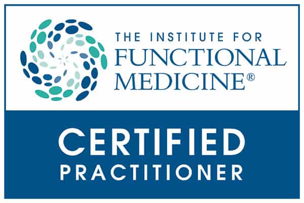
IFM's Find A Practitioner tool is the largest referral network in Functional Medicine, created to help patients locate Functional Medicine practitioners anywhere in the world. IFM Certified Practitioners are listed first in the search results, given their extensive education in Functional Medicine
Online Booking & Appointments 24/7*

All providers work within legal jurisdiction and have an established clinical scope of practice.*
COMPLETE ONLINE HISTORY 24/7*
