Restore Flexibility and Mobility with Integrative Chiropractic Care and Shockwave Therapy at El Paso Back Clinic
Many El Paso residents wake up with stiff joints or tight muscles, making simple daily tasks feel hard. Reaching overhead, bending down, or walking for long stretches can become painful or limited. At El Paso Back Clinic, integrative chiropractic care combined with Extracorporeal Shockwave Therapy (ESWT) offers a natural solution. This approach restores proper joint alignment, reduces muscle tension, and resolves soft-tissue restrictions, allowing patients to move freely again. Led by Dr. Alexander Jimenez, DC, APRN, FNP-BC, the clinic’s team uses gentle adjustments, stretching, exercises, and advanced shockwave treatments to help people regain flexibility and enjoy life in El Paso.
What Integrative Chiropractic Care Does for Flexibility at El Paso Back Clinic
Integrative chiropractic care at El Paso Back Clinic treats the whole body instead of just one problem area. It corrects small misalignments, called subluxations, in the spine and joints. These misalignments put pressure on nerves and tighten muscles. Regular adjustments gently move everything back into place. This restores proper joint alignment, eases tension, and lets the nervous system send clearer signals to the muscles.
When joints line up correctly, range of motion improves right away. Stiffness fades, and daily movements become smoother and more efficient. Patients at the clinic often say they feel looser and more energetic after just a few visits. (Gentle Chiro, n.d.) The care also includes stretching and therapeutic exercises to maintain gains over time. Muscles and joints start working together as a team, building resilience that lasts.
How Chiropractic Adjustments Restore Joint Alignment and Reduce Stiffness
Adjustments form the core of care at El Paso Back Clinic. The team uses precise, gentle pressure to correct subluxations. This simple step brings clear benefits that patients notice quickly:
Better range of motion, so joints glide freely without catching
Less muscle tension around the back, neck, and limbs
Improved nervous system function for better balance and coordination
Smoother daily activities like turning your head while driving or reaching for groceries
Lower risk of future stiffness because proper alignment trains the body to stay balanced
Many people in El Paso report that these changes make physical activities feel easier and less tiring. (Rodgers Stein Chiropractic, n.d.) The adjustments help the body move more efficiently without pain, supporting an active lifestyle.
Adding Stretching and Therapeutic Exercises for Long-Term Results
Adjustments open the door to better movement, but stretching and exercises keep it open. At El Paso Back Clinic, the rehabilitation team creates simple home programs that match each patient’s needs. Dynamic stretches warm up the body before activity. Static stretches hold the new mobility after adjustments. Therapeutic exercises strengthen the muscles that support the joints.
These steps build endurance and agility. Patients find they can stay active longer without soreness. The clinic’s sports medicine approach helps people return to hiking in the Franklin Mountains, playing with family, or working without the same old limitations. (Chiropractic Fitness, n.d.) Consistent practice turns short-term gains into lasting flexibility.
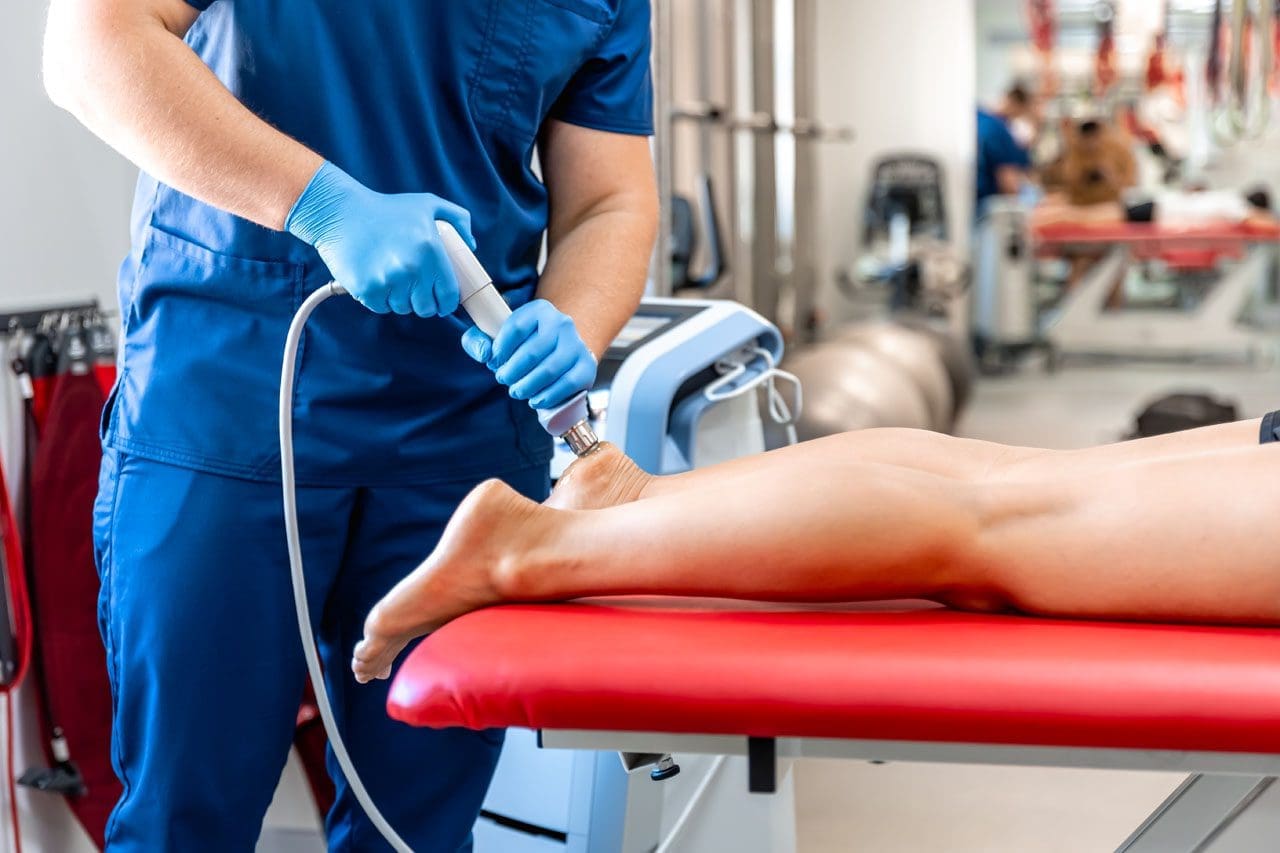
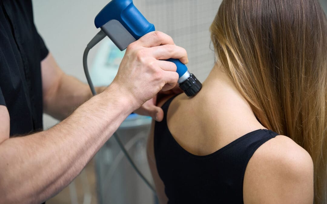
Introducing Extracorporeal Shockwave Therapy (ESWT) at El Paso Back Clinic
ESWT uses focused sound waves to reach deep into muscles, tendons, and ligaments. The waves create tiny pulses that restart healing in areas stuck with scar tissue or chronic tightness. This noninvasive treatment increases blood flow, breaks down old buildup, and reduces inflammation. At El Paso Back Clinic, ESWT is available as a key component of advanced care plans for patients who need additional support for soft tissue problems.
Why Combining Chiropractic Care and ESWT Delivers Stronger Flexibility Gains
The real power at El Paso Back Clinic comes from pairing chiropractic adjustments with ESWT. Adjustments fix the mechanical side—joint position and nerve signals—while ESWT handles the soft-tissue side—scar tissue, poor circulation, and stubborn tension. Together, they create faster, longer-lasting results than either method alone.
This dual approach works in several key ways:
Chiropractic restores spinal and joint mobility
ESWT breaks down scar tissue and releases tight fascia
The pair reduces inflammation and collagen cross-linking that causes stiffness
Blood flow improves, helping muscles and tendons heal
Patients regain a greater range of motion because both structure and tissue health get better at once
Clinic reports show that this combination can significantly improve outcomes compared with standard care. Many El Paso patients with ongoing tightness notice a real return of freedom of movement.
Common Conditions That Benefit from This Integrated Approach
El Paso Back Clinic uses this combined approach to treat several conditions that rob people of flexibility. Here are some of the most common:
Frozen shoulder – Adjustments free stuck joints while ESWT dissolves scar tissue and calcium deposits. Patients often regain full arm motion without pain.
Achilles tendinopathy – Chiropractic realigns the lower body to ease strain. Shockwave therapy stimulates the growth of new blood vessels and clears chronic buildup, so walking and running feel normal again.
General chronic muscle tension – Tightness in the back, neck, or legs from stress, work, or old injuries—responds well. The therapies release trigger points and restore smooth movement.
Post-injury stiffness from car accidents or sports – The clinic specializes in personal injury care. The combination speeds recovery and safely rebuilds mobility.
Other issues, such as plantar fasciitis and tennis elbow, also improve because the care addresses both alignment and tissue damage. (Bend Total Body Chiropractic, n.d.)
Clinical Insights from Dr. Alexander Jimenez at El Paso Back Clinic
Dr. Alexander Jimenez, DC, APRN, FNP-BC, leads El Paso Back Clinic with more than 30 years of experience. As both a Doctor of Chiropractic and a board-certified Family Nurse Practitioner, he brings a unique integrative perspective to every patient. In his clinical work in El Paso, Dr. Jimenez sees how chiropractic adjustments correct subluxations and improve nervous system function, thereby boosting flexibility and range of motion. When combined with ESWT, the results are even stronger for soft tissue injuries from accidents or overuse.
Dr. Jimenez often notes that this teamwork helps patients break down scar tissue, reduce inflammation, and restore proper movement patterns faster than traditional methods alone. His approach includes personalized functional medicine, nutritional support, and rehabilitation exercises to help patients build lasting resilience. At the clinic’s convenient El Paso locations, patients receive complete care that addresses the root causes of stiffness and helps them return to daily life and favorite activities with confidence.
Tips to Get the Most from Care at El Paso Back Clinic
Start with a full evaluation so the team can build a plan that fits your body and lifestyle. Attend regular adjustments and ESWT sessions as recommended. Follow the simple stretching and exercise routine at home every day. Support your progress with good posture, daily walks, proper hydration, and enough rest. The friendly staff at El Paso Back Clinic makes the process easy and supportive. Many patients see big improvements in flexibility within just a few weeks when they stay consistent.
A Natural Path to a More Flexible, Resilient Life in El Paso
Integrative chiropractic care and ESWT at El Paso Back Clinic offer a powerful, drug-free way to fight stiffness and reclaim natural movement. By correcting joint alignment, releasing muscle tension, and healing soft tissues, this approach makes daily life and physical activity feel effortless again. Muscles and joints work in harmony, the nervous system functions smoothly, and the body stays strong through the years.
Whether you deal with occasional tightness or a specific injury, the experienced team at El Paso Back Clinic can help. Contact the clinic today to schedule an evaluation and discover how these natural tools can work for you. With the right plan, better flexibility and mobility are well within reach for El Paso residents.
Can Athletes Keep Training with Integrative Chiropractic Care at El Paso Back Clinic? Safe Modifications for Faster Recovery
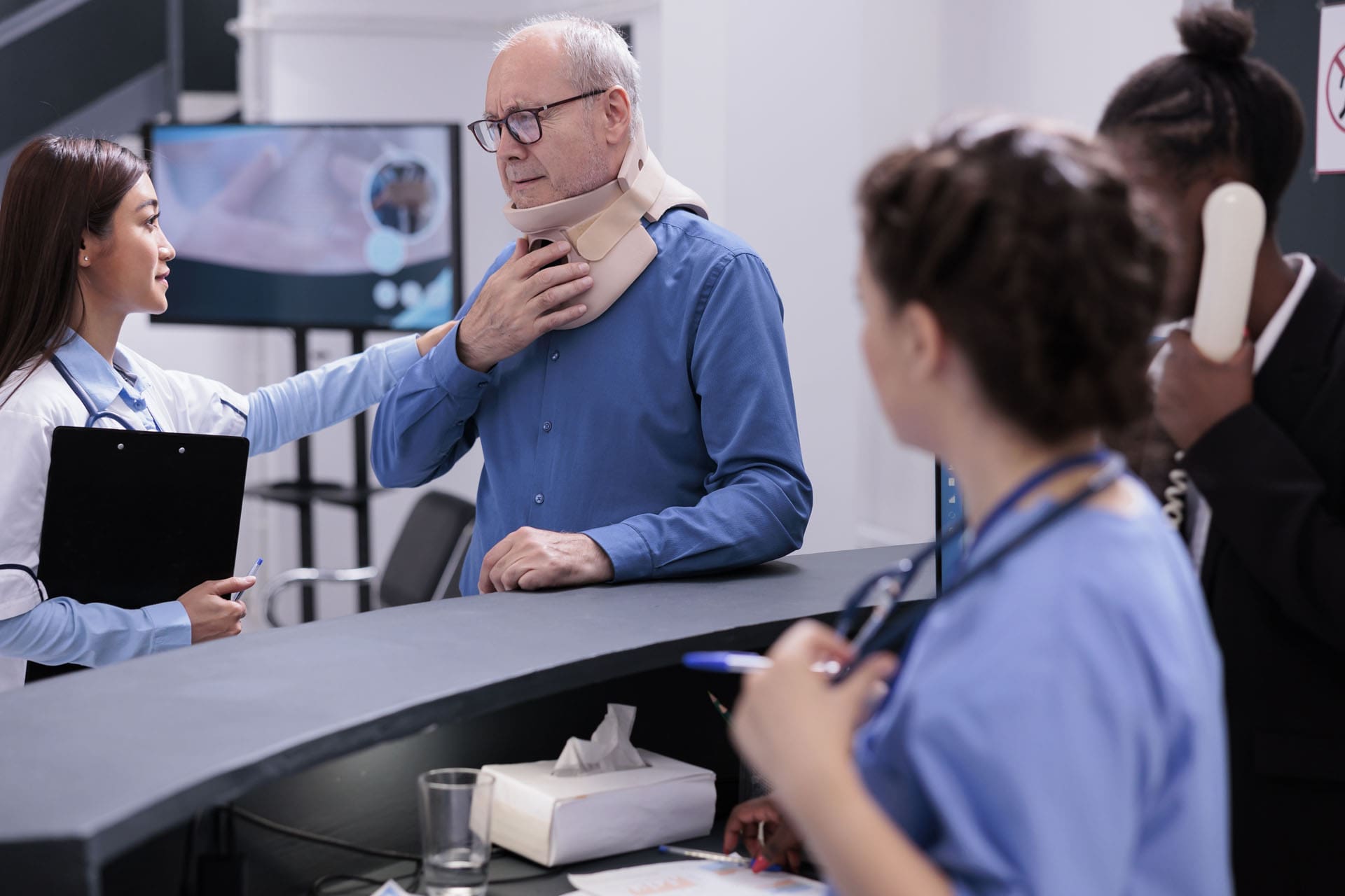
Athletes in El Paso often worry when pain slows them down
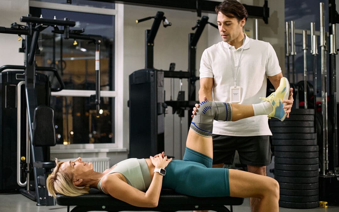
They do not want to lose strength or miss games. The good news is clear. While receiving treatment from an integrative chiropractor at El Paso Back Clinic, athletes can usually continue training or participating in sports; however, activity modification is often necessary to promote healing and prevent further injuries. “Complete rest is rarely the answer,” according to an integrative approach, which promotes “optimal loading”—applying just enough stress to promote healing without overtaxing injured structures.
To recover to full, pain-free performance more quickly, the athlete should see the chiropractor as a partner who offers a customized, structured strategy that shifts the goal from “complete rest” to “controlled, modified training.” At El Paso Back Clinic, led by Dr. Alexander Jimenez, DC, APRN, FNP-BC, this teamwork happens every day. The clinic blends chiropractic adjustments, functional medicine, sports rehab, and the PUSH Functional Fitness System to keep athletes moving safely while their bodies heal.
El Paso Back Clinic sits right here in El Paso, Texas. The team treats back pain, sports injuries, and chronic issues with a whole-person plan. Dr. Jimenez and his staff check posture, movement, and daily habits. They create plans that fit each athlete’s sport and life. Adjustments ease joint pressure. Nutrition tips fight swelling. Light fitness drills keep strength high. The result is faster healing and stronger returns to the field or court.
Many athletes fear losing fitness during recovery
At El Paso Back Clinic, modified training transforms that fear into steady progress. Gentle movement helps deliver blood and nutrients to injured areas. This speeds repair and stops muscles from getting weak. Clinic experience and research show athletes who stay active the smart way return sooner and stay healthier longer.
• Check how your body feels before and after activity
• Warm up with five minutes of easy walking every time
• Keep pain mild—no more than a 2 out of 10
• Write down small daily improvements
• Meet with your provider each week to adjust the plan
These simple steps make recovery feel active and hopeful instead of frustrating
Optimal loading is the heart of care at El Paso Back Clinic. Tissues heal best with the right amount of stress. No stress slows rebuilding. Too much stress causes new problems. Dr. Jimenez guides athletes to that perfect balance. A runner with knee pain might skip long runs but keep swimming and light cycling. A football player with a shoulder issue might pause heavy lifts but continue band work and core drills. This method protects overall fitness while targeted areas mend.
One trusted guide notes that gradually reintroducing exercise helps avoid high-impact or strenuous moves at first. Athletes who follow this advice stay ready for their sport instead of starting over later.
Chiropractic adjustments at the clinic realign the spine and joints, so nerves fire cleanly, and pain drops fast. Sessions often add soft-tissue release, stretches, and in-office exercises. These steps make everyday movement smoother. Many patients notice less stiffness after just a few visits. The clinic’s sports rehab programs incorporate mobility-agility training and the PUSH Functional Fitness System to safely rebuild power.
• Use ice for ten minutes on swollen areas
• Drink plenty of water to keep joints flexible
• Try low-impact cardio like pool walking or biking
• Stretch tight muscles each morning
• Choose meals high in protein and colorful vegetables
These easy habits work with the clinic’s functional medicine approach and boost results between visits
A clear step-by-step return plan keeps everything safe. Experts recommend building activity in stages. Begin with light aerobic moves that gently raise your heart rate. Add moderate effort next. Then move to sport-specific drills without contact. A full return occurs only after pain-free testing.
The Centers for Disease Control and Prevention outlines a similar graduated path that fits many injuries. Each stage lasts at least twenty-four hours. If symptoms flare, step back and rest briefly. This safety net stops athletes from rushing and builds real confidence.
• Stage 1: Short walks or stationary bike sessions
• Stage 2: Light jogging plus easy resistance moves
• Stage 3: Faster drills and full weights with no contact
• Stage 4: Skill practice by yourself
• Stage 5: Full practice or competition
Athletes at El Paso Back Clinic who follow these stages often feel stronger and more prepared when they return to games
Personalized plans set the clinic apart. No two athletes are the same. A soccer player’s ankle plan looks different from a weightlifter’s back plan. Dr. Jimenez reviews movement patterns, lab results, and daily routines. Then he builds a custom roadmap. Weekly check-ins let the plan grow with healing.
Clinical observations from Dr. Alexander Jimenez, DC, APRN, FNP-BC, demonstrate powerful real-world results. At El Paso Back Clinic, he sees athletes recover fastest when chiropractic care teams up with functional fitness and whole-body support. Instead of ordering full rest, Dr. Jimenez uses tailored rehab that mixes mobility drills, core stability, light conditioning, and nutrition guidance. His patients return to sport more quickly because the plans address root causes and keep controlled training alive. Many gain better movement habits that last long after recovery (Jimenez, n.d.).
Active recovery days keep momentum going. Light walks, foam rolling, or gentle yoga replace couch time. These sessions improve blood flow, clear muscle waste, and keep nerve pathways sharp. One recovery tip explains that active recovery involves engaging in low-intensity activities to promote blood flow and reduce muscle soreness. Staying hydrated makes these sessions even better.
• Foam roll tight spots for five minutes daily
• Stretch big muscle groups after light work
• Add simple balance drills
• Use compression sleeves for mild swelling
• Aim for seven to nine hours of sleep each night
Small actions like these prevent weakness and support the clinic’s goal of optimal mobility and fitness
Nutrition plays a huge role at El Paso Back Clinic. Food acts as fuel for repair. Protein rebuilds tissue. Anti-inflammatory choices calm swelling. The team shares easy meal ideas that fit busy training schedules. When athletes eat and drink right, soreness drops, and progress speeds up between appointments.
Early inflammation needs smart handling. Light ice and compression calm the area at first. Gentle motion then keeps fluids moving. Adjustments improve circulation and ease nerve pressure. The focus stays on guiding healing with the right activity.
Timing after an adjustment matters. Most athletes can start light movement soon, but waiting 20 to 30 minutes lets the joints settle. Begin easy and build slowly. Pain stays the guide—keep it low and slow down if needed.
• Warm up lightly before every session
• Focus on perfect form over heavy weights
• Cross-train to rest injured areas
• Log workouts in a simple notebook
• Celebrate wins like easier daily movement
These habits turn recovery into real progress
Beyond healing, care at El Paso Back Clinic lifts performance. Adjustments improve range of motion, balance, and power. Many athletes notice faster speed and better endurance after regular visits. The same tools that fix today’s injuries also prevent tomorrow’s.
Knowing when to pause is key. Sharp pain, growing swelling, or numbness means you should rest that spot. The team teaches self-checks so athletes stay safe between visits. Plans work for every sport—runners cut miles but add hills slowly, contact athletes drill form with lighter loads, swimmers focus on technique.
The biggest shift is mental. Athletes stop fearing rest and start partnering with experts for smart progress. The goal moves from “complete rest” to “controlled, modified training.” This builds trust and keeps the drive high.
Results show quickly. Shorter breaks mean more practice time and better seasons. Lower re-injury rates extend careers. Many athletes learn smarter movement habits that help them reach new levels.
El Paso Back Clinic welcomes players of all levels
—from weekend players to serious competitors. Plans adjust for age, background, and goals. The integrative style fits busy lives in El Paso and beyond, with clear in-person and follow-up support.
Modern research confirms smart loading beats total rest for most injuries. The clinic stays current by mixing classic chiropractic with functional science and sports medicine. Athletes gain body knowledge that lasts a lifetime. Dr. Jimenez and his team become ongoing partners for wellness and peak performance.
Recovery no longer means sitting out. With guidance from El Paso Back Clinic, athletes train smarter, heal naturally, and return stronger. Optimal loading, custom plans, and whole-person support turn every setback into a powerful comeback.
Integrative Chiropractic Care at El Paso Back Clinic: Boosting Body Function, Easing Pain, and Building Lasting Wellness
Living in El Paso can mean long days on your feet, heavy lifting at work, or weekend sports that leave your back sore and your energy low. Many people deal with nagging pain, stiff joints, slow healing, and constant tiredness. At El Paso Back Clinic, integrative chiropractic care offers a natural path to resolve these problems and help your body work at its best. This approach improves human body function by removing nerve interference through safe spinal adjustments. It also enhances mobility and calms the nervous system. Patients often feel less pain, more energy, better blood flow, and smoother movement right away. The team at El Paso Back Clinic pairs gentle adjustments with soft tissue work and simple exercises for real, long-term health gains.
What sets El Paso Back Clinic apart is its full-body focus. Care extends beyond a single spot to support your overall physical and emotional well-being. The clinic may add helpful therapies like massage and acupuncture. When chiropractic joins forces with functional medicine and advanced nursing, the results get even stronger. This team effort lines up your spine and structure with your nutrition, metabolism, and nerve health. Pain and swelling drop fast. Nervous system signals sharpen. Mobility improves, so you can move freely again. The collaborative model at El Paso Back Clinic combines biomechanical fixes with biochemical support to deliver truly lasting comfort and strength.
How Spinal Adjustments at El Paso Back Clinic Clear Nerve Interference
Spinal adjustments sit at the center of care at El Paso Back Clinic. When bones in your spine shift out of place, they can press on nerves and block signals traveling between your brain and body. This nerve interference causes pain, weakness, and slow recovery. A quick, controlled adjustment uses gentle force to guide the bones back into proper alignment. Once pressure lifts, nerves fire clearly again.
The science behind these moves is clear and simple. Joints regain smooth motion and lose stiffness almost instantly. Tight muscles relax, easing strain on nearby tissues. Many patients at El Paso Back Clinic notice quick relief because their bodies can now heal themselves without blocked signals. The clinic’s advanced tools, such as digital motion X-rays, help Dr. Alex Jimenez pinpoint exactly where help is needed.
Top Benefits of Clearing Nerve Interference at El Paso Back Clinic
Adjustments ease back, neck, and joint pain by fixing misalignments and relaxing tight muscles.
Soft-tissue work and custom exercises reduce swelling and prevent problems from returning.
Functional medicine adds nutrition plans to lower whole-body inflammation for steady results.
These steps do far more than treat one ache. They help your entire system run more smoothly every day.
Improving Mobility and Calming the Nervous System
Good mobility means bending, walking, lifting, and playing without limits or pain. At El Paso Back Clinic, integrative chiropractic care unlocks this freedom. Spinal adjustments restore normal joint range so your hips, shoulders, and back move easily again. Patients often say they can walk farther, play sports longer, and handle daily tasks with confidence.
The nervous system also settles down beautifully. Clear nerve signals improve the brain-body connection. Stress that used to tighten your shoulders or trigger headaches fades away. Your body shifts out of “fight or flight” mode into a calm, healing state. This balance supports better sleep, steadier moods, and faster recovery from everyday wear and tear. The clinic’s sports rehabilitation and functional training lock these gains in place.
Mobility and Calm Benefits Patients Love
Spinal adjustments improve joint range of motion and reduce stiffness, making daily activities easier.
Functional strength exercises and rehab build support, so injuries stay away.
Combined therapies help people stay active at work, in sports, or around the house.
Better movement creates a positive loop. More activity keeps the nervous system relaxed and your body strong.
Reducing Pain, Raising Energy, and Boosting Circulation
Pain relief is the number one reason El Paso residents visit El Paso Back Clinic. Adjustments trigger your body’s natural pain-fighting mechanisms while addressing the root cause. Issues like sciatica, headaches, or lower back strain often improve after just a few visits. When pain drops, energy rises because your body stops wasting strength fighting constant discomfort.
Blood circulation gets a major lift, too. Proper spinal alignment lets blood flow freely, delivering oxygen and nutrients to every cell. Waste leaves faster. Patients report warmer hands and feet, sharper thinking, and less fatigue. This improved flow supports heart health and helps muscles recover more quickly after physical activity.
Energy and Circulation Wins at El Paso Back Clinic
Care boosts blood flow so oxygen reaches muscles and the brain more easily.
Less muscle tension and nerve pressure bring higher energy and clearer focus.
Regular sessions leave patients feeling refreshed instead of drained.
These changes add up quickly. Less pain plus steady energy makes life in El Paso feel lighter and more fun.
Optimizing Movement with Soft Tissue Work and Exercises
Integrative chiropractic care at El Paso Back Clinic goes way beyond quick adjustments. Soft tissue techniques, such as targeted massage, loosen tight muscles and break up scar tissue. This works hand in hand with spinal changes to keep your body balanced for longer. Simple exercises then strengthen the muscles around your newly aligned spine. The clinic’s rehab centers teach stretches and core moves you can do at home to maintain your progress.
This complete package prevents old problems from coming back. Instead of chasing symptoms, care at El Paso Back Clinic builds a rock-solid foundation for active living. Over time, patients enjoy better posture, stronger balance, and real confidence in their movements.
Complementary Therapies for Full-Body Wellness
Massage and acupuncture blend perfectly into plans at El Paso Back Clinic. Massage relaxes muscles and improves blood flow right after an adjustment. Acupuncture calms the nervous system and eases emotional stress that often shows up as tight shoulders. Together, these tools address both the physical ache and the hidden tension many people carry. The result is a complete sense of balance that touches every part of life.
Patients who add these therapies often sleep more deeply, feel happier, and handle daily stress with ease. Body and mind work together instead of against each other.
The Power of Chiropractic, Functional Medicine, and Advanced Nursing Together
The strongest results occur when chiropractic, functional medicine, and advanced nursing come together at El Paso Back Clinic. Functional medicine looks deep into nutrition, gut health, and hormones to fix issues at their source. Advanced nursing brings medical checks, lab tests, and personalized plans. Dr. Alex Jimenez, DC, APRN, FNP-BC, leads this team with his dual training, making El Paso Back Clinic one of the most complete injury and wellness centers in Texas.
This trio aligns structural fixes with inner-chemistry support. Pain and inflammation drop fast. Nervous system function sharpens. Mobility improves, and long-term health becomes normal. The model combines biomechanical care with nutritional and neurological support for lasting results.
Dr. Alexander Jimenez’s Clinical Observations at El Paso Back Clinic
Dr. Alexander Jimenez, DC, APRN, FNP-BC, CFMP, IFMCP, has helped thousands of El Paso patients at his clinic. He sees spinal adjustments helping even complex herniated discs and severe sciatica heal naturally without surgery. When he combines chiropractic care with functional medicine, nutrition, and advanced nursing, inflammation in the joints and gut drops quickly. Patients gain more energy and far less pain.
Dr. Jimenez notes that proper alignment restores nerve signals, helping the body heal faster from injuries such as whiplash, sports strains, or work-related back issues. His patients with chronic conditions regain mobility and strength through custom plans that blend structure, diet, and lifestyle. He often points out that clear nerve pathways plus metabolic support improve sleep, lower stress hormones, and strengthen immune health. People enjoy lasting gains in posture, agility, and daily function when structural care is combined with nutritional and neurological support at El Paso Back Clinic.
His work proves that this integrated style delivers results far beyond what any single treatment can offer. Patients leave feeling empowered to stay healthy and active for years ahead.
Linking Movement, Recovery, and Stress Relief
Care at El Paso Back Clinic also connects movement with faster recovery. After an adjustment, guided rehab exercises rebuild strength while your body heals. This stops new injuries before they start and keeps you moving. Stress from work or daily life often shows up as tight muscles or poor posture. The clinic eases both kinds of tension so your nervous system stays balanced. Patients perform at their best because their bodies handle pressure without breaking down.
Long-Term Health and Immune Support
Regular visits to El Paso Back Clinic support your immune system naturally. Clear nerve signals help your body fight illness more effectively. Reduced inflammation and better circulation keep energy high and sick days low. Over months and years, patients report fewer health setbacks, stronger resilience, and a brighter outlook. This natural boost comes from your body’s own healing power once nerve interference is gone.
Many people stay with the clinic because it delivers steady improvements without drugs or surgery. They gain simple tools to manage their own wellness while knowing expert help is always close by.
Why El Paso Residents Choose Integrative Chiropractic Care at El Paso Back Clinic
Integrative chiropractic care at El Paso Back Clinic truly transforms how your body functions. It clears nerve interference, improves mobility, calms the nervous system, reduces pain, boosts energy, improves circulation, and optimizes movement. By blending spinal adjustments with soft-tissue work, exercises, massage, acupuncture, functional medicine, and advanced nursing, this approach delivers comprehensive physical and emotional support. Dr. Alexander Jimenez and the team at El Paso Back Clinic show that this collaborative style creates real, lasting health for people of all ages in our community.
If you live with ongoing back pain, sciatica, stiff joints, or just want to feel stronger every day, El Paso Back Clinic offers a safe, effective path forward. Small changes in your spine lead to big wins across your whole body. Call today or visit https://elpasobackclinic.com/ to start your journey toward pain-free living and lasting wellness.
How PRP Supports Tissue Repair and Recovery at El Paso Back Clinic
Platelet-Rich Plasma, or PRP, is a treatment that uses a concentrated portion of your blood to support healing in a specific injured area. Platelets are best known for helping blood clot, but they also carry growth factors and signaling proteins that help guide tissue repair. PRP is made by drawing a small amount of blood, spinning it in a centrifuge, and then placing the platelet-rich portion back into the area that needs help healing. Reviews of PRP describe it as an autologous therapy, meaning it comes from the patient, with platelet levels above baseline and a strong supply of growth factors and cytokines that can affect inflammation, angiogenesis, and cell proliferation.
For El Paso Back Clinic, this topic fits naturally with the clinic’s broader identity as a multidisciplinary injury and recovery practice. The clinic presents itself as a center for chiropractic care, functional medicine, injury care, rehabilitation, imaging and diagnostics, and wellness support, with a strong focus on injury recovery and musculoskeletal problems. That makes PRP a logical part of a larger recovery conversation rather than a stand-alone trend.
What PRP Really Does
PRP is often described in popular language as helping the body “clean up” damaged tissue. That idea can be helpful, but it needs to be explained carefully. PRP is not a whole-body cleanse or a detox program. The better scientific explanation is that PRP supports local tissue healing in a targeted area by releasing growth factors and signaling molecules that help coordinate repair. These signals may encourage cell recruitment, help regulate inflammation, support blood vessel growth, and improve the rebuilding of connective tissue.
In simple terms, PRP helps the body do three major things at an injured site:
Signal that healing needs to begin
Support the cleanup of damaged material
Help rebuild healthier tissue
That is why PRP is often used for tendons, ligaments, muscles, joints, and other slow-healing structures. Hospital for Special Surgery explains that PRP is injected into injured or diseased tissue to accelerate healing of tendons, ligaments, muscles, bones, and joints.
PRP and the Early Healing Response
Every injured tissue needs an organized healing response. In many chronic injuries, that response becomes weak, disorganized, or incomplete. PRP helps by creating a stronger healing signal in the injured area. A major review on PRP explains that platelets release growth factors and cytokines that influence inflammation, angiogenesis, stem cell migration, and cell proliferation. Another HSS review states that activated concentrated platelets release growth factors that stimulate the body to produce more reparative cells.
This is one of the reasons PRP is attractive in conservative and regenerative care. Instead of only covering pain, it aims to support the body’s own repair process. That does not mean results are guaranteed. PRP outcomes vary by tissue type, injury severity, preparation method, and the patient’s health. Still, the basic goal is clear: support better healing instead of simply masking symptoms.
How PRP Supports Tissue “Cleanup”
When people talk about PRP helping with detoxification or cleansing, the best way to describe it is local biologic cleanup. Injured tissue often contains damaged cells, inflammatory byproducts, and disorganized matrix material. Research shows that PRP helps create a regenerative microenvironment that supports both structural repair and functional recovery. A 2025 review describes key PRP pathways, including immune modulation, angiogenesis, and support for M2 macrophage polarization, which is linked to tissue repair.
Macrophages are important because they help remove damaged material. In healing tissues, they act like cleanup and coordination cells. They help phagocytose, or break down and remove, debris and necrotic material while also supporting repair signals. So when PRP is used in an injured joint, tendon, or soft-tissue area, it may help the body more effectively clear damaged tissue while also moving the area toward repair. That is much more accurate than saying PRP “flushes toxins” out of the whole body.
Angiogenesis: Bringing Better Blood Supply to Injured Tissue
A major part of healing is circulation. If tissue has a poor blood supply, healing can be slower and less complete. PRP has been linked to angiogenesis, which means the formation of new blood vessels. A major review of PRP biology reports that platelets release factors, including vascular endothelial growth factor and fibroblast growth factor, both of which are involved in angiogenesis. A newer PRP review also states that PRP’s overall effect is predominantly pro-angiogenic in therapeutic settings such as wound repair and tissue regeneration.
This matters because new blood vessel growth can help the injured area receive:
More oxygen
More nutrients
More signaling molecules
Better support for tissue remodeling
For a spine, joint, tendon, or sports-injury practice like El Paso Back Clinic, angiogenesis is one reason PRP may fit into broader musculoskeletal recovery plans. Better blood flow support can help move tissue from a stuck or slow-healing state toward active repair.
Fibroblasts, Collagen, and Matrix Remodeling
PRP is also important because healing is not only about cleanup. It is also about rebuilding. Fibroblasts are connective tissue cells that help produce collagen and organize the extracellular matrix. Research reviews show that PRP can stimulate fibroblast proliferation, collagen production, and extracellular matrix remodeling. These effects are part of why PRP is studied in wound care, scar remodeling, skin repair, and musculoskeletal recovery.
This rebuilding phase is important for injuries in which tissues have become weak, irritated, or degenerated over time. In those situations, PRP may help encourage a better repair environment by supporting stronger collagen organization and more orderly tissue remodeling. In practical terms, that can support recovery in tissues that need structure as well as symptom relief.
Inflammation: Starting It, Then Regulating It
Some people get concerned when they hear that PRP can create a healing response that includes inflammation. But a short and controlled inflammatory response is a normal part of repair. The goal is not endless inflammation. The goal is an organized healing phase followed by better regulation of the tissue environment. The 2025 PRP review notes that PRP can reduce pro-inflammatory cytokines while promoting tissue-repair pathways. This is part of why PRP is described as both reparative and immunomodulatory.
This balanced effect is important for chronic injuries. A tissue that has been irritated for a long time may need a better biologic signal to restart and organize healing. PRP can support that process by helping shift the local environment away from ongoing dysfunction and toward recovery.
Why Image Guidance and Clinical Precision Matter
PRP is only as useful as the way it is applied. Cleveland Clinic notes that providers may use ultrasound to locate the appropriate injection site. Hospital for Special Surgery also notes that ultrasound imaging is sometimes used to guide the injection directly into the area of injury.
That point matters for a clinic like El Paso Back Clinic because the site emphasizes injury care, diagnostics, imaging, rehabilitation, and multidisciplinary support. When PRP is paired with careful diagnosis and precise placement, the treatment is more likely to target the tissue that actually needs help. This is especially important in complex cases of back pain, sports injuries, ligament problems, and other musculoskeletal conditions where multiple structures may be involved.
An Integrative Recovery Approach
One of the strongest ways to frame PRP for El Paso Back Clinic is as part of a bigger recovery plan. The clinic site highlights chiropractic care, functional medicine, rehabilitation, injury care, wellness medicine, and diagnostic services. That kind of setting supports the idea that tissue repair works best when the injection is not treated like a one-step fix.
A full PRP recovery plan may also include:
A clear diagnosis
Image-guided placement when needed
Activity modification
Rehabilitation exercises
Joint and spine support
Nutrition and metabolic support
Follow-up to track healing progress
This broader model lines up well with Dr. Alexander Jimenez’s public clinical approach, which emphasizes injury recovery, rehabilitation, imaging, wellness, and integrated musculoskeletal care through the El Paso Back Clinic platform and related services. Based on that public positioning, PRP can be described as one piece of a comprehensive repair strategy rather than a stand-alone solution.
What Patients Should Keep in Mind
PRP has real potential, but it also has limits. HSS notes that one of the main uncertainties with PRP is that effectiveness can vary from patient to patient. The same source notes that the risk of infection is low but still possible, as with any injection. Because PRP comes from the patient’s own blood, side effects are usually limited, but results are not identical for everyone.
So the most honest summary is this:
PRP supports local tissue repair, not a whole-body detox
PRP may help damaged tissue move through the cleanup and rebuilding phases
PRP can support angiogenesis, fibroblast activity, and collagen remodeling
PRP often works best when paired with diagnosis, rehab, and follow-up care
PRP is promising, but patient response can vary
That kind of balanced explanation is helpful for patients who want both hope and realism.
Final Thoughts
For El Paso Back Clinic, PRP is best suited as a biologic support tool within a broader musculoskeletal and wellness model. It uses the patient’s own platelets to deliver growth factors and signaling molecules into injured tissue. Those signals can help start healing, support local immune cleanup, encourage angiogenesis, stimulate fibroblasts, and improve collagen and matrix remodeling. In other words, PRP may help the body clear damaged tissue and build healthier tissue in the same area.
That message matches the clinic’s public identity as a multidisciplinary injury and recovery center in El Paso. When PRP is paired with careful diagnosis, image-guided precision, rehabilitation, chiropractic and wellness support, and a thoughtful follow-up plan, it can be presented as a practical part of an integrative recovery strategy for back pain, sports injuries, and other musculoskeletal conditions.
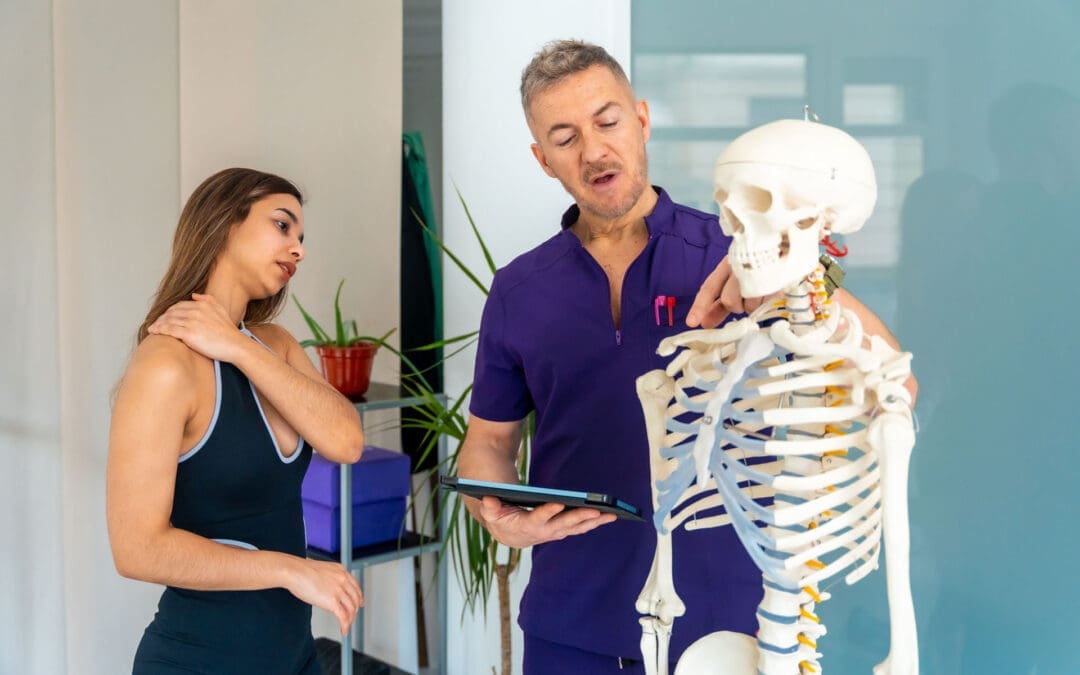
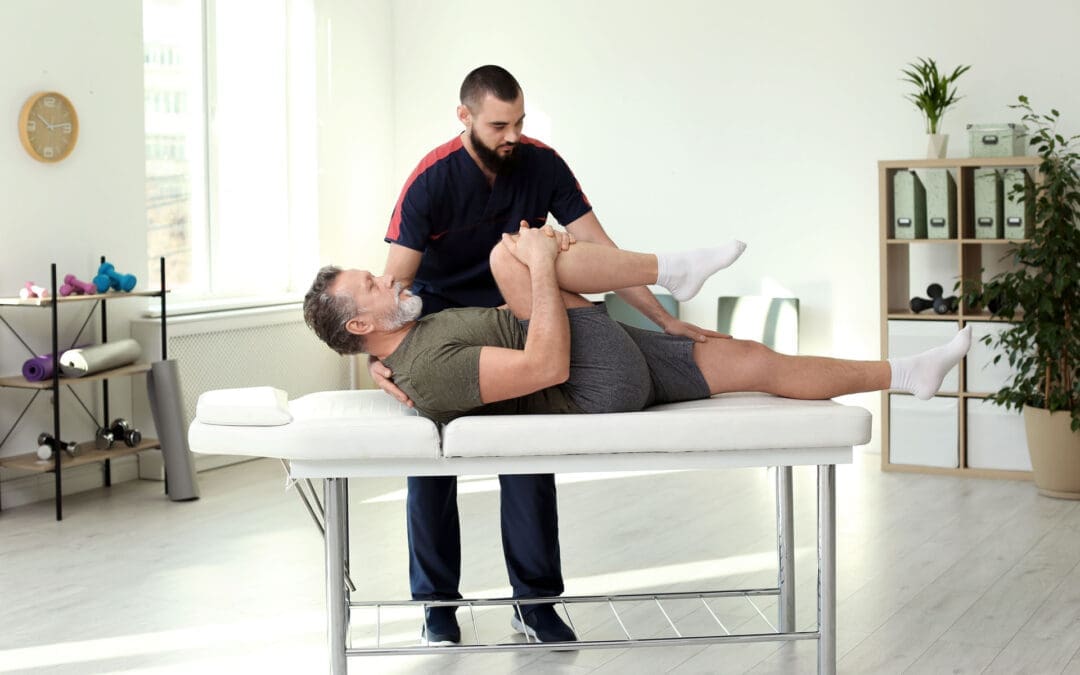
Understanding Chiropractic Spinal Adjustments: Techniques, Benefits, and Integrated Care
Chiropractic spinal adjustments, also known as spinal manipulations or reductions, offer a natural way to address back pain, improve mobility, and support overall health. These procedures focus on aligning the spine to reduce discomfort and enhance body function without surgery or heavy reliance on medications. Many people seek chiropractic care for issues like chronic back pain, neck strain, or injury recovery. This article explores what happens during an adjustment, its effects on the body, common techniques, and how team-based care can boost results.
What Is a Chiropractic Spinal Adjustment?
A chiropractic spinal adjustment involves a trained practitioner using their hands or a tool to apply a quick, controlled force to misaligned parts of the spine. This helps restore proper alignment and movement to the joints. The goal is to ease pain, improve joint function, and reduce pressure on the nerves and surrounding muscles (Cleveland Clinic, n.d.). It’s a non-surgical method that stretches the joint, often releasing gas bubbles like nitrogen, oxygen, and carbon dioxide, which create that familiar cracking sound—similar to when you crack your knuckles (Chiro One, n.d.).
Adjustments target areas of restriction, called subluxations, where vertebrae are out of place or not moving well. By correcting these, the procedure can improve nervous system function, leading to reduced irritation and better overall health (NCCIH, n.d.). Patients often feel an increase in range of motion right away, along with looser muscles.
Key Aspects of a Chiropractic Adjustment
Here are some main features of this treatment:
Procedure: The chiropractor first checks the spine for problem spots. Then, they use a sudden but precise push to fix the joint (Revive Chiropractic DSM, n.d.).
Sensations: You might hear a pop or crack, but it’s just gas escaping the joint fluid, not bones breaking (Cleveland Clinic, n.d.).
Physical Effects: The thrust stretches tight joints, relaxes tense muscles, and frees trapped gases, reducing built-up pressure (Physicians Group LLC, n.d.).
Benefits: It restores normal joint motion, supports nerve health, and reduces pain from nerve compression (Spine Health, n.d.).
What It Feels Like: Most find it painless, though some notice mild soreness afterward, like after a workout. Many report quick relief and easier movement (Complete Care, n.d.).
These elements make adjustments a popular choice for managing pain without invasive options.
Techniques Used in Chiropractic Adjustments
Chiropractors use different methods based on the patient’s needs. Common ones include:
Manual Adjustment: This is a high-velocity, low-amplitude (HVLA) thrust done by hand. It’s direct and aims to realign the spine quickly (Towson Chiro, n.d.).
Instrument-Assisted: Tools provide gentle taps to the spine, ideal for those who prefer less force (Visit Chiro First, n.d.).
Spinal Decompression: Using a specialized table, the spine is stretched to create space between the vertebrae, helping with issues such as herniated discs (Get Adjusted Columbia, n.d.).
These techniques can be tailored to conditions such as whiplash or back injuries sustained in accidents (Utah Therapeutic Massage, n.d.).
What Happens During a Chiropractic Spinal Adjustment
A typical session starts with an assessment. The chiropractor reviews your health history, performs a physical exam, and may use X-rays to identify subluxations (Dubuque Chiropractic, n.d.). Once identified, the adjustment begins.
The practitioner positions you on a table and applies a fast, targeted thrust to the specific joint. This might cause cavitation—the popping sound from gas release in the joint fluid (Starkwood Chiropractic, n.d.). Right after, muscles relax, nerve irritation drops, and joint motion improves (Personal Injury Doctor Group, 2024).
Sessions often include additional therapies such as soft-tissue work, trigger-point release, or stretches to support the adjustment (Boca Chiropractic SW, n.d.). The whole process is quick and focused on comfort.
Benefits of Chiropractic Spinal Adjustments
Regular adjustments offer several advantages:
Pain Relief: They reduce mechanical stress on the spine and ease nerve compression, helping with back, neck, and headache pain (Chiro One, n.d.).
Improved Function: By fixing alignment, they enhance posture and spinal health, preventing future issues (Boca Chiropractic SW, n.d.).
Nervous System Support: Adjustments promote improved nerve signaling, supporting overall bodily function (Physicians Group LLC, n.d.).
Faster Recovery: For injuries like car accidents, this approach speeds healing by addressing root causes (Dallas Accident and Injury Rehab, n.d.).
Studies show these benefits lead to higher patient satisfaction when combined with other care (My Chiro, n.d.).
Incorporating an Interdisciplinary Team for Better Results
Bringing in a team of experts—like advanced practice registered nurses (APRNs), family nurse practitioners (FNP-BC), certified functional medicine providers (CFMP and IFMCP), advanced translational nutrigenomics specialists (ATN), and certified chiropractic spinal trauma experts (CCST)—makes treatment more effective. This approach combines structural fixes with medical and nutritional support to provide holistic care (Health Coach Clinic, n.d.).
For complex cases, such as auto injuries or chronic pain, this team provides comprehensive plans. It focuses on root causes rather than just symptoms, leading to lasting improvements (LinkedIn, n.d.).
How Each Role Contributes
APRN/FNP-BC: These nurses offer medical checks, diagnose issues, and manage meds if needed. They educate patients and integrate chiropractic with traditional medicine to improve pain control (Nursing World, n.d.; Goodwin University, n.d.).
CFMP/IFMCP: They dig into metabolic and nutritional roots of problems, using functional medicine to heal the musculoskeletal system faster (LinkedIn, n.d.).
ATN: By studying genetics and nutrition, they create custom diets and supplements to cut inflammation and aid repair (Jimenez, n.d.).
CCST: Experts in spinal trauma handle tough injuries like whiplash or disc herniations with advanced techniques (Spine Stop, n.d.).
This teamwork enhances outcomes, especially in recovery from accidents or ongoing conditions (Dallas Accident and Injury Rehab, n.d.).
Clinical Observations from Dr. Alexander Jimenez, DC, APRN, FNP-BC
Dr. Alexander Jimenez, with his dual roles in chiropractic and nursing, observes that adjustments restore function in conditions such as sciatica and herniated discs by reducing nerve compression without surgery (Jimenez, n.d.). He notes that patients often experience rapid pain relief and improved mobility after sessions, especially when combined with functional nutrition.
In trauma cases, such as car accidents, Jimenez highlights how spinal decompression and shockwave therapy speed recovery by addressing inflammation and nerve damage (LinkedIn, n.d.). His integrated approach, blending chiropractic with nutrigenomics, helps address root causes such as gut issues that affect spinal health. Patients report reduced symptoms in fibromyalgia and neuropathies through personalized plans that include team input from therapists and nutritionists.
Jimenez emphasizes holistic care for all ages, using assessments to uncover environmental factors. His observations show that interdisciplinary teams lead to sustained health, with testimonials praising relief from chronic pain and improved vitality (Jimenez, n.d.).
Conclusion
Chiropractic spinal adjustments provide a safe, effective way to manage pain and improve spinal health. By understanding the process, techniques, and benefits, you can see why many choose this path. Adding an interdisciplinary team takes it further by offering comprehensive care for better long-term results. If you’re dealing with back issues or injuries, consider consulting a qualified chiropractor.
ESWT for Car Accident Injuries in El Paso: How El Paso Back Clinic Uses Shockwave Therapy With Integrative Chiropractic + NP Care
Motor vehicle accidents (MVAs) can cause injuries that do not always show up clearly on basic imaging. You might be told, “Nothing is broken,” but still feel real pain, stiffness, tightness, and limited movement. That is because many car accident injuries involve soft tissue injuries such as muscle strains, tendon irritation, ligament sprains, fascia tightness, and painful scar tissue (adhesions). These injuries can lead to chronic pain when tissues remain inflamed, circulation remains poor, and the body continues to guard the area.
At El Paso Back Clinic, an integrative approach can help people recover more completely. The clinic’s content emphasizes non-invasive care, structural assessment, chiropractic and rehab, and broader healing support as part of a multi-disciplinary recovery plan. This matters because post-MVA pain is rarely caused by just one issue. It is often a combination of tissue injury, movement dysfunction, and ongoing sensitivity.
One tool that can make a big difference in stubborn cases is genuine Extracorporeal Shockwave Therapy (ESWT). True ESWT delivers therapeutic acoustic waves into injured tissues to help break down tight scar tissue, reduce pain signaling, improve circulation, and stimulate tissue repair. Mayo Clinic describes shockwave therapy as a noninvasive option used in musculoskeletal care with generally minimal adverse effects when appropriately applied.
This article explains, in plain language, how genuine ESWT can help with MVA injuries and why it works even better when combined with integrative chiropractic care and nurse practitioner (NP) oversight, a care model frequently discussed across El Paso Back Clinic content.
What “genuine ESWT” means (and why it matters)
Not all “shockwave” or “acoustic wave” treatments are the same. Real ESWT is designed to deliver a measurable therapeutic dose of acoustic energy into tissue. In simple terms, it is meant to do more than feel like a massage tool. The goal is to create a controlled mechanical stimulus that tells your body, “Restart repair here.”
A major review in the medical literature describes ESWT as working through mechanotransduction, meaning the mechanical stimulus triggers biological healing responses in the tissue. These responses can include improved signaling for healing, pain modulation, and tissue remodeling.
At El Paso Back Clinic, ESWT is presented as a non-surgical option that can be especially useful for deeper, stubborn pain patterns and chronic soft tissue problems.
Why car accident injuries can linger for months
After an accident, your body tries to protect you. It tightens muscles, limits motion, and increases inflammation around the injured area. That is normal at first. The problem happens when this protective pattern sticks around too long.
Common reasons MVA injuries become chronic include:
Scar tissue and adhesions that limit motion and pull on pain-sensitive tissue
Poor micro-circulation around the injury, slowing repair
Trigger points and muscle guarding that keep joints stiff
Altered biomechanics (compensation patterns) that overload nearby areas
Nervous system sensitivity, where pain signals stay “turned up”
El Paso Back Clinic’s approach highlights that many chronic pain cases improve when you combine structural assessment, conservative care, and a plan that supports true recovery rather than temporary relief.
How ESWT helps MVA injuries heal
Genuine ESWT can help through several overlapping effects. Think of it as improving the tissue environment so your body can complete the healing process.
It helps break down thick, painful scar tissue
Many chiropractic and rehab clinics describe shockwave therapy as useful for breaking down scar tissue and adhesions that form after injuries, especially when those tissues stay tight and painful.
It increases circulation to injured tissue
Better blood flow helps deliver oxygen and nutrients needed for repair. This is one reason ESWT is often used for chronic injuries that feel “stuck.” UCHealth describes shockwave therapy as promoting a reparative healing process that includes changes in circulation and tissue response.
It stimulates tissue remodeling and collagen repair
Tendons, ligaments, and fascia rely heavily on collagen structure. ESWT is commonly discussed as supporting tissue regeneration and collagen-related remodeling in musculoskeletal injuries.
It can reduce pain signaling
Pain relief from ESWT is not just “numbing.” Research reviews describe pain reduction effects that may involve changes in nerve sensitivity and local biochemical signaling.
It can support recovery in stubborn muscle injuries
Some reviews describe ESWT as associated with improvements in pain and function in certain muscle injury contexts (including sports-related muscle injuries), which can be relevant when car accidents result in deep strains and protective tightness.
MVA conditions that may respond well to ESWT
ESWT is commonly used for soft tissue and chronic pain patterns. In post-accident care, it may be considered for:
Whiplash-related muscle strain patterns (neck/upper back tightness)
Shoulder strain and rotator cuff irritation
Thoracic and rib region soft tissue pain and stiffness
Low back sprains/strains and persistent tight bands
Hip and glute strain patterns (piriformis-type tightness, trigger points)
Hamstring and calf strains from bracing during impact
Tendon irritation that does not respond well to rest alone
Chronic “knots” and trigger points that restrict motion
El Paso Back Clinic’s ESWT-focused content specifically points toward accident-related soft tissue injury and stubborn pain that has not improved as situations where this approach may fit well.
How many sessions does ESWT usually take?
Many patients report improvement early, but full remodeling can take time. A common pattern described in clinic-based educational resources is:
Noticeable changes often occur within 2–3 sessions
Full treatment plans commonly range from 4 to 12 sessions, depending on severity and how long the injury has been present
What often improves first:
Reduced sharpness or intensity at the worst pain points
Better range of motion (turning the neck, lifting the shoulder, bending)
Less stiffness the next morning
Improved tolerance to rehab exercises and daily activities
Why ESWT works best when paired with integrative chiropractic + NP care
ESWT helps tissue repair, but most MVA injuries also involve movement dysfunction. If a joint is not moving well, the tissue around it can stay irritated. That is why combining tissue work and structural care often produces better results.
Clear documentation of progress and functional improvement
El Paso Back Clinic’s content highlights the value of an integrated chiropractic + nurse practitioner approach.
Why the combination accelerates healing
When ESWT improves tissue quality and pain sensitivity, it often becomes easier to:
Move better
Accept and benefit from adjustments and mobility work
Build strength and stability through rehab
Return to work, training, and daily life with fewer flare-ups
Some integrative therapy articles describe combining chiropractic care with shockwave therapy (and sometimes laser therapy or rehab) to address both tissue injury and mechanical contributors.
What an ESWT session is like at a practical level
ESWT is typically done with a handheld applicator placed on the skin over the injured area. You may feel a tapping or pulsing sensation that can be intense in tight spots.
Many people experience:
Mild soreness afterward (similar to deep tissue work)
Temporary redness or sensitivity
A sense of looseness or improved motion over the next day or two
Mayo Clinic notes that shockwave therapy is generally associated with minimal adverse effects when used appropriately in musculoskeletal care.
Simple ways to get more out of ESWT after a car accident
ESWT is not magic by itself. It works best as part of a plan. Helpful steps often include:
Hydrate and walk after treatment (gentle circulation support)
Avoid overloading the area the same day (do not “test it” aggressively)
Track function, not just pain (turning your neck, lifting, walking, sitting tolerance)
Signs your plan is working:
You can do more with less flare-up
Your range of motion is improving
Pain is less frequent or less intense
Rehab feels more doable and less aggravating
Clinical perspective aligned with Dr. Alexander Jimenez’s educational approach
Across El Paso Back Clinic’s content, Dr. Alexander Jimenez presents a multidisciplinary, evidence-informed style that connects tissue healing, biomechanics, rehab, and whole-person factors. In this framework, ESWT fits as a regenerative tool that supports deeper tissue recovery, while chiropractic and rehab restore movement quality.
The practical takeaway is simple:
ESWT supports tissue repair and pain reduction
Chiropractic care supports structure and motion
NP oversight supports safer decision-making and whole-body recovery planning
That combination is often what helps MVA patients move from “surviving day to day” to building a stable recovery.
That “Reset Pain” After You Sit or Hold a Weird Position: What It Is and How El Paso Back Clinic Approaches It
Have you ever held your body in an awkward position—like slouching on a couch, twisting in a chair, leaning on one hip, or sleeping with your neck turned—then you stand up and feel a sharp ache, tightness, or a “catch”? Sometimes it feels like a joint or muscle has to “reset” before you feel normal again. You might even feel clumsy for a minute, then things settle down.
At El Paso Back Clinic, this pattern is commonly discussed as a mix of postural strain, muscle guarding, myofascial tightness (trigger points), and sometimes joint restriction—especially when movement has been limited for too long or posture has been stressing the same tissues over and over.
This article explains what that “reset” feeling usually means, why it happens, and how integrative chiropractic care—like the approach described at El Paso Back Clinic—can help restore smoother motion and reduce the chances of it happening again.
What Do You Call This “Reset” Feeling?
There isn’t one single official name that covers every case, because different tissues can create the same sensation. But the most common clinical labels include:
Postural strain (tissues overloaded by a sustained position)
Muscle stiffness (tightness and reduced ease of motion)
Muscle guarding (protective tension driven by the nervous system)
Myofascial trigger points (irritable “knots” in muscle/fascia)
Joint restriction / joint dysfunction (a joint that temporarily doesn’t glide well)
Many people casually call it a “stuck joint” or “something out of place.” In reality, it’s often less dramatic than it feels—more like a temporary movement problem plus a protective muscle response.
Why It Often Hurts When You Return to Neutral (Not While You’re Sitting)
This surprises many people: “If the posture was the problem, why didn’t it hurt until I moved?”
Because your body adapts to the position you hold. While you’re still:
Your muscles settle into a holding pattern
Your joints move less
Your fascia (connective tissue) can get less “slippery” with inactivity or repeated stress
Your nervous system may “turn down” certain signals until movement starts again
Then you stand, rotate, or straighten up—and your tissues have to slide, load, and coordinate again. That’s when you feel the catch, the sting, or the awkward “reset” moment.
What’s Actually Happening: 5 Common Mechanisms Behind the “Reset”
Most cases are a combo, not just one thing.
Postural Strain: You Overloaded a Region
When you hold a position that isn’t friendly to your body—like forward head posture, slumped sitting, or a rotated spine—you can stress:
muscles
ligaments
joint capsules
fascia
Over time, those tissues complain when you ask them to move again. El Paso Back Clinic describes how repetitive positions and mechanical issues can contribute to stiffness and restriction patterns.
Muscle Guarding: Your System “Braces” for Safety
Muscle guarding is your nervous system’s way of saying, “I’m not sure this movement is safe, so I’m going to tighten things up.” It can feel like:
locked
braced
hard to relax
stiff even when you try to stretch
El Paso Back Clinic notes that pain patterns can keep muscles guarded and that stiffness may involve more than “tight muscles.”
Trigger Points: The “Knot” That Bites When You Move
Trigger points are sensitive spots in tight muscle bands. When you change position, those fibers stretch and can cause sharp, deep, or referred pain.
Fascia health is closely tied to this, because fascia surrounds muscle and helps movement feel smooth. Johns Hopkins Medicine explains that fascia can become “gummy,” stiff, and painful with limited movement, repetitive movement, or trauma.
Fascial Stiffness: The “Gummy Tissue” Effect
Fascia is like a body-wide web. When you don’t move much or repeat the same posture all day, fascia can get less elastic and less hydrated. That can make motion feel “sticky.”
Johns Hopkins Medicine specifically lists limited activity, repetitive movement, and trauma as factors that can contribute to fascia adhesions and stiffness.
Joint Cavitation: The Pop or Release
Sometimes the reset comes with a pop. A well-known imaging study found evidence that joint cracking is linked to cavity formation in the joint fluid (not bones grinding).
A pop isn’t automatically “good” or “bad.” What matters more is:
Do you move more easily afterward?
Does pain decrease?
Or does pain increase and function drop?
Why You Feel Awkward for a Bit After the “Reset”
That lingering weirdness—seconds to minutes—is often your body downshifting from protection back into normal movement.
Common reasons include:
muscles slowly letting go of guarding
irritated tissue calming down
fascia rehydrating and sliding better with movement
your brain re-mapping posture and balance (proprioception “recalibration”)
This is one reason many people feel better after a short walk post-sitting.
A Quick Self-Check: Is This Normal Stiffness or Something More?
Muscle stiffness is common and often improves with gentle movement and better posture habits. The Cleveland Clinic notes that stiffness often improves without medical treatment, but it should be taken more seriously if it comes with concerning symptoms such as fever, weakness, swelling, or persistent worsening.
Consider getting evaluated if you notice:
pain that’s getting worse over days/weeks
tingling, numbness, or weakness
pain that wakes you up repeatedly
symptoms after a significant fall or crash
the “reset pain” keeps happening in the exact same spot
What You Can Do Right Away (Safe, Simple, and Usually Helpful)
The 2–3 minute “reset without forcing it”
Stand up and walk 30–90 seconds
Do small, slow movements in a pain-free range
Try a long exhale breathing pattern (relaxes guarding)
Use gentle heat if it helps you relax
Simple posture habits that reduce repeat episodes
Change position every 30–60 minutes
Avoid “camping” in end-range posture (deep slouch, deep twist)
Use a supportive setup for workstations when possible
Build basic endurance in the muscles that hold posture (core, glutes, upper back)
How El Paso Back Clinic Approaches This Pattern (Integrative Chiropractic Style)
El Paso Back Clinic describes an integrative model that blends chiropractic care with rehab-style strategies and multidisciplinary support for spine and soft tissue problems.
Identify what’s actually driving the “reset”
Sometimes stiffness isn’t just “tight muscles.” It may involve:
joint restrictions
spine or pelvis mechanics
inflammation around a joint
pain patterns that keep muscles guarded
nerve-related problems
That’s why an exam matters—so the plan matches the cause.
Restore motion with chiropractic adjustments or mobilization
A chiropractic adjustment is a controlled force applied to a spinal joint to improve motion and movement ability.
When a joint isn’t moving well, nearby muscles often overwork and tighten. Improving joint motion can reduce the need for your body to “force” a painful reset.
Address myofascial tightness (muscle + fascia)
Because fascia can become stiff due to limited movement or repetitive strain, integrative care often includes hands-on work and guided movement to improve tissue glide.
Stabilize the area so it doesn’t keep “getting stuck”
If a joint repeatedly feels like it “locks,” the missing piece is often:
strength
endurance
timing/control
movement habits
El Paso Back Clinic frequently emphasizes rehabilitation and conditioning alongside chiropractic care to restore normal function after spine and soft-tissue issues.
A “Stop the Reset Cycle” Plan (2–3 Weeks)
These are general strategies that many patients tolerate well. Keep it gentle and pain-free.
Daily (2–5 minutes, 1–2 times/day)
1 minute easy walking
5 slow neck turns each side (easy range)
8 shoulder blade squeezes (2–3 sec hold)
8 hip hinges (small, smooth)
3 slow breaths with long exhale
During the day (30–60 seconds every hour)
stand up
10–20 steps
reset your sitting position (hips back, chest relaxed, neck tall)
3 days/week (10–15 minutes)
core stability (dead bug / modified plank)
glute strength (bridges / step-ups)
upper back endurance (band rows)
If stretching makes symptoms worse, or if stiffness keeps returning the same way, that’s a good reason to get assessed—El Paso Back Clinic even notes that persistent stiffness may signal joint restrictions or mechanics issues beyond “tight muscles.”
When to Reach Out to El Paso Back Clinic
If your “reset pain” is frequent, sharp, or starting to change your daily routine, it’s reasonable to get an evaluation—especially if you suspect joint restriction, posture-related mechanics, or muscle guarding patterns.
El Paso Back Clinic lists multiple El Paso locations and a main phone line for help and questions.
Phone: (915) 850-0900
Location (example listing): 11860 Vista Del Sol, Ste 128, El Paso, TX 79936
Key Takeaway
The experience of “I held a posture → now it hurts → then it resets” usually indicates that your body is showing a predictable pattern:
posture overloads tissues
fascia and muscle tension increase
a joint may move less smoothly
the nervous system guards
returning to neutral triggers a brief recalibration
The goal isn’t to chase pops or force releases. The goal is to restore smooth motion + stable control, so your body doesn’t keep needing that painful “reset.”
IFM's Find A Practitioner tool is the largest referral network in Functional Medicine, created to help patients locate Functional Medicine practitioners anywhere in the world. IFM Certified Practitioners are listed first in the search results, given their extensive education in Functional Medicine