Elbow pain from lifting is a common symptom among individuals who lift weights, heavy objects, children, grocery bags, etc. Depending on the underlying cause, can conservative treatments relieve and heal elbow pain?
Elbow Pain Caused By Lifting
Elbow pain from lifting can result from weight training, repetitive daily tasks, or job duties like lifting small children or heavy objects. Pain can manifest at the sides or the front of the elbow. Most minor injury cases can be treated with ice, rest, and medications at home. However, pain after lifting can also be a sign of a serious injury, such as a tendon rupture/tear.
Minor Pain From Lifting
Lifting puts pressure on the tendons connecting the wrist and upper arm to the bones in the elbow joint. Minor elbow pain can occur from temporary inflammation in any of these structures after lifting an object. Tendonitis occurs when a tendon becomes inflamed, often from overuse or lifting something too heavy, and ranges from mild to severe. Mild tendonitis typically causes pain during the activity and improves with rest. (American Academy of Orthopaedic Surgeons, 2020) Common forms of tendonitis include:
Tennis elbow – tendonitis on the outside of the elbow
Golfer’s elbow – tendonitis on the inside of the elbow.
Add ice to the affected area for up to 20 minutes daily to decrease elbow pain.
Rest
Avoid lifting heavy objects as much as possible when pain is present.
Wearing A Brace
If the pain is at the tendons on the inside or outside of your elbow, try wearing a wrist brace to limit the use of your wrist muscles that connect to this area.
Stretching
Gently stretching the wrist flexors and extensors can help reduce elbow pain after lifting. Stretches can be performed several times daily, even after symptoms have resolved. (American Academy of Orthopaedic Surgeons, 2024)
Hold the arm out in front with the palm down. Keep the elbow straight.
Bend the wrist down so that the fingers are pointing toward the ground.
With the other hand, gently pull the wrist further down until a stretch is felt along the back of the forearm.
Hold this position for 15 seconds.
Repeat five times.
Next, bend the wrist upward so the fingers point toward the ceiling.
Using the other hand, gently pull the hand backward until the stretch is felt along the front of the forearm.
Mild cases can improve after a few days of self-care, whereas more pronounced elbow symptoms can take several weeks, months, or even a year. (Kheiran A. Pandey, A. & Pandey R. 2021) If self-care doesn’t work, physical therapy may be recommended. A physical therapy team can use various modalities and treatments to help reduce pain and inflammation from elbow injuries. The therapy can include targeted exercises to strengthen weak muscles and stretch tight muscles that might contribute to the condition. In addition, the therapy team will help individuals modify their lifting technique to help prevent further injury.
A biceps tendon rupture is a rare but serious injury usually caused from lifting. In addition to other visible signs of the injury, there will be a bulge at the top of the upper arm because the muscle bunches up as it is no longer attached to the elbow. (American Academy of Orthopaedic Surgeons, 2022) Individuals may hear an audible popping sound if an elbow ligament or tendon gets torn while lifting. (Johns Hopkins Medicine, 2024)
Treatment
Treatment depends on the severity of the injury, but most cases resolve on their own with rest and, if necessary, physical therapy. Conditions that cause severe pain require orthopedic surgeon expertise. These physicians specialize in treating musculoskeletal system injuries. Imaging such as X-rays, MRIs, or CT scans are often used to determine the extent of damage. Individuals with tendon or ligament tears in the elbow may need surgery to regain full range of motion and strength in their arm. After surgery, physical therapy will help restore function.
Injury Medical Chiropractic and Functional Medicine Clinic
Injury Medical Chiropractic and Functional Medicine Clinic works with primary healthcare providers and specialists to develop an optimal health and wellness solution. We focus on what works for you to relieve pain, restore function, and prevent injury. Regarding musculoskeletal pain, specialists like chiropractors, acupuncturists, and massage therapists can help mitigate the pain through spinal adjustments that help the body realign itself. They can also work with other associated medical professionals to integrate a treatment plan to improve the body’s flexibility and mobility and resolve musculoskeletal issues.
Shoulder Pain Chiropractic Treatment
References
American Academy of Orthopaedic Surgeons. (2020). Sprains, strains, and other soft-tissue injuries. https://orthoinfo.aaos.org/en/diseases–conditions/sprains-strains-and-other-soft-tissue-injuries/
Kheiran, A., Pandey, A., & Pandey, R. (2021). Common tendinopathies around the elbow; what does current evidence say?. Journal of clinical orthopaedics and trauma, 19, 216–223. https://doi.org/10.1016/j.jcot.2021.05.021
American Academy of Orthopaedic Surgeons. (2024). Therapeutic exercise program for epicondylitis (tennis elbow/golfer’s elbow). https://orthoinfo.aaos.org/globalassets/pdfs/2024-therapeutic-exercise-program-for-lateral-and-medial-epicondylitis.pdf
American Academy of Orthopaedic Surgeons. (2023). What are NSAIDs? https://orthoinfo.aaos.org/en/treatment/what-are-nsaids/
American Academy of Orthopaedic Surgeons. (2022). Biceps tendon tear at the elbow. https://orthoinfo.aaos.org/en/diseases–conditions/biceps-tendon-tear-at-the-elbow
Can healthcare professionals implement H.E.A.R.T. protocols for trafficked individuals while providing a safe space?
Introduction
Across the world, many local media and organizations are paying close attention to a phenomenon that many people should be aware of. This phenomenon is known as trafficking, and it can be associated with numerous activities, from forced labor to sex labor, and can affect a person’s sense of self-worth. While many people will correlate that trafficking affects many women and children, it can affect many individuals regardless of age, gender, and background. While many survivors of trafficking are dealing with the psychological and physical injuries that they obtain from their traffickers, many medical professionals can implement protocols and roles through the implementation of H.E.A.R.T. to provide a safe space for individuals suffering from trafficking. Today’s article focuses on the definition of trafficking, what H.E.A.R.T. is, and how it is used in a clinical setting. We discuss with certified associated medical providers who consolidate our patients’ information to assess and identify trafficking in a clinical approach while providing a safe space. We also inform and guide patients while asking their associated medical provider intricate questions to formulate customized treatment plans for their pain and provide them with a safe space and positive experience. Dr. Jimenez, D.C., includes this information as an academic service. Disclaimer.
The Definition Of Trafficking
When it comes to defining trafficking, it can be challenging as it is frequently associated with other issues. However, the main definition for trafficking is “recruiting, transporting, transferring, or harboring many individuals or a person that are threatened or forced to achieve the consent of a person having control of the individuals for exploitation.” With human trafficking being a pressing public concern that affects all races, social classes, demographics, and genders, it can impact society and the individual who is being trafficked. (Toney-Butler et al., 2024) Additionally, many people often mistake trafficking and smuggling as they are completely different. Smuggling requires a person to be transported into a nation through voluntary illicit means. While trafficking can come in two forms, which are labor and commercial sex, it can happen within the person’s own home. (Rambhatla et al., 2021) This is because many survivors who are going to get healthcare services will feel various emotions of fear or shame that can prevent them from asking for help due to what they have been through with their trafficker. However, when many individuals who are trafficking survivors are suffering from significant physical, mental, and social health problems and are seeking healthcare services, many healthcare professionals play an important role by creating a safe and responsive space for them. (The Lancet Regional Health-Western, 2022)
Beyond the Surface: Understanding the Effects of Personal Injury- Video
What is H.E.A.R.T In A Clinical Setting
When it comes to creating a safe and positive space in a clinical setting, many healthcare professionals often miss the signs of trafficking due to a lack of training or confidence to identify and treat patients who are trafficking victims. (Lee et al., 2021) However, healthcare protocols should be implemented, and H.E.A.R.T. should be incorporated into a clinical approach to assess and develop a customized treatment plan for the patient. Healthcare professionals can engage with the patient in a one-on-one discussion away from their trafficker and can offer important medical and psychological care resources. (Exeni McAmis et al., 2022) By incorporating H.E.A.R.T. protocols in a healthcare clinic, many doctors and medical professionals can help many patients be in a safe environment. Below is what H.E.A.R.T. stands for.
H-Hearing
The “H” in H.E.A.R.T. is for hearing as many medical professionals not only to hear but to see what is going on in the clinic and to establish environmental awareness. This is due to looking at the patient and who is accompanied by them. With healthcare providers being at the front, they interact with patients and may not know what health concerns are affecting them. This could be due to the following:
By incorporating the hearing aspect in H.E.A.R.T., many healthcare professionals can provide a safe, thoughtful, and engaging approach to the patient and know what to look for when a patient is coming in for treatment.
E-Evaluating
The “E“ in H.E.A.R.T. is used to evaluate its importance in enhancing patient interactions in a trauma-informed care facility. This is highly important because the individual is seeking health care. For the patients being trafficked, it is important to notice the red flags the individual is experiencing. Some of the red flags that many healthcare providers should look for are:
Physical health
Behavioral Health
The patient is with a controlling person
The patient does not have possession of their I.D.
Additionally, it is always important to show compassion, be sensitive to the individual while addressing their needs and concerns, and use a non-judgmental approach during the interview process. This helps the individual ensure they are in a safe environment when discussing sensitive topics. At the same time, it is important not to let the patient be re-traumatized while avoiding the impulse to rescue and overpromise the patient to mental health as we want them to have their self-worth. At the same time, it is best to remember the four “Rs“ when doing a trauma-informed approach; they are:
Realize: Understanding how trauma can affect people.
Recognize: Recognizing the signs of trauma.
Respond: Have all staff trained, use evidence-based practices, and provide a safe environment.
Resist Re-trauma: Recognizing how some practices may trigger painful memories while avoiding re-traumatizing the patient.
By implementing the four “Rs“ and the “E“ in H.E.A.R.T., many healthcare professionals can provide valuable resources to trafficking survivors with a strong support system.
A-Activating
The “A“ in H.E.A.R.T. stands for activating, where healthcare professionals must have proper protocols to engage all employees. This allows the healthcare providers to understand how beneficial it is to develop a protocol for a person who is being trafficked, understand their state and federal reporting laws, and list key elements of effective trauma-informed screening procedures when assessing the patient. This allows a foundational structure to support a response for suspected patients who are being trafficked. At the same time, by following HIPAA laws and organization policies, many healthcare providers must explain the reporting process to the right officials. Additionally, the benefits of developing a protocol for trafficking are by:
Clarifying procedures
Enhance staff training
Optimize the interactions with the trafficking patients
Improve staff confidence
Prepare for any threatening situations
Maximizing preparedness to aid trafficking patients
Optimize support for patients
Develop collaborative outside resources
R-Resourcing
The “R“ in H.E.A.R.T. stands for resourcing, as many healthcare providers must identify the referral systems. This allows healthcare professionals to understand the important message to convey when assessing trafficking victims and the importance of responding to safety, emergency, and reporting requirements. When assessing and interviewing the patient, many will have to recognize that their patient may be a possible victim of trafficking, what their immediate needs are, and what long-term resources can help.
T-Training
The “T” in H.E.A.R.T. stands for training, as it is important that many healthcare providers continuously train to spot trafficking; this provides confidence to many healthcare workers and can help save a person’s life. By implementing H.E.A.R.T. protocols, the “T” allows the doctor to respect the individual’s decision to want help, providing a positive support system while encouraging them to come back, offering to help with a safety plan, and building a resource network. This is because if the patient is accompanied by someone who is controlling and answering for the patient, handing out information discreetly can provide a bit of hope to the individual to make the move. At the same time, providing local and immediate assistance resources can help the individual in the long run. This allows healthcare providers to build a trusting relationship and even help individuals to have a safe and positive experience on their health and wellness journey.
References
Exeni McAmis, N. E., Mirabella, A. C., McCarthy, E. M., Cama, C. A., Fogarasi, M. C., Thomas, L. A., Feinn, R. S., & Rivera-Godreau, I. (2022). Assessing healthcare provider knowledge of human trafficking. PLOS ONE, 17(3), e0264338. https://doi.org/10.1371/journal.pone.0264338
Gutfraind, A., Yagci Sokat, K., Muscioni, G., Alahmadi, S., Hudlow, J., Hershow, R., & Norgeot, B. (2023). Victims of human trafficking and exploitation in the healthcare system: a retrospective study using a large multi-state dataset and ICD-10 codes. Front Public Health, 11, 1243413. https://doi.org/10.3389/fpubh.2023.1243413
Lee, H., Geynisman-Tan, J., Hofer, S., Anderson, E., Caravan, S., & Titchen, K. (2021). The Impact of Human Trafficking Training on Healthcare Professionals’ Knowledge and Attitudes. J Med Educ Curric Dev, 8, 23821205211016523. https://doi.org/10.1177/23821205211016523
Rambhatla, R., Jamgochian, M., Ricco, C., Shah, R., Ghani, H., Silence, C., Rao, B., & Kourosh, A. S. (2021). Identification of skin signs in human-trafficking survivors. Int J Womens Dermatol, 7(5Part B), 677-682. https://doi.org/10.1016/j.ijwd.2021.09.011
For individuals and athletes with a gluteal contusion with severe bruising, can a healthcare provider determine if there are any other injuries to underlying structures, including muscle or tendon tears?
Gluteal Contusion
A gluteal contusion is an injury, in this case, a bruise to the buttocks’ gluteal muscles caused by damage to muscle fibers and blood vessels. A buttock bruise is caused by direct bodily impact, typically from falls, automobile collisions, accidents, bumping into something, or being struck by an object or person. Like all bruises, a gluteal bruise most often results in pain and visible discoloration of the skin at the injury site, varying in severity from grade I to grade III, with higher-graded bruises requiring more time to heal. Most butt bruises can heal on their own with time and rest, but if bruising is severe, individuals may require physical therapy to restore full muscle function.
Symptoms
A contusion is a muscle injury that can affect the body’s skeletal muscles. A gluteal contusion can be painful, with a black and blue mark that changes color over time. Other symptoms may include: (Mount Sinai, 2024)
Tenderness to touch over the injury site
Increased pain with contraction of the glutes
Swelling
Discomfort with sitting
Causes
A contusion occurs from direct trauma and forceful impact on the gluteal muscles, causing damage to underlying blood vessels, muscle fibers, and sometimes bone, resulting in bleeding under the skin. (MedlinePlus, 2016) Direct impacts to the gluteal muscles that can cause a contusion include:
Falls
Car accidents
Direct hits to the buttocks from a piece of sports equipment or person.
Bumping into furniture, a door, or a counter.
Intramuscular injections into the gluteal muscles.
Individuals who take blood thinners or anticoagulant medication have an increased risk of bruising from direct contact injuries.
Diagnosis
A gluteal contusion is usually diagnosed through a physical examination and is generally straightforward to diagnose based on physical appearance, symptoms, and type of injury. Contusions can be graded based on the severity according to the following criteria (Fernandes, T. L. et al., 2015)
Grade I
An injury that affects only a small amount of muscle fibers, resulting in minimal pain, tenderness, and possible swelling.
Causes minimal or no loss of strength in the affected muscle or range of motion limitations.
Muscle use is typically unaffected.
Grade II
An injury that causes significant damage to muscle fibers, resulting in increased pain and impaired muscle contraction.
A small muscle defect can be felt to the touch.
Discoloration increases over the first few days after injury.
Grade III
An injury that involves extensive muscle fiber damage and bleeding across an entire area of a muscle that results in severe, and sometimes total, loss of muscle function.
Causes severe pain and significant discoloration of the skin.
When contusions are larger, deeper, and involve significant blood pooling and swelling, they are called hematomas.
If the bruising is severe, a diagnostic ultrasound, CT scan, or MRI may be used to determine whether any underlying structures are damaged.
Treatment
Contusions are generally mild injuries. Treatment typically involves rest to allow the muscles to heal from the bleeding and the bruising to dissipate.
Applying ice to the injury site can help relieve pain and inflammation.
If the bruising is severe, significant physical activity like sports, dancing, running, jumping, and weight lifting should be avoided until the muscles heal. (Mount Sinai, 2024)
With more severe bruising, contraction and stretching of the glutes are painful and can require longer healing and recovery time.
Physical therapy rehabilitation may be needed for more significant injuries to restore muscle function.
Prognosis
A mild injury usually heals on its own with time and rest. More significant injuries take longer to heal and may require physical therapy to build strength and range of motion if muscle function is affected.
Healing Time and Recovery
Healing and recovery times for gluteal contusions vary depending on the severity of the injury (Fernandes T. L. et al., 2015)
Grade I
Minor injuries that cause minimal discomfort typically heal fully in five days to two weeks.
Grade II
During the first two to three days, contusions develop, increasing discoloration under the skin, and complete healing can take two to three weeks.
Return to sport is typically resumed after a month.
Grade III
Contusions can take up to four to six weeks to heal, often requiring rehabilitation to restore muscle strength and range of motion.
Injury Medical Chiropractic and Functional Medicine Clinic
At Injury Medical Chiropractic and Functional Medicine Clinic, we passionately focus on treating patients’ injuries and chronic pain syndromes. We focus on improving ability through flexibility, mobility, and agility programs tailored to the individual. We use in-person and virtual health coaching and comprehensive care plans to ensure every patient’s personalized care and wellness outcomes. Our providers use an integrated approach to create personalized care plans that include Functional Medicine, Acupuncture, Electro-Acupuncture, and Sports Medicine principles. Our goal is to relieve pain naturally by restoring health and function to the body. If he feels the individual needs other treatment, they will be referred to a clinic or physician best suited for them as Dr. Jimenez has teamed up with the top surgeons, clinical specialists, medical researchers, and premier rehabilitation providers to provide our community with the best clinical treatments.
Building a Stronger Body
References
Mount Sinai. (2024). Bruise. https://www.mountsinai.org/health-library/injury/bruise
MedlinePlus. (2016). Bruises. Retrieved from https://medlineplus.gov/bruises.html
Fernandes, T. L., Pedrinelli, A., & Hernandez, A. J. (2015). MUSCLE INJURY – PHYSIOPATHOLOGY, DIAGNOSIS, TREATMENT AND CLINICAL PRESENTATION. Revista brasileira de ortopedia, 46(3), 247–255. https://doi.org/10.1016/S2255-4971(15)30190-7
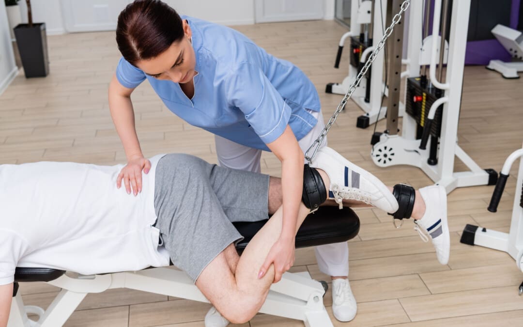
Individuals dealing with symptoms like sudden pain, weakness, and tenderness in the back of the knee could have a hamstring injury. Can knowing the symptoms and performing self-care help bring relief?
Hamstring Pain Behind The Knee
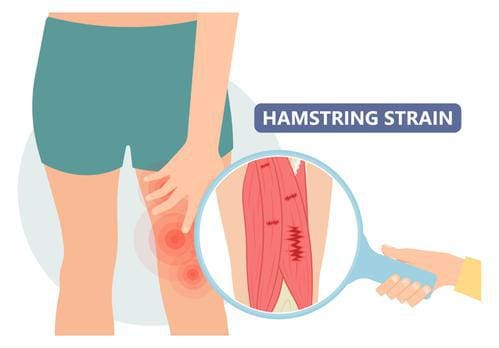
The hamstrings consist of three long muscles that run down the back of the thigh, cross over the back of the knee, and connect to bones in that area. A hamstring injury, such as a strain or tear, tendonitis, or biceps femoris tendinopathy, can cause pain in the back of the knee, difficulty bending the knee, swelling, and bruising. A hamstring strain occurs when the muscle is stretched too far or torn completely. This can happen from sudden, forceful movements or overstretching. Hamstring tendonitis develops over time, usually after a sudden increase in activity, when the hamstring tissue cannot recover from too much loading. Pain is often felt after physical activity and exercise and, in severe cases, during the activity or throughout the day. Biceps femoris tendinopathy can also cause pain in the back of the knee. Strains, tendonitis, bursitis, and muscle tears are all possible explanations for a hamstring injury that leads to pain behind the knee. Discussing pain symptoms with a healthcare provider is recommended, especially if it occurs suddenly during physical activity or exercise. They can help identify the exact cause and offer guidance for rehabilitation, including physical therapy referrals.
Causes and Triggers
Individuals may experience hamstring pain behind the knee when the muscles in that area are overworked, inflamed, or injured, such as from activities like running, walking, dancing, soccer, or basketball. Possible types of injuries and their causes.
A primary cause of muscle strain occurs when the muscle is stretched too far or has to handle a sudden force like sprinting or kicking. (American Academy of Orthopaedic Surgeons, 2021)
Severe Cases
Most causes of pain behind the knee are easily treatable at home with self-care and rest. However, it can be more severe, signaling a blood clot, infection, torn muscle or tendon/ligament. Hamstring knee pain may be serious if any of the following is experienced (American Academy of Orthopaedic Surgeons, 2021)
Sudden pain during physical activity, often during a full stride.
Feeling a pop or sharp pain that causes falling or limping.
Pain that worsens over time and prevents or hinders walking or exercising as normal.
If pain is severe and does not improve with rest and anti-inflammatory medications, evaluation by a healthcare professional is necessary.
Assesses Hamstring Pain
A healthcare provider will ask about symptoms and injury, including what happened when the pain began. They will perform a physical examination, which may include pressing on the back of the thigh to look for swelling, bruising, tenderness, or bunched-up muscles. (American Academy of Orthopaedic Surgeons, 2021) The healthcare provider will ask the patient to perform specific resisted movements, such as the manual muscle test, and measure the range of motion. Diagnostic testing includes an X-ray or MRI to determine the degree of the injury and which soft tissues or bones may be involved.
Self-Care
The first line of treating hamstring knee pain is the RICE protocol, which includes: (Mount Siani, 2024)
Rest
Stop any activity that causes symptoms and pain.
A healthcare provider may recommend crutches or a knee scooter in severe cases.
Ice
Apply cold packs to the swollen or painful area for 20 minutes throughout the day.
Compression
A knee brace, wrap, or bandage that applies gentle pressure to the injured area can help reduce and prevent swelling.
Elevation
Lifting the leg higher than the heart will help reduce swelling and blood accumulation.
Individuals may need to lie on a bed or sofa and elevate their legs with pillows.
Individuals can use at-home pain relievers like acetaminophen or NSAIDs like ibuprofen or naproxen. Over time, and depending on the severity of the injury, a healthcare provider will advise on gentle hamstring stretches and how to ease back into physical activity.
A healthcare provider will advise immobilizing the knee to help with muscle healing, which could involve wearing a knee brace or using crutches.
Physical therapy
A healthcare provider may refer the patient to a physical therapist, who will perform a personalized evaluation and prescribe targeted exercises to heal the injury and regain strength, flexibility, and movement.
Surgery
Tendon avulsion injuries are when the hamstring tendon completely tears away from the bone, and surgery is required to reattach the tendon.
Platelet-rich plasma – PRP
Platelet-rich plasma has become an additional treatment for hamstring muscle strain or tendonitis. (Seow D. et al., 2021)
The treatment involves injecting a solution from the patient’s blood into the muscle to heal the injury.
Recovery
Predicting how long a hamstring injury takes to heal and how long the pain will linger depends on the type, location, and severity. The most severe type is the hamstring coming unattached around the knee. This surgical repair and rehabilitation take at least three months before returning to sports and exercise (American Academy of Orthopaedic Surgeons, 2021). Lesser injuries like tendonitis or a mild strain can take less time to heal. However, it’s essential to avoid reinjuring the area so the condition does not become chronic. This includes: (American Academy of Orthopaedic Surgeons, 2021)
Stretching to encourage and maintain flexibility.
Fixing muscle imbalances between the quadriceps and hamstring.
Endurance and conditioning.
Avoiding overuse.
Injury Medical Chiropractic and Functional Medicine Clinic works with primary healthcare providers and specialists to develop personalized treatment programs. We focus on what works for you and use an integrated approach to treating injuries and chronic pain syndromes to improve flexibility, mobility, and agility, relieving pain and helping individuals return to normal activities. If other treatments are needed, Dr. Jimenez has teamed up with top surgeons, clinical specialists, medical researchers, and rehabilitation providers. Our providers use Functional Medicine, Acupuncture, Electro-Acupuncture, and Sports Medicine principles.
Chiropractic Care for Leg Instability
References
National Library of Medicine. (2017). Tendinitis Also called: Tendonitis. Retrieved from https://medlineplus.gov/tendinitis.html
American Academy of Orthopaedic Surgeons. OrthoInfo. (2020). Sprains, strains, and other soft tissue injuries. https://orthoinfo.aaos.org/en/diseases–conditions/sprains-strains-and-other-soft-tissue-injuries/
American Academy of Orthopaedic Surgeons. OrthoInfo. (2021). Hamstring muscle injuries. https://orthoinfo.aaos.org/en/diseases–conditions/hamstring-muscle-injuries/
American Academy of Orthopaedic Surgeons. OrthoInfo. (2021). Pes aserine (knee tendon) bursitis. https://orthoinfo.aaos.org/en/diseases–conditions/pes-anserine-knee-tendon-bursitis/
Mount Siani. (2024). Hamstring strain – aftercare. https://www.mountsinai.org/health-library/selfcare-instructions/hamstring-strain-aftercare
Seow, D., Shimozono, Y., Tengku Yusof, T. N. B., Yasui, Y., Massey, A., & Kennedy, J. G. (2021). Platelet-Rich Plasma Injection for the Treatment of Hamstring Injuries: A Systematic Review and Meta-analysis With Best-Worst Case Analysis. The American journal of sports medicine, 49(2), 529–537. https://doi.org/10.1177/0363546520916729
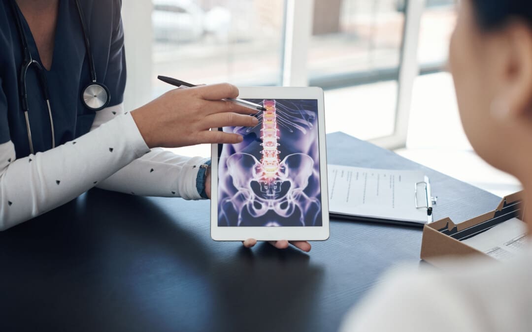
An annular fissure is caused by age-related changes to the spine, which often do not cause symptoms but can cause back pain. Can understanding the causes help individuals manage lower back pain and help healthcare providers develop an effective treatment program?
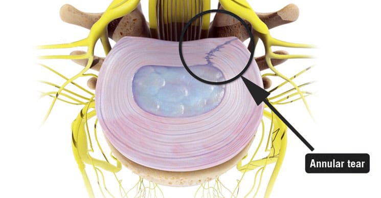
Annular Fissure
An annular fissure is a discogenic condition that affects the spine and can cause lower back pain. Also called an annular tear, it’s usually a wear-and-tear condition that happens over time rather than a condition caused by trauma. It usually happens when the fibers that make up the annulus or the tough outer covering of the intervertebral disc break or separate. To manage it, healthcare providers may recommend:
Making lifestyle changes.
Staying aware of how you go about daily activities and take steps to make adjustments, such as being mindful of unhealthy posture.
Start doing exercises that help make the back stronger.
Medical care if pain and other symptoms need to be managed.
Symptoms
Lower back pain may be a sign of an annular fissure, or there may be no symptoms. Symptoms can include:
Pain
Weakness
Numbness
Electrical sensations travel down one leg or arm if a cervical/neck tear is present.
Numbness and weakness may be caused by the nerves getting irritated or compressed near an annular tear. (Stadnik, T. W. et al., 1998)
These symptoms can also be similar to a herniated disc, which can be a complication of an annular fissure.
However, studies have shown that annular tears and herniated discs often go unnoticed because they have few obvious symptoms. (Jarvik, J. G. et al., 2005)
Annulus Function
The annulus comprises several layers of tough fibers/fibrocartilage that surround, contain, and protect the soft, liquid nucleus inside the disc. The layers of the annulus fibrosus crisscross to provide support. The nucleus is a shock absorber cushions the body’s weight on the spinal joints when sitting, standing, or moving. Its strength also allows the disc to buffer the jolts and jars it experiences. It also helps maintain the integrity of the intervertebral joint by supporting the space between the two vertebrae. When an annular fissure occurs, the fibers separate or tear off from insertion on the nearby spinal bone. A fissure can also be a break in the fibers of one or more layers. (Jarvik, J. G. et al., 2005)
Causes
An annular tear is not the standard term medical professionals use to describe or diagnose a fissure because the word tear suggests that trauma has led to the separation or break in the fibers. While an injury can cause an annular fissure, it’s usually caused by long-term wear and tear. (Guterl, C. C. et al., 2013) The tears are typically caused by age-related degenerative changes in the disc, which can also lead to degeneration in other areas of the spine. Wear and tear are caused by annular fissures due to an individual’s daily living habits, such as sitting, standing, walking, climbing stairs, and performing other routine movements.
Treatment
While a large annular fissure is not likely to improve without treatment, a small one could heal independently. However, once an area has torn, it becomes more likely to continue tearing. (Virginia Spine Institute, N.D.) Conservative treatment is usually enough to control pain and symptoms. Physical therapy and anti-inflammatory medication are the first line of treatment. (Cheng, J. et al., 2019) Medication can be over-the-counter or prescription. Physical therapy treatment includes exercises, traction, and other therapies. If these do not help with the symptoms, the provider may suggest a steroid injection to reduce inflammation and pain. It can take three to six months to recover from degenerative disc problems if doing a standard treatment plan that includes rest, low-impact therapy exercises, and anti-inflammatory treatments. (Cheng, J. et al., 2019)
In severe cases, surgery may be recommended, including disc replacement surgery. An annular tear is not a reason to have disc replacement surgery alone; it is only when there are degenerative changes in the vertebral disc that surgery might be necessary. (Yue, J. J. et al., 2012)
Improving Body Alignment
Not paying attention and being aware of how the body performs everyday activities can, over time, set the stage for an annular fissure and other musculoskeletal injuries. However, fixing daily movement and posture habits to prevent injuries can be done through simple adjustments. For example, strengthening the core and back muscles can reduce pressure on the spine and help prevent injuries. (Camp, C. L. et al., 2016) The idea is to improve joint and overall body alignment. Activities can include:
Strength training
Walking
Pilates classes
Yoga
Tai chi
Somatic exercises
These activities help with muscle balance and joint alignment, which are recommended prevention strategies that physical therapists use when working with individuals who need help with spinal problems.
Visiting a chiropractic and physical therapy team can help treat injuries and chronic pain syndromes, relieve pain, resolve musculoskeletal issues, and prevent future symptoms. Injury Medical Chiropractic and Functional Medicine Clinic works with primary healthcare providers and specialists to develop a personalized care program for each patient through an integrated approach to treating injuries, improving flexibility, mobility, and agility to help return to normal and optimal function. If other treatments are needed, Dr. Jimenez has teamed up with top surgeons, clinical specialists, medical researchers, and rehabilitation providers to provide the most effective treatments.
Back Pain Specialist
References
Stadnik, T. W., Lee, R. R., Coen, H. L., Neirynck, E. C., Buisseret, T. S., & Osteaux, M. J. (1998). Annular tears and disk herniation: prevalence and contrast enhancement on MR images in the absence of low back pain or sciatica. Radiology, 206(1), 49–55. https://doi.org/10.1148/radiology.206.1.9423651
Jarvik, J. G., Hollingworth, W., Heagerty, P. J., Haynor, D. R., Boyko, E. J., & Deyo, R. A. (2005). Three-year incidence of low back pain in an initially asymptomatic cohort: clinical and imaging risk factors. Spine, 30(13), 1541–1549. https://doi.org/10.1097/01.brs.0000167536.60002.87
Guterl, C. C., See, E. Y., Blanquer, S. B., Pandit, A., Ferguson, S. J., Benneker, L. M., Grijpma, D. W., Sakai, D., Eglin, D., Alini, M., Iatridis, J. C., & Grad, S. (2013). Challenges and strategies in the repair of ruptured annulus fibrosus. European cells & materials, 25, 1–21. https://doi.org/10.22203/ecm.v025a01
Virginia Spine Institute. (N.D.). Annular disc tear Understanding the Symptoms, Causes, and Treatments. https://www.spinemd.com/conditions/annular-disc-tear/
Cheng, J., Santiago, K. A., Nguyen, J. T., Solomon, J. L., & Lutz, G. E. (2019). Treatment of symptomatic degenerative intervertebral discs with autologous platelet-rich plasma: follow-up at 5-9 years. Regenerative medicine, 14(9), 831–840. https://doi.org/10.2217/rme-2019-0040
Yue, J. J., Telles, C., Schlösser, T. P., Hermenau, S., Ramachandran, R., & Long, W. D., 3rd (2012). Do presence and location of annular tear influence clinical outcome after lumbar total disc arthroplasty? A prospective 1-year follow-up study. International journal of spine surgery, 6, 13–17. https://doi.org/10.1016/j.ijsp.2011.09.001
Camp, C. L., Conti, M. S., Sgroi, T., Cammisa, F. P., & Dines, J. S. (2016). Epidemiology, Treatment, and Prevention of Lumbar Spine Injuries in Major League Baseball Players. American journal of orthopedics (Belle Mead, N.J.), 45(3), 137–143.
Competitive swimmers, recreational, and swimming enthusiasts who experience pinching and sharp shoulder pain while swimming may suffer from shoulder impingement. Can understanding symptoms can help healthcare providers develop an effective treatment program?
Swimmer’s Shoulder
Swimmer’s shoulder, medically known as rotator cuff impingement syndrome, is a common injury among swimmers. It can limit swimming ability and normal arm use for functional tasks. It is caused by persistent and abnormal rubbing and pinching of the structures in the shoulder, causing pain and irritation of the shoulder’s rotator cuff tendons and the bursa. The injury affects 40% to 90% of swimmers at some point. (Wanivenhaus F. et al., 2012) Self-care treatment involves rest, anti-inflammatory medication, and exercise to restore normal shoulder mobility. Most cases resolve within a few months, but physical therapy may be needed along with continued exercises and stretches to maintain pain relief.
Anatomy
The shoulder is a complex joint with extreme mobility. It is comprised of three bones:
The scapula or shoulder blade.
The clavicle or collar bone.
The humerus or upper arm bone.
These three bones combine at various places to make up the joint. Several muscles attach to and move the joint. (Kadi R. et al., 2017) The rotator cuff is one group of four muscles deep in the shoulder surrounding the joint. When lifting the arm, these muscles contract to hold the ball in the joint’s socket, allowing the arm to be raised in a fluid and smooth motion. Several ligaments hold the shoulder joint together and connect the various bones of the shoulder, giving the joint stability when moving. (Kadi R. et al., 2017)
Shoulder pain when bearing weight through the arm.
Symptoms tend to be worse during or immediately after swimming.
This is due to the position of the arms and upper extremities while swimming. (Wanivenhaus F. et al., 2012) Reaching overhead and turning the hand inward can cause the rotator cuff tendons or shoulder bursa to become pinched underneath the acromion process of the shoulder blade, similar to the motion that occurs during the crawl or freestyle stroke. When pinching/impingement occurs, the tendons or bursa can become inflamed, leading to pain and difficulty with normal arm use. (Struyf F. et al., 2017) The condition may also occur due to the laxity of the shoulder ligaments. (Wanivenhaus F. et al., 2012) It is theorized that the ligaments in swimmers become stretched and lax, leading to shoulder joint instability. This can cause the shoulder joint to become loose and compress the shoulder structures.
Diagnosis
A clinical examination can diagnose cases of swimmer’s shoulder. (Wanivenhaus F. et al., 2012) The exam can include:
Palpation
Strength test
Specialized tests
One shoulder test that is often used is called Neer’s test. A physician elevates the arm overhead to the maximum degree during this examination. If this results in pain, the rotator cuff tendons may be compressed, and the test is positive. Individuals may begin treatment after the examination, but a doctor may also refer them for diagnostic testing. An X-ray may be taken to examine the bone structures, and an MRI may be used to examine the soft tissue structures, such as the rotator cuff tendons and the bursa.
Treatment
Appropriate treatment of swimmer’s shoulder involves managing pain and inflammation in your shoulder and improving the way your shoulder moves so you avoid pinching structures inside the joint. (Wanivenhaus F. et al., 2012) There are various treatments available and can include:
Rest
Physical therapy
Acupuncture
Non-surgical decompression
Targeted exercises and stretches
Medications
Injections
Surgery for serious cases
Physical Therapy
A physical therapist can treat shoulder impingement. They can assess the condition and prescribe treatments and exercises to improve mobility and strength. (Cleveland Clinic, 2023) They may use various treatment modalities to decrease pain and improve circulation to facilitate and expedite healing. Physical therapy treatments can include:
Ice
Heat
Trigger point release
Joint mobilizations
Stabilization
Stretching
Exercise
Electrical stimulation
Ultrasound
Taping
Medication
Medication may include over-the-counter anti-inflammatory medicine to help decrease pain and inflammation. A physician may prescribe stronger medication to manage inflammation if the condition is severe. While taking medication, the shoulder will need rest, so avoiding swimming or other shoulder movements for a week or two may be necessary.
Injections
Cortisone is a powerful anti-inflammatory medicine. Individuals may benefit from cortisone injections into their shoulders. (Wanivenhaus F. et al., 2012) When injected, cortisone decreases pain, reduces swelling in the rotator cuff and bursa, and improves shoulder mobility.
Surgery
If symptoms are persistent and fail to be alleviated with conservative treatments, surgery may be recommended. An arthroscopic procedure called subacromial decompression may be performed. (Cleveland Clinic, 2023) This type of surgery is done with small incisions, inserting a camera, and tiny tools. During this procedure, inflamed tissue and bone spurs are removed from the underside of the acromion process of the shoulder blade, allowing more space to the shoulder joint. Post-surgery, individuals can gradually return to swimming and all other activities in about eight weeks.
Recovery
Most episodes last about eight to ten weeks, and severe cases last up to three months. (Struyf F. et al., 2017) Often, the symptoms slowly resolve with rest and gentle stretching. As symptoms improve, individuals can slowly return to normal activity and swimming. However, performing prescribed exercises two to three times a week may be necessary to maintain shoulder strength and mobility and help prevent future episodes of shoulder impingement. Individuals experiencing any of these symptoms should visit their physician for an accurate diagnosis of their condition to begin proper treatment. Discuss goals with a healthcare professional and physical therapist.
Sports Injuries Rehabilitation
References
Wanivenhaus, F., Fox, A. J., Chaudhury, S., & Rodeo, S. A. (2012). Epidemiology of injuries and prevention strategies in competitive swimmers. Sports health, 4(3), 246–251. https://doi.org/10.1177/1941738112442132
Kadi, R., Milants, A., & Shahabpour, M. (2017). Shoulder Anatomy and Normal Variants. Journal of the Belgian Society of Radiology, 101(Suppl 2), 3. https://doi.org/10.5334/jbr-btr.1467
Struyf, F., Tate, A., Kuppens, K., Feijen, S., & Michener, L. A. (2017). Musculoskeletal dysfunctions associated with swimmers’ shoulder. British journal of sports medicine, 51(10), 775–780. https://doi.org/10.1136/bjsports-2016-096847
How do healthcare professionals provide a clinical approach in the role of nursing to reducing pain in individuals?
Introduction
The practice of Registered Nurses (RN), Advanced Practice Registered Nurses (APRN), and Licensed Practical Nurses (L.P.N.) is governed by the Nurse Practice Act. Nurses working in the specializations above must keep up their practice skills and knowledge, which includes familiarity with the rules and regulations that pertain to their profession. Practicing practical nursing is authorized for Licensed Practical Nurses (L.P.N.s). Today’s article looks at the role of nursing. We discuss with certified associated medical providers who consolidate our patients’ information to assess any pain or discomfort they are experiencing. We also inform and guide patients while asking their associated medical provider intricate questions to integrate into their personalized treatment plan to manage the pain. Dr. Jimenez, DC, includes this information as an academic service. Disclaimer.
The Roles In Nursing
The Nurse Practice Act describes practical nursing as “the performance of selected various actions, including the administration of numerous treatments and medications, in the care of the ill, injured, and providing the promotion of wellness, health maintenance and prevention of illnesses while following under the direction of a registered nurse, a licensed physician, osteopathic physician, podiatric physician, or a licensed dentist.” It was revised in 2014 and now teaches broad health and wellness concepts to non-nursing students and the public. The main goal for an RN is to complement the access to health care for individuals in pain or who are dealing with chronic issues. (Cassiani & Silva, 2019)
Many individuals are under the supervision of a registered nurse, doctor, or dentist, individuals who have completed a prelicensure practical nursing education program approved by the Board, a professional nursing education program, and graduate practical nursing students qualifying as professional nursing students; however, licensed practical nurses who have not completed the specified course under Rule 64 B9-12.005, FAC, may perform a limited scope of intravenous therapy. This range consists of:
Intravenous Therapy Within the Scope of the Practical Nurse:
Calculate and adjust the flow rate of IV therapy.
Observe and report both subjective and objective signs of various reactions to IV administration to the patient.
Must inspect the insertion site, change the dressing, and remove the intravenous needle or catheter from the peripheral veins
Hanging bags or bottles of hydrating fluid.
Intravenous Therapy Outside the Scope of the Practical Nurse:
Initiation of blood and blood products
Initiation or administration of cancer chemotherapy
Initiation of plasma expanders
Initiation of administration of investigational drugs
Making IV solution
IV pushes, except for heparin flushes and saline flushes
It is appropriate for licensed practical nurses to provide treatment for patients undergoing such therapy, even though this rule restricts the practice of licensed practical nurses. 64B-12.005 Requirements for Competency and Knowledge required for the LPN to be qualified to give IV therapy. If the IV Therapy Course Guidelines published by the National Federation of Licensed Practical Nurses Education Department are completed, an LPN may be certified to administer IV therapy. The LPN can take part in further training to provide IV therapy via central lines while supervised by an RN. “The Central Lines. The Board acknowledges that a Licensed Practical Nurse, as defined in subsection 64B9-12.002, FAC, may provide intravenous therapy via central lines under a registered professional nurse’s supervision with the necessary education and training. Four hours of instruction is the minimum required for appropriate education and training. The thirty hours of education for intravenous therapy needed for this rule’s subsection may include four hours of training. At the very least, didactic and clinical practicum instruction in the following areas must be included in the education and training mandated by this subsection:
Central venous anatomy and physiology
CVL site assessment
CVL dressing and cap changes
CVL flushing
CVL medication and fluid administration
CVL blood drawing
CVL complications and remedial measures
The Licensed Practical Nurse will be evaluated on clinical practice, competency, and theoretical knowledge and practice after completing the intravenous therapy course via central lines. A Registered Nurse must witness the clinical practice assessment and file a proficiency statement on a Licensed Practical Nurse. The Licensed Practical Nurse will be evaluated on clinical practice, competence, and theoretical knowledge and practice. A Registered Nurse who oversees the clinical practice assessment must sign a proficiency statement attesting to the Licensed Practical Nurse’s competence in administering intravenous treatment through central lines. The applicant’s Licensed Practical Nurse personnel file must contain the proficiency statement. 64B9-12.005 code.
Professional nursing is practiced by registered nurses (RNs). The Nurse Practice Act defines this as “the performance of those numerous acts requiring substantial specialized knowledge, judgment, and nursing skill based upon the applied principles of psychological, biological, physical, and social sciences.” Professional nursing goes beyond hands-on care to include nursing diagnosis, planning, supervision, and training other staff members in the theory and execution of any tasks mentioned above. Additionally, nurses must use numerous experiences to assist patients with an understanding of empathy to make them feel comfortable and safe. (Torres-Vigil et al., 2021)
Delegations & Certificates For Nursing
The delegation of responsibilities to another healthcare provider or a competent unlicensed individual is permitted by the Florida Nurse Practice Act. When assigning a task or activity, the registered nurse (RN) or licensed practical nurse (L.P.N.) must consider appropriateness. They had to consider the possibility of patient injury, the difficulty of the work, the outcome’s predictability or unpredictability, and the resources—including staff and equipment—available in the patient environment. The RN and the LPN may assign tasks outside the supervising or delegating nurse’s scope of practice. These tasks include determining the nursing diagnosis or interpreting nursing assessments, developing the plan of care, establishing the goals of nursing care, and assessing the progress of the care plan. The role of nursing is to promote advocacy and create a direct relationship with patients. (Ventura et al., 2020)
464.0205 Retired Volunteer Nurse Certificate
A retired practical or registered nurse may apply for a retired volunteer certificate from the Board of Nursing to work with underprivileged, impoverished, or critically ill populations. They are directly supervised by a physician, advanced practice registered nurse, registered nurse, director of a county health department, and:
Provides services under the certificate only in sponsored settings that the Board has approved
The scope of practice for a certified volunteer is limited to primary and preventive health care by the Board.
A retired volunteer nurse shall not:
Administer controlled substances
Supervise other nurses
Receive monetary compensation
464.012 Advanced Practice Registered Nurse (APRN)
“The Barbara Lumpkin Prescribing Act” was proposed towards the end of 2018. This Act helps many practitioners convert a certificate to a license, and it takes effect on October 1, 2018. This Act established a transition timeline and process for practitioners certified as advanced registered nurse practitioners or clinical nurse specialists as of September 30, 2018, to practice as advanced practice registered nurses (APRNs). Until the department and Board complete the transition from certification to licensure, established under this Act, an advanced registered nurse practitioner who is holding a certificate to practice on September 30, 2018, may continue to practice with all the rights, authorizations, and responsibilities under this licensure section as an advanced practice registered nurse. They may also use the applicable title under s.464.015 after this Act’s effective date.
The Board of Nursing requires the following to establish an APRN license:
A nurse who wants to become an advanced practice registered nurse must apply to the APRN department, provide documentation that they meet the requirements set out by the Board, and have a valid license to practice professional nursing or an active multistate license to practice professional nursing by s. 464.0095.
Accreditation by a relevant specialty board. To become a certified nurse in any nursing department and to renew your current state license, you must first obtain this certification. For a duration deemed suitable for preparing for and passing the national certification examination, the Board may, by rule, grant certified registered nurse anesthetists, clinical nurse specialists, certified nurse practitioners, psychiatric nurses, and certified nurse midwives provisional state licensure.
Completing a master’s program in a clinical nursing specialty field and training in particular practitioner skills. For candidates who will graduate on or after October 1, 1998, paragraph (4)(a) requires completion of a master’s degree program to be eligible for initial certification as a certified nurse practitioner.
The Board of Nursing defines APRN’s role/duties:
Prescribe, dispense, administer, or order any medication; however, an advanced practice registered nurse is only permitted to prescribe or dispense the controlled substance as specified in s.893.03 if they have completed a master’s or doctoral program that provides training in specialized practitioner skills and leads to a master’s or doctoral degree in clinical nursing.
Initiate appropriate therapies for certain conditions.
Performed additional functions as may be determined by rule under s.464.003.
Order diagnostic tests and physical and occupational therapy.
Order any medication for administration to a patient in a facility.
Beyond the general duties mentioned in subsection (3), an APRN is qualified to carry out the following tasks within their area of expertise:
Within the confines of established protocol, the certified nurse practitioner may carry out any or all of the following actions:
Manage selected medical problems.
Order physical and occupational therapy.
Initiate, monitor, or alter therapies for certain acute illnesses.
To monitor and manage patients with stable chronic diseases.
Established behavioral problems and diagnoses and made treatment recommendations.
The Stature goes on to define the functions of anesthetists and nurse midwives. Refer to the Statue for more details.
Obtaining & Maintaining Nursing License
A license may be acquired through testing, endorsement, or the Nurse Licensure Compact’s enactment. Upon application and a non-refundable payment fee determined by the Board, the department will grant the necessary license by endorsement to engage in professional or practical nursing to the applicant who can provide proof to the Board that they:
Possesses a valid license to practice professional or practical nursing in another state or territory in the United States, provided that the requirements for licensure in that state were either more stringent or substantially equivalent to those in Florida when the applicant obtained their original license.
Fulfills the requirements outlined in s.464.008 for licensing and has passed a state, regional, or national exam that is at least as difficult as the one administered by the department.
Has spent two of the previous three years actively practicing nursing in a different state, territory, or jurisdiction within the United States without having any action taken against their license by any jurisdiction’s licensing body. Under this paragraph, applicants who obtain a permit must finish a board-approved Florida laws and rules course within six months of receiving their license. After reviewing the findings of the national criminal background check, the applicant will be granted the relevant license by endorsement as soon as the department determines that the applicant has no criminal history.
It will be assumed that any exams and requirements from other US states and territories are roughly the same or more demanding than those from this state. This assumption will materialize on January 1, 1980. The Board may, however, establish rules designating some states and territories, the qualifications and exams for which shall not be deemed to be substantially similar to those of this state.
When an individual submission of the appropriate application and fees, as well as the successful completion of the criminal background check that is required under subsection (4), an applicant for licensure by endorsement who is relocating to this state due to the official military orders of their spouse with a military connection and who is a member of the Nurse Licensure Compact in another state will have all the requirements satisfied.
The applicant must submit a set of fingerprints to the department on a form and per departmental rules. The applicant must also pay the department a sum equal to the expenses the Department of Health paid for the applicant’s criminal background check. For a statewide criminal history check, the Department of Health will send the applicant’s fingerprints to the Florida Department of Law Enforcement, and the Florida Department of Law Enforcement will forward the fingerprints to the FBI for a nationwide criminal history check. When an applicant satisfies all other requirements for licensure and has no criminal record, the Department of Health will review the results of the criminal history check, issue a license, and refer all other applicants who have a criminal history back to the Board for a decision on whether or not to issue a permit and under what circumstances.
Until the investigation is finished, at which point the requirements of s.464.018 will take effect, the department will not grant an endorsement license to any applicant who is being investigated in another state, jurisdiction, or territory of the United States for an act that would violate this part or chapter 456. After completing all necessary data collection and verification, the department will issue a license within 30 days. It will also develop an electronic applicant notification process and provide electronic notifications upon application receipt and completion of background checks. Suppose the applicant must appear before the Board because of information on their application or because of screening, data gathering, and verification procedures. In that case, the 30-day license issuance time will be extended. The qualifications for licensure by endorsement in this section do not apply to an individual with an active multistate license in another state under s. 464.0095.
Licensure By Examination
Anyone who wants to take the licensing exam to become a registered nurse must apply to the department. The department will assess each candidate who:
The applicant has fulfilled the requirements by filling out the application form and paying the $150 fee set by the Board. Additionally, they have paid the $75 examination fee set by the Board and the actual cost per applicant to the department for purchasing the exam from the NCSBN (National Council of State Boards of Nursing) or a comparable national organization.
Possesses enough information as of October 1, 1989, or later, which the department needs to provide to conduct a statewide criminal records correspondence check with the Department of Law Enforcement.
Possesses a high school diploma or its equivalent, is in good mental and physical health, and has fulfilled the prerequisites for:
Graduation from an approved program
Graduation from a pre-licensure nursing education program equivalent to an approved program determined by the Board.
Graduated on or after July 1, 2009, from an accredited program
Graduation before July 1, 2009, from a pre-licensure nursing education program whose graduates were eligible for examination.
Completing courses in a professional nursing education program may satisfy the educational criteria for licensing as a licensed practical nurse. Possesses the ability to communicate in English, as assessed by a department exam. Unless rejected by s.464.018, any applicant who passes the exam and has completed the educational requirements listed in subsection (1) is eligible to become a licensed practical nurse or registered professional nurse, as the case may be.
Regardless of the jurisdiction in which the examination is administered, any applicant who fails the test three times in a row will need to finish a remedial course approved by the Board to be eligible for reexamination. The candidate may be permitted to attempt the test up to three times after completing the remedial course before being forced to undertake remediation. After the remedial process, the applicant has six months to petition for a reexamination. By regulation, the Board will set requirements for remedial education.
An applicant who completes an approved program must be enrolled in and complete a board-approved licensure examination preparing course if they choose not to take the license examination within six months of graduation. The applicant cannot use federal or state financial aid to cover any course-related expenses; they are solely responsible for covering them. The Board will set rules for the preparatory courses for licensing exams. Section 464.0095 exempts an individual from the licensure requirements if they currently have an active multistate license in another state (2).
Licensure Upon Enactment of the Nurse Licensure Compact
Florida passed the Nurse Licensure Compact into law. This allows nurses to participate in 26 states’ licensing compacts. The call to remove the burdensome and redundant system of duplicate licensure and to advance public safety and health advantages led to the enactment of this law. The official statement is as follows:
“This agreement becomes operative and legally binding on December 31, 2018, whichever comes sooner, or on the day it is enacted into law by at least 26 states. Within six months following the implementation date of this compact, any member states that were also parties to the previous Nurse Licensure Compact (“prior compact”) that this compact replaced are considered to have withdrawn from the previous compact.”
Until a party state is withdrawn from the prior compact, each party state to this one shall respect a nurse’s multistate licensure privilege to practice in that party state granted under the preceding compact. Any party state may opt out of the compact by passing a law canceling it. A party state’s departure becomes effective six months after the repealing Act is passed. Any cooperative arrangement, including nurse licensure agreements, between a party state and a nonparty state that complies with the other conditions of this compact remains valid and unaffected by this compact. The party states may alter this contract. Only when it is incorporated into the laws of every party, state a modification to this compact is binding on the party states and becomes effective. Before all party states adopt this compact, representatives of nonparty states to the agreement will be invited to engage in commission activities without being able to vote.
Unlocking Vitality: Chiropractic Wisdom & The Science of Functional Healing-Video
Continuing Nursing Education Requirement
Licenses need to be renewed every biennium or every two years. One contact hour must be completed for each calendar month of the licensure cycle in a given year. The hours stipulated in subsection (1) at the designated times must include the following continuing education courses as a necessary component:
A 2-hour course in prevention of medical errors must be completed each biennium.
A 1-hour course in HIV/AIDS in the first biennium only
A 2-hour course in Florida laws and rules in each biennium
Effective August 1, 2017, a 2-hour course in recognizing impairment in clinical approach and every other biennium after that.
On or after January 1, 2019, a 2-hour course on human trafficking and each biennium after that.
A 2-hour course in domestic violence is required every third biennium.
In addition, the Florida Board of Nursing requires general hours of continuing education to fulfill the requirement of one contact hour for each calendar month of the licensure cycle. These hour requirements are updated on their website. In addition to the courses mentioned above, they currently demand 16 hours of continuing education in general nursing.
Nurse Licensee With Two Licenses & CE Requirements
A licensee with an RN and an LPN license may fulfill CE requirements by completing the necessary RN-specific continuing education. Visit the Board of Nursing website for further information regarding the rules, as mentioned earlier, and the exceptions.
Standards For Continuing Education
Learner Objectives: The objectives should outline the anticipated behavioral outcomes of the learners and be measurable, reachable, and pertinent to the state of nursing practice today. The goals will dictate the curriculum, mode of instruction, and assessment strategy.
Subject Matter: The content must be specifically created to satisfy the participants’ learning needs, levels, and objectives. The information will be arranged logically and incorporate advice from subject-matter experts. Appropriate subject matter for continuing education offerings should include information from one or more of the following. It should represent the learner’s professional educational needs to address the consumer’s health care demands:
Nursing areas and special health care problems.
Biological, physical, behavioral, and social sciences.
Legal aspects of healthcare
Management/administration of health care personnel and patient care
Teaching/ learning process of health care personnel and patients
Evaluation: It must be demonstrated in a way that satisfies the Board that participants are given the chance to assess the educational opportunities, delivery strategies, facilities, and resources utilized in the offering. At the end of the learning process, self-directed learning activities—such as computer programs, web-based courses, internet research, and home study—must be used to assess student knowledge. There must be ten questions or more in the assessment. For the learner to be eligible for the contact hours, they must receive an evaluation score of at least 70%. The provider is required to grade the assessment.
References
Cassiani, S. H. B., & Silva, F. (2019). Expanding the role of nurses in primary health care: the case of Brazil. Rev Lat Am Enfermagem, 27, e3245. https://doi.org/10.1590/1518-8345.0000.3245
Torres-Vigil, I., Cohen, M. Z., Million, R. M., & Bruera, E. (2021). The role of empathic nursing telephone interventions with advanced cancer patients: A qualitative study. Eur J Oncol Nurs, 50, 101863. https://doi.org/10.1016/j.ejon.2020.101863
Ventura, C. A. A., Fumincelli, L., Miwa, M. J., Souza, M. C., Wright, M., & Mendes, I. A. C. (2020). Health advocacy and primary health care: evidence for nursing. Rev Bras Enferm, 73(3), e20180987. https://doi.org/10.1590/0034-7167-2018-0987
IFM's Find A Practitioner tool is the largest referral network in Functional Medicine, created to help patients locate Functional Medicine practitioners anywhere in the world. IFM Certified Practitioners are listed first in the search results, given their extensive education in Functional Medicine