El Paso Back Clinic®: Premier Wellness Chiropractic Care in El Paso, TX
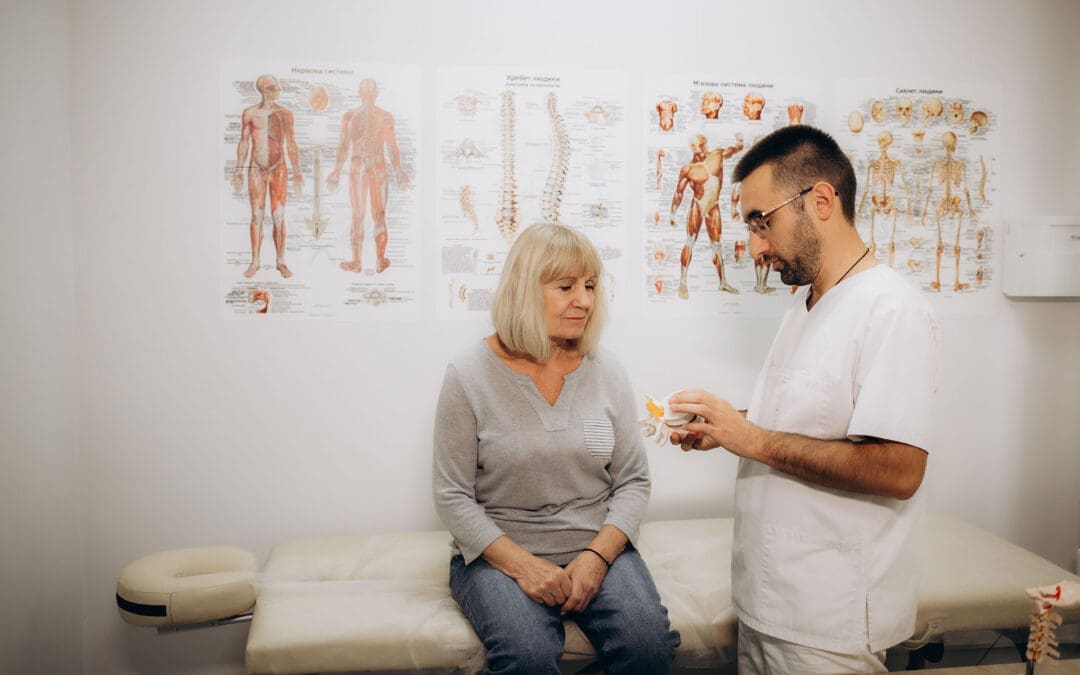
Spine problems are evident in the model. A woman is in consultation with the doctor in the clinic.
At El Paso Back Clinic®, led by Dr. Alexander Jimenez, DC, APRN, FNP-BC, we’re dedicated to transforming lives through advanced chiropractic care and integrative wellness in El Paso, TX. Specializing in recovery from motor vehicle accidents (MVAs), sports injuries, and chronic conditions such as neuropathy, our team utilizes cutting-edge neuromusculoskeletal imaging and dual-scope diagnostics to identify the root causes of injuries. From whiplash to gastrointestinal trauma, we craft personalized plans blending spinal adjustments, nutrition, and therapies like acupuncture to restore mobility and vitality. With a focus on holistic healing and legal support for injury claims, we empower El Pasoans to live pain-free and thrive.
Personal Injuries and Their Impact in El Paso
Living in El Paso’s bustling community means navigating busy roads and an active lifestyle, where accidents—from car crashes to sports mishaps—can disrupt one’s health. MVAs, common on rainy I-10 days, often cause spinal misalignments, leading to sciatica or numbness (Jimenez, 2025a). Sports injuries, like joint strains and workplace falls, add to the toll, risking chronic issues like neuropathy if untreated (Mana.md, n.d.).
At El Paso Back Clinic®, Dr. Jimenez’s chiropractic expertise targets these musculoskeletal and nerve disruptions. Using advanced imaging, we link injuries to symptoms, ensuring precise care. Our integrative approach, which combines adjustments with wellness coaching, helps prevent long-term pain and includes legal documentation to support insurance claims for patients in El Paso.
Nerve Pain and Neuropathy Post-Collision
Car accidents frequently trigger nerve compression, causing tingling, burning, or weakness that mimics peripheral neuropathy. Even minor collisions in El Paso’s unpredictable weather can cause vertebrae to shift, potentially pinching nerves (Jimenez, 2025b). Our clinic employs EMG and dynamic X-rays to map these injuries, correlating crash forces to nerve damage for accurate diagnosis.
We use spinal decompression and laser therapy to relieve pressure and promote healing, with patients often seeing 40-50% symptom improvement in weeks (Miami Chiropractors, n.d.). Detailed biomechanical reports strengthen personal injury claims, ensuring fair compensation for El Paso residents.
Chiropractic Care for Joint and Ligament Injuries
Injuries like ACL tears from sports or MVA dashboard impacts require targeted restoration. At El Paso Back Clinic®, we realign joints, boost circulation, and strengthen muscles to speed recovery without surgery (Jimenez, 2025c). Dr. Jimenez’s functional assessments prevent compensatory patterns, vital for El Paso’s athletes.
We integrate acupuncture and custom orthotics, helping patients resume activities in six months, rather than a year. Nutritional support, like collagen-rich diets, enhances ligament repair, tailored to El Paso’s active community.
Five Musculoskeletal Challenges We Address
Accidents hit muscles and bones hard. Our chiropractic team tackles five common issues:
Neck and Back Pain: Whiplash from MVAs causes stiffness; gentle adjustments restore motion (Jimenez, 2025d).
Sciatica: Pinched nerves from disc issues ease with traction therapy.
Joint Inflammation: Post-injury arthritis responds to ultrasound and anti-inflammatory nutrition.
Sports Strains: Overuse injuries can be effectively treated through myofascial work and gait analysis.
These protocols, customized for El Pasoans, cut recurrence by half, blending wellness education for lasting health.
Spinal Misalignment: Recovery After Crashes
El Paso’s slick roads amplify MVA risks, often misaligning spines and compressing discs, leading to radiating pain (Jimenez, 2025a). We utilize high-velocity adjustments and flexion-distraction techniques to realign the vertebrae, paired with massage to relax the muscles.
Dr. Jimenez’s imaging links crash dynamics to disc damage, guiding non-surgical plans that preserve mobility in 70% of cases (Knecht Chiropractic, n.d.). Legal reports detail injury causation, supporting the claims of El Paso patients.
Reducing Inflammation for Pain Relief
Inflammation fuels post-injury pain. Our chiropractic care enhances lymphatic drainage via soft-tissue therapy and cryotherapy, breaking the cycle (Jimenez, 2025e). Patients adopt home strategies, such as taking turmeric supplements, which can reduce swelling by 40% (Miami Chiropractors, n.d.).
For workers’ compensation cases, we monitor biomarkers, aligning treatments with recovery goals to help El Paso workers return to their feet.
Cyclist Recovery After Bike-MVA Collisions
Biking on El Paso’s scenic trails poses risks from urban traffic, which can lead to fractures or nerve injuries. Our integrative care includes bike-fit corrections and vestibular training for balance (Jimenez, 2025f). Cyclists return to riding in three months, supported by endurance nutrition and legal advocacy.
Massage Therapy for MVA Trauma Healing
MVAs cause soft-tissue damage, from bruises to adhesions. Massage therapy, paired with adjustments, boosts circulation and endorphins, reducing whiplash recovery time by 30% (Jimenez, 2025). We progress from gentle strokes to deep tissue, documenting for El Paso insurance claims.
Spinal Trauma from 18-Wheeler Accidents
Semi-truck crashes deliver intense force, fracturing vertebrae or tearing ligaments. We use dynamic imaging to assess damage, guiding bracing and neuromodulation (Jimenez, 2025h). Legal reports link crash mechanics to injuries, aiding settlements for El Paso patients.
Nutrition for Tissue Repair Post-MVA
Injured tissues require nutrients such as protein and antioxidants. Dr. Jimenez designs diets with salmon and berries, using genetic insights to optimize healing (Jimenez, 2025i). This reduces fibrosis, strengthening tissues for El Paso’s active residents.
Durable Medical Equipment for Recovery
Following a motor vehicle accident (MVA), tools such as TENS units or cervical collars can support healing. We select evidence-based equipment, such as ergonomic chairs, to offload spines (Jimenez, 2025). Tele-rehab ensures compliance, with invoices bolstering El Paso claims.
Comprehensive Musculoskeletal Recovery
MVAs strain muscles and joints, from sprains to dislocations. Our pain mapping and multi-modal care—adjustments, PT, mindfulness—restore 80% function in six weeks (Jimenez, 2025k). Legal narratives ensure fair compensation.
Whiplash-Associated Disorders (WAD) Recovery
WAD from crashes causes neck pain or dizziness. We use Doppler ultrasound for vascular checks and treat with mobilization for 90% relief (Jimenez, 2025). Immediate post-accident icing and evaluations ensure thorough El Paso claims.
Gastrointestinal Injuries from MVAs
Car accidents can disrupt digestion, causing nausea or organ strain. Our integrative care, which includes visceral manipulation and nutrition, restores gut health, backed by legal support for claims (Jimenez, 2025).
Why Choose El Paso Back Clinic®?
Our team, led by Dr. Jimenez, combines chiropractic precision with medical expertise, utilizing tools such as digital motion X-rays. We offer acute-to-chronic care, transparent billing, and testimony for legal cases. Patients reduce their reliance on medication, regaining vitality through holistic plans tailored for El Paso’s vibrant community.
Sciatica Relief for Teachers: El Paso Back Clinic’s Chiropractic Solutions
A teacher helping an elementary school girl using a tablet computer
Introduction: Supporting Teachers’ Health in El Paso
Teaching is a rewarding yet demanding profession, especially in vibrant communities like El Paso, Texas. Teachers spend long hours standing, sitting, and moving in ways that strain their bodies. These daily tasks can lead to sciatica, a painful condition caused by irritation of the sciatic nerve, which runs from the lower back down the legs. Symptoms like sharp leg pain, numbness, or tingling can disrupt lesson plans and classroom energy.
At El Paso Back Clinic®, led by Dr. Alexander Jimenez, DC, APRN, FNP-BC, we understand the unique challenges educators face. Prolonged sitting during grading, standing for lessons, poor posture over desks, and the physical demands of managing classrooms increase sciatica risks. Our clinic specializes in chiropractic care, integrative medicine, and functional rehabilitation to help teachers manage pain and prevent flare-ups. Using manual adjustments, ergonomic advice, and targeted exercises, we aim to restore spinal health and enhance quality of life.
This article examines why teachers are prone to sciatica, how our clinic’s chiropractic and integrative approaches can provide relief, and offers practical steps for achieving lasting wellness. Drawing on Dr. Jimenez’s 30+ years of expertise, we’ll share clinical insights and real-world solutions tailored for El Paso’s educators.
What Is Sciatica and Why Does It Affect Teachers?
Sciatica occurs when the sciatic nerve, the body’s longest nerve, becomes compressed or irritated. This nerve starts in the lower spine, travels through the hips, and extends down each leg. Common symptoms include burning pain, tingling, or weakness in one leg, often worsening with sitting or standing. For teachers, this can mean discomfort during classes or while grading at home.
The teaching environment in El Paso schools, from bustling elementary classrooms to high school lecture halls, creates perfect conditions for sciatica. Standing for long periods during lessons or playground duty fatigues back muscles, pressing on spinal discs (Bomberg Chiropractic, 2023). Sitting at desks or in cramped staff rooms can shorten hip muscles, tilt the pelvis, and pinch the nerve (East Bay Chiropractic Office, 2023). Poor posture, like slouching over lesson plans, further irritates the nerve roots (Scoliosis Center of Utah, n.d.).
Dr. Jimenez sees this often at El Paso Back Clinic. His advanced neuromusculoskeletal imaging, such as X-rays and MRIs, pinpoints disc bulges or muscle imbalances that cause sciatica in teachers. By addressing these root causes, our clinic helps educators stay active without pain.
How Teachers’ Daily Routines Trigger Sciatica
Teachers’ days are a mix of physical and mental demands. Standing to deliver lessons or monitor halls strains the lower back, increasing nerve pressure (Boyne Ergonomics, n.d.). Sitting for hours on outdated chairs compresses the spinal discs, a key factor in triggering sciatica (Bomberg Chiropractic, 2023). Bending to assist students or lifting heavy teaching materials—such as projectors or book boxes—can strain the piriformis muscle, which is located near the sciatic nerve.
Poor posture is a major culprit. Leaning over desks or hunching at computers curves the spine unnaturally, squeezing nerve roots (Scoliosis Center of Utah, n.d.). Stress from managing classrooms or meeting tight deadlines can cause muscle tension, leading to inflammation (Paragon Chiropractic, n.d.). In El Paso, where teachers often juggle bilingual classes and extracurricular duties, these risks accumulate.
Dr. Jimenez’s clinic frequently treats educators with sciatica from these habits. His dual-scope approach—combining chiropractic exams with diagnostic imaging—reveals how daily tasks, such as carrying heavy bags, can lead to spinal misalignment. Our tailored treatments at El Paso Back Clinic, including adjustments and massage, address these issues directly, helping teachers move freely.
The Impact of Prolonged Sitting and Standing
Teachers switch between sitting and standing constantly—standing for morning assemblies, sitting for parent-teacher meetings, then standing again for labs. Prolonged sitting, especially on hard classroom chairs, increases disc pressure by up to 30%, irritating the sciatic nerve (Bomberg Chiropractic, 2023). Standing too long without breaks tightens hip flexors, pulling the spine out of alignment (Boyne Ergonomics, n.d.).
This back-and-forth strains stabilizing muscles, risking micro-tears in discs that pinch nerves. In El Paso’s active school settings, teachers may stand for over four hours daily, increasing the odds of back pain by 50% (Abundant Life Chiropractor, 2023). At El Paso Back Clinic, Dr. Jimenez utilizes advanced imaging to identify these strains, which are often seen in teachers following minor classroom injuries or slips. Our spinal decompression therapy gently stretches the spine, relieving nerve pressure and promoting healing.
Simple fixes can help: switch positions every 20 minutes, use cushioned mats for standing, or adjust your desk height to a comfortable level. These small changes, guided by our clinic’s ergonomic coaching, significantly reduce the risk of sciatica.
Poor Posture: A Hidden Cause of Nerve Pain
Posture shapes spinal health. Teachers often slouch over their desks or lean forward to engage students, curving their spines into a “C” shape. This compresses the lumbar vertebrae, irritating sciatic nerve roots (Scoliosis Center of Utah, n.d.). Low computer screens can cause neck craning, which can lead to lower back strain.
In El Paso classrooms, crouching to help young students or writing on low boards exacerbates this issue. Over time, uneven muscle pull misaligns the spine, trapping the nerve. At El Paso Back Clinic, Dr. Jimenez uses posture assessments to spot these habits early. His chiropractic adjustments realign the vertebrae, while acupuncture relaxes tight muscles, such as the piriformis, easing nerve pressure (Jimenez, n.d.a).
Posture tips: Keep your ears over your shoulders, use a lumbar-support chair, and raise screens to eye level. Our clinic offers workshops for El Paso teachers to build these habits, preventing chronic pain.
Physical Demands: The Active Side of Teaching
Teaching isn’t just standing or sitting—it’s dynamic. Lifting stacks of textbooks, bending to pick up dropped items, or dashing to manage recess chaos strains the back. These motions can herniate discs or inflame muscles near the sciatic nerve (East Bay Chiropractic Office, 2023). In El Paso, where teachers may handle heavy bilingual materials or sports equipment, risks grow.
Sudden twists, such as grabbing a falling projector, mimic sports injuries that Dr. Jimenez treats. His clinic documents these as work-related injuries for insurance purposes, utilizing massage and exercise to aid in tissue healing. Advanced imaging ensures an accurate diagnosis, detecting layered issues such as sprains and nerve compression (Jimenez, n.d.b).
Safe habits reduce risks: Lift with bent knees, use carts for supplies, and stretch before engaging in active duties. El Paso Back Clinic’s tailored plans help teachers stay strong and pain-free.
Chiropractic Care at El Paso Back Clinic: Targeted Relief
Chiropractic care is a cornerstone for sciatica relief. At El Paso Back Clinic, our manual adjustments realign the spine, reducing nerve irritation and inflammation (Active Health Center, n.d.). Teachers notice less leg pain and better mobility after sessions. Our spinal decompression therapy gently stretches the spine, retracting bulging discs that pinch nerves (Bomberg Chiropractic, 2023).
Dr. Jimenez’s expertise shines here. With over 30 years of experience treating patients in El Paso, including educators, Dr. [Last Name] combines chiropractic care with integrative methods, such as acupuncture, to provide natural pain relief. Advanced imaging ensures precise adjustments, targeting the exact cause of sciatica (Jimenez, n.d.a). Regular visits prevent flare-ups, letting teachers focus on students, not pain.
Restoring Spinal Alignment and Nerve Function
Adjustments are quick, targeted thrusts that realign vertebrae, freeing the sciatic nerve. This boosts blood flow and reduces inflammation, key for teachers battling daily strain (AFC Adherence, n.d.). At our clinic, Dr. Jimenez pairs adjustments with soft tissue work to release tight hips, a common issue among educators.
Our approach restores function holistically. Teachers regain flexibility for classroom tasks, and consistent care prevents future issues. Jimenez’s diagnostic tools, such as MRIs, ensure that treatments match each patient’s needs, offering El Paso educators reliable relief (Jimenez, n.d.b).
Reducing Inflammation Naturally
Inflammation fuels sciatica pain and swelling of the tissues around the nerve. Our adjustments improve spinal motion, reducing this swelling (Active Health Center, n.d.). We add ice or heat therapy to speed relief, tailored to each teacher’s symptoms.
Dr. Jimenez enhances this approach with nutrigenomics, recommending anti-inflammatory foods, such as salmon, to support the healing process. For El Paso teachers, this integrative approach means less pain and faster recovery from classroom strains (Jimenez, n.d.a).
Lifestyle Changes: Ergonomics and Exercises for Teachers
El Paso Back Clinic goes beyond adjustments, offering practical advice. Ergonomic tips include adjustable chairs, footrests, and raised monitors to reduce strain (Boyne Ergonomics, n.d.). For teachers, we recommend lumbar pillows and standing desks for grading.
Exercises are key: planks strengthen the core, and piriformis stretches loosen the hips (Alliance Orthopedics, n.d.). Dr. Jimenez designs home routines, such as knee-to-chest stretches, to accommodate busy schedules. Our massage therapy supports recovery, ensuring El Paso educators stay active.
Preventing Flare-Ups: Daily Habits for Long-Term Relief
Preventing sciatica means tracking triggers. Long sits or heavy lifts? Take breaks or use carts. Heat eases tight muscles; cold calms acute pain (Abundant Life Chiropractor, 2023). Weekly core workouts and posture apps help maintain proper alignment.
Dr. Jimenez’s clinic emphasizes prevention. Our exercise plans, paired with stress-reducing yoga, help teachers avoid chronic issues. Legal documentation supports work-injury claims, ensuring access to care (Jimenez, n.d.b).
Integrative Care: A Team Approach at El Paso Back Clinic
We combine chiropractic care with physical therapy, acupuncture, and massage to facilitate a comprehensive recovery. Physical therapy builds strength with moves like bridges (Active Health Center, n.d.). Acupuncture calms the nerves, making it ideal for reducing teachers’ stress (Jimenez, n.d.a). Short movement breaks, such as stretching during class, boost circulation.
Our clinic’s integrative model, led by Dr. Jimenez, treats sciatica holistically, addressing work or personal injuries with detailed records for insurance.
Dr. Alexander Jimenez’s Expertise: A Beacon for El Paso Teachers
Dr. Jimenez, with dual credentials as a chiropractor and nurse practitioner, brings unmatched care to El Paso. His clinic treats sciatica from classroom strains, sports injuries, or accidents, using imaging to diagnose precisely. Treatments such as adjustments, massage, and tailored exercises can help the body heal naturally, thereby preventing long-term issues.
For teachers, Jimenez’s legal documentation supports work claims, ensuring coverage. His functional medicine approach, including nutrition and acupuncture, empowers educators to thrive (Jimenez, n.d.a; Jimenez, n.d.b).
Practical Tips for El Paso Teachers
Morning Stretch: Try cat-cow (10 reps) to loosen the spine.
Classroom Ergonomics: Use lumbar-support chairs; raise boards to waist height.
Breaks: March in place every 30 minutes to ease nerve pressure.
Nutrition: Eat berries and fish to combat inflammation, according to Jimenez’s advice.
Conclusion: Empowering El Paso Educators
Sciatica doesn’t have to slow down El Paso’s teachers. At El Paso Back Clinic, Dr. Jimenez and our team offer chiropractic care, integrative therapies, and practical tips to relieve pain and prevent issues. From adjustments to ergonomic tweaks, we help educators stay healthy and focused on inspiring students.
Visit us at 11860 Vista Del Sol Dr, Suite 128, El Paso, TX, or call 915-850-0900 to start your pain-free journey.
Preventing Sports & Back Injuries: The El Paso Back Clinic Approach
Athletes, weekend warriors, and active individuals often push their bodies to the limit. Without smart preparation and care, minor misalignments or imbalances can lead to back pain, sprains, or more serious injuries. At El Paso Back Clinic, our mission is to prevent injuries before they occur, maintain spine health, and support long-term performance and wellness.
In this article, you’ll learn how a multifaceted strategy—involving movement, conditioning, chiropractic, integrative therapies, and recovery—can reduce injury risk. We’ll also show how El Paso Back Clinic applies these principles in real-world care.
Why Back & Sports Injuries Occur
Biomechanical Stress & Misalignment
Even small spinal misalignments or joint restrictions can change movement mechanics. Over time, stresses that should spread evenly across tissues become concentrated on certain discs, muscles, or ligaments, making them vulnerable (Mount Sinai, n.d.; Emery & Meeuwisse, 2008).
Overuse and Repetition
Playing the same sport repeatedly without variation often leads to overuse injuries—microtears that accumulate faster than the body can heal. Many youth and amateur athletes suffer from this because they skip rest phases (Nationwide Children’s, n.d.; CHOP, n.d.).
Fatigue, Poor Technique, and Weakness
When muscles fatigue, the muscle fibers break down. A runner might collapse inward at the knee, or a basketball player might land with improper form. These movement faults under fatigue cause injury (Walker Physical Therapy, n.d.; PWR Physio, n.d.).
Insufficient Recovery
Without proper rest, nutrition, and tissue repair, microdamage lingers. Eventually, the body’s threshold is crossed, and a dramatic injury occurs.
Core Prevention Pillars
At El Paso Back Clinic, we emphasize these foundational pillars:
1. Dynamic Warm-Up & Mobility Routines
Warm-ups aren’t just stretching—they’re activation drills, joint movements, and controlled progressions that prepare muscles and joints. Cooling down, stretching, and mobility work afterward help flush byproducts and reduce stiffness (First Physio Plus, n.d.; Garden State Pain, n.d.).
2. Technique Monitoring and Movement Quality
We routinely analyze movement—such as running gait, jumping, squatting, and twisting—to identify harmful patterns. By coaching technique and correcting faults, we reduce stress on the back and joints (GPOA, n.d.; Walker Physical Therapy, n.d.).
3. Balanced Strength, Stability & Flexibility
Having a strong core, glutes, and stabilizers protects the lumbar spine. We design programs that incorporate strength, balance, flexibility, and endurance to create a well-rounded system (PWR Physio, n.d.; Walker Physical Therapy, n.d.).
4. Strategic Rest and Load Management
We guide patients and athletes in periodization, which involves alternating high and low loads, scheduling rest days, and monitoring fatigue to prevent overtraining (Bayfront Health, n.d.; Fick PT & Performance, n.d.).
5. Nutrition, Hydration & Recovery Support
Good hydration and nutrients (protein, vitamins, minerals) are essential for tissue repair. A poor diet hinders recovery and increases the risk of injury (LI Spine Med, 2024).
The Role of Chiropractic & Back Clinic Services
El Paso Back Clinic (under Dr. Jimenez) stands out by combining back/spine care with integrative therapies. Here’s how chiropractic and back-clinic services help prevent injuries:
Spinal Alignment & Joint Function
Chiropractic adjustments and spinal mobilizations help maintain vertebral alignment, ease restrictions, and ensure joints move properly. This reduces compensatory stress on surrounding tissues (Dallas Accident & Injury Rehab, n.d.; Evolved Health Chiropractic, n.d.).
Posture, Movement Pattern Correction & Neuromuscular Feedback
We assess posture and movement patterns across the kinetic chain. Correcting compensations (e.g., pelvic tilt, scoliosis curves) helps protect the spine during sport demands (Dallas Accident & Injury Rehab, n.d.; Evolved Health Chiropractic, n.d.).
Proper nerve input from spinal segments supports muscle activation and timing. By improving the communication between the spine and joints and the surrounding muscles, we help the body respond more effectively under stress (Fremont Chiropractic, n.d.; Young Chiropractic, n.d.).
Versatile Soft-Tissue & Myofascial Work
Muscles, fascia, and connective tissues often tighten, pulling on the spine. Techniques, such as soft-tissue work, instrument-assisted release, and myofascial release, help reduce tension and restore balance (Garmon Chiropractic, n.d.).
Monitoring & Maintenance Care
We often schedule preventive “maintenance” visits. Even when patients feel fine, small dysfunctions can arise. Regular check-ins allow us to catch them early—before they develop into problems.
Integrative Therapies & Supportive Methods
To maximize prevention, El Paso Back Clinic layers on integrative and complementary care:
Physical Therapy & Exercise Therapy
Sometimes muscles need retraining. Our clinic can collaborate with or provide therapeutic exercise programs that focus on weakness, imbalance, mobility deficits, and sport-specific drills (Current Physical Therapy, 2025).
Massage, Trigger Point Work & Soft-Tissue Modulation
Massage and trigger point therapy enhance circulation, alleviate adhesions, and promote muscular recovery. These help tissues remain supple and resilient (Primary Health & Wellness, n.d.).
Acupuncture & Electro-Acupuncture
Using needles or micro-current stimulation, we stimulate healing, reduce inflammation, and modulate pain. These methods pair well with structural work (clinic’s integrative model).
Kinesio Taping & Supportive Bracing
Taping techniques provide gentle support, reduce stress on soft tissues, and enhance proprioception during dynamic phases of sports (Premier Injury Clinics of DFW, n.d.).
Nutritional & Functional Medicine Guidance
As part of Dr. Jimenez’s broader practice, we assess systemic contributors—such as nutrition, inflammation, and hormonal balance—to optimize the body’s healing environment.
Putting It Together: How El Paso Back Clinic Builds a Preventive Protocol
Here’s how our clinic might structure a prevention plan for an athlete or active individual:
Watch performance metrics, fatigue trends, and pain signals
Adjust load or interventions accordingly
Over time, this layered approach builds resilience—spines become more stable, tissues more durable, and neuromuscular control more refined.
Why Choose El Paso Back Clinic
Dual Expertise for Spine & Whole-Body Health
At El Paso Back Clinic, Dr. Jimenez offers both advanced back-centric care and integrative medicine. The clinic’s services extend beyond symptom relief to encompass systemic wellness, functional movement, and injury prevention (El Paso Back Clinic, n.d.).
Local Focus, Tailored to El Paso Athletes
We are familiar with the terrain, climate, demands, and sports culture in El Paso. Our protocols are adapted to local conditions—heat, elevation, sports trends—and we serve individuals, teams, schools, and sports clubs.
Evidence-Informed, Patient-Centered Approach
Our protocols integrate best practices from sports medicine, chiropractic research, and functional health models. We emphasize care plans unique to each patient—not cookie-cutter templates.
Support for Injury, Recovery & Prevention
Whether someone has already been injured or is simply seeking preventive care, our clinic handles a spectrum: back pain, sports injuries, work injuries, and even personal injury/auto trauma.
Summary & Next Steps
Preventing back and sports injuries is not about a single fix. It’s about a synergistic strategy: warm-ups, monitoring technique, balanced conditioning, spinal care, integrative therapies, and smart recovery. El Paso Back Clinic weaves these together in a real-world, locally tuned model.
If you are an athlete or an active person looking to protect your spine and enhance your performance, consider a preventive evaluation. Contact us to begin your tailored, resilience-building program.
El Paso Back Clinic’s Guide to Ergonomic Mice for Pain-Free Hands
Spending hours at a computer can strain your hands, wrists, and arms, especially after injuries from accidents or repetitive tasks. At El Paso Back Clinic in El Paso, TX, led by Dr. Alex Jimenez, DC, APRN, FNP-BC, we specialize in providing holistic solutions to help patients overcome pain. An ergonomic mouse, designed to fit your hand’s natural shape, reduces strain and helps prevent conditions like carpal tunnel syndrome and tendonitis. Paired with our chiropractic care, advanced diagnostics, and integrative therapies, it supports recovery and long-term wellness. This article explains how El Paso Back Clinic uses ergonomic tools and expert care to restore health and prevent future issues.
Why Choose an Ergonomic Mouse?
Unlike standard flat mice, an ergonomic mouse curves to match your hand, often tilting upright in a manner similar to a handshake grip. This keeps your wrist straight, easing muscle and nerve strain (Goldtouch, 2023a). At El Paso Back Clinic, we recommend these for patients with desk jobs or those recovering from accidents.
Traditional mice twist your forearm, pinching nerves. Ergonomic designs hold your arm neutrally, reducing fatigue (Logitech, n.d.). For example, Logitech’s MX Vertical tilts at 57 degrees, cutting wrist tension (Logitech, n.d.). Our patients report less pain after switching, helping them work or recover comfortably.
Pick a mouse with thumb rests or adjustable angles to suit your hand. Our clinic guides you to the best choice for your needs (ProtoArc, 2023).
Supporting Natural Posture for Comfort
Your hand’s position affects your entire arm. Regular mice force your wrist to bend inward, stressing bones and nerves (ZDNet, 2023). An ergonomic mouse reduces this twist, called pronation, keeping your hand in a relaxed position (Goldtouch, 2023a).
Studies show these mice cut muscle effort by up to four times (Logitech, n.d.). They also help ease shoulder and neck tension, which is crucial for those recovering from injuries (Kosak Chiropractic, n.d.). At El Paso Back Clinic, we have seen patients benefit from this switch, especially those who have experienced motor vehicle accidents (MVAs) or repetitive strain injuries.
Reducing Repetitive Strain Injuries
Repetitive strain injuries (RSI) from constant clicking cause tingling, numbness, or pain (EffyDesk, 2023). Ergonomic mice minimize hand movements, featuring curves that allow fingers to rest naturally (Goldtouch, 2023b).
Thumb rests stop over-gripping, and lightweight designs make moving easier (ProtoArc, 2023). Our patients, from office workers to MVA survivors, use these to avoid worsening injuries. This supports healing during rehabilitation.
Preventing Carpal Tunnel and Tendonitis
Carpal tunnel syndrome squeezes the wrist’s median nerve, causing tingling or a weak grip. Tendonitis inflames tendons from overuse (FlexiSpot, n.d.). Both are common in desk workers and individuals who have been in accidents. Ergonomic mice open the wrist’s tunnel, reducing pressure by up to 30% (Goldtouch, 2023a).
They also limit bends that inflame tissues (ZDNet, 2023). For tendonitis, less forearm twist eases elbow strain, preventing long-term damage (Lowery Chiropractic, n.d.). El Paso Back Clinic patients who use these mice often stop the progression of injury early.
Setting Up Your Workstation for Health
An ergonomic mouse works best with a properly set-up desk. At El Paso Back Clinic, we recommend adjusting your chair to a 90-degree elbow angle with your feet flat. Keep your mouse at elbow height to avoid reaching (Kosak Chiropractic, n.d.).
Use a keyboard tray to maintain a straight wrist position and set your monitor at eye level to prevent neck strain (Kosak Chiropractic, n.d.). Take hourly breaks—stretch wrists, roll shoulders—to boost blood flow (EffyDesk, 2023). Our team offers personalized tips to make your workspace support recovery.
El Paso Back Clinic’s Holistic Healing Approach
Our clinic blends chiropractic adjustments, acupuncture, and rehabilitation to treat pain holistically. Adjustments realign joints, easing nerve pressure and swelling (Rozenhart Chiropractic, n.d.). For wrist pain, we target hand-to-elbow alignment to relieve carpal tunnel (Lowery Chiropractic, n.d.).
We utilize integrative therapies, such as ultrasound to warm tissues and electrical stimulation to calm nerves (Lowery Chiropractic, n.d.). Nutrition counseling helps reduce inflammation, thereby aiding recovery (Evolve Chiropractic, n.d.). Dr. Jimenez creates custom plans to address the causes of injuries, not just their symptoms.
Dr. Alex Jimenez’s Expertise in Injury Care
Dr. Alex Jimenez, a chiropractor and nurse practitioner, leads El Paso Back Clinic with dual expertise. He treats work, sports, personal, and MVA injuries using advanced neuromusculoskeletal imaging and dual-scope diagnosis to pinpoint issues like nerve compression (Jimenez, n.d.a).
For MVAs, he links whiplash to arm pain, using scans to guide treatment (Jimenez, n.d.b). Care includes adjustments, exercises, and massage to restore function. Acupuncture boosts natural healing (Evolve Chiropractic, n.d.). We also manage legal documentation for injury claims, easing patient stress (Jimenez, n.d.a).
A recent patient, following a motor vehicle accident (MVA), utilized an ergonomic mouse and our care plan. Pain dropped 70% in weeks, avoiding surgery (Jimenez, n.d.b). Dr. Jimenez focuses on natural healing over medication.
Targeted Therapies for Lasting Relief
We pair ergonomic tools with rehab. Grip exercises strengthen the hands, while wrist stretches build flexibility (EffyDesk, 2023). Acupuncture targets specific pain points, and massage helps loosen muscles (Rozenhart Chiropractic, n.d.).
Dr. Jimenez utilizes electro-acupuncture for nerve recovery, which has been shown to be effective for chronic pain (Jimenez, n.d.a). Patients track their progress with pain logs to achieve steady improvement. Our El Paso clinic provides these therapies for seamless care.
Success Stories at El Paso Back Clinic
Anna, a receptionist, switched to an ergonomic mouse and received our adjustments. Her wrist pain faded in weeks, improving her work (Goldtouch, 2023a). Carlos, an MVA survivor, worked with Dr. Jimenez. Adjustments and exercises restored his arm strength (Jimenez, n.d.b).
These stories show our approach delivers. Small changes, combined with expert care, transform lives.
Building a Pain-Free Future
Start with an ergonomic mouse and a tuned workspace. Experience the benefits of our chiropractic care, acupuncture, and nutrition for lasting health. Short walks and breathing exercises boost recovery (Evolve Chiropractic, n.d.).
Visit El Paso Back Clinic for a custom plan. Dr. Jimenez’s team treats all injuries naturally, from desk strain to MVAs (Jimenez, n.d.a). Act early to stay pain-free.
Conclusion: Heal with El Paso Back Clinic
An ergonomic mouse supports natural hand posture, cutting strain. Paired with our chiropractic and integrative care, it helps prevent and manage issues such as carpal tunnel syndrome. Dr. Jimenez’s expertise ensures effective recovery. Call +1 (915) 850-0900 to start your pain-free journey today.
Strumming Without Pain: Chiropractic Solutions for Guitarists and Bassists at El Paso Back Clinic
Playing guitar or bass fills life with rhythm and joy. The thrill of strumming chords or plucking deep notes creates unforgettable moments. But for many string players in El Paso, Texas, this passion can lead to pain. Hours of practice can strain hands, wrists, forearms, elbows, and shoulders, leading to repetitive strain injuries (RSIs) such as tendonitis. These injuries bring swelling, stiffness, and aches that make playing tough. At El Paso Back Clinic, led by Dr. Alexander Jimenez, DC, APRN, FNP-BC, we offer integrative chiropractic care to tackle these issues, helping musicians heal naturally and keep the music alive.
This article explains why guitarists and bassists are prone to RSIs, how tendonitis affects key areas, and how our clinic’s holistic approach—combining chiropractic adjustments, massage, acupuncture, and nutrition—restores health. With insights from Dr. Jimenez’s 30+ years of expertise, we’ll show how El Paso Back Clinic helps local musicians recover from injuries and prevent future pain, so they can strum and pluck without worry.
Why String Players Face Repetitive Strain Injuries
Guitarists and bassists repeat the same motions for hours: fretting chords, strumming strings, or plucking heavy bass lines. These actions stress tendons—the tough bands connecting muscles to bones. Over time, small tears form, which can lead to inflammation or tendonitis. Unlike a one-time injury, RSIs develop gradually from overuse, making them common among musicians (Pianucci et al., 2021).
The fretting hand curls tightly to press strings, while the strumming or plucking arm moves fast. Bassists face extra strain from thicker strings that need more force. Poor posture, like slouching over a guitar, adds pressure to the shoulders and neck. Heavy instruments—guitars at 7-10 pounds and basses up to 12—strain the body more during gigs (Pain Free NY, n.d.). Cold El Paso nights or long jam sessions at local venues like Lowbrow Palace can worsen symptoms by stiffening muscles.
Other factors increase risks. Older players over 40 have less flexible tendons (Bend Total Body Chiropractic, n.d.). Poor habits, such as gripping picks too hard or skipping warm-ups, can speed up strain. Diet matters too—sugary or fatty foods fuel inflammation, slowing recovery (Healthline, 2022). El Paso’s active music scene, with frequent gigs and rehearsals, means local players often push their limits, increasing the risk of RSI.
Where It Hurts: Tendonitis in Musicians’ Bodies
Tendonitis hits specific spots based on how guitarists and bassists play. Here’s where pain strikes:
Hands and Fingers: Fretting chords strains finger tendons, especially at the thumb base. Thumb tendonitis (De Quervain’s) causes sharp pain when gripping the neck. Swelling or a gritty feel signals trouble (Guitar Strength Project, n.d.).
Wrists: Strumming and plucking bend wrists repeatedly, inflaming tendons on top (extensor) or below (flexor). Stiffness after waking or a weak grip are signs. Carpal tunnel syndrome may add tingling or numbness (Rawlogy, n.d.).
Forearms: Constant flexing causes the forearm muscles to burn. Redness, warmth, or lumps show tendonitis. Bassists feel it more from forceful plucks (Healthline, 2022).
Elbows: “Guitar elbow” mimics tennis elbow, with pain on the outer elbow from strained tendons. Inner elbow pain (golfer’s elbow) also hits. Both weaken grip, making it hard to hold picks or instruments (Tennis Elbow Classroom, n.d.).
Shoulders: Holding arms out for chords strains the rotator cuff tendons, causing aches that spread down the arm. Slouching worsens it (Smithsonian Folkways, n.d.).
These areas link up. Hand pain can trigger elbow issues, and shoulder misalignment can strain wrists. Catching early signs—such as soreness or fatigue—prevents more severe problems.
Symptoms That Stop the Show
Tendonitis symptoms creep in but hit hard. Pain starts as a dull ache during play, then sharpens at rest. Swelling puffs up joints, and stiffness locks fingers, especially in the morning. Numbness or tingling buzzes in cold venues, sometimes with fingers turning blue from poor blood flow (Pain Free NY, n.d.). Weakness, drops, and fatigue, as well as burning or throbbing sensations, often linger after gigs. A grating sensation hints at the presence of scar tissue.
For El Paso musicians, long practices for gigs at Tricky Falls or house shows can exacerbate symptoms. Stress from late-night sets or cold weather can cause muscles to tighten, exacerbating pain. If symptoms last for weeks, it’s time to visit El Paso Back Clinic for expert care.
Quick Relief at Home
Before professional help, try these steps to ease tendonitis:
RICE Method: Rest by avoiding play and using splints. Ice for 15 minutes, wrapped, several times daily. Compress with elastic wraps, not too tight. Elevate arms on pillows (Mayo Clinic, 2023).
Meds: Ibuprofen reduces swelling, but ask a doctor first.
Stretches: Gentle wrist circles, finger spreads, or forearm pulls, held 15-30 seconds (Healthline, 2022).
Massage: Use massage balls to roll out knots gently (Rawlogy, n.d.).
Diet: Eat berries, fish, and leafy greens to help combat inflammation. Avoid sugary snacks common at El Paso food trucks.
These help, but don’t address the root cause. For lasting relief, see the experts at El Paso Back Clinic.
Chiropractic Care at El Paso Back Clinic
At El Paso Back Clinic, Dr. Alexander Jimenez and his team utilize chiropractic care to effectively treat RSIs. Adjustments realign joints in the wrist, elbow, or shoulder, freeing nerves and boosting blood flow. For elbow tendonitis, specific adjustments reduce pain and swelling, with patients often regaining full motion in weeks (Stamford Spine, n.d.).
Our clinic checks the whole body. A misaligned shoulder can strain wrists, so we adjust the entire chain. Soft tissue work, such as Graston therapy, breaks down scar tissue in the wrists. Laser therapy reduces inflammation, and taping supports joints during physical activity (Pinnacle Hill Chiropractic, 2024). Regular visits help keep the body aligned, reducing the risk of re-injury by up to 50% (Chiro One, n.d.).
Dr. Jimenez’s dual training as a chiropractor and nurse practitioner ensures precise diagnosis and treatment. Using advanced imaging like MRIs, we pinpoint tendon tears or nerve issues. Treatments are safe, with only mild soreness possible, and tailored to each musician’s needs (Bend Total Body Chiropractic, n.d.).
Integrative Healing for El Paso Musicians
Our integrative approach goes beyond adjustments. We combine:
Massage Therapy: Kneads forearm knots, easing tension from long sets (Beech Street Health, n.d.).
Acupuncture: Calms nerves, reducing wrist tingling for smoother playing.
Exercises: Wrist curls with light weights or finger bands build strength (Chiro One, n.d.).
Nutrition: Collagen supplements and omega-3 fatty acids accelerate tendon repair. We guide patients to local El Paso markets for healthy foods.
Ergonomics: Adjust guitar straps or use lighter picks. Take breaks every 20 minutes during practice (Smithsonian Folkways, n.d.).
This mix helps heal faster and prevents future pain, allowing musicians to stay on stage.
Dr. Jimenez’s Expertise at El Paso Back Clinic
Dr. Alexander Jimenez brings over 30 years of experience to El Paso Back Clinic. His dual-scope approach—combining chiropractic and functional medicine—targets the root causes of injuries. We provide personalized plans for musicians, workers, athletes, and individuals who have experienced accidents. Advanced tools, such as neuromusculoskeletal imaging, can reveal hidden damage, while assessments also consider lifestyle and genetics (Jimenez, n.d.a).
For a local guitarist who was injured in a car accident, we utilized adjustments, massage, and nutrition to restore their fretting ability. Our clinic also handles legal documentation for injury claims, ensuring smooth insurance processes (Jimenez, n.d.b). From whiplash to wrist strain, we help El Paso’s music community heal naturally.
Preventing Pain for Lifelong Playing
Prevention keeps musicians playing. Try these:
Exercises: Wrist stretches, towel twists, or 1-pound weight curls, 10 reps, three times weekly (Healthline, 2022).
Warm-Ups: 10-minute finger flexes and arm circles before gigs.
Technique: Use loose grips and neutral wrists. Alternate hands for songs (No Treble, 2011).
Gear: Ergonomic straps and lighter instruments ease shoulder strain.
Breaks: Rest every 20 minutes. Relax with meditation to cut stress.
El Paso Back Clinic offers tailored plans to keep your body gig-ready.
Keep the Music Playing
Tendonitis doesn’t have to silence your strings. At El Paso Back Clinic, Dr. Jimenez and our team use chiropractic and integrative care to heal RSIs and prevent pain. From hands to shoulders, we address the root causes so you can play without fear. Visit us in El Paso to get back to strumming and plucking with ease.
Contact El Paso Back Clinic at 915-850-0900 or dralexjimenez.com to schedule your consultation today.
Exploring Integrative Chiropractic Care: Benefits, Techniques, and More
Introduction
Integrative chiropractic care is gaining popularity as people seek natural ways to address health issues beyond back and neck pain. This approach combines traditional chiropractic adjustments with other therapies to promote overall wellness. Many wonder about its effectiveness, safety, costs, and how to choose the right provider, especially in a city like El Paso, Texas. This article answers common questions about integrative chiropractic care, with insights from Dr. Alexander Jimenez, a chiropractor and nurse practitioner in El Paso, whose clinical expertise highlights how this care can treat various conditions, injuries, and promote long-term health.
What Is Integrative Chiropractic Care?
Integrative chiropractic care goes beyond traditional spinal adjustments. It focuses on the whole person, combining chiropractic techniques with therapies like massage, acupuncture, and exercise to improve health. According to Integrative Chiropractic Center, this approach emphasizes natural healing and healthy living, addressing the root causes of health issues rather than just symptoms (Integrative Chiropractic Center, n.d.). Dr. Alexander Jimenez, DC, APRN, FNP-BC, a leading practitioner in El Paso, uses this holistic method to treat injuries and chronic conditions, tailoring care to each patient’s needs (Jimenez, n.d.).
Is Integrative Chiropractic Effective for More Than Just Back and Neck Pain?
Yes, integrative chiropractic care can help with more than just back and neck pain. It’s effective for conditions like migraines, dizziness, allergies, and chronic pain. Research shows that up to 75% of migraine sufferers experience neck stiffness or pain, which chiropractic care can address through spinal adjustments and soft tissue therapies (Healthgrades, 2025). A 2019 study found that spinal manipulation reduced migraine frequency and intensity, making it a promising option for headache relief (Healthgrades, 2025).
For dizziness, chiropractic care can improve balance by addressing spinal misalignments that affect the nervous system. While evidence for allergies is less clear, some patients report relief due to improved immune function from reduced stress on the body (Mile High Spine, n.d.). Dr. Jimenez’s clinic in El Paso has successfully treated patients with chronic pain from work, sports, personal, or motor vehicle accident (MVA) injuries, using a combination of chiropractic adjustments and targeted therapies to address underlying issues (Jimenez, n.d.).
What Techniques Are Used in Integrative Chiropractic Care?
Integrative chiropractors use a variety of techniques to promote healing. Common methods include:
Spinal Manipulation: Adjusting the spine to improve alignment and nerve function.
Soft Tissue Therapies: Techniques like myofascial release to relax tight muscles.
Massage Therapy: Relieves muscle tension and improves circulation.
Acupuncture: Stimulates specific points to reduce pain and promote healing.
Targeted Exercise: Strengthens muscles and improves mobility.
Lifestyle Advice: Guidance on posture, nutrition, and stress management.
Dr. Jimenez incorporates these techniques, tailoring them to each patient. For example, he uses the Diversified Technique for spinal adjustments and combines it with therapies like massage to treat injuries from sports or MVAs (Jimenez, n.d.). These methods work together to address both symptoms and their causes, promoting natural healing (Pivotal Chiropractic, n.d.).
Can Integrative Chiropractic Help with Chronic or Severe Pain?
Integrative chiropractic care is effective for chronic and severe pain, especially when caused by musculoskeletal issues. A 2019 case study of a 23-year-old woman with chronic migraines and neck pain showed significant improvement after chiropractic care using Diversified and Thompson Techniques (Vertebral Subluxation Research, 2019). Dr. Jimenez’s approach involves dual-scope diagnosis, combining his expertise as a chiropractor and nurse practitioner to assess injuries thoroughly. He uses sophisticated imaging, like X-rays or MRIs, to identify the root causes of pain, ensuring targeted treatment (Jimenez, n.d.).
For chronic pain from work or sports injuries, integrative care combines adjustments with exercise and massage to reduce inflammation and restore function. This approach helps prevent long-term consequences by addressing misalignments and promoting healing (DE Integrative Healthcare, n.d.).
Is Integrative Chiropractic Safe for Specific Populations?
Integrative chiropractic care is generally safe for most people, including children, pregnant women, and older adults, when performed by a licensed chiropractor. More research is necessary, but a case study suggests that chiropractic care is a safe and effective treatment option for migraines during pregnancy (Medical News Today, n.d.). For older adults, chiropractic adjustments can improve balance and reduce dizziness by addressing spinal stiffness (ScienceDirect, n.d.).
Dr. Jimenez ensures safety by conducting thorough assessments, including diagnostic imaging, to customize treatments. Patients with conditions like osteoporosis or recent surgeries may need gentler techniques, and Dr. Jimenez’s dual training as a nurse practitioner allows him to evaluate these risks carefully (Integrative Services, n.d.).
Can Integrative Chiropractic Help with Conditions Like Migraines, Dizziness, or Allergies?
As mentioned, integrative chiropractic care can help with migraines by reducing neck tension and improving spinal alignment. A 2019 meta-analysis found that spinal manipulation decreased migraine days and pain intensity (Healthgrades, 2025). Dizziness, often linked to neck issues or poor balance, can improve with adjustments that enhance nervous system function (ScienceDirect, n.d.).
For allergies, the evidence is less conclusive; however, some patients report benefits from reduced stress and an improved immune response. Dr. Jimenez’s clinic employs a comprehensive approach, combining adjustments, acupuncture, and lifestyle modifications to address these conditions, with a focus on overall well-being (Jimenez, n.d.).
How Much Does Integrative Chiropractic Care Cost?
Costs for integrative chiropractic care vary depending on location, treatment type, and session frequency. In El Paso, a single session may range from $50 to $150, with initial visits costing more due to assessments and imaging (DE Integrative Healthcare, n.d.). Packages or memberships can reduce costs for ongoing care. Dr. Jimenez’s clinic offers transparent pricing, and patients can inquire about costs during their first visit (Jimenez, n.d.).
Does Integrative Chiropractic Accept Insurance?
Many chiropractic clinics, including Dr. Jimenez’s, accept insurance; however, coverage varies depending on the specific plan. Some insurance providers cover chiropractic care for specific conditions like back pain or injuries, but coverage for integrative therapies like acupuncture may be limited. Patients should verify with their insurance provider and the clinic to confirm coverage. Dr. Jimenez’s office assists with insurance claims and provides options for those without coverage (Integrative Services, n.d.).
What Should Be Expected on the First Visit?
During your initial visit to an integrative chiropractor, you can anticipate a thorough evaluation. This includes:
Health History Review: Discussing Past Injuries, Conditions, and Symptoms.
Physical Exam: Assessing posture, spine, and range of motion.
Diagnostic Assessments: X-rays or other imaging to identify issues.
Treatment Plan Discussion: Outlining therapies and goals.
Dr. Jimenez uses sophisticated imaging and his dual expertise to create personalized plans. The first visit may include an adjustment or other therapies, depending on the assessment (Jimenez, n.d.). Expect the visit to last 30–60 minutes (Pivotal Chiropractic, n.d.).
Is Treatment Ongoing?
Integrative chiropractic care is often ongoing, especially for chronic conditions or injury recovery. Initial treatment may involve multiple sessions per week, transitioning to maintenance care (e.g., monthly visits) as symptoms improve. Dr. Jimenez designs long-term plans to prevent re-injury and promote health, incorporating exercise and lifestyle changes (DE Integrative Healthcare, n.d.).
How Do I Choose the Right Integrative Chiropractor in El Paso?
Choosing the right chiropractor in El Paso involves several steps:
Check Credentials: Ensure the chiropractor is licensed and has relevant training. Dr. Jimenez, for example, is a Doctor of Chiropractic and a Family Nurse Practitioner (Jimenez, n.d.).
Ask for Referrals: Consult your primary care doctor or friends for recommendations (Healthgrades, n.d.).
Research Experience: Look for expertise in your condition, like migraines or sports injuries.
Read Reviews: Check platforms like Healthgrades for patient feedback.
Ask About Techniques: Confirm the chiropractor uses integrative methods like acupuncture or massage.
Verify Insurance: Ensure the clinic accepts your insurance or offers affordable options.
Schedule a Consultation: Meet the chiropractor to discuss your needs and comfort level.
Evaluate Communication: Choose someone who listens and explains clearly.
Dr. Jimenez stands out in El Paso due to his dual credentials and focus on integrative care, making him a strong choice for complex cases (LinkedIn, n.d.).
Dr. Alexander Jimenez’s Expertise and Approach
Dr. Alexander Jimenez, DC, APRN, FNP-BC, brings a unique perspective to integrative chiropractic care in El Paso. His dual training as a chiropractor and nurse practitioner enables him to bridge the gap between medical and chiropractic approaches. His clinic specializes in treating injuries from work, sports, personal incidents, and MVAs, using a comprehensive process:
Clinical Correlation: Dr. Jimenez connects patient symptoms to specific injuries, ensuring accurate diagnosis.
Dual-Scope Diagnosis: Combining chiropractic and medical assessments for a complete picture.
Treatment Procedures: Using adjustments, acupuncture, massage, and exercise to address injuries.
Diagnostic Assessments: Employing sophisticated imaging like X-rays and MRIs to pinpoint issues.
Legal Documentation: Providing detailed reports for MVA or work injury cases, supporting insurance claims, or legal proceedings.
His clinic emphasizes natural healing, addressing injury causes to prevent chronic issues. For example, a sports injury might involve spinal adjustments to restore alignment, massage to reduce muscle tension, and exercises to strengthen the area, ensuring long-term recovery (Jimenez, n.d.).
How Integrative Chiropractic Assists with Injuries
Integrative chiropractic care is highly effective for work, sports, personal, and MVA injuries. Dr. Jimenez’s clinic uses a multi-faceted approach:
Chiropractic Adjustments: Correct spinal misalignments to reduce pain and improve function.
Targeted Exercise: Strengthens muscles to support recovery and prevent re-injury.
Massage Therapy: Reduces inflammation and promotes relaxation.
Acupuncture: Alleviates pain and enhances healing.
Integrative Medicine: Incorporates nutrition and lifestyle changes to support overall health.
This approach not only treats injuries but also prevents long-term consequences like chronic pain or reduced mobility. For MVA cases, Dr. Jimenez provides thorough documentation for insurance or legal needs, ensuring patients receive proper support (Jimenez, n.d.).
Conclusion
Integrative chiropractic care offers a holistic approach to health, addressing conditions like migraines, dizziness, allergies, and chronic pain beyond traditional back and neck issues. Techniques like spinal manipulation, massage, acupuncture, and exercise promote natural healing. Dr. Alexander Jimenez’s clinic in El Paso exemplifies this approach, using dual-scope diagnosis and sophisticated imaging to treat injuries effectively. By understanding costs, insurance options, and what to expect, patients can make informed decisions about their healthcare. Choosing the right chiropractor involves researching credentials, experience, and patient reviews, with Dr. Jimenez being a trusted option in El Paso.
Sport-Specific Chiropractic Care at El Paso Back Clinic: Building Strength, Restoring Balance, and Preventing Injuries
Moving Beyond One-Size-Fits-All Training
Every athlete faces unique physical demands. A sprinter’s body requires explosive power, while a baseball pitcher depends on shoulder rotation and stability. At El Paso Back Clinic, we emphasize that performance training should reflect those realities. Sport-specific chiropractic care addresses both the physical and neurological patterns behind athletic success, offering customized recovery and prevention strategies instead of generalized routines (Trainerize, n.d.; Seaver College, n.d.).
This approach enhances coordination, balance, and endurance by targeting the specific movements athletes use in their sport. More importantly, it builds resilience—protecting the musculoskeletal system against injuries that can derail progress (Physio-Pedia, n.d.).
Chiropractic Care as a Performance Tool
Chiropractic adjustments are often seen as a way to ease back or neck pain, but in athletics, they play a far greater role. Spinal and joint alignment improves nervous system efficiency, helping muscles fire correctly during sport-specific actions (Nansledan Chiropractic, n.d.).
At El Paso Back Clinic, chiropractic care goes hand-in-hand with soft tissue therapies, mobility exercises, and recovery strategies. Athletes benefit from:
This integrative approach makes chiropractic care a cornerstone for both rehabilitation and peak performance.
From Pain to Play: How Athletes Heal
Injury recovery is never just about repairing one area of the body; it’s about restoring overall function. At El Paso Back Clinic, we use chiropractic integrative care to restore overall function. For example, an athlete recovering from an ACL injury might receive adjustments for pelvic alignment alongside agility drills to re-train proper knee mechanics (Jag PT, n.d.).
Our recovery process follows clear steps:
Pain and Inflammation Control – through chiropractic adjustments, soft tissue work, and supportive therapies.
Strength and Mobility Restoration – using targeted, sport-specific rehabilitation exercises (HQPT, n.d.).
Neuromuscular Re-education – training the nervous system to move efficiently and avoid re-injury (ECU Research Online, n.d.).
Return-to-Sport Readiness – functional assessments ensure athletes are prepared for real-world demands (Marygrove Mustangs, n.d.).
By integrating rehabilitation and chiropractic strategies, athletes heal faster and more safely while regaining confidence on the field.
The El Paso Back Clinic Advantage: Integrative, Collaborative Care
What sets El Paso Back Clinic apart is the collaborative nature of care. Our providers don’t just focus on short-term relief; they build long-term health through a mix of therapies:
Chiropractic Adjustments – for alignment, pain reduction, and improved function
Acupuncture – reducing inflammation and supporting natural recovery
Nutritional Guidance – Promoting Anti-Inflammatory Eating to Accelerate Healing (Avance Care, n.d.)
Performance Training – customized sport-specific drills that build functional strength (Prevent PT, 2023)
Together, these therapies ensure athletes receive complete care that supports both the body and mind.
Prevention: Protecting Athletes Before Injuries Happen
Many injuries develop gradually, often due to poor posture, muscular imbalances, or repetitive stress. Regular chiropractic evaluations allow us to detect and correct these issues before they become painful setbacks (Hyperhealth, n.d.).
For instance, a runner may develop pelvic instability that, if untreated, leads to knee pain. At El Paso Back Clinic, chiropractic adjustments stabilize the pelvis while sport-specific training reinforces single-leg balance and stride efficiency. This proactive approach not only prevents injuries but also enhances performance (Essential Chiropractic, n.d.).
Legal and Medical Support for Injury Cases
Beyond athletics, El Paso Back Clinic also supports patients recovering from motor vehicle accidents, workplace injuries, and personal injury cases. Our providers deliver:
Thorough diagnostics using imaging and functional testing
Comprehensive injury documentation for legal cases
Collaborative care plans that integrate chiropractic treatment, exercise therapy, and functional medicine (Perrone Wellness, n.d.; RxWellness, n.d.)
By combining advanced care with precise documentation, we help patients heal physically while supporting them through legal processes that often follow accidents.
The Lasting Benefits of Sport-Specific Chiropractic Care
Athletes who embrace an integrated model of chiropractic care and tailored training experience benefit that extend well beyond the field. They gain:
Enhanced performance through better biomechanics
Faster and more complete recovery after injuries
Greater resilience against future injuries
A foundation for long-term musculoskeletal health
At El Paso Back Clinic, our mission is simple: to keep athletes, workers, and accident patients moving safely, confidently, and at their highest potential.
IFM's Find A Practitioner tool is the largest referral network in Functional Medicine, created to help patients locate Functional Medicine practitioners anywhere in the world. IFM Certified Practitioners are listed first in the search results, given their extensive education in Functional Medicine