Can various stretches be beneficial for individuals dealing with wrist and hand pain by reducing pain and discomfort to the extremities?
Introduction
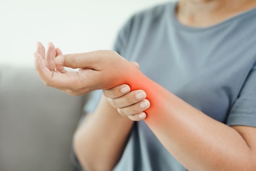
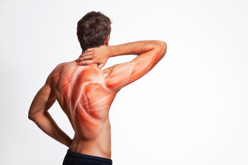
In a technological-driven world, it is common for people to experience wrist and hand pain at some point in their lives. The hands are part of the body’s upper extremities and are used for various tasks and chores throughout the entire day. The forearms provide a causal relationship with the hands and wrists for the upper extremities since they offer very important motor functions to the body. The hands support the body when carrying something; the various muscles, ligaments, tendons, and joints help the wrist with mobility and flexibility. However, when injuries or everyday movements begin to affect the forearms and cause issues with the hands and wrist, it can be difficult to do simple tasks and negatively impact a person’s way of life. Fortunately, numerous ways exist to reduce the pain and discomfort of the wrist and hands. Today’s article focuses on what causes wrist and hand pain, how to prevent wrist and hand pain from returning, and how incorporating various can help reduce the pain-like effects. We discuss with certified medical providers who consolidate our patients’ information to assess the multiple causes that lead to the development of wrist and hand pain. We also inform and guide patients on how various stretches and techniques can help reduce the chances of wrist and hand pain from returning. We also encourage our patients to ask their associated medical providers many intricate and important questions about incorporating these stretches and techniques into their daily routines to live healthier lives. Dr. Jimenez, D.C., includes this information as an academic service. Disclaimer.
What Causes Hand and Wrist Pain?
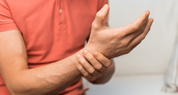
Do you often feel pain or stiffness in your wrist after typing all day on the computer or phone? Do you have trouble gripping items in your hands? Or how often do your hands ache that massaging them causes temporary relief? Many people, including older adults, have experienced pain at some point, and most of the time, it affects the hands and wrists. Since everyone uses their hands and wrists when performing various tasks, when injuries or repetitive movements start to affect the hands and wrists, it can have a huge impact on simple tasks. When dealing with wrist and hand pain, it can make life unbearable for the person. Since pain is a normal protective response to any injuries and potentially harmful stimuli in its acute form, when prolonged or dysfunctional neuromuscular issues start to affect the body, it may contribute to disability and pain. (Merkle et al., 2020) For wrist and hand pain, many occurrences that lead to its development result from micro-stress or repetitive tear usage.
This is because since the world is technological-driven, many people are using computers or smartphones to communicate with each other, which can be one of the causes of the development of wrist and hand pain. When many people frequently use electronic devices, the frequent movements and uses of the thumbs will increase their load and become a higher prevalence of musculoskeletal disorders. (Baabdullah et al., 2020) Other studies stated that when many individuals begin to do repetitive movements constantly and have different positions of their wrist joints while using their electronic devices continually, it can cause pain to their wrist joints and affect the structure. (Amjad et al., 2020) Additionally, when repetitive vibration exposures or forceful angular motions affect the hands and wrists, it can lead to carpal tunnel syndrome and affect the hands. (Osiak et al., 2022) The various joints, tendons, and muscles also become affected in the hands and wrist as trigger points in the forearm. Fortunately, there are multiple ways that many people can reduce the pain-like effects of wrist and hand pain.
The Benefits of Stretching-Video
How To Prevent Wrist & Hand Pain From Returning
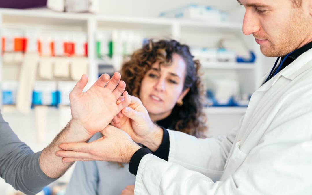
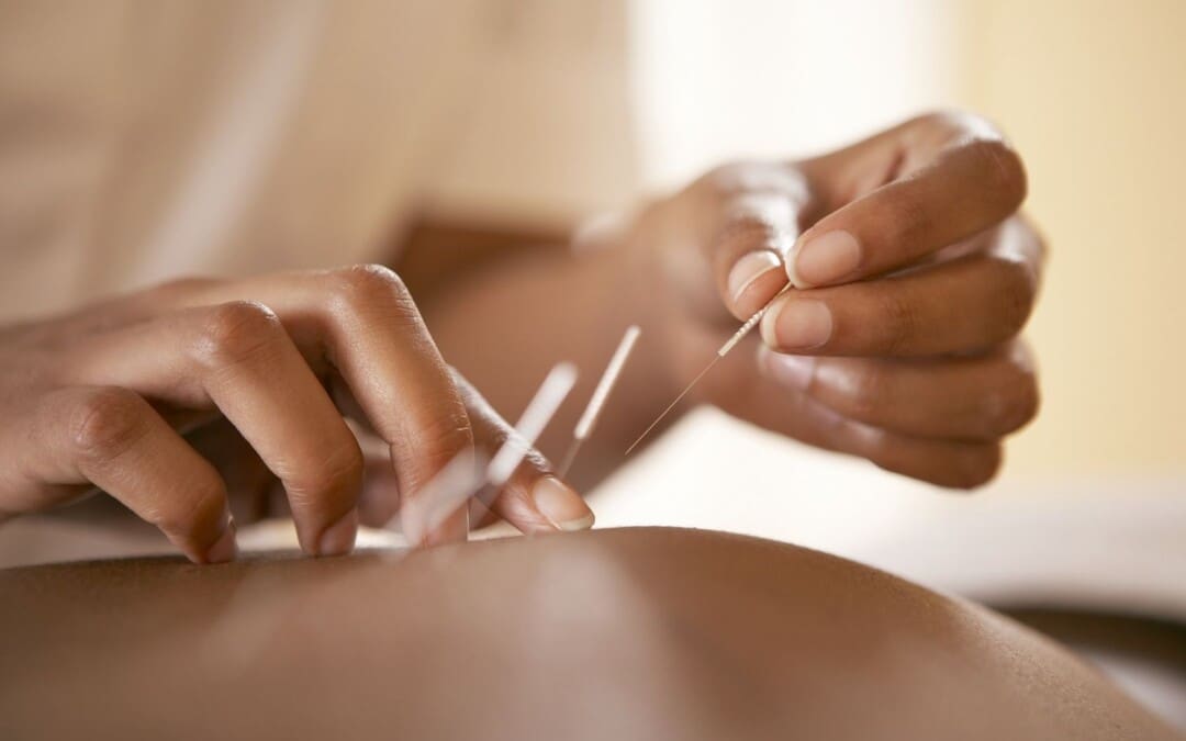
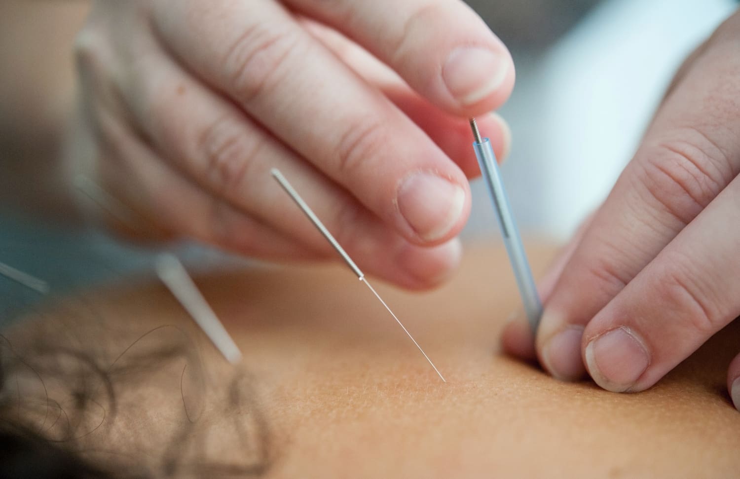
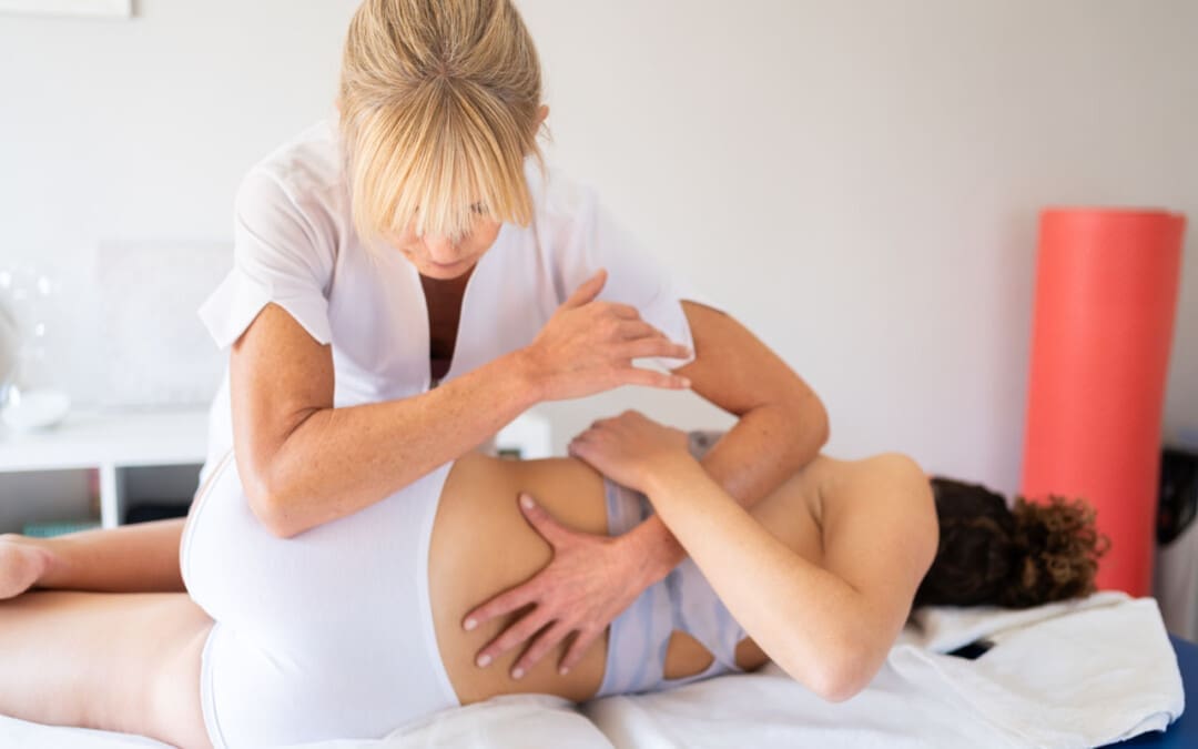
There are numerous ways to reduce wrist and hand pain, and many people try to find therapeutic solutions to mitigate the pain. Non-surgical treatments like manual therapy can help with wrist and hand pain by using mobilization forces to allow wrist flexion and extension to improve motor function. (Gutierrez-Espinoza et al., 2022) Another non-surgical treatment that can help with wrist and hand pain is acupuncture. Acupuncture utilizes small, solid, thin needles to be placed in various acupoints in the forearm to reduce the pain intensity and bring back the mobility function to the hands and wrist. (Trinh et al., 2022)
Various Stretches For Wrist & Hand Pain
Fortunately, there’s a simple and accessible way for many individuals to reduce the effects of wrist and hand pain-stretching and incorporating yoga into their routine. Yoga stretches for the hands and wrists can help decompress and reduce stiffness, and these stretches can be done for just a few minutes, providing beneficial results. (Gandolfi et al., 2023) Below are some of these stretches that can be easily incorporated into anyone’s routine, making it easier for you to take control of your wrist and hand health.
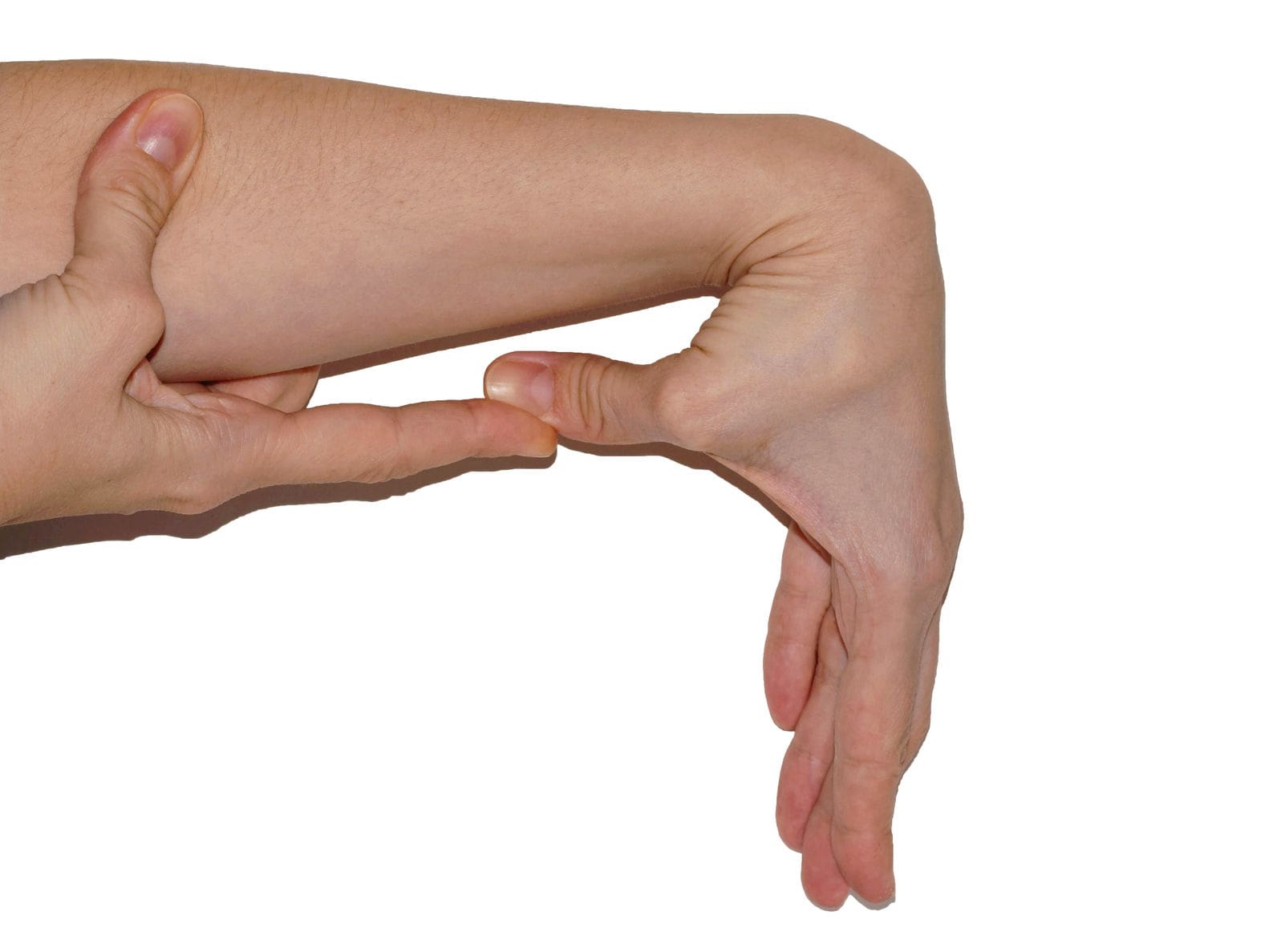
Wrist Flexor Stretch
How to Do It:
Extend your arm in front of you with your palm up.
Use your other hand to gently pull the fingers back toward the body until you feel a stretch in your forearm.
Hold this position for about 15 to 30 seconds.
Repeat 2-3 times with each wrist.
Wrist Extensor Stretch
How to Do It:
Extend your arm in front of your body with your palm facing down.
Gently pull the fingers towards your body with your other hand until you feel a stretch on the outside of your forearm.
Hold for 15 to 30 seconds.
Do this 2-3 times per wrist.
Prayer Stretch
How to Do It:
Put the palms together in a prayer position in front of the chest, below the chin.
Slowly lower the conjoined hands towards the waistline, keeping the hands close to your stomach and your palms together until you feel a stretch under your forearms.
Hold for at least 30 seconds and repeat a few times.
Tendon Glides
How to Do It:
Start with your fingers extended straight out.
Then, bend your fingers to form a hook fist; you should feel a stretch but no pain.
Return to the starting position and bend your fingers to touch the top of your palm, keeping your fingers straight.
Finally, bend your fingers into a full fist.
Repeat the sequence ten times.
Thumb Stretch
How to Do It:
Extend your hand with your fingers together.
Pull your thumb away from your fingers as far as comfortable.
Hold for 15 to 30 seconds.
Repeat 2-3 times with each thumb.
Shake It Out
How to Do It:
After stretching, shake your hands lightly as if trying to dry them off. This helps reduce tension and promote circulation.
References
Amjad, F., Farooq, M. N., Batool, R., & Irshad, A. (2020). Frequency of wrist pain and its associated risk factors in students using mobile phones. Pak J Med Sci, 36(4), 746-749. https://doi.org/10.12669/pjms.36.4.1797
Baabdullah, A., Bokhary, D., Kabli, Y., Saggaf, O., Daiwali, M., & Hamdi, A. (2020). The association between smartphone addiction and thumb/wrist pain: A cross-sectional study. Medicine (Baltimore), 99(10), e19124. https://doi.org/10.1097/MD.0000000000019124
Gandolfi, M. G., Zamparini, F., Spinelli, A., & Prati, C. (2023). Asana for Neck, Shoulders, and Wrists to Prevent Musculoskeletal Disorders among Dental Professionals: In-Office Yoga Protocol. J Funct Morphol Kinesiol, 8(1). https://doi.org/10.3390/jfmk8010026
Gutierrez-Espinoza, H., Araya-Quintanilla, F., Olguin-Huerta, C., Valenzuela-Fuenzalida, J., Gutierrez-Monclus, R., & Moncada-Ramirez, V. (2022). Effectiveness of manual therapy in patients with distal radius fracture: a systematic review and meta-analysis. J Man Manip Ther, 30(1), 33-45. https://doi.org/10.1080/10669817.2021.1992090
Merkle, S. L., Sluka, K. A., & Frey-Law, L. A. (2020). The interaction between pain and movement. J Hand Ther, 33(1), 60-66. https://doi.org/10.1016/j.jht.2018.05.001
Osiak, K., Elnazir, P., Walocha, J. A., & Pasternak, A. (2022). Carpal tunnel syndrome: state-of-the-art review. Folia Morphol (Warsz), 81(4), 851-862. https://doi.org/10.5603/FM.a2021.0121
Trinh, K., Zhou, F., Belski, N., Deng, J., & Wong, C. Y. (2022). The Effect of Acupuncture on Hand and Wrist Pain Intensity, Functional Status, and Quality of Life in Adults: A Systematic Review. Med Acupunct, 34(1), 34-48. https://doi.org/10.1089/acu.2021.0046
Individuals suffering from a jammed finger: Can knowing the signs and symptoms of a finger that is not broken or dislocated allow for at-home treatment and when to see a healthcare provider?
Jammed Finger Injury
A jammed finger, also known as a sprained finger, is a common injury when the tip of a finger is forcefully pushed toward the hand, causing the joint to become compressed. This can cause pain and swelling in one or more fingers or finger joints and cause ligaments to stretch, sprain, or tear. (American Society for Surgery of the Hand. 2015) A jammed finger can often heal with icing, resting, and taping. This is often enough to allow it to heal in a week or two if no fractures or dislocations are present. (Carruthers, K. H. et al., 2016) While painful, it should be able to move. However, if the finger cannot wiggle, it may be broken or dislocated and require X-rays, as a broken finger or joint dislocation can take months to heal.
Treatment
Treatment consists of icing, testing, taping, resting, seeing a chiropractor or osteopath, and progressive regular use to regain strength and ability.
Ice
The first step is icing the injury and keeping it elevated.
Use an ice pack or a bag of frozen vegetables wrapped in a towel.
Ice the finger in 15-minute intervals.
Take the ice off and wait until the finger returns to its normal temperature before re-icing.
Do not ice a jammed finger for over three 15-minute intervals in one hour.
Try To Move The Affected Finger
If the jammed finger does not move easily or the pain gets worse when trying to move it, you need to see a healthcare provider and have an X-ray to check for a bone fracture or dislocation. (American Society for Surgery of the Hand. 2015)
Try to move the finger slightly after swelling, and the pain subsides.
If the injury is mild, the finger should move with little discomfort for a short time.
Tape and Rest
If the jammed finger is not broken or dislocated, it can be taped to the finger next to it to keep it from moving, known as buddy taping. (Won S. H. et al., 2014)
Medical-grade tape and gauze between the fingers should be used to prevent blisters and moisture while healing.
A healthcare provider may suggest a finger splint to keep the jammed finger lined up with the other fingers.
A splint can also help prevent a jammed finger from re-injury.
Resting and Healing
A jammed finger must be kept still to heal at first, but eventually, it needs to move and flex to build strength and flexibility.
Targeted physical therapy exercises can be helpful for recovery.
A primary care provider might be able to refer a physical therapist to ensure the finger has a healthy range of motion and circulation as it heals.
A chiropractor or osteopath can also provide recommendations for helping rehabilitate the finger, hand, and arm to normal function.
Easing The Finger Back to Normal
Depending on the extent of the injury, the finger and hand can be sore and swollen for a few days or weeks.
It can take some time to start feeling normal.
Once the healing process begins, individuals will want to return to using it normally.
Avoiding using a jammed finger will cause it to lose strength, which can, over time, further weaken it and increase the risk of re-injury.
If the pain and swelling persist, see a healthcare provider to get it checked for a possible fracture, dislocation, or other complication as soon as possible, as these injuries are harder to treat if the individual waits too long. (University of Utah Health, 2021)
At Injury Medical Chiropractic and Functional Medicine Clinic, we passionately focus on treating patients’ injuries and chronic pain syndromes and improving ability through flexibility, mobility, and agility programs tailored to the individual. Our providers use an integrated approach to create personalized care plans that include Functional Medicine, Acupuncture, Electro-Acupuncture, and Sports Medicine protocols. Our goal is to relieve pain naturally by restoring health and function to the body. If the individual needs other treatment, they will be referred to a clinic or physician best suited for them. Dr. Jimenez has teamed up with the top surgeons, clinical specialists, medical researchers, and premier rehabilitation providers to provide the most effective clinical treatments.
Treatment for Carpal Tunnel Syndrome
References
American Society for Surgery of the Hand. (2015). Jammed finger. https://www.assh.org/handcare/condition/jammed-finger
Carruthers, K. H., Skie, M., & Jain, M. (2016). Jam Injuries of the Finger: Diagnosis and Management of Injuries to the Interphalangeal Joints Across Multiple Sports and Levels of Experience. Sports health, 8(5), 469–478. https://doi.org/10.1177/1941738116658643
Won, S. H., Lee, S., Chung, C. Y., Lee, K. M., Sung, K. H., Kim, T. G., Choi, Y., Lee, S. H., Kwon, D. G., Ha, J. H., Lee, S. Y., & Park, M. S. (2014). Buddy taping: is it a safe method for treatment of finger and toe injuries?. Clinics in orthopedic surgery, 6(1), 26–31. https://doi.org/10.4055/cios.2014.6.1.26
University of Utah Health. (2021). University of Utah Health. Should I worry about a jammed finger? University of Utah Health. https://healthcare.utah.edu/the-scope/all/2021/03/should-i-worry-about-jammed-finger
Can individuals with Ehlers-Danlos syndrome find relief through various non-surgical treatments to reduce joint instability?
Introduction
The joints and ligaments surrounding the musculoskeletal system allow the upper and lower extremities to stabilize the body and be mobile. The various muscles and soft connective tissues that surround the joints help protect them from injuries. When environmental factors or disorders start to affect the body, many people develop issues that cause overlapping risk profiles, which then affect the stability of the joints. One of the disorders that affect the joints and connective tissue is EDS or Ehlers-Danlos syndrome. This connective tissue disorder can cause the joints in the body to be hypermobile. It can cause joint instability in the upper and lower extremities, thus leaving the individual to be in constant pain. Today’s article focuses on Ehlers-Danlos syndrome and its symptoms and how there are non-surgical ways to manage this connective tissue disorder. We discuss with certified medical providers who consolidate our patients’ information to assess how Ehlers-Danlos syndrome can correlate with other musculoskeletal disorders. We also inform and guide patients on how various non-surgical treatments can help reduce pain-like symptoms and manage Ehlers-Danlos syndrome. We also encourage our patients to ask their associated medical providers many intricate and important questions about incorporating various non-surgical therapies as part of their daily routine to manage the effects of Ehlers-Danlos syndrome. Dr. Jimenez, D.C., includes this information as an academic service. Disclaimer.
What Is Ehlers-Danlos Syndrome?
Do you often feel extremely tired throughout the day, even after a full night of sleep? Do you bruise easily and wonder where these bruises are coming from? Or have you noticed that you have an increased range in your joints? Many of these issues are often correlated with a disorder known as Ehlers-Danlos syndrome or EDS that affects their joints and connective tissue. EDS affects the connective tissues in the body. The connective tissues in the body help provide strength and elasticity to the skin, joints, as well as blood vessel walls, so when a person is dealing with EDS, it can cause a significant disruption to the musculoskeletal system. EDS is largely diagnosed clinically, and many doctors have identified that the gene coding of the collagen and proteins that interact in the body can help determine what type of EDS affects the individual. (Miklovic & Sieg, 2024)
The Symptoms
When understanding EDS, it is essential to know the complexities of this connective tissue disorder. EDS is classified into numerous types with distinct features and challenges that vary depending on the severity. One of the most common types of EDS is hypermobile Ehlers-Danlos syndrome. This type of EDS is characterized by general joint hypermobility, joint instability, and pain. Some of the symptoms that are associated with hypermobile EDS include subluxation, dislocations, and soft tissue injuries that are common and may occur spontaneously or with minimal trauma. (Hakim, 1993) This can often cause acute pain to the joints in the upper and lower extremities. With its broad range of symptoms and the personal nature of the condition itself, many often don’t realize that joint hypermobility is common in the general population and may present no complications that indicate that it is a connective tissue disorder. (Gensemer et al., 2021) Additionally, hypermobile EDS can lead to spinal deformity due to the hyperextensibility of the skin, joints, and various tissue fragility. The pathophysiology of spinal deformity associated with hypermobile EDS is primarily due to muscle hypotonia and ligament laxity. (Uehara et al., 2023) This causes many people to reduce their quality of life and daily living activities significantly. However, there are ways to manage EDS and its correlating symptoms to reduce joint instability.
Movement Medicine: Chiropractic Care-Video
Ways To Manage EDS
When it comes to looking for ways to manage EDS to reduce pain and joint instability, non-surgical treatments can help address the physical and emotional aspects of the condition. Non-surgical treatments for individuals with EDS commonly focus on optimizing the body’s physical function while improving muscular strength and joint stabilization. (Buryk-Iggers et al., 2022) Many individuals with EDS will try to incorporate pain management techniques and physical therapy anduse braces and assistive devices to reduce the effects of EDS and improve their quality of life.
Non-surgical Treatments For EDS
Various non-surgical treatments like MET (muscle energy technique), electrotherapy, light physical therapy, chiropractic care, and massages can help strengthen while toning the surrounding muscles around the joints, provide sufficient pain relief, and limit long-term dependence on medications. (Broida et al., 2021) Additionally, individuals dealing with EDS aim to strengthen the affected muscles, stabilize the joints, and improve proprioception. Non-surgical treatments allow the individual to have a customized treatment plan for the severity of EDS symptoms and help reduce the pain associated with the condition. Many individuals, when going through their treatment plan consecutively to manage their EDS and reduce the pain-like symptoms, will notice improvement in symptomatic discomfort. (Khokhar et al., 2023) This means that non-surgical treatments allow individuals to be more mindful of their bodies and reduce the pain-like effects of EDS, thus allowing many individuals with EDS to lead fuller, more comfortable lives without feeling pain and discomfort.
References
Broida, S. E., Sweeney, A. P., Gottschalk, M. B., & Wagner, E. R. (2021). Management of shoulder instability in hypermobility-type Ehlers-Danlos syndrome. JSES Rev Rep Tech, 1(3), 155-164. https://doi.org/10.1016/j.xrrt.2021.03.002
Buryk-Iggers, S., Mittal, N., Santa Mina, D., Adams, S. C., Englesakis, M., Rachinsky, M., Lopez-Hernandez, L., Hussey, L., McGillis, L., McLean, L., Laflamme, C., Rozenberg, D., & Clarke, H. (2022). Exercise and Rehabilitation in People With Ehlers-Danlos Syndrome: A Systematic Review. Arch Rehabil Res Clin Transl, 4(2), 100189. https://doi.org/10.1016/j.arrct.2022.100189
Gensemer, C., Burks, R., Kautz, S., Judge, D. P., Lavallee, M., & Norris, R. A. (2021). Hypermobile Ehlers-Danlos syndromes: Complex phenotypes, challenging diagnoses, and poorly understood causes. Dev Dyn, 250(3), 318-344. https://doi.org/10.1002/dvdy.220
Hakim, A. (1993). Hypermobile Ehlers-Danlos Syndrome. In M. P. Adam, J. Feldman, G. M. Mirzaa, R. A. Pagon, S. E. Wallace, L. J. H. Bean, K. W. Gripp, & A. Amemiya (Eds.), GeneReviews((R)). https://www.ncbi.nlm.nih.gov/pubmed/20301456
Khokhar, D., Powers, B., Yamani, M., & Edwards, M. A. (2023). The Benefits of Osteopathic Manipulative Treatment on a Patient With Ehlers-Danlos Syndrome. Cureus, 15(5), e38698. https://doi.org/10.7759/cureus.38698
Can understanding the body’s hinge joints and how they operate help with mobility and flexibility problems and manage conditions for individuals with difficulty fully bending or extending their fingers, toes, elbows, ankles, or knees?
Hinge Joints
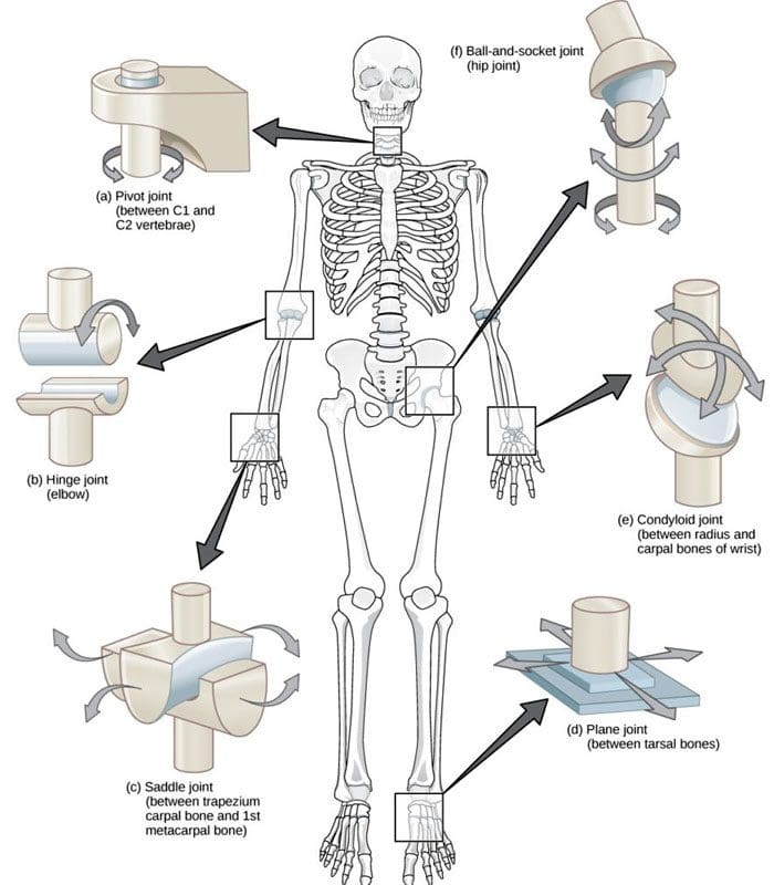
A joint forms where one bone connects to another, allowing motion. Different types of joints differ in structure and movement depending on their location. These include hinge, ball and socket, planar, pivot, saddle, and ellipsoid joints. (Boundless. General Biology, N.D.) Hinge joints are synovial joints that move through one plane of motion: flexion and extension. Hinge joints are found in the fingers, elbows, knees, ankles, and toes and control movement for various functions. Injuries, osteoarthritis, and autoimmune conditions can affect hinge joints. Rest, medication, ice, and physical therapy can help alleviate pain, improve strength and range of motion, and help manage conditions.
Anatomy
A joint is formed by the joining of two or more bones. The human body has three main classifications of joints, categorized by the degree to which they can move. These include: (Boundless. General Biology, N.D.)
Synarthroses
These are fixed, immovable joints.
Formed by two or more bones.
Amphiarthroses
Also known as cartilaginous joints.
A fibrocartilage disc separates the bones that form the joints.
These movable joints allow for a slight degree of movement.
Diarthroses
Also known as synovial joints.
These are the most common freely mobile joints that allow movement in multiple directions.
The bones that form the joints are lined with articular cartilage and enclosed in a joint capsule filled with synovial fluid that allows for smooth motion.
Synovial joints are classified into different types depending on differences in structure and the number of motion planes they allow. A hinge joint is a synovial joint that allows movement in one plane of motion, similar to a door hinge that moves forward and backward. Within the joint, the end of one bone is typically convex/pointed outward, with the other concave/rounded inward to allow the ends to fit smoothly. Because hinge joints only move through one plane of movement, they tend to be more stable than other synovial joints. (Boundless. General Biology, N.D.) Hinge joints include:
The finger and toe joints – allow the fingers and toes to bend and extend.
The elbow joint – allows the elbow to bend and extend.
The knee joint – allows the knee to bend and extend.
The talocrural joint of the ankle – allows the ankle to move up/dorsiflexion and down/plantarflexion.
Hinge joints allow the limbs, fingers, and toes to extend away and bend toward the body. This movement is essential for activities of daily living, such as showering, getting dressed, eating, walking, standing up, and sitting down.
Conditions
Osteoarthritis and inflammatory forms of arthritis can affect any joint (Arthritis Foundation. N.D.) Autoimmune inflammatory forms of arthritis, including rheumatoid and psoriatic arthritis, can cause the body to attack its own joints. These commonly affect the knees and fingers, resulting in swelling, stiffness, and pain. (Kamata, M., Tada, Y. 2020) Gout is an inflammatory form of arthritis that develops from elevated levels of uric acid in the blood and most commonly affects the hinge joint of the big toe. Other conditions that affect hinge joints include:
Injuries to the cartilage within the joints or ligaments that stabilize the outside of the joints.
Ligament sprains or tears can result from jammed fingers or toes, rolled ankles, twisting injuries, and direct impact on the knee.
These injuries can also affect the meniscus, the tough cartilage within the knee joint that helps cushion and absorb shock.
Rehabilitation
Conditions that affect hinge joints often cause inflammation and swelling, resulting in pain and limited mobility.
After an injury or during an inflammatory condition flare-up, limiting active movement and resting the affected joint can reduce increased stress and pain.
Applying ice can decrease inflammation and swelling.
Once the pain and swelling start to subside, physical and/or occupational therapy can help rehabilitate the affected areas.
A therapist will provide stretches and exercises to help improve the joint range of motion and strengthen the supporting muscles.
For individuals experiencing hinge joint pain from an autoimmune condition, biologic medications to decrease the body’s autoimmune activity are administered through infusions delivered every several weeks or months. (Kamata, M., Tada, Y. 2020)
Cortisone injections may also be used to decrease inflammation.
At Injury Medical Chiropractic and Functional Medicine Clinic, we passionately focus on treating patients’ injuries and chronic pain syndromes and improving ability through flexibility, mobility, and agility programs tailored to the individual. Our providers use an integrated approach to create personalized care plans that include Functional Medicine, Acupuncture, Electro-Acupuncture, and Sports Medicine protocols. Our goal is to relieve pain naturally by restoring health and function to the body. If the individual needs other treatment, they will be referred to a clinic or physician best suited for them. Dr. Jimenez has teamed up with the top surgeons, clinical specialists, medical researchers, and premier rehabilitation providers to provide the most effective clinical treatments.
Chiropractic Solutions
References
Boundless. General Biology. (N.D.). 38.12: Joints and Skeletal Movement – Types of Synovial Joints. In. LibreTexts Biology. https://bio.libretexts.org/Bookshelves/Introductory_and_General_Biology/Book%3A_General_Biology_%28Boundless%29/38%3A_The_Musculoskeletal_System/38.12%3A_Joints_and_Skeletal_Movement_-_Types_of_Synovial_Joints
Kamata, M., & Tada, Y. (2020). Efficacy and Safety of Biologics for Psoriasis and Psoriatic Arthritis and Their Impact on Comorbidities: A Literature Review. International journal of molecular sciences, 21(5), 1690. https://doi.org/10.3390/ijms21051690
Can individuals dealing with muscle pain find relief from acupuncture therapy to get back to their daily activities and well-being?
Introduction
Many people worldwide have dealt with pain in their musculoskeletal system that has affected their daily routine. Some of the most common factors that people have experienced muscle pain include sedentary lifestyles from working at a desk job or physical demands from an active lifestyle. The muscles, tendons, ligaments, and soft tissues can become overstretched and overworked, causing the muscles to become weak. At the same time, visceral somatic issues in the neck, shoulders, and back can affect the upper and lower extremities, leading to a life of disability. Many factors that can contribute to the development of muscle pain can impact a person’s routine and cause them to find various techniques to reduce the muscle pain in their bodies. Since muscle pain can be in acute or chronic form, many individuals who are seeking treatment for their ailments can look into non-surgical therapies like acupuncture to not only reduce muscle pain but also find the relief they are looking for. Today’s article focuses on how muscle pain can affect a person’s well-being, how the essence of acupuncture can be beneficial for muscle pain, and how people can integrate acupuncture therapy as part of a wellness routine. We talk with certified medical providers who consolidate our patients’ information to assess how muscle pain can impact a person’s well-being. We also inform and guide patients on how acupuncture therapy can benefit the body by reducing the effects of muscle pain. We encourage our patients to ask their associated medical providers intricate and important questions about incorporating acupuncture therapy into a wellness routine to reduce muscle pain and its referred symptoms. Dr. Jimenez, D.C., includes this information as an academic service. Disclaimer.
How Muscle Pain Can Affect A Person’s Well-Being
Do you feel the effects of tiredness and weakness in your upper and lower extremity muscles? Have you experienced general soreness or aches in your neck, shoulders, or back? Or does twisting and turning your body cause temporary relief to your body, only for it to be worse throughout the day? When it comes to muscle pain can be a multi-factorial condition where that can involve complex interactions on a person’s structure, physical, social, lifestyle, and comorbid health factors that can come into play as contributing factors for people to experience long-term pain and disability. (Caneiro et al., 2021) As many individuals start to do repetitive motions or stay in sedentary positions, muscle pain can develop when they stretch or try to move their muscles while doing their routine. The burden of muscle pain often correlates with socioeconomic factors that can cause many people, both young and old, to substantially limit their mobility and engagement in their routine, which predisposes increased risk factors to other chronic conditions they may have. (Dzakpasu et al., 2021)
When many individuals are dealing with muscle pain in its acute or chronic form, many often don’t realize that when the affected muscles in the upper and lower body quadrants are coping with pain, there is associated pain and stiffness from how active or inactive the muscles are can affect the soft tissue causing high mechanical stress to the affect the skeletal joints. (Wilke & Behringer, 2021) When this happens, many people will start to experience referred muscle pain in their bodies, causing issues with their mobility, flexibility, and stability. Coincidentally, muscle pain can also be a symptom of many people who have various pains in their bodies that have impacted their lives prior; seeking treatment can reduce the effects of muscle pain and help them take back their routine to lead a healthier lifestyle.
Movement Medicine- Video
The Essence Of Acupuncture For Muscle Pain
When many people are dealing with muscle pain, they are seeking treatments that are not only affordable but also can be effective in reducing the overlapping risk profiles that are affecting the body, causing muscle pain. Many treatments like chiropractic care, decompression, and massage therapy are non-surgical and are effective through consecutive sessions. One of the oldest and most effective treatments that can help reduce muscle pain in the body is acupuncture therapy. Acupuncture is a holistic treatment derived from Traditional Chinese Medicine that utilizes small, solid, thin needles inserted by professional acupuncturists to various acupoints. The main philosophy is that acupuncture provides relief to the body as it helps improve the body’s energy flow while maintaining a person’s overall health and vitality. (Zhang et al., 2022) When a person is dealing with muscle pain, the muscle fibers can develop tiny nodules known as trigger points that can induce pain in the affected muscle quadrants. With acupuncture needles placed in the affected area, local and referred pain is reduced, muscle blood flow and oxygen are returned to the body, and the muscle’s range of motion is improved. (Pourahmadi et al., 2019) Some of the benefits that acupuncture therapy provides include:
Increased circulation
Inflammation reduction
Endorphin release
Relaxing muscle tension
Integrating Acupuncture As Part Of A Wellness Routine
Many individuals who are seeking acupuncture therapy as part of their wellness journey can see the positive benefits of acupuncture and can combine it with other therapies to reduce the chances of muscle pain from returning. While acupuncture can help stimulate the nerves and restore motor function, treatments like joint mobilization can help stretch the affected muscles and joints to improve the body’s range of motion. (Lee et al., 2023) With many individuals seeking acupuncture treatment to reduce muscle pain, many can make small changes in their routine to prevent the pain from causing overlapping risk profiles to their bodies. When addressing the root causes of pain and promoting the body’s innate healing abilities, acupuncture can help restore balance, alleviate discomfort, and enhance overall well-being.
References
Caneiro, J. P., Bunzli, S., & O’Sullivan, P. (2021). Beliefs about the body and pain: the critical role in musculoskeletal pain management. Braz J Phys Ther, 25(1), 17-29. https://doi.org/10.1016/j.bjpt.2020.06.003
Dzakpasu, F. Q. S., Carver, A., Brakenridge, C. J., Cicuttini, F., Urquhart, D. M., Owen, N., & Dunstan, D. W. (2021). Musculoskeletal pain and sedentary behaviour in occupational and non-occupational settings: a systematic review with meta-analysis. Int J Behav Nutr Phys Act, 18(1), 159. https://doi.org/10.1186/s12966-021-01191-y
Lee, J. E., Akimoto, T., Chang, J., & Lee, H. S. (2023). Effects of joint mobilization combined with acupuncture on pain, physical function, and depression in stroke patients with chronic neuropathic pain: A randomized controlled trial. PLOS ONE, 18(8), e0281968. https://doi.org/10.1371/journal.pone.0281968
Pourahmadi, M., Mohseni-Bandpei, M. A., Keshtkar, A., Koes, B. W., Fernandez-de-Las-Penas, C., Dommerholt, J., & Bahramian, M. (2019). Effectiveness of dry needling for improving pain and disability in adults with tension-type, cervicogenic, or migraine headaches: protocol for a systematic review. Chiropr Man Therap, 27, 43. https://doi.org/10.1186/s12998-019-0266-7
Wilke, J., & Behringer, M. (2021). Is “Delayed Onset Muscle Soreness” a False Friend? The Potential Implication of the Fascial Connective Tissue in Post-Exercise Discomfort. Int J Mol Sci, 22(17). https://doi.org/10.3390/ijms22179482
Zhang, B., Shi, H., Cao, S., Xie, L., Ren, P., Wang, J., & Shi, B. (2022). Revealing the magic of acupuncture based on biological mechanisms: A literature review. Biosci Trends, 16(1), 73-90. https://doi.org/10.5582/bst.2022.01039
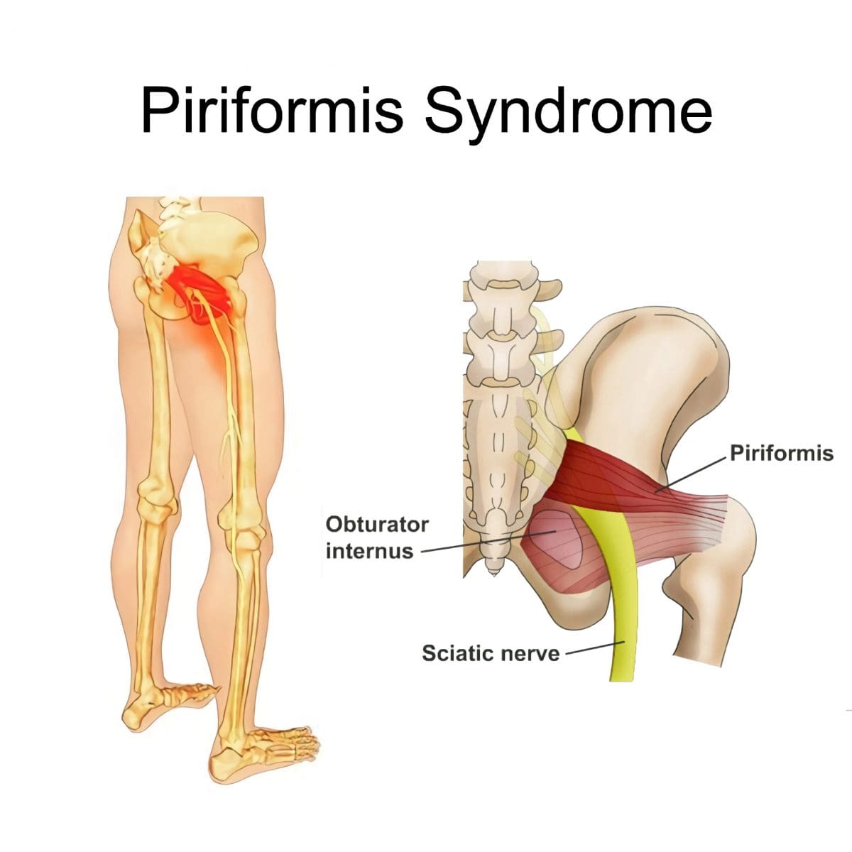
Can physical therapy treatment protocols aimed at improving range of motion and flexibility around the hip and relieving inflammation around the sciatic nerve help individuals experiencing deep buttock pain or piriformis syndrome?
Deep Buttock Pain
Piriformis syndrome, a.k .a. deep buttock pain, is described as sciatic nerve irritation from the piriformis muscle.
The piriformis is a small muscle behind the hip joint in the buttocks.
It is about one centimeter in diameter and functions in the hip joint’s external rotation or turning outward.
The piriformis muscle and tendon are close to the sciatic nerve, which supplies the lower extremities with motor and sensory functions.
Depending on an individual’s anatomic variation of the muscle and tendon:
The two cross over, under, or through each other behind the hip joint in the deep buttock.
This relationship is thought to irritate the nerve, leading to sciatica symptoms.
Piriformis Syndrome
When diagnosed with piriformis syndrome, it is thought that the muscle and tendon bind to and/or spasm around the nerve, causing irritation and pain symptoms.
The theory supported is that when the piriformis muscle and its tendon tighten, the sciatic nerve becomes compressed or pinched. This decreases blood circulation and irritates the nerve from the pressure. (Shane P. Cass 2015)
Tenderness with pressure on the piriformis muscle.
Discomfort in the back of the thigh.
Deep buttock pain behind the hip.
Electric sensations, shocks, and pains travel down the back of the lower extremity.
Numbness in the lower extremity.
Some individuals develop symptoms abruptly, while others go through a gradual increase.
Diagnosis
Doctors will order X-rays, MRIs, and nerve conduction studies, which is normal.
Because piriformis syndrome can be challenging to diagnose, some individuals with minor hip pain may receive a piriformis syndrome diagnosis even if they don’t have the condition. (Shane P. Cass 2015)
It is sometimes referred to as deep buttock pain. Other causes of this type of pain include back and spinal problems like:
Herniated discs
Spinal stenosis
Radiculopathy – sciatica
Hip bursitis
A piriformis syndrome diagnosis is usually given when these other causes are eliminated.
When the diagnosis is uncertain, an injection is administered in the area of the piriformis muscle. (Danilo Jankovic et al., 2013)
Different medications can be used, but the injection itself is used to help determine the specific location of the discomfort.
When an injection is given into the piriformis muscle or tendon, it is often administered by ultrasound guidance to ensure the needle delivers the medication to the correct location. (Elizabeth A. Bardowski, J. W. Thomas Byrd 2019)
Avoiding activities that cause symptoms for at least a few weeks.
Physical Therapy
Emphasize stretching and strengthening the hip rotator muscles.
Non-Surgical Decompression
Gently pulls the spine to release any compression, allowing optimal rehydration and circulation and taking the pressure off the sciatic nerve.
Therapeutic Massage Techniques
To relax and release muscle tension and increase circulation.
Acupuncture
To help relax the piriformis muscle, sciatic nerve, and surrounding area.
Relieve pain.
Chiropractic Adjustments
Realignment rebalances the spine and musculoskeletal system to alleviate pain.
Anti-Inflammatory Medication
To decrease inflammation around the tendon.
Cortisone Injections
Injections are used to decrease inflammation and swelling.
Botulinum Toxin Injection
Injections of botulinum toxin paralyze the muscle to relieve pain.
Surgery
Surgery can be performed in rare cases to loosen the piriformis tendon, known as a piriformis release. (Shane P. Cass 2015)
Surgery is a last resort when conservative treatments have been tried for at least 6 months with little to no relief.
Recovery can take several months.
Sciatica Causes and Treatment
References
Cass S. P. (2015). Piriformis syndrome: a cause of nondiscogenic sciatica. Current sports medicine reports, 14(1), 41–44. https://doi.org/10.1249/JSR.0000000000000110
Jankovic, D., Peng, P., & van Zundert, A. (2013). Brief review: piriformis syndrome: etiology, diagnosis, and management. Canadian journal of anaesthesia = Journal canadien d’anesthesie, 60(10), 1003–1012. https://doi.org/10.1007/s12630-013-0009-5
Bardowski, E. A., & Byrd, J. W. T. (2019). Piriformis Injection: An Ultrasound-Guided Technique. Arthroscopy techniques, 8(12), e1457–e1461. https://doi.org/10.1016/j.eats.2019.07.033
Knee injuries can present in physically active individuals that lift weights. Can understanding the types of weightlifting knee injuries help in prevention?
Weightlifting Knee Injuries
Weight training is very safe for the knees as regular weight training can improve knee strength and prevent injury as long as the correct form is followed. For Individuals with knee injuries from other activities, incorrect weight-training exercises could worsen the injury. (Ulrika Aasa et al., 2017) As well as, sudden twisting movements, poor alignment, and pre-existing injuries can increase the risk of worsening or creating further injuries. (Hagen Hartmann et al, 2013) The body and the knees are designed to support vertical forces on the joints.
Common Injuries
Weightlifting knee injuries occur as the knee joints endure a wide range of stresses and strains. In weight training, the ligaments that attach to the complex bone system of the knee joint can be damaged by incorrect movements, overloading the weight, and increasing the weight too soon. These injuries can result in pain, swelling, and immobility that can range from minor to severe, from a sprain or a slight tear to a complete tear in serious cases.
Anterior Cruciate Ligament – ACL – Injury
This ligament attaches the thigh’s femur bone to the lower leg’s shin bone/tibia and controls excessive rotation or extension of the knee joint. (American Academy of Family Physicians. 2024)
Anterior means front.
ACL injuries are seen mostly in athletes but can happen to anybody.
Severe damage to the ACL usually means surgical reconstruction and up to 12 months of rehabilitation.
When weightlifting, try to avoid twisting knee movements, intentionally or accidentally, under excessive load.
Posterior Cruciate Ligament – PCL – Injury
The PCL connects the femur and tibia at different points to the ACL.
It controls any backward motion of the tibia at the joint.
Injuries occur most with high-impact forces as a result of accidents and sometimes in activities where forceful trauma to the knee occurs.
Medial Collateral Ligament – MCL – Injury
This ligament maintains the knee from bending too far to the inside/medially.
Injuries mostly occur from impact to the outside of the knee or from accidental bodyweight force on the leg that bends at an unusual angle.
Lateral Collateral Ligament – LCL – Injury
This ligament connects the smaller bone of the lower leg/fibula to the femur.
It is opposite to the MCL.
It maintains excessive outward movement.
LCL injuries occur when a force pushes the knee out.
Cartilage Injury
Cartilage prevents bones from rubbing together and cushions impact forces.
Knee menisci are cartilage that cushions the knee joints inside and outside.
Other types of cartilage protect the thigh and shin bones.
When cartilage gets torn or damaged, surgery may be required.
Tendonitis
Aggravated and overused knee tendons can lead to weightlifting knee injuries.
A related injury known as iliotibial band syndrome/ITB causes pain to the outside of the knee, usually in runners, but it can occur from overuse.
Rest, stretching, physical therapy, and anti-inflammatory medication are a common treatment plan.
The condition causes the cartilage to deteriorate and bones to rub together, resulting in pain and stiffness.
Prevention
Individuals can minimize their risk of weightlifting knee injuries and pain by following their doctor’s and personal trainers’ recommendations.
Individuals with an existing knee injury should follow their doctor’s or physical therapist’s recommendations.
A knee sleeve can keep the muscles and joints secure, providing protection and support.
Stretching the leg and knee muscles can maintain joint flexibility.
Avoid sudden lateral movements.
Possible recommendations can include:
Avoiding Certain Exercises
Isolation exercises like leg curls, standing, or on a bench, as well as using the leg extension machine, can stress the knee.
Deep Squat Training
Research shows that the deep squat can protect against lower leg injury if the knee is healthy. However, this is when done with proper technique, under expert supervision, and with a gradual progressive load. (Hagen Hartmann et al, 2013)
Individuals should talk to their doctor before beginning a new exercise routine. A personal trainer can provide training in learning the proper technique and weightlifting form.
How I Tore my ACL Part 2
References
Aasa, U., Svartholm, I., Andersson, F., & Berglund, L. (2017). Injuries among weightlifters and powerlifters: a systematic review. British journal of sports medicine, 51(4), 211–219. https://doi.org/10.1136/bjsports-2016-096037
Hartmann, H., Wirth, K., & Klusemann, M. (2013). Analysis of the load on the knee joint and vertebral column with changes in squatting depth and weight load. Sports medicine (Auckland, N.Z.), 43(10), 993–1008. https://doi.org/10.1007/s40279-013-0073-6
American Academy of Family Physicians. ACL injury. (2024). ACL injury (Diseases and Conditions, Issue. https://familydoctor.org/condition/acl-injuries/
Mellinger, S., & Neurohr, G. A. (2019). Evidence based treatment options for common knee injuries in runners. Annals of translational medicine, 7(Suppl 7), S249. https://doi.org/10.21037/atm.2019.04.08
Driban, J. B., Hootman, J. M., Sitler, M. R., Harris, K. P., & Cattano, N. M. (2017). Is Participation in Certain Sports Associated With Knee Osteoarthritis? A Systematic Review. Journal of athletic training, 52(6), 497–506. https://doi.org/10.4085/1062-6050-50.2.08
IFM's Find A Practitioner tool is the largest referral network in Functional Medicine, created to help patients locate Functional Medicine practitioners anywhere in the world. IFM Certified Practitioners are listed first in the search results, given their extensive education in Functional Medicine