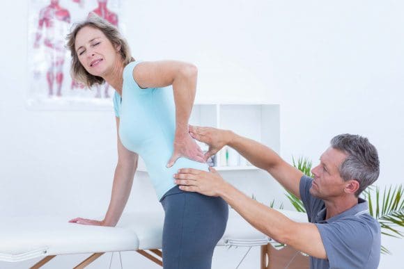
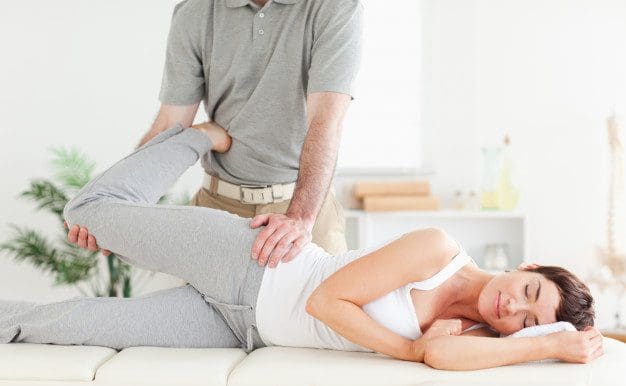
Piriformis Syndrome Treatment: Sandra Rubio defines sciatica, a collection of symptoms caused by the compression of the longest nerve in the body: the sciatic nerve. As further explained, symptoms of sciatica include, low back pain, followed by painful symptoms which radiate down the hips, buttocks, legs and feet. A lot of patients walk-in to Dr. Alex Jimenez’s office with sciatica caused by a variety of health issues. Dr. Alex Jimenez is the non-surgical choice for piriformis syndrome treatment with chiropractic care.
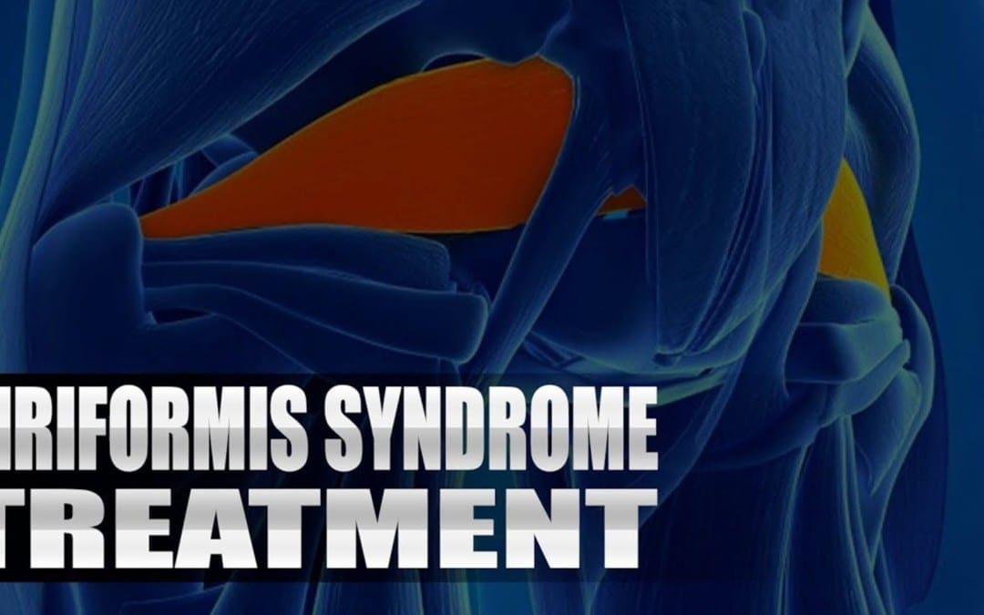
Piriformis Syndrome Treatment
Sciatica may feel like pain similar to an electrical sensation, or sharp. Until it goes off, the cramp can last for weeks. You might have pain, particularly when you move, sneeze, or cough. You might also have a tingling or burning sensation, numbness, pins and needles; weakness down your leg. You are likely to get sciatica between the ages of 50 and 30 years. It may happen as a consequence of the overall wear and tear of aging, and any sudden strain on the discs that cushion the bones (vertebrae) of your lower spine.
We focus on what works for you. We also strive to create fitness and better the body through researched methods and total wellness programs. These programs are natural, and use the body�s own ability to achieve goals of improvement, rather than introducing harmful chemicals, controversial hormone replacement, surgery, or addictive drugs.
We want you to live a life that is fulfilled with more energy, positive attitude, better sleep, less pain, proper body weight and educated on how to maintain this way of life. I have made a life of taking care of each and every one of my patients.
I assure you, I will only accept the best for you�
God Bless You & Your Health�?
If you have enjoyed this video and/or we have helped you in any way please feel free to subscribe and share us.
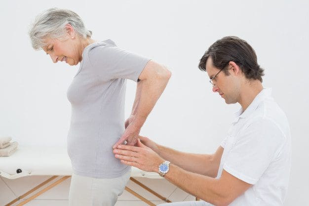
Sciatica Pain: Sandra Rubio discusses sciatica, its causes and its symptoms. Sciatica is the collection of symptoms caused by the compression of the sciatic nerve, the longest nerve in the human body which extends from the lower back to the feet. Sandra Rubio describes how she’s witnessed many patients come into Dr. Alex Jimenez’s office feeling painful and often severe symptoms of sciatica caused by a variety of spinal health issues. Fortunately, Dr. Alex Jimenez is the non surgical choice for the safe and effective treatment of sciatica symptoms.
Sciatica Pain Explained
Based upon how it’s defined, approximately 2 percent to 40 percent of individuals will experience sciatica symptoms at some point in their lifetime. It is most frequent during people’s 40’s and 50’s, and men are more frequently affected than women. About 90 percent of the time, sciatica symptoms are because of a disc herniation. Other issues that may bring about sciatica comprise of spondylolisthesis, spinal stenosis, piriformis syndrome, pelvic tumors, and compression by a baby’s head during pregnancy, among other spinal health issues.
When your body is truly healthy, you will arrive at your optimal fitness level proper physiological fitness state. �We want to help you live a new and improved lifestyle. Over the last 2 decades while researching and testing methods with thousands of patients we have learned what works effectively at decreasing pain while increasing human vitality.
We focus on what works for you. We also strive to create fitness and better the body through researched methods and total wellness programs. These programs are natural, and use the body�s own ability to achieve goals of improvement, rather than introducing harmful chemicals, controversial hormone replacement, surgery, or addictive drugs.
We want you to live a life that is fulfilled with more energy, positive attitude, better sleep, less pain, proper body weight and educated on how to maintain this way of life. I have made a life of taking care of each and every one of my patients.
I assure you, I will only accept the best for you�
If you have enjoyed this video and/or we have helped you in any way please feel free to subscribe and share us.
Experiencing foot pain, there’s no doubt you checked out your foot to make sure it’s not injured or hurting from�improper fitting shoes, corns, plantar fasciitis, etc. This may seem counterintuitive, but you may want to check the condition of the�lumbar spine (lower back)?� Most foot problems are caused from issues with the foot itself, but you might be surprised to find that pressure on the sciatic nerve can cause intense foot pain.
Sciatic Nerve Pain
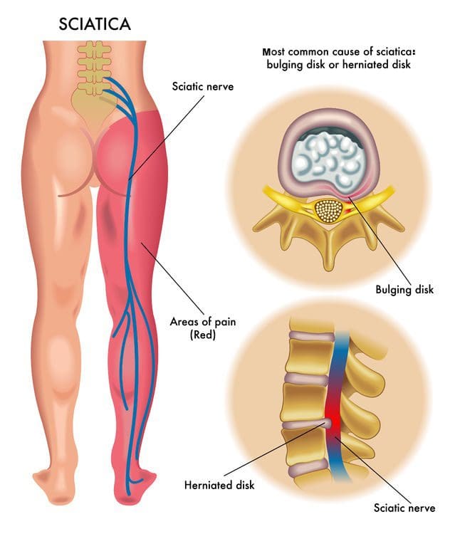
The sciatic nerve is the largest nerve in the body and consists of five nerves that come together at the lower spine and then extend all the way down the back of the legs into the toes. If the lumbar spine is compressed, it presses on the sciatic nerve, thus causing radiating pain down the leg and sometimes all the way into the big toe. Foot pain without leg pain is often due to an issue located within the foot. However, it is possible that the foot pain could be the only symptom of sciatica.
Sciatica can be caused by lumbar spine disc herniation, lumbar spinal stenosis, and spondylolisthesis. There are various types of sciatica, which present differently according to which spinal disc is affected. If the L5 disc is compressed, Foot Drop can occur. This refers to the heavy, weak feeling that makes flexing the foot almost impossible. Foot Drop usually results in pain radiating down along the outside of the leg, crossing over the foot and into the big toe. If the S1 nerve root is affected, the pain is likely on the sole of the foot. An accurate diagnosis is first priority in order to address the pain correctly and properly.
What To Do About The Foot Pain
Addressing the root of the problem is most important. Nearly three million people a year suffer from sciatic pain along with other dysfunctions. An experienced chiropractor or physician will demonstrate exercises to help lengthen and stretch the spine. This along with massage, acupuncture, and medication are all helpful in the management of sciatic pain. The foot pain will be addressed by a doctor or chiropractor who will to tell which treatment is most effective for the situation.
Treatment for foot pain varies depending on the condition/injury. Treatment can go from rest and ice to physical therapy, chiropractic and in severe cases surgery. Reflexology can provide relief, as well as, stretching exercises. Over the counter pain medication is often used. If the pain is too intense that it prevents sleep, a physician may prescribe non-addictive pain medication. Wear shoes with good arch supports, and if pain persists, see a podiatrist for special orthotic shoe inserts. Insurance often covers orthotics.
Further Considerations
Don�t forget that most pain in the body is caused from inflammation and can be helped with anti-inflammatory diet and lifestyle stressors. Concentrate on eating whole, unprocessed foods. Stay away from sugar, alcohol, artificial sweeteners, and white flour. Make sure to drink enough water every day, and get eight hours of sleep. This is one of the most effective ways to address inflammation. Bring the body back into balance.
Sciatica Pain: The sciatic nerve is the largest single nerve found within the human body, running from each side of the lumbar spine, through the area of the lumbar plexus, and trailing down into the buttocks, the back of the thigh and into the foot.
Sciatica is a medical term used to define a group of symptoms rather than a single injury or condition. The most common symptom for sciatica is pain in the lower back and, although low back pain can be the result of numerous lumbar spine injuries or conditions, various other common symptoms associated with sciatica can closely suggest its presence. Often a result of damage or impingement of the sciatic nerve, many people affected with sciatica experience burning and tingling sensations along the back of the thigh, followed by numbness or cramping. People suffering from sciatica may have difficulty going through their regular activities but chiropractic care can help relieve the symptoms and treat many other underlying conditions causing the pain and discomfort.
Sciatica Pain: Surgery Vs. Chiropractic
Chiropractic treatment for mild to severe cases of sciatica most frequently involves chiropractic adjustments and manual manipulations, followed by a specialized series of stretches and exercises accommodated to each individual�s level of injury or condition and its symptoms. Both of these treatments together may speed up the rehabilitation process as well as improve the health of the spine and ultimately reduce the symptoms of sciatica.
Sciatica is used to identify a set of symptoms on the region of the lumbar spine, generally as a result of a previous injury or underlying condition. Regular symptoms of low back pain, stiffness, and burning or tingling sensations could indicate the presence of sciatica. For more information, please feel free to ask Dr. Jimenez or contact us at (915) 850-0900.
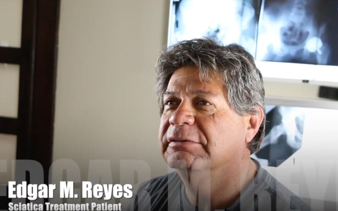
Sciatica Nerve Pain:�Edgar M. Reyes works for the city of El Paso and his ability to properly engage in his occupation is an essential part of his job, however, Mr. Reyes developed sciatica, which affected his everyday performance. Unable to walk due to his sciatica nerve pain, Edgar M Reyes found chiropractic treatment with Dr. Alex Jimenez. Chiropractic care provided Mr. Reyes with the relief he deserved from his sciatica and restored his ability to walk as well as his health and wellness.
Sciatica is a set of symptoms characterized by radiating pain from the lumbar spine. This pain may go down the back, into the buttocks, hips, legs and feet. Onset is frequently sudden following tasks like heavy lifting, though slow onset may also occur. Symptoms may occur on one or both sides of the body. Pain, numbness and weakness can occur depending on the type of compression on the sciatic nerve. About 90% of sciatica cases are often due to a spinal disc herniation pressing on one of the lumbar or sacral nerve roots. Other issues that may cause sciatica include spondylolisthesis, spinal stenosis and piriformis syndrome.
Please Recommend Us: If you have enjoyed this video and/or we have helped you in any way please feel free to recommend us. Thank You.
A herniated disc, also known as a slipped or ruptured disc, is a healthcare condition which occurs when a tear in the outer, fibrous ring of an intervertebral disc causes its soft, central portion to bulge out from the damaged, surrounding cartilage. Disc herniations are generally due to the degeneration of the outer ring of an intervertebral disc, known as the anulus fibrosus. Trauma, lifting injuries or straining may also cause a herniated disc. A tear in the intervertebral disc may result in the release of chemicals which may cause irritation and ultimately become the direct cause of severe back pain, even without nerve root compression.
Disc herniations also commonly develop following a previously existing disc protrusion, a healthcare condition in which the outermost layers of the anulus fibrosus remain intact, however, these can bulge if the disc is placed under pressure. Unlike a disc herniation, none of the gel-like section escapes the intervertebral disc. Herniated discs often heal on their own within several weeks. Severe disc herniations may require surgery, however, a variety of research studies have demonstrated that nonoperative treatment may help improve and manage the recovery process of a herniated disc without the need for surgical interventions.
Surgical vs Nonoperative Treatment for Lumbar Disk Herniation Using The Spine Patient Outcomes Research Trial (SPORT): A Randomized Trial
Abstract
Context: Lumbar diskectomy is the most common surgical procedure performed for back and leg symptoms in US patients, but the efficacy of the procedure relative to nonoperative care remains controversial.
Objective: To assess the efficacy of surgery for lumbar intervertebral disk herniation.
Design, Setting, and Patients: The Spine Patient Outcomes Research Trial, a randomized clinical trial enrolling patients between March 2000 and November 2004 from 13 multidisciplinary spine clinics in 11 US states. Patients were 501 surgical candidates (mean age, 42 years; 42% women) with imaging-confirmed lumbar intervertebral disk herniation and persistent signs and symptoms of radiculopathy for at least 6 weeks.
Interventions: Standard open diskectomy vs nonoperative treatment individualized to the patient.
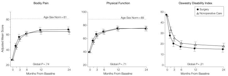
Main Outcome Measures: Primary outcomes were changes from baseline for the Medical Outcomes Study 36-item Short-Form Health Survey bodily pain and physical function scales and the modified Oswestry Disability Index (American Academy of Orthopaedic Surgeons MODEMS version) at 6 weeks, 3 months, 6 months, and 1 and 2 years from enrollment. Secondary outcomes included sciatica severity as measured by the Sciatica Bothersomeness Index, satisfaction with symptoms, self-reported improvement, and employment status.
Results: Adherence to assigned treatment was limited: 50% of patients assigned to surgery received surgery within 3 months of enrollment, while 30% of those assigned to nonoperative treatment received surgery in the same period. Intent-to-treat analyses demonstrated substantial improvements for all primary and secondary outcomes in both treatment groups. Between-group differences in improvements were consistently in favor of surgery for all periods but were small and not statistically significant for the primary outcomes.
Conclusions: Patients in both the surgery and the nonoperative treatment groups improved substantially over a 2-year period. Because of the large numbers of patients who crossed over in both directions, conclusions about the superiority or equivalence of the treatments are not warranted based on the intent-to-treat analysis.
Lumbar diskectomy is the most common surgical procedure performed in the United States for patients having back and leg symptoms; the vast majority of the procedures are elective. However, lumbar disk herniation is often seen on imaging studies in the absence of symptoms[1,2] and can regress over time without surgery.[3] Up to 15-fold variation in regional diskectomy rates in the United States[4] and lower rates internationally raise questions regarding the appropriateness of some of these surgeries.[5,6]
Several studies have compared surgical and nonoperative treatment of patients with herniated disk, but baseline differences between treatment groups, small sample sizes, or lack of validated outcome measures in these studies limit evidence-based conclusions regarding optimal treatment.[7-12] The Spine Patient Outcomes Research Trial (SPORT) was initiated in March 2000 to compare the outcomes of surgical and nonoperative treatment for lumbar intervertebral disk herniation, spinal stenosis, or degenerative spondylolisthesis.[13] The trial included both a randomized cohort and an observational cohort who declined to be randomized in favor of designating their own treatment but otherwise met all the other criteria for inclusion and who agreed to undergo follow-up according to the same protocol. This article reports intent-to-treat results through 2 years for the randomized cohort.
Methods
Study Design
SPORT was conducted at 13 multidisciplinary spine practices in 11 US states (California, Georgia, Illinois, Maine, Michigan, Missouri, Nebraska, New York, New Hampshire, Ohio, Pennsylvania). The human subjects committee of each participating institution approved a standardized protocol. All patients provided written informed consent. An independent data and safety monitoring board monitored the study at 6-month intervals.[13]
Patient Population
Patients were considered for inclusion if they were 18 years and older and diagnosed by participating physicians during the study enrollment period as having intervertebral disk herniation and persistent symptoms despite some nonoperative treatment for at least 6 weeks. The content of preenrollment nonoperative care was not prespecified in the protocol but included education/counseling (71%), physical therapy (67%), epidural injections (42%), chiropractic therapy (32%), anti-inflammatory medications (61%), and opioid analgesics (40%).
Specific inclusion criteria at enrollment were radicular pain (below the knee for lower lumbar herniations, into the anterior thigh for upper lumbar herniations) and evidence of nerve-root irritation with a positive nerve-root tension sign (straight leg raise�positive between 30� and 70� or positive femoral tension sign) or a corresponding neurologic deficit (asymmetrical depressed reflex, decreased sensation in a dermatomal distribution, or weakness in a myotomal distribution). Additionally, all participants were surgical candidates who had undergone advanced vertebral imaging (97% magnetic resonance imaging, 3% computed tomography) showing disk herniation (protrusion, extrusion, or sequestered fragment)[14] at a level and side corresponding to the clinical symptoms. Patients with multiple herniations were included if only one of the herniations was considered symptomatic (ie, if only one was planned to be operated on).
Exclusion criteria included prior lumbar surgery, cauda equina syndrome, scoliosis greater than 15�, segmental instability (>10� angular motion or >4-mm translation), vertebral fractures, spine infection or tumor, inflammatory spondyloarthropathy, pregnancy, comorbid conditions contraindicating surgery, or inability/unwillingness to have surgery within 6 months.
Study Interventions
The surgery was a standard open diskectomy with examination of the involved nerve root.[15,16] The procedure agreed on by all participating centers was performed under general or local anesthesia, with patients in the prone or knee-chest position. Surgeons were encouraged to use loupe magnification or a microscope. Using a midline incision reflecting the paraspinous muscles, the interlaminar space was entered as described by Delamarter and McCullough.[15] In some cases the medial border of the superior facet was removed to provide a clear view of the involved nerve root. Using a small annular incision, the fragment of disk was removed as described by Spengler.[16] The canal was inspected and the foramen probed for residual disk or bony pathology. The nerve root was decompressed, leaving it freely mobile.
The nonoperative treatment group received �usual care,� with the study protocol recommending that the minimum nonsurgical treatment include at least active physical therapy, education/counseling with home exercise instruction, and nonsteroidal anti-inflammatory drugs, if tolerated. Other nonoperative treatments were listed, and physicians were encouraged to individualize treatment to the patient; all nonoperative treatments were tracked prospectively.[13,17]
Study Measures
The primary measures were the Medical Outcomes Study 36-Item Short-Form Health Survey (SF-36) bodily pain and physical function scales[18-21] and the American Academy of Orthopaedic Surgeons MODEMS version of the Oswestry Disability Index (ODI).[22] As specified in the trial protocol, the primary outcomes were changes from baseline in these scales at 6 weeks, 3 months, 6 months, and 1 and 2 years from enrollment.
Secondary measures included patient self-reported improvement, work status, and satisfaction with current symptoms and with care.[23] Symptom severity was measured by the Sciatica Bothersomeness Index (range, 0-24; higher scores represent worse symptoms).[24,25]
Recruitment, Enrollment, and Randomization
A research nurse at each site identified potential participants and verified eligibility. For recruitment and informed consent, evidence-based videotapes described the surgical and non-operative treatments and the expected benefits, risks, and uncertainties.[26,27] Participants were offered enrollment in either the randomized trial or a concurrent observational cohort, the results of which are reported in a companion article.
Enrollment began in March 2000 and ended in November 2004. Baseline variables were collected prior to randomization. Patients self-reported race and ethnicity using National Institutes of Health categories.
Computer-generated random treatment assignment based on permuted blocks (randomly generated blocks of 6, 8, 10, and 12)[28] within sites occurred immediately after enrollment via an automated system at each site, ensuring proper allocation concealment. Study measures were collected at baseline and at regularly scheduled follow-up visits. Short-term follow-up visits occurred at 6 weeks and 3 months. If surgery was delayed beyond 6 weeks, additional follow-up data were obtained 6 weeks and 3 months postoperatively. Longer-term follow-up visits occurred at 6 months, 1 year from enrollment, and annually thereafter.
Statistical Analyses
We originally determined a sample size of 250 patients in each treatment group to be sufficient (with a 2-sided significance level of .05 and 85% power) to detect a 10-point difference in the SF-36 bodily pain and physical functioning scales or a similar effect size in the ODI. This difference corresponded to patients’ reports of being �a little better� in the Maine Lumbar Spine Study (MLSS).[29] The sample size calculation allowed for up to 20% missing data but did not account for any specific levels of nonadherence.
The analyses for the primary and secondary outcomes used all available data for each period on an intent-to-treat basis. Predetermined end points for the study included results at each of 6 weeks, 3 months, 6 months, 1 year, and 2 years. To adjust for the possible effect of missing data on the study results, the analysis of mean changes for continuous outcomes was performed using maximum likelihood estimation for longitudinal mixed-effects models under �missing at random� assumptions and including a term for treatment center. Comparative analyses were performed using the single imputation methods of baseline value carried forward and last value carried forward, as well as a longitudinal mixed model controlling for covariates associated with missed visits.[30]
For binary secondary outcomes, longitudinal logistic regression models were fitted using generalized estimating equations[31] as implemented in the PROC GENMOD program of SAS version 9.1 (SAS Institute Inc, Cary, NC). Treatment effects were estimated as differences in the estimated proportions in the 2 treatment groups.
P<.05 (2-sided) was used to establish statistical significance. For the primary outcomes, 95% confidence intervals (CIs) for mean treatment effects were calculated at each designated time point. Global tests of the joint hypothesis of no treatment effect at any of the designated periods were performed using Wald tests[32] as implemented in SAS. These tests account for the intraindividual correlation due to repeated measurements over time.[32]
Nonadherence to randomly assigned treatment may mean that the intention-to-treat analysis underestimates the real benefit of the treatment.[33,34] As a preplanned sensitivity analysis, we also estimated an �as-treated� longitudinal analysis based on comparisons of those actually treated surgically and nonoperatively. Repeated measures of outcomes were used as the dependent variables, and treatment received was included as a time-varying covariate. Adjustments were made for the time of surgery with respect to the original enrollment date to approximate the designated follow-up times. Baseline variables that were individually found to predict missing data or treatment received at 1 year were included to adjust for possible confounding.
Results
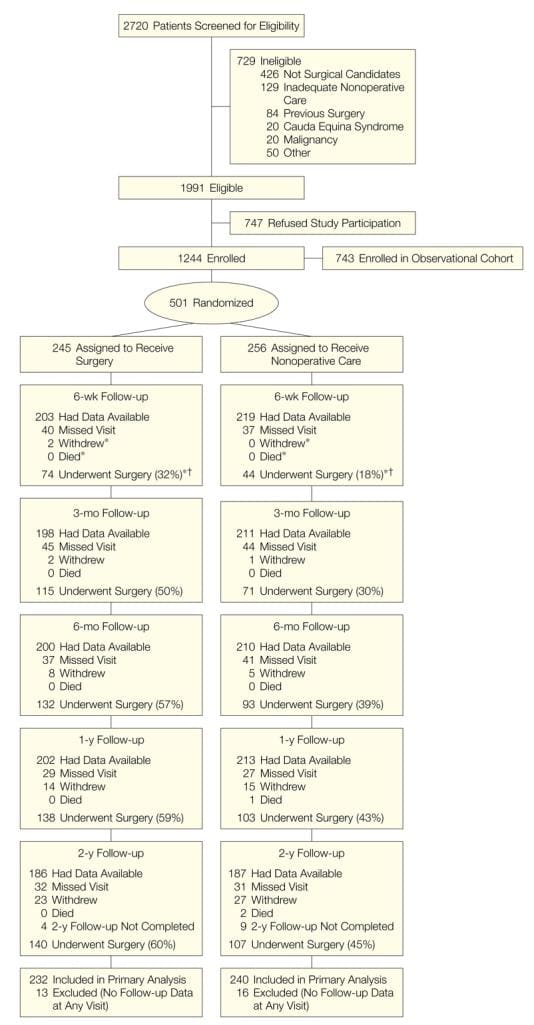
SPORT achieved full enrollment, with 501 (25%) of 1991 eligible patients enrolled in the randomized trial. A total of 472 participants (94%) completed at least 1 follow-up visit and were included in the analysis. Data were available for between 86% and 73% of patients at each of the designated follow-up times (Figure 1).
Figure 1: Flow Diagram of the SPORT Randomized Controlled Trial of Disk Herniation: Exclusion, Enrollment, Randomization, and Follow-up.
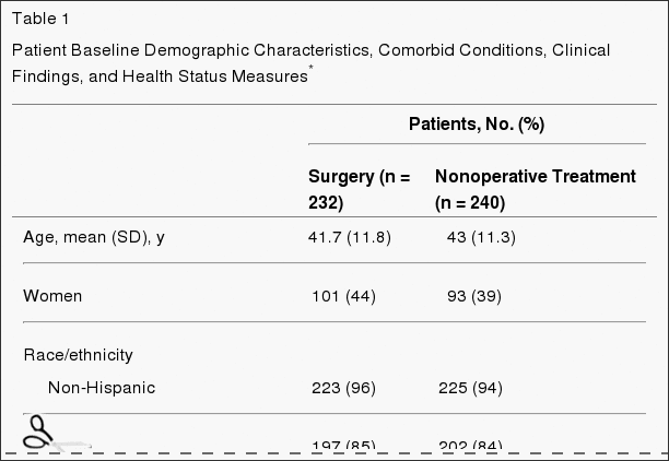
Patient Characteristics
Baseline patient characteristics are shown in Table 1. Overall, the study population had a mean age of 42 years, with majorities being male, white, employed, and having attended at least some college; 16% were receiving disability compensation. All patients had radicular leg pain, 97% in a classic dermatomal distribution. Most of the herniations were at L5-S1, posterolateral, and were extrusions by imaging criteria.[14] The 2 randomized groups were similar at baseline.
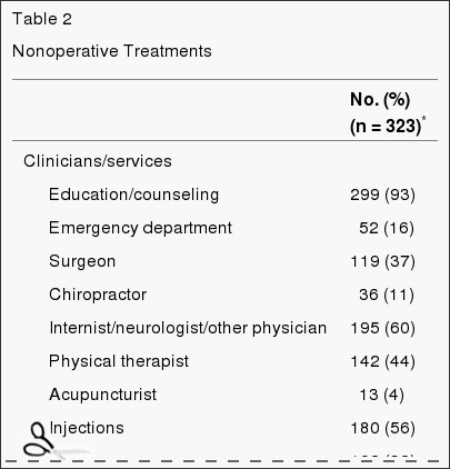
Nonoperative Treatments
A variety of nonoperative treatments were used during the study (Table 2). Most patients received education/counseling (93%) and anti-inflammatory medications (61%) (nonsteroidal anti-inflammatory drugs, cyclooxygenase 2 inhibitors, or oral steroids); 46% received opiates; more than 50% received injections (eg, epidural steroids); and 29% were prescribed activity restriction. Forty-four percent received active physical therapy during the trial; however, 67% had received it prior to enrollment.
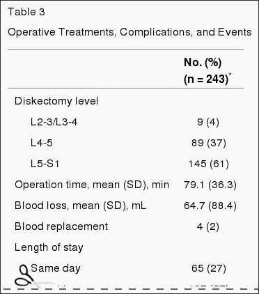
Surgical Treatment and Complications
Table 3 gives the characteristics of surgical treatment and complications. The median surgical time was 75 minutes (interquartile range, 58-90), with a median blood loss of 49.5 mL (interquar-tile range, 25-75). Only 2% required transfusions. There were no perioperative deaths; 1 patient died from complications of childbirth 11 months after enrollment. The most common intraoperative complication was dural tear (4%). There were no postoperative complications in 95% of patients. Reoperation occurred in 4% of patients within 1 year of the initial surgery; more than 50% of the reoperations were for recurrent herniations at the same level.
Nonadherence
Nonadherence to treatment assignment affected both groups, ie, some patients in the surgery group chose to delay or decline surgery, and some in the nonoperative treatment group crossed over to receive surgery (Figure 1). The characteristics of crossover patients that were statistically different from patients who did not cross over are shown in Table 4. Those more likely to cross over to receive surgery tended to have lower incomes, worse baseline symptoms, more baseline disability on the ODI, and were more likely to rate their symptoms as getting worse at enrollment than the other patients receiving nonoperative treatment. Those more likely to cross over to receive nonoperative care were older, had higher incomes, were more likely to have an upper lumbar disk herniation, less likely to have a positive straight leg�raising test result, had less pain, better physical function, less disability on the ODI, and were more likely to rate their symptoms as getting better at enrollment than the other surgery patients.
Missing Data
The rates of missing data were equivalent between the groups at each time point, with no evidence of differential dropout according to assigned treatment. Characteristics of patients with missed visits were very similar to those of the rest of the cohort except that patients with missing data were less likely to be married, more likely to be receiving disability compensation, more likely to smoke, more likely to display baseline motor weakness, and had lower baseline mental component summary scores on the SF-36.
Intent-to-Treat Analyses
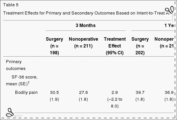
Table 5 shows estimated mean changes from baseline and the treatment effects (differences in changes from baseline between treatment groups) for 3 months, 1 year, and 2 years. For each measure and at each point, the treatment effect favors surgery. The treatment effects for the primary outcomes were small and not statistically significant at any of the points. As shown in Figure 2, both treatment groups showed strong improvements at each of the designated follow-up times, with small advantages for surgery. However, for each primary outcome the combined global test for any difference at any period was not statistically significant. This test accounts for intraindividual correlations as described in the �Methods� section.
Figure 2: Mean Scores Over Time for SF-36 Bodily Pain and Physical Function Scales and Oswestry Disability Index.
Table 5: Treatment Effects for Primary and Secondary Outcomes Based on Intent-to-Treat Analyses*
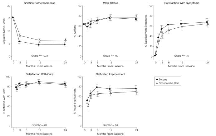
For the secondary outcome of sciatica bothersomeness, Table 5 and Figure 3 show that there were greater improvements in the Sciatica Bothersomeness Index in the surgery group at all designated follow-up times: 3 months (treatment effect, ?2.1; 95% CI, ?3.4 to ?0.9), 1 year (treatment effect, ?1.6; 95% CI, ?2.9 to ?0.4), and 2 years (treatment effect, ?1.6; 95% CI, ?2.9 to ?0.3), with results of the global hypothesis test being statistically significant (P=.003). Patient satisfaction with symptoms and treatment showed small effects in favor of surgery while employment status showed small effects in favor of nonoperative care, but none of these changes was statistically significant. Self-rated progress showed a small statistically significant advantage for surgery (P=.04).
Figure 3: Measures Over Time for Sciatica Bothersomeness Index, Employment Status, Satisfaction With Symptoms, Satisfaction With Care, and Self-rated Improvement.
As-treated analyses based on treatment received were performed with adjustments for the time of surgery and factors affecting treatment crossover and missing data. These yielded far different results than the intent-to-treat analysis, with strong, statistically significant advantages seen for surgery at all follow-up times through 2 years. For example, at 1 year the estimated treatment effects for the SF-36 bodily pain and physical function scales, the ODI, and the sciatica measures were 15.0 (95% CI, 10.9 to 19.2), 17.5 (95% CI, 13.6 to 21.5), ?15.0 (95% CI, ?18.3 to ?11.7), and ?3.2 (95% CI, ?4.3 to ?2.1), respectively.
Sensitivity analysis was performed for 4 different analytic methods of dealing with the missing data. One method was based on simple mean changes for all patients with data at a given time point with no special adjustment for missing data. Two methods used single imputation methods�baseline value carried forward and last value carried forward.[32] The latter method used the same mixed-models approach for estimating mean changes as given in Table 5 but also adjusted for factors affecting the likelihood of missing data. Treatment effect estimates at 1 year ranged from 1.6 to 2.9 for the SF-36 bodily pain scale, 0.74 to 1.4 for the physical function scale, ?2.2 to ?3.3 for the ODI, and ?1.1 to ?1.6 for the sciatica measures. Given these ranges, there appear to be no substantial differences between any of these methods.
Dr. Alex Jimenez’s Insight
Herniated disc symptoms vary on the location of the condition and on the surrounding soft tissues affected along the spine. Lumbar disc herniations, one of the most common area for herniated discs to occur, are characterized by the compression of the nerve roots along the lower back and can generally cause symptoms of sciatica. Surgery is commonly recommended to treat disc herniations, however, numerous treatment methods can help manage the condition without the need of surgical interventions. A research study conducted on sciatica caused by herniated discs determined that about 73 percent of participants experienced an improvement in symptoms with nonoperative treatment. The results of this article concluded that nonoperative treatment can be as effective as surgery in the treatment of herniated discs.
Comment
Both operated and nonoperated patients with intervertebral disk herniation improved substantially over a 2-year period. The intent-to-treat analysis in this trial showed no statistically significant treatment effects for the primary outcomes; the secondary measures of sciatica severity and self-reported progress did show statistically significant advantages for surgery. These results must be viewed in the context of the substantial rates of nonadherence to assigned treatment. The pattern of nonadherence is striking because, unlike many surgical studies, both the surgical and nonoperative treatment groups were affected.[35] The most comparable previous trial[8] had 26% crossover into surgery at 1 year, but only 2% crossover out of surgery. The mixing of treatments due to crossover can be expected to create a bias toward the null.[34] The large effects seen in the as-treated analysis and the characteristics of the crossover patients suggest that the intent-to-treat analysis underestimates the true effect of surgery.
SPORT findings are consistent with clinical experience in that relief of leg pain was the most striking and consistent improvement with surgery. Importantly, all patients in this trial had leg pain with physical examination and imaging findings that confirmed a disk herniation. There was little evidence of harm from either treatment. No patients in either group developed cauda equina syndrome; 95% of surgical patients had no intraoperative complications. The most common complication, dural tear, occurred in 4% of patients, similar to the 2% to 7% noted in the meta-analysis by Hoffman et al,7 2.2% seen in the MLSS,[29] and 4% in the recent series from Stanford.[36]
One limitation is the potential lack of representativeness of patients agreeing to be randomized to surgery or nonoperative care; however, the characteristics of patients agreeing to participate in SPORT were very similar to those in other studies.[29,36] The mean age of 42 years was similar to the mean ages in the MLSS,[29] the series of Spangfort,[37] and the randomized trial by Weber,[8] and only slightly older than those in the recent series from Stanford (37.5 years).[36] The proportion of patients receiving workers’ compensation in SPORT (16%) was similar to the proportion in the Stanford population (19%) but lower than that in the MLSS population (35%), which specifically oversampled patients receiving compensation. Baseline functional status was also similar, with a mean baseline ODI of 46.9 in SPORT vs 47.2 in the Stanford series, and a mean baseline SF-36 physical function score of 39 in SPORT vs 37 in the MLSS.
The strict eligibility criteria, however, may limit the generalizability of these results. Patients unable to tolerate symptoms for 6 weeks and demanding earlier surgical intervention were not included, nor were patients without clear signs and symptoms of radiculopathy with confirmatory imaging. We can draw no conclusions regarding the efficacy of surgery in these other groups. However, our entry criteria followed published guidelines for patient selection for elective diskectomy, and our results should apply to the majority of patients facing a surgical decision.[38,39]
To fully understand the treatment effect of surgery compared with nonoperative treatment, it is worth noting how each group fared. The improvements with surgery in SPORT were similar to those of prior series at 1 year: for the ODI, 31 points vs 34 points in the Stanford series; for the bodily pain scale, 40 points vs 44 in the MLSS; and for sciatica bothersomeness, 10 points vs 11 in the MLSS. Similarly, Weber[8] reported 66% �good� results in the surgery group, compared with the 76% reporting �major improvement� and 65% satisfied with their symptoms in SPORT.
The observed improvements with nonoperative treatment in SPORT were greater than those in the MLSS, resulting in the small estimated treatment effect. The nonoperative improvement of 37, 35, and 9 points in bodily pain, physical function, and sciatica bothersomeness, respectively, were much greater than the improvements of 20, 18, and 3 points reported in the MLSS. The greater improvement with nonoperative treatment in SPORT may be related to the large proportion of patients (43%) who underwent surgery in this group.
The major limitation of SPORT is the degree of nonadherence with randomized treatment. Given this degree of crossover, it is unlikely that the intent-to-treat analysis can form the basis of a valid estimate of the true treatment effect of surgery. The �as-treated� analysis with adjustments for possible confounders showed much larger effects in favor of surgical treatment. However, this approach does not have the strong protection against confounding that is afforded by randomization. We cannot exclude the possibility that baseline differences between the as-treated groups, or the selective choice of some but not other patients to cross over into surgery, may have affected these results, even after controlling for important covariates. Due to practical and ethical constraints, this study was not masked through the use of sham procedures. Therefore, any improvements seen with surgery may include some degree of �placebo effect.�
Another potential limitation is that the choice of nonoperative treatments was at the discretion of the treating physician and patient. However, given the limited evidence regarding efficacy for most nonoperative treatments for lumbar disk herniation and individual variability in response, creating a limited, fixed protocol for nonoperative treatment was neither clinically feasible nor generalizable. The nonoperative treatments used were consistent with published guidelines.[17,38,39] Compared with the MLSS, SPORT had lower use of activity restriction, spinal manipulation, transcutaneous electrical nerve stimulation, and braces and corsets, and higher rates of epidural steroid injections and use of narcotic analgesics. This flexible nonoperative protocol had the advantages of individualization that considered patient preferences in the choice of nonoperative treatment and of reflecting current practice among multidisciplinary spine practices. However, we cannot make any conclusion regarding the effect of surgery vs any specific nonoperative treatment. Similarly, we cannot adequately assess the relative efficacy of any differences in surgical technique.
Conclusion
Patients in both the surgery and nonoperative treatment groups improved substantially over the first 2 years. Between-group differences in improvements were consistently in favor of surgery for all outcomes and at all time periods but were small and not statistically significant except for the secondary measures of sciatica severity and self-rated improvement. Because of the high numbers of patients who crossed over in both directions, conclusions about the superiority or equivalence of the treatments are not warranted based on the intent-to-treat analysis alone.
Manipulation or Microdiskectomy for Sciatica? A Prospective Randomized Clinical Study
Abstract
Objective: The purpose of this study was to compare the clinical efficacy of spinal manipulation against microdiskectomy in patients with sciatica secondary to lumbar disk herniation (LDH).
Methods: One hundred twenty patients presenting through elective referral by primary care physicians to neurosurgical spine surgeons were consecutively screened for symptoms of unilateral lumbar radiculopathy secondary to LDH at L3-4, L4-5, or L5-S1. Forty consecutive consenting patients who met inclusion criteria (patients must have failed at least 3 months of nonoperative management including treatment with analgesics, lifestyle modification, physiotherapy, massage therapy, and/or acupuncture) were randomized to either surgical microdiskectomy or standardized chiropractic spinal manipulation. Crossover to the alternate treatment was allowed after 3 months.
Results: Significant improvement in both treatment groups compared to baseline scores over time was observed in all outcome measures. After 1 year, follow-up intent-to-treat analysis did not reveal a difference in outcome based on the original treatment received. However, 3 patients crossed over from surgery to spinal manipulation and failed to gain further improvement. Eight patients crossed from spinal manipulation to surgery and improved to the same degree as their primary surgical counterparts.
Conclusions: Sixty percent of patients with sciatica who had failed other medical management benefited from spinal manipulation to the same degree as if they underwent surgical intervention. Of 40% left unsatisfied, subsequent surgical intervention confers excellent outcome. Patients with symptomatic LDH failing medical management should consider spinal manipulation followed by surgery if warranted.
In conclusion, a herniated disc causes the soft, central portion of an intervertebral disc to bulge out a tear in its outer, fibrous ring as a result of degeneration, trauma, lifting injuries or straining. Most disc herniations can heal on their own but those considered to be severe may require surgical interventions to treat them. Research studies, such as the one above, have demonstrated that nonoperative treatment may help the recovery of a herniated disc without the need for surgery. Information referenced from the National Center for Biotechnology Information (NCBI). The scope of our information is limited to chiropractic as well as to spinal injuries and conditions. To discuss the subject matter, please feel free to ask Dr. Jimenez or contact us at 915-850-0900 .
Curated by Dr. Alex Jimenez
Additional Topics: Back Pain
According to statistics, approximately 80% of people will experience symptoms of back pain at least once throughout their lifetimes. Back pain is a common complaint which can result due to a variety of injuries and/or conditions. Often times, the natural degeneration of the spine with age can cause back pain. Herniated discs occur when the soft, gel-like center of an intervertebral disc pushes through a tear in its surrounding, outer ring of cartilage, compressing and irritating the nerve roots. Disc herniations most commonly occur along the lower back, or lumbar spine, but they may also occur along the cervical spine, or neck. The impingement of the nerves found in the low back due to injury and/or an aggravated condition can lead to symptoms of sciatica.
1.�Boden SD, Davis DO, Dina TS, Patronas NJ, Wiesel SW. Abnormal magnetic-resonance scans of the lumbar spine in asymptomatic subjects: a prospective investigation.�J Bone Joint Surg Am.�1990;72:403�408.�[PubMed]
2.�Jensen MC, Brant-Zawadzki MN, Obuchowski N, Modic MT, Malkasian D, Ross JS. Magnetic resonance imaging of the lumbar spine in people without back pain.�N Engl J Med.�1994;331:69�73.[PubMed]
3.�Saal JA, Saal JS. Nonoperative treatment of herniated lumbar intervertebral disc with radiculopathy.�Spine.�1989;14:431�437.�[PubMed]
4.�Weinstein JN, Dartmouth Atlas Working Group .�Dartmouth Atlas of Musculoskeletal Health Care.American Hospital Association Press; Chicago, Ill: 2000.
5.�Deyo RA, Weinstein JN. Low back pain.�N Engl J Med.�2001;344:363�370.�[PubMed]
6.�Weinstein JN, Bronner KK, Morgan TS, Wennberg JE. Trends and geographic variations in major surgery for degenerative diseases of the hip, knee, and spine.�Health Aff (Millwood)�2004;(suppl Web exclusive):var81�89.�[PubMed]
7.�Hoffman RM, Wheeler KJ, Deyo RA. Surgery for herniated lumbar discs: a literature synthesis.�J Gen Intern Med.�1993;8:487�496.�[PubMed]
8.�Weber H. Lumbar disc herniation: a controlled, prospective study with ten years of observation.�Spine.�1983;8:131�140.�[PubMed]
9.�Buttermann GR. Treatment of lumbar disc herniation: epidural steroid injection compared with discectomy: a prospective, randomized study.�J Bone Joint Surg Am.�2004;86:670�679.�[PubMed]
10.�Gibson JN, Grant IC, Waddell G. The Cochrane review of surgery for lumbar disc prolapse and degenerative lumbar spondylosis.�Spine.�1999;24:1820�1832.�[PubMed]
11.�Gibson JN, Grant IC, Waddell G. Surgery for lumbar disc prolapse.�Cochrane Database Syst Rev.�2000;(3):CD001350.�[PubMed]
12.�Jordan J, Shawver Morgan T, Weinstein J, Konstantinou K. Herniated lumbar disc.�Clin Evid.�2003 June;:1203�1215.
13.�Birkmeyer NJ, Weinstein JN, Tosteson AN, et al. Design of the Spine Patient Outcomes Research Trial (SPORT)�Spine.�2002;27:1361�1372.�[PMC free article]�[PubMed]
14.�Fardon DF, Milette PC. Nomenclature and classification of lumbar disc pathology: recommendations of the Combined Task Forces of the North American Spine Society, American Society of Spine Radiology, and American Society of Neuroradiology.�Spine.�2001;26:E93�E113.�[PubMed]
15.�Delamarter R, McCullough J. Microdiscectomy and microsurgical laminotomies. In: Frymoyer J, editor.�The Adult Spine: Principles and Practice.�2nd ed. Lippincott-Raven Publishers; Philadelphia, Pa: 1996.
16.�Spengler DM. Lumbar discectomy: results with limited disc excision and selective foraminotomy.�Spine.�1982;7:604�607.�[PubMed]
17.�Cummins J, Lurie JD, Tosteson T, et al. Descriptive epidemiology and prior healthcare utilization of patients in the Spine Patient Outcomes Research Trial’s (SPORT) three observational cohorts: disc herniation, spinal stenosis, and degenerative spondylolisthesis.�Spine.�2006;31:806�814.�[PMC free article][PubMed]
18.�Ware JE, Jr, Sherbourne D. The MOS 36-item short-form health survey (SF-36), I: conceptual framework and item selection.�Med Care.�1992;30:473�483.�[PubMed]
19.�Ware JE., Jr .�SF-36 Health Survey: Manual and Interpretation Guide.�Nimrod Press; Boston, Mass: 1993.
20.�McHorney CA, Ware JE, Jr, Lu JF, Sherbourne CD. The MOS 36-item Short-Form Health Survey (SF-36), III: tests of data quality, scaling assumptions, and reliability across diverse patient groups.�Med Care.�1994;32:40�66.�[PubMed]
21.�Stewart AL, Greenfield S, Hays RD, et al. Functional status and well-being of patients with chronic conditions: results from the Medical Outcomes Study.�JAMA.�1989;262:907�913.�[PubMed]
22.�Daltroy LH, Cats-Baril WL, Katz JN, Fossel AH, Liang MH. The North American Spine Society lumbar spine outcome assessment instrument: reliability and validity tests.�Spine.�1996;21:741�749.[PubMed]
23.�Deyo RA, Diehl AK. Patient satisfaction with medical care for low-back pain.�Spine.�1986;11:28�30.[PubMed]
24.�Atlas SJ, Deyo RA, Patrick DL, Convery K, Keller RB, Singer DE. The Quebec Task Force classification for spinal disorders and the severity, treatment, and outcomes of sciatica and lumbar spinal stenosis.�Spine.�1996;21:2885�2892.�[PubMed]
25.�Patrick DL, Deyo RA, Atlas SJ, Singer DE, Chapin A, Keller RB. Assessing health-related quality of life in patients with sciatica.�Spine.�1995;20:1899�1908.�[PubMed]
26.�Phelan EA, Deyo RA, Cherkin DC, et al. Helping patients decide about back surgery: a randomized trial of an interactive video program.�Spine.�2001;26:206�211.�[PubMed]
27.�Weinstein JN. Partnership: doctor and patient: advocacy for informed choice vs. informed consent.�Spine.�2005;30:269�272.�[PubMed]
28.�Friedman L, Furberg C, DeMets D.�Fundamentals of Clinical Trials.�3rd ed. Springer-Verlag; Cambridge, Mass: 1998. The randomization process; pp. 61�81.
29.�Atlas SJ, Deyo RA, Keller RB, et al. The Maine Lumbar Spine Study, II: 1-year outcomes of surgical and nonsurgical management of sciatica.�Spine.�1996;21:1777�1786.�[PubMed]
30.�Little R, Rubin D.�Statistical Analysis With Missing Data.�2nd ed. John Wiley & Sons; Philadelphia, Pa: 2002.
31.�Diggle P, Haeagery P, Liang K, Zeger S.�The Analysis of Longitudinal Data.�2nd ed. Oxford University Press; Oxford, England: 2002.
32.�Fitzmaurice G, Laird N, Ware J.�Applied Longitudinal Analysis.�John Wiley & Sons; Philadelphia, Pa: 2004.
33.�Altman DG, Schulz KF, Moher D, et al. The revised CONSORT statement for reporting randomized trials: explanation and elaboration.�Ann Intern Med.�2001;134:663�694.�[PubMed]
34.�Meinert CL.�Clinical Trials: Design, Conduct, and Analysis.�Oxford University Press; New York, NY: 1986.
35.�Kuppermann M, Varner RE, Summitt RL, Jr, et al. Effect of hysterectomy vs medical treatment on health-related quality of life and sexual functioning: the medicine or surgery (Ms) randomized trial.�JAMA.�2004;291:1447�1455.�[PubMed]
36.�Carragee EJ, Han MY, Suen PW, Kim D. Clinical outcomes after lumbar discectomy for sciatica: the effects of fragment type and anular competence.�J Bone Joint Surg Am.�2003;85:102�108.�[PubMed]
37.�Spangfort EV. The lumbar disc herniation: a computer-aided analysis of 2,504 operations.�Acta Orthop Scand Suppl.�1972;142:1�95.�[PubMed]
38.�Agency for Health Care Policy and Research .�Acute Low Back Problems in Adults.�US Dept of Health & Human Services; Bethesda, Md: 1994.
39.�North American Spine Society .�North American Spine Society Phase III Clinical Guidelines for Multidisciplinary Spine Care Specialists.�NASS; LaGrange, Ill: 2000. Herniated disc.
Constipation is an uncomfortable and common side effect of lower back and leg pain conditions. Sciatic nerve pain can occur at the same time as constipation does, but can also alternate where constipation ensues followed by sciatica.
Finding lasting relief is crucial, but understanding the exact reasons why the symptoms occur is just as important. These two conditions can be related or they may be completely coincidental. But the more they occur together, or in succession, there is greater chance that some structural or body connection is happening between the two.
The Facts: Sciatica & Constipation
Investigate why the source process may be the same for both conditions in some.
Constipation,�known as a recurrent and chronic health concern which plagues some people their entire lives. It can be caused by a variety of anatomical reasons, but many of these are fairly easy to diagnose, despite being difficult to cure using traditional medical therapy.
Sciatica is very much the same in that it can be chronic, recurrent and sometimes treatment-resistant.
What these disorders have in common is that they are often linked by nerve compression conditions within the spine. The source can be central or foraminal stenosis, which leads to compression of one or more of the lumbar nerve roots.
It is also possible for cervical central spinal stenosis to cause sciatica and may contribute to constipation, as well.
Both conditions are associated with the mind and body processes, that is physical illness caused or aggravated by mental factors, i.e. stress or some type of conflict. Constipation can be linked to conscious and subconscious emotional issues, while sciatica is just starting to receive the same recognition as a possible mind and body disorder.
Constipation/Sciatica: Solutions
Sciatica cases where constipation is also present involves the nerve roots in the lower spinal regions. These types of symptomatic expressions will be blamed on a variety of structural abnormalities in the lumbosacral region, which include degenerative discdisease, herniated discs and spinal osteoarthritis.
An alternative explanation for many cases of constipation accompanied by sciatica is regional oxygen deprivation. The solution to this condition is the treatment option invented by Dr. John Sarno. This simple treatment can usually solve even the most harmful of sciatica concerns. But the therapy remains controversial as it helps some and not others.
Sciatica/Constipation: Analysis
Once the symptoms have been diagnosed, if symptoms are structural, then treatments should resolve them or at least help in controlling the pain. If various treatments have been utilized with no relief, then it could be misdiagnosis.
Another anatomical condition that could be responsible for the symptoms or the cause could be a combination of the aforementioned mind and body issues working together. An epidemic problem that the healthcare system and one of the underlying reasons why so many with back, neck and sciatica pain never find a lasting cure. Don’t be surprised if to find out the pain was inaccurately diagnosed. This happens to millions every day.
Constipation can also be a result of serious internal diseases or organ malfunctions. Request a complete workup, which includes appropriate diagnostic testing for any significant or chronic constipation case.
Sometimes, this combination of symptoms may indicate the first signs of cauda equina syndrome.�This is a medical emergency and must be treated immediately.
Many will disregard any notion that sciatica is caused by constipation. Constipation can cause sciatica check other websites. Doctors do agree that constipation is one of a many of causes of sciatica.
But the bowels and the lower back are different parts of the body. It is important to understand that all parts of the body are connected in some way or other.
Sciatica?
If there is pain in the lower back near the buttocks and that pain travels down one or both legs, then chances are sciatica is present. Sciatica has become a common lower back pain that doctors, chiropractors, acupuncturists and physical therapists treat frequently. The pain is characterized with a combination of dull and sharp aches that create a feeling of pins and needles. With nerve conditions pins and needles are the most common type of pain.
Sciatica is the result of sciatic nerve compression. Constipation is a non-spinal condition that can cause sciatica. Just trying to use the bathroom can cause pain by irritating the sciatic nerve.
Sciatica happens when the sciatic nerve, which is the largest in the body, is compressed by an external pressure. Women in child birth and men who carry their wallets in the back pocket can experience sciatica.
Sciatica is treatable; if experiencing constipation and lower back pain at the same time, ask a doctor to test for sciatica. Doctors will order a CT scan, MRI, X-Ray or nerve conduction test.
Solving The Problem:
Experiencing sciatica related to constipation, then the first course of action is diet change. A fiber-infused diet that combines fruits and vegetables can relieve constipation. Or consider a fiber supplement.
Pain Reduction:
While waiting for constipation relief, there are various ways to reduce pain.
Take aspirin or ibuprofen, Anti-inflammatory medications reduce nerve and muscle inflammation, which alleviate nerve irritation.
Alternate hot and cold compresses, which reduce inflammation and sooth the pain. Can also be applied to the legs if the pain travels down the body.
Consider a firm mattress to support the back and alleviate any sciatica that may be the result from back strain.
A doctor may recommend several days of rest in order to allow the nerve damage time to heal.
Rules To Remember:
Do not bend or sit in a soft chair. Back support is critical.
Do not ignore the pain. Nerve pain heals within a week or gets worse.
Move slowly when standing or getting in and out of bed.
No heavy lifting & sometimes no lifting at all.
Good Nutrition & Chiropractic Treatment Contribute To Overall Well-Being
IFM's Find A Practitioner tool is the largest referral network in Functional Medicine, created to help patients locate Functional Medicine practitioners anywhere in the world. IFM Certified Practitioners are listed first in the search results, given their extensive education in Functional Medicine
 We focus on what works for you. We also strive to create fitness and better the body through researched methods and total wellness programs. These programs are natural, and use the body�s own ability to achieve goals of improvement, rather than introducing harmful chemicals, controversial hormone replacement, surgery, or addictive drugs.
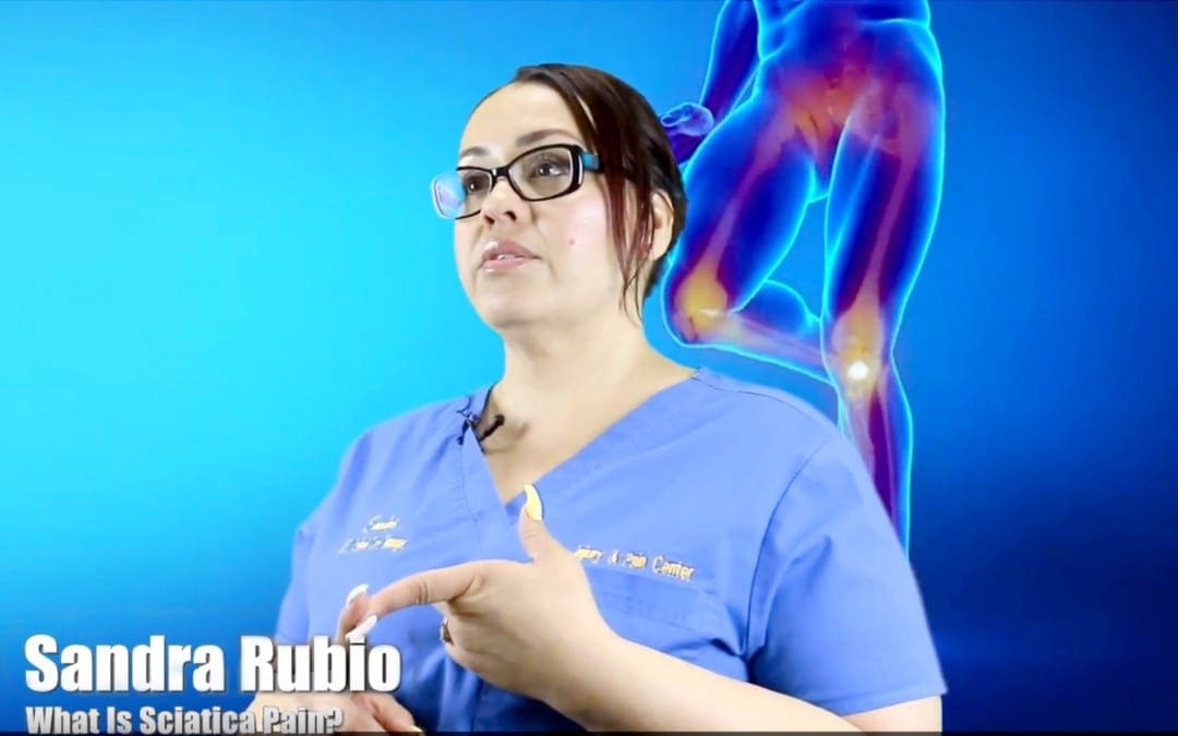
 When your body is truly healthy, you will arrive at your optimal fitness level proper physiological fitness state. �We want to help you live a new and improved lifestyle. Over the last 2 decades while researching and testing methods with thousands of patients we have learned what works effectively at decreasing pain while increasing human vitality.
 Sciatic Nerve Pain
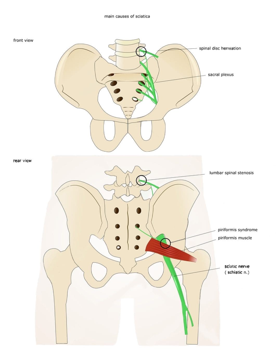
 The sciatic nerve is the largest nerve in the body and consists of five nerves that come together at the lower spine and then extend all the way down the back of the legs into the toes. If the lumbar spine is compressed, it presses on the sciatic nerve, thus causing radiating pain down the leg and sometimes all the way into the big toe. Foot pain without leg pain is often due to an issue located within the foot. However, it is possible that the foot pain could be the only symptom of sciatica.
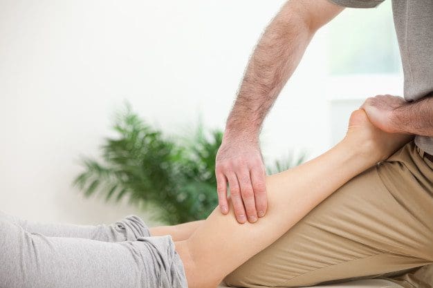


 Sciatica is a set of symptoms characterized by radiating pain from the
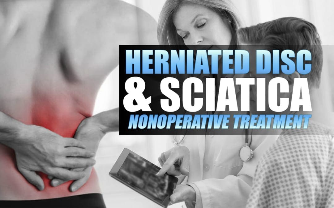


 Investigate why the source process may be the same for both conditions in some.
 Sciatica cases where constipation is also present involves the nerve roots in the lower spinal regions. These types of symptomatic expressions will be blamed on a variety of structural abnormalities in the lumbosacral region, which include degenerative disc disease, herniated discs and spinal osteoarthritis.