After pulling out a tree root Muntathar experienced excruciating pain which forced him into a extreme hunched position. If he tried to stand up he would get terrible pain and numbness all down his leg. Despite being to emergency 9 times, he had found no help in the medical world. So as a last resort he decided to travel from America to Australia to see Dr. Ian. Watch as after 2 weeks of specific Gonstead Chiropractic care, Mun stands straight and tall once more and gets his life back. We hope you enjoy this very special case.
The video�(see at bottom of post) was posted on Reddit by user Duggerdean with the comment: �I used to be a skeptic about chiropractic care until I started watching this channel. His latest video is so amazing.�
The video they refer to is that of Dr Ian Watch, who practices at Gonstead Chiropractic in Victoria, Australia � and his teenage patient�Muntathar Altaii from the US.
Despite going to A&E nine times, he says no one was able to help him.
Day 1
Dr Ian X-rays Mun�s back and sees one of the joints isn�t functioning properly. The measurements are also quite a long way out which suggests the sacrum (the large triangular bone at the base of the spine) has rotated.
Mun Also Has No Feeling In His Right Leg
Dr Ian makes what he calls �a very small correction� at the first appointment. After just 10 minutes, he�s already got a small amount of feeling back in his leg.
Mun then confesses he hopes to be better by his graduation � on June 17. No pressure.
Day 2
Mun Reports He�s Slept Well For The First Time In Three Months
Dr Ian identifies a huge amount of upper cervical nerve pressure in his neck. His head sits higher on one side than the other. So, Dr Ian makes another �adjustment�.
Day 4
Mun has a lot more movement. He can move his leg easily, whereas before he would have to pick it up even to walk.
Day 7
After further manipulation of Mun�s spine (with assistance to keep it stable), Mun tells Dr Ian how desperate he�d become after his injury. �At first I didn�t want to even live anymore,� he says, �because I couldn�t walk anywhere.�
Day 10
‘Now he just walks like he�s been playing too much X-Box� jokes Dr Ian.
For The First Time, Mun�s Taller Than The Doctor
Transformation In Just 10 Days Is Pretty Incredible
Call Today!
Photo Mun Sent Back To His Family That Night
Before & After
Mun�s High School Graduation Picture � He Got His Wish Last Week
Mun, Second From Left, With His Dad
Now Mun Wants To Be A Chiropractor When He�s Older, Of Course!
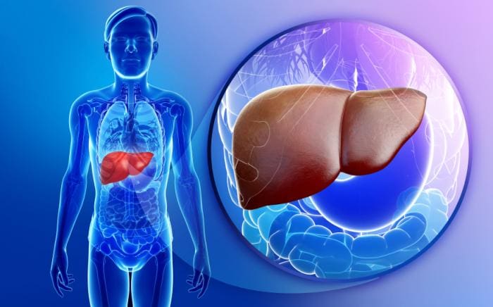
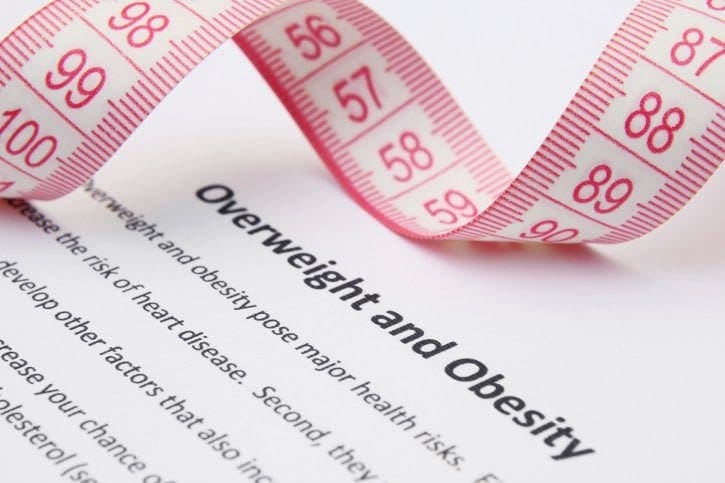
Throughout the United States, U.K., and Australia, more and more cases of liver disease are arising in the absence of alcohol abuse. Decades ago, we only saw conditions like Fatty Liver Disease and cirrhosis occur as a direct result of excessive alcohol indulgence, however, this trend has changed in the current day. Today, more and more adults and children are being diagnosed with NON-ALCOHOLIC FATTY LIVER DISEASE (NAFLD).
NAFLD is a medical condition that is characterized by an excessive accumulation of fats, within liver cells. This means normal, healthy liver tissue becomes partly replaced with fatty tissue. The fat starts to invade the liver, gradually infiltrating the healthy liver areas, decreasing the amount of healthy active liver tissue.
While it�s normal for your liver to contain some fat, accumulations of more than 5 percent to 10 percent of your liver�s weight are problematic.
70 million Americans have fatty liver disease and don�t even know it.
Anatomy & Function of the Liver
The liver is one of the hardest-working organs in the body, working tirelessly day in and day out. So here�s what your liver does, in a nutshell. Your liver regulates most chemical levels in the blood and excretes bile. Bile is necessary to break down fats. All of the blood leaving the stomach and intestines must pass through the liver for filtering. It�s the liver�s responsibility to detoxify this blood. Here are several other important functions of the liver:
Detoxifies chemicals and metabolizes (breaks down) drugs.
Manufactures proteins important for the regulation of blood clotting
Breaks down excess hormones circulating in bloodstream
Produces cholesterol (necessary for vitamin D and hormone production and for healthy nerves)
Stores and releases glucose, as needed
Stores iron
Converts harmful ammonia to urea (urea is an end product of protein metabolism that gets excreted in the urine)
Clears the blood of alcohol, medications, drugs and other harmful chemicals
Produces immune factors and removes bacteria from the bloodstream
Clears and removes bilirubin (excessive buildup causes jaundice -yellowing of skin and eyes)
It�s the liver�s responsibility to process (store) nutrients, such as vitamins, minerals and iron, so they�re more efficiently absorbed.
Nonalcoholic fatty liver (NAFLD) has become increasingly common in the United States and Western Europe as weight gain, obesity, insulin resistance, diabetes and metabolic syndrome have risen in epidemic proportions. It is now the most common cause of liver disorders in the United States and other Western industrialized countries, such as Australia and the United Kingdom. It�s estimated that 1 in 5 people (25%) throughout these regions have NAFLD.
Although research has shown that NAFLD is most commonly caused by excess weight & obesity, metabolic syndrome and diabetes, studies have also revealed that the excessive use of prescribed medications and pain killers (or the toxicity of these) can lead to fatty liver disease, as well.
Symptoms of Liver Disease
A non-alcoholic fatty liver is often referred to as a �Silent Disease�. Initially there may be no symptoms, meaning, you can live with the condition for many years, even decades, and not realize it. Over time, however, some signs may begin to surface. These symptoms include:
feeling tired
fatigue
weight loss
loss of appetite
weakness
nausea
confusion
trouble concentrating
pain in the center or right upper part of belly
enlarged liver
bloating and gas
dark urine
bruising easily
sweating, excessively
constipation
dry and dark patches on neck and under arms
Over time, fatty liver disease can lead to cirrhosis of the liver. This occurs when scar tissue develops in the liver, preventing the liver from functioning properly. The scar tissue blocks the flow of blood through the liver and slows the processing of nutrients, hormones, drugs and naturally produced toxins, as well as the production of proteins and other substances made by the liver. Symptoms of cirrhosis are severe and include the buildup of fluid in the body (especially the abdominal cavity called ascites), muscle weakness, internal bleeding, yellowing of the skin and eyes, and liver failure.
Fatty Liver Diagnosis
The best way to diagnose a fatty liver is with an abdominal ultrasound or a biopsy, although an ultrasound is far less invasive. Often, people with NAFLD will not have elevated liver enzymes, so the blood tests may look normal. Elevated liver enzymes however, do indicate that you have inflammation of the liver which may be do to NAFLD or a more serious condition called NASH.
Root Causes & Risk Factors of Liver Disease
There are a number of risk factors that increase your chances of having NAFLD:
Obesity
Gastric bypass surgery
High cholesterol
High levels of triglycerides in the blood
Type 2 diabetes
Metabolic syndrome
Medications
Sleep apnea
Polycystic ovary syndrome (PCOS)
Underactive thyroid (hypothyroidism)
Underactive pituitary gland (hypopituitarism)
Hemachromatosis (excess iron accumulation)
A 2006 review published in the Journal of Clinical Gastroenterology states that NAFLD is a common finding among patients undergoing bariatric surgery, with an occurrence ranging between 84 percent to 96 percent. The review also noted that the disease seems to be most common among men, and it increases with menopause in women.
Foods That Can Lead to Fatty Liver Disease
High-Carbohydrate & Refined Foods
Foods such as bread, rice, and corn should be avoided. All white bread and carbs should be eliminated or significantly, reduced from your diet, and even whole grains should be consumed in moderation (because grains convert to sugar). All refined When we consume too many refined carbohydrates, insulin levels spike, and insulin sensitivity is a major factor in the cause of liver disease.
Sugary Drinks
Sports drinks (Gatorade/powerade), soda, energy drinks and fruit juices are full of sugar and artificial sweeteners. This sugar that enters your body causes fatty liver disease. The average 12-ounce can of soda, for example, has 10 teaspoons of sugar! Your body isn�t able to break down the amount of sugar that most Americans consume every day, and it�s impacting the liver, big time.
The American Heart Association (AHA) recommends no more than 6 tsp (25g) of sugar per day for women and 9 tsp (38g) per day for men. A child�s sugar intake should not exceed 3 tsp per day.
The average person consumes 20 tsp or more of sugar per day � equating to 66 pounds and more of sugar per year.
According to a study conducted at Emory University School of Medicine in Atlanta, sugars, particularly fructose, are suspected to contribute to the development of NAFLD and its progression. Fructose has been shown in research to do extensive damage to liver cells. There have also been substantial links between increased fructose consumption and obesity, dyslipidemia and insulin resistance.
Processed Foods
Hydrogenated oils, refined sugar, convenience foods and lunch meats are notoriously toxic to your system. Nitrates and nitrites, for example, are commonly found in processed foods and lunch meat, and they have been linked to serious conditions, including cancer. The high fructose corn syrup found in our processed foods is the single biggest cause of fatty liver; you must stay away from these products in order to heal liver disease.
Foods That Improve Fatty Liver Disease
A review published in the European Journal of Medicinal Chemistry states that natural enzymes found in vegetables, as well as fruits, plant extracts and herbs, have been traditionally used for treating liver diseases. It�s incredibly important to add vegetables to your everyday diet.
An easy way to do this is by juicing vegetables for near-perfect health. With impaired liver function, juicing vegetables has the added benefit of making the vegetables easier to digest and more readily available for absorption. Vegetables ideal for a liver detox include kale, cabbage, lettuce, cauliflower, broccoli, Brussels sprouts, asparagus, beets and celery.
Beets
Beets naturally cleanse and purify the blood, which boosts liver function and nutrient production in your body. Beets are also high in antioxidants, folate, iron, fiber and betaine (a natural digestive enzyme). Beets go great in juicing recipes and thrown into smoothies (a little goes a long way). Shred some beets and throw on your salads, daily.
Broccoli
Broccoli and other members of the cruciferous family (brussel sprouts, cauliflower, arugula, cabbage, collard greens, kale, bok choy) are high in fiber and glucosinolates, which help the liver naturally cleanse the body of carcinogens and other toxins.
Ginger Root
Ginger has powerful antioxidant and anti-inflammatory properties, especially necessary with a dysfunctional liver due to NAFLD. Ginger has also been found to drastically lower blood sugar levels. Elevated glucose and insulin resistance are 2 key factors in the development of a fatty liver. Make ginger tea by boiling ginger slices in green tea or water. You can also add ginger to a stir-fry, salad or smoothie.
Sweet Potatoes
Sweet potatoes, along with carrots, butternut squash and pumpkin) are rich in beta-carotene, a natural anti-inflammatory. A deficiency of potassium can disrupt liver function. Sweet potatoes, naturally high in potassium, are beneficial because they help support liver function. One sweet potato contains nearly 700 milligrams of potassium! It�s also rich with vitamins B6, C, D, magnesium and iron. Sweet potatoes are easy to eat because they�re naturally sweet, and the sugars are slowly released into the bloodstream through the liver, so it won�t cause a spike in blood sugar.
Lemons
Lemons are great for your liver. They provide a wealth of antioxidants and help your liver produce more enzymes giving you more energy and help with digestion.. Lemons are also naturally high in electrolytes. Although lemons are acidic, once they enter the body they become alkalinizing, which helps neutralize toxins, excrete wastes. Juice 1 fresh lemon, daily and drink-undiluted on an empty stomach every morning.
Bananas
Containing 470 milligrams of potassium, banana nutrition is also great for cleansing the liver and overcominglow potassium levels; plus, bananas assist in digestion and help release toxins and heavy metals from the body. A great way to decrease the liver�s burden.
Garlic, Whole Cloves
Garlic is rich in allicin and selenium, two powerhouse nutrients for your liver. They act in cleansing and in nourishing the entire body, especially the blood. Selenium is a naturally detoxifying mineral and allicin helps ward off immune system invaders, which helps lighten the load on your liver. Garlic also activates enzymes in the liver which help with overall digestion and flushing out toxins. Use whole garlic cloves as the best option, instead of processed minced garlic or powder.
Leafy Greens
The nutritional all-star ingredients for just about every health issue are leafy greens. Spinach, kale, chard, romaine, arugula, and collards are all some of the most nutrient dense leafy greens to enjoy. They�re packed with chlorophyll, which assists in liver function by purifying the blood, alleviating toxins, decreasing inflammation and promotes wound healing. Chlorophyll is also amazing at neutralizing heavy metals, toxic chemicals, and even pesticides that burden the liver.
Supplements That Improve Fatty Liver Disease
Dandelion Root
The vitamins and nutrients present in dandelions help cleanse our livers and keep them working properly. Dandelions also aid our digestive system by maintaining the proper flow of bile. They�re natural diuretics and allow the liver to eliminate toxins quickly. Dandelion tea or stems are also high in vitamin C, which helps with mineral absorption, reduces inflammation and prevents the development of disease.
Milk Thistle
As a liver support and aid, milk thistle is a powerful detoxifier. It helps rebuild liver cells while removing toxins from the body that are processed through the liver. According to a study published in Digestive Diseases and Sciences, milk thistle has the power to improve mortality in patients with liver failure; it�s able to naturally reverse the harmful effects of alcohol consumption, pesticides in our food supply, heavy metals in our water supply, pollution in the air that we breathe in and even poisons. According to a 2010 study, milk thistle benefitshelp treat alcoholic liver disease, acute and chronic viral hepatitis, and toxin-induced liver diseases.
Vitamin D
Recent studies have indicated that deficiencies in vitamin D can result in Non-alcoholic fatty liver disease (NAFLD). Vitamin D deficiency was shown to cause severe degrees of NAFLD along with liver inflammation and liver fibrosis (hardening). This research also revealed that vitamin D deficiencies also resulted in insulin resistance and metabolic syndrome. All of these factors play a significant role in the development of peripheral neuropathy (nerve damage). Optimum vitamin D levels should be between 70-100 ng/ml.
Curcumin
Curcumin, the active component of turmeric is arguably the most powerful herb on the planet at fighting and potentially reversing disease. Currently there have been over 6,000 peer-reviewed published articles proving the health benefits. Studies have also shown that curcumin may prevent the progression of fatty liver disease and reduces inflammation of the liver and body.
Black Seed Oil
This amazing oil can greatly speed the healing process for people with fatty liver disease. A study published in the European Review for Medical and Pharmaceutical Sciencesmeasured black seed oil�s ability to inhibit liver oxidative stress markers. The results of the study indicated that black seed oil benefitsliver disease patients because it�s able to reduce the complications and progression of fatty liver disease.
The best thing you can do to treat fatty liver disease is maintain a healthy diet. Many people with fatty liver disease are overweight and malnourished. A healthy diet that provides the vitamins and nutrients that your body needs to function is very important.
The number one treatment of fatty liver disease is weight loss and a healthy diet. It�s essential that you eat a well-balanced diet that is predominately plant-based; plus, you should exercise regularly � shoot for doing physical activity for at least 30 minutes a day, even if it�s taking a walk.
Sources:
Bedogni G, Miglioli L, Masutti F, Tiribelli C, Marchesini G, Bellentani S. Prevalence of and risk factors for nonalcoholic fatty liver disease: the Dionysos nutrition and liver study. Hepatology. 2005;42:44�52. [PubMed]
Adams LA, Lymp JF, Sauver J, St, et al. The natural history of nonalcoholic fatty liver disease: a population-based cohort study. Gastroenterology. 2005;129:113�121. [PubMed]
Peripheral Neuropathy and Fatty Liver Disease
Nonalcoholic fatty liver disease (NAFLD) is considered the most common liver disorder in the Western world. It�s recognized as one of the most common forms of chronic liver disease across the globe.
A study published in the Journal of Gastroenterology and Hepatology (2003) reported a link between non-alcoholic fatty liver disease (NAFLD) and peripheral neuropathy. The research revealed that 73% of people with NAFLD would develop peripheral nerve damage leading to the symptoms of peripheral neuropathy.
As if the development of peripheral neuropathy isn�t bad enough, science shows that the longer you have NAFLD, the more likely it is to progress into liver fibrosis (accumulation of abnormal fibrous tissue), cirrhosis (accumulation of scar tissue in the liver) and NASH (severe liver inflammation and cell damage).
Although, NAFLD is most likely to happen in people who are overweight with metabolic syndrome or type 2 diabetes, recently there are more and more cases of children with NAFLD. This is a direct result of the standard American diet. Pediatric NAFLD have been reported in children as young as 3 years old.
If you have been diagnosed with NAFLD or are overweight, suffer from metabolic syndrome, insulin resistance or diabetes, it�s important to take action. The good news is � The liver is the only organ capable of fully regenerating itself. As long as you have at least 15% of your liver that is working and functional, your body can repair and regenerate your liver.
For more information, please feel free to ask Dr. Jimenez or contact us at 915-850-0900 .
Whole Body Wellness
Following a balanced nutrition, participating in regular physical activity and getting plenty of rest are fundamental factors for maintaining whole body wellness. While all of these can make you look and feel healthy, its also essential to address the health of your spine in order to maintain the proper function of all the body�s structures. Chiropractic care is a well-known alternative treatment option utilized by many individual�s to restore the health of the spine as well as maintain it. Chiropractic can also help prevent complications related to spinal injuries and conditions.
Peripheral neuropathy may be more common in patients with pre-diabetes than previously thought, and early interventions may be warranted in this patient population, according to researchers from the University of Utah.
Currently, 86 million adults � more than one in three U.S. adults � have prediabetes, according to CDC estimates. Without weight loss and moderate physical activity, 15% to 30% of these people will develop full-blowntype 2 diabetes within 5 years.1
�We know now a lot more than we did 3 or 5 years ago about neuropathic pain in patients with prediabetes. Neuropathy affects patients with prediabetes in a continuum,� said J. Rob Singleton, MD, who is a professor of neurology at the University of Utah in Salt Lake City. �We think it is more obesity and dysfunction of lipids (fats) that is causing the problem.�
Research Shows the Link between Obesity, Pre-diabetes and Neuropathy
In another study conducted by researchers from the University of Michigan, peripheral neuropathy was also common in obese patients, even if they had normal blood sugar levels, when compared with lean control participants. This same study also confirmed that rates of neuropathy were increased in participants with prediabetes and diabetes, leading the researchers to conclude that diabetes, prediabetes, and obesity are likely metabolic drivers of peripheral neuropathy. The findings were published in JAMA Neurology.1
Dr. Singleton and his team have been studying peripheral neuropathy associated with prediabetes and metabolic syndrome as well as what treatments may work best. Metabolic syndrome is the name for a group of risk factors that raise the risk for heart disease, diabetes and stroke. Risk factors include high blood pressure, elevated blood glucose, elevated cholesterol, and abdominal fat. Through their research, they have found that many patients with metabolic syndrome have pre-diabetes and peripheral neuropathy. Therefore, a multi-pronged approach to managing these patients is essential.
�We have shown that, in pre-diabetics with neuropathic pain, exercise reduces neuropathic pain and increases the intradermal nerve fibers in the thigh and ankle. We are in the process now of replicating that study,� Singleton said in an interview withEndocrinology Advisor. �You need to improve lipid (cholesterol) function and glucose levels. So, lifestyle issues have to be addressed.�
Relationship Between Nerve Damage and Pre-diabetes
New studies evaluating the link between prediabetes and peripheral neuropathy are filling in some of the gaps in knowledge.
In a study recently published in Diabetes Care, C. Christine Lee, PhD, of the University of Toronto, and colleagues reported that prediabetes was associated with similar risks for nerve dysfunction and damage leading to peripheral neuropathy as one develops with �new-onset� diabetes.2
While the exact mechanisms behind these associations are unclear, a growing body of evidence suggests that peripheral neuropathy begins in the early stages of diabetes pathogenesis, the researchers noted.
Lee and colleagues analyzed data on 467 individuals. The researchers found that the prevalence of peripheral neuropathy was 29% in adults with normal glucose levels, as compared with 49% in adults with prediabetes and 50% in adults with new-onset diabetes.
The researchers also found that progression of elevated glucose (pre-diabetes) over 3 years predicted a higher risk for peripheral neuropathy and nerve dysfunction.
Early intervention with lifestyle changes involving diet and exercise may be vital to preventing the severity of nerve damage, Dr. Lee stated. This had previously been backed up by another study published in 2006 in Diabetes Care, by Dr. Singleton. Singleton and his colleagues found that dietary changes and exercise can result in cutaneous reinnervation and improved pain in patients with prediabetes.3
Nerve Damage Occurs Long Before Diabetes
It is imperative to realize that the nerve damage seen in peripheral neuropathy can actually occur long before diabetes sets in. In fact the most current research has shown that obesity, even with normal glucose (blood sugar) levels has been linked with causing peripheral neuropathy as well as pre-diabetes. Although it is important to strive for maintaining fasting glucose levels between 70 � 80 mg/dL, it is equally important to keep your weight down, lower LDL cholesterol and triglycerides. All of this can be accomplished without the use of medication or bariatric procedures.
For more information, please feel free to ask Dr. Jimenez or contact us at 915-850-0900 .
Additional Topics: Neck Pain and Auto Injury
Neck pain is characterized as the most prevalent symptom after being involved in an automobile accident. During an auto collision, the body is exposed to a sheer amount of force due to the high speed impact, causing the head and neck to jolt abruptly back-and-forth as the rest of the body remains in place. This often results in the damage or injury of the cervical spine and its surrounding tissues, leading to neck pain and other common symptoms associated with whiplash-related disorders.
Promises of more volume, shine, and botanical extracts may lure you in as you browse the�shampoo�aisle, but you may want to turn your attention to the tiny ingredients lists on the bottles to make sure you�re not choosing a product that will have you showering yourself in a neuro-toxic and carcinogenic chemical every day.
According to a new report from the Center for Environmental�Health, dozens of shampoos, soaps, and other personal care products (the nonprofit group tested) contained cocamide diethanolamine, otherwise known as cocamide DEA. The basis of the chemical�coconut oil�seems innocent enough. But scientists tinker with the ingredient, modifying it into an unnatural, toxic form, merely for the purpose �foaming agent.
University of North Carolina researchers found�that when Diethanolamine (DEA), a chemical used as a thickening agent in most shampoos, is applied to the skin of pregnant mice, it interferes with their offspring�s normal brain development.
The Common Natural Ingredient You Must Avoid
DEA blocks�absorption of the nutrient choline, which is essential to brain development and peripheral nerve function.� Choline deficiencies can lead to peripheral nerve damage, metabolic syndrome, NAFLD (non-alcoholic fatty liver disease), insulin resistance, and hypertension.� All of these disorders can result in peripheral nerve damage and nerve pain, also known as peripheral neuropathy.
California listed cocamide DEA as a known carcinogen in 2012 under its Prop 65 law, which requires warning labels on consumer products containing carcinogens or reproductive toxicants.� In fact, The Center for Environmental�Health�recently filed a California lawsuit against four companies (Walmart, Target, Trader Joe�s, Kohl�s) that sell�shampoo�and personal care products containing the toxic chemical without a warning label.
��Most people believe that products sold in major stores are tested for safety, but consumers need to know that they could be doused with a cancer-causing chemical every time they shower or�shampoo,� said Michael Green, executive director of the Center for Environmental�Health. �We expect companies to take swift action to end this unnecessary risk to our children�s and families�health.�
Some other things uncovered through the center�s independent testing:
A store brand children�s bubble bath from Kmart and a children�sshampoo�and conditioner from Babies �R� Us also contained cocamide DEA.
Falsely labeled organic products from Organic by Africa�s Best also tested for high levels of the cancer-causing chemical
One�shampoo�tested contained a whopping 20% cocamide DEA.
It�s important to know that cocoamide DEA can masquerade under other names, so here�s what you should look out for on all of your personal care labels:
Cocamide DEA
Cocamide MEA
Cocamidopropyl Betaine*
DEA-Cetyl Phosphate
DEA Oleth-3 Phosphate
Lauramide DEA
Linoleamide MEA
Myristamide DEA
Oleamide DEA
Stearamide MEA
TEA-Lauryl Sulfate
Triethanolamine
Cocamidopropyl betaine, or CAPB, has been replacing cocamide DEA because it is thought to cause less skin irritations in people who are sensitive; however, it does not reduce the amount of neuro-toxicity or cancer risk.
SHAMPOOS with COCAMIDE DEA
Bed Head (TIGI)
Biosilk
CVS brand shampoos
Fekkai
Garnier Fructis
Head & Shoulders
JASON shampoo
John Frieda
Johnson & Johnson baby shampoo
L�Anza
Loreal
Matrix Biolage
Neutrogena
Nexxus
Nick Chavez
Redken
Selsun Blue Dandruff
TIGI (all shampoos)
Tresemme
Walgreens brand shampoos (adult & baby)
Additionally, the most common chemical compounds in shampoos are�straight-chain alkyl benzene sulfonates. Benzene is a chemical that is responsible for neurological symptoms, headache, nausea, dizziness, drowsiness and confusion and worst of all � oftentimes linked to leukemia and many types of cancers.
Most conventional shampoos contain 1,4-dioxane, a highly toxic�carcinogen. According to the California Environmental Protection Agency, 1,4-dioxane is known to cause cancer and may cause kidney, respiratory, and neurological toxicity. The Environmental Working Group (EWG) has also stated that 1,4-dioxane is a groundwater contaminant.
MAKE YOUR OWN ORGANIC SHAMPOO
With hundreds of available shampoos on the shelf to buy, why on earth would you consider making your own? �I�m going to give you a few reasons which you won�t be able to refute.
First of all, the FDA �does not regulate what companies put in personal care products.
The majority of large companies like Suave, Pantene and Aussie (to name just a few) use chemicals that have been linked to cancer, nerve damage, immunotoxicity, and allegies.
Secondly,�It�s cheaper and doesn�t take any time to make. �That�s correct, you can make your own shampoo in under 5 minutes (no exageration) and save a boat-load of money, too.
RECIPES: Here are some of my favorite recipes for homemade shampoo.
8 oz of Dr. Bronner�s Castille Soap
13 drops Lavender essential oil (EO)
7 drops Peppermint (EO)
7 drops Rosemary (EO)
3 drops Tea Tree Oil
Rosemary Shampoo (stimulates hair growth)
Ingredients:
6 oz Dr. Bronner�s liquid castille soap
15 drops Rosemary essential oil (eo)
10 drops Geranium (eo)
BPA free plastic or glass dispenser bottle
Hydrating Shampoo
1/2 cup coconut milk
2/3 cup�Dr. Bronner�s liquid castille soap
15 drops of essential oil of your choice (see below)
2 teaspoons of olive oil
Anti-Dandruff Shampoo
1 1/2 cups coconut milk
1/2 cup Dr. Bronners liquid castille soap
1/2 cup purified water
1/2 teaspoon virgin coconut oil
1 teaspoon apple cider vinegar
1 teaspoon baking soda
20 drops Rosemary (eo)
15 drops Tea Tree Oil
1 tablespoon ground fenugreek seeds
BPA free plastic or glass dispenser bottle
Your Own Formulation
6 oz Dr. Bronners Castille Soap (liquid)
Essential oils (EO) of your choice (30 drops, may use single essential oil or multiple oils totaling 30 drops)
Did you ever let your foot fall asleep and suffer first from numbness and then from a tingling, pins-and-needles sensation while it �awakened�? People with peripheral neuropathy suffer from those types of sensations all the time. And there�s growing evidence that peripheral neuropathy is linked with celiac disease and gluten sensitivity.
The Prevalence of Neuropathy
Peripheral neuropathy is a condition that occurs from damaged nerves in the arms, legs, hands, and feet. Commonly, symptoms experienced as a result of this are numbness, tingling, burning, and pain. The condition has a number of different causes, such as, diabetes, chemotherapy, statin medications, disc herniation and traumas, toxic metal exposure, chronic alcohol consumption and vitamin deficiencies. Now, however, scientists have linked peripheral nerve damage to gluten sensitivity and celiac disease.
Gluten is a protein found in wheat, rye, spelt, kamut and barley. Celiac disease is an autoimmune disorder that wreaks havoc on the digestive tract. When a person afflicted with celiac�s eats even the tiniest bit of gluten it causes damage to the small intestine and interferes with nutrient absorption. In many cases, the inability to absorb nutrients can stunt growth, weaken bones and damage peripheral nerves resulting in neuropathy.
Celiac disease affects one out of every 100 people throughout the world. In America, two-and-a-half million Americans are undiagnosed and at risk for serious health problems, according to the Celiac Foundation. If it goes untreated, after a while a person can develop disorders like type 1 diabetes, multiple sclerosis, dermatitis herpetiformis (itchy skin rash), anemia, osteoporosis, infertility, miscarriage, neurological conditions like epilepsy, migraines, short stature, intestinal cancers, and now nerve damage.
It was approximately five years ago that researchers first discovered a possible link between celiac disease and neuropathy. A new study published in the Journal of the American Medical Association Neurology has found celiac disease patients are at an increased risk for nerve damage. �It�s quite a high figure, compared to many other outcomes in celiac disease,� the study�s coauthor Dr. Jonas Ludvigsson, a pediatrician and professor at Karolinska Institutet in Sweden, said in a statement. �There is a real association between celiac disease and neuropathy� [and] we have precise risk estimates in a way we haven�t had before.�
Furthermore, Swedish researchers studied medical records between 1969 and 2008 from over 28,000 patients with celiac disease and compared them to 139,000 people who were never diagnosed with the autoimmune disorder. Those with celiac disease were 2.5 times more likely to suffer from nerve damage also known as neuropathy.
Meanwhile, non-celiac gluten sensitivity is a newly-recognized condition, and physicians who are performing research on this topic say tingling and numbness in the extremities represents one of the most common gluten sensitivity symptoms.
In another study, researchers screened 215 patients with peripheral neuropathy. A total of 140 of these had �idiopathic neuropathy,� meaning there was no apparent medical reason for their peripheral neuropathy.
The researchers tested those 140 people for antibodies to gluten using two celiac disease blood tests, the AGA-IgA test and the AGA-IgG test. Although these tests are not thought to be very specific to celiac disease, they can detect if your body views gluten as an invader and is generating antibodies against the protein.
Thirty-four percent of those tested � 47 people � had high antibodies to gluten in one or both of those tests, compared with a 12% rate of high antibodies to gluten in the overall population.
The researchers also performed endoscopies and biopsies on those people in the study suspected to have celiac disease, and found that 9% of those in the �unexplained neuropathy� group actually had celiac. The celiac disease genes � i.e., HLA-DQ2 and HLA-DQ8 � were found in 80% of all peripheral neuropathy patients.
Celiac, Gluten Sensitivity Symptoms & Neuropathy
New research has revealed that peripheral neuropathy actually is one of the most common non-digestive symptoms of celiac disease, and gluten sensitivities, according to the University of Chicago Celiac Disease Center. In fact, it�s possible to have no noticeable gastrointestinal symptoms of celiac disease, but instead to have mainly peripheral neuropathy and other neurological symptoms.
Researchers analyzed medical records of over 28,000 patients with biopsy-confirmed celiac disease and then they followed up with all the study participants after a median of 10 years to see if they had developed nerve damage. They found that those with celiac disease had a 2.5-fold increased risk of developing nerve damage over a period of time as compared to the control population.
How Gluten Sensitivity Causes Nerve Damage
Neurological symptoms such as peripheral neuropathy, migraines and brain fog are even more common in non-celiac gluten sensitivity, according to Harvard Medical School�s Dr. Alessio Fasano, one of the lead researchers in the field of gluten sensitivity. Dr. Fasano says up to 30% of people he�s diagnosed with gluten sensitivity have neurological symptoms � a much larger percentage than people with neurological symptoms in celiac disease.
Dr. Fasano: Gluten Sensitivity May Affect 6% to 7% Overall
Dr. Fasano, director of the University of Maryland Center for Celiac Research, published the first study looking at the molecular basis for gluten sensitivity and how it differs from celiac disease. He also participated in the research concluding that celiac disease incidence is one in every 133 people.
According to Dr. Fasano, gluten sensitivity potentially affects far more people than celiac disease. He estimates about 6% to 7% of the U.S. population may be gluten-sensitive, meaning some 20 million people in the United States alone could be sensitive to gluten.
Symptoms of gluten sensitivity in this population can include digestive problems, headaches, rashes and eczema-like skin symptoms, brain fog, fatigue, and peripheral neuropathy. Almost one-third of those he�s diagnosed as gluten-sensitive report brain fog and headaches as symptoms, he says.
Dr. Ford and Dr. Fine Say Percentage Could Be Far Higher � Up To 50%
Dr. Ford, a pediatrician in Christchurch, New Zealand and author of The Gluten Syndrome, says he believes the percentage of people who are gluten-sensitive actually could be much higher � potentially between 30% and 50%.
�There are so many people who are sick,� he says. �At least 10% are gluten-sensitive, and it�s probably more like 30%. I was sticking my neck out years ago when I said at least 10% of the population is gluten-sensitive. My medical colleagues were saying gluten sensitivity didn�t exist. We�ll probably find it�s more than 50% when we finally settle on a number.�
Dr. Fine, a gastroenterologist who founded and directs the gluten sensitivity testing service Enterolab, agrees that gluten sensitivity probably affects half the population.
Another large percentage of Americans have autoimmune disorders, irritable bowel syndrome, chronic headaches and/or microscopic colitis, which place them at high risk for gluten sensitivity. About 60% to 65% of people with those conditions test positive for gluten sensitivity through Enterolab, Meanwhile, about 20% to 25% of people with no symptoms are diagnosed with gluten sensitivity based on Enterolab testing results, says Dr. Fine.
�When we did the math, we came up with the number of about one in two are gluten-sensitive,� he says.
Neuropathy Found in People with Gluten Sensitivity
A study published in 2010 in the journal of Neurology found that a gluten free diet led to stabilization of the neuropathy for many of the patients in this study.
Over the past many years, gluten has been shown to induce an autoimmune antibody response to nerve cells, myelin sheath (protective coating around nerves, as well as receptor sites on cells that bind neurotransmitters (chemicals that allow nerves to communicate).
It has also been discovered that gluten can contribute to the breakdown of the blood brain barrier. This allows chemical toxins to leak into the blood supply of the brain itself .
In addition, it has become a well researched fact that Gluten sensitivity can damage the gut inducing malabsorption of vitamins and minerals (such as vitamins B1 and B12). Gluten sensitivity has been linked to the following list of neurologic conditions:
So it goes without saying, if you have been diagnosed with Celiac disease or gluten sensitivity/intolerance or if you suspect you may have these conditions, going gluten free is imperative for the health of your nerves and your GI tract. If you are unsure, then try the � GLUTEN FREE FOR 3 � challenge. Go completely gluten free for just 3 days and keep a journal to log in how you feel and sleep during those 3 days. If you feel better, overall, then chances are high that you are gluten sensitive.
For more information, please feel free to ask Dr. Jimenez or contact us at 915-850-0900 .
Additional Topics: Early Intervention After Auto Injury
When a person is involved in an unexpected automobile accident, the most common type of injury which often results from the incident is whiplash. Whiplash is identified as a neck injury caused by the sudden, back-and-forth motion of the head during a car crash. Whiplash can cause a variety of symptoms and complications if left untreated, which is why seeking medical treatment immediately after being involved in an auto accident is essential in order to help people recover quickly without developing further issues.
It�s often assumed that in order to develop type 2 diabetes, you have to be overweight. While it�s true that excess weight is clearly associated with insulin resistance and diabetes, it�s the insulin resistance � not necessarily the weight gain � that drives the disease.
As such, many people with a healthy weight are not metabolically healthy, putting them at risk of diseases like type 2 diabetes � even without being overweight or obese.
One of the greatest risk factors, according to University of Florida researchers, is actually inactivity, which drives up your risk of pre-diabetes regardless of your weight.
Inactivity Is Associated with Diabetes
If you were looking for motivation to get moving, this study, published in the American Journal of Preventive Medicine, is as good as it gets.
In a survey of more than 1,100 healthy-weight individuals, those who were inactive (physically active for less than 30 minutes per week) were more likely to have an A1C level of 5.7 or higher, which is considered to be pre-diabetic.
The researchers suggested that people who live a largely sedentary lifestyle yet have a healthy weight may have �normal-weight obesity or �skinny fat,’� which they described as a �high proportion of fat to lean muscle.�
�Don�t focus solely on the scale and think you�re OK. If you have a sedentary lifestyle, make sure you get up and move,� lead author Arch Mainous III, chair of health services research, management and policy in the University of Florida�s College of Public Health and Health Professions, said in a news release.
Weight Doesn�t Always Reveal Metabolic Health
Weight isn�t always an accurate tool by which to gauge metabolic health, and research by Dr. Robert Lustig, professor of pediatric endocrinology at the University of California, San Francisco (USCF), bears this out.
Lustig is perhaps best known for speaking out about the health risks of sugar, but in our 2015 interview he explained the problem with �judging a book by its cover� in terms of weight and health.
More than two-thirds of the American population is overweight or obese. About 50 percent have diabetes or pre-diabetes, and 1 out of every 3 have high blood pressure. Many also have high serum triglycerides, which is a risk factor for heart disease and stroke. Insulin resistance is a component of all of these health issues.
According to Lustig, at least 50 percent of Americans have some form of insulin resistance � whether you�re overweight or not � and that is what�s driving our seemingly out-of-control disease statistics.
Exercise Is Important
The evidence is clear that regular physical activity, which includes reducing your time spent sitting and exercising, is crucial to lower your risk of diabetes (and treat it if you�ve already been diagnosed).
For instance, sitting for more than eight hours a day has been shown to increase your risk of type 2 diabetes by 90 percent, while people with diabetes who engaged in a six-month moderate-intensity exercise program experienced significant health improvements, including decreased fat in the abdomen, liver and around the heart.
How to Determine if You�re Pre-Diabetic
If you�re reading this and aren�t sure what your fasting insulin and glucose levels are, these are blood tests I recommend receiving annually. Your fasting insulin level reflects how healthy your blood glucose levels are over time.
A normal fasting blood insulin level is below 5, but ideally you�ll want it below 3. A fasting glucose level below 100 mg/dl suggests you�re not insulin resistant, while a level between 100 and 125 confirms you have pre-diabetes. If this, or your A1C level, confirms you either have or are at risk of pre-diabetes or diabetes, the time to take action is now. You might also find a hip-to-waist size index chart helpful.
This is far better than body mass index (BMI) for evaluating whether or not you may have a weight problem, as BMI fails to factor in both how muscular you are and your intra-abdominal fat mass (the dangerous visceral fat that accumulates around your inner organs), which is a potent indicator of insulin/leptin sensitivity and the associated health problems.
You Can Improve Your Insulin Sensitivity in Just Two Weeks
Fortunately, proper exercise and attention to diet can reverse the course of this disease, with benefits seen in as little as two weeks (and to some extent after just one exercise session).
For instance, unfit but otherwise healthy middle-aged adults were able to improve their insulin sensitivity and blood sugar regulation after just two weeks of interval training (three sessions per week). A follow-up study also found that interval training positively impacted insulin sensitivity.
The study involved people with full-blown type 2 diabetes, and just one interval training session was able to improve blood sugar regulation for the next 24 hours.10 You can actually reap much greater benefits by exercising in short, high-intensity bursts known as intervals than you can exercising for longer periods at a slower steady pace.
The high-intensity interval training (HIIT) approach I personally use and recommend is the Peak Fitness method, which consists of 30 seconds of maximum effort followed by 90 seconds of recuperation, for a total of eight repetitions. I also recommend super slow weight lifting for your resistance training.
Getting Up From Your Chair Is Also Important
When you hear the term sedentary, it�s important to understand that exercising for 20 or 30 minutes a day, and then sitting for much of the rest, is not enough to pull you out of this category. Long hours spent sitting are linked to chronic diseases including diabetes, and this may be, in part, because it increases aging at the cellular level.
In a study of 64- to 95-year-old women, those who sat for more than 10 hours a day and got less than 40 minutes of moderate-to-vigorous physical activity had shorter telomeres and were, on average, eight years older, biologically speaking, than women who moved around more often.
Every time a cell divides, the telomeres get shorter, which is why they�re used as a measure of biological aging. Short telomeres have also been linked with chronic diseases such as cancer, heart disease and diabetes.
In addition, your body�s ability to respond to insulin is affected by just one day of excess sitting, which leads your pancreas to produce increased amounts of insulin. Research published in Diabetologia also found that those who sat for the longest periods of time were twice as likely to have diabetes or heart disease, compared to those who sat the least. I recommend replacing the majority of your sedentary sitting time with active movement, keeping sitting to three hours a day or less.
What to Do if You Have Pre-Diabetes or Diabetes
You may be thin but that doesn�t mean you have more lean muscle than fat in your body. Having a higher percentage of fat than lean muscle can set the stage for insulin resistance.
The take-home message to remember is that you shouldn�t assume you�re metabolically healthy just because you�re not overweight or obese � especially if you live a largely sedentary lifestyle. You could actually be �skinny fat,� with many of the same health risks as someone who�s overweight or obese and sedentary.
The good news is that there�s plenty you can do to not only reduce your risk of type 2 diabetes and pre-diabetes but also improve your metabolic health at the same time.
During the three-year Diabetes Prevention Program study, for instance, lifestyle interventions were found to be more effective than the diabetes drug metformin at preventing or delaying the development of diabetes in people at high risk of the disease. A follow-up study monitored the group for 15 years � and lifestyle interventions were still more effective than metformin at preventing diabetes.13
One of the most important dietary recommendations is to limit net carbs (total carbohydrates minus fiber) and protein, replacing them with higher amounts of high-quality healthy fats, like seeds, nuts, raw grass-fed butter, olives, avocado, coconut oil, organic pastured eggs and animal fats (including animal-based omega-3s).
If you�re insulin resistant or diabetic, I also strongly suggest you limit your total fructose intake to 15 grams per day until your insulin/leptin resistance has resolved (then it can be increased to 25 grams) and start intermittent fasting as soon as possible.
As mentioned, exercise and reduced sitting time are also crucial, along with attention to proper sleep, optimized vitamin D levels and gut health. Taken together, this plan will lower your risk of diabetes and related chronic diseases and help you to avoid becoming victim to a health condition you might not even realize you have.
For more information, please feel free to ask Dr. Jimenez or contact us at 915-850-0900 .
Additional Topics: Early Intervention After Auto Injury
When a person is involved in an unexpected automobile accident, the most common type of injury which often results from the incident is whiplash. Whiplash is identified as a neck injury caused by the sudden, back-and-forth motion of the head during a car crash. Whiplash can cause a variety of symptoms and complications if left untreated, which is why seeking medical treatment immediately after being involved in an auto accident is essential in order to help people recover quickly without developing further issues.
High level performers are always looking for strategies that will give them an edge in their field of endeavor. Athletes want to run a split second faster and jump an inch higher while business executives want to have sharper mental clarity and improved working efficiency. Specific meal timing and superfood strategies have been shown to optimize performance and recovery.
We all want to perform at our peak no matter whether we are a teacher, stay at home mom, doctor or athlete. It is also integral that we recover fast and effectively. The goal is peak performance and quick and effective recovery so we can get up the next day and do it all over again.
Optimize Your Performance
Performance in any field depends upon high level mental activity and often kinesthetic activity whether that be running or jumping or eye-hand coordination. The keys for healthy function include good fats, anti-oxidants and clean protein sources.
Providing lots of clean healthy foods and meal timing properly can make a huge difference in your overall energy and daily performance. Use the principles in this article to help guide you in preparing your body for great energy and daily performance in your daily life.
Coconut
This superfood is loaded with medium chain triglycerides (MCT�s) that break down for energy very easily. Unlike most fat sources which consist of long chain fatty acids these MCT�s do not depend upon bile and instead go right to the liver and are metabolized immediately for energy.
Muscle cells are also able to store MCT�s and use them immediately for energy during exercise sessions.
Bone Broth Protein
Bone broth contains a wide variety of valuable nutrients including collagen, gelatin, hyaluronic acid, chondroitin sulfate, glycosamino glycans, proline, glycine, calcium, phosphorus, magnesium and potassium. These all help with the development of healthy joints, bones, ligaments and tendons as well as hair and skin.
These nutrients are considered beauty foods because they help the body with proper structural alignment and beautiful skin and hair. They also help to prevent against injuries by strengthening joints, tendons and ligaments. Additionally, bone broth is great for the immune system.
If you are unable to make your own bone broth, than you can try our Bone Broth Protein which comes with the same benefits as homemade broth, but it�s even more versatile in so many recipes. Bone Broth Protein begins as a true bone broth liquid. It�s then dehydrated, making it into a concentrated source of high-quality, tasty powder.
Berries
Berries are loaded with anti-oxidants and are low in sugar. Berries contain unique phytonutrients called anthocyanin�s that give them their strong pigments and allow them to handle intense sunlight. Consuming these anthocyanin�s helps our bodies adapt to stress effectively.
Be sure to get your berries organic as the thin skin makes them susceptible to pesticides and they are highly contaminated with dangerous pesticides when produced on conventional farms.
Spinach
This superfood is loaded with blood purifying chlorophyll and the anti-oxidants lutein and zeaxanthin. Spinach also contains its own plant based steroids called phyoecdysteroids that boost the bodies ability to adapt to stress. Phytoecdysteroids are similar to insect molting hormones and have been shown to dramatically increase glucose metabolism.
This keeps blood sugar levels stable and minimizes the need for the critical fat-storage hormone insulin. Additionally, phytoecdysteroids increase human muscle tissue growth rates by 20% when applied in a culture medium.
Rice & Pea Proteins
For sensitive cases where people have pronounced sensitivities I prefer to use either a brown rice or pea protein. Pea protein appears to be the most hypoallergenic of all protein powders and has a 98% absorption rate. This combination of 1% brown rice and 99% pea protein has a great blend of branched chain amino acids, lysine and arginine for lean body tissue development and good circulation
Pea & brown rice protein has also been shown to be very easy on the digestive system and the protein is fairly quickly assimilated into muscle tissue. This is important because slower digesting and assimilating proteins have a greater chance of producing gas and making one feel bloated. Most plant proteins have poor assimilation rates but pea, brown rice and hemp proteins are the rare exceptions.
These are loaded with sulfur containing amino acids like methionine and cysteine which are critical for glutathione production. They are also a rich vegetarian source of branched chain amino acids. They contain essential fatty acids and edestin and albumin which are similar to the makeup of human blood and these help with critical functions like DNA repair.
Grass-Fed Butter
Dairy from grass-fed cows is extraordinarily rich in essential fats and critical nutrients like vitamin K2 and magnesium. The best foods from this group include grass-fed butter and ghee which are loaded with anti-inflammatory fats and anti-oxidants.
Grass-fed butter and ghee are also rich in conjugated linoleic acid, which helps burn fat and is anti-carcinogenic. It is the best food source of the anti-inflammatory short chain fatty acid butyrate. Finally, it is full of choline which supports healthy brain and neurotransmitter production. I use butter and ghee generously each day. Look for Kerry Gold grass-fed butter here
Red Onions
These are loaded with the flavonoid anti-oxidant quercetin. They also contain sulfur containing amino acids that boost cysteine and glutathione production within the body. They also have chromium which helps to regulate blood sugar levels.
Grass-Fed Beef
Cows that are fed a 100% green diet are loaded with anti-oxidants like carnosine and essential fatty acids. Because cows have multiple stomachs and are able to fully digest grass (humans cannot) we are getting the benefits of this fully digested super-greens in a high protein food.
A fantastic source for grass-fed beef and pastured poultry is US Wellness Meats here
Avocados
Avocados are loaded with good fats and carotenoid anti-oxidants like lutein and zeaxanthin. It is very easy on the digestive system and helps support optimal hormone function, muscle development and exercise recovery.
Raw Chocolate
This is rich in good fats and polyphenol anti-oxidants for fuel and recovery. Chocolate contains theobromine which is a natural cardiovascular stimulant that helps improve circulation throughout the body.
Proper Meal Timing
It is best to eat light before our times of performance to keep as much energy focused on the specific performance rather than on digestion. Instead of food, turn to water, as optimal hydration correlates very strongly with great performance. I try to stay super hydrated throughout the day and notice that as my water intake drops, my energy and mental acuity declines.
After the performance we want to have our largest meal to replenish our system with nutrients. Ideally, you have your largest meal at the end of your day as opposed to the beginning of your day. This helps your body to recover from the stresses of the day. You are in a relaxed position at this point, as you no longer have to work hard and perform, so it allows you to digest and absorb the nutrients you need more effectively.
This helps to take stress off of your gut lining and improves the integrity of your digestive system. When you eat in a hurry or have a large meal before a stressful event or a busy day, you compromise your digestive system and cause more inflammation in the body.
These ideas and much more I teach in our Navigating the Ketogenic diet program. Opt-in here for more details and a FREE video training series I give you on advanced strategies to improve your performance.
Diet and Nutrition for a Healthy Back
As most patients are well aware, good nutrition and a balanced diet are important components of overall health. What may surprise people with back problems is that diet, nutrition and maintaining a healthy weight also play a major role in the back � including preventing many problems and healing from injuries.
The bones, muscles and other structures in the spine need good nutrition and vitamins so that they are strong enough to support the body and to perform their other functions. Using these nutritional guidelines, patients can integrate back-friendly vitamins and nutrients into their diets.
Choosing the Right Foods for Optimal Nutrition
Eating a balanced diet that includes the right amount and variety of vitamins and nutrients will reduce back problems by nourishing the bones, muscles, discs and other structures in the spine. Particular importance is given to calcium, which can be obtained through a variety of healthy food choices as well as nutritional supplements.
While a healthy diet calls for many vitamins and nutrients, this partial list highlights a number of healthy choices that can be directly beneficial for helping back patients.
Role of Vitamin or Nutrient Food Sources
Vitamin A
An antioxidant that assists the immune system in fighting off diseases. It is good for the back because it helps repair tissue and in the formation of bone. It also helps the body use protein effectively.
Additionally, the body can convert beta-carotene into vitamin A. Beta-carotene can be found in dark green leafy vegetables and most orange vegetables and fruits.
It�s important not to get more then the recommended daily allowance of vitamin A, as too much can promote bone fractures. Beta-carotene does not increase the risk of fracture.
Vitamin A can be found in beef, calf and chicken liver; dairy products like milk, butter, cheese and eggs; orange fruits such as apricots, nectarines and cantaloupe; orange or green vegetables such as carrots, sweet potatoes and spinach.
Vitamin B12
Necessary for healthy bone marrow and for the body � and the spine � to grow and function normally. Vitamin B12 can be found in meat products, such as liver, fish, red meat and poultry; dairy products, such as milk, yogurt and cheese; and eggs.
Vitamin C
Necessary for the development of collagen, which is an important part of the process that allows cells to be able to form into tissue. This is extremely important for healing problems caused by injured tendons, ligaments and vertebral discs, as well as for keeping bones and other tissues strong.
Vitamin C can be found in fruits, such as strawberries, kiwi fruit and citrus fruits (e.g. oranges, guavas, grapefruits) and tomatoes; many vegetables, such as broccoli, spinach, red and green peppers, sweet potatoes and white potatoes.
Vitamin D
Improves calcium absorption, which is important for the development of strong and healthy bones. Adequate calcium absorption is particularly important to help prevent development of osteoporosis, a disorder characterized by weak and brittle bones in the spine that can results in painful vertebral fractures. Vitamin D is naturally occurring in egg yolks and fish oils; also found in most brands of fortified milk in the US; can also be obtained by spending time in the sunlight.
Vitamin K
Needed for the bones to properly use calcium. The combination of vitamin K and calcium works to help bones throughout the body stay strong and healthy. Vitamin K is found in liver, pork, green leafy vegetables such as spinach, kale and broccoli, and dairy products.
Iron
Needed for cells to remain healthy as it helps them receive oxygen and get rid of carbon dioxide. It also aids in the production of myoglobin, an important element of healthy muscles that are needed to support the spine. Iron is found in meat products such as liver, pork, fish, shellfish, red meat and poultry; lentils, beans, soy, eggs, grains, and green leafy vegetables such as spinach, kale and broccoli.
Magnesium
Important for the relaxing and contracting of muscles. It also helps maintain muscle tone and bone density, which in turn can help prevent back problems. Further, it assists in the body�s use of protein. Magnesium is found in whole grains and whole-grain breads, beans, seeds, nuts, potatoes, avocados, bananas, kiwi fruit, shrimp, and green leafy vegetables such as spinach, kale and broccoli.
Calcium
Essential for bone health and helps maintain the necessary level of bone mass throughout the lifespan and especially in old age. Adequate calcium intake is particularly important to help prevent development of osteoporosis, which results in weak and brittle bones in the spine that can results in painful vertebral fractures. Calcium is found in dairy products such as yogurt, cheese and especially milk; dark green leafy vegetables such as spinach, broccoli and kale; tofu, peanuts, peas, black beans and baked beans; some types of fish (salmon and sardines); a variety of other foods such as sesame seeds, blackstrap molasses, corn tortillas, almonds and brown sugar.
For more information, please feel free to ask Dr. Jimenez or contact us at 915-850-0900 .
Additional Topics: Choosing the Right Vitamins
Following a balanced nutrition can help ensure we receive the necessary vitamins and minerals we need to maintain our overall health and wellness. Although many people may successfully follow a healthy diet, it might not always be easy to intake all the right foods we require. More than half of Americans report taking a multivitamin or dietary supplement. There are a wide variety of supplements available in the market and knowing which of these are the correct ones to take can be challenging for many, according to research studies.
IFM's Find A Practitioner tool is the largest referral network in Functional Medicine, created to help patients locate Functional Medicine practitioners anywhere in the world. IFM Certified Practitioners are listed first in the search results, given their extensive education in Functional Medicine