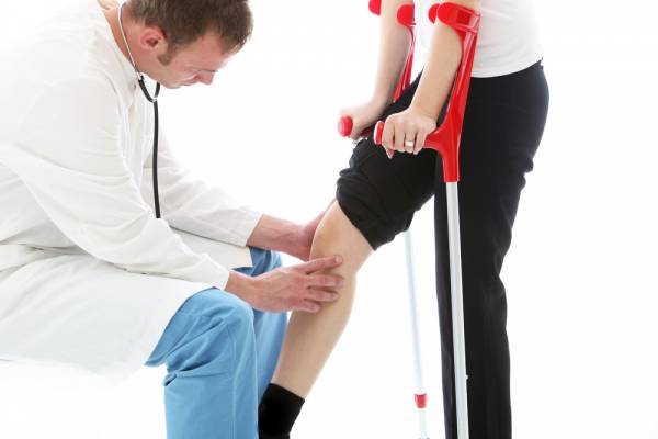
Patients who go straight home from the hospital following hip or knee replacement surgery recover as well as, or better than, those who first go to a rehabilitation center, new research indicates.
And that includes those who live alone without family or friends, one of three studies shows.
“We can say with confidence that recovering independently at home does not put patients at increased risk for complications or hardship, and the vast majority of patients were satisfied,” said that study’s co-author, Dr. William Hozack. He is an orthopaedic surgery professor with the Rothman Institute at the Thomas Jefferson University Medical School in Philadelphia.
Hozack noted that while in the past it was “not uncommon for patients to enter a rehabilitation facility in order to receive additional physical therapy,” most patients today do not end up going to a secondary facility. In fact, roughly 90 percent of Hozack’s joint replacement patients are discharged directly home following surgery, he said. “Considerable evidence has now shown that most patients do just as well at home,” he noted.
Hozack and his colleagues are scheduled to present their findings in San Diego at a meeting of the American Academy of Orthopaedic Surgeons (AAOS).
Home Recovery Following Surgery
Two other studies being presented at the meeting also found that recovering at home may be the better option.
One study found that patients who are discharged directly home following a total knee replacement face a lower risk for complications and hospital readmission than those who first go to an inpatient rehab facility. The study was led by Dr. Alexander McLawhorn, an orthopaedic hip and knee surgeon at the Hospital for Special Surgery in New York City.
McLawhorn was also part of a second Hospital for Special Surgery study, led by Michael Fu. That study found that hip replacement patients admitted to an inpatient facility rather than being sent home faced a higher risk for respiratory, wound and urinary complications, and a higher risk for hospital readmission and death.
Dr. Claudette Lajam is chief orthopaedic safety officer with NYU Langone Orthopaedics in New York City. She was not involved with the studies, but agrees that home recovery is the best option for most patients.
“The home setting is the single best way to get people back into their routines as quickly as possible after surgery,” she said. “In some cases, this cannot be done,” Lajam acknowledged. “Some patients live in settings that are inaccessible, [such as] a 5th-floor walk-up apartment where the patient would need to go downstairs to let the visiting nurse and therapist in the door.” For some patients, anxiety about the recovery process could also pose a challenge, she added. But “being in an institutional setting after surgery only reinforces the idea that the patient is ‘sick,’ ” Lajam added. “We have learned that this type of thinking slows down recovery. We want our total joint patients to start using their new joints as quickly as possible, and staying in bed at a nursing facility is not the way to do this.”
Hozack and his colleagues set out to see whether patients who live alone fare as well as those who live with others. All 769 patients enrolled in the study by Hozack’s team went home following either a total hip replacement or a total knee replacement. Of those, 138 lived alone (about 18 percent). Once home, all were assessed on multiple levels, including functionality (ability to move); pain levels; hospital readmissions; emergency department visits; unscheduled doctor visits; dependency on assisted-walking devices; and time before returning to work or being able to drive again.
Hozack’s team observed no differences by any measure. And while those who lived with others indicated relatively higher satisfaction levels at the two-week mark, by the three-month point there was no appreciable difference between the two groups.
“We feel that giving patients back their independence early on is the best way to promote a safe and effective recovery,” said Hozack. His team concluded that single-household patients who go straight home can expect to fare as well as those who have live-in support.
A recent Mayo Clinic study calculated that between 2000 and 2010, the number of Americans who underwent hip replacement surgery more than doubled, rising from just under 140,000 to more than 310,000 per year.
Meanwhile, AAOS figures indicate that in 2010 more than 650,000 knee replacement procedures were performed, with about 90 percent involving total knee replacement. AAOS estimates from 2014 show that 4.7 million Americans now live with an artificial knee and 2.5 million have an artificial hip.
Findings presented at meetings should be viewed as preliminary until published in a peer-reviewed journal.
SOURCES: William J. Hozack, M.D., professor of orthopaedic surgery, Rothman Institute, Thomas Jefferson University Medical School, Philadelphia; Claudette Lajam, M.D. assistant professor and chief orthopedic safety officer, NYU Langone Orthopedics, New York City; March 14-18, 2017 presentations, American Academy of Orthopaedic Surgeons meeting, San Diego
The scope of our information is limited to chiropractic and spinal injuries and conditions. To discuss options on the subject matter, please feel free to ask Dr. Jimenez or contact us at 915-850-0900 .
Additional Topics: What is Chiropractic?
Chiropractic care is a safe and effective, alternative treatment option utilized to diagnose, treat and prevent a variety of injuries and conditions associated with the musculoskeletal and nervous system. A chiropractor, or doctor of chiropractic, commonly uses spinal adjustments or manual manipulations to help correct the spine and it’s surrounding structures, improving and maintaining the patient’s strength, mobility and flexibility.
Surgery is a common approach to treat carpal tunnel syndrome. But, physical therapy may work just as well, a new study indicates.
Researchers found that physical therapy improved hand and wrist function and reduced pain as effectively as a standard operation for the condition. Moreover, after one month, physical therapy patients reported better results than those who underwent surgery.
“We believe that physical therapy should be the first therapeutic option for almost all patients with this condition,” said lead study author Cesar Fernandez de las Penas. “If conservative treatment fails, then surgery would be the next option,” said de las Penas, a professor of physical therapy at King Juan Carlos University in Alcorcon, Spain.
Also, one extra benefit of therapy over surgery may be cost savings, he noted.
Treatments for Carpal Tunnel Syndrome
Carpal tunnel syndrome occurs when the median nerve, which runs from the forearm into the palm of the hand, becomes squeezed at the wrist. It often arises from repetitive motions required for work, such as computer use or assembly line work. Symptoms usually start gradually, with patients noticing numbness and weakness in the hand and wrist.
Surgery for the condition generally involves cutting a ligament around the wrist to reduce pressure on the median nerve, according to the U.S. National Institutes of Health.
Results of Physical Therapy vs Surgery
For this study, de las Penas and his colleagues followed 100 women from Madrid who had carpal tunnel syndrome. Half were treated with physical therapy and half underwent surgery.
For three weeks, the therapy patients received weekly half-hour manual therapy sessions — meaning therapists only used their hands. The therapists focused on the neck and the median nerve. They also applied manual physical therapy to the shoulder, elbow, forearm, wrist and fingers. On their own, patients performed neck-stretching exercises at home.
After one month, the therapy group reported greater daily function and greater “pinch strength” between the thumb and forefinger compared to the surgery patients. After three, six and 12 months, however, improvements were similar in both groups. All participants experienced similar reductions in pain.
Study co-author Joshua Cleland is a professor with the physical therapy program at Franklin Pierce University in Rindge, N.H. “Manual physical therapy may be just as beneficial in improving function and symptom severity as surgery despite the severity of their condition,” he said, noting that 38 percent of those in the therapy group had “severe” carpal tunnel syndrome.
“These manual physical therapy techniques are commonly used here in the United States as well and should become a standard of practice for physical therapists working with patients who have carpal tunnel syndrome,” Cleland said.
Dr. Daniel Polatsch is co-director of the New York Hand and Wrist Center at Lenox Hill Hospital in New York City. He treats several hundred cases of carpal tunnel syndrome each year, of which 15 to 20 percent require surgery. Treatment should be decided on a case-by-case basis, Polatsch said. Mild cases may be treated with conservative approaches that can include splinting, injections, therapy and activity modification, he added.
“Surgery is necessary when there is muscle weakness or atrophy from the nerve being compressed at the wrist,” he said.
Polatsch added that this type of surgery is generally safe and effective.
Still, operations can have complications, said Cleland. He cited a previous research finding that “approximately 25 percent of individuals undergoing surgery for carpal tunnel syndrome experience treatment failure with half of those requiring an additional surgical procedure.”
According to the researchers, almost half of all work-related injuries are linked to carpal tunnel syndrome. And, more than one-third who undergo surgery for the condition are not back at work eight weeks later.
Because this was a small study focusing only on women, the study authors said that future studies need to examine men.
The study results were published in the March issue of the Journal of Orthopaedic & Sports Physical Therapy.
SOURCES: Cesar Fernandez de las Penas, P.T., Ph.D., professor, physical therapy, King Juan Carlos University, Alcorcon, Spain; Joshua Cleland, P.T., Ph.D., professor, physical therapy program, Franklin Pierce University, Rindge, N.H.; Daniel Polatsch, M.D., co-director, New York Hand and Wrist Center, Lenox Hill Hospital, New York City; March 2017, Journal of Orthopaedic & Sports Physical Therapy
The scope of our information is limited to chiropractic and spinal injuries and conditions. To discuss options on the subject matter, please feel free to ask Dr. Jimenez or contact us at 915-850-0900 .
Additional Topics: Chiropractic and Carpal Tunnel Syndrome
Carpal tunnel syndrome, which occurs when the median nerve, found between the forearm and the palm of the hand, becomes compressed at the wrist, can be treated in a variety of ways, including physical therapy and even surgery. New research has also determined that chiropractic care can be effective towards treating carpal tunnel syndrome and its symptoms. Chiropractor utilize manual manipulations to relieve the painful symptoms.
Doctor of Chiropractic, Dr. Alexander Jimenez offers some tips for back pain.
Our patients in El Paso have always appreciated our 5 Tips for Back Pain. Have you missed work, had to give up a recreational activity that you enjoy, or had trouble sleeping at night because of back pain? If so you�re not alone. In fact, it was recently found that 80% of Americans will experience back pain at some point throughout their lives. In addition to this startling statistic, back pain has also risen to capture the number one spot as the leading cause of disability in the United States. While pain is the primary concern for sufferers of back pain, it often causes a significant financial burden as well. In 2012 alone, it was estimated that the American people spent nearly 30 billion dollars seeking treatment for their back pain.
With back pain rising to epidemic proportions, patients, doctors, and researchers are searching high and low for a cost effective solution. �The field of Chiropractic hopes this article will give you some information on the latest discoveries in research about back pain.
Five Simple Tips to Help You Manage Your Back Pain:
Weight
1. While being overweight or obese has been shown to be correlated with a greater incidence of heart attack, stroke, and diabetes, it has also been found to be one of the biggest contributing factors for the development of back pain. Since our body’s frame is designed to only carry a certain amount of weight, excess weight puts an immense strain not only on our spine, but also on other joints throughout our body. This excess strain has been shown to increase the rate of degeneration of the vertebrae in our back, leading to the early development of back pain.
In addition to the degenerative effects of being overweight, those extra pounds have been shown to increase the odds of developing osteoarthritis, a herniated disc, and sciatica. Unfortunately, people who are obese also have a greater tendency to undergo unwanted back surgeries. So the next time you feel the urge to stop at your favorite fast food establishment, think twice and head home to get some of those fresh fruits and veggies.
Smoking
2. Since the time that cigarettes were invented there has always been someone saying smoking is bad for you. While many people have heard that smoking increases the risk of cancer, these same people may be surprised to hear that research is showing it also contributes to the development of back pain. In fact, smoking is related to spinal pain in a couple of different ways. First of all, smoking has been identified as one of the main factors in causing atherosclerosis (blockage of the small arteries throughout the body). The spine and its related tissues such, as the intervertebral discs, primarily receive their blood supply and nutrients from these small vessels. As these structures become obstructed due to smoking, the tissues are unable heal properly leading to early degeneration and pain.
In addition to the effects of atherosclerosis, the nicotine that is found in cigarettes has been shown to decrease the activity of the bone forming cells called osteoblasts. This can be considered another contributing factor to the spines decreased healing capability and as a direct result the presence of pain. These findings only give you another reason to quit smoking.
Posture
3. I bet you can still remember your parents yelling at you to sit up straight while at the dinner table or doing your homework. While you may have rebelled against your parents then, you should listen now; your posture has a large effect on your spine and the development of back pain. With the increased time people spend in the seated position at work or on the computer at home, learning how to correct your posture will go a long way in helping you obtain relief from back pain.
The effects of poor posture range from putting extra strain on the discs, vertebrae, and muscles throughout your back to causing an increase in pressure on the nerves exiting the spine. All of these factors contribute to pain not only in your back, but also throughout other areas of your body. So the next time you think about slouching in your chair, sit up straight and follow your chiropractors advice.
Sitting Too Long
4. Have you ever noticed that you back pain becomes worse when sitting in one position for too long? It has been shown through research that inactivity is one of the primary factors for the development of long-term musculoskeletal pain. Not only does inactivity lead to weight gain (which causes back pain in itself), it also causes the structures that support the spine to weaken, resulting in a greater incidence of back pain. In addition to weakening, the muscles and discs tend to shorten as certain positions are maintained for long periods of time. Simply developing a daily exercise and stretching program can go a long way in helping you gain relief from your back pain.
Pain: Body’s Mechanism Telling You Something Is Wrong
5. While many people wait until they can hardly stand the pain to visit a chiropractor, it is important to understand that pain is your body’s last mechanism for letting you know something is wrong. While the effectiveness and safety of chiropractic for the treatment of lower back pain is undebatable, many people are still unaware of exactly how chiropractic helps. Chiropractors simply focus on allowing the body to function properly, typically concentrating on the musculoskeletal and nervous system. While each patient is treated individually depending on their condition, chiropractors are skilled at identifying and correcting spinal misalignments. Since every message from your brain to your body travels through your spinal cord you can imagine how important the alignment of your spinal bones is for protecting this important structure. While chiropractic has been shown to be one of the most cost effective treatments for back pain, chiropractors are even better at preventing back pain from beginning in the first place.
Doctor of Chiropractic, Dr. Alexander Jimenez offers insights into choosing a chiropractic office.
Whether you are looking to change from your current chiropractor or you�re trying chiropractic treatment for the first time, it�s important to choose the right one to suit your needs and your lifestyle. Chiropractic care is an effective way to eliminate scores of health issues naturally, but it�s still important to find a chiropractor you feel at ease with. Here are tips on how to choose a El Paso chiropractic office.
Ask the Right Questions
When you have your initial consultation with a new chiropractor it�s important to ask questions. Find out how long he or she has been practicing, ask if they have a special area of expertise, and make sure to ask about their experience with your specific health issue. You�ll also get a good sense of whether they�re the right one by watching how they respond to your basic questions. Ideally, you�d like someone that is patient, friendly, and courteous throughout the consultation.
If you notice the chiropractor seems agitated or isn�t allowing you to finish your sentences before answering, you might want to shop around. Since chiropractic treatment is foreign to a lot of people, it�s important for the chiropractor to take the time necessary to explain the entire process clearly until you understand it.
Follow Your Instincts
Sometimes everything seems to check out but you just have a bad feeling for one reason or another. You�re always in control when it comes to selecting a El Paso chiropractic office, so follow your instincts whether they are good or bad.
You’ll find that most chiropractors are great people that are looking to provide you with the best possible care.� If you do your homework you’ll find one the resonates with you.
Travel arrangements are in place for the UTEP men�s basketball team�s trip to Costa Rica Aug. 15-20.
The Miners have games scheduled versus McGill University on Aug. 16 and Aug. 17 (both at 7 p.m.), and the University of British Columbia on Aug. 18 (11:30 a.m.). All three games will be played at the Ciudad Deportiva, Av. Francia Hatillo #2 in San Jose and all games will be governed by FIBA rules.
Cultural aspects of the trip will include a city tour, visits to the Holy Spirit Orphanage and Crocodile Bridge, and a Zip Line experience.
The Miners will play three games on their first foreign tour in 12 years.
The Miners will leave El Paso on Tuesday morning, Aug. 15, and fly to Dallas. They will connect on American Airlines Flight #986 to San Jose (Costa Rica), departing DFW Airport at 2:50 p.m. Arrival at San Jose Juan Santamaria International Airport in Costa Rica is set for 6:02 p.m. local time.
The team hotel for the first three nights (Aug. 15-17) is the Costa Rica Marriott San Jose. The team hotel for the last two nights (Aug. 18-19) is the Los Suenos Marriott Ocean & Golf Resort. The Miners will return to El Paso on Sunday, Aug. 20. The return flight to Dallas from San Juan (Costa Rica) is American #2436, departing at 6:55 a.m.
The Miners will scrimmage at the Don Haskins Center on Saturday, Aug. 12 prior to embarking on their trip. Details will be announced in July.
Doctor of Chiropractic, Dr. Alexander Jimenez discusses chiropractic care for back pain.
Back pain, especially lower back pain, is a constant for many people in El Paso and around the world. Chiropractors have always been known as �back pain doctors� to a certain degree, and while they can treat a much wider range of ailments, this moniker is certainly true. El Paso chiropractic care for back pain includes a range of individualized treatments designed to remove the pain permanently.
Some Facts About Back Pain
One estimate places the number of back pain sufferers in America at roughly 31 million.
Here are some other interesting facts that illustrate just how serious the problem is:
A 2010 report entitled Global Burden of Disease placed lower back pain as the leading cause of disability around the world.
Back pain is one of the most common reasons for missed work days and the second-leading reason for doctor�s visits behind infections in the upper-respiratory tract.
50 percent of all working Americans say they experience back pain symptoms each year.
Mechanical issues cause the majority of back pain cases, meaning they are not the result of an underlying health condition.
Conservatively, Americans spend about $50 billion on back pain.
Some experts say that as much as 80% of the population will experience back pain at some point in their lives.
Why Do I Have Back Pain?
The causes of back pain can vary quite a bit. With so many muscles, bones, ligaments, and joints all working together in a complex structure, strains and other injuries leading to back pain are common. You may be experiencing back pain due to improper lifting, poor posture, stress, being overweight, a past sports injury, or a car accident. Conditions like arthritis and osteoporosis also cause back pain, as does disease that originates in certain internal organs.
Chiropractic To The Rescue
Chiropractic care is a safe and effective way to treat many types of back pain. With proper chiropractic care for back pain, you will have greater mobility and fewer flare ups. For many patients eliminating the side effects of prescription pain medication is a welcomed benefit they never even considered. Chiropractic treatment focuses on the underlying cause of the back pain while most conventional treatments focus on the symptoms. When only treating the symptom or the pain you may experience relief, but whatever is causing that pain will still be there.
Reputable chiropractors will also suggest lifestyle changes you can make to help prevent back pain from returning. These include maintaining a healthy weight, avoiding prolonged sitting, staying active throughout your life, staying hydrated, wearing low-heel shoes, stretching your body, and using proper body posture. By helping yourself and receiving high quality chiropractic care, back pain should be a thing of the past.
Doctor of Chiropractic, Dr. Alexander Jimenez shares a few tips on how to prevent back pain, treating injuries and stretching.
Since chiropractors see the results of poor lifestyle choices on a daily basis, it�s only natural to formulate opinions and offer tips to patients so they can help themselves. From sore backs, necks, shoulders, irregular sleeping patterns to back pain, we provide help in a wide range of areas. Here are 3 tips from a El Paso chiropractor that will help keep your body operating efficiently.
1) Try Not To Sit So Much
Sitting seems like a relatively innocent activity, but the negative effects that prolonged sitting creates are numerous. Extensive sitting has always been associated with back pain and spinal issues, but recent research also suggests a link between too much sitting and heart disease. If you have a sedentary job like so many people do, make a point of getting up and moving around at least once per hour. You can take phone calls standing up, buy an adjustable standing desk, do deep knee bends, jumping jacks or just go for a quick walk. The key is to stand up and move around to relieve pressure and stay healthy.
2) Get Injuries Treated Promptly
Another important tip from a El Paso chiropractic team is to get quick treatment if you�ve suffered an injury. A little twist or tweak now can lead to years of discomfort and improper muscle function if you just leave it alone. It�s always wise to apply ice to injuries to help reduce swelling, but visiting a chiropractor as soon as possible will help with the healing process and keep your muscles and joints functioning at full capacity.
Leaving minor injuries may not cause a great deal of pain, but the effects will be felt in the future. Many people end up using various pain medications or having reduced mobility as they get older because they chose to leave an injury alone.
3) Incorporate Stretching Into Your Day
Treating injuries promptly is a good idea, but preventing them altogether is even better. Keeping your muscles, tendons, and ligaments flexible with daily stretching will help you avoid many common injuries. You can incorporate the stretches into your morning routine or as part of your daily workout regimen. As you age, those muscles will become tighter and tighter leaving you prone to injury. Working for long hours hunched over a desk also shortens muscles and opens the door to injury. Stretching tips from a El Paso chiropractor include your hamstrings, quadriceps, calves, chest, hips, and back. It only takes a few minutes a day, but you�ll notice the results for the rest of your life.
IFM's Find A Practitioner tool is the largest referral network in Functional Medicine, created to help patients locate Functional Medicine practitioners anywhere in the world. IFM Certified Practitioners are listed first in the search results, given their extensive education in Functional Medicine