The Miners Hockey Club has announced they will open the�2017-18 season on the road against Texas A&M on September 29, 2017 at the Spirit Ice Arena. This matchup kicks off conference play for the new season.
The Miners and Aggies opened up their season last year in College Station. The first game saw the two teams trade goals back and forth and ended up going into overtime. Neither team scored in OT, however the Miners were able to win the game 7-6 in a shootout.
The following night, the Miners and Aggies were back at it. Again both teams didn�t have issues scoring early. However, the Aggies outscored the Miners to win 5-3.
Opening up the second half of the season, the Miners and Aggies faced off in El Paso. The first game once again saw the Miners come out strong. They took the first game 6-3. However, they couldn�t get the home sweep against the Aggies. The Aggies would win 2-1.
�We are starting our conference play against one of the strongest teams. It�s no secret A&M has had a strong club for several years. Although we have only played them a handful of times, the games are always exciting and something our team looks forward too.� commented Coach Herman.
Last season, the Miners finished 1st in the South Division and the Aggies were a close 2nd. During the TCHC tournament, the teams were in opposite brackets for a potential matchup for the championship. However, the Aggies were upset by UT in overtime the opening game.
The Miners went on to win the TCHC Championship by defeating the DBU Patriots by a score of 6-0.
Both teams will look to build on their success from last season as the TCHC enters it�s second year.
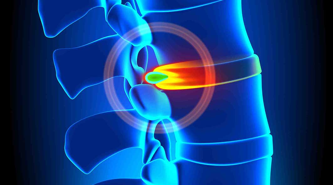
“The clinical diagnosis shows a disc bulge in their neck and some arthritis, so their neck symptoms are not associated with the crash. Lots of folks have those and do not have pain although it could be a minimal herniation. It’s our diagnosis that it was there before the crash.” This statement from an adjuster is an argument that has been made for many years, allowing insurance companies to inappropriately reduce settlements to their clients based on the client’s inability to prove when or how the damage or injury occurred. To factually counter this sort of statement, an individual must use imaging and age dating to discuss causality. Without medical experts utilizing the current medical and academic research available, it will continue to be difficult for any argument to be made explaining effects of these injuries and their mechanism based on fact vs. rhetoric.
Imaging of the spine is critically important in most cases of injured clients. In cases, imaging is necessary for proper diagnosis and future management of injuries. Imaging needs to be performed as per the academic and modern criteria to ensure an accurate diagnosis. The most common injuries in car accidents are spinal related, and the simple imaging available includes x-rays, CAT scans and magnetic resonance imaging (MRI), allowing medical providers to make an accurate diagnosis, when medically indicated.
Every medical provider has a permit to see and treat automobile related injuries. However a “license” is not the same as “specialization.” By way of example, though psychiatrists may have a license to do heart surgery and are MDs, it would not be in the patient’s best interest. Nor would I go to a spine surgeon for psychological concerns although they are licensed to treat medical conditions. In spinal trauma, certain suppliers specialize in connective tissue injuries of the spine, allowing us to go one step farther in diagnosis, prognosis and management, including “age-dating” these generally found disc and ligament injuries.
Understanding Age-Dating of Injuries
To understand age-dating, one wants to have a basic medical understanding of anatomy and physiology, and what tissue is commonly injured and the probable “pain generator”. Since neck injuries are the most common injuries cervical joints will be our focus. Related to anatomy, every set of two vertebrae in the neck is connected with three joints; two facet joints and a single disc. These joints allow for normal movement of the spine (mobility). There are multiple ligaments that are responsible for stability and hold together these joints. The correct balance of mobility and stability is critical when looking at the part of patient’s injuries, meaning that too little or too much movement in spinal joints can lead to pain, secondary to damaged tissue. The tissue most commonly hurt in a car crash is nerve, ligament, disc, facet and muscle/tendon. Spinal cord and bone injuries also happen although less frequently. To determine causality, the supplier should comment on what tissue is injured, and also use imaging to help determine if this injury occurred (age-dating).
There are two fundamental problems that must be addressed. Fardon and Milette (2001) reported, “The phrase ‘herniated disc’ does not infer knowledge of cause, relation to trauma or activity, concordance with symptoms, or need for treatment” (p. E108). Simply having a disc herniation’s presence, without a physical exam or without symptom documentation that is appropriate, does not allow one to comment on the cause of the injury. In a rear impact collision by way of example, even if the diagnosis is confirmed, additional criteria will need to be fulfilled to answer the question of “Was there sufficient force generated into the vehicle and the occupant to induce the cervical/lumbar herniation?” Fardon, in a follow-up study (2014) reported that disc injury “in the absence of significant imaging evidence of associated violent injury, should be classified as degeneration rather than trauma.” (p. 2531). Thus, we must more objectively define the subjective connotations of “violent injury” and address the issue of “degeneration as opposed to trauma”. Although this statement can frequently be misleading, it gives the trauma trained expert doctor a basis in going forward understanding that every patient’s physiology is unique and not subject to rhetoric, but clinical findings.
Violent injury to the occupant can occur when there are sudden acceleration and deceleration forces (g’s) generated to the neck and head which overwhelm connective tissue or pull them past their physiological limit. To determine the acceleration force, ?V (delta V) is utilized. ?V is the change in speed of the occupant vehicle when it is hit from behind (i.e., going from a stopped position to seven mph in 0.5 seconds because of forces moved from the “bullet” vehicle to the “target” vehicle). Utilizing these data, research allows us to make specific comments related to violent injury. Since the cervical spine is subjected to shearing forces, and compression, tension we are oversimplifying. Along with g-forces and the elastic nature of the majority of rear impact crashes makes it almost impossible to discover an actual minimum threshold for injury even though the literature has given us many examples of low-speed crashes which are dependent not simply on speed, but the mass (weight) of the subject vehicles. Each individual’s susceptibility to injury is unique. While g-forces alone are insufficient to predict injury, Krafft et al. (2002) reported that in low-speed collisions there’s an injury threshold of 4.2 g’s for males and 3.6 g’s for females. Krafft’s analysis is unique in that she has access to insurance data inaccessible to researchers. Panjabi (2004) revealed that forces as low as 3.5g impacts would lead to damage to the front of the disc, and 6.5g and 8g impacts would lead to disc damage posteriorly where the neurological components are.
Diagnosis for Disc and Ligament Injuries
A spinal biomechanical expert can look for evidence that is conclusive by disc and pathology, according to two phenomena. First, it is recognized that the body is electric. We’re measuring activity to diagnose when an EMG is done. Second, there are bioelectrical fields in all tissues. This typical field is disrupted when an injury occurs, and in the case of joints calcium is drawn to the damaged tissue. Issacson and Bloebaum (2010) reported “The particular loading pattern of bone has been documented as a significant piezoelectric parameter since potential gaps in bone have been known to be due to charge displacement during the deformation period” (p. 1271). For the patient, we have the ability to tell just how much of this process has occurred before or after their crash, especially if we take into consideration the tissue damage and signs of bone/calcium deposition.
In addition, the body begins a healing process that includes regeneration and remodeling of the soft and hard tissue as reported by Issacson and Bloebaum (2010). Spinal vertebrae have a unique structure of bone which allows it to adapt to abnormal mobility and stability (injury) by changing shape, which can be found on radiographs or MRI. Moreover, shape will change according to patterns based on the pressure or load it undergoes post-injury. Issacson and Bloebaum stated that “Physical forces exerted on a bone change bone structure and is a well-established principle…” (p. 1271). This is a further understanding of a scientific principle called Wolff’s law established in the 1800’s. Because we know what “normal” is, when we see “abnormal” findings as a result of mechanical stress we could broach the topic of an acute injury versus a degenerative process being the cause of the abnormality and create specific medical predictions accordingly.
He and Xinghua (2006) studied the predictability of the bone remodeling process and were able to make predictions of pathological changes that will occur in bone, specifically the osteophyte (bone spur) on the edge of a bone structure. Significantly, they noted their findings “confirmed that osteophyte formation was an adaptive process in response to this change of mechanical environment”. They noted that factors are crucial to the morphology of bones, particularly bones such as the femur and vertebrae.
For readers familiar with current academic and medical accepted nomenclature for disc injury, recognized from the combined task forces of the North American Spine Society (NASS), the American Society of Spine Radiology (ASSR) and the American Society of Neuroradiology (ASNR), disc herniations must have a directional component. When this occurs, the additional and abnormal pressure at the level of the disc damage matched with the direction of the herniation will cause that section of the vertebrae.
Thus, if there’s a C5/6 right sided herniation (protrusion/extrusion) secondary to a cervical acceleration/deceleration injury, then only that side of the vertebrae will change shape, creating an osteophyte. Facet arthritis is additionally caused by this compounded loading on the facet joint. This process is very similar to the formation of a callous on your hand or foot. The callous is a recognized and expected tissue response to increased load/friction exposure. Similarly, an osteophyte is a known and anticipated bone response to a rise in load/friction exposure.
At a basic level, the body has an electrical and mechanical response to injury leading to additional stress that leads to calcium (bone) to flow in the region of injury to further support the joint. The joint then abnormally grows, developing a called hypertrophy, degeneration, disc osteophyte complex, or arthritis/arthropathy, common terms seen in the reports of doctor and radiology.
Everybody is subject to these morphological (structural) changes, always and predictably determined by mechanical imbalances in the spine. He and Xinghua (2006) concluded that, “…it will actually take about over half a year to discover the bone morphological changes…” (p. 101). This indicates that it takes approximately six months to get an osteophyte (bone spur) to be demonstrable post-mechanical breakdown or failure. This again provides a time frame to better understand whether pathology of the intervertebral disc has been present for a long period of time (pre-existing) or has been produced as the direct result of the specific traumatic event by deficiency of the existence of an osteophyte, meaning the disc pathology is less than six months old, dependent on location and management of the pathology.
Conclusion
In conclusion, that by definition, a disc is a ligament connecting a bone to a bone and it has the structural responsibility to the vertebrae above and below to maintain the spinal system in equilibrium. Damage to the disc because of a tear (herniation or annular fissure) or a bulge will create abnormal load-bearing forces in the injury site. These present differently based on [1] if traumatic failure on the side of the disc lesion, or [2] if age related, as a general complex. Since other research and human subject crash testing have defined the term “violent trauma” as not being dependent upon the amount of damage done to the vehicle but rather to the forces to which the neck and head are exposed, we can now accurately predict in a demonstrable way the timing of causality of this disc lesion. This depends upon the symptomatology of the the morphology of the structure and is a subject that can be predicated upon speculation or rhetoric.
The scope of our information is limited to chiropractic and spinal injuries and conditions. To discuss options on the subject matter, please feel free to ask Dr. Jimenez or contact us at 915-850-0900 .�
References:
Fardon, D. F., & Milette, P. C. (2001). Nomenclature and classification of lumbar disc pathology: Recommendations of the combined task forces of the North American Spine Society, American Society of Spine Radiology, and American Society of Neuroradiology.�Spine, 26(5), E93�E113.
Fardon, D. F., Williams, A. L., Dohring, E. J., Murtagh, F. R., Rothman, S. L. G., & Sze, G. K. (2014). Lumbar Disc Nomenclature: Version 2.0:�Recommendations of the combined task forces of the North American Spine Society, American Society of Spine Radiology, and American Society of Neuroradiology.�Spine,�14(11), 2525-2545.
Krafft, M., Kullgren, A., Malm, S., and Ydenius, A. (2002). Influence of crash severity on various whiplash injury symptoms: A study based on real life rear end crashes with recorded crash pulses.� In�Proc. 19th�Int. Techn. Conf. on ESV, Paper�No. 05-0363, 1-7
Batterman, S.D., Batterman, S.C. (2002). Delta-V, Spinal Trauma, and the Myth of the Minimal Damage Accident.�Journal of Whiplash & Related Disorders, 1:1, 41-64.
Panjabi, M.M. et al. (2004). Injury Mechanisms of the Cervical Intervertebral Disc During Simulated Whiplash.�Spine 29 (11): 1217-25.
Issacson, B. M., & Bloebaum, R. D. (2010). Bone electricity: What have we learned in the past 160 years?�Journal of Biomedical Research, 95A(4), 1270-1279.
Studin, M., Peyster R., Owens W., Sundby P. (2016) Age dating disc injury: Herniations and bulges, Causally Relating Traumatic Discs.
Frost, H. M. (1994). Wolff’s Law and bone’s structural adaptations to mechanical usage: an overview for clinicians.�The Angle Orthodontist, 64(3), 175-188.
He, G., & Xinghua, Z. (2006). The numerical simulation of osteophyte formation on the edge of the vertebral body using quantitative bone remodeling theory.�Joint Bone Spine 73(1), 95-101.
Additional Topics: Weakened Ligaments After Whiplash
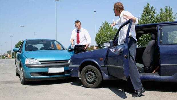
Whiplash is a commonly reported injury after an individual has been involved in an automobile accident. During an auto accident, the sheer force of the impact often causes the head and neck of the victim to jerk abruptly, back-and-forth, causing damage to the complex structures surrounding the cervical spine. Chiropractic care is a safe and effective, alternative treatment option utilized to help decrease the symptoms of whiplash.
UTEP softball head coach Tobin Echo-Hawk announced the addition of pitcher Kira McKechnie on Wednesday. McKechnie played her first two years at Fresno State and will have two years of eligibility with the Miners.
McKechnie will join fellow transfer pitcher Julia Wright, and UTEP sophomore hurlers Devyn Cretz and Allie Johnson for the 2018 season.
�We are excited to have Kira on our roster for the upcoming season,� Echo-Hawk said. �It is always nice to add some depth to your pitching staff.�
McKechnie, a native of Sacramento, Calif., made a relief appearance in the circle during the 2017 campaign against San Diego and recorded a strikeout. In 2016, McKechnie made a pair of appearances in the circle, throwing 1.1 innings, while allowing two hits and no runs.
McKechnie attended Christian Brothers High School and was a dual-sport athlete. She was a four-year letterwinner in both softball and basketball. McKechnie was voted softball team captain in 2015 and capped her senior year with multiple accolades, which includes Sacramento Bee�s 2015 All-Metro first team, Cal-Hi first team All-State, CAL All-Optimist All-Star team, MaxPreps first team All-State and Cal-Hi D3 Athlete of Honor.
She was named Female Athlete of the Year by Character Combine, Bee Preps Show and MaxPreps Christian Brothers, while earning the Credit Union Athlete of the Week in 2015.
During her senior season, McKechnie hit .462 with 40 RBI, 11 doubles, a triple and eight home runs. She added 18 runs and was walked nine times. In the circle, McKechnie (12-7) fashioned a 1.95 ERA and recorded 195 Ks 140 innings (23 starts/25 appearances).
McKechnie has competed for the California Breeze (2005-09), Capital City Comets (2009-12), Nor Cal Patriots (2012-14) and Central Cal Dirt Dogs (2014-15). She also attended the 2012 OnDeck Elite Futures Camp and 2013 Colorado Sparkler All-Star/All-American game.
Concussion, also known as mild traumatic brain injury (MTBI), has been a poorly understood condition known to the majority of healthcare providers as difficult to objectify and manage.
Historically, there has been no testing available to conclude an accurate diagnosis. In the absence of objective imaging findings of bleeding in the brain, a diagnosis of “mild traumatic brain injury” has been affixed to the condition, whereas if there’s evidence of traumatic bleeding then the diagnosis “traumatic brain injury” is applied.
Although Hartvigsen, Boyle, Cassidy and Carroll (2014) reported that 600 out of 100,000 Americans are affected every year by concussion, Jeter et al, (2012) reported that close to 40 percent of people experiencing a mild brain injury do not report it to their doctor, making accurate statistics very tricky to conclude. Despite potential under reporting in the people, we realize concussion is an issue that has consequences that are important from the perspective of a clinical result and we cannot afford to ignore this condition.
Mechanism of Injury: Mild Traumatic Brain Injury
Mild traumatic brain injury or concussion results from transfer of mechanical energy from the outside environment to the brain due to traumatic events where there’s a sudden acceleration and then a sudden deceleration of the mind and brain, such as in a Coup/Contrecoup injury during a whiplash scenario. As the brain is freely moving to a degree because it’s only surrounded by cerebral spinal fluid, it continues moving in the original direction and as the head “whips” rapidly in the opposite direction, the brain bounces off parts of the inner skull, which in turn rebounds shortly after the head changes direction. This is one easily defined mechanism of MTBI that doesn’t cause gross bleeding, yet leaves the brain injured through direct compression or overstretching (axonal shearing) of central nervous system components.
Although this has been examined extensively in the military, it’s been recently investigated in professional sports, where after several lawsuits and lives at risk, there are now definitive “concussion protocols” in place. Part of the protocols as reported from the British Journal of Sports Medicine (2016) is the Sports Concussion Assessment Tool 2 or SCAT2 that’s been adopted by numerous professional sports leagues. However, the majority of concussion victims are not active participants in the military or a professional sports team and many find their way into chiropractic practices as a consequence of sports injuries, car accidents, slip and falls and every other sort of head trauma etiology. Even though the mechanisms might vary, the induced end results are the same.
For generalized patient intake protocols, according to both Medicare and academia standards, a questionnaire outlining a summary of body systems is mandated, and part of those questions center on brain function. As reported by Jeter et al behavioral and cognitive symptoms, signs and symptoms are reported on standard patient intake questionnaires and require consideration of a diagnosis of concussion.
Prominent symptoms of concussion include: balance issues, vomiting, nausea, headache, drowsiness, dizziness, fatigue, vision, light or noise sensitivity and sleep disturbances. Cognitive symptoms include deficits in attention, concentration, memory, mental processing speed, and working memory or decision making. Behavioral symptoms include anxiety, depression, irritability, depression and aggression. The researchers went on to report that approximately 25 percent of the cases can have these symptoms persist.
Diagnosis and Treatment for MTBI
As a profession, chiropractic is a important part of the rehabilitation for the concussion population as the post-traumatic patient typically presents to the average chiropractic practice. As chiropractors (along with all healthcare providers), even if you mix the history with the above symptoms inclusive of neurological, behavioral and cognitive traits, you then have the direction or “triage road map” of the way to conclusively differentially diagnose your individual, including what tests to consider conducting in order to do so. The first line of testing is to consider imaging to rule out bleeding and ensure the patient does not require an immediate consultation. Treating blindly can place your patient in risk that is possible.
Imaging of the brain requires either MRI or CAT scans, MRI being the more sensitive, and in the absence of bleeding, the diagnosis is limited to MTBI or concussion (used interchangeably). More recently, diffusion tensor imaging (DTI) has been a tool available to picture mTBI victims that uses tissue water diffusion speeds to determine bleeding at a very small level giving demonstrable evidence to brain injury. As reported by Soares, Marques, Alves, and Sousa, (2013), DTI has several issues to overcome to certify accuracy including, but not limited to, tissue type, integrity, barriers and quantitative diffusion rates that are required to infer molecular diffusion prices. DTI is a model based upon assumption with a outlook as a tool.
Historically, MTBI was exclusively diagnosed by an omission of advanced imaging findings and the presence and persistence of the neurology, cognitive and behavioral signs and symptoms. Today, brain-derived neurotrophic factors (BDNF) offer responses about carpal brain pathology that is both conclusive and reproducible. Based on Korley et al. (2015), brain-derived neurotrophic factors is a secreted autocrine (compound hormone or messenger in blood) which promotes the development, maintenance, survival, differentiation and regeneration of neurons. BDNF also is important for synaptic plasticity (strengthening of synapses over time) and memory processing. Germane to MTBI and concussion, BDNF has been implicated in decreasing brain injury, with elevations and restoring traumatic brain injury.
Korley went on to report that BDNF levels were the highest in the normal group with lower values in mTBI and even lower in traumatic brain injury (TBI) subjects. In addition BDNF values were associated with incomplete recovery of patients that were MTBI compared to moderate or severe TBI patients. Because of this, it has been ascertained that BDNF has for identifying associated sequelae at 6 23, a prognostic value.
Korley stated that BDNF is the most abundantly secreted brain neurotrophin and as a secreted protein and can be readily measured using well-established immune-assay methods, identifying it as a non-necrosis brain injury biomarker. This distinguishes BDNF from other biomarkers which are components of neurons and myelin based proteins among other structures. In order for structural fibers to be found in high abundance in circulation, adequate cellular necrosis and damage to the blood barrier membrane must be observed, however BDNF does not require cellular damage or necrosis to be observed in circulation enabling DDNF to be more plentiful in flow than structural proteins.
Following a traumatic brain event, BDNF supports synaptic reorganization and recovery during the brain circuitry “reconnection” phase. Therefore, a better prognosis is indicated by lowered values. In patients with a co-morbidity of BDNF of anxiety, depressive disorders and schizophrenia BDNF values on the day of injury predispose this population to incomplete recovery as a risk element. Korley et al.. Concluded that serum BDNF discriminates between MTBI and TBI cases. Also, diminished BDNF values are associated with recovery in identifying and useful symptoms 6-months post-trauma.
Conclusion
Simply put, a blood test could assist providers in concluding the existence and/or severity of traumatic brain injury or mild traumatic brain injury. An early diagnosis is afforded by the results so you can devise a treatment plan inclusive of changing activities of everyday living to prevent additional damage and optimize the repair procedure with minimizing further chemical, physical or emotional stressors.
Based upon interviews with leading neurologists and neurosurgeons who understand and have first-hand expertise of both receiving chiropractic care and handling and treating MTBI patients, it is strongly recommended that until the signs and symptoms of the neurologic, cognitive and behavioral abate that high-velocity rotational cervical adjustments be avoided to enable the brain to “repair and rewire” the connections without additional possibilities of and Coup/ Contrecoup energy to the mind. This is a recommendation which we agree while recognizing that chiropractic care should not be avoided adapted to allow the brain to heal.
The scope of our information is limited to chiropractic and spinal injuries and conditions. To discuss options on the subject matter, please feel free to ask Dr. Jimenez or contact us at 915-850-0900 .�
References:
1. Hartvigsen, J., Boyle, E., Cassidy, J. D., & Carroll, L. J. (2014). Mild traumatic brain injury after motor vehicle collision: What are the symptoms and who treats them? A population-based 1-year inception cohort study. Archives of Physical Medicine and Rehabilitation, 95(Suppl. 3), S286-S294.
2. Jeter, C. B., Hergenroeder, G. W., Hylin, M. J., Redell, J. B., Moore, A. N., & Dash, P. K. (2013). Biomarkers for the diagnosis and prognosis of mild traumatic brain injury/concussion. Journal of Neurotrauma, 30(8), 657-670.
3. British Journal of Sports Medicine. (2016). Sport concussion assessment tool 2. Retrieved from http://bjsm.bmj.com/content/43/Suppl_1/i85.full.pdf
4. Soares, J. M., Marques, P., Alves, V., & Sousa, N. (2013). A hitchhiker�s guide to diffusion tensor imaging. Frontiers in Neuroscience, 7(31), 1-14.
5. Korley, F. K., Diaz-Arrastia, R., Wu, A. H. B., Yue, J. K., Manley, G. T., Sair, H. I., Van Eyk, J., Everett, A. D., Okonkwo, D. O., Valadka, A. B., Gordon, W. A., Maas, A. I., Mukherjee, P., Yuh, E. L., Lingsma, H. F., Puccio, A. M., & Schnyer, D. M., (2015). Circulating brain-derived neurotrophic factor has diagnostic and prognostic value in traumatic brain injury. Journal of Neurotrauma, 32, 1-11.
Additional Topics: Weakened Ligaments After Whiplash
Whiplash is a commonly reported injury after an individual has been involved in an automobile accident. During an auto accident, the sheer force of the impact often causes the head and neck of the victim to jerk abruptly, back-and-forth, causing damage to the complex structures surrounding the cervical spine. Chiropractic care is a safe and effective, alternative treatment option utilized to help decrease the symptoms of whiplash.
Emmanuel Korir, Michael Saruni and Mickael Hanany qualified to the IAAF (International Association of Athletics Federation) World Championships over the weekend.
Running in Nairobi, Kenya, Korir qualified to the 800m final with a time of 1:45.50 in the first heat, Saruni followed with a time of 1:46.10 in the second. In the men�s final, Korir (1:43.86) notched the crown and Saruni took third with a personal best of 1:44.61.
The All-Americans garnered a spot on the Kenya national team which heads to London, England to compete at the World Championships on August 5-8.
Also making his way to London will be former UTEP track and field star Mickael Hanany (France). Hanany took gold at the 2017 Euro Superleague with a leap over 2.26m (7-5) in the high jump. The seven time All-American will compete in his fourth IAAF World Championship.
The Nigerian trails will take place on July 7-8.
For more information on UTEP track and field, follow the Miners on Twitter (@UTEPTrack) and on Instagram (uteptrack).
Check Also
New UTEP Tennis Head Coach Ivan Fernandez announced his first two signees on Friday. Erandi �
A good read to understanding alteration of motion segment integrity (AOMSI) is the article �Biomechanical Analysis of clinical instability in the cervical spine� White, et al., Clin Ortho Relat Res, 1975;(109):85-96.
AOMSI is a biomechanical analysis. It�s all about numbers that have clinical meaning and significance. Threshold values have been determined that quantify without a doubt the patient has serious injury. It is a test of structural integrity of the ligaments interconnecting the motion segments. In this case, structural integrity has to do with the material properties of ligament tissue. Those properties include strength and flexibility. When a material is both strong and flexible, it�s called a semi-rigid material. Strength is related to the composition of the material. Strength might be thought of as load carrying capacity before failure.
Mechanism of Injury: Ligaments
Ligament tissue has previously been bench tested to describe its physical characteristics of stress/strain. That is, given so much load (stress) how much elongation will occur (strain). During normal physiologic loads the ligament remains intact and recoils to its original length when the load is removed. If the load becomes too large the materials (ligaments) begin to yield. They go past their elastic limit. When this happens the (strained) ligament fibers will not return to their original shape. The ligament loses its restraining capacity to hold the joint in normal stabilization and hypermobility occurs.
The ligaments, if sufficiently strained or avulsed results in AOMSI. The following paragraphs illustrates that if AOMSI is found there must be gross destruction or yielding of multiple ligaments. We need to build a BIG motion segment with Velcro ligaments. When you tear them off, they make a really nice ripping noise. That drives home the point.
In the White et al work, they found that the motion segment stayed intact i.e., less than 11 degrees� rotation (angualr mtion) and less than 3.5 mm translation, until they transected over 50% of the ligaments from an anterior or posterior approach. And when they transected from either approach the loss of stability was not linear but suddenly catastrophic. And they meant that suddenly the two vertebra totally separated in rotation or translation.
Suddenly Separated: pulled apart, head off of body, all neural components compromised, paralysis. Keeping that in mind, what are the injuries of someone just under the threshold? Severe to very severe. They stand the possibility of a serious event with much less force.
Prevalence of Ligament Injury: AOMSI
If AOMSI is detected, think about more than 50% of ligaments transected. That will start to explain the seriousness of the finding. In a patient/child that demonstrates hypermobility everywhere, then you take a statistical average of all segments, and look at the aberrant statistical finding if it exists. There are clues to injury everywhere when you understand what the numbers mean in reference to stability and function.
To diagnose ligament laxity, it is imperative that imaging be performed and a basic flexion-extension x-ray is all that is required. In today�s medical economy, advanced imaging of MRI or CT Scan, although accurate becomes an unnecessary expenditure and an x-ray renders very accurate demonstrative images to conclude a definitive diagnosis. In determining if there is an impairment, it is necessary to follow the AMA Guides to the Evaluation of Permanent Impairment as the 4th, 5th and 6th editions all render an impairment for AOMSI as sequella to ligament laxity, which is damage to the ligament from trauma.
This document is intended to serve as a simple explanation as to the severity of ligament damage and how to demonstrably diagnose the injury. It is also critical to remember that ligament do �wound repair.� In normal physiology, ligaments grow during puberty from cells within the ligaments called fibroblasts. They produce both collagen (white) and elastin (yellow) tissue, which gives the ligaments both tensile and elastic strength. Upon puberty the cells stop producing tissue and remains dormant. Upon injury, the fibroblast reactivates, but can only produce collage leaving the joint wound repaired in an aberrant juxtaposition (place) with poor movement abilities due to the lack of the requisite elastin. In turn, according to Hauser et. Al (2013) this leads to permanent loss of function of the ligament and arthritis of the joint. This is not a speculative statement; it is based upon Wolff�s that dates back to the late 1800�s and has been a guiding principle in healthcare for more than a century.
The scope of our information is limited to chiropractic and spinal injuries and conditions. To discuss options on the subject matter, please feel free to ask Dr. Jimenez or contact us at 915-850-0900 .�
References:
White, et al., Clin Ortho Relat Res, 1975;(109):85-96
Hauser, Dolan,Phillips, Newlin, Moore Woldin, B.A.(2013) Ligament injury and healing: A review of current clinical diagnostics and therapeutics.The Open Rehabilitation Journal, 6,1-20.
Additional Topics: Weakened Ligaments After Whiplash
Whiplash is a commonly reported injury after an individual has been involved in an automobile accident. During an auto accident, the sheer force of the impact often causes the head and neck of the victim to jerk abruptly, back-and-forth, causing damage to the complex structures surrounding the cervical spine. Chiropractic care is a safe and effective, alternative treatment option utilized to help decrease the symptoms of whiplash.
When the aberrant sequela to victims in car crashes has been investigated, providers often overlook and concurrently underestimate the tissue pathology and resultant biomechanical failures of spinal ligamentous damages commonly known as �strain � sprain.� In addition, the courts have been �blinded� by rhetoric in allowing this pathology to be deemed transient. There is an ever growing body of scientific literature that verifies strain – sprain as permanent pathology, which is the standard being taught in today�s medical and chiropractic academia.
In addition, strain � sprain as sequela to whiplash, renders a 25% whole person impairment based upon the American Medical Association�s Guide to the Evaluation of Permanent Impairment fifth and sixth editions.
Whiplash Associated Disorder Sequela Injuries
Juamard, Welch and Winkelstein (2011) reported:
��Rear end accelerations have been used to study the response of a variety of soft tissues in the cervical spine, including the facet capsular ligament. For simulations of whiplash exposures, the strains in the capsular ligament were found to be two � five times greater than those sustained during physiological motions of the cervical spine. In a similar but separate study, the facet joints of the cervical spine�s that were previously exposed to a whiplash injury ridden exercise under low � level tension and found to undergo elongations nearly 3 times greater than on exposed ligaments for the same tensile loads. Those capsular ligaments were also found to exhibit greater laxity after the purported injury. Since increased laxity may be linked to a reduction in the joints ability to stabilize the motion segment during sagittal motion, this finding suggests that whiplash exposure may alter the structure of the individual�s tissues of the facet, such as the capsular ligament, and/or the mechanotransduction processes that could maintain and repair the ligamentous structure. Accordingly, such an injury exposure could initiate a variety of signaling cascades that prevent a full recovery of the mechanical properties of the tissues of the facet joint.� (Pg 15)
Simply put, if we focus on the last sentence above, this �prevents a full recovery of the mechanical properties of the tissues of the facet joint,� which is referencing the ligaments of the spine that make up the tissues of the facet joint. In lay terms; it means that once injured, a joint is permanently damaged and it is demonstrable on x-rays with an extension and flexion view that does not have to show a full dislocation. Therein lies the core of the issue. Most radiologists are not trained in the latest literature on biomechanical tissue failures and therefore underreport the pathology.
Last month I attended a presentation by Michael Modic MD, Neuroradiology, a nationally renowned educator in neuroradiology who focuses on spondylolisthesis (vertebral segmental abnormal movements) and I asked a simple question �why don�t radiologist report more on abnormal positioning due to biomechanical failure as a result of ligament pathology� and his answer was �because their training focuses more on disease pathology.� Although I agree that is critical, so are biomechanical failures that lead to chronic degeneration, which is epidemic in our society. Simply look at the posture of our elderly for verification and much of that started with a simple �fender bender� years ago where the strain-sprain was either undiagnosed or deemed transient and not treated.
Ligament Pathology Diagnosis and Prognosis
The above scenario is why the American Medical Association values ligament pathology at 25% whole body impairment. There is also a growing body of doctors who are trained and credentialed in Spinal Biomechanical Engineering that understand how to create a diagnosis and prognosis, along with treatment plans around ligament pathology and fully understand the long-term effects of damaged facet joint tissues. These doctors are currently educating, based upon the current scientific literature their respective radiology communities to be able to diagnose and document the full extent of the injuries sustained.
We must also recognize that there is a significant amount of evidence in the scientific literature that verifies ligamentous damage as permanent and refutes the rhetorical claim of �transient.� In the end, it must be the facts of human physiology verified by science that sets the standards of healthcare and not deceptive rhetoric at any level.
The scope of our information is limited to chiropractic and spinal injuries and conditions. To discuss options on the subject matter, please feel free to ask Dr. Jimenez or contact us at 915-850-0900 .�
References:
Cocchiarella L., Anderson G., (2001) Guides to the Evaluation of Permanent Impairment, 5th Edition, Chicago IL, AMA Press
Juamard N., Welch W., Winkelstein B. (July 2011) Spinal Facet Joint Biomechanics and Mechanotransduction in Normal, Injury and Degenerative Conditions, Journal of Biomechanical Engineering, 133, 1-31
Additional Topics: Weakened Ligaments After Whiplash
Whiplash is a commonly reported injury after an individual has been involved in an automobile accident. During an auto accident, the sheer force of the impact often causes the head and neck of the victim to jerk abruptly, back-and-forth, causing damage to the complex structures surrounding the cervical spine. Chiropractic care is a safe and effective, alternative treatment option utilized to help decrease the symptoms of whiplash.
IFM's Find A Practitioner tool is the largest referral network in Functional Medicine, created to help patients locate Functional Medicine practitioners anywhere in the world. IFM Certified Practitioners are listed first in the search results, given their extensive education in Functional Medicine