Degenerative disc/s is a condition where the wearing down of the spinal discs causes pain and discomfort. All of our spines go through this, but not everybody�feels pain. Chiropractic care is a treatment option for degenerative disc disease (DDD). The first step is to determine the disc-related problem.
Chiropractic Diagnosis
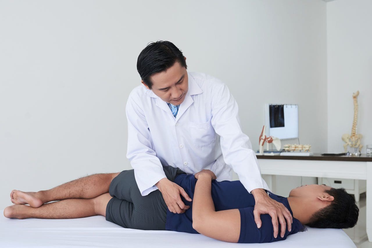
During an initial evaluation, your chiropractor will review your medical history, along with a discussion about current symptoms and the possible causes.
With degenerative disc/s��the main symptom is back pain. A chiropractor will examine potential causes:
How the mechanics of the spine are operating.
Thinning discs could bulge and place added pressure on the nerves of the spine.
Spinal stenosis can also cause back and leg pain.
A physical and neurological exam can pinpoint problem areas, like:
Where joint motion is restricted
Range of motion
Abnormal spinal curvature
Muscle spasms
Trigger points
An injury like a sprain or strain
There are different reasons why your neck or back may hurt, and your examination may include some basic tests. Observance of how you walk, and overall posture. This will help to observe body mechanics and how your spine moves. Imaging tests like x-rays or MRI are common to find the problem area and cause. The chiropractor will diagnose your condition and create a treatment plan for pain alleviation.�
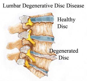
Degenerative Disc/s
The disc/s can lose their integrity, and begin to thin and possibly tear. This increases unwanted pressure on nerves and creates friction between the vertebrae. Degenerative disc disease can go through various stages. With the progression, the negative symptoms become more pronounced.
Stages
1
The first stage of degenerative disc disease may go unnoticed by the individual but can be identified by a chiropractor or other medical professional. The loss of the natural curvature of the spine can indicate the beginning of degenerative disc disease. Pain may not be apparent, but extra pressure is being placed on the spine which can lead to more rapid aging of the spine, nerves, joints, etc.
2
The degradation of discs becomes more apparent in the second stage. They may look thinner, and it is common to see deformations in the bone, such as bone spurs. The curvature of the spine will become more unnatural and the spinal canal may become more narrow. Stage 2 is often where you will begin to notice some pain and discomfort.
3
Stage 3 is marked by a more extreme change in the posture and curvature of the spine, along with more pain and loss of mobility. Nerve damage is common and scar tissue typically begins to form. Discs are even thinner than before, which can sometimes cause even more deformation of the bones.
4
The final stage of degenerative disc disease is the most severe and is typically considered irreversible. Discs are at their thinnest or gone altogether. The flexibility of the spine is extremely limited and pain is often considerable. Nerve damage can be severe and the bones of the spine may even begin to fuse together.
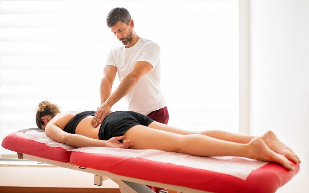
Care
Chiropractic care is a preferred treatment for degenerative disc/s disease. Because it is gentle and non-invasive, chiropractic does not create undesirable side effects like prescription meds and surgery are more prone to do. The earlier the stage of the disease, the more successful the treatment. But chiropractic can help even in the most extreme cases. The goal of chiropractic is to improve joint mechanics with improved spinal motion and reduced inflammation. The chiropractor may also work on improving the function of the intervertebral discs.
Treatments can include:
Adjustment/s
One of the most effective treatments is to ensure the spine is in proper alignment. Loss of alignment, which can happen due to injury or just regular wear and tear, puts extra stress on the spine which can accelerate disc/s degradation. Adjustments bring the body back into proper alignment.
Spinal manipulation will identify the joints that are restricted or those that have abnormal motion. They will use a gentle thrusting technique.
Flexion-distraction technique uses a gentle, non-thrusting technique that is typically used to treat herniated discs and spinal stenosis.
Instrument-assisted manipulation uses a hand-held instrument. The chiropractor or therapist applies gentle force without thrusting directly into the spine.
Therapeutic massage
A physical therapist can perform different types of massage to reduce muscle tension. Manual joint stretching and resistance techniques can also relieve pain and other symptoms.
Trigger point therapy
Here tight painful points on a muscle/s are identified and places direct pressure on these points to relieve tension.
Decompression
Spinal decompression uses gentle but firm pressure to bring space back between the vertebrae. Space allows blood flow to return and healing to happen. Decompression is an important treatment to complement adjustments.
Electrical stimulation
A low-frequency electrical current stimulates your muscles and reduces inflammation.
Ultrasound
This type can help reduce muscle spasms, stiffness, and pain with sound waves that penetrate deep into your muscle tissues. This creates a gentle heat and enhances blood circulation.
With chiropractic, prevention is the key, and therapeutic exercises can prevent symptoms from getting worse. Seek treatment for your back and neck pain now. Our team can help you feel better and educate you on living a healthier life. Your chiropractor will work hard to treat you and address all your symptoms. Chiropractors treat the whole body and not just symptoms.�
Herniated Disc El Paso, TX
NCBI Resources
A disc can also�herniate and place pressure on the nerves that flow into the feet, and cause pain, tingling, and numbness. The wear and tear of the spine combined with a herniated disc�can pinch the nerves that go to the feet.
Metabolic syndrome is a collection of risk factors that can ultimately increase the risk of developing a variety of health issues, including heart disease, stroke, and diabetes, among other problems. Central obesity, high blood pressure, high blood sugar, high triglycerides, and low HDL are the 5 risk factors associated with metabolic syndrome. Having at least three of the five risk factors may suggest the presence of metabolic syndrome. Dr. Alex Jimenez and Dr. Mario Ruja explain the 5 risk factors associated with metabolic syndrome, in further detail, as they recommend diet and lifestyle modification advice and guidelines to help people with metabolic syndrome improve their overall health and wellness. From eating fiber and staying hydrated to exercise and better sleep, Dr. Alex Jimenez and Dr. Mario Ruja discuss how diet and lifestyle modifications can help improve the 5 risk factors associated with metabolic syndrome to ultimately prevent the risk of developing a variety of other health issues, including heart disease, stroke, and diabetes. – Podcast Insight
[00:00:07] And we are live. Yes, we are. Hi, this is Dr. Alex Jimenez. Today we’re gonna be talking with Dr. Mario Ruja. We’re here together today. We’re testing out a new technology of head to head conversations regarding the whole process. Mario, how you feeling, baby?
[00:00:24] Feeling incredibly metabolic, Alex.
[00:00:29] Yes, really metabolic. I’m about to go through this mic right now. That’s what I’m talking about. Hey, we’re here.
[00:00:37] Mario and I are, you know, we’re gonna be hitting you every day. Every week. Every time we can. As much as we can. We’re gonna be going through the airways. Yeah. And we’re gonna be using the new technology to discuss exactly what we’re up to. Today, we’re focusing on an interesting disorder called metabolic syndrome. Many of you have heard the word. But really, you know, tying in exactly what it is that we’re talking about requires kind of elaborate conversation. You’ve seen it in many pictures. Mario, you can pop up the picture there PIP and you can see that a lot of times people see this gut thing going on. And that’s one of the components of it. Metabolic syndrome, when you break it down is ultimately and people notice it when they go to their doctors. Doctors are very good at assessing clinical assessments at the point where they show up in the lab work. Now, metabolic syndrome is one of these issues that many people have. And when they’re diabetic, well, they are pretty much in that range already.
[00:01:39] But before it happens, the body can stray into a metabolic area where a lot of times, for example, if your blood sugar is over 100 and you’re starting to feel like really bad, your bellies are really large. We need to have some parameters to determine it. But most people end up having metabolic syndrome and just feeling like crap. So the idea behind this process and understanding what metabolic syndrome is, understanding that there are some underlying pathologies with it. So what we’re going to talk about today is we’re going to talk about issues that are related to it. Now, in the areas of diabetes, we have, you know, complicating issues such as sleep apnea, large waistline, people who take metformin, liver disorders, nonalcoholic delivered diseases that we have heard of all fall under the realm of metabolic. But we have certain criteria that we can do that actually determines what metabolic syndrome is. Now, Mario, you’ve noticed some things regarding blood pressure. Yeah. Now, if you can show the PIP and when we see this, we can actually determine if you can kind of explain that a little bit.
[00:02:44] Yeah, it’s very simple, when you’re looking at blood pressure, your whole system. When you are out of balance in terms of your sugar, Alex, and your gut is overflowing your belt and you have issues tucking in your shirt. Now that blood has to pump hard. It has to work as a turbo. So what happens is this, at that point, this is what we call the breaking point. Anything over 140. OK, and over 90.
[00:03:27] Systolic, diastolic. Now you’re running into problems that that engine has so much pressure to make up for the resistance.
[00:03:38] Yeah, OK. Yes. The overweight, the diabetic factor, the inflammatory factor. You’re talking about triglycerides over 150. You’re talking about type 2 diabetes. OK, again, type 2 diabetes, basically, you know, you’re not born with it. It’s something that you create. You create that diabetes. Where that insulin is out of balance. And now you’re talking about, again, a very large waistline, abdominal obesity. So a lot of times if you look at people, Alex, they look great.
[00:04:16] From the chest up. Yeah. And that mid-abdomen. That torso is scary. Yeah. Yeah. It’s a showstopper, as they say. OK. So this is where the high blood pressure comes in because again, that abdominal aorta, that pressure on it puts so much pressure that it goes above 140 and sometimes it goes over 180, which is like critical, critical. And again, with these characteristics, again, it creates abdominal cholesterol. It creates blood glucose over. We mentioned that over a plus 100 and again, high blood pressure connected with what stroke? So you have triglycerides.
[00:05:15] Clogging up. Triglycerides and yes, this is huge when the arteries clog up.
[00:05:21] Yeah. We have an issue with ultimately all the roads metabolically lead to the liver. Right. So one of the things that we’ve noticed is that when we assess the liver, sometimes they look pretty good and the symptoms may be highly elevated. There’s a huge range of liver enzymes. But what we’re noticing is that if we start having a blood sugar that is elevated. If we start having enzymes, if we start having disorders like what we call nonalcohol, this was a new disease that actually just came aboard. We always knew about alcoholic liver disease or cirrhosis. Now we have nonalcoholic liver cirrhosis and liver disease. Now, how did that happen? Because our sugar was too high and the triglycerides add these fats into the fat level of the liver and start actually destroying the liver. So we started having this disorder and it’s a huge issue, as you indicated, when we start having HDL levels and that which we measure, we start noticing greater levels in 40, lower levels than 40 for men and 50 for women. We start noticing little trends. We also start noticing abdominal obesity, high blood pressure. There are other areas like ovarian cysts.
[00:06:35] Mario, you’ve noticed that there are other areas that are indicated that are collaborative or even equal or what we use to determine metabolic syndrome.
[00:06:44] What are the ones you have noticed? The two major ones where we’re looking at the studies that you see. I’m going to pull this up for you. So we can kind of get a grasp on that. We’re looking at two just very simple.
[00:07:04] Let’s make things simple for the listeners and viewers. Number one.
[00:07:11] Right away. You’re talking about.
[00:07:15] Abdominal obesity. OK, that’s number one. Number two, insulin resistance. So what happens is that your whole sugar balance within your system is not tolerating, the insulin is not effective in your body. So this is where people, Alex, are always hungry and are always eating. And so what I call it. It’s almost like you’re overeating and you’re starving at the same time. Yeah. Because that sugar is in your blood vessels. Okay. And it’s not being taken into the cells. So the cells are starving. But your whole body is overeating. Does that make sense?
[00:08:01] Mario, you know, in your practice. Like, how many people? What percentage of people? Do you notice that even have metabolic syndrome?
[00:08:10] Just a scan and again, the literature says 23 percent of the population now, I would say and in our past community here, I would say at least double that to 40 to 45. And it’s really, really a point. This is why we’re here today. You know, we’re here to educate, inspire and most of all, give people simple understanding and solutions on what to do. And one thing that I can tell you with a lot of my patients, number one, increase your fiber intake. Like, eat more vegetables. You know, I tell them that. So what do you eat? Yeah. OK. You know, so, I mean, you come in and people want to get on treadmills and they want to climb mountains. They want to do burpees. It’s like, yeah, you know what? You’re 100 pounds overweight. Those burpees are going to kill you. Okay. You’re going to wake up tomorrow morning. You can’t get out of bed. So the major factor to really start to address this is not getting in and starting a workout in gyms. First, we’ve gotta handle and we need to really educate the public on, the food intake is the solution. That is the primary solution. Medicine of the body. The food is primary medicine. And this is what we’re talking about, becoming more vegetarian, increasing fiber, reducing alcohol intake. I know I’m hurting some folks right now. Yeah. Yeah. And the point is, you know, again, reducing. It doesn’t say, you know, if you want to have a beer or something, that’s great. But again, let’s be mindful. This metabolic syndrome is a beast. OK.
[00:10:07] It’s a beast that’s affecting our parents, our grandparents. And now, Alex, I can see these patterns in elementary school kids, OK? And what they’re eating, they’re eating a lot of sugars, right? They’re eating a lot of fast foods, processed foods. This is one thing like right now I’m looking at you and you’re drinking this green. Green, you know?
[00:10:32] Yeah. There you go. Yeah.
[00:10:35] It’s like a jolly green giant drink. OK. That is a live food. It’s uncooked. It’s raw. The vitamins are there. The nutrition is there. It’s not denatured. OK. Anytime we cook food, you kill it. Right. Anytime you bottle it up and you preserve it for a month and two months.
[00:11:02] I can tell you right now, you are not eating live. So the rule that I share with my patients and when I do seminars all over the nation and people invite me over, I say, look, if you want to be alive, why are you eating dead? Simply, why are you eating dead? Right. And like right now, you know, let’s make it simple. Number one, increase hydration. Right. OK. Have a gallon of water or more is a must. Excellent. That’s number one. Number two, increase live foods. Live foods are what? Vegetables. Fruits, right? Juice them. Eat them.
[00:11:38] I mean, from what I understand and what I do is that everything leads to, any sort of nutritional component, whether it’s a treatment for diabetes or a treatment for, let’s say, rheumatoid issues when there is a nutritional component, many times it’s hard getting the foods that you need. So the world has turned on smoothies. Smoothies can date different formats. And these smoothies, as we work them, ultimately have the solution. Now, what kind of smoothies? What’s the best type of approach with them? Well, we have that stuff in terms of awareness in our offices and gladly we’ll share that stuff whenever it’s necessary. However, the reason is that those inner parts of the cells, those living enzymes, those DNA molecules, those proteins, those…
[00:12:37] Actually, probiotics, even in the fiber, prebiotics, because when you do these smoothies or even do juicing that fiber you eat, that fiber that sometimes is lost while you do it juicy, juicing. This is important for the bacteria. So that helps even with disorders, such as leaky gut or intestinal dysbiosis, because they all come together. Someone who has metabolic syndrome most likely has leaky gut and vise versa. And not every single time, we can assess that, but what we want to do is we want to assess a person completely in terms of the drugs. You pretty much know that your doctor is trying to get you away from diabetes because you’re in that losing control place when you’re taking metformin.
[00:13:20] Metformin is very powerful, it’s a special medication that ultimately guides to restores the blood sugar back to where or it makes it more effective or makes you less insulin tolerant and more sensitive. So there’s a lot of things that we’re looking at that are useful for us. But one of the things is, well, what am I going to do with this?
[00:13:41] How am I going to get better? Well, diet? Diet has everything to start with. You start with your diet. You start with being vegetarian. You start with Mediterranean style foods. What kind of techniques do you use in terms of diets? Because I can go off and explain those things. But I want you to kind of get. Simple.
[00:13:56] You know, simplicity is golden. Complexity is chaos, Alex. The more complex we make it for ourselves, the more likely it is that we’re going to quit. OK. You can’t sustain complex things. We need to make them simple. So number one, as much as possible, eat raw, eat live foods. That’s number one. Number two. Number two, stop eating processed things like you’re talking about like high corn sirup, like simple, stop drinking Cokes and all of these fruity drinks and everything else, you know. And it tells you right there on the bottle, right there on a can. It’s 10 percent fruit. Do you know what 10 percent means? It’s not 100 percent. There’s a missing zero, baby. OK. It’s missing. So you know what? After a while, you’re gonna be missing off the planet. OK. You’re going to be extinct. Yeah. So, yeah. We need to get real, you know, this is real stuff. Like I can tell you, you know, I visit, I do home visits sometimes because I give back to the community and I go to families and I sit there and, you know, people are losing legs. People are losing limbs. They have wound care, you know. They have, you know, they have issues in terms of that. And that’s painful, not only for the person but for the whole family. So you know what? I take this very seriously. We take this very seriously at the show. You know, we want to make it live. We want to make it interesting. But I want to tell you right now, I’m not playing games. This is not game time. This is showtime. And so in terms of that, get off the Cokes, get off the fruity drinks, get off the candy bars. OK. You know, I think that commercial with Snickers, they need to change that. You know, instead of the Snickers satisfy, how about the Snickers gonna kick your ass? How about that one? Yeah, yeah. I said it. Thank you. I think we are physicians so we can say that word. Right. So that’s what’s happening. And then again, the lifestyle. If you’re drinking, the two things I can tell you right now. Yeah. Two things that are going to kick you in your gut, no pun intended. Metabolic boys and girls. OK. Number one is going to be smoking. And number two is going to be alcohol. You do those two.
[00:16:16] OK. And here it is. I’m going to tell you what it’s, you know, can you find that on the slide. OK. It’s gonna be the statistic. Oh, here it is. There. Bam, right there. Pull that up. Yeah, ok. Bam. OK. So if you’re looking at that, I’m going to tell you this is scary. Lifestyle factors, the two factors right there. Can you enlarge that? So we can see that. We can do that. OK. Excellent. I appreciate that. That’s all big. There it is. So now watch this. Do you see this? OK. Here it is. The number one life factor right here. Number one, is that? Yes. The one right here? OK. Now watch. I want to circle this. OK, where is it? It’s right there. Tools. I’ll get it for you. OK. Go ahead. Circle that, smoking and heavy drinking. Okay. I just want everyone to kind of take note, smoking and heavy drinking. That is one of the most destructive things that you can do right now. And guess what? Most people do them together, don’t they? Yes, sir. That’s it. So now watch, the ratio that metabolic syndrome affecting the man. OK. This is, again, something new. Mm-hmm. It’s affecting the man less than the women. Do you see that, guys? Yes, I do. Look at that. The women is 4.45. The women are affected most out of everyone. Out of everyone. And the men are at1.85. Now, the lesser evil is heavy drinking and poor diet, and the less one is smoking and physical activity. But if you really look at it, that’s what’s scary, smoking and heavy drinking. And this has really come down to a shift.
[00:18:04] You know, you used to be that men smoked and men drink. Now it’s changed, Alex. This is scary because it’s affecting, you know, momma is the boss. And to me, you know, mom is the doctor in the house. OK. And no, I don’t want to take responsibility for the man because you know what? We need to be the head, not the tail, but at the same time, who is going to take care of the kids?
[00:18:28] It’s got to be mama. You know, most of the time. Who’s going to take the kids to the doctor? Who’s gonna be wrong? So we need moms healthy. We need moms healthy. OK. We can, you know, because there’s a saying that says this, Alex. When mom ain’t happy. Dad ain’t happy.
[00:18:44] Nobody’s happy. No, thank you. Here, even the dog ain’t happy, Alex. He’s leaving now. He’s gone. He’s gone.
[00:18:52] I happen to know a lot of, I think after about 40, I think that in general there’s a tendency for the love of wine and it gets a little crazy for a wine to three a night. Yeah. This leads to metabolic syndrome. So we need to. Moderation is key. Right. Yeah. So we’re going to you know, if you’re if one of the biggest treatments is cardio, well why give yourself those extra calories and doing their process?
[00:19:19] Now one glass is fine. I understand that. But we don’t have to go crazy with the wine in the evening because it’s a more relaxing thing. You know, there’s always women’s night out, right? You know, I mean, it’s shot here, but women’s night out. You know, and when there’s women’s night out. And for many women, it’s a little bit of vino. So we need to kind of cater to those things, shall I say.
[00:19:39] And it’s woman’s night out. But it’s not all night, baby, you know?
[00:19:42] Yeah, OK. I mean, you know, there is.
[00:19:45] Exactly. I mean, you know, let’s have a glass but not the bottle, baby. Come on. Well, you know. Right. If you have 4 people a bottle is OK. OK. I said wine. Okay. I’m sorry. I lost. I lost the meaning of that. Go ahead Alex. Yeah.
[00:19:57] So the bottom line is, that we’re here to bring in awareness of this disorder, which is metabolic syndrome.
[00:20:05] Honestly, I’ve been going to school for a long time. And this is a new revelation of the last decade.
[00:20:12] The gastroenterologist is really focusing on. They’re the first to see it, the dimensions of. And here’s the thing. Crazy metabolic syndrome leads to nonalcoholic, fatty liver disease. And you were seeing this in rampant levels, literally. Gallstones. Exactly. Because the liver, the cholesterol issues, all these dynamic changes are affecting even our children. We’re having kids with nonalcoholic fatty liver disease. Why? Too much sugar? Too much sugar? We have to control the sugars. And there are things that we can do in our diets. Plan on bringing all those concepts.
[00:20:46] But we want to bring awareness as to what happens. Lack of sleep, cortisol raising, you know, all this kind of stuff alters the blood sugar in our system. So it’s very important to do the best we can. Exercising is awesome for this stuff in terms of cardiogenic exercises, cardiometabolic dynamics, though. That’s where we want to still focus on. We have to do a little bit of cardio. We got to, you know, eat more of vegetables, greens, juicing, those kinds of things sleep better. It’s important to sleep.
[00:21:15] Alex. Oh, OK. I want to jump in because I know we’re jumping here, you know, and people and I like the fact what you mentioned earlier, you know, we want to, you know, get some wine and some things to relax. Why don’t we do this? I encourage people to meditate. OK. And to try to create some stress management strategies. OK. Right.
[00:21:38] Like a nice, you know, nice warm cup of tea an hour before we go to sleep. You know, some chamomile tea and I mean, you know, chamomile, you know, and.
[00:21:50] Yeah. Good stuff. Yerba buena. You know, stuff like that. You heard about it? Yeah. Yeah. So.
[00:21:56] So you know, all of these things. Meditation because why? Stress levels, as you mentioned, I’m on one accord with that stress level increase cortisol, which contracts, arterial function and then decreases dopamine.
[00:22:13] OK, and oxytocin, which is like the love thing going on. OK. And so now all of this creates sleep apnea. And how many people do you know, Alex, that suffers from sleep apnea and instead of dealing with the causation? OK. So this is why chiropractic is such a beautiful thing.
[00:22:35] You know, for 25 plus years, a quarter of a century plus, you know, between both of us, we’re like, gosh, 60 plus years. Correct? Yes, 60 plus years. All right. Chiropractic and chiropractors have such a beautiful story and beautiful platform because we’re all about natural healing and helping our community and our country at very, very cost-effective.
[00:23:09] You know, we do one of the most cost-effective ways of not only treating health but preventing health. And we are ambassadors of health. I mean. And so this is where when we’re talking about, again, sleep apnea, meditation, I see so many people in my practice, they’re taking a pill to go to sleep.
[00:23:32] Every night.
[00:23:34] They suffer from depression. They suffer from anxiety. Okay. And then I look and I go, you know what? Let’s talk about your lifestyle. What are you doing? What are you doing every day to put your body in a high, intense, inflammatory system in high, intense stress? Yeah, you’re redlining. I always tell people. Right now you’re redlining it’s just like a car. You cannot maintain it. You better change gears, otherwise, you’re going to blow the engine. And this is what I see, sleep apnea. Sleep is again, all the way from athletics to life function. That is where the neuroreceptors neuroplasticity, Alex. Okay. That’s where we heal. We recover. We reset for the next fight, which is early in the morning. And if we don’t do that, we go to the next day with that fog. Yeah. You know that mental fog. Alex. Hey, you know, and this is where people say, you know, I can’t focus. I’m forgetting, you know, and I don’t know what’s happening, you know, and I’m going, you know why you’re not sleeping?
[00:24:47] Exactly. You know, we’ve done the studies and the studies specifically about sleep. If you’re a person that you need seven hours of sleep and you miss one hour, just one hour of sleep.
[00:24:58] The mind is very just speaking about just the brain fog that happens with this metabolic syndrome because it really starts disrupting everything. One hour of sleep actually diminishes your ability to be creative. Creativity. Yes, the brain is altered. And you may think that you know, 25 percent by one hour, it makes a difference. But literally two hours of sleep, you lose 50 percent of your creativity if you lose four hours of sleep. Well, no one of those when you just push it like four hours. Your creativity is so low that even just trying to figure out how to find where your keys are or creativity, how to solve problems, how to deal with issues, you go to work and someone’s got some drama. It stresses you out and metabolically and then what would a lot of people do? You go to work. You jack yourself up with some coffee and then you grab what? Many people grab the donut instantly, send in the blood sugar into chaos. This constant repetitive issue of habits leads to the disorder of metabolic syndrome.
[00:26:02] The body, the body. Alex cannot sustain us. You know, you can’t. As I mentioned before, you can’t drive that, that your car in first gear going 80 miles an hour. You cannot. And so it’s going to rip at the seams. The gasket is going to come off. The oil is going to spray out. OK. And this is what’s happening. If you really look at the body, we look at the HDL cholesterol, it’s lower than 40. They should be the highest, high density.
[00:26:33] High density should be the highest. Cholesterol, good cholesterol stuff.
[00:26:37] You know, you want, LDL should be low and the HDL should be high. You’re looking at insulin. You’re looking at strokes. You’re looking at again, you know, triglycerides over 150, you know, sleep apnea again, more than ever. Now I’m hearing about it. Polycystic ovaries. I just hear this, you know. You know, I have another surgery. You know, I have cysts.
[00:27:04] I have this with all of these things. If we really look at it, we are seeing that we are pushing ourselves over the cliff. Yeah, OK. And then I’m going to throw this in there with the onset of overuse of cell phones by adults and by youth. Now it is disrupting the cognitive pattern, the focus pattern. And it’s throwing people where they don’t go to sleep because they have a cell phone in front of them at 11 o’clock at night in bed. You know, and then you’re talking about like one hour less than two hours. Well, let me tell you, the worst thing you could do, Alex, is have your cell phone next to your head with the radiation, with the sounds every time that phone beeps. Tweaks a bell, sounds for an email or your Facebook. Guess what your subconscious in your brain does? It wakes up. It wakes up and bam, it blows up.
[00:28:11] That REM sleep you only have. What is it, Alex? What, ten, 10 minutes, less than 15 minutes, of REM sleep? Oh, there’s a lot of. What is it?
[00:28:20] What happens is the body goes into a bunch of different stages as it does. What we’ve learned over the years is that we used to believe that REM was something intermittent. It goes into these deep, deep levels throughout the night.
[00:28:31] Your body temperature, what your blood sugars at, what’s your mental state, what’s your tired levels is, what your electrolyte balance is. This matters. So sleeping is very important. So in the restoration of your body through to get it back in order. Sleep is one of the greatest ways to be able to restore the body. So it’s important to try to you know, if you go to bed at 10, push yourself to start working the process, to go to bed probably about an hour earlier or start working. And the TV is going to be there. They’re going to keep on going 24/7. But, you know, we’re the ones dealing with the issue later on in the next day where we are brain doesn’t work well.
[00:29:10] Our bodies are needing carbohydrates, our foods are. And don’t eat, you know, and put yourself on one of the things that I encourage. Again, my children and, you know, Karen and the kids. Is this. Put your cell phone in the kitchen and get a real alarm clock. I don’t think they make them anymore, do they? They don’t make alarm clocks anymore. Now, forget it. What’s an alarm clock? It’s kind of like a pager, Alex. You know what? We’re gonna get to the point where we’re going to say, hey, do you have an alarm clock? And I go, now I’ve got a pager. All right. Okay. It’s ridiculous. We need to have a phone in the kitchen. You remember back in the days with that long cord. About like 80 yards. You used to take it in your room for those private sessions, you know, with your girlfriend and all of that.
[00:29:58] Well, let me tell you, that cell phone needs to be in the kitchen. It needs to be turned off. Okay. And then you need to have an alarm clock next to your bed. You need to honor sleep. You cannot eat foods before you go to sleep. Go to sleep hungry.
[00:30:16] One of the things that I’m reading more and more in terms of research and in terms of data. Go to sleep, hunger, you’re not going to die. All right. Calm down. Well, I know what you’re talking about. Yes. Intermittent fasting. Absolutely. OK. Yeah. Yeah. You can’t. I mean, at the end, you know, let’s say 8:00. That’s enough. Put the burger down.
[00:30:35] Yeah. Look at that. You know.
[00:30:37] You know, 300 years ago, we would be we many of us were nomadic. You know, people live culturally on the sides of the earth. And the terrain was different. We got the food during the daytime. Nighttime was a time when you kind of just, you know, settled in. All right. If you did, have you had some grains, some nuts, and it was different. So just by using the sunlight in order to kind of like you, as soon as the sun comes up, you can eat and shut off at night if you get really good and you start using what we’re learning, that is a great method, which is intermittent fasting. The body has the ability to rectify itself. This is an amazing, metabolic syndrome or not. The body even activity stream has the ability to rectify itself.
[00:31:23] So if you allow the body to cleanse itself throughout the day, let’s say you do an eating cycle of only eight hours on a window, so to speak. Well, you got a good, you know, 16 hours of what period of where your body breaks things down. Well, the metabolic processes of the breakdown of usually the mitochondria, the mitochondria, as it starts working, the process needs to rest. Our bodies need to rest. I mean, if I told you to cut the grass, Mario, you know, cut the grass. And as soon as you walked in the house and you were done and I gave you some lemonade and you were just kinda, that was rough. And I said, go back and cut it again. Right. And then also you go back out and you’re like, wow, this is crazy. When what kind of guy? And then right when you’re tired and you’re shaking because you have no energy and you’re about to go to bed. All right. And you go get up because you’re gonna go cut the grass again and you never let the body rest. Eventually, your body breaks down. That’s what happens with mitochondria. If you’re constantly eating, you’re constantly going through a process, burn out, burning, burning and burning. It needs time to settle and relax, to recover. And that’s what metabolic syndrome recovery process is. Do we try to calm the body through sleep, through lifestyle changes, through blood sugar issue changes? And little by little, we’ll start getting back control of your life because otherwise you have increased steroid or what we call a cortisol steroid production, which then makes the body store fat. Right. And then that’s where you get the belly issues because one of the first indicators of metabolic syndrome is a big belly. Right. For a woman, it’s over a certain amount, over 35 inches. And for men, anywhere over 40, that’s a lot of people over 40 inches. You pretty much have a kickoff towards metabolic syndrome.
[00:33:00] And how many times have you heard in your practice, Alex? You know, people come in and go, you know, I just need to lose weight. I just need to lose weight. And, you know, I look at him and I go, you know, forget about the weight.
[00:33:14] What are you doing?
[00:33:15] Are you sleeping well? All right. Right. Exactly. I mean, first. I mean, let’s forget about the weight. The body knows how to calibrate, Alex. It knows how to calibrate. It knows the zone, the sweet zone, the target zone. It knows genetically DNA, RNA. We have an innate intelligence. OK. We learned this in school and chiropractic school.
[00:33:38] The body has innate intelligence to know how to heal, to know how to recover, to know how to grow. And to know how to survive when we put undue stress to our system, to that fiber optics, to that circuit, we blow it and then it goes a wall. And that’s what happened. So I tell people, you know, no, the answer is not for you to go to a gym for 20 hours a day. No. Why don’t you start with letting go of high corn, syrup high fructose corn, syrup drinks?
[00:34:17] Why don’t you do that first? That’s what I tell them right away. Do that first. Number two, eat more fiber. What does fiber mean? Eat more legumes. Spinach, OK. Like broccoli. And instead of cooking them. And I know most people don’t want to eat raw. Why don’t you just steam them? OK. Just kind of steam them a little bit. OK. Don’t fire. Don’t kill them. Steam them. The other thing you want to do is, you know, get up on a Mediterranean diet, you know, fresh seafood, things like that, regular exercise, just get up in the morning. The first thing that I would share with people is just make things simple.
[00:34:53] Wake up when the rest of the animals wake up, OK? Wake up. And just go for a walk.
[00:35:02] At least, you know, let’s not get on a treadmill and break a record, OK? Let’s not do that because you’re gonna do that twice. You’re gonna get hurt and you’re gonna quit for the next three months to recover. So why don’t you just get up and walk? And then next time walk a little more and walk a little faster and next time walk a little faster and jog. So we need to create that patterning, rituals. We need healthy rituals, Alex, in our lives instead of rituals that are killing us slowly. OK. And so in terms of that, I look at eating more fresh vegetables, Mediterranean, sleep better and honor that sleep. Get up earlier, get up before 5 o’clock. OK. Get up. Get into a routine that way when you when you’re not sleeping late. OK. Your body’s tired. So now you can go to sleep at nine-thirty, at ten o’clock, OK. And then create a new pattern to where you let go of the cell phone. And I would say, you know how people do like fasting or cleansing. I recommend at least two cleanses a year, two cleanses a year. You know, stop eating meats.
[00:36:15] OK. Stop eating meats.
[00:36:17] And just go vegetarian. Okay. Let your body cleanse, I know you have wonderful programs at your clinic. Alex, in terms of detox, I believe in that twice a year. You gotta do it. It’s just like flushing your car. Alex Right.
[00:36:31] From detoxing. We start we really begin the process at the intestine. So from the things we start eating. So one of the best ways to do it is to go through a detox program and the detox programs help us cleanse it.
[00:36:45] You know what? If your garage is full of mud, if you’re you know, you just open the garage, you see it’s just a mess and your body’s metabolically a wreck similar to a garage as an analogy. Well, you can have a couple of things. The first thing we do is we open up the garage and we got to plunge that garage. We’ve got to get the hose. Or you can take the broom. And you know what? Many times you’ll realize that taking the hose is probably the best thing. So when we do detox programs, we begin by helping with the colon cleanse. And we basically clean out the colon and eventually also repurpose the direction where the probiotics go. We actually add probiotics in the diet. We add the fibers, the prebiotics, the post probiotics. And we start working from the intestine out. The body instantly starts recovering. And little things that we can do in the mornings on a regular basis such as increase water, take a lemon, for example. We start with water. We start with let’s say you squeeze four or five, three lemons, just three lemons, squeeze the juice, put it in a drink that you’re gonna spend all day. Take one whole lemon to squeeze it nice and just take it as a shot. Some people use apple cider vinegar. Exactly. So what we do is we start that well, that goes into the intestines, into the stomach. It is much it just basically showers the liver with a process that begins the process of cleansing as the body starts recovering. You start eating better. You let’s say you do a fast and you begin your process of eating at around 12:00 and you stop at six or maybe seven or maybe eight. And it gives you an eight-hour window, six to eight-hour window and that’s it. And then you start recovering in that process. So early on, we start doing the changes with the beautiful thing about the body if you give it time, it’s all recoverable to do it. And if you teach the body, you don’t have to be taken metformin the rest of your life. Now, if your doctor thinks it’s appropriate, well, then, you know, you speak to your doctor. And here’s the other thing is, is that when you’re dealing with metabolic issues and you’ve got someone that’s giving you metformin and as it or other diabetes or different types of medications that help with diabetes, it is appropriate to have it under a doctor’s control. Now, the constant taking medications without, having lifestyle changes or not having dietary alterations is inappropriate. It’s malpractice, I believe, in my opinion, after being here so long, you just don’t give people pills and not change their diet. You got to have a doctor who’s willing to sit down and help you out with lifestyle changes, diet or someone that has some staff to support you or a dietician or a registered dietetic dietitian. So that can help you or a nutritionist. Sorry. Or a health coach that can help you. We have to have people that can do that. So to help you with the process, because it’s not just saying, hey, take a pill and go off on your own, that confuses a lot of people. We have to have a change and we’re here to help you with that. So as we go through this process over the next couple of weeks, Mario, as we discuss each one of these particular topics, we’re going to bring it to you. We’re going to talk about ways that you can change your diet that simple. Just make it simple and it adapts. So I know you’ve got some points you want to mention again.
[00:39:50] Absolutely. You know, with metabolic syndrome, the two things that are markers are number one, your belly fat. OK, so let’s get going in at home. Yeah. Let’s wrap it up. You know, let’s button this thing up and make it simple. First of all, it’s belly fat. So. So if your belt, when you’re buying a belt and you’re looking at the little notches and the little holes and they’re over 40 inches, OK, 40, you’re in trouble. For male. Yeah, for male. And if you’re over 35 for a female, you’ve got issues.
[00:40:24] Metabolic syndrome. Absolutely. Absolutely. And the other point is this. If you have a hard time and you can’t even wear a belt, then I don’t need to explain any further. It’s time to come in and get a coach, get a health coach, get someone to make you accountable because time is ticking. Time is ticking and we need to be proactive instead of reactive. And at the same time, when you are always hungry, you’re overeating and you keep being hungry. You cannot lose weight. Now the things are coming to fruition. And on top of that, if you have blood pressure, over 140, over 90. So let’s say 145 over 100, let’s say 150 over 105. It’s time to get real. You can do this. Just go buy a blood pressure cuff, OK? Quit playing games. Get-go. Go to Walgreens. Get a blood pressure cuff and check it tonight. And if it’s over. Check it at least. I always tell people, don’t do it once. Keep a record and do it for at least three, three to four days. OK. And take it in the morning. In the afternoon at night so you can see a cycle. Bring that over and find yourself a team. Find yourself a team that will that has a program for you to naturally empower you and give you the right tools to get back to your best life ever. It is up to you. It’s called self-responsibility. No pill is going to take the responsibility from you. And at the same time, you can enjoy your family more, enjoy your life, and be happier in the process.
[00:42:19] So I really appreciate it, Alex. And I want to thank our listeners and viewers and we look forward to continue the conversation of health and wealth. Because I want to tell you, the message for today is your health is your ultimate wealth. Thank you. And we’ll leave it there.
[00:42:38] All right, guys. Thank you.
[00:42:39] And we’ll come back tomorrow and we’ll add another health nugget, so to speak, for you guys. Two miners.
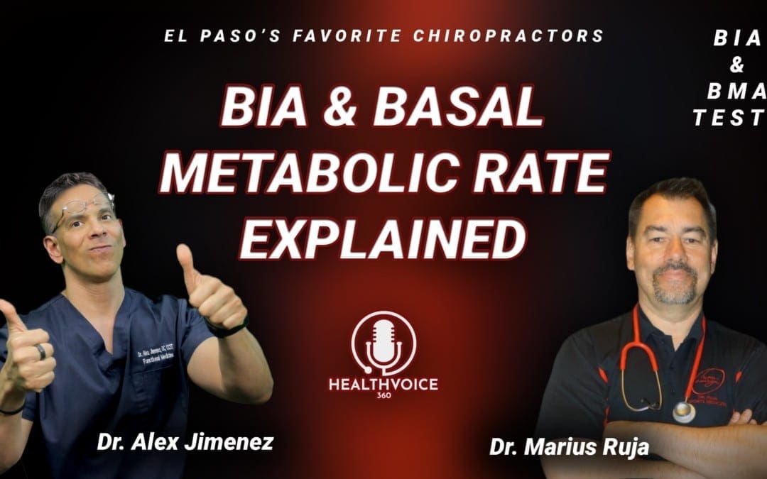
Podcast: BIA and Basal Metabolic Rate Explained
Dr. Alex Jimenez and Dr. Mario Ruja discuss basal metabolic rate, BMI, and BIA. Body mass and body fat can be measured in a variety of ways, however, several measurement tools may ultimately be inaccurate for many athletes. According to Dr. Alex Jimenez and Dr. Mario Ruja, calculating an individual’s body mass and body fat utilizing various tools is essential to determine overall health and wellness. BMI uses a person’s height divided by twice their weight. The results may be inaccurate for athletes because their body mass and body fat are different, in terms of weight, compared to the average person. Dr. Alex Jimenez and Dr. Mario Ruja demonstrate that BIA, or bioelectrical impedance analysis, and various other tools, such as the DEXA test, the Tanita scale, and the InBody, among others, can help more accurately determine an athlete’s body mass and body fat. Basal metabolic rate, BMI, and BIA are essential for parents that have young athletes as well as for the general population. Healthcare professionals that have these tools available can ultimately help provide individuals with the results they may need to maintain overall health and wellness.
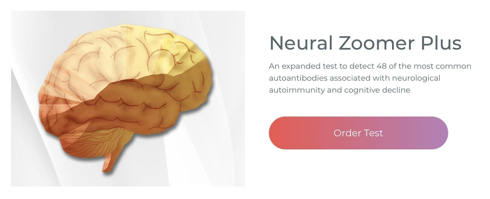
Neural Zoomer Plus for Neurological Disease
Dr. Alex Jimenez utilizes a series of tests to help evaluate neurological diseases. The Neural ZoomerTM Plus is an array of neurological autoantibodies which offers specific antibody-to-antigen recognition. The Vibrant Neural ZoomerTM Plus is designed to assess an individual�s reactivity to 48 neurological antigens with connections to a variety of neurologically related diseases. The Vibrant Neural ZoomerTM Plus aims to reduce neurological conditions by empowering patients and physicians with a vital resource for early risk detection and an enhanced focus on personalized primary prevention.
Food Sensitivity for the IgG & IgA Immune Response
Dr. Alex Jimenez utilizes a series of tests to help evaluate health issues associated with a variety of food sensitivities and intolerances. The Food Sensitivity ZoomerTM is an array of 180 commonly consumed food antigens that offers very specific antibody-to-antigen recognition. This panel measures an individual�s IgG and IgA sensitivity to food antigens. Being able to test IgA antibodies provides additional information to foods that may be causing mucosal damage. Additionally, this test is ideal for patients who might be suffering from delayed reactions to certain foods. Utilizing an antibody-based food sensitivity test can help prioritize the necessary foods to eliminate and create a customized diet plan around the patient�s specific needs.
Gut Zoomer for Small Intestinal Bacterial Overgrowth (SIBO)
Dr. Alex Jimenez utilizes a series of tests to help evaluate gut health associated with small intestinal bacterial overgrowth (SIBO). The Vibrant Gut ZoomerTM offers a report that includes dietary recommendations and other natural supplementation like prebiotics, probiotics, and polyphenols. The gut microbiome is mainly found in the large intestine and it has more than 1000 species of bacteria that play a fundamental role in the human body, from shaping the immune system and affecting the metabolism of nutrients to strengthening the intestinal mucosal barrier (gut-barrier). It is essential to understand how the number of bacteria that symbiotically live in the human gastrointestinal (GI) tract influences gut health because imbalances in the gut microbiome may ultimately lead to gastrointestinal (GI) tract symptoms, skin conditions, autoimmune disorders, immune system imbalances, and multiple inflammatory disorders.
Formulas for Methylation Support
XYMOGEN�s Exclusive Professional Formulas are available through select licensed health care professionals. The internet sale and discounting of XYMOGEN formulas are strictly prohibited.
Proudly,�Dr. Alexander Jimenez makes XYMOGEN formulas available only to patients under our care.
Please call our office in order for us to assign a doctor consultation for immediate access.
If you are a patient of Injury Medical & Chiropractic�Clinic, you may inquire about XYMOGEN by calling 915-850-0900.
For your convenience and review of the XYMOGEN products please review the following link. *XYMOGEN-Catalog-Download
* All of the above XYMOGEN policies remain strictly in force.
Modern Integrated Medicine
The National University of Health Sciences is an institution that offers a variety of rewarding professions to attendees. Students can practice their passion for helping other people achieve overall health and wellness through the institution’s mission. The National University of Health Sciences prepares students to become leaders in the forefront of modern integrated medicine, including chiropractic care. Students have an opportunity to gain unparalleled experience at the National University of Health Sciences to help restore the natural integrity of the patient and define the future of modern integrated medicine.
Dr. Alex Jimenez and Dr. Mario Ruja discuss basal metabolic rate, BMI, and BIA. Body mass and body fat can be measured in a variety of ways, however, several measurement tools may ultimately be inaccurate for many athletes. According to Dr. Alex Jimenez and Dr. Mario Ruja, calculating an individual’s body mass and body fat utilizing various tools is essential to determine overall health and wellness. BMI uses a person’s height divided by twice their weight. The results may be inaccurate for athletes because their body mass and body fat is different, in terms of weight, compared to the average person. Dr. Alex Jimenez and Dr. Mario Ruja demonstrate that BIA, or bioelectrical impedance analysis, and various other tools, such as the DEXA test, the Tanita scale, and the InBody, among others, can help more accurately determine an athlete’s body mass and body fat. Basal metabolic rate, BMI, and BIA is essential for parents that have young athletes as well as for the general population. Healthcare professionals that have these tools available can ultimately help provide individuals with the results they may need to maintain overall health and wellness.
Podcast Insight
[00:00:08] All right. It’s Mario and Alex time. The two favorite chiropractors from El Paso, TX. Ok. We’re going to be… Functional medicine, Alex. That’s what we’re gonna do. It’s about functional medicine in 2020, baby.
[00:00:21] This 2020, we’re gonna be focusing on BMI and we’re gonna be focusing on everything. Mario, my awesome co-host here we’re tearing it up. We’re gonna give some points of view. We’re gonna be discussing certain things. Today our focus is going to be on anthropometric measurements and measuring the body composition rationale and its interpretation.
[00:00:46] Now I’m afraid of that. All right.
[00:00:49] I’m afraid of measurements, Alex, I’m telling you right now, I don’t want measurements around my body.
[00:00:55] Okay. Thank you. All right Mario. Yeah.
[00:01:00] Mario, we’ve got to get a little bit of knowledge here. Okay. Well, what we’re not going to do is we’re not going to try to make this boring. No. If you really want to see boring. I think we have plenty of examples of what boring looks like. Yeah. Have you seen those boring guys, Mario? You know, it’s like the measurement of what’s going on. Yeah. Here you go.
[00:01:20] Video plays in the background.
[00:01:31] You know what? I can go to sleep with that one, Alex. Now, that’s what I’m talking about Mario. I can go to sleep and just shut it off.
[00:01:40] But, you know, learning has to be fun. It has to be interactive and it has to be functional.
[00:01:47] So that’s what we’re… Absolutely I totally agree. So what we’re gonna do is we’re gonna try to bring the facts as it can be and we’re gonna try to bring it with a little bit of slapstick fun.
[00:01:56] So it’s gonna be fun. Mario, tell me a little bit about your interpretation of BMI as how people understand basal metabolic rate.
[00:02:05] Well, this is what I understand and what I hear about basal metabolic rate.
[00:02:13] Bottom line is, can you put your belt around your pants and can you tuck your shirt in? How about that?
[00:02:25] You know, that’s pretty scientific. Right. That is scientific. Yes, that is scientific. Yes. We could talk pear, we could talk apple, sizes, apple-shaped bodies types.
[00:02:33] But we’re going to get specific here because people want to know, Ok, what’s going on. Let’s start. One of the things that we can do is we can start discussing calculating energy requirements, because one of the things that we want to see, as you can see, I put up here a little bit of facts so that it can help us out a little bit in terms of figuring out what’s the best approach in terms of what we do. Now, you can tell here that sedentary, no exercise, what we want to do is talk about basal metabolic rate. Ok. So this is a measurement that has occurred by height as well as weight index. So it comes out to that number and we can start looking at calorie, caloric intake burn. But when we do a BMR and we calculate this number, we typically want to get about a 1.2. And that’s what would be normal in most situations if you’re sedentary, light activity, we start noticing that there’s an increased activity expenditure and BMR should be one point 1.375. If you are moderately active, you should start doing that. So in its interpretation…
[00:03:33] Mario, when you see these kind of things and these kind of figures, what does it bring to mind for you in terms of these numbers? As we keep on going back to this, we’ll be able to see exactly what’s going on. What’s your incentive sense of the rates and the metabolic processes?
[00:03:52] Well, again, very simple, when you look at it as the more active you are, the higher your metabolic rate is. That’s it. So at the end of the day, we want to put it in very simplistic terms to the public. We want to be more active about that. So science is supporting that, you know, park the car as far away as possible from the Wal-Mart entrance and your work. So by doing that every day, you are creating a higher function. Ok, metabolic, that’s the burn. That’s your whole system burning fuel within yourself. So it’s simple. And the studies are showing that the more active you are, the higher your metabolic rate is. It can go up to a 1.9 from a 1.2. Correct.
[00:04:50] Exactly. So what we’re looking at here is that the requirements are going to be pretty high. If you are one of those people that are very active. So ultimately, our goal is to get you as active or what you’re your lifestyle could require. So, you know, if you’re a mechanic, you say moderately active. If you’re someone who works in, let’s say, an office, your BMR is going to be calculable. Using these numbers for the body mass index, the whole idea is to try to figure out the body mass index using the BMR. So the BMR allows us to kind of give an estimate, the best estimate as to where you’re BMR should be at and then we can use the same number, this BMR to assess your body mass index. So our goal is to continue with kind of learning about this thing. And as we kind of go through that, we look at body measurement types. Now, in the past, what we’ve looked at in terms of this, we assess the body in a bunch of different ways. Historically, we’ve been able to do a weight, underwater weight assessment. Remember, Mario, we used to have like a tank and put someone in water, have them float, actually measure the oxygen consumption. Those were the old methods, the true standard way of doing our fat analysis.
[00:05:57] Pretty expensive. Sometimes, though, we use the DEXA test. The DEXA test is a similar test that is used for bone density. We can actually do that. We also have, historically the body pod test. Now, I know that you have noticed different types of tests and we’re going to put up here.
[00:06:13] What are the other tests that you’ve seen? Alex, on that one. When you’re talking about the underwater weighing and DEXA and even the body pod, those are again, more research-based, more scientific.
[00:06:30] Exactly. In that. So when you’re looking at that, I look at it from my perspective.
[00:06:38] You know what’s functional? What’s can everyone do? Exactly. Skinfold is easy. Yeah. You know, skinfold and the BIA and the Tanita scale. Yeah. I mean that one, electrical impulses going through and you’re looking at resistance and impedance. Those are simple. You can’t just buy them from Wal-Mart or anywhere and step on it. Make sure you don’t eat and make sure you don’t drink before you do your test. So do it early morning. Let’s say six, seven o’clock. Right. On an empty stomach so you can get some good readings with the scan. And also, you know, skin fold is easy.
[00:07:21] And again, with the BMI, you’re looking at weight divided by twice your height, your height squared. Exactly.
[00:07:31] So that’s kind of like a simplistic view in terms of BMI. Anyone can do this. Yes. So those are right now. Those are the standards. Those are things, most of the time, when you go to your trainer. Most of the time when you go workout in your CrossFit gym or your, you know, what I call functional gym. Now people are going into more a functional aspect of fitness.
[00:07:55] So they incorporate less wear-and-tear and trauma. Now they’re looking at skin fold and InBody. They even have the new InBody systems that are very popular that give you a nice ratio even of your hydration, which is really nice.
[00:08:13] You know, when you actually say that, when we look at this thing like the Tanita, these scales, like you said, that you can get them at home. The BIA is where it’s at. What we’re finding is that a lot of the studies are reflecting that the BIA actually shows quite a correlation with accuracy with these more complex underwater weighing as well as the DEXA test. So these standards research-based, you’d always want to maintain some sort of research-based, at least collaborative information that makes sense. Right. So now the BIA assessment machines, they can actually determine through OHMS, through impedance to fat analysis to actually measuring the electrical current of the body, a very accurate approach to weight assessment. And by, you know, basal metabolic rates. So now the studies are actually better and they’re easier for people to do. And we don’t have to do some real complex things.
[00:09:09] Yeah. And, you know, if you can show everyone the body part, I think that’s really cool. That’s like a cool thing. You know, I mean, look at that. Can you. Yeah.
[00:09:21] Yeah. That’s really cool. So when you look at a body pod. Right.
[00:09:24] This is an incredible thing. But this is not something you would want to have in your office. Right? Thirty, Forty-thousand dollars. Right. Jesus, man.
[00:09:31] Yeah, you know, it’s crazy, I mean, they’re probably looking at you like they should have you on an alien channel or something. But the simple one, if you can scroll up on the BIA, it’s a simple machine and the readings are awesome. You know, the readings are very good. They’re portable. And you can see the resistance level and you can see the phase angle, which is really nice because then you’re looking at very specific patterns and turns your metabolism.
[00:10:06] Absolutely. These tests now are available in most clinics, or at least the clinics that focus on functional fitness. We have them at the fitness centers and many fitness centers have them. And you and I are used to using these things in our offices. So as we do these things, as we assess these things, we really can give kind of the patients a quantitative point of view that really helps them figure out exactly how everything is.
[00:10:38] You’re exactly right, Alex. You know, in my work, you know, working with athletes and also what I call performance professions, where we’re talking about military S.F., Special Forces, Rangers, things like that. It’s all about performance. So in that, we use calipers. You know, those are very, very useful, easy to use. And the one that I particularly like, which.
[00:11:08] Again, with BMI, there are a lot of discrepancies, Alex, and you know, this being, you know, in the world of bodybuilding and athletics and all of our kids are athletes. I mean, they’re, that’s just part of the family structure. That’s who we are. So now you got to run, jump, catch a ball or kick a ball or do something. Right. So the point is in that what I have found out is that the BMI is not very accurate. Not very accurate at all Alex, when it comes down to athletes. Right. So this is where the discrepancy comes in, where it gets crazy because now you go to a regular assessment, a regular assessment or a regular, I don’t want to say regular doctor, but, you know, your doctor and then he’ll test your BMI and you’re gonna be off, you’re going to be high and you’re going to say, you know, you need to get your BMI lower. Yeah, the point is that the BMI is the mass, right? So again, muscle is heavier than fat. So in your environment of bodybuilding, what do you think about that?
[00:12:22] I mean because I’m sure it was crazy. Well, one of the things that I’ve been able to see over the years is that when you have someone, as we understand this, that the BMR is obviously the thing that we’re using to assess height and weight. But those numbers get skewed when you have an athlete and they don’t work well for the muscular individual, someone that’s I mean, my son, for example, he was 195 pounds, 5′ 8″. In all reality, he’s clinically obese. Right. Yet he’s shredded and ripped. And he was a national champion in wrestling. Literally had no body fat. So the caliper method, the BMR, the BMI based on height and weight has deficiencies. And that’s where the BIA came in and the body impedance assessment. That’s where the studies became very popular. And as what we see, Mario is that in essence, when we look at these situations, we find out that there are great assessment tools out there. These tools are the ones that are actually going to give us the ability to kind of come up with an accurate for a large range of individuals, whether they’re bodybuilders, whether they’re women. There’s a standard between, you know, a good 13 percent body fat and 29 percent body fat for females. Women typically have a larger number of between 18 and 29 percent body fat. At times, that’s a range that is kind of in there. Hopefully, they can stick around 22 to 24, boys in the 13 range just because the body density is different in a female. Right. So what we look at is what’s the norm? One of the things that we can do is try to calibrate people for their numbers so that they make sense for that individual and be able to work them towards it because a true athlete will be able to almost blow the BMR, BMI into the wrong number skew. And if we can get it to a nice number, we’re gonna have to use a lot of different tools. Now, what we’re going to present today are our ideas and fundamental philosophies and knowledge points that we use to determine actual true health. Right. So we’re going to be discussing those particular issues and we’re going to go over those particular areas here. Now, the BIA is the body impedance. Okay. So when we look at the bioimpedance areas, we can see that these kinds of tests are not only just affordable, but they actually determine the electrical current. And because of the body amount of muscle fat and the fat that occurs, we are using the fat as kind of like the thing that allows us to assess body dynamics as well as body density. Right. So as the more, there’s more impedance or more ohms or more resistance in the body, the greater the body fat. So it’s very important that these tests be done properly. Many of the times before you do a BIA, you’ve got to kind of, you know, you’ve got to not take, first of all, you’ve got to be dry. Ok. Because if you’re sweaty, it throws it off. Right. If you eat too much or too many fluids. So typically you try to keep away from foods, eating food prior to this and you try to get this thing to work. So resistance, as we look at it, are the things that we’re trying to measure. So one of the things that, when you look at these particular graphs, you see low resistance associated with large amounts of body fat mass, which is where the body is stored. Right. So when we look at this, this is one of the areas we can kind of put together when we look at the resistance numbers. Now, as we look at different angles, let’s say we got the phase angles. We also look at the ability. This is the new number that is assessing actually the intracellular and extracellular activity as well as the permeability of the cells. Ok. Now, as we range this. They’re looking at ranges between 0 and 20 percent. But the higher the phase angle, Ok, the higher the number where it pops, the better it is for the individual, the lower it is. It’s not as good. So what we want to do is we want to see where your phase angle is and we want to be able to assess it as it gets calculated. So one of the things that we look at, we assess this and our tools that we use, such as the BIA assessments, such as the InBody testing systems, we can actually determine the ranges that are for the individuals. But here’s where things make sense. But what we’re in general, when you look at this, Mario, what is your take from when we assess this particular type of under fundamental research technology as we can apply to athletes? Your daughters are athletes, right? And do you? What have you used in the past for this?
[00:17:07] Usually, when they go on to programs, I mean, they’re super fit, first of all. So they’re looking more at anywhere between like performance in terms of speed, agility, and sustainability. Right. Like, you know, vertical in terms of explosiveness, those types of things. In the area of recovery and energy. This is where I can tell you with the girls and even the boys, they really focused on the energy consistency. Ok. And I can see even with this, which is critical that the phase angle, again, the lower the phase angle, it shows the inability of the cell to store, you know, energy.
[00:18:09] So that’s why that storage of energy, Alex, is real critical because why that is where we get the maximum output and everyone is talking about performance and performance is about what, output. So if that cell can not store the energy, it cannot release the energy and perform. So that’s how nice these are nice markers. I would say that with the latest technology, we need to use them. We need to use them and we need to have benchmarks where it’s not just generalities. A lot of times we talk about generalities. How do you doing? I’m doing good. You know, I had a good workout. Well, what does it mean to you to have a good workout? And what does it mean to have a great workout? The difference is, show me proof. Show me results. It’s all about results. So the better, I guess a good takeaway. A good, good. Kind of, you know, assessment for people. Look at number one. Go to a professional and get your BMR and BMI done. That’s number one. And use the equipment.
[00:19:26] And the specifics so you can mark and you can assess them afterward.
[00:19:34] If you don’t have a straight baseline of pre, you will not have a post. And this is the same thing in performance. If you don’t have your electronic time and track your pre, then your post is meaningless. You really don’t know where you’re going. So for a lot of the performance, you know, to me, life is performance. You’re going to have to perform either at work or at home or you’re going to perform on the field, whatever that may be. On a mat. On a field, you know, in your sports. It’s about keeping track of markers, your pre and post. That way, you know where you’re going and you know your performance in our world. We love scores. Just imagine, go into a game and you never have a score. We don’t keep score. We just want to have fun. It doesn’t. It’s not fun anymore. Right. So.
[00:20:34] So for the things that we’re covering today in terms of the instruments, the methods of measuring body composition all the way from professional, DEXA and water displacement and body pods to skin folds, you know, everyday use, that you can just buy it at your local Wal-Mart anywhere and do the count protest.
[00:21:02] That’s a great baseline.
[00:21:06] And with a lot of the trainers, make sure that when you are training with someone, make sure that they do a baseline so you know and they know where you’re at and the performance and the programming.
[00:21:23] It’s really important to understand programming. There has to be a scaling. There has to be a periodicity in that development. And I know when little Alex was training for state, you know, in the wrestling, there has to be a periodicity. You can’t just go hard and go home like everybody says. No. You have to have your point of performance and you’ve got to have your track, your flow to that. Just like when Mia is training for nationals or international competition in tennis, there has to be a plan where she is developing to peak at that time. Is that correct? Yes, yes, yes, yes. That’s so critical. And we, you, cannot create that plan to peak at that specific if you’re in the dark in terms of having a knowledge of where you’re at. And I think for our listeners and our viewers, it’s critical and it’s very, very easy to get. I think sometimes people get lost, like all, you know, BMI. I would venture to say 80 percent of the people that are listening today. Right. That are watching this video. Have no clue what BMI means. They’ve heard about it, but they have no clue what it is. Yeah, they think it’s some scientific something. No, it’s not. All right. We want to bring it down to earth, down into your living room, where you can actually do a BMI for your kids, right? Yeah. Why don’t we do that? Why don’t we do a BMI for your kids? Do it for your husband, your wife. Make sure you know where you’re at again, with a BMI. And this, you know, refresh my memory. The target is from 19 to 20. Ok, 19 to 20. Anything beyond that is obesity. If you’re talking about 25 BMI, you’re in the obesity range. Right. If you’re talking about 30, you are morbidly obese. And the word morbidly obese means death. That should get everyone’s attention. Oh, yes. Yes, it does. It kinda like wakes you up. So what we’re looking at is, number one, understand where you are. Then measurements and then also understand that these measurements fit the profile of a person. So if you’re a bodybuilder, if you are very heavy muscle-bound. Ok. Then you already know you need to go into impedance. Not measurements. But what I have found out. A very reliable measurement is. The measurement for your waist and that’s where, Alex, I want to kind of share this with our listeners and viewers. Just a simple waist measurement is so powerful because it is actually…
[00:24:24] Some people say it’s better than BMI. It sure is. Right. I mean, actually, yes, it’s yes, it’s very much. That waist measurement gets down and makes it so simple because that abdominal mass, that abdominal fat is the one that’s gonna kill you.
[00:24:41] That’s the one that has the highest risk. Is that correct?
[00:24:44] That’s correct. And if your belly is wide. If it sticks over your belt, we got issues. Ok. So we’re noticing that if there is a certain distance between the chest and the waist, those are better measurements in general. Yeah. So as those numbers are calculated, you don’t need a high-level test. To do this. Ok. I like that. So it’s a very important component to look at. But as we advance and we’re dealing with high-performance athletes, people want to know and you can take a sport like, let’s say, just wrestling, for example, you got these individuals. Or soccer. Huge. We’re dealing with to assess a tight BMI or in a tight body mass index. You got to have body fat. You got to have body fat to be able to sustain the loads of an exercise routine. You’re going to see that during season you got some guys that got some good body fat density. Right. And let’s say their weight class is 198, for example. And the guy is about 215 pounds. Well, if he drops from 215 to 198 overnight, he’s going to be exorbitantly exhausted. And this is something that we’re going to see now if he slowly works towards the goal towards the arena of 198 over a period of two weeks. Or he is better off. But let’s assume he gets there to the exact bodyweight 198 and its 3 days before competition, right? It’s going to be exhausting. He’s gonna be tired. However, if he can get there two weeks earlier and adapt his body as his body starts getting better, it will be able to respond better during the loads that it needs.
[00:26:31] And this is what we are talking about, that it needs to be sports specific. You follow me Alex? Exactly. So that same conversation cannot be held with a soccer player. Exactly. A football player and a tennis player or anything in that what I call long aerobics exertion of over, you know, over, let’s say 10, 15 minutes. And this is what’s happening is and I love it when you said that example with wrestlers, you know, I would say the same goes towards MMA fighters, which I take care of. Yes. MMA fighters in Phoenix and in different areas that then you’re talking about also boxers. Again, they have to make weight. Yes. Ok. Though the world of making weight is a beast, that is a world where you have to be on or you’re going to die. Exactly. You either go into that fight feeling like a beast or you’re praying that it ends quickly. And so. Yeah. Yeah. You gotta pin him in the first 10 seconds. Yes. So. So this is where it’s so important that the training, the measurements, the analytics, and metrics. We’re in a world of analytics and metrics, Alex. We’re not in a world of. Oh, he looks good.
[00:28:09] No, no, we’re past that. We’re way past. No, Mario, we’re in the world of making sure that when we wait, when we compare the athlete, we can measure their changes. And every stage down the road as they compete, as they become more and more in tune to that moment of competition, their body changes, their bodies adapt, their bodies become more refined. And as the season gets better or further along in the season, towards the competitions, towards the season, towards the heavy loads. Yeah. That’s when we can kind of see how the body’s changing. So these tests can actually help us determine how the body reacts. And once these competitors have years of competing and during those years they have offseason and on the season and we need to be able to measure those things in an easy way. That’s what these tests do in terms of tennis, for example, when you’ve done these kind of things. What have you noticed in terms of, let’s say, just the athlete of tennis or even the boxers that you deal with? What have you noticed in terms of the, specifically the…
[00:29:15] Progression through the season. It’s critical, it’s critical and Alex, I can tell you this, that it’s not just performance. The other conversation that I think really needs to be. Dialed in is recovery, recovery, Alex. Ok. And the other one that fits together with recovery is the phase angle. Yes. And decreasing injuries. Exactly. That’s where it kind of gets real, real crazy because you can not have this sustainable pattern. Without recovery and without that specificity and knowing when to push it, one to max out, as they say, and when to shut it down or when to go half-speed, and these are conversations that are really, really critical for young athletes. Alex. Yeah, I see a lot of them, you know, and they’re starting nowadays. They’re starting earlier. They’re starting at six and seven years old. Six and seven. I mean, tell your body hasn’t even woke up to the conversation of sports yet. And they are practicing three times a week, having games every weekend, or some of them practice three times a week with one team and then go with another team and practice the other two days just so they can be at their best peak.
[00:30:48] What sports are you dealing with that kids are doing at six or seven?
[00:30:53] They’re running like right now. I have patients that are doing basketball and track at the same time.
[00:31:01] Yeah. And during middle school.
[00:31:05] That’s amazing. This is crazy. Yeah. So this is my question. Our question. We’re here to help the community. We’re here to help the parents because their vision is my little kid’s gonna be a superstar, right. He’s going to sign a D1 contract. UT Austin, Texas tag, guns up, baby. Yeah, guns up or U of A. You have Wildcats wildcat.
[00:31:34] No, you know walk-ins.
[00:31:35] Yes. And I’m thinking you’re not gonna make it past high school. I mean, you’re not gonna make it past Montwood or past Franklin. I mean, you are going to hit the wall so hard, so hard with repetitive traumas. Ok. And so those are the components that to me as a health care provider, as a, you know, a sports functional medicine…
[00:32:05] Cognitive.
[00:32:08] Coach, I mean, I need to teach people this, forget taking care of injuries. I want to teach you so you don’t get injured. It’s critical. And then they go into middle school and high school and there’s no season off. There is no season off.
[00:32:24] So in your opinion, what have you seen these tests do in order to help the parent or the athlete or the individual or the coach, for that matter? Understand, as a form of betterment for them? What do we get out of these tests in terms of the athlete?
[00:32:46] Very simple. There is a time to turn it on and a time to turn it off. Ok. So, you reach your goal, rest. Ok. You’ve done the tournament, recover, get the recovery, get the mind and body to recover, Alex. A lot of times we don’t even think about the mind. Yeah, the mind gets beat up in the war, in the battlefield of performance, the mind gets beat up. Yes. Ok. It affects your sleep pattern. It affects your focus. Emotions, anger management, all of those things. So what I would say is we’re here to share knowledge and tools or health. But most of all, for performance. Yes. So that way. Each child and each person, let’s say you’re not in middle school, high school. Let’s say you’re in your 20s and 30s and 40s. Well, you’re performing for life. And so let’s really invite everyone to learn more to look up BMI, BMR, all of these and incorporate them into their plan of workouts and challenge them and ask them, when’s the last time you got measured? How about that? Yeah.
[00:34:13] When’s the last time? We have to kind of teach people that these tests are not, you know, at any point. Just one test. You have to follow through these tests for a lifetime to see what’s actually going on. If you really have a center where you can go and the BIA tests are so simple now that we and the correlation between the highest level of research show that we’re very, very tight. Less than 1 percent variation from clinical research methods. So we know that the BIA works in terms of extremity inflammation, in terms of joint swelling, in terms of the metabolic processes for the mass density in the…
[00:34:56] In each extremity. So if you have one muscle that is larger on one side as a result of an injury from the other extremity, we’ll be able to see the changes.
[00:35:05] So the studies are very clear now. We use phase angles to determine health. We use fat analysis. We use the changes and the progression during a very athletic era or a very athletic season is very important to be able to determine. So that today we’re starting the children a lot younger. We’re starting them at four, five, six years old as the child has to around 4 years old, as long as he can focus is in long as he can pay attention. That’s when we start him active. So it is wise to start the process of understanding the metabolism methods that we use to calculate body mass index through their ages so that we have a measurement of what’s normal for that particular child. Because what we really have to see is what’s good for that individual. Specific gravity is another method to determine if you’re cutting down too much. But that’s another topic running. This particular issue is, particularly on the body mass index. And what we want to do is we want to bring that to the towns and to El Paso, particularly because we have those research capacities here, specifically the ones that we have liked is, you know, body mass index so InBody is one of the most top used. They use it at UTEP. They use it at the top research centers. And it’s pretty much the standard now. And, you know, and since we use it, it offers us an ability to quickly assess an individual. I’ve been at UTEP. I’ve seen the types that they use and it’s very accurate. And since we’ve seen the research said that it follows now we know that this stuff is very accurate. And specifically, now you can actually assess your own and have it online and the determinant through methods where you can keep up with your child, see what’s going on. Any other ideas, any other comments that you have, Mario, in terms of bringing this logic or this kind of approach to understanding basal metabolic indexes to the public?
[00:37:10] I would say, Alex. Number one, let’s make it very simple. You know, let’s make it very simple. So with that, this is as simple as getting on a scale to see how much you weigh. That’s it. So let’s bring that conversation to everyone so everyone gets a scan. Minimal. Minimal. I would say seasonal every season. You should get a scan. You should get a BMI. You should have you should log it in just like your weight. You know, let’s be functional. Let’s think of ourselves as important as our cars. Right. So. So I look at it as you have a little tag up on your windshield that says oil change, you know. So why don’t we do this? Why don’t we have? And I really challenge everyone listening. And, you know, we’re here because we need to take care of our community. You know, our community is probably one of the highest rates of diabetes in the nation. Ok. And all of that starts… Mario. Mario. Yeah. Yeah.
[00:38:20] I’m sorry. I don’t want to say it, but you have to. There’s a big elephant in the room. But El Paso, our town was considered the fattest, sweatiest town in the whole United States at one point. That sickened me when I heard it. It was a different town. We are much more advanced. There were very few gyms. Now we’re all about fitness. So if we’re gonna be the leaders out there and man, I gotta tell you, we got some beautiful athletes coming out of El Paso now. Absolutely. We are one of the tops. We can put our athletes against the best, even the most. Well-bred. Top schools. So as we compete in those areas, we really want to use the tools that all the other places use in order to assess our athletes, our children, and our high-performance individuals. So it’s very important we do that kind of stuff now because we have the technology. And no longer is El Paso going to be the fattest, sweetest town of the United States. That’s unforgivable. You definitely agree with that.
[00:39:23] So just bring in that and the division that I would like to share. Is that the measurement, the simplicity of just getting your weight and your height is now complemented with a BMI that you understand. You have some goals. It’s 2020. Yeah, yeah. It’s 2020, baby. You know what, 2020 means that let’s do better than last year. Let’s be healthier than last year and let us integrate and have a better understanding and better objective plan for our own health. And with this, I would say this test and the body measurement index is a word and an understanding that needs to be spread throughout families. So the family can talk about that, like, hey, what are we doing? How are we doing? Ok. And then with that, use it accordingly. Ok. Accordingly. To create positive outcomes where there is just to be able to play with your child if you have children. That’s your sport. Your sport is not to sit and watch. Your sport is to participate. Throw the ball. Kick the ball. Run with your child. Or if your child is really into sports. Give him the tools. Give her the best tools. They’re not that expensive. Now they’re available. So that way they can get training that is on point and results that are extraordinary.
[00:41:04] Exactly. I couldn’t have said it better myself. We have the technology. It’s here. This is not the six million dollar man, kind of world or this is not outside of our realm. We can give it to our kids. We can show them, parents become the educators.
[00:41:22] They are the ones that seek out the coaches. They are the ones that are the nutritionist for the children. They are the ones that are the psychologists that every aspect of developing a child requires a lot of different aspects. So those parents that have athletes, athletes that want to learn more about their bodies and the world of heavy tech research methods are over. Now, it’s simple. You get on scale really accurate methods and you can monitor your body a few times a year, two, three, four times a year, depending on your type of sport and your level of performance. These are the things we can do. And we need to provide that information so that you have tools in order to gage.
[00:42:11] You can’t get in a car without looking at a speedometer. So if don’t know how fast you’re going. You don’t know if you’ve gone too far. You don’t know if you’re having protein metabolic catabolism, which is breakdown or if you’re anabolic. So these are the tools that help us figure things out. You don’t know if certain joints or certain extremities are swollen because of just water or if it’s this protein breakdown. These tools we can actually see inside the body and monitor the improvement or changes. So the world changed. So now El Paso, we have the ability to change the way we understand our own physiology as well as the patient’s physiology and our client’s physiology. So I welcome this technology. And by no means is it limited to anything that we do. This is many providers in the town who can do this. Many hospitals have it. But for a facility, it’s within our practices as well. So we use those things. So I look forward to being able to share this with the patients as well as the town.
[00:43:15] Absolutely.
[00:43:16] I second emotion on that, Alex, and the challenge and the motivation and passion that we’re going to have this year in 2020. Absolutely.
[00:43:26] As to not only motivate and be cheerleaders for functional health and fitness, but also to educate and empower the community with the latest technology and knowledge so they can do their best.
[00:43:43] Amen, brother. This is awesome. And I look forward to being able to continue. We’re going to be coming at you often because we’re motivated.
[00:43:53] We’re parents and we want to be able to touch our El Paso and make it a better place because, you know, without getting too crazy, we’re pretty badass, as they say.
[00:44:11] They’re gonna shut me down. No, no, no, no.
[00:44:16] We won’t do that later, guys. We’ll go ahead and see the show. And it’s been a blessing. So from all of us here, we can actually see how you guys are doing. So. Blessings to you guys. Thank you, guys. Bye-bye.
Additional Topic Discussion: Chronic Pain
Sudden pain is a natural response of the nervous system which helps to demonstrate possible injury. By way of instance, pain signals travel from an injured region through the nerves and spinal cord to the brain. Pain is generally less severe as the injury heals, however, chronic pain is different than the average type of pain. With chronic pain, the human body will continue sending pain signals to the brain, regardless if the injury has healed. Chronic pain can last for several weeks to even several years. Chronic pain can tremendously affect a patient’s mobility and it can reduce flexibility, strength, and endurance.
Neural Zoomer Plus for Neurological Disease
Dr. Alex Jimenez utilizes a series of tests to help evaluate neurological diseases. The Neural ZoomerTM Plus is an array of neurological autoantibodies which offers specific antibody-to-antigen recognition. The Vibrant Neural ZoomerTM Plus is designed to assess an individual�s reactivity to 48 neurological antigens with connections to a variety of neurologically related diseases. The Vibrant Neural ZoomerTM Plus aims to reduce neurological conditions by empowering patients and physicians with a vital resource for early risk detection and an enhanced focus on personalized primary prevention.
Food Sensitivity for the IgG & IgA Immune Response
Dr. Alex Jimenez utilizes a series of tests to help evaluate health issues associated with a variety of food sensitivities and intolerances. The Food Sensitivity ZoomerTM is an array of 180 commonly consumed food antigens that offers very specific antibody-to-antigen recognition. This panel measures an individual�s IgG and IgA sensitivity to food antigens. Being able to test IgA antibodies provides additional information to foods that may be causing mucosal damage. Additionally, this test is ideal for patients who might be suffering from delayed reactions to certain foods. Utilizing an antibody-based food sensitivity test can help prioritize the necessary foods to eliminate and create a customized diet plan around the patient�s specific needs.
Gut Zoomer for Small Intestinal Bacterial Overgrowth (SIBO)
Dr. Alex Jimenez utilizes a series of tests to help evaluate gut health associated with small intestinal bacterial overgrowth (SIBO). The Vibrant Gut ZoomerTM offers a report that includes dietary recommendations and other natural supplementation like prebiotics, probiotics, and polyphenols. The gut microbiome is mainly found in the large intestine and it has more than 1000 species of bacteria that play a fundamental role in the human body, from shaping the immune system and affecting the metabolism of nutrients to strengthening the intestinal mucosal barrier (gut-barrier). It is essential to understand how the number of bacteria that symbiotically live in the human gastrointestinal (GI) tract influences gut health because imbalances in the gut microbiome may ultimately lead to gastrointestinal (GI) tract symptoms, skin conditions, autoimmune disorders, immune system imbalances, and multiple inflammatory disorders.
Formulas for Methylation Support
XYMOGEN�s Exclusive Professional Formulas are available through select licensed health care professionals. The internet sale and discounting of XYMOGEN formulas are strictly prohibited.
Proudly,�Dr. Alexander Jimenez makes XYMOGEN formulas available only to patients under our care.
Please call our office in order for us to assign a doctor consultation for immediate access.
If you are a patient of Injury Medical & Chiropractic�Clinic, you may inquire about XYMOGEN by calling 915-850-0900.
For your convenience and review of the XYMOGEN products please review the following link. *XYMOGEN-Catalog-Download
* All of the above XYMOGEN policies remain strictly in force.
Modern Integrated Medicine
The National University of Health Sciences is an institution that offers a variety of rewarding professions to attendees. Students can practice their passion for helping other people achieve overall health and wellness through the institution’s mission. The National University of Health Sciences prepares students to become leaders in the forefront of modern integrated medicine, including chiropractic care. Students have an opportunity to gain unparalleled experience at the National University of Health Sciences to help restore the natural integrity of the patient and define the future of modern integrated medicine.
Lumbago is a term that means mild to extreme pain in the muscles and joints of the lower spine/back. The pain can be acute or chronic and affects both young and older individuals. The spine is a very intricate structure and when one component fails to function properly it can affect the entire body, causing pain and loss of mobility. Back pain costs about $100 billion in medical bills, disability, and lost workdays.
Tiny bones, known as vertebrae, are stacked on top of each other, which forms the spine. The way these bones are joined facilitates movement, flexibility, and range of motion. In between�each vertebra are small, fluid-filled discs that are the padding/cushions between the bones. When these discs become damaged, injured, or shift out of place it can affect the surrounding nerves, muscles, and ligaments causing pain and making movement difficult.
At some point, an individual will experience some form of back pain. This could be a sore back from yard work, tough manual labor or hard workout, etc. This is a normal occurrence and can be taken care of at home. If you have a sprained or strained back but not a serious injury icing can help reduce swelling, muscle spasms, and pain for the first 24 to 48 hours after the injury. After 48 hours, switch to heat to warm and relax the sore muscle tissues.
Causes
There are a variety of conditions that can cause low back pain. The more common conditions:
Herniated Disc is one of the most common spinal disc issues.
Osteoarthritis or Spondylosis
Osteoporosis
Rheumatoid Arthritis
Scoliosis
Spinal Stenosis
Spinal Tumor
Factors that can contribute to back pain and can exacerbate the pain during episodes of lumbago.
Exposure to a cold damp environment
Poor posture
Sudden movement/s
Coughing
Sneezing
Lumbago Symptoms
Common symptoms of lumbago.
The pain can radiate into the buttocks, thigh/s, and the groin and can cause tingling/numbing/electrical sensations to be felt in the low back, buttocks, and legs and feet.
The pain can be aggravated when moving around. Bending forward, backward and side-to-side can limit mobility.
Spine muscle spasms can cause the back to feel stiff with a constant soreness.
The pain can affect proper posture, which causes individuals to walk bent or hunched to one side.
Lumbago rarely causes serious symptoms that need immediate medical attention.
See A Chiropractor
A chiropractor can help you create a natural, non-surgical treatment/exercise/health plan to control low back pain and prevent future episodes of lumbago. Treatments include physical therapy, chiropractic, supplements, and alternative medicine like acupuncture.
The “LIFE CHANGING” Foot Orthotics | El Paso, Tx (2020)
NCBI Resources
Everyone needs to take care of their back/spine because it is what holds us up as long as we keep it straight and strong.�Strengthening the core�can help prevent a weak and misaligned spine from getting worse�and helps to eliminate pain. Back safety should be priority one, as it affects all aspects of your life. Not only are you protecting your back, but preventing future injury.
Sacroiliac joint dysfunction and its symptoms can also be a cause for low back pain conditions and disorders.
This condition is also known as:
Sacroiliitis
SI joint inflammation
SI joint syndrome
Sacroiliac joint dysfunction
SI joint strain
It can make regular activities like sitting, standing, walking, and sleeping frustrating, difficult and unbearable. It has been found in around 30-35% of individuals.�Many individuals can spend months or even years dealing with symptoms but are never aware that it’s not the correct diagnosis. Sacroiliac joint dysfunction can sometimes be difficult to diagnose. These joints are so close to the hip and low back, that it is common for sacroiliac joint dysfunction to be mistaken for other causes/conditions of low back pain, like a herniated, slipped or bulging disc.
Sacroiliac Joint Dysfunction Symptoms
When low back pain is present, it can be quite difficult for a doctor to figure out the exact source/cause of your pain.
These are a few recognizable symptoms of sacroiliac joint dysfunction.
Low back pain is the most common.
Pain could also present in the hips, buttocks, thighs, and possibly the groin area.
The pain can be so severe that just touching the area is unbearable.
Other activities that can cause symptoms:
Common movements like climbing stairs, sitting down/getting up out of a seat, can cause pain. Pain can also be aggravated from standing or walking for extended periods but improve when relaxed or lying down. Stiffness or a burning sensation in the pelvis can also present.
Causes of Joint Dysfunction
A variety of conditions can cause sacroiliac joint dysfunction. The most common:
Arthritis
Joint pain can be caused by spinal osteoarthritis because as the cartilage around the sacroiliac joint wears down, the bones can start to rub against each other. Ankylosing spondylitis is an inflammatory arthritis that affects the spine that can also cause joint dysfunction.
Pregnancy
Pregnant women can experience low back pain or pelvic pain from sacroiliac dysfunction because the joints get stretched and lose their elasticity during pregnancy. Hormone changes and added weight during pregnancy can place added stress on the sacroiliac joints.
Accident Trauma
High impact from an auto accident, slip and fall injury, or sports injury could damage the sacroiliac joint/s.
Viral Infection
Infections are extremely rare but could be another cause of joint pain.
There are a variety of treatment options that can help reduce and prevent low back pain.
Exercise/Physical Therapy
Exercise and physical therapy are therapies that doctors highly recommend before other treatments like pain medications or surgery. Exercise, chiropractic and physical therapy can help manage pain and other symptoms. Consult your doctor before starting any physical therapy or exercise program. Your doctor can recommend a chiropractor or therapist that can help get you started with the proper exercise treatment plan.
Physical Therapy
Physical therapy can help reduce stress on the joints, that are strained and tight. It also helps maintain joint flexibility. A physical therapist will perform passive and active treatment therapies to help manage joint pain.
Passive treatments – the physical therapist does the majority of the work. An example of passive treatment applying heat or ice along with massaging the area.
Active treatments – the patient participates in the treatment with at-home exercise routines is an example of active treatment.
A combination of passive and active treatment provides the best outcomes and offers the most benefits, as the patient has the know-how of proper posture, exercises that they can do and injury prevention. A chiropractor and physical therapist can build a physical therapy program to help address your symptoms.
Exercise
With sacroiliac joint dysfunction, you do not have to do hard intense exercise. The benefits come from a consistent gentle exercise routine with an emphasis on consistency. Exercising stretches and strengthens muscles of the low back muscles and helps maintain joint flexibility.
An overall exercise plan should incorporate the three main types:
Sacroiliac joint dysfunction is known to cause low back pain, but diagnosing can be hard for some doctors. Especially those that do not have a great deal of experience in sacroiliac joint pain. However,�chiropractors specialize in this area as the SI joint is an important part of the musculoskeletal system.
[00:00:08] Welcome to the Dr. J. Hour with our crew here and our entire staff. We’re gonna be talking today about basal metabolic rate, BMI, and BIA. What does BIA stand for?
[00:00:33] BIA is a bioelectrical impedance analysis. Right. And we’re gonna be talking about weight analysis and figuring things out. We got a couple of guests online. We have Alexander Jimenez. Which is going to come in, pop him in, please. Let’s hear and as you can see him here on the line there. He’s on the corner side of the HDMI. So on the HDMI, you can actually see him. Hey, Alex, how are you doing? Where are you calling from? Where are we getting in with you?
[00:00:58] I’m doing good. I’m calling here from Lombard, Illinois. And what is it you do, Alexander? Right now I’m going to school for chiropractic and ND, which is naturopathic doctor, kind of doing a little bit of a dual degree program here up north.
[00:01:11] All right. That sounds good. We also have on the line here we have Kenna Vaughn, who is the senior health coach over at our domain. We also have two wonderful individuals. We have Truide who is basically the director and the patient flow analysis individual. And we have Astrid, who is the chief editor at Injury Medical Clinic, where she writes and documents everything according to that. Can everyone hear okay? Yeah, yeah, yeah. All right. We’re all live. All right. So what we’re gonna be discussing today is the basal metabolic index. Okay. And one of the things that we do here is we’re gonna be able to assess the findings. Ok, so our goal today is to determine exactly what the primary assessment is and the assessment. So stay on me. Stay on me. You know you got that. And what we want to do is we want to see what BMI is and why is it useful? Ok. The main topics are what we’ve used to assess body fat analysis and determine the indications and the reasons…
[00:02:11] We use BMI alongside BIA, along with we got waist circumference variables allows also with waist-hip circumference variables and what it means because what we’re finding today is that these are great analysis tools that can be used to determine nature’s kind of a healthy cadence. It’s like blood pressure. We can kind of assess it. Then as long as we have other assessments and other evaluations, we can come up with a plan to kind of comparative. Now, all of them have their issues and we’re gonna discuss each one of them in the costs and what’s the easiest and most cost-effective methods that we can use. But one of the things that we can do is to see, why would we do this and what are the purposes. Now, there are quite a few reasons for this now in terms of it, what I’ve noticed is I’d like for you to, let’s look at the HDMI, we have the assessments here and we can actually see on the live the origination of this. We can see that there are different types over the history of BMI. We’ve gone from calipers that were just a method of measuring behind the arm, behind the waist, along with kind of in the thigh region. We would come up with a measurement of which we were probably about five to 10 percent away from accuracy. But in that process, even using those as long as you did use it on a consistent basis, you had a comparative accuracy and a method of kind of determining a change in your body dynamics and composition. It may not be accurate, but it was accurately inaccurate if that makes sense. Right. So what we did here is we’ve looked at here on our graph here. We have one thing that’s called the underwater way. But that was the standard. It is a research method. It is one of the most important things. And to determine actually what your body metabolism is we have to use things of oxygen consumption because that’s what determines the great spot of metabolism. The metabolism is controlled by the amount of cellular activity that is happening in the body. So calculating how much oxygen debt and how much oxygen use is a great way of researchers finding it out. But we don’t always have a laboratory that has I’d say, you know, one hundred million dollars of stuff and like a university. So it’s pretty much used in a research setting, the DEXA test, you may have heard it because it’s used as the clinical standard to look at bone density specifically on the hip side and the lumbar spine. We use that to determine things such as osteoporosis, osteopenia. We get a variable number and based on that number we can calculate the changes in bone density. There are certain types of medications we use to determine a better outcome for those that have estrogen deficiency. Some people have radical hysterectomy where they do remove all of both ovaries at a young age for whatever the reason is and the estrogen level is changed at that point a DEXA test every few years is determinable. The standard of care for the DEXA is one every two to three years after the age of about 50 to determine the changes in bone density. Now the skin fold that we just looked at here and you can actually, as I kind of draw it out, forgive me that I’m not drawing this, but we can actually see that the BIA and the skin fold tests are other tests that are easy to find, but as we discussed, are inaccurate. The Body Pod test, as you see here, you can see the cost of it, it’s about thirty to forty thousand. It’s extremely accurate and it allows for your body to relax. Each one of these tests has certain standards. One of the things that we have to do is prep for a BIA test or a BMI test. Now, the BMI is something that calculates an easy way of figuring out just height versus weight. However, someone like Alexander, you are how tall Alex? I am 5′ 8″. So a 5’8″ individual. How much do you weigh when you were at your deep competition and you were at your healthiest. Probably around 195 to 197. If you have a person that is 5′ 8 and 187 and I’ve seen him range from 210 and he’s still shredded as can be, he would be having a very high and he would characteristically show as minimally obese. Now you can tell that he’s not obese. So in essence, the BMI has flaw issues and bone density issues. Also, there are other disorders that sometimes fool you like sarcopenia, which is muscle wasting, and you wouldn’t be able to see the differences between those on the BMI. So what clinical assessments have determined is that the BMI and the BIA can be used together. But we have led a new technological world where we actually measure impedance. Impedance is kind of like the filtering or the kind of slowing of electrons or how fast and how quickly they go through. If electrons travel faster or something, there is low impedance or if it goes slowly through it or takes a longer time, there’s higher impedance or actually a lower flow, which allows us to understand that fat is not good a good conductor.
[00:07:03] So the more fat you have, the indirect relationship is, the slower the electrical current goes, therefore it’s less healthy. So using these electronic methods and what we have used in recent technology, we’ve used different companies such as Tanita, also InBody has an awesome system. We will be discussing those that assess the BIA in a way that it’s the most accurate if the most accurate is underwater weighing. And let’s give that relative value of 1.0 the actual BIA test are so accurate they are at .98. OK, so once we see that ratio, we can see that we’re the closest with the BIA test, specifically with an eight-point contact. Now any point contact means with your fingers, your palm of your hand, that’s two, both hands before. And then you have two points of the foot that can actually hit on each foot and that would be four from the feet if you can send that electrical current through the body. In that way, the BIA assessments such as done by the InBody design, we can actually determine a much more accurate assessment. So as we look at these tests, we can actually see what’s actually going on. So one of the things is we’re going to start looking at different companies and we can actually look at what a BMI is looked at. So BMI is used so you can look at it from a relative number. And it’s measuring kilograms per meter squared. And it’s measuring basically the height of the body, base of the body weight. Right. So the higher the number, as you can see, 25 to 29, you’re going to notice that the person is pretty much overweight. OK. I would venture to say that Alex would just because he’s in a situation where he would actually rate because he’s a much more muscular guy. He would not be healthy. He would basically almost reach close to 30 just because he has a large size and weight. But it’s clearly measuring only height and weight. So in order to determine the true health, that’s where the BIA and its bioelectrical impedance analysis, okay. Right. I should remember the words. So this is very typical. So one of the things is I like to show you here on these things, what kind of, Astrid, show a copy of what a BIA, kind of machine would look like. Okay. We have like here, here we have bioelectrical impedance analysis and this is what it looks like. If you notice, you’ll be able to see. And I’ll kinda take you through here and we can kind of see the ability to look at this particular region. You can see that the palm of the hand has one and you’ll notice the two points on each foot. What we’re missing is the final contact, but it’s eight and it takes only 15 seconds to be .98 versus one. Accuracy is a pretty efficient way.
[00:09:48] It also takes seconds to do this and not 45 minutes to do something like the Body Pod. So one of the things that we have to do is ask ourselves, why would someone do these kinds of things? Why would we do this? There are more accurate, more powerful ones, such as the 550 model, which we do not have in our office. And the model that we have is the 770 in our office. So you can see it’s much more accurate and we’re gonna discuss exactly what goes on. But one of the things we want to do is how could someone do something at home that is logical? Kenna, you got some thoughts on how someone could check out their basic health by doing some measurements? What are those measurements?
[00:10:25] Right. So one thing that’s really easy to do at home is gonna be to check your waist circumference, which is relatively cheap. You just need one of those flexible tape measurers you can get at the dollar store or it comes in almost all your basic sewing kits. And what you’re going to do is just gonna stand up nice and straight and you’re going to take that tape measurer, you’re going to want to make sure that you have no bulky clothes on. It’s best done just right on the skin. And you’re going to take it and you’re gonna fold it around where your belly button lies because that’s generally the largest part of your waist. So you’re just gonna wrap it around. You don’t want it to be too tight where it’s squeezing you or anything like that, because that’s going to give you inaccurate results. Take a deep breath in and on your exhale. That’s when you’re gonna get the best measurement and it’s best to do it about three times that way you make sure you’re really getting that accurate read.
[00:11:12] And what is that going to give us in terms of, what’s the circumference going to give us in terms of the assessment?
[00:11:18] What will we get from measuring our girth. That’s gonna help to see if you’re high risk for cardiovascular disease or type 2 diabetes. If you hold more of your fat around your abdomen, it shows that individuals are at higher risk for developing these later on in their life.
[00:11:34] Got it. Got it. There’s another one that we’ve used that most people can use at home.
[00:11:40] It’s a really simple one, which is that one? That one’s gonna be the waist to hip ratio. So you are going to start with that waist ratio that I just explained. But in addition to that measurement, you’re going to go ahead and also take your hip circumference.
[00:11:53] Alexander, one of the things that you did when you did your competition, when you were doing your stuff as a competitor, one of the things that you were doing is you were competing and weight was very important. Tell us what how that kind of stuff affected you and how could the BIA be useful in the world of, let’s say high-performance collegiate wrestling?
[00:12:15] So when it came to wrestling, there were a couple of different things that you just do to strategically approach the season, so the season is around six months. Most of the time wrestlers tend to gain weight in the offseason. So you do a couple of different things at the beginning of the season, they would have you use a caliper by a trained individual and they would measure your body fat and do a pretty much a calculation to see how much weight you could lose in a timely manner that was considered healthy for an average individual. And these individuals were allowed to cut a certain amount of weight within that time frame. So you kind of they pretty much gave you a plan of what you could and could not cut. I think the lowest I was ever allowed to cut was 184. And kind of just showing you kind of what’s going on here. So what they would do is kind of show you what your BMI is. I’m going to switch over to my screen here and you can kind of see just kind of just the reference ranges here. So below 18.5, they have underweight 18.5 to the rest of this kind of going into those reference ranges. And like Kenna was saying, they were doing an analysis depending on what type you had. So there’s a couple you have, your android, which is more of that apple-shaped and then your pear, which is gynoid. So they would measure for females and males completely different. So as your approach, you would either take a couple different dietary regimens and kind of approach to a different aspect of how you would go to cutting weight. So whether it was certain people did better just doing fats and protein. Some people did better just managing their carbs. Some people did better just lowering their caloric intake. So by having that BMI, you can see what your total calorie output for the day for living costs is a good way of saying it. So what’s the cost of living for every day for all your cells to function and what or how much exercise you would do. So most of us were burning between 1000 to 1800 calories between running in the morning or working out and then a two and a half hour to three-hour wrestling practice, which is all cardio intensive training. So as the month went on, we were allowed to cut weight and they would measure the weight that we were cutting at each tournament. So we were kind of scanned in, in a way to allow for us to cut weight correctly. One thing that they did start implementing when I joined, when I started wrestling in high school, was morning weigh-ins, which prevented people from really cutting weight the wrong way and not having a good benefit, because before you could cut all this amount of weight and you’d still have a 15, 16-hour window to replenish all those glycogen storages and gain 15 to 20 pounds depending on how dehydrated you are. So it was not a very healthy approach to cutting weight in that sense. So now with everything that’s going on, athletes are training smarter.
[00:15:01] They’re cutting weight better. They’re managing pretty much themselves a lot better in that aspect.
[00:15:07] So this is a great example of when you use these assessments. I remember as we’ve done a lot with the wrestling, one of the way that the university got involved and they do BIAs for people, but for the athletes, they start them out with the standard specific gravity test and that’s a urine test to determine which protein is within their urine. Also, there are electrolytes such as potassium, sodium, calcium. If the person is highly dehydrated, what happens is the specific gravity rises and they also measure specific gravity. In the beginning, it’s something to be able to measure. And we can say that the kid who weighs 200 pounds, his specific gravity is a certain amount. Right. So we don’t want a kid cutting down with the specific gravity of their body concentration. Blood constriction is too high because that means they’re dehydrated. Those individuals can, once we got that number, we can assess the person, not just with body weight, but we can use BIA to follow them through the season in a much more accurate way. So that allows a very expensive test, which is a specific gravity to go through the BIAs. We also have issues and we’re going to touch upon these. But there are other special dynamics that parents can be involved and parents like Truide, you’re a parent of an athlete. What would you want to know in a BIA?
[00:16:22] Well, I have two athletes. Well, basically, I guess at what point is it safe with children to start watching, you know, and being concerned about, you know, BIA? Because certainly as young adolescents, you don’t want them prone to having eating disorders or being so self-conscious about their body composition that then you have other types of side effects of psychological approaches. So that will be one of my concerns. When is it safe from a parental stand to be watching about BIA and BMI?
[00:16:59] You know, that’s a very good question. And I guess what we would want to know is that we know that our kids are healthy when they’re with us, but when they’re going through certain dynamic changes. We want to follow up. We want to see when is it getting too concentrated? When is the kid losing too much protein? When is the child running the risk of hurt because we do have the stories of the 180 pounder cutting down to 135. That individual is not only running themselves at risk. But in the world that they’re wrestling someone, their own body weight, they’re running the risk of being injured. So as a parent, I see that very important. The BIA offers us the ability to be able to quantify and to see the measurable changes as the child goes through the process. Now, if you can look at these areas, now there are other areas. And one of the things that we have here, our chief editor has found out some really interesting dynamics, specifically how assessing BMI actually has other consequences and things that we can assess for those things. Astrid, one of the things that you’ve seen in the process when you have gone through the process, tell us a little bit about what you’ve found out about the BMI and BIA in terms of brain health.
[00:18:09] OK. So there are actually quite a few research studies that have been conducted on pretty much the importance of BMI and BIA and why it’s, you know, we all want to know these, I guess, these values so that, you know, because obesity or having excess weight, you know, which you use BMI and BIA to determine, you know, it can affect a variety of functions. And like you said, research studies have actually found that obesity can actually affect your brain health. And there’s actually like probably one of the biggest things like there are several things that obesity, can affect brain health because it increases inflammation in your body and it could even speed up the neurodegeneration process like your brain ages a lot faster.
[00:19:09] Wait a second. Wait a second. So, it actually makes your brain kind of lose its intelligence.
[00:19:15] I wouldn’t say it makes you lose your intelligence. It just makes it work slower. Slower. Yes. Ok.
[00:19:21] So brains that get smaller. Or let’s say dementia. Well, let’s say the brain becomes, loses its size and dimension.
[00:19:29] So those things we could, are correlated to BMI as well.
[00:19:34] Well, you want to know your, it’s important to know your BMI or your BIA.
[00:19:39] You know, interchangeably like depending on what would it be more important to know or the person? Because like research studies have found that if you have excess weight or if you have obesity, that it can actually change the size of your brain and it can decrease or just alter the gray matter and the white matter of the brain and that can age your brain a lot faster and it can slow down your cognitive functions and it can even affect your memory. And you’ll have a harder time remembering things simply by having excess weight.
[00:20:25] I read something today that said even dopamine is affected by, the production of dopamine potentially is correlatable or associated with BMI values.
[00:20:36] Ok. So what scientists have found is that if you have a lot of excess weight, you know, if you’re overweight or if you have, you’re a person with obesity.
[00:20:49] The thing is that.
[00:20:52] Excess weight or obesity, it can go as far as affecting what’s known as your pleasure and reward center of the brain. And that is, it’s pretty much the region of the brain that controls the release of dopamine. Just being like having obesity in itself could even cause your brain to release less amount of dopamine. And that can cause a variety of mental health issues. And it could even lead to depression just by having excess weight.
[00:21:26] So BMI, you know, let’s pop it up first to Alexander, make his screen bigger, let’s go to the HDMI. He’s noticed some things here. Alex, can you tell us a bit about what you’re actually putting up on there?
[00:21:36] Perfect. So you guys can see my screen right there? Yes, we sure can. Awesome. So like Astrid was saying that adipose tissue does have an effect on certain things.
[00:21:46] We have to take a look at adipose tissue at the cellular level. Normally, adipose tissue, large amounts in certain areas were meant for producing hormones and not all adipose tissue is created equally. So we could see that, here’s the average individual that would have what people call a beer gut. And here’s the pear-shaped as what a normal individual would have as well, for most females. So we could see that we have pear, which would be gynoid, and android, which would be apple. But we could see that males who tend to have these tend to produce a little bit more estrogen, affects the males differently. We can also see the ratio of significant health risks. So males have a 95 percent increase in percentage for developing other health risks while females are different. Now, when it comes to losing this type of way, we have to keep in mind that not all adipose tissue is created equally, as my professor likes to say. So we can see that even though we have the same amount of adipose tissue in both areas, they’re in completely different areas.
[00:22:48] So most of the time you’ll hear the saying that some women have trouble losing weight, which is correct because of their adipose tissue located in different areas. It’s not as metabolically active, which is good for them because they don’t produce as much hormones from it. They don’t get much stress from it. Now, on the other hand, the visceral fat releases hormones and this is the one that is problematic to individuals but can also be reduced a lot quicker. So when you’re kind of coming down to the breakdown of the main macromolecules of the bodies, we could see there were 5 percent minerals, calcium, 15 percent fat, 1 percent carbs, and 60 percent water mostly. So we can kind of see what the reference ranges are here for the reference man aged 20 to 24 years and the reference woman 20 to 24. And coming back to Mrs. Truide when she was mentioning what is the safe and recommended way? Well, it’s hit and miss. It’s different from males and females. For males you want to keep anywhere between always stay above 3 percent body fat, anything below that becomes detrimental and your body starts to break down. But for females, on the other hand, we can see that once they start dropping below 15 percentage, they actually will miss their period if it drops below that. So if a female athlete or a tri triathlete does this, it will, honestly. So there’s been cases where female athletes develop osteoporosis just because they produce less estrogen, only because they drop below that 15 percent body weight. And females need more adipose tissue than men. It’s just the way we came through evolution and we were designed and it’s just the way it is and needs to be. So for females, as long as they’re above 15 percent body fat, they’re always gonna be healthy. For males, it’s a long as long as it’s above 3 percent, they’re always going to be healthy. It’s very hard to attain 3 percent for males, but as long as they usually stay between this, you can kind of see what body masses, lean body masses muscle on kind of the breakdown of everything from there.
[00:24:48] Answering the question regarding Truide’s particular question, cause that’s the bottom line is, what is the 25-year-old mom? What is the mom that’s 27, 28 years old with a five year old little boy, with a 10 year old little boy. What is she got to do? Go ahead and put up the 550 up there for me. What we’re looking at here is we want to be able to quantify these things. And one of the things I’d like to show you that is available in today’s research is the ability to determine certain things. Body composition analysis takes a lot of different studies and a lot of different dynamics in its research. We’ve been able to look at questions that are more specific for someone like Truide and her child to be able to assess the variable changes, body composition. And we’re going to follow up with the follow up videos discussing this particular issue. But the studies today, we can actually see in this particular area that we can assess muscle body fat analysis and the ranges versus the total body fat. We can look at obesity analysis and we can measure the ranges, Ok? We can look at percent body fat. We can look at extremity segmental lean body analysis versus one arm versus the other. This particularly comes in and true because let’s assume someone has an ACL injury on the right leg or left leg. You’ll notice that there will be a variation of body mass changes and to be able to determine if the body protein in that leg is getting more developed, in a leg that has noted atrophy there will be obvious changes in terms of the amount of protein or the amount of difference in percent body fat as well as water retention in that area. If someone has a swollen knee, you can also see the difference. Now the total area and the body weight, we can start measuring and just start noticing this. This is what helps mother Truide’s around the world understand the continuous. You’ll notice down here on the bottom that there are reference dates here where we can actually see the changes as time goes by. And once you see the changes as time goes by, it gives you a lot better tools. Now, the other thing that we’ve noticed here in terms of this particular area is the awareness that we can actually do visceral body fat that Alexander referred to. We want to be able to see what is inside the body. Now, visceral body fat is if you open a body up, there’s fat that surrounds each area, like the liver, the intestines. We call it the peritoneal area, Pernille, area that basically holds the body fat and holds the amount of tissue to protect it. And that’s how nature has done it. But we want to know how much it is. And if it does produce hormones that are bad like cortisol or produces hormones, that we know about that for a different video, we can see that that would mean not good. So the person with fat that’s around the viscera is at higher risk. So that analysis and to be able to give a point and see the changes is very important. So one of the things is that we can do. And as we talked about here, you can actually see the impedance by the individual depending on which extremity it is. So the tests on a regular basis, those simple as that may be standing on a thing. They’re very complex and very accurate. So it does bode well for someone that really needs it, that has a family that is going through these kinds of techniques so that we can reassess. Initially, you can start someone with a, to be real detailed and to be accountable to the individual by specific gravity to determine the concentration of body, metabolites in the body, and the proteins, and the level of dehydration the person may have. Then you can follow up with a BIA repetitively through the history of a season and you can really get a lot of information as to how your child is going if it’s a healthy form of weight loss. So, Alex, I notice that you put up some things up there. Can you open that up? And you’re putting up some interesting dynamics there. What is it that you’re noticing there? What did you put there?
[00:28:32] So we can see that these are some of the graphs that I just got from classes here so we can see that correlation is not cause for causation, but it does correlate to the data of many individuals. This was taken from a study. And what they found is that people with a higher body mass index tend to have a higher risk for certain diseases, such as cardiovascular disease or cancer or all other causes, just illnesses in general. And we also have to keep in mind that women do have more receptors for those hormones produced by adipose tissue. So they are more susceptible to cancers. So we have to ask ourselves, what is the point of, you know, losing weight besides just the cancer thing? So we come down here, we can just see the correlation of the lifestyle of individuals just if they lose 10 percent body weight. So if their obesity is decreased, their life expectancy increases for about seven years. But not only that, but you can see where the circle of lifestyle changes for this individual who not only begins to change their lifestyle but also operates those healthy choices into their daily environment.
[00:29:42] Makes sense. So these are very, very important things to assess on a regular basis. So what I’ve learned is the BIA, BMI, in general, is a very important method to be able to assess the overall health and the progression of health. So we want to get a better BMI and we want to get a better, be able to assess the regions of the body and to assess the dynamics of what someone can do to monitor their true health. We’re going to be following through with follow-up studies and the follow-up information and on these follow-up programs to teach about what we use here in our little town of El Paso, which is a big town. But we’re gonna be bringing families. We’re gonna be bringing athletes to discuss those things. We’re gonna get real technical right on the screen right now. We have a real interesting dynamics to the level that we’re gonna be pushing the knowledge of. Can you pull back to the screen? You actually can see it right there. You can actually see biochemistry. And we have our resident and biochemistry expert right there. Alexander, tell us what we’re looking at there.
[00:30:46] So here we’re kind of going through the synthesis of cholesterol and what it takes to actually produce cholesterol. So it’s kind of a very complex pathway. But we can also see that it takes your body a lot of stress to really produce high cholesterol levels as well as cortisol levels. So kind of coming into the main breakdown of what kind of goes down, if you have a very increased dietary cholesterol intake, not only are you putting more stress on the liver, but you’re also increasing more LDL. LDL just tends to be bad in general because the LDL is…
[00:31:23] I’m sorry, Alex was is LDL?
[00:31:25] Low-density lipoprotein. Got it. So the problem with the low-density lipoproteins is that their main job is to drop off the correct cholesterol to the proper levels. Now if they turn into LDLs, the problem is that if they don’t get reabsorbed by the liver or a good way, I think of it as, they are thought of as a suicide bomber. Pretty much so. What they do is they deposit. If they don’t deposit and don’t get absorbed by the liver because the liver is being bombarded by too much cholesterol, they will actually deposit themselves into other tissues, specifically the epithelial lining of the extracellular part of capillaries. Then at that point macrophages will actually try to eat them up and create these things called foam cells. Foam cells tend to aggregate and they’re really large cells that have no purpose other than to aggregate within the layers of the capillaries.
[00:32:24] Is that what the protective mechanism, is that what the body does?
[00:32:27] It pretty much just tries to seal it off so it can’t affect anything else. But in doing so, it clogs arteries.
[00:32:33] Okay. So BMI would be able to determine the overall health of the body and fat. So it would tend to, we would tend to say that a person with a, let’s say a high BMI, extremely high BMI and the obese level would have more of this action happening. Right?
[00:32:49] Exactly. But we also have to take a look at a different thing. So let’s say this person is already morbidly obese and their cholesterol is high. So they go to an M.D., their M.D. gives them a statin. So statins are good to an extent. So, yes, they’re going to decrease the level of cholesterol, but they’re also going to decrease. See if I can find it here. The levels of all these other hormones being produced because cholesterol is the driving force for all these hormones. So if you are decreasing your cholesterol, you are decreasing for women their progesterone for men, their testosterone, their dihydrotestosterone was kind of what people describe as the old man’s strength. As you get older, you produce more DHT, but you’re also decreasing these. Not only are you decreasing cholesterol, but you’re also decreasing by 50 percent coenzyme-Q. Coenzyme-Q it was the main electron transporter for the electron transport chain and it transports electrons that certain parts in the pathway which we won’t go into. But if you decrease that by 50 percent, most of these people feel tired all the time. And what’s the tissue that has the most mitochondria in the entire body? Muscle. The heart. Oh, okay. So the heart. So not only are they decreasing their energy levels, they’re decreasing the ability of their actual lifeline, pretty much, their heart to produce and pump blood and get energy. So by just decreasing body weight, decreasing cholesterol intake in a moderate amount because cholesterol is good, we need it for everything. Too much of one thing can kill you, though. So within moderate amounts, exercise, dietary regimens, you won’t need to be put on statins and you can be weaned off of those in time as your cholesterol levels get to an appropriate level.
[00:34:37] That’s amazing, Alex. So that’s at the microscopic level. At the macroscopic level, we can assess that by visceral fat. So the more visceral fat you got, the more hormones you got going haywire. And it’s fair to say that we have a situation where you’re less healthy with visceral fat. Ok. So does anyone else have any input in here? Any other questions that you would want to know about Mrs. Truide as a mom of athletes?
[00:35:02] No, I guess just the woman on a side note, if all of this is very complex. The bottom line is if you’re having to lay down in your bed to tie your jeans, you’re not having a healthy BMI.
[00:35:14] That’s very true. Right. So basically, if your gut hangs over, you’re in trouble, right? So that’s where you can use the simple tests of the waist circumference or the waist-hip balance. And if pretty much the belly is three inches bigger than the hips, you are probably at a BMI that you shouldn’t have. So I hope. OK, go ahead. Astrid, you want to say something.
[00:35:36] So essentially, it’s just super important to find out your, I guess, your BMI or your BIA interchangeably pretty much for overall health, because just having excess weight, as we’ve found out here, it can affect a whole variety of functions. Brain health, cardiovascular, health, and finding your BMI and your BIA seems like such an easy, you know, like an easy way to kind of find out if your weight is like at a healthier range. And it all kind of goes like hand-in-hand with all these things, as you’ve explained and as Alex explained. Pretty much like being, having obesity or excess weight, it can just disrupt our entire body, our entire system.
[00:36:37] Exactly. Kenna. Any other comments in terms of specifics on questions?
[00:36:43] Not for questions, just for that waist to hip ratio. We’re gonna want to make sure that for women, they’re under one. And the same thing for males. So you’re gonna divide your waist by your hip ratio to get that number. Right.
[00:36:58] So the numerator hopefully won’t be too bigger than the denominator, right? Yeah. So that would be the numerator would be the waist and the denominator would be the hip. Right. So we want to keep those in line. So all those kinds of things are very important. We’ve learned a lot today. We’re going to come back with a much more elaborate and we’re gonna splice this up in many ways. I want to thank Alexander for coming in from Illinois at the National University of Health Sciences way out there. This probably about 2000 miles away. We have Truide, Mrs. Truide who’s giving us a point of view. We have Astrid who’s given us the scientific approach of the brain and dopamine connection. But we also need to know that there’s a lot of reasons why the BMI. As we learn every day through the NCBI research institutes of information, that there are lots of reasons why to keep a BMI low and also to make sure you watch your BIAs in time. So we look forward to bringing it to you. And we thank you for watching us and we look forward to giving more information. Alexander, thank you so much for coming from a distance and making it real cool for us all.
[00:38:01] Ok, thank you. Thank you for having me. You’re welcome. Appreciate it. Thanks.
Additional Topic Discussion: Chronic Pain
Sudden pain is a natural response of the nervous system which helps to demonstrate possible injury. By way of instance, pain signals travel from an injured region through the nerves and spinal cord to the brain. Pain is generally less severe as the injury heals, however, chronic pain is different than the average type of pain. With chronic pain, the human body will continue sending pain signals to the brain, regardless if the injury has healed. Chronic pain can last for several weeks to even several years. Chronic pain can tremendously affect a patient’s mobility and it can reduce flexibility, strength, and endurance.
Neural Zoomer Plus for Neurological Disease
Dr. Alex Jimenez utilizes a series of tests to help evaluate neurological diseases. The Neural ZoomerTM Plus is an array of neurological autoantibodies which offers specific antibody-to-antigen recognition. The Vibrant Neural ZoomerTM Plus is designed to assess an individual�s reactivity to 48 neurological antigens with connections to a variety of neurologically related diseases. The Vibrant Neural ZoomerTM Plus aims to reduce neurological conditions by empowering patients and physicians with a vital resource for early risk detection and an enhanced focus on personalized primary prevention.
Food Sensitivity for the IgG & IgA Immune Response
Dr. Alex Jimenez utilizes a series of tests to help evaluate health issues associated with a variety of food sensitivities and intolerances. The Food Sensitivity ZoomerTM is an array of 180 commonly consumed food antigens that offers very specific antibody-to-antigen recognition. This panel measures an individual�s IgG and IgA sensitivity to food antigens. Being able to test IgA antibodies provides additional information to foods that may be causing mucosal damage. Additionally, this test is ideal for patients who might be suffering from delayed reactions to certain foods. Utilizing an antibody-based food sensitivity test can help prioritize the necessary foods to eliminate and create a customized diet plan around the patient�s specific needs.
Gut Zoomer for Small Intestinal Bacterial Overgrowth (SIBO)
Dr. Alex Jimenez utilizes a series of tests to help evaluate gut health associated with small intestinal bacterial overgrowth (SIBO). The Vibrant Gut ZoomerTM offers a report that includes dietary recommendations and other natural supplementation like prebiotics, probiotics, and polyphenols. The gut microbiome is mainly found in the large intestine and it has more than 1000 species of bacteria that play a fundamental role in the human body, from shaping the immune system and affecting the metabolism of nutrients to strengthening the intestinal mucosal barrier (gut-barrier). It is essential to understand how the number of bacteria that symbiotically live in the human gastrointestinal (GI) tract influences gut health because imbalances in the gut microbiome may ultimately lead to gastrointestinal (GI) tract symptoms, skin conditions, autoimmune disorders, immune system imbalances, and multiple inflammatory disorders.
Formulas for Methylation Support
XYMOGEN�s Exclusive Professional Formulas are available through select licensed health care professionals. The internet sale and discounting of XYMOGEN formulas are strictly prohibited.
Proudly,�Dr. Alexander Jimenez makes XYMOGEN formulas available only to patients under our care.
Please call our office in order for us to assign a doctor consultation for immediate access.
If you are a patient of Injury Medical & Chiropractic�Clinic, you may inquire about XYMOGEN by calling 915-850-0900.
For your convenience and review of the XYMOGEN products please review the following link. *XYMOGEN-Catalog-Download
* All of the above XYMOGEN policies remain strictly in force.
Modern Integrated Medicine
The National University of Health Sciences is an institution that offers a variety of rewarding professions to attendees. Students can practice their passion for helping other people achieve overall health and wellness through the institution’s mission. The National University of Health Sciences prepares students to become leaders in the forefront of modern integrated medicine, including chiropractic care. Students have an opportunity to gain unparalleled experience at the National University of Health Sciences to help restore the natural integrity of the patient and define the future of modern integrated medicine.
[00:00:10] You know what keeps them moving and growing and living? Tell me. It’s other catfish or that predator. So, if we never had predators in our lives. We’d stay stuck and we progress nothing. So every time we ask, “God, take away the stress, God, take away this issue,” we’re asking God to make us weaker, not stronger.
[00:00:33] Because instead of asking like, “Hey, God, make me more creative, make me more passionate, make me more patient”. We ask, “Hey, take away this”.
[00:00:45] But then we still want everything else that comes along with it. How does that work?
[00:00:49] It’s easy. I don’t know. I mean, if you think about it, you know, from the first time we’re born, Danny. It’s not easy. Right. You’ve got to be one in a trillion sperm, really, and only to God, it’s very, very clear that, if you don’t get to that egg first? Done. Right. You’re done. So from the moment we’re given a chance, we’re on the point of destruction from the beginning. Exactly. So, in essence, why did that sperm get to that egg? Right. So you asked. It fought through. Fought through, right.
[00:01:27] So, when you think of everything else as far as people complain, how people say, “oh, you know, I want more money, I want this”. But they don’t look at everybody’s backstory, the back end. And then behind the curtains, they think, “oh, man, Dr. Jimenez, you’re a doctor”. Sure, you don’t know how many times you’ve lost and rebuilt your practice or you’re a gym owner and you have it made. You don’t know how many times you have to go in at 4:00 in the morning to get a work out because you have to train people all day long to ensure that this business stays afloat. You know? Right. People don’t see that back end. Right. You know, they’re quick to say, “oh, it must be easy”. No, it’s not easy until you step into the person’s shoes because you’re the one that has to sign the checks. You’re the one that has to stay up at night and figure out payroll. You’re the one who has to be creative and figure out how you’re going to make ends meet. You’re the one that constantly has to be on it. You know, as much as you want to kick back and say whatever and do this. Well, I would love to work out four to five hours a day. That my passion. Is it your passion? It’s my passion. And do we? No, no. Right. What do we have to do? We have to be meticulous. We have to be disciplined. And we have to keep, I’m sure, we have to keep a proper order to ensure that we stay on top of schedule. Yes or no? Absolutely. Exactly. You know, so I’m saying at the end of the day is, if you don’t have something chasing you, you become fat and dormant and you become lazy.
[00:02:54] I think nature is designed to eliminate you. Alex would say, you know, it’s survival of the fittest limiting the species or whatever he’d call it when he’s in biochemistry. You know, I got to tell you, it’s not easy to be a business owner. It’s not. It’s not easy when you have no sleep.
[00:03:15] Ever since I’ve known you, you’ve put time in from early hours and you’re here from 430 in the morning and here. What time is it now? You’re here. You’re here. And we’re here sharing some stories. You know, it’s one of those things where it’s going to be nonstop all our lives.
[00:03:29] But here’s the thing. If you don’t do it, it doesn’t stimulate you to become good at what you do. Right. You become lethargic and everything goes bad. Literally, you slowly begin the process of ceasing to exist. Right.
[00:03:45] So, we all need rest. Right? Right. To rejuvenate. Get creative. It’s scientifically proven. You need that to reset. You have to. Otherwise, you burn out. Right? Right. But after how many days of rest? One. Two. Where you just get, disconnected, spastic. You know, you’re kind of like, “All right, cool. I rested enough. I’m good”. You don’t stay stuck there.
[00:04:12] No, no, no, no. And I pray for vacation. Right. And when I get it, after about three days, I’m like, OK. Right. All right. I’m done. Let’s go. Yeah, OK. What am I going to break? What am I going to do? That’s how we are. Exactly. But that’s what makes you so successful, right? Yeah, well, it drives us. And it drives us in creating who we are. And it also gives us a vision as to what we’re going to do. You know, Danny, when we start these podcasts, we want to get a little bit or tell the people a little bit about the story of what you do. And tell them about, you know, where you’ve been and what’s been happening with you. OK. So for me, it’s very important to share with the people what is actually happening.
[00:04:59] I’ve always been one to say, you know, I see how hard you work. And I see how much effort you put into things. But I’d like to know a little bit about you as to what actually made you and what kind of makes you click a little bit. When I actually discuss these things, I want to ask you what made you begin PUSH, what made you start this massive, massive organization.
[00:05:25] I want to reach the masses of people and help people.
[00:05:33] So, in all reality.
[00:05:39] My sister, my brother-in-law, my brother. We’ve all come from platforms, as far as, speaking, preaching, singing. Whatever it is. I was always, kind of the black sheep, so to speak, and I mean that like in a good way because I wasn’t treated differently. I just was very much a rebel. If that makes any sense.
[00:06:00] I wanted to create my own. So if my family went right, I’d go left. The people go right. I’d go left. I was always trying to find a different way. And I was just stubborn like that. But I think that’s what allowed me to become the most successful. But that’s what allowed me to create this place so I can reach the masses of people and have my own platform of changing people’s lives.
[00:06:23] Let me ask you. When you first started PUSH. What was the reason you started it out? You know? Did you? You were always in the fitness. Ever since I’ve known you, you’ve always been into a deep understanding. You know, I love sharing that story with people about when I first met you. You were driven. I mean, you were this kid, it’s just like you were hunting for knowledge. You were trying to figure out what it was that made people tick. And you wanted to teach people. A little cocky. I’d say. But being 18 years old, I mean, who isn’t? Right. At that age? You haven’t even thumped in the head a couple of times, but you did and you shared it with people and you did that. But what made you, what drove you? Because I got to tell you, I’m a big believer, Danny. About like, when you evaluate families, I see how hard your dad works. I see how your mom is incredible in terms of what she does. She just, she wins these crossfit competitions just on mere, literally, you have to turn off the lights to get her off the wall because she just keeps on going. Right? I mean, what is it that you feel drove you and what started the whole philosophy of trying to help people out?
[00:07:32] I mean, like you put it, in my my parents work ethic. They just never stop. They still don’t stop. They still try to move forward despite what life throws at them and they’re successful in their own way. They never stop working towards their marriage, towards their love, towards serving each other. The biggest thing they showed me is that, well, we always have to serve people and they serve each other. They serve at the church, they serve wherever they go. My dad, it doesn’t matter where it is. He’s always trying to, if it’s not your house. It doesn’t matter. Try to take out your trash, clean your table, whatever it is. You’ve got to tell him, “Hey day, just chill. But that’s where I learned it from. You don’t just go anywhere and just be. Wherever you go, you always serve. And that’s my sense of faith mentality. You know, it’s biblical. You know? Wherever you are, we are supposed to serve people. As husbands and wives, we’re supposed to serve each other. That’s what makes us so successful. You know, you look at Jesus in the Bible and he’s, “What do you do? You serve people.
[00:08:44] He helped people.
[00:08:47] Not the norm. The most unorthodox non-religious people, you know, other people that needed the most help, not the most religious. And I think that’s what I love to do. I love helping the people that need the most help. The unconventional. Not the people that are already athletic. I mean, don’t get me wrong. I do love helping them, but I guess I like helping the unorthodox, so to speak.
[00:09:17] Yeah. You know what? When you mentioned that about your dad, one of things I noticed is that I came here to work out around 6:00 in the morning and it was freezing outside, literally freezing.
[00:09:29] You had a flat tire. Your dad was lifting the car by himself to get that tire up. Yeah. It was crazy. I go, you know, by the time I got there, I was like, there’s this guy working on that. There was no jack. He’s actually picking up the car himself. He’s pushing that thing up and lifting the car to fit the tire on. I was like, you gotta be kidding me. You didn’t even know. I told you and you go, man, my dad never asked or, you know, he just does it. That’s one of the things you said. And that’s who we are. We are our parents. We eventually become our parents to some extent. And that’s very much how you are. Your philosophies that have guided the PUSH fitness, you know, entourage and the people that come here have been like extreme athletes. Tell me a little bit about that. In terms of what actually drove you to pick athleticism as your way of serving.
[00:10:20] I think seeing the potential of.
[00:10:24] Well, what people can be pushed to if you believe in them. A lot of times people, you know, do believe in themselves but it’s amazing what you see people become or individuals or athletes when you say, hey, I believe you. Someone that, not your mom not your dad, because it’s kind of like, it’s expected, sister, relatives, you know. And you know, not that they have to tell you that but, you know, it’s kind of sometimes expected. Yes, exactly. But then you have this stranger saying, I believe in you genuinely, wholeheartedly. And it brings out that much more of you. I know. That’s how I was. I still remember various times where you tapped me on the shoulder and said, you know.
[00:11:15] What are you doing? You can, and I’m very different.
[00:11:20] I don’t need someone to preach to me. To tell me how to run the place it’s kind of like a little, just a little hug. A kick, a get going. And that gets you going to move on to the next level of the mountain. And that’s what allowed, that’s what I love seeing as a potential that you could bring out in all individuals. When you see that pretty much you’ve been able to see everyone crack.
[00:11:45] What is it you look for when you see them kind of hit that wall? When you start working with an individual with a specific set, whatever sport they’re in or whatever their dreams are, a weight loss or whatever it is. What is it you look for?
[00:11:59] To see the reason why they’re quitting. They’re genuinely tired or have they’ve been?
[00:12:07] Babied so much by society that they don’t know how to push themselves anymore, and it’s really a sensitive society nowadays you can’t, you know, push kids because they get their feelings hurt or they feel this way or that way, and sometimes it’s like.
[00:12:25] Dude, you got to wake your ass up.
[00:12:28] If not, you’re not making it in this life. Nothing comes easy. And then we’re all expecting for things to become easy because we’re, you know, a microwave generation, everything we do we want to do so quick. So I look for the reason as to why they’re quitting. This genuinely like, the why, they’re tired and they’re gonna throw up. Oh, alright. But you remember firsthand when I worked out with you, I went to go to the restroom, threw up. I came right back. Why? Because it’s what you build with that person, that respect. You know, why would you want someone to quit on you when it gets hard, you know? Yeah. That’s exanctly right. How are you gonna count on them. I know you depend on them. No, when it gets tough. You know, they’re just gonna jump the wagon. That’s it. You’re left alone.
[00:13:18] You know, you’re given a responsibility, a huge one, with a lot of the El Paso kids in whatever sports they do, in whatever the sport, whether it be agility sport based or just some sort of sport based system where where they’re just kind of, you know, let’s say hockey or even things like tennis or golf.
[00:13:37] But they all have a moment of trying to reach within. I love the way you do that in terms of, you can go ahead and see the depths of what is wrong with them and you can actually connect with them like no other. I’ve seen that every single time, my own kids too when you train them. You asked why? So really like that boats to the point? No one cares what you know. They care that you care. And that caring really allows them to open up, huh?
[00:14:04] Right. Yeah, definitely it does. You know, it makes them see like I do have it in me. I, you know, need to quit babying myself. Right. And I need to get up and get after this because no one’s gonna give it to me. I gotta get up after it and work for it, period.
[00:14:20] When I would tell my daughter, when they would come in, I’d say, you know what? You know, I’m not coming and, you know, I’m not going today. Right. I’d say, all right let me call, Danny. No, no. They’d feel like they have a, well.
[00:14:34] They sense the obligation that and the trust that you have put into their hearts like no other. Because that’s what they want. They want someone to believe in them. Exactly. You know, push them. That’s why the push. Push. You know, there’s the added edge of the push. You know, these are very important points. What got you into, do you have to deal with, like the mind-stuff and like work? How do you work on developing a child or working them through their own mental impediments or their own mental kind of dynamics to make them better of who they are? If that makes sense.
[00:15:13] You have to build a foundation with them first. You have to build trust with them. You can’t just go in and yell at them. Hey, let’s go, let’s go. Like, move your ass. Don’t be a little, bitch. You know, you can’t just do that. You have to build a relationship first, have them trust you and have them understand why you’re pushing them. And then when they’re at the brink of giving up, you yell at their ass and they know why you’re yelling at them. A good parent, after they discipline their kid or ground them, they’ll tell them the reason why they did that. But they don’t stop loving them. They appreciate because they know they’re wrong.
[00:15:48] Same concept here. Obviously I don’t hit them. But I do yell at them after they know like hey. Yeah, I was sucking and I need to stop feeling sorry for myself and get after it. Right?
[00:16:01] You know, in my own experience with what you did. You know, you have a lot of moms watching you train their kids. Moms are really sharp. There’s nothing smarter than a mother in this world. Right.
[00:16:14] And they intuitively understand and they feel the depths of the change in the child. Right. So when they see the depths of the change in the child, they trust you. And this is in mass because I have like a whole wall of families of moms, dads. They bring their kids no matter what. Tired, cold, sleet, rain, snow. They bring their kids here to train with you and the entire crew with the philosophies of pushing to those limits. You know, how does that feel when you see those kids, you know, kind of, excel?
[00:16:54] Proud.
[00:16:57] Completely, pretty much over the moon because you see the hard work that you took to instill that time into them and make sure their full potential came out. So it’s well, it’s rewarding. It’s inexplicable.
[00:17:11] Let me ask you this. You’ve been now, you know, you’re not young, like 16 years more. You’re in your 30s, which is a very young age. However, you’ve lived long enough to see some of these kids go on and do their their thing.
[00:17:27] Tell me how that feels in terms of you watching them develop, in terms of who they are and what they are, what they develop because of the foundation or at least influenced by the foundation of just don’t give up and keep on pushing through it. How does it feel? What do you think?
[00:17:45] It feels very, in a lot of sense, a lot of pride, because you can see what they could have been and what they couldn’t have been. A lot of times some kids do come from poor or poor extremities. And so to see them excel, believe in themselves, to go to college, to get a successful job, to be something of a higher profession, that other wise they thought they couldn’t build or settling for less and not letting them settle for less is really amazing. It’s of most, that’s why I keep doing what I’m doing.
[00:18:26] These kids keep on calling you? They talk to you personally?
[00:18:30] Yeah, they do. They still kind of keep up with me as far as like what they’re doing, how they’re doing, the’ll come in work out. Still, you know, to chat with me, everything. It’s fun. You build that long lasting relationship.
[00:18:43] If you could come up with a couple of words vindicating what makes PUSH unique and you can look deep inside your heart and figure out what it would be. Say you were to had an obituary being read about you. What would they say about PUSH and you? What would you want them to say?
[00:19:04] Honestly.
[00:19:06] That they have somebody other than their parents believe in them.
[00:19:11] That’s amazing. That’s really a huge component to everything that’s going on. When do you think someone should actually be coming out to this place and enjoying the kind of lifestyle that this place, you know, helps enhance their lives with? When is that time?
[00:19:29] Whenever. Whenever you want to be a better version of yourself.
[00:19:33] Ok. Ok.
[00:19:34] What do you think people sometimes think about, you know, why shouldn’t they come in? What should not be an impediment? Of them coming in here?
[00:19:43] Their image, that they can’t do it. That they’re not athletic enough. That they’re obese. That they have any problems, low back problems. That they look foolish. You know, the whole thing is that at the day, we all look foolish to one extent or another.
[00:19:56] But the point is that if I always assume what others thought and paid attention to how I felt, as far as being embarrased and not being good enough, then I wouldn’t be where I’m at.
[00:20:11] I tell you, I’ve learned a lot from you, and if anything, my kids have learned a lot from you by just your persistence. You know, I can honestly tell you that my son is better as an athlete because of his relationship with you.
[00:20:29] But let me ask you, in terms of what kind of physical changes as well as emotional changes have happened with you in terms of watching your clients attain their goals?
[00:20:43] Hearing people say you saved me from.
[00:20:48] From diabetes, medications, diabetic medications, I mean you hear people say like.
[00:20:55] I would have died being in this obese state. You saved my life. Man. How do you not get emotional with things like that?
[00:21:04] How do you not get emotional? People saying like, you know, I thought I couldn’t walk or had this muscle imbalance or muscle type of. How do you say?
[00:21:18] I have this one client that couldn’t build muscle. I can’t remember terminology, but the fact that she can build muscle now where the doctor told her she wouldn’t be able to squat a bar and now she’s squatting over one hundred and thirty five pounds, that’s phenomenal.
[00:21:31] How does that not keep you motivated and getting up every single day when you don’t feel like getting up, you know, and I’ll say it again. I am in the King David. You know, when you had to encourage yourself, because somebody is not always going to be there to encourage you. You do have to encourage yourself so you can be the best or somebody else that needs it more than you. Ultimately, at the end of the day, someone has it harder than you and you can always help somebody under you.
[00:22:01] Oh, Danny, you’ve basically said it in very short and very important keywords. You know, we appreciate you. We’re here at the PUSH Fitness Center. You know, we’ve got some information there that you can use to find Mr. Alvarado, the PUSH Fitness Center is a monster center with a lot of people that care and make changes in people’s lives.
[00:22:25] If you guys have any questions, any comments, any ideas in terms of what we do for people, let us know. And we’re here to serve as Danny is. Danny, thank you very much, brother. And I appreciate everything you’ve done.
[00:22:38] And God bless, brother. God bless. Thank you.
Additional Topic Discussion: Chronic Pain
Sudden pain is a natural response of the nervous system which helps to demonstrate possible injury. By way of instance, pain signals travel from an injured region through the nerves and spinal cord to the brain. Pain is generally less severe as the injury heals, however, chronic pain is different than the average type of pain. With chronic pain, the human body will continue sending pain signals to the brain, regardless if the injury has healed. Chronic pain can last for several weeks to even several years. Chronic pain can tremendously affect a patient’s mobility and it can reduce flexibility, strength, and endurance.
Neural Zoomer Plus for Neurological Disease
Dr. Alex Jimenez utilizes a series of tests to help evaluate neurological diseases. The Neural ZoomerTM Plus is an array of neurological autoantibodies which offers specific antibody-to-antigen recognition. The Vibrant Neural ZoomerTM Plus is designed to assess an individual�s reactivity to 48 neurological antigens with connections to a variety of neurologically related diseases. The Vibrant Neural ZoomerTM Plus aims to reduce neurological conditions by empowering patients and physicians with a vital resource for early risk detection and an enhanced focus on personalized primary prevention.
Food Sensitivity for the IgG & IgA Immune Response
Dr. Alex Jimenez utilizes a series of tests to help evaluate health issues associated with a variety of food sensitivities and intolerances. The Food Sensitivity ZoomerTM is an array of 180 commonly consumed food antigens that offers very specific antibody-to-antigen recognition. This panel measures an individual�s IgG and IgA sensitivity to food antigens. Being able to test IgA antibodies provides additional information to foods that may be causing mucosal damage. Additionally, this test is ideal for patients who might be suffering from delayed reactions to certain foods. Utilizing an antibody-based food sensitivity test can help prioritize the necessary foods to eliminate and create a customized diet plan around the patient�s specific needs.
Gut Zoomer for Small Intestinal Bacterial Overgrowth (SIBO)
Dr. Alex Jimenez utilizes a series of tests to help evaluate gut health associated with small intestinal bacterial overgrowth (SIBO). The Vibrant Gut ZoomerTM offers a report that includes dietary recommendations and other natural supplementation like prebiotics, probiotics, and polyphenols. The gut microbiome is mainly found in the large intestine and it has more than 1000 species of bacteria that play a fundamental role in the human body, from shaping the immune system and affecting the metabolism of nutrients to strengthening the intestinal mucosal barrier (gut-barrier). It is essential to understand how the number of bacteria that symbiotically live in the human gastrointestinal (GI) tract influences gut health because imbalances in the gut microbiome may ultimately lead to gastrointestinal (GI) tract symptoms, skin conditions, autoimmune disorders, immune system imbalances, and multiple inflammatory disorders.
Formulas for Methylation Support
XYMOGEN�s Exclusive Professional Formulas are available through select licensed health care professionals. The internet sale and discounting of XYMOGEN formulas are strictly prohibited.
Proudly,�Dr. Alexander Jimenez makes XYMOGEN formulas available only to patients under our care.
Please call our office in order for us to assign a doctor consultation for immediate access.
If you are a patient of Injury Medical & Chiropractic�Clinic, you may inquire about XYMOGEN by calling 915-850-0900.
For your convenience and review of the XYMOGEN products please review the following link. *XYMOGEN-Catalog-Download
* All of the above XYMOGEN policies remain strictly in force.
Modern Integrated Medicine
The National University of Health Sciences is an institution that offers a variety of rewarding professions to attendees. Students can practice their passion for helping other people achieve overall health and wellness through the institution’s mission. The National University of Health Sciences prepares students to become leaders in the forefront of modern integrated medicine, including chiropractic care. Students have an opportunity to gain unparalleled experience at the National University of Health Sciences to help restore the natural integrity of the patient and define the future of modern integrated medicine.
IFM's Find A Practitioner tool is the largest referral network in Functional Medicine, created to help patients locate Functional Medicine practitioners anywhere in the world. IFM Certified Practitioners are listed first in the search results, given their extensive education in Functional Medicine