PODCAST: Dr Alex Jimenez, chiropractor in El Paso, TX, Kenna Vaughn, health coach, Truide Torres, Alexander Jimenez, and Astrid Ornelas discuss metabolic syndrome. The following podcast will focus on a deeper look at understanding metabolic syndrome. Metabolic syndrome is a collection of conditions which can increase the risk of developing a variety of health issues, including diabetes, stroke, and heart disease. Moreover, risk factors such as excess waist fat, high blood sugar, high blood pressure, high triglycerides, and low HDL levels. Diet and lifestyle modifications can ultimately help promote weight loss which can help improve metabolic syndrome and its associated health issues. Several different types of nutraceuticals, including Niacin or vitamin B3, vitamin D, DHEA, Nrf2, and green tea, among others. Weight loss is important to help improve metabolic syndrome. – Podcast Insight
If you have enjoyed this video and/or we have helped you in any way please feel free to subscribe and share us.
Thank You & God Bless.
Dr. Alex Jimenez RN, DC, MSACP, CCST
When dealing with back pain, it’s not just the pain that has to be dealt with. It is stress, anxiety, and depression that can make coping even harder. Learn how to manage pain and mental health. Dealing with chronic back pain is difficult for anyone.
All-around mental distress can exacerbate pain and worsen the stress you are already experiencing creating a vicious cycle. There are treatments available for mental health and chronic back pain that can help get a handle on both at the same time. What you should know about the connection, along with the therapies that can help.
Dealing with Chronic Back Pain and Mental Health
Back pain is very common and it is estimated that about 90% of Americans will experience back pain. A small portion will develop chronic back pain or pain that continues more than 12 weeks. Chronic back pain can be caused by a variety of medical problems. Injuries to illness are all are pathways to chronic pain. Pain is different for everyone, depending on the cause, the area affected and the individual. For some, the pain might feel like a mild, persistent ache. While others, the pain could be a continual throbbing.
One factor of chronic back pain is the emotional response that happens when it presents. If you stress or fixate on the pain, you are perceiving it to be much worse. This can lead to more stress, and:
Anxiety
Appetite changes
Depression
Fatigue
Mood swings
Sleep issues
These problems then feedback into the pain and together significantly affect relationships, work, ability to function and your quality of life. The single step to take is to reach out to a doctor. They can check for mental health issues, begin treatment for your psychological/physical issues and refer you to specialists.
Therapy
There are many approaches to treating chronic back pain and the psychological issues that come with it. Not every treatment regimen works for everybody. The best approach is usually a combination of techniques. Psychotherapy, specifically the talking therapy can help treat both physical and emotional pain. One of the most-researched forms is cognitive-behavioral therapy (CBT).
During a session, you learn how to identify negative reactions and work to change them into positive thoughts and actions. The idea is to alter the initial response to better manage how the pain affects you. This therapy is directed by a therapist and can be done individually or in a group.
Medication
For many medications are an effective way to manage mental health issues and some can help relieve the pain itself.
While these drugs can be helpful, many can come with side effects. Antidepressants can cause:
Blurry vision
Drowsiness
Dizziness
Bathroom issues
Pain Rehabilitation
Chronic pain rehabilitation programs are another option. With rehab, a team of doctors/physical therapists from different areas of medicine, work together addressing the medical, physical and mental issues that come with the pain. Every treatment program is customized to the patient, and while treatments are usually conducted at a medical clinic, they can also be done online.
Pain rehab includes:
Addressing any underlying conditions
Improving physical function
Reducing reliance on pain medication
Helping you cope with stress, anxiety, and more
Integrative Health
Alternative health approaches can help control back pain and ease the mind. Research has shown that certain alternative practices do work to relieve pain. There is evidence that the following therapies can help reduce chronic back pain, according to the National Center for Complementary and Integrative Health:
Acupuncture
Chiropractic
Low-level laser therapy
Mindfulness-based stress breathing exercises and imagery
Muscle relaxation
Tai chi
Yoga
Other treatments
Electromyography biofeedback is a therapy where low-level electric signals are used to help gain control over muscle movement. Some patients find journaling, massage, prayer and other relaxation techniques to be helpful in coping. Speak with your doctor if you have questions or health issues before beginning complementary treatments.
Lifestyle
A most effective and widely recommended method for relieving stress, anxiety, depression and chronic pain improving physical function is regular exercise and a healthy diet.
Low-impact workouts like:
Stretching
Walking
Swimming
Yoga
These all are helpful for people with chronic back issues. Talk with a doctor about physical exercises that are safe. Proper sleep can help, like poor sleep and sleep deprivation increase stress, which leads to more pain. Adults should go for 7 to 9 hours regularly, according to the National Sleep Foundation. Wake up and go to bed at the same time every night and turn off electronic devices.
Eating healthy can boost mood and help relieve back pain by promoting weight loss. Enjoy complete meals full of lean proteins, whole grains, and vegetables while limiting the intake of processed foods, added sugars, and saturated fats. Avoid excess alcohol and smoking, as both are linked to chronic back pain. Learning and dealing with chronic back pain along with re-searching successful treatment options can be a long and frustrating process. Understanding the condition and cutting yourself plenty of slack can go a long way to helping you feel better.
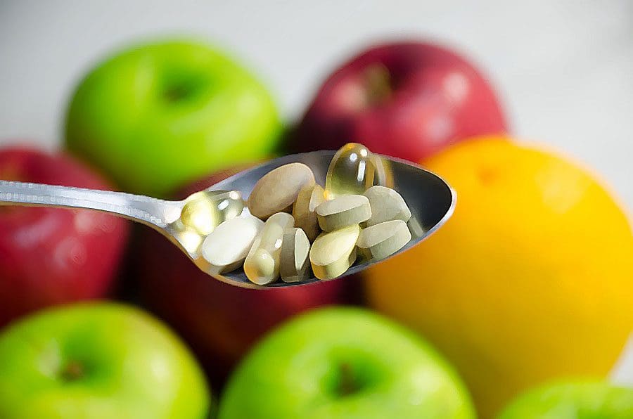
Smart supplementation along with proper nutrition is one of the biggest factors and plays a large role in an individual’s health. You are what you eat is true. Feeding the body with vital vitamins and minerals promotes growth and healing. Filling up on processed, fatty foods does the opposite. The bones in the spine and throughout the body need nutrients to continue to rebuild and maintain strength throughout life.
A balanced diet rich in:
�Calcium
Vitamin D
Magnesium
This is the best way to nourish the body’s bones and ward off spinal problems, like spinal fractures, and osteoporosis. Inadequate diets or medical issues can create nutritional gaps. This is where vitamins or mineral supplements come in. Supplements are not a cure-all, but they can create a safetyhealth net when taken properly.
Supplement Smart
These supplements for bone health, fill in nutritional gaps. They are not necessary if the key nutrients the body needs from a proper diet are already there. However, individuals use supplements as a replacement for certain foods, this is not how they should be used. Actual food supplies multiple nutrients, along with minerals, and vitamins that are beneficial for health and are not found in supplements.
Taking a Supplement to Strengthen the Spine
The body’s dietary needs change throughout life, so adding supplements as you age or during pregnancy can help maintain health. Calcium and vitamin D requirements vary based on age and sex.When it comes to protecting spine bone health, certain individuals may need supplements to ensure their bodies are processing calcium and vitamin D properly.
This includes:
Individuals who had intestinal bypass procedure
Those with food absorption conditions, like Celiac or Crohn�s disease
People who eat few or no dairy products, like vegans or those that are lactose intolerant
Is a calcium supplement right for you?
The only way to definitively know is by having a conversation with your doctor. Then you can supplement smart.
Supplement Safety
Because nutritional supplements can be purchased over the counter, individuals assume�incorrectly�that they are completely safe. Dietary supplements can interfere with absorption, other supplements, medications, and can be toxic if taken in high doses.
For example:
Calcium and iron supplements can prevent each other from being fully absorbed when taken together. This is true of many minerals, including magnesium, because they get into�absorption�competition with each other and so are best taken separately.
Supplements taken together can cause too much of either one to be absorbed. This is the case with high-dose vitamin D supplements, which can cause too much calcium absorption.
Too much calcium can increase raise the risk of having a heart attack or stroke. Taking more than 1,000-1,200 mg of calcium per day is pointless because the body cannot process that much calcium at once.
Are your supplements helping or hurting you? The best way to know is to talk to your doctor whenever you change medication or a supplement program, even when just adding a new vitamin to the mix. An underused resource for supplement advice is a pharmacist. They will know whether the mix of supplements and medicines being taken pose any risk of negative interaction. And a pharmacist can help recommend a trustworthy product.
Supplement Success
Here are a few tips to get the most benefits if you and your doctor think a calcium supplement can help support spinal bone health.
Buy supplements with the USP symbol. This indicates that the supplement has been independently evaluated and certified.
Take your supplement as directed, ideally with a meal.
Take doses no higher than 500-600 mg, no more than 2-3 times a day, for a maximum of 1,000-1,200 mg.
Drink plenty of water as some supplements can cause constipation.
Do not take calcium supplements with a high-fiber meal or laxative. This can interfere with calcium absorption.
Supplements Support Spine Health
Remember that supplements are exactly that supplements. Eating foods rich in calcium, vitamin D, and magnesium is the best way to build/maintain strong healthy spinal bones and prevent debilitating health problems. If you are concerned about your diet, talk with your doctor or a health coach about a smart supplementation regimen to meet your nutritional needs.
Reduce stress, reduce pain. Life creates stress, and while some stress can be good, too much causes health problems. Everyone experiences stress. However, now it is becoming a new normal in today�s hectic, fast-paced, high-pressure society. Most individuals equate stress with high blood pressure, heart attacks, or stroke. However, neck and back pain, insomnia, and weight gain can be stress-related, as well. And a lot of stress can make already-existing back/neck pain worse.
73% of individuals report experiencing stress-related psychological symptoms including anxiety and depression. These are not accurate numbers because most do not seek help for their stress issues. Stress symptoms should not be taken lightly. It is important to address the symptoms and find ways to reduce stress. Chiropractic is an effective stress reliever.
Stress
Financial pressures, kids, long work weeks, and medical problems are common anxieties. Prolonged stress can become chronic, which results in muscle tension that can feel stiff, achy and uncomfortable. Stress can develop into neck or back pain.
Stress is the state of:
Emotional
Mental
Pressure
Tension
That results from difficulties, adverse situations, or extremely demanding circumstances. The very nature of stress by definition makes it very subjective. A “stressful” situation for one person might not phase another. This makes it difficult to pin down a precise definition.
More often, the term stress is more often used to describe the set of symptoms that are caused by stress and those symptoms can be as varied as the people who experience them.
Symptoms
Stress symptoms can affect the entire body physically and mentally. Common symptoms include:
Anxiety
Chest pain
Depression
Fatigue
Gastrointestinal problems
Irritability
Lower back pain
Muscle tension
Overeating
Headache
Restlessness
Sleep problems
Unable to focus
Undereating
Health
Technically, stress itself does not have a negative impact on health. Some individuals deal with situations that others would consider to be stressful, yet they never exhibit symptoms. This speaks to the subjective nature of stress. Different people experience different symptoms and are a combination of stress symptoms, how the person handles those symptoms that adversely affect health.
Ultimately, stress symptoms can lead to some very serious conditions including:
Heart disease
Hypertension
Diabetes
Obesity
Cancer/s
Psychologically, it can lead to social withdrawal and social phobias and is directly linked to alcohol and drug abuse.
Tips
These can help you reduce stress, and reduce pain.
Vital Signs
Get a medical checkup if possible through Telemedicine and talk to a doctor/therapist about your stress, along with medical history. Side effects from medications (prescription or over-the-counter), herbal products, or other supplements can cause restlessness, insomnia, and anxiety.
Physical therapy combines pain-relieving non-invasive treatments with therapeutic exercise, posture correction, and preventive body mechanics.
Consider conversational therapy with a stress counselor, psychologist, or support group online.
Get Moving
Yoga and relaxation movements help reduce stress and stretch muscles. Viniyoga blends breathing and movement together to quiet body and mind. These movements are less precise and adapted to a person’s physical condition. Talk to a doctor about trying yoga or other stretches.
Swimming combined with a sauna or steam bathing can relieve stress-induced pain.
Take frequent stretch breaks to loosen up tight neck or back muscles.
Go for short walks at break or lunchtime to get the circulation going.
Learn to Relax
Kick back, put your feet up, and empty your mind of everything.
Wrap an ice pack and hot pack (or hot water bottle) individually in towels. Apply the ice pack for 10 minutes and then the hot pack for 5 minutes. Alternate several times.
Massage, aromatherapy and spa treatments you can do at home.
Aromatic massage oils containing eucalyptus can help ease muscle pain.
Meditation or visualization therapy combines meditation practices that focus on breathing and calming the mind.
Visualization techniques combine imagery with breathing exercises.
Take Control of the Little Things
Break up problems into smaller manageable pieces and work on resolving the easier parts first.
Learn your limits, how to delegate responsibility and not take the entire load on your shoulders so as not to get overwhelmed.
Allow yourself to fail, we all have to fail in order to learn in order to apply what was learned.
Eat and Drink for Life
Make mealtime less stressful. Pick nourishing foods, eat slowly, and savor each other’s company.
Caffeinated coffee, soda, and other drinks do not help reduce stress or promote restful sleep.
Avoid drinking at night because it can make falling and staying asleep a challenge.
Proper sleep or naps can help relieve stress.
Dealing with Stress Is Good for Your Back
We may not be able to control life’s stressors, but don’t let everyday demands interfere with your health. Incorporate exercise, relaxation techniques, and healthy foods to reduce stress and pain and promote stress prevention.
Reduce stress reduce pain with chiropractic
Chiropractic cannot get rid of stress, but it can help relieve stress symptoms. The more stress the body endures, the more sensitive it becomes to pain and physical imbalances. Chiropractic helps by bringing the body back into balance, aligning the spine, and relieving pain.
The simple act of aligning the spine helps relieve stress in the body that you may not even be aware of. The physical stress of a misaligned spine can exacerbate symptoms and make a person more susceptible to stressful stimuli in their environment. Chiropractic helps to improve circulation which is essential in relieving muscle tension and helps shuts down the fight or flight response allowing the body to rest and heal.
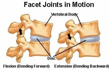
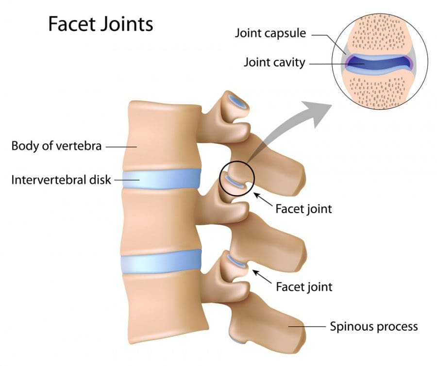
Facet joint syndrome also called facet joint sprain is a common cause of back pain. The joint/s in our bodies connect two or more bones and their primary function is to promote motion. In the spine, the joints connecting each vertebra are known as facet joints. Like any other joint, each facet joints� job is to generate healthy movement and provide stability for each motion segment.
The disc/s function as spacers that support motion between the vertebral bodies. This creates a tri-relationship between the intervertebral disc and the facet joints. Degeneration or damage/injury to one affects the other. The effects of aging and/or traumatic injury can damage the facet joints and is the leading cause of back pain known as facet joint syndrome.
There are a variety of treatments that are used, but the most mainstream involves pain medication which can have undesirable side effects and can lead to addiction. Chiropractic is a proven, reliable treatment for pain relief and discomfort of facet joint syndrome. It restores mobility and flexibility while providing pain relief. Individuals notice significant alleviation from the pain and inflammation with chiropractic and is often recommended to those with the condition.
Facet joint syndrome
The joints are located at the back of the spine. At each level, there are two joints, one on each side of the spine. The facet joints are enclosed in a capsule. The capsule contains synovial fluid and the surface is covered with hyaline cartilage. These joints are constructed in this fashion because of their role in the body. This role is to control excessive or extensive movement, which includes hyperextension and rotation. This helps to stabilize the spine.
Facet joint syndrome happens when there is an injury or damage to the joints. There are a variety of causes, but basically, it is a sprain that is brought on by excessive movement. This damages the joint capsule and results in inflammation, swelling, and pain.
The pain triggers a protective mechanism in the spine called a reactive muscle spasm which causes difficulty moving comfortably and severe, sudden pain. It is difficult to sit and rest because of its integral function of supporting the body. Severe sprains can take weeks to heal, normally 2 to 6 weeks. The�daily pain and lack of mobility make everyday activities difficult and a normal lifestyle almost impossible.
Chiropractic/Physical Therapy
Chiropractic is a proven, effective treatment for facet syndrome.� A chiropractor will conduct a physical exam, discuss medical history, and send you for diagnostic tests like x-rays and MRIs. Once they have a clear picture of the condition and a facet joint syndrome diagnosis has been confirmed, they will discuss a recommended course of treatment that can include:
Exercise
They recommend exercises�specific�to the condition that helps relieve the pain while strengthening the muscles in the back.
Poor Posture
Posture is extremely important for spinal health and general wellness.
A chiropractor will help you achieve a proper, healthy posture along with exercises to do at home to maintain posture and retrain your body.
Heat/Ice
Heat wraps, hot showers or ice packs and cold pads can be recommended to help control pain and swelling.
Changes
Depending on job type, you may be advised to take frequent breaks if you sit all day or shorten your commute. Certain activities won�t be do-able for some time until your back heals.
Spinal manipulation
Spinal manipulation is the most standard chiropractic treatment. A chiropractor may include other treatments/therapies depending on the specific condition and lifestyle.
Chiropractic is a safe, effective, non-invasive, and drug-free way to treat facet joint syndrome, relieve back pain, and help you regain mobility. Talk to your chiropractor about treatment options. Our uplifting southwest community surrounded by its infinite beauty is a fantastic place to live and enjoy our families; it is, therefore, our mission to help each of our patients to live,�to�love,�to�matter�and�to�thrive�pain-free�in this beautiful special place.
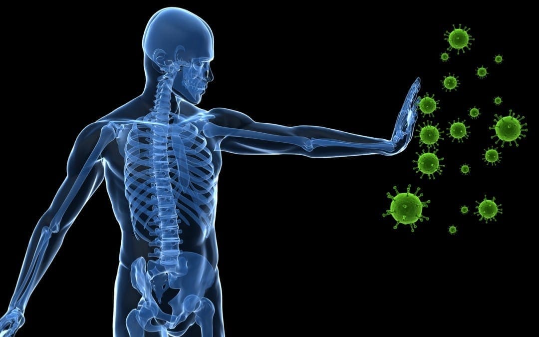
With everything that is going on in today’s world immunity is especially important. Without a properly functioning immune system, our bodies can become inflamed and more susceptible to viruses. Inflammation can cause a weakened immune system, joint pain, headaches, fatigue and more!
So what can we do to build up our immunity and help give our bodies a fighting chance? First off, washing your hands is highly important. Not just now, but always. Be sure to wash your hands with warm water and scrub everywhere. Second, get plenty of sleep. Rest is how the body recovers. If you do not give your body adequate sleep, the strength you’re cells have to fight off infection lessens. Third, eat healthy food, hydrate, and exercise. Finally, last but not least help kick up your immune system by supplementing the body with all-natural supplements.
There are many supplements that will be beneficial to the body. However, two of the most important are NAC and Glutamine.
What Are They?
NAC stands for N-acetyl-Cystine. NAC is an amino acid that the body can produce but the body can also greatly benefit from taking additional NAC in supplemental form. NAC plays an important role in helping the liver to detox. In addition to this, NAC helps to replenish the glutathione levels in the lungs and can help to reduce the inflammation. This is highly beneficial in helping to relieve the symptoms of a respiratory infection.
NAC is also greatly beneficial in boosting brain health. NAC helps to regulate glutamate levels and replenish glutathione. However, one of the most important factors of NAC is its ability to boost Glutathione levels.
Glutamine is an amino acid that helps the body perform many functions. Glutamine plays a crucial part in the immune system.
The Connection & How It Impacts Immunity
However, one of the most important factors of NAC is its ability to booze Glutathione levels. NAC and glutathione can help to boost an individual’s immune health. In research studies shown, NAC has been shown to lessen the effects of a virus and its ability to replicate. When it comes to immunity NAC and Glutamine are powerful molecules. Stoping the replication of a virus can help reduce the spread and the length of the virus in an individual.
Many infections and diseases have been linked to low glutathione levels. When the glutathione levels are low this is typically due to enhanced oxygen radicals. Studies have been done and show that when supplementing NAC to those who have low glutathione levels, it directly boosts their levels and helps with infection.
Especially with everything happening today, we want to increase our immunity and decrease the inflammation in the body.� Essentially, think of the body as a road trip. For this trip we need two main things: the gas for the car, and the car to take you to the end destination.� NAC is the gas that drives the car. We need the gas to get to our end destination. Our end destination is being healthy and giving our body the best chance to fight off infection (increased Glutathione). So by giving our body gas (NAC) we provide it with what it needs to take us to where we want to go (increased Glutathione, leading to increased immunity).
How Can I Benefit?
Overall, NAC is great to decrease inflammation. Inflammation is an extremely common underlying issue relating to other health conditions individuals suffer from. By providing your body with additional supplements, you can help increase your immunity and decrease your chances of contracting a virus and/or the length of the virus. Always discuss supplements with your primary care doctor before you begin them, but consider adding these into your daily routine!
I always recommend talking to your primary care provider and taking supplements daily. Supplements, in general, are a great way to help provide the body with the essential vitamins and minerals you may be missing. However, now more than ever supplementation is key. By building up and providing the body with the nutrients it needs for proper function, it will help prepare your body to fight off an infection. Supplementation like NAC is great to have already running in your system to help combat an infection if you were to catch one. Remember to be smart, talk to a primary care doctor before beginning supplementation, and keep in mind that not all supplements are created equal.� -Kenna Vaughn, Senior Health Coach��
The scope of our information is limited to chiropractic, musculoskeletal, and nervous health issues or functional medicine articles, topics, and discussions. We use functional health protocols to treat injuries or disorders of the musculoskeletal system. Our office has made a reasonable attempt to provide supportive citations and has identified the relevant research study or studies supporting our posts. We also make copies of supporting research studies available to the board and or the public upon request. To further discuss the subject matter above, please feel free to ask Dr. Alex Jimenez or contact us at 915-850-0900.�
References:
Dinicola S, De Grazia S, Carlomagno G, Pintucci JP. N-acetylcysteine as powerful molecule to destroy bacterial biofilms. A systematic review.�Eur Rev Med Pharmacol Sci. 2014;18(19):2942�2948.
Wessner B, Strasser EM, Spittler A, Roth E. Effect of single and combined supply of glutamine, glycine, N-acetylcysteine, and R,S-alpha-lipoic acid on glutathione content of myelomonocytic cells.�Clin Nutr. 2003;22(6):515�522. doi:10.1016/s0261-5614(03)00053-0
Dr. Alex Jimenez, a chiropractor in El Paso, TX, Kenna Vaughn, Truide Torres, Alexander Jimenez, and Astrid Ornelas, discuss how chiropractic care can ultimately help treat sciatica or sciatic nerve pain. The sciatic nerve is the largest and longest nerve in the human body. It runs from the lower back, down the buttocks and hips, and into the legs, knees, and feet. Sciatica can be caused by a variety of health issues which result in the compression or impingement of the sciatic nerve. Dr. Alex Jimenez, Kenna Vaughn, Truide Torres, Alexander Jimenez, and Astrid Ornelas discuss sciatica or sciatic nerve pain in further detail to ultimately help educate patients on their symptoms. Diet and lifestyle modifications, including nutraceuticals and exercise or physical activity, can be beneficial for patients with sciatic nerve pain. Furthermore, sciatica or sciatic nerve pain is a collection of symptoms rather than a single injury or underlying condition. Dr. Alex Jimenez, Kenna Vaughn, Truide Torres, Alexander Jimenez, and Astrid Ornelas conclude the podcast by describing how they each can help patients achieve overall health and wellness. – Podcast Insight
If you have enjoyed this video and/or we have helped you in any way
please feel free to subscribe and share us.
Thank You & God Bless.
Dr. Alex Jimenez RN, DC, MSACP, CCST
IFM's Find A Practitioner tool is the largest referral network in Functional Medicine, created to help patients locate Functional Medicine practitioners anywhere in the world. IFM Certified Practitioners are listed first in the search results, given their extensive education in Functional Medicine