Weightlifting Fitness and Chiropractic, The Perfect Team
Weightlifting and chiropractic go hand in hand as a perfect team. Everyone in some way can utilize weightlifting, whether it’s for general exercise, strength training, rehabilitation, bodybuilding, looking and feeling good, spinal health matters. When the spine and the body’s central nervous system work in harmony, muscle function is at its optimum.
Many individuals consider health care to be a reactionary function. The proverb if it’s not broken, then don’t fix it, is an approach currently being applied to various health conditions. Only after an individual exhibits or feels ailment symptoms is when they’ll see a medical professional. Weightlifters are thought to be generally more in tune with their bodies. But they are no different in that many do not seek medical attention until symptoms present.
Bodybuilding involves lifting heavy weights while maintaining proper posture and balance. Weightlifters, athletes, and fitness enthusiasts know that balance includes a healthy diet and combining fitness training with a positive mindset. Individuals involved in exercise/fitness regimens know that the muscles need time to recover and build new tissue.
Weightlifters, athletes, and overall fitness enthusiasts are discovering chiropractic medicine and its benefits. Fear of the unknown is usually the biggest reason for people not seeing a chiropractor. But for athletes, weightlifters, etc., not seeing a chiropractor, they’re usually worried they will have to stop training/competing for a while. Whatever the reason/s for not seeing a chiropractor, here are five for seeing one that everybody and every bodybuilder should know.
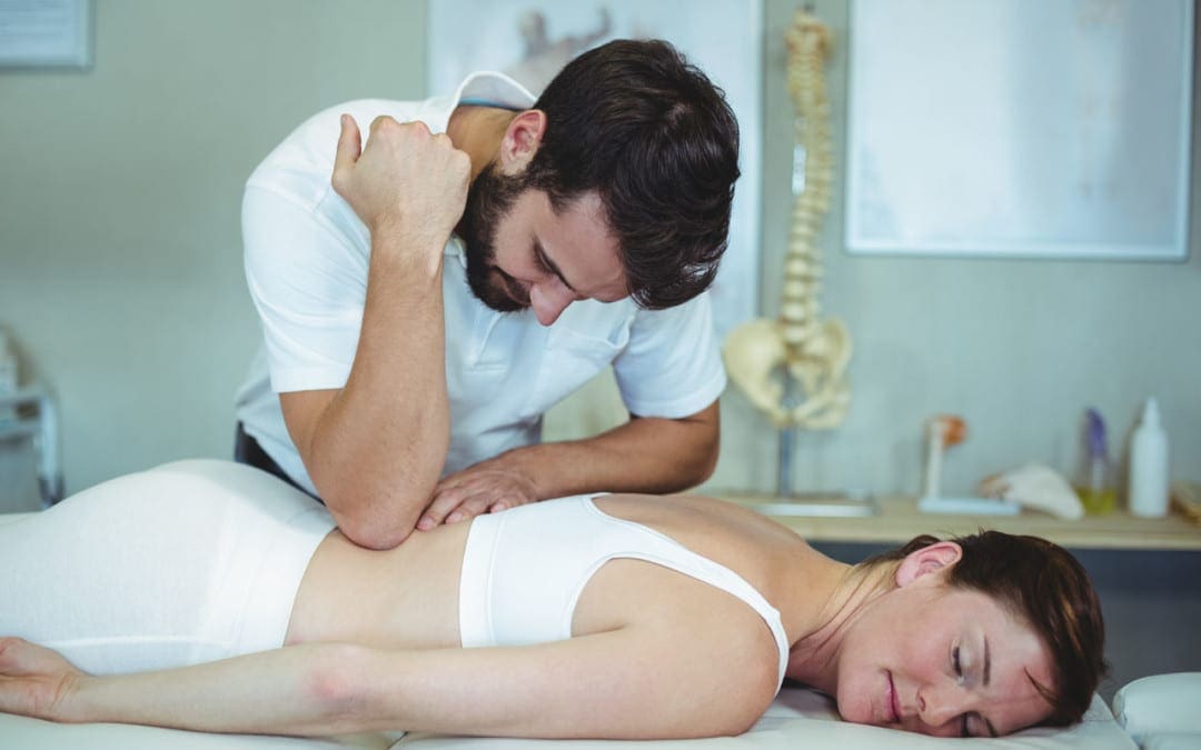
Chiropractic Mind and Muscle
Distractions in weightlifting will almost always result in an injury. The mind and body need to be balanced when working out. Just adding more weight or doing more repetitions will not create the best bodybuilder. Professional weightlifters know that it’s not about working harder but working smarter. This is where chiropractic enters the picture.
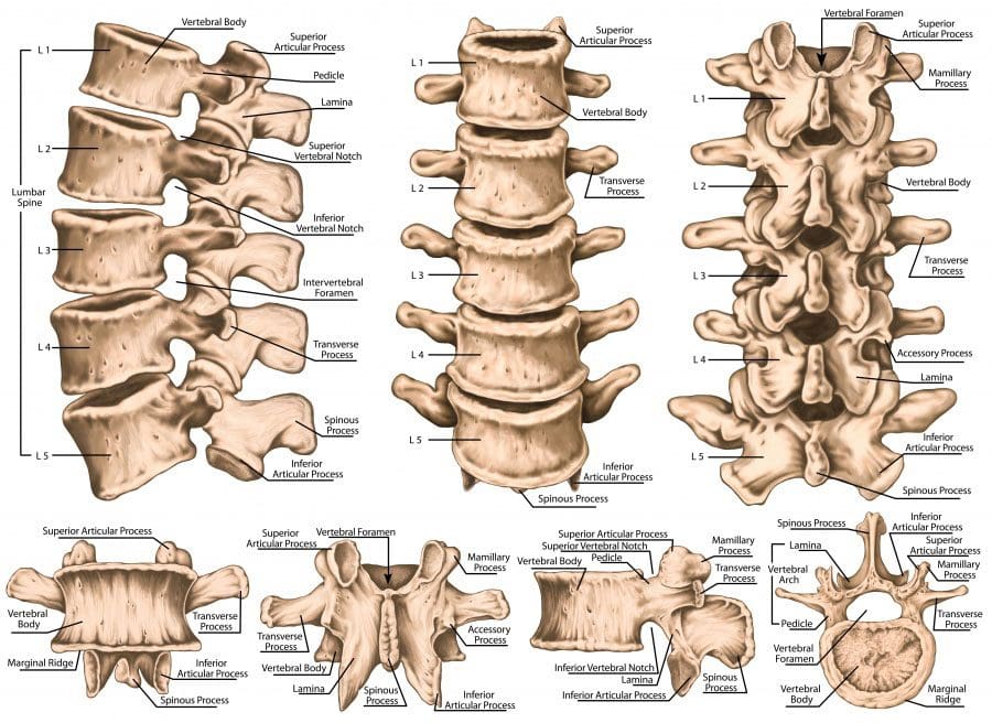
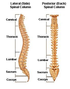
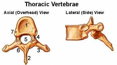
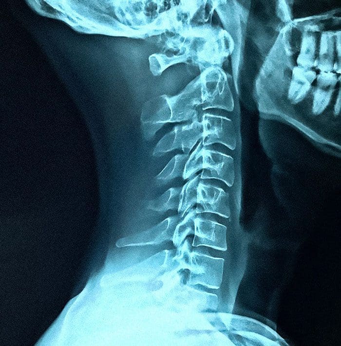
All the body’s muscles connect to joints or the spine. The joints and spine must be properly aligned for the muscles to work in proper balance. In today�s world, it’s all about the quick fix. Whether it’s a pill for whatever or fast food, however, some things need time and proper care to flourish. Chiropractic and bodybuilding are two of those things.
A Spinal Shift Makes WeightLifting Harder
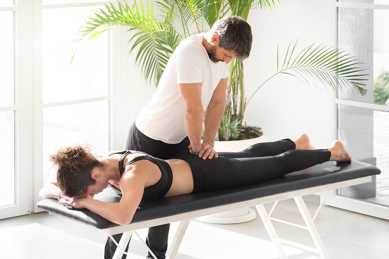
When the spine is not aligned, the muscles on one side of the body are forced to work harder than the other side. This is a perfect injury set-up. An example is doing bench presses with one foot firmly on the ground, with the other using only the toes. That is the picture when the spine is out of alignment. Working out with an uneven foundation opens the doors to injury/s.
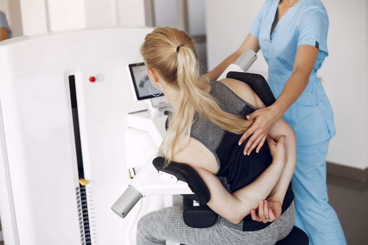
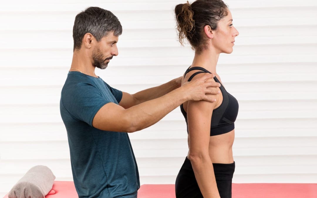
A chiropractor can discuss the best bodybuilding routine that will get results, give diet/supplement recommendations, and advice on proper lifting techniques, as well as stretches and flexibility exercises. They see the changes in the body before any pain is felt. Based on this, they can decrease the potential for injury.
Minor Injuries Lead to Severe Injuries
Many weightlifters believe if they feel pain after a workout, it means it was a good workout and is considered a sign that the muscles worked to the maximum. However, this is not always true. Microtrauma injuries are not always detected because they can hide behind minor muscle pain after a heavy workout.
Microtrauma injuries are small tears in the connective tissue and the fibers of the muscle itself. These micro-tears can cause swelling that is not seen but can be felt. This type of trauma needs proper recovery time to heal. And if treatment is not sought out, it can increase the risk for severe injuries later. These include:
- Ruptured ligaments
- Joint function loss
- Fractures
Bodybuilders who receive regular chiropractic adjustments also benefit from having one-on-one discussions about strength, diet, power, or pain they’re experiencing and get sound advice/recommendations. The chiropractor will know the difference and will know how to prevent further injury.
Weightlifting and Maximum Potential
Professional weightlifters understand that a combination of natural approaches and utilizing these resources will bring optimal results. Bodybuilders, athletes, and fitness lovers are utilizing chiropractic to stay healthy, fit, and aligned. It is a perfect team, fitness, and chiropractic.
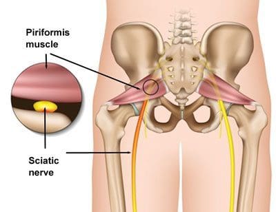
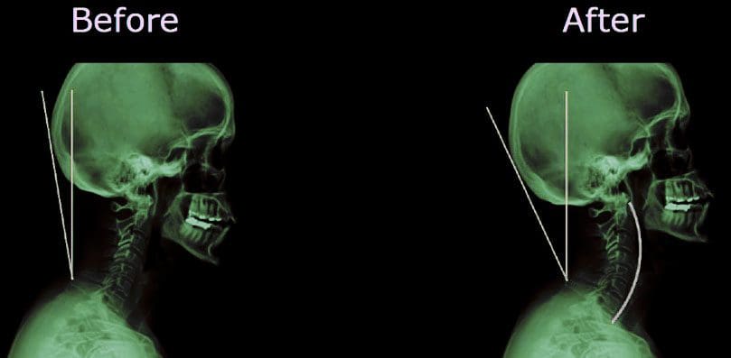
Weightlifting makes the body stronger. This comes from the added stress on the bones, muscles, and joints, which causes them to adapt. However, there is also an added strain that can misalign the spine and pinch the nerves. Pinched nerves cause lower levels of muscle strength and the development of scar tissue. Individuals might not be aware as this condition does not always cause pain.
Chiropractic involves adjusting the spine back into its natural, proper position. This allows the muscles to achieve maximum potential. Protein supplements and powders can also help. Chiropractic relieves the stress that occurs from weightlifting and releases the subluxations. Regular chiropractic prevents injuries, helps injuries heal quickly, and allows for continued training with modifications depending on the patient’s case.

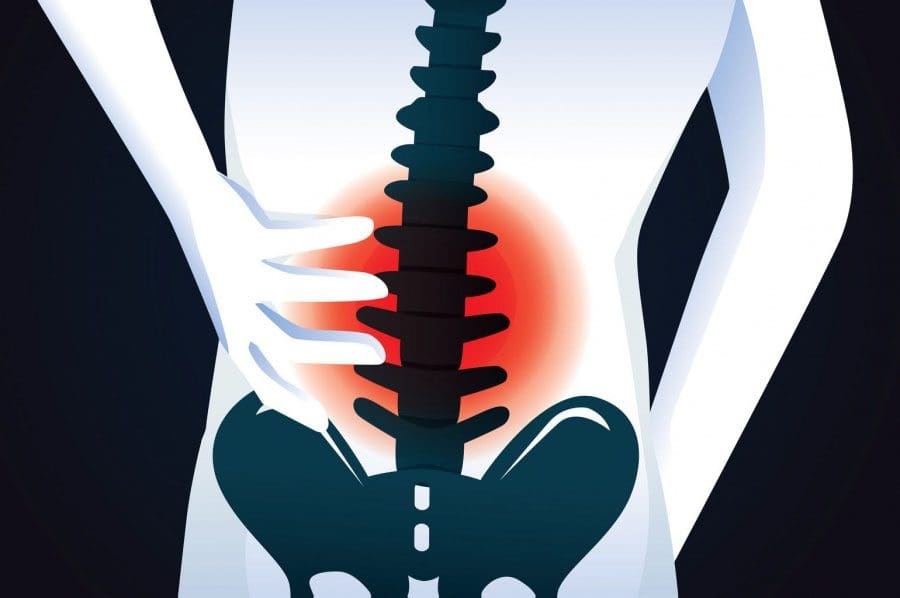
Alleviating Pain and Preventing Injury
The muscles function and perform based on the signals sent and received through the central nervous system. When an injury occurs to the muscles, ligaments, tendons, or other body areas, inflammation and swelling occur. Inflammation is not all bad and is a good sign that the body has been injured, needs attention, and deals with the injury.
But communication needs to be relayed for this to happen. When the joints in the spine are out of place or are not moving properly, the information can be scrambled or cut off. This can make it feel as if everything is fine, when there should be pain or when something hurts in one area when the pain is located in another area. Chiropractic restores function to joints, re-aligns the spine, and improves range of motion. This opens up the communication lines completely and allows the body to heal on its own.
Frequently Asked Questions
- How soon can I go back to lifting? It depends on the individual case, but most go back to training the following day if there are no injuries. However, discuss the matter with a doctor.
- Can a chiropractor adjust a big muscular individual? A chiropractor knows how to manipulate the body to don’t have to be stronger than the individual, no matter their size.
- Can I adjust myself? Chiropractors are trained doctors that know where to apply specific movement and pressure to a joint that is causing problems.
- Do I need chiropractic because my back doesn’t hurt? An individual does not have to be hurt to benefit from chiropractic. Chiropractic can be utilized to improve performance and as a preventative treatment.
- Can chiropractic help with difficulty sleeping after workouts? Tension and stress, as well as tight muscles, are irritating to the central nervous system. Hot baths can help relax the muscles. Chiropractic helps release tension, relieve stress, leading to a better night’s sleep.
Strong Chiropractor
The information herein is not intended to replace a one-on-one relationship with a qualified health care professional, licensed physician, and is not medical advice. We encourage you to make your own health care decisions based on your research and partnership with a qualified health care professional. Our information scope is limited to chiropractic, musculoskeletal, physical medicines, wellness, sensitive health issues, functional medicine articles, topics, and discussions. We provide and present clinical collaboration with specialists from a wide array of disciplines. Each specialist is governed by their professional scope of practice and their jurisdiction of licensure. We use functional health & wellness protocols to treat and support care for the injuries or disorders of the musculoskeletal system. Our videos, posts, topics, subjects, and insights cover clinical matters, issues, and topics that relate to and support, directly or indirectly, our clinical scope of practice.* Our office has made a reasonable attempt to provide supportive citations and has identified the relevant research study or studies supporting our posts. We provide copies of supporting research studies available to regulatory boards and the public upon request.
We understand that we cover matters that require an additional explanation of how it may assist in a particular care plan or treatment protocol; therefore, to further discuss the subject matter above, please feel free to ask Dr. Alex Jimenez or contact us at 915-850-0900.
Dr. Alex Jimenez DC, MSACP, CCST, IFMCP*, CIFM*, ATN*
email: [email protected]