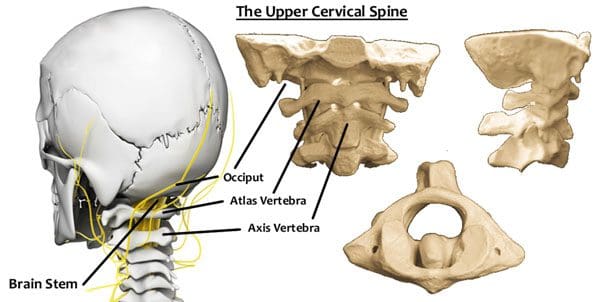
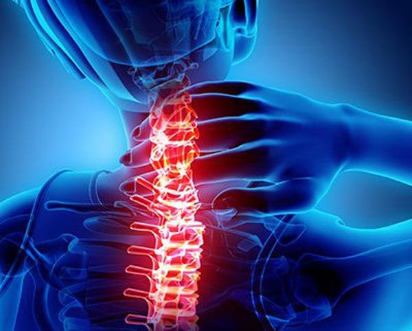
The Atlas vertebra is named for the mythological figure who held the world on their back/neck. The vertebrae are located at the top of the spine, where the cranium and spine connect. More than just a foundation for support, the vertebrae could be the most important vertebrae of the body. It consists of a complex bundle of nerves, vertebral arteries, and is the point where the entire weight of the cranium makes contact.
The myth requires Atlas to be careful while holding the world carefully and confidently at all times, otherwise it will come crashing down. The key is being able to balance it perfectly. The vertebra has the same job to hold the head up properly and maintain posture. If not problems with balance and alignment will begin to develop, and affect the entire spine.
The Atlas Vertebra
Balance
The Atlas vertebrae’s role in maintaining balance is based on its ability to adjust to the weight of the head. The actual vertebra is wider than the other cervical vertebrae. This creates a center of gravity that is reinforced through proper posture. It distributes the weight of the head (10-12lb) evenly to centralize the weight and is supported by the natural curvature of the spine.
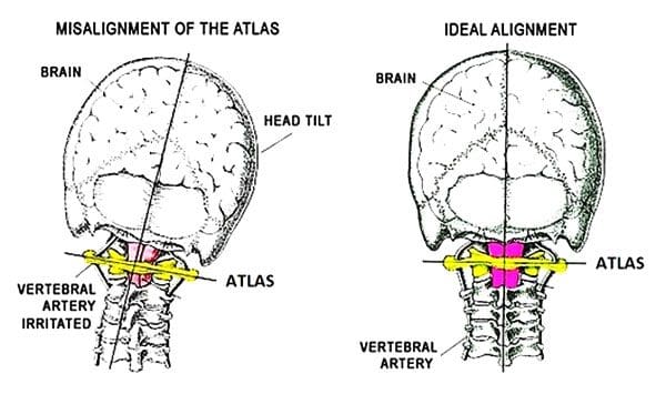
If the center of gravity shifts, the Atlas vertebra will tilt in that direction as well. This creates instability in the cervical spine and can increase the amount of weight the spine is taking and trying to redistribute. This creates spinal issues and leads to everything from poor posture, overcompensation that leads to injury.
Shifting Causes
Disruption to the vertebra and its ability to balance can come from a variety of causes and can occur as a result of chronic and acute conditions. Some include:
Auto accidents, sports, work injuries can cause cervical soft tissue damage
Dislocation of cervical vertebrae below the Atlas results in instability
Poor posture/s make individuals overcompensate to one side of the body straining muscles, ligaments, tendons causing pain and other issues
Herniated, bulging, and slipped discs
Unbalanced effects
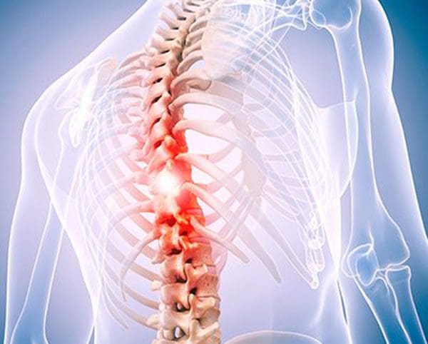
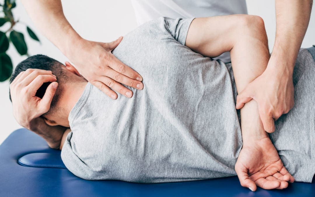
Spinal issues range from simple neck pain and soreness to full-on chronic pain. Because the Atlas can alter the balance of the entire spine, combined with cranium support, issues can be localized and referred creating further complications. Addressing the root problems requires a comprehensive chiropractic approach. Chiropractic will assess the position of the spine and determine the degree to which Atlas has shifted out of place. An adjustment treatment plan makes it possible to undo the widespread damage.
Body Composition
Muscle Loss
Individuals do not realize that muscle loss occurs throughout their lifetime. This is because muscles, like other tissues in the body, must go through cell turnover and protein synthesis. This means that the body is constantly breaking down protein in the muscles and rebuilding them.
Skeletal muscle can be developed with proper nutrition and includes consuming a proper amount of protein to provide the necessary amino acids and from physical activity. The reverse is also true, if an individual becomes less physically active and/or their diet no longer supports the development of increased muscle tissue, the body enters a catabolic/tissue-reducing state known as muscle atrophy.
Dr. Alex Jimenez�s Blog Post Disclaimer
The scope of our information is limited to chiropractic, musculoskeletal, physical medicines, wellness, and sensitive health issues and/or functional medicine articles, topics, and discussions. We use functional health & wellness protocols to treat and support care for injuries or disorders of the musculoskeletal system. Our posts, topics, subjects, and insights cover clinical matters, issues, and topics that relate and support directly or indirectly our clinical scope of practice.*
Our office has made a reasonable attempt to provide supportive citations and has identified the relevant research study or studies supporting our posts. We also make copies of supporting research studies available to the board and or the public upon request. We understand that we cover matters that require an additional explanation as to how it may assist in a particular care plan or treatment protocol; therefore, to further discuss the subject matter above, please feel free to ask Dr. Alex Jimenez or contact us at 915-850-0900. The provider(s) Licensed in Texas& New Mexico*
References
Woodfield, H Charles 3rd et al. �Craniocervical chiropractic procedures – a pr�cis of upper cervical chiropractic.��The Journal of the Canadian Chiropractic Association�vol. 59,2 (2015): 173-92.
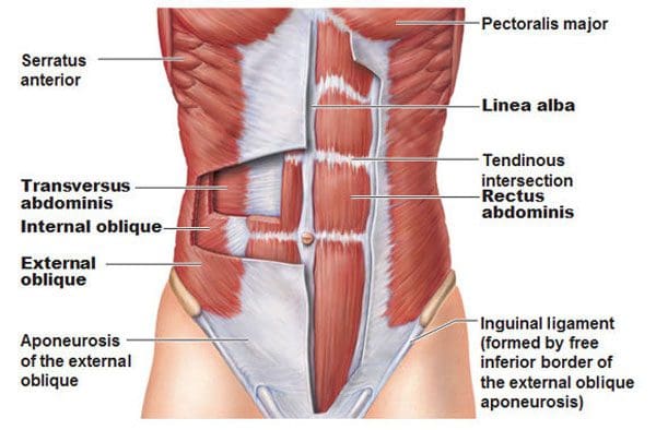
The core and the muscles involved are a group of muscles that wrap around the body’s torso. The front, back, and sides. Strengthening these muscles will improve and ultimately alleviate lower back pain. One of the main muscles that are overlooked is the transverse abdominis muscle. It is vital to a healthy core, especially if back pain is presenting. It’s known as the seatbelt muscle as it is deep in the abdomen and wraps around the waist. It has everything to do with long-term core strength and function. A properly developed transverse abdominus functions like a lumbar support belt that protects the spine. When the transverse abdominus is strong the muscle contracts to generate the correct amount of support and stability when in motion. �
�
For example, individuals that do not have low back pain engage the transverse abdominus around 30 milliseconds before moving the shoulder, while individuals that have low back pain have a delayed contraction of the transverse abdominus muscles that makes them take on awkward postures, and move in an awkward fashion contributing to back pain and continuing to weaken the core muscles. Individuals that regularly do transverse abdominus strengthening exercises greatly reduce the risk of experiencing low back pain for the first time and reduce the recurrence of those already with back pain. �
Core Muscle Anatomy
The first step to strengthening is understanding the moves and how to do them correctly with basic anatomy. Think of the core as a muscle box where the:
The body flexes and extends whenever bending forward and standing up
The body does a lateral side bend when bending the trunk to one side
The body rotates the trunk when twisting the torso
�
Muscle Weakness
The transverse abdominus tends to suffer from neglect which is one reason why it becomes weakened. This increases the risk of developing back pain. Another reason is that individuals have a weak muscle is they exercise in one-plane of movement. Not working out the core muscles in all planes of motion can contribute to back pain. For example, if an individual performs pelvic tilts, they are only moving in one plane when tilting the hips forward and back, known as flexion and extension. To achieve optimal/functional strength, the core workout needs to include side bending and twisting movements. �
Strengthening The Transverse Abdominus
Pigeon Pose
Many individuals sit for extended amounts of time and are excessively tight along the sides and hips. The first step should be to increase the hip’s mobility before strengthening the core. If the hip muscle’s fibers become shortened, it can affect hip joint function and efficiency during core movement. The Pigeon Pose is a hip opener. How to do it:
Get on the floor with the knees and palms on the ground.
Slide the left leg back so the hip is extended, then externally rotate the right hip/turn the right leg out from the hip. Focus on positioning the right shin perpendicular to the body.
Extend the trunk so the body is upright, lifting the chest, arching the back, and looking toward the ceiling, while resting the fingertips on the floor a little forward of the hips.
Hold the pose for 30 seconds and switch sides.
This stretches the hip flexor muscles in the extended leg and the rotator and outer hip muscles in the flexed leg.
�
Pay Attention To The Engaged Muscles
Individuals can train the transverse muscles to activate faster and more effectively throughout the day by slowing down and paying attention to moving with more intent. Place the hands around the waist and engage the core to feel the muscles contracting. This will help get a feel for the movement. Once comfortable remember to engage these abdominal musclesbefore and while reaching, twisting, or lifting items. �
Pelvic Tilt
This exercise is vital for building the smaller muscles that support a healthy core. How to do it:
Lie on back with knees bent and feet on the floor.
Engage the transverse muscles and gently tilt the pelvis upward.
Return the pelvis to a neutral position.
Repeat.
Start with 3 sets of 10-12 reps.
When this is no longer challenging and can be completed without increasing back pain, advance to more challenging exercises like the bird dog, planks, or plank variations.
�
Standing Exercises
Translating core strength into functional strength and pain-free movement progresses to standing exercises that require rotation. One example is a standing lunge with rotation. How to do it:
Get into a lunge stance with the front leg flexed 90 degrees at the hip, knee, and ankle. The rear leg should be extended at the hip with the knee touching or almost touching the floor.
Twist from the waist. When comfortable doing this movement, hold weight like a dumbbell, medicine ball, or gallon jug of water in both hands, and gradually increase the weight as the muscles get stronger.
When in the process of strengthening the core, consistency is the key. Commit to a short workout every day instead of one massive workout once or twice a week. Ten minutes a day is enough to build strength, improve function, and decrease back pain. �
Improved Body Composition
Functional fitness and the ability to move about comfortably not only benefit physical wellness but also improves body composition. The aging process reduces the metabolic rate, which leads to increased body fat. Lean Body Mass gets lost from age and inactivity. Lean Body Mass contributes to the overall Basal Metabolic Rate, also known as the body’s metabolism. It is the number of calories the body needs to support essential functions. Engaging in strength training or resistance exercises will help regain the muscle loss from aging/inactivity, and can lead to an increase in lean body mass.
Dr. Alex Jimenez�s Blog Post Disclaimer
The scope of our information is limited to chiropractic, musculoskeletal, physical medicines, wellness, and sensitive health issues and/or functional medicine articles, topics, and discussions. We use functional health & wellness protocols to treat and support care for injuries or disorders of the musculoskeletal system. Our posts, topics, subjects, and insights cover clinical matters, issues, and topics that relate and support directly or indirectly our clinical scope of practice.*
Our office has made a reasonable attempt to provide supportive citations and has identified the relevant research study or studies supporting our posts. We also make copies of supporting research studies available to the board and or the public upon request. We understand that we cover matters that require an additional explanation as to how it may assist in a particular care plan or treatment protocol; therefore, to further discuss the subject matter above, please feel free to ask Dr. Alex Jimenez or contact us at 915-850-0900. The provider(s) Licensed in Texas& New Mexico*
References
People who regularly engaged in TVA-strengthening exercises were less likely to experience a recurrence of low back pain:�Australian Journal of Physiotherapy�(2002), �Specific spinal exercise substantially reduces the risk of low back pain recurrence��https://www.sciencedirect.com/science/article/pii/S000495141460283X?via%3Dihub
The health of the spine and optimal body performance go hand in hand. When thinking about chiropractic treatment thoughts of cracking, popping, or fixing spinal kinks are what usually come to mind. This is true, however, chiropractic goes much deeper than just adjustments. Regular chiropractic will help maintain overall/optimal health and prevent:
Learn more about the treatment options and benefits that chiropractic can provide.
How Chiropractic Supports The Health Of The Spine
The main focus of chiropractic is on the health of the spine. This is because the spine’s health is the base for all other aspects of the body’s health. When the spine is properly aligned, nerve energy/communication and blood circulation are optimized to support the body’s function. When the body is in proper form the health benefits are numerous. These include:
Pain alleviation
Increased energy
Increased productivity
Clear thinking
Improved biomechanics movement
Improved mood
The chiropractic approach is science-based and is highly recommended for achieving positive health results.
Treatment Options
Chiropractic builds-up the body’s nervous system, immune system, and strengthens the musculoskeletal system. What to expect from treatment sessions depends on each individual’s specific conditions, needs, and goals. Treatment can include:
Spinal adjusting
Spinal adjustments combined with other treatment techniques are needed to maintain the body’s health long term.
Health Coaching
Understanding the causes of ill health/conditions will help manage and prevent issues. A chiropractor and a health coach can discuss positive lifestyle modification factors that will help like:
Posture education
Exercise tips
Stress management techniques
Sleep modification
Developing healthy diet habits
Deep/Soft tissue massage
Soreness, stiffness, and connective tissue flexibility problems are common with misalignment. Addressing these areas with physical therapeutic massage will help reinforce proper body alignment.
Prescribed exercise program
A chiropractic practitioner will recommend exercises for strengthening and stretching muscles, tendons will provide support for optimal body alignment and overall health.
Health Potential
Seeking treatment options for improved health, consider a chiropractic provider. They will address the root cause to help bring out the best on every level. Chiropractic providers are in high demand and will get the body back to its full potential. See what the possibilities can be.
Body Composition Benefits
Best Diet for Weight Loss
Life is about balance and is important to remember that all macronutrients are needed to function properly. Protein does not just affect muscle, it also acts on hormones, enzymes, and antibodies. Carbs are an energy source that the red blood cells use, and without enough fat in the diet, an individual can gain weight by eating more filler foods. The body is a well-refined machine that does not function on fad diets.
The function is obtained through balance and variety in a healthy diet. The best diet for weight loss is not complicated or high-tech. The focus should be on whole unrefined foods, reduced red meat consumption, and plenty of vegetables. But, the food does not have to taste bad or have no taste at all. A too rigid diet can make individuals more likely to revert back into unhealthy eating habits. Try a salad with leafy greens, brown rice, sweet potatoes, avocado, black beans, and cilantro lime dressing. It may be a surprise, how healthy eating can taste amazing.
Dr. Alex Jimenez�s Blog Post Disclaimer
The scope of our information is limited to chiropractic, musculoskeletal, physical medicines, wellness, and sensitive health issues and/or functional medicine articles, topics, and discussions. We use functional health & wellness protocols to treat and support care for injuries or disorders of the musculoskeletal system. Our posts, topics, subjects, and insights cover clinical matters, issues, and topics that relate and support directly or indirectly our clinical scope of practice.*
Our office has made a reasonable attempt to provide supportive citations and has identified the relevant research study or studies supporting our posts. We also make copies of supporting research studies available to the board and or the public upon request. We understand that we cover matters that require an additional explanation as to how it may assist in a particular care plan or treatment protocol; therefore, to further discuss the subject matter above, please feel free to ask Dr. Alex Jimenez or contact us at 915-850-0900. The provider(s) Licensed in Texas& New Mexico*
References
Meeker, William C, and Scott Haldeman. �Chiropractic: a profession at the crossroads of mainstream and alternative medicine.��Annals of internal medicine�vol. 136,3 (2002): 216-27. doi:10.7326/0003-4819-136-3-200202050-00010
Spinal traction, both mechanical and manual are treatment options that are based on the application of force to the axis of the spinal column. A region of the spinal column is pulled in opposite directions to stabilize or change the position of herniated, slipped, bulging, discs, and/or nerve injury/damage to the spine. Traction treatment is crucial to spinal adjustments, especially with disc or nerve compression.
It allows the chiropractor to alleviate any stress that could lead to disc problems like herniation, rupture, or displacement. However, traction is a general term. The concepts can apply to all forms of traction, but the application itself can be drastically different in terms of static positioning and inverse force.
Mechanical vs. Manual Cervical Traction
Mechanical force is typically applied through a series of weights or a fixation device and requires the patient to stay in bed or is placed in a halo vest. The techniques and methodologies can vary, but the objectives/results are the same.
The utilization is developed on a case-by-case basis and the chiropractor’s diagnosis/recommendations. Many chiropractors implement both mechanical and manual traction approaches. Choosing the right traction plan comes from a thorough examination, medical history, and understanding of each method’s strengths.
Traction approach
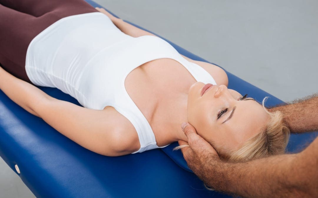
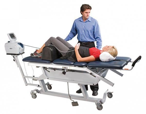
The difference between mechanical and manual traction is simple. Mechanical traction is directed by the use of machines, weights, and pulleys, while manual traction is performed by a professional chiropractor. With mechanical traction, an individual’s head is cradled into a sling, then positioned at the optimal position for the adjustment. The sling is counterweighted to hold the head/neck in that position, leveraging mechanical pressure and affecting change.
Manual traction has the individual lie down on a table, with the chiropractor pulling the head away from the neck to decompress the cervical spine. The adjustment/s can be a continuous pull, or a series of low-force pulls in different directions. Again these depend on the individual’s condition and nature of the adjustment.
Techniques and methodologies
Mechanical and manual traction can have similar results, but both offer different benefits based on the individual. Mechanical traction is a hands-free technique for decompression that allows chiropractors to focus on the patient’s needs when working on complex cases. This method is more applicable for severe cases, where the traction could last for 20-30 minutes.
Mechanical traction is helpful when teaching healthy posturing. Manual traction benefits come from the control that a chiropractor has over the technique. With the manual pulling, the chiropractor can increase or decrease the countering force. A hands-on approach enables chiropractors to feel the spinal adjustments, and understand the effects of the traction.
The proper form of traction
The overall ability of traction to decompress the spine makes it a valuable approach to treat various conditions. The exact nature of the condition determines whether mechanical or manual traction will be used along with the recommendation/treatment plan of the chiropractor. Injury Medical Chiropractic Clinic is committed to implementing the best approach for spinal correction for every patient. Mechanical and manual traction are just two adjustment modalities.
Body Composition Health
Resistance Training For Everyone
Even if not an athlete resistance training is important for functional fitness. Functional strength training attempts to emulate the physiological demands of real day-to-day activities. Traditional strength training focuses on specific muscle groups during the exercise, while functional training focuses on whole muscle groups to train the body for daily responsibilities.
Individuals might believe they are too old for resistance training. But research shows the benefits of improving an individual’s functional fitness level, specifically for older adults. Functional training resistance exercises and bodyweight movements can help the body become stronger, more flexible, more agile, and better equipped to handle day-to-day responsibilities. Plus, it can help with injury prevention.
Reference
Afzal, Rabia et al. �Comparison between Manual Traction, Manual Opening technique, and Combination in Patients with cervical radiculopathy: Randomized Control Trial.� JPMA. The Journal of the Pakistan Medical Association�vol. 69,9 (2019): 1237-1241.
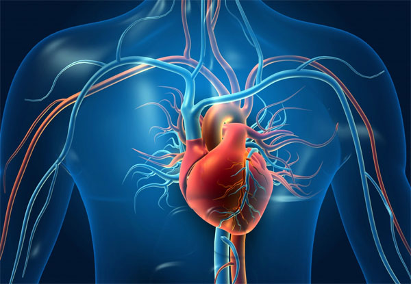
Heart health and proper function circulate millions of gallons of blood to the entire body. The circulation moves:
Oxygen
Fuel
Hormones
Essential cells
Other compounds
Removes metabolic waste products
If the heart stops, vital functions can fail almost instantly. Family history and genetics play a role in the development of heart disease, but lifestyle choices also play a part. Heart health disease prevention focuses on:
Chiropractic treatment can help improve overall heart health.
Heart Health
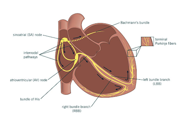
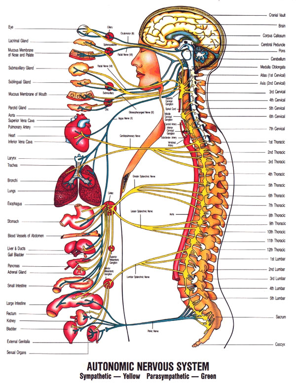
If every nerve was disconnected, the heart would continue to beat. There is a small node of the heart muscle that rhythmically contracts and relaxes inherently, and sets the heartbeat pace. It can be thought of as a natural pacemaker and is called the sinoatrial node.
In an average adult, the node maintains a rhythm of around 70 beats per minute. This natural pacemaker keeps the heart working, while the nerves that accelerate and decelerate (the sympathetic and parasympathetic nerves) can affect the sinoatrial node affecting the heartbeat.
Sympathetic and Parasympathetic Nerves
The sympathetic and parasympathetic nerves are located in the thoracic and upper cervical spine. With chiropractic, any spinal misalignments, pressure, stress, and restrictions are properly addressed, and able to normalize the cardiac rhythm and heart rate. Corrective treatment of the cervical spine will also help lower blood pressure and remove any stress on the cardiovascular system. Heart and spinal health are vital, contact a local chiropractor today.
Healthy Body Composition
Aerobic Training Strengthens The Heart
Aerobic exercise will strengthen the heart, as well as, train the heart to be more efficient in circulating blood. The chamber of the heart that pumps blood to the rest of the body literally gets larger and squeezes out more blood with each pump, meaning the stroke volume gets increased.
This improves cardiac output, which is the quantity of blood pumped by the heart per minute. A strong, efficient heart is the objective to live a long and healthy life. When the heart is stronger and pumps more blood it doesn�t have to beat as much and as rapidly. Lowering the resting heart rate is associated with a reduced risk of cardiovascular disease.
Cardiac adaptations are helped with an increase in blood volume that happens with aerobic exercise training. What happens is the expanded blood volume improves the heart�s contractility/fill capacity pumping more blood per beat. The heart contracts to move blood throughout the body. By making it stronger and more efficient, the heart�s responsibilities are lightened by decreasing the different types of resistance.
Dr. Alex Jimenez�s Blog Post Disclaimer
The scope of our information is limited to chiropractic, musculoskeletal, physical medicines, wellness, and sensitive health issues and/or functional medicine articles, topics, and discussions. We use functional health & wellness protocols to treat and support care for injuries or disorders of the musculoskeletal system. Our posts, topics, subjects, and insights cover clinical matters, issues, and topics that relate and support directly or indirectly our clinical scope of practice.*
Our office has made a reasonable attempt to provide supportive citations and has identified the relevant research study or studies supporting our posts. We also make copies of supporting research studies available to the board and or the public upon request. We understand that we cover matters that require an additional explanation as to how it may assist in a particular care plan or treatment protocol; therefore, to further discuss the subject matter above, please feel free to ask Dr. Alex Jimenez or contact us at 915-850-0900. The provider(s) Licensed in Texas& New Mexico*
References
Yang, Jian et al. �Physical Exercise Is a Potential “Medicine” for Atherosclerosis.��Advances in experimental medicine and biology�vol. 999 (2017): 269-286. doi:10.1007/978-981-10-4307-9_15
Participating in any sports or physical activities strengthens the mind and body. But working out and engaging in these types of activities too much or without rest periods wears down the body. There is the feeling of a good workout with some sore muscles and achiness that lets you know the activity is working positively.
However, soreness can quickly lead to pain and further injury if ignored. The lower back is a common area of soreness after working out playing sports, and where muscle spasms, pulls, and pinches occur. Being able to distinguish between workout soreness and pain is critical for maintaining a healthy spine.
A constant-sore back or feelings of sharp pains is not normal. If there is a feeling of low back pain during or after a physical routine, stop and take a moment to examine the tingling, discomfort, or pain being experienced. If unsure if the soreness or pain is a cause for concern call or video conference with a chiropractor to discuss what is going on.
Physical activity and pain
Individuals participating in physical/sports activities have an increased risk of low back pain because of the consistent running, twisting, and jumping. Any of these movements place pressure on the spine along with the surrounding ligaments and muscles, which can lead to injury.
Repetitive twisting and turning, stresses the muscles around the spine, which can cause frequent muscle sprains. Running and jumping also wears down the vertebrae and discs. Impact activities can also cause injuries to the spine, nerve roots, and surrounding tissues. The most common back problems include:
Muscle sprains
Osteoarthritis
Bulging discs
Herniated discs
Sciatica
Fractures are less common but still pose a risk
Individuals should watch for achiness or stiffness that lasts longer than a few days and does not alleviate with ice or anti-inflammatory over the counter medication, or sharp pain that happens with specific movement/s, along with any pain, numbness, tingling that runs down the leg/s or to other areas should consult a medical professional.
Treatment and prevention
Maintaining the body’s health is critical. If the lower back begins to present discomfort or hurts, do not ignore it. Many will play through the pain when they should be taking a break. And ignoring any back pain could create new injuries or worsen the condition. Continued pressure on the back will worsen any strains or fractures and will hinder the body from healing properly.
Individuals tend to take on awkward/uncomfortable postures and move in awkward ways to avoid or compensate for the pain. This places added pressure in the wrong places and can cause/worsen an injury or condition. Pay attention to the pain. Try ice and heat therapy at home to see if it eases up. Using a foam roller or self-massage device can help if the back pain is muscular. However, if the pain is sharp, shooting, or does not go away, visit a chiropractor for diagnosis and treatment.
A chiropractor will conduct imaging tests and physical exams to identify the root cause. Once a diagnosis has been reached a treatment plan will be implemented through:
Massage
Stretches
Therapeutic exercises
Spinal adjustments
Health coaching
Visiting a chiropractic professional will improve the condition and strengthen the spine.
Fit Body Composition
Muscle recovery
When engaging in physical activity there is microscopic damage to the muscle cells. The stress and fatigue the body goes through during physical activity cause hormone and enzyme levels to fluctuate, increasing inflammation. This leads to:
Fat loss
Increased metabolism
Increased strength
Muscle growth
However, it happens through proper recovery. There are different types of recovery: immediate, short-term, and training.
Immediate recovery is the short time between movements. For example, when jogging, immediate recovery is the time between each stride.
Short-term is the time between sets of exercises. For example, the rest periods between exercise intervals.
Training recovery is the period between one workout session ending and the next beginning.
Research has shown that rest time is not a one size fits all. Everyone is different and therefore should consult a fitness trainer, or sports chiropractor and experiment with what feels right. For some individuals, 24 hours works. For others, it can be 48 or 72 hours to feel fully recovered. It depends on age, fitness level, physical activity intensity, diet, sleep, and more.
Dr. Alex Jimenez�s Blog Post Disclaimer
The scope of our information is limited to chiropractic, musculoskeletal, physical medicines, wellness, and sensitive health issues and/or functional medicine articles, topics, and discussions. We use functional health & wellness protocols to treat and support care for injuries or disorders of the musculoskeletal system. Our posts, topics, subjects, and insights cover clinical matters, issues, and topics that relate and support directly or indirectly our clinical scope of practice.*
Our office has made a reasonable attempt to provide supportive citations and has identified the relevant research study or studies supporting our posts. We also make copies of supporting research studies available to the board and or the public upon request. We understand that we cover matters that require an additional explanation as to how it may assist in a particular care plan or treatment protocol; therefore, to further discuss the subject matter above, please feel free to ask Dr. Alex Jimenez or contact us at 915-850-0900. The provider(s) Licensed in Texas& New Mexico*
References
Smith, Jo Armour et al. �Risk Factors Associated With Low Back Pain in Golfers: A Systematic Review and Meta-analysis.��Sports health�vol. 10,6 (2018): 538-546. doi:10.1177/1941738118795425
Individuals are tired of feeling sick. Many doctors prescribe medications to just control the symptoms of the various ailments.
Headaches
Migraines
Nausea
Fatigue
Acid reflux
Asthma
Allergies
Chiropractic care combined with Health coaching will:
Bring the body back into balance
Restore optimal circulation
Detox the body
Increase immune system response
Tired Nervous System
Many of these problems are rooted deep within the nervous system. This system controls pain, movement, organ function, and action/reaction in the body and needs consistent maintenance to continue to operate at an optimal level. Chiropractors are trained to detect nerve interference brought on from spinal misalignment. Chiropractic along with body scanning/imaging can detect nerve interference, and help identify any issues.
Nerve Interference
Nerve interference along the spine can lead to being tired, weakness, pain, discomfort, organ dysfunction, and disease if it is not addressed by a professional chiropractor. The interference can be a result of poor postural habits that have caused the spine to misalign. This places added and dangerous pressure on the delicate nerves flowing throughout the spine.
Chiropractors can determine the root cause back to the region of the spine that is causing any type of impediments. Spinal rehabilitation and realignment will restore the spine back to health eliminating nerve interference. As the body is realigned health coaching recommendations that include diet, supplements, and learning healthy habits will enhance chiropractic maintenance. The end result is a healthy energetic body free of disease, dysfunction, and pain.
Body Composition
Body composition is a way of breaking down the body into components, which are: fat, protein, minerals, and body water. It describes an individual’s accurate weight and provides a new perspective on overall health than traditional methods. Proper body composition analysis will show changes in fat mass, muscle mass, and body fat percentage.
The scope of our information is limited to chiropractic, musculoskeletal, physical medicines, wellness, and sensitive health issues and/or functional medicine articles, topics, and discussions. We use functional health & wellness protocols to treat and support care for injuries or disorders of the musculoskeletal system. Our posts, topics, subjects, and insights cover clinical matters, issues, and topics that relate and support directly or indirectly our clinical scope of practice.*
Our office has made a reasonable attempt to provide supportive citations and has identified the relevant research study or studies supporting our posts. We also make copies of supporting research studies available to the board and or the public upon request. We understand that we cover matters that require an additional explanation as to how it may assist in a particular care plan or treatment protocol; therefore, to further discuss the subject matter above, please feel free to ask Dr. Alex Jimenez or contact us at 915-850-0900. The provider(s) Licensed in Texas& New Mexico*
References
Stochkendahl, Mette Jensen, et al. �Can chiropractors contribute to work disability prevention through sickness absence management for musculoskeletal disorders? – a comparative qualitative case study in the Scandinavian context.� Chiropractic & manual therapies�vol. 26 15. 26 Apr. 2018, doi:10.1186/s12998-018-0184-0
Westerterp, Klaas R. �Exercise, energy balance, and body composition.��European journal of clinical nutrition�vol. 72,9 (2018): 1246-1250. doi:10.1038/s41430-018-0180-4
IFM's Find A Practitioner tool is the largest referral network in Functional Medicine, created to help patients locate Functional Medicine practitioners anywhere in the world. IFM Certified Practitioners are listed first in the search results, given their extensive education in Functional Medicine