Getting dizzy can happen, usually after standing up too fast or staring at an optical illusion then looking away. The unsteadiness can be troubling but is minimal when compared to vertigo symptoms. Vertigo is a symptom rather than a condition that causes dizziness combined with a spinning sensation, even when an individual stands completely still. Vertigo can make everyday life a debilitating nightmare:
It causes individuals to feel nauseous.
It makes it difficult to walk.
It interrupts vision and hearing.
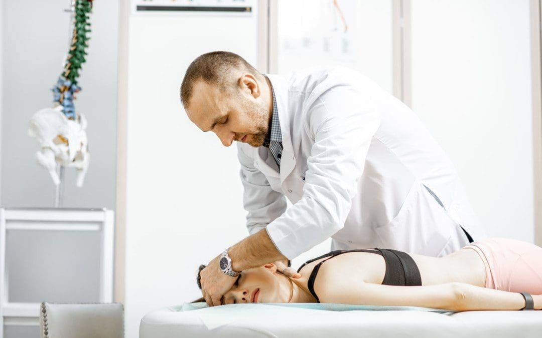
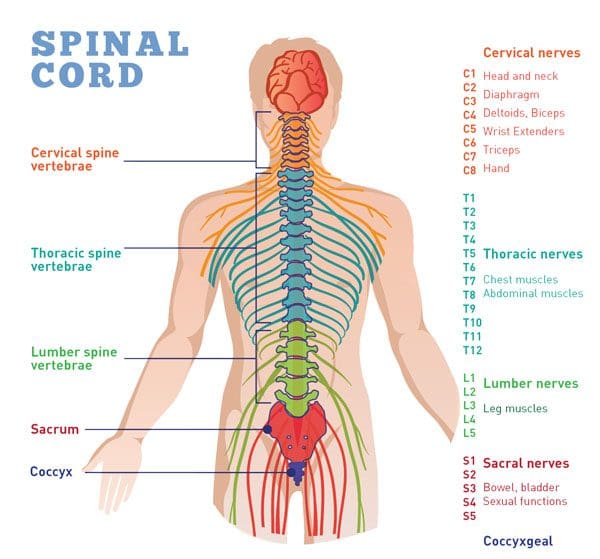
Vertigo symptoms usually begin with a communication issue with the inner ear and brain. This neurological connection involves the spinal cord, which chiropractic can treat and cure. A chiropractor will implement the necessary techniques to help alleviate vertigo symptoms.
Causes
The most common signs and symptoms are dizziness accompanied by a spinning sensation. Vertigo symptoms can also include:
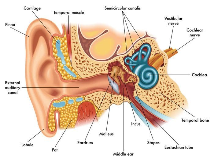
Vertigo is a common symptom in individuals that have gone through trauma to the neck and/or head. A disturbance/interruption occurs in the nerve pathways in the spinal column. Disruption, injury, or damage to the vestibular system/inner ear also causes vertigo symptoms. Other causes include:
Ear infections
Pressure changes
Movement of particles within the inner ear
Chiropractic Treatment
Chiropractic treatment can cure vertigo symptoms through various exercises and spinal adjustments. When nerve signals don’t transmit correctly, it can cause a feeling of dizziness along with the other symptoms. Adjustments or manipulations help alleviate the symptoms by allowing nerve energy to circulate properly. Spinal adjustments realign the joints and vertebrae in the cervical spine. This opens the nerves pathways and allows for clear communication.
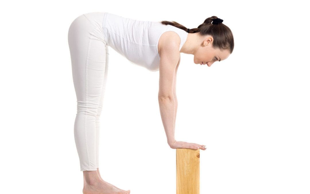
A common type of vertigo is known as benign paroxysmal positional vertigo or BPPV. This is the movement of particles around the inner ear. These particles come from otolith organs. The particles break loose and fall into other parts of the vestibular labyrinth. This alters the center of balance and can cause dizziness. A chiropractor may use the Epley maneuver, which maneuvers the head into different positions. A chiropractor will recommend specific exercises at home to benefit the vestibular system and its communication with the brain.
Body Composition
A Common Cold
The common cold, aka upper respiratory tract inflammation. It is the most common infectious respiratory disease. It is called this because of the effects on the nose and throat. According to the Center for Disease Control and Prevention, an adult will catch 2–3 colds a year. A cold-causing virus enters the respiratory tract directly when an individual inhales droplets or direct skin contact from an infected person. Cold symptoms include:
Runny nose
Stuffy nose
Sneezing
Coughing
Headaches
Body aches
The duration of a cold differs, but most individuals with a healthy immune system recover in 7–10 days. However, individuals with compromised immune systems, asthma, or COPD have an increased risk of developing serious illnesses like bronchitis or pneumonia. Quickly recovering from a cold requires boosting the immune system with proper rest, a nutritious diet, and drinking plenty of water.
References
Collins, Matthew E, and Tom M Misukanis. “Chiropractic management of a patient with post-traumatic vertigo of complex origin.” Journal of chiropractic medicine vol. 4,1 (2005): 32-8. doi:10.1016/S0899-3467(07)60110-4
Dalby, B J. “Chiropractic diagnosis and treatment of closed head trauma.” Journal of manipulative and physiological therapeutics vol. 16,6 (1993): 392-400.
Sajko, Sandy S et al. “Chiropractic management of benign paroxysmal positional vertigo using the Epley maneuver: a case series.” Journal of manipulative and physiological therapeutics vol. 36,2 (2013): 119-26. doi:10.1016/j.jmpt.2012.12.011
Poor spinal health in adolescence can lead to chronic pain in adulthood. Teenagers, just like adults, can experience back pain from accidents, sports injuries, a sedentary lifestyle, part-time jobs, chores, etc. However, sitting too long in school along with heavy backpacks can also contribute to compromised spinal health. Chiropractic professionals can help these young individuals address and prevent spinal issues/injuries to maintain a healthy spine.
Teenagers Spine Issues
If discomfort or pain is present, much push through, as they and their spines are young. There are common spinal dysfunctions that teens and parents should be aware of. These include:
Disc injuries
Teenagers can put a serious strain on the spine from various forms of physical activity, jumping, dancing, and playing. This pressure gets transmitted through the spine. During a teenager’s development, this can result in permanent disc damage.
Scoliosis
A spinal deformity or exaggerated curvature of the spine is common and affects young children and teens. It usually happens during the growth spurt just before puberty. This is why it is important to have a teenager’s spine checked regularly and analyzed for signs/symptoms of scoliosis.
Spondylolysis
This condition is often associated with sports injuries. It happens when teenagers overextend/overreach their backs. It’s most common in gymnastics, weight lifting, tennis, football, diving, and other similar sports.
Protection and Prevention
There are several ways that parents and healthcare providers can help teenagers make healthy decisions to achieve and maintain optimal spinal health.
Sitting less, moving more.
Children are taught to sit from a very young age. In school, watching t.v., or doing homework, teenagers spend more time sitting than their bodies should. Teenagers need to stand, walk and move around just like adults to protect their spines from degeneration and injury.
Playing sports is healthy. However, there is a risk associated with teen sports. Although they are taught to play safely, encourage them to continue to educate themselves about sports injuries and know how to address them.
Chiropractic Support
At Injury Medical Chiropractic and Functional Medicine Clinic, we’re committed to helping young adults and adolescents overcome and prevent spinal injuries that could turn into chronic pain conditions. We are continually developing our chiropractic, and physical therapy treatment approaches to achieve optimal results.
Body Composition
Sleep and Growth Hormone In Children
Growth hormones primarily control growth. The hypothalamus and the pituitary gland regulate this hormone. Sleep plays an important role in the proper function of these glands. A review showed that:
Growth hormone levels rise and peak at the onset of deep sleep
Multiple but smaller peaks were seen during other sleep stages
Individuals that have a delay in the onset of deep sleep have delayed peaks in growth hormone levels
For children to grow properly, they need to have adequate levels of growth hormone. This means they need to have a sufficient amount of sleep. The proper amount of sleep is vital for healthy body composition. A study measured the body composition of preschool-aged children. The study found that children who had proper sleep levels had less overall fat mass and reduced body fat. Children and teenagers need to get the proper amounts of sleep for their bodies to grow healthily.
References
Clement, R Carter et al. “What are normal radiographic spine and shoulder balance parameters among adolescent patients?.” Spine deformity vol. 8,4 (2020): 621-627. doi:10.1007/s43390-020-00074-9
Driehuis, Femke et al. “Spinal manual therapy in infants, children and adolescents: A systematic review and meta-analysis on treatment indication, technique, and outcomes.” PloS one vol. 14,6 e0218940. 25 Jun. 2019, doi:10.1371/journal.pone.0218940
Manansala, Christian et al. “Change in young people’s spine pain following chiropractic care at a publicly funded healthcare facility in Canada.” Complementary therapies in clinical practice vol. 35 (2019): 301-307. doi:10.1016/j.ctcp.2019.03.013
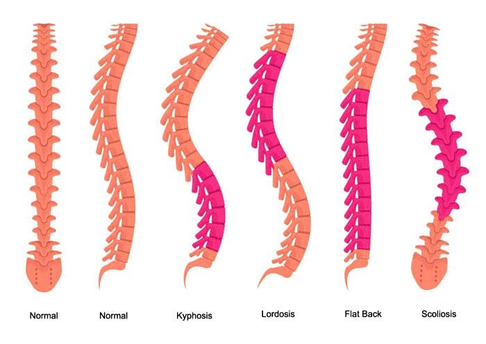
A non-invasive method of treating scoliosis. Yoga Has Been Shown To Help Reverse Scoliosis. Scoliosis is the lateral curvature of the spine. The spine bends inward toward the front of the body at the neck region and lower back region. This curve is known as lordosis and bows outward in the middle-back region. This is known as kyphosis. If the spine curves to the side, this could indicate curvature that could be scoliosis. It can be painful and often can affect an individual’s appearance once the measurement goes beyond 25 – 30 degrees. One shoulder is usually higher than the other, and clothing cannot fit properly. If the curve goes beyond 60 degrees, it can affect breathing and cardiac function.
Idiopathic Causes Unknown
This condition can consist of various components, especially with more intense curves. The ribs can shift backward on the side where the curve bulges. Most cases consist of adolescent idiopathic (without a known cause) scoliosis. Because the cause is unknown, there are not a variety of effective treatment besides surgery. Physicians carefully keep an eye for:
Curves under 25 degrees.
Bracing between 25 and 45 degrees.
Consider surgery for intense curvature.
Curves in individuals typically appear between 12 and 20 years old.
Yoga Shown To Reverse Scoliosis
Individuals are recommended to do just one yoga pose daily. However, depending on the type and severity of the curves, it could be more than one. They are asked to perform the pose for 5 minutes or less, depending on the condition. A yoga therapist, chiropractor, and physical therapist can generate significant spinal improvement. This could mean that a curve of 30 degrees could be reduced to around 18 degrees in 10-12 months. Individuals that do the poses at least 4 times a week have shown 80-90% improvement. The pose can be done at work during breaks, etc.
The biggest advantage of this technique is that it is non-invasive; it can help individuals with developing curves, reversing the curvature early. Most curves do not reach the point of surgery. In late adolescence and teen years, the spine is still quite flexible. This can help accelerate the effectiveness of the yoga pose to straighten the spine. The technique reduces the curve from worsening. X-rays will show if the curvature has improved or not. Patients could be asked to do the pose/s twice or more daily depending on the direction the condition is taking.
Body Composition
Gluten Effects
Gluten causes digestive issues for individuals that have celiac disease or autoimmune thyroid disease. Individuals with these conditions could experience a variety of uncomfortable and/or painful effects. These symptoms can vary based on their presentation. They fall into classifications.
Classical Celiac Disease
With classical celiac disease, symptoms include:
Diarrhea
Discolored stools
Constipation
Abdominal bloating and pain
Weight loss
However, these symptoms are more common in children than adults. In adults, symptoms are more similar to non-classical celiac disease.
Non-Classical Celiac Disease
With non-classical celiac disease, severe digestive symptoms may not present as classic celiac disease symptoms but develop other symptoms. These include:
Silent celiac disease is less visible. Individuals might not see any symptoms. However, damage to the intestines is still happening from gluten consumption.
Autoimmune Thyroid Disease
Autoimmune Thyroid Disease or ATD. Autoimmune thyroid disease includes conditions like Hashimoto’s disease. This affects the thyroid gland and causes:
Extreme fatigue
Sensitivity to cold
Hair loss
Body aches
Joint aches
Negative health effects
Studies have shown that gluten-free helps alleviate symptoms.
References
Loren M. Fishman, M.D., B.Phil. (oxon). Healing Yoga. (New York: W.W. Norton, 2014).
Loren M. Fishman, M.D., B.Phil. (oxon). “Isometric Yoga-Like Maneuvers Improve Adolescent Idiopathic Scoliosis—A Nonrandomized Control Trial.” Global Advances in Health and Medicine. February 24, 2021. https://journals.sagepub.com/doi/full/10.1177/2164956120988259
Fishman LM, Groessl EJ, Sherman KJ, “Serial Case Reporting Yoga for Idiopathic and Degenerative Scoliosis.” Global Advances in Health and Medicine. September 1, 2014. https://journals.sagepub.com/doi/10.7453/gahmj.2013.064
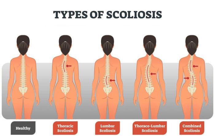
Scoliosis in adolescents and teens can be corrected with proper bracing, adjustments and lead a normal life. For adult scoliosis correcting the problem is more difficult. Fortunately, cases of adult scoliosis are rare. Scoliosis cases that follow from childhood into adulthood require a comprehensive diagnosis to determine severity. Thoracolumbar scoliosis adult-onset scoliosis requires an understanding of the catalysts to develop an effective treatment plan. Chiropractors use a full range of diagnostic tools to measure the severity of adult scoliosis.
Diagnosis
Adult scoliosis is the presentation of abnormal curvature of the spine. It can happen in the thoracic, lumbar spine, or both. This can have varying degrees of severity. Severe adult scoliosis can be apparent through visual assessment and examination. Cases that are not as obvious require utilizing diagnostic tools. These include:
Imaging
X-rays will show any asymmetry that is associated with scoliosis. This asymmetry can be present in the hips or shoulder and is usually qualified by spinal misalignment.
Walking Gait Examination
Inspecting how worn out an individual’s shoe/s are and having them perform various walking tests can reveal problems with gait. In adults, this can present instability. For example, having problems with balance or fast-twitch muscle response.
Neuromotor Exams
These exams are general and first performed to get a baseline diagnosis for the presence of adult scoliosis. Tests look at the left and right coordination along with the sense of touch capabilities. This measures the severity of the improper spinal curvature and how much it has affected the development of an individual’s motor functions. It is also done in the context of how it’s affecting the body’s biomechanics. Following these exams are quantitative tools/techniques for measuring the severity of adult thoracolumbar scoliosis. These include:
Cobb Angle Measurement
This tool determines the maximum degree of spinal curvature variation and provides a context for severity.
King Classification Tool
This examines the vertebral alignment to determine the spinal variance in specific vertebrae from the neutral center position.
Lenke Classification Tool
This spinal exam relies on measurements of three positions and looks for flexibility.
Combined Approaches
When assessing adult scoliosis, this is important to understand and helps determine how to proceed with treatment. The body is no longer in development as an adolescent. This means bracing does not come with a one-size-fits-all approach. Chiropractic can help with the assessment modalities used to investigate adult scoliosis cases. These measurement and analyses tools are often used in combination to develop a complete picture of what is going on.
Body Composition
Fill Up With Prebiotics
Individuals can help their gut bacteria thrive in the digestive tract by consuming prebiotics. Prebiotics are a form of soluble fiber. The body cannot digest these prebiotics, but gut bacteria can. Recommended sources of fiber-rich prebiotics can be found in nutrient-dense foods like:
Leeks
Garlic
Onions
Fruits
Legumes
Raw chicory
A diet with various fiber types has been shown to reduce the risk of obesity and prevent weight gain. Resistant starches like plantains, green bananas, and cooled potatoes have increased beneficial bacteria in the colon. Barley, oats, and wheat bran are insoluble high-fiber grains that are also recommended sources.
References
Aebi, Max. “The adult scoliosis.” The European spine journal: official publication of the European Spine Society, the European Spinal Deformity Society, and the European Section of the Cervical Spine Research Society vol. 14,10 (2005): 925-48. doi:10.1007/s00586-005-1053-9
Haenen, Daniëlle et al. “A diet high in resistant starch modulates microbiota composition, SCFA concentrations, and gene expression in pig intestine.” The Journal of nutrition vol. 143,3 (2013): 274-83. doi:10.3945/jn.112.169672
Lowe, Thomas et al. “The SRS classification for adult spinal deformity: building on the King/Moe and Lenke classification systems.” Spine vol. 31,19 Suppl (2006): S119-25. doi:10.1097/01.brs.0000232709.48446.be
Surgery options when back pain is becoming chronic or so severe that an individual cannot function normally and negatively affects their life. Pretty much everyone experiences back pain at some point. This is often from:
Lifting heavy/non-heavy objects incorrectly
Improper posture
Twisting in an awkward way
Overreaching
Muscle spasms
Physical activity the body is not used to doing
Most cases of backaches and pain go away by themselves or with conservative treatment. But sometimes, surgery is necessary.
When Surgery Is Necessary
Acute back pain can last for days or weeks and can often resolve with physical therapy, chiropractic, and self-care. Back pain that continues for 12 weeks or longer is considered chronic. Around twenty percent of individuals who experience acute low back pain after a year begin developing chronic back pain. Doctors try to treat most back pain cases with non-surgical approaches.
They usually begin with physical therapy/chiropractic.
If that doesn’t work, then medication is incorporated.
However, many individuals do not want to take long-term medication, which is when surgery may be recommended.
In most cases, surgery is a last resort.
When the pain radiates to the legs or if it is causing problems with bladder and/or bowel function, these are definite signs/symptoms that surgery is needed. If the pain/dysfunction continues after thorough and effective non-surgical treatment, surgery could be recommended to preserve the spine to improve spinal strength and function before the problem worsens, causing further injury and damage. Some of the most common and effective spine surgery options include.
Surgery Options
Microdiscectomy
Microdiscectomy is the most common back surgery in the United States. It is minimally invasive spine surgery. Microdiscectomy patients have low back pain combined with leg pain, tingling, numbness, and weakness. In between the vertebrae are the body’s shock-absorbing discs. The discs can begin to bulge out, a bulging or herniated disc, and press on the surrounding nerve roots, causing pain, tingling, numbness, or weakness. A microdiscectomy removes the portions of the disc pressing on the nerve.
It is called micro because the surgeon wears specialized glasses known as loupes that act as microscopes. This is so the surgeon can see the details when they’re operating. The surgery is performed through a small incision in the middle of the back or on the affected side. Patients can go home a few hours after the surgery and return to normal activities within two weeks. The success rate is 85 – 95%, especially if the surgery is done early before the damage begins to spread out.
Laminectomy
The spinal canal contains a special lining. This is where the nerves and ligaments run through. Age, along with normal wear and tear on the body, causes the ligaments to thicken. This is when bone spurs can develop from osteoarthritis, and the discs can begin to bulge or rupture/herniate. This clogs and impinges the space where the nerves should easily flow through. This narrowing is called spinal stenosis. A laminectomy opens up the space relieving the compression/pressure. The procedure requires removing part of the back of a vertebra called the lamina. This enlarges the spinal canal and relieves the pressure on the nerves. The procedure is done through a small incision in the middle of the back but can also be done through a minimal incision. Leg pain improves after surgery. A traditional incision full recovery takes 6 to 12 weeks. The success rate is around 85 percent.
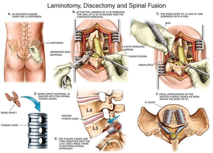
Spinal Fusion
A spinal fusion joins/fuses two or more bones in the spine. This is done when an individual has severe compression of the nerves, severe instability, or spinal revision surgery. A fusion can help stabilize spinal fractures. Other reasons for a spinal fusion are spine deformity, cancer of the spine, and sometimes used for intractable pain. A fusion stabilizes the spine with screws and rods. The disc causing the compression is replaced with a fusion device and bone graft. The surgery is often performed in combination with a laminectomy. Recovery and returning to activities can take around 3-4 months after the procedure. The success rate is 85-90% with pain improvement.
Kyphoplasty
Spinal compression fractures are common in individuals with osteoporosis. When they happen, the pain can be so intense that braces and medication don’t help. Kyphoplasty can bring pain relief. It can be performed by a pain management doctor, interventional radiologist, or surgeon in an outpatient X-ray facility and operating room. The procedure involves conscious sedation, sometimes accompanied by general anesthesia. A small instrument is inserted into the vertebra, and a balloon is inflated to make room for bone cement. After the bone cement is injected, patients can go home within a few hours. The success rate is around 85%, and recovery time could be several days.
Disc Replacement
This is a procedure that can replace spinal fusion for certain cases. A disc replacement can be done in the lumbar/low back or the cervical/neck spine. This procedure is performed to treat a pinched nerve and/or spinal cord compression. The injured/damaged disc is removed and is replaced with an artificial disc. The device allows for motion, whereas fusion procedures fuse the bones to stabilize and immobilize the area. Disc replacement is recommended for younger patients that don’t have serious arthritis. This is because they still have mobility. If significant arthritis is present, the patient could experience more pain and require spinal fusion. Recovery takes around six weeks. Intense physical activity is off-limits for two or three weeks after the initial recovery period. The success rate is more than 90%.
Anterior Cervical Discectomy and Fusion – ACDF
This is a common neck/cervical spine procedure. This surgery is for pain relief, weakness, tingling, and numbness of the arms caused by a pinched nerve or stenosis. The damaged disc is removed through a small incision in the side of the front of the neck. The disc is replaced with a bone graft or specialized spacer and a small plate with screws. This is to stabilize the spine. It is highly effective in relieving pain and in preventing neurological decline from spinal cord compression. Recovery time is around 12 weeks before a full return to normal activities. However, individuals report feeling better after two weeks.
Back Surgery Options
The majority of cases involving back pain get better on their own or with conservative treatment. But if an individual cannot find relief, there are safe and effective surgery options that can help.
Body Composition
When The Immune System Activates
When the body gets sick from a bacterial infection, virus, etc., the body’s defense system activates, causing inflammation. This immune response serves as the first wave of defense against foreign invaders. The infected area becomes red and swollen from increased blood flow. For example, when the nose gets red from a cold, this is inflammation. The reaction is caused by white blood cells known as macrophages, and the proteins they emit called cytokines encourage inflammation. Inflammation that’s triggered by the immune system is normally a good thing. It means the body is releasing a proper amount of hormones and proteins. These activate the white blood cells to start the healing process and work to fight the infection.
References
Low Back Pain Fact Sheet. National Institute of Neurological Disorders and Stroke. https://www.ninds.nih.gov/Disorders/Patient-Caregiver-Education/Fact-Sheets/Low-Back-Pain-Fact-Sheet
A review of complication rates for Anterior Cervical Diskectomy and Fusion (ACDF). Surg Neurol Int. 2019. https://pubmed.ncbi.nlm.nih.gov/31528438/
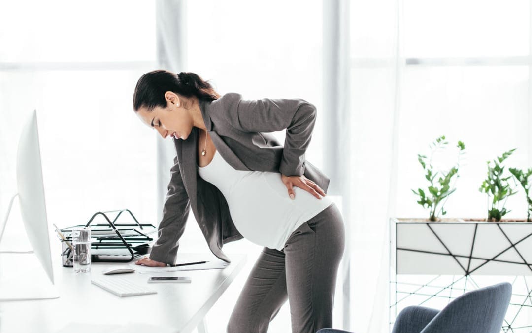
Pregnancy is an exciting time in a woman’s life. A healthy pregnancy is the objective, and therefore, essential to maintain a healthy diet and stay physically active. However, it can be easy to forget about the strain that pregnancy places on the body and push through the aches and pain with everything going on. Specifically the spine and pelvis. During pregnancy, the body goes through various changes to accommodate the growing baby. When pregnant, a hormone known as relaxin is released. This relaxes the joints for labor and delivery. Sometimes the ligaments can become too loose that they become structurally unstable. This causes pain. Other factors that can lead to spine misalignment and pain:
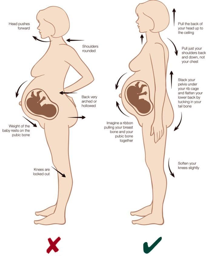
25 – 30 lbs of added weight and pressure on the spine, pelvis, legs, feet.
Weakened abdominal muscles.
Lower back curvature becomes more prominent.
Pregnant women going through discomfort/pain can benefit from chiropractic pregnancy adjustments. These maintain spinal alignment while keeping the baby safe. Chiropractors are trained to treat pregnant women; however, there is additional certification to specialize in this area. A prenatal chiropractor also provides information on:
Preparing the body for labor and birth
Maintaining healthy posture
Ensure the body is properly aligned throughout the pregnancy.
Diet
Health coaching
Postpartum care
Chiropractic Benefits For A Healthy Pregnancy
The obvious benefits include back pain relief, quality of life improvement, and reducing the need for pain medications. Other benefits include:
Alleviates sciatic nerve pain
Helps to control urinary incontinence
Reduces pregnancy-related nausea
Aligns the pelvis for optimal delivery
Reduces labor and delivery time
There is little risk associated with prenatal chiropractic care. However, it is not recommended for pregnant women with health complications like vaginal bleeding or ectopic pregnancy.
Repositioning
If the baby does not have enough room or is going into a breeched position, a chiropractor will work on the ligaments and muscles in the pelvic region to decrease any intrauterine restrictions. Getting the pelvis aligned will allow for an optimal delivery position. This will help move the head down and give the baby more space. If there is a breech position, it is recommended to see a chiropractor once a week during the eighth and ninth months.
When to Start
After receiving approval from the obstetrician, a woman can see a chiropractor at any stage during pregnancy. Many women start chiropractic in the first trimester. This develops a relationship early and benefits the woman as her body changes throughout the pregnancy.
How Often
Most patients visit a chiropractor once a month during the first trimester. Then they increase the sessions as the pregnancy progresses. Every woman’s pregnancy is different, requiring a personalized/customized treatment plan. The chiropractor will best advise on the frequency of treatment.
Pregnancy Technique
A chiropractor’s job is to manipulate the woman’s spine, joints, and muscles into alignment.
The adjustments utilize gentle pressure. The techniques are adjusted accordingly. Depending on how far along the woman is, as well as musculoskeletal health. For example, they could emphasize focus on the pelvis to reduce stress on the uterus and ligaments for a few sessions. Then shift back to the spine. They will not exert excessive pressure on the abdomen and possibly incorporate specialized equipment specifically for pregnant women.
Sciatica
Symptoms of sciatica are common during pregnancy, typically in the later months. This comes from the added pressure on the sciatic nerve. It can be sharp, burning pain from the hip to the foot. This makes walking, sitting, and sleeping uncomfortable to unbearable. Some women are fortunate enough to experience relief if the baby moves off the sciatic nerve. However, most will need some form of treatment to manage and heal the symptoms. This usually includes:
Series of adjustments
Hot and cold therapies
Stretches
Chiropractic can continue to help after giving birth, providing post-natal care helping the body return to its pre-pregnancy state healthily and optimally.
Body Composition
Diet, Nutrition During Pregnancy
From a nutritional perspective, a high GI diet during pregnancy increases the chances of excessive weight gain and overweight babies. Intake of low-glycemic carbs is associated with weight gain in the normal range. If planning to get pregnant or are pregnant, pay attention to the quality of carb intake for a healthy pregnancy. Health care providers recommend avoiding extreme diets for the first four to six weeks after delivery. This gives the body time to recover and helps to establish a consistent milk supply, as rapid weight loss could interfere with breastfeeding. Before going on a diet or starting an exercise program, check with a healthcare provider to rule out any medical conditions.
References
30 of the Most Surprising (And Alarming) Back Pain Statistics. The Good Body. https://www.thegoodbody.com/back-pain-statistics/. Last updated May 30, 2017. Accessed September 22, 2017.
Bernard, Maria, and Peter Tuchin. “Chiropractic Management of Pregnancy-Related Lumbopelvic Pain: A Case Study.” Journal of chiropractic medicine vol. 15,2 (2016): 129-33. doi:10.1016/j.jcm.2016.04.003
Gostine M. Is Lower Back Pain Normal During Pregnancy? BabyQ. https://www.babyq.com/lens/lifestyle/is-lower-back-pain-normal-during-pregnancy/. Published February 4, 2017. Accessed September 22, 2017.
Sabino J, Grauer JN. Pregnancy and low back pain. Curr Rev Musculoskelet Med. 2008; 1(2): 137–141. Published online February 26, 2008. doi: 10.1007/s12178-008-9021-8.
Individuals and doctors have praised the anti-inflammatory, pain-relieving properties of drinking tea. Inflammation is the body’s natural immune response when injury and infection present. This is good. However, it’s meant to be a temporary response that deactivates when there is no longer any danger. When the body is exposed to various irritants like industrial chemicals, inflammatory foods like sugar, refined carbohydrates, and autoimmune disorderscan cause the immune system to go into overdrive. Chronic inflammation can develop, circulating powerful hormones and chemicals through the body, causing damage to the cells. One consequence of chronic inflammation is back pain. Besides standard backaches, some chronic conditions are directly tied to inflammation. These include forms of arthritis:
Ankylosing spondylitis
Rheumatoid arthritis
Transverse myelitis
Multiple sclerosis
These conditions involve inflammation of the central nervous system.
Drinking tea can help with back pain and pain in general.
Teas With Anti-Inflammatory Properties
Certain teas contain anti-inflammatory compounds. These compounds are called polyphenols and work to decrease the chemicals in the body responsible for pain and inflammation. There are varieties of teas that contain anti-inflammatory properties.
Certain Teas Reduce Inflammation
Drinking specific teas with more polyphenols can better decrease inflammation. For example, green tea is higher in polyphenols than black tea. Recent studies centered on individuals with rheumatoid arthritis over six months found significant improvement in symptoms in those who drank green tea. Green tea works best when part of an anti-inflammatory and nutritional lifestyle adjustment. This supports combating inflammation. Other teas that are believed to reduce inflammation include:
Turmeric
Holy basil
Ginger
Three Cups a Day
The amount of tea depends on the quality of the tea and how it is prepared. Doctors recommend around three cups a day for individuals with rheumatoid arthritis. However, these could contain caffeine. If this is an issue, there are decaffeinated versions with the same anti-inflammatory properties.
Drinking Tea Works Best When Combined with Other Treatments
If experiencing back pain or looking to combat a specific condition, it’s recommended to utilize various treatment approaches combined with drinking tea. This includes:
Certain back conditions benefit from drinking tea regularly; however, spine structural issues or fractures will not benefit from tea’s mild anti-inflammatory properties. It is vital for individuals with back pain that a spine specialist or chiropractor perform a proper and thorough examination, especially for Individuals that take medication that could directly interact with anti-inflammatory teas.
Drinking Tea for Back Pain
For most individuals, drinking tea is safe to help treat back pain conditions and added health benefits. For example, studies have found that green tea has mild anti-cancer, anti-diabetic properties and can help in maintaining a healthy weight. If tea helps reduce pain, it’s worth trying. Remember, pain is the body’s way to alert the individual that something is wrong.
Body Composition
Alcohol and Heart Health
According to the Mayo Clinic, consuming more than three alcoholic drinks in one sitting causes a temporary blood pressure elevation. Foods often served with alcohol are usually high in salt, which can also raise blood pressure. A few alcoholic beverages on a night out is fine, but heavy or binge drinking can lead to short-term spikes in blood pressure that could cause cardiac health problems. These are the short-term effects of alcohol on blood pressure. Heavy alcohol consumption can lead to long term health risks like:
Hypertension
Heart disease
Digestive issues
Liver disease
Stroke
It’s recommended that individuals incorporate regular exercise/physical activity and healthy diet changes and watch alcohol intake to improve heart health.
References
The Clinical Journal of Pain. (October 2019) “Nonspecific Low Back Pain:
Inflammatory Profiles of Patients With Acute and Chronic Pain” https://journals.lww.com/clinicalpain/fulltext/2019/10000/nonspecific_low_back_pain__inflammatory_profiles.2.aspx
Certain Teas Bring Down Inflammation More Than Others: Journal of Physical Therapy Science. (October 2016) “Green tea and exercise interventions as nondrug remedies in geriatric patients with rheumatoid arthritis” https://www.ncbi.nlm.nih.gov/pmc/articles/PMC5088134/
The Bottom Line: Proceeding of the Japan Academy, Series B Physical and Biological Sciences. (March 2012) “Health-promoting effects of green tea” https://www.ncbi.nlm.nih.gov/pmc/articles/PMC3365247/
IFM's Find A Practitioner tool is the largest referral network in Functional Medicine, created to help patients locate Functional Medicine practitioners anywhere in the world. IFM Certified Practitioners are listed first in the search results, given their extensive education in Functional Medicine