ClickCease
+1-915-850-0900 spinedoctors@gmail.com
Select Page

Cannabinoids

Back Clinic Cannabinoids. Plants are medicine, and as research continues with these alternative medicines, more information is available when it comes to medical options for various ailments, conditions, diseases, disorders, etc… Chiropractor Dr. Alex Jimenez investigates and brings insight into these developing medicines, how they can help patients, what they can do, and what they cannot do.

The marijuana plant is how most know about cannabinoids. It is the most recognized cannabinoid tetrahydrocannabinol (THC), which is the compound that causes feelings of euphoria.

Scientists identified cannabinoids only in cannabis. However, new research has found these same medicinal qualities in many plants, including black pepper, broccoli, carrots, clove, echinacea, and ginseng.

These vegetables or spices won’t get you high, but understanding how these different plants affect the human body can lead to vital health discoveries.


A Deeper Look Into Metabolic Syndrome | El Paso, TX (2021)

A Deeper Look Into Metabolic Syndrome | El Paso, TX (2021)

In today’s podcast, Dr. Alex Jimenez, health coach Kenna Vaughn, chief editor Astrid Ornelas discuss about metabolic syndrome from a different point of view as well as, different nutraceuticals to combat inflammation.


Dr. Alex Jimenez DC*: Welcome, guys, welcome to the podcast for Dr. Jimenez and crew. We’re discussing today’s metabolic syndrome, and we’re going to be discussing it from a different point of view. We will give you excellent, useful tips that can make sense and are easily doable at home. Metabolic syndrome is a very vast concept. It contains five major issues. It has high blood glucose, it has belly fat measurements, it has triglycerides, it has HDL issues, and it pretty much has a whole conglomeration of dynamics that have to be measured in the whole reason we discuss metabolic syndrome because it affects our community very much. So, we’re going to be discussing these particular issues and how we can fix them. And give you the ability to adapt your lifestyle so that you don’t end up having. It’s one of the most important disorders affecting modern medicine today, let alone once we understand it. Everywhere you go, you’re going to see a lot of people having metabolic syndrome. And it’s part of a society, and that’s something you see in Europe as much. But in America, because we do have a lot of foods and our plates are usually bigger, we have the ability to adapt our bodies differently by just what we eat. No disorder will change so quickly and fast as a good mechanism and a good protocol to help you with metabolic disorders and metabolic syndrome. So having said that, today, we have a group of individuals. We have Astrid Ornelas and Kenna Vaughn, who will discuss and add information to help us through the process. Now, Kenna Vaughn is our health coach. She’s the one who works in our office; when I’m a practicing physician on physical medicine and when I’m working with people one on one, we have other people working with dietary issues and dietary needs. My team here is very, very good. We also have our top clinical researcher and the individual who curates much of our technology and is at the cutting edge of what we do and our sciences. It’s Mrs. Ornelas. Mrs. Ornelas or Astrid, as we call her, she’s ghetto with the knowledge. She gets nasty with science. And it’s really, really where we are. Today, we live in a world where research is coming and spitting out of the NCBI, which is the repository or PubMed, which people can see we use this information and we use what works and what does it. Not all information is accurate in PubMed because you have different points of view, but it’s almost like a finger on a pulse when we have our finger in. We can see the things that affect it. With certain keywords and certain alerts, we get notified of changes for, let’s say, dietary sugar issues or triglyceride issues with fat issues, anything about metabolic disorders. We can kind of come up with a treatment protocol that is live adapted from doctors and researchers and PhDs around the world almost instantaneously, literally even before they’re published. For example, today happens to be February 1st. It’s not, but we’ll be getting results and studies presented by the National Journal of Cardiology that will come out in March if that makes sense. So that information is early hot off the press, and Astrid helps us figure these things out and sees, “Hey, you know, we found something really hot and something to help our patients” and brings the N equals one, which is patient-doctor equals one. A patient and therapist equal one that we don’t do specific protocols for everyone in general. We do specific protocols for each person as we go through the process. So as we do this, the journey of understanding metabolic syndrome is very dynamic and very deep. We can start from just looking at someone to the bloodwork, all the way to dietary changes, to metabolic changes, all the way down to the cellular activity that it’s actively working. We measure issues with BIAs and BMI, which we have done with previous podcasts. But we can also get into the level, the genomics and the changing of the chromosomes and the telomeres in the chromosomes, which we can affect by our diet. OK. All roads lead to diets. And what I say in some weird way, all roads lead to smoothies, OK, smoothies. Because when we look at smoothies, we look at the components of smoothies and come up with dynamics that are abilities to change now. What I look for is when I look for treatments, I look at things that make people’s lives better, and how can we do this? And for all those mothers, they understand that they may not realize that they do this, but a mom doesn’t wake up saying, I’m going to give my kid food. No, she’s kind of doing a mental lavage of bringing the whole kitchen because she wants to infuse the best nutrition for their child and offer their best kind of options for their baby to go through the world or daycare or elementary school, through middle school, through high school so that the child can develop well. Nobody goes out thinking that I’m going to give my kid just junk and. And if that’s the case, well, that’s probably not good parenting. But we won’t talk about that well; we will talk about good nutrition and adapting those things. So I’d like to introduce Kenna right now. And she’s going to be discussing a little bit of what we do when we see someone with metabolic disorders and our approach to it. So as she goes through that, she’s going to be able to understand how we evaluate and assess a patient and bring it in so that we can start getting a little bit of control for that individual. Kenna, it’s all yours.


Kenna Vaughn: All right. So first, I just want to talk about the smoothies a little bit more. I am a mom, so in the morning time, things get crazy. You never have as much time as you think you do, but you need those nutrient nutrients and so do your kids. So I love smoothies. They’re super fast. You get everything you need. And most people think that when you’re eating, you’re eating to fill your stomach, but you’re eating to fill your cells. Your cells are what need those nutrients. That’s what carries you on with the energy, the metabolism, all of that. So those smoothies are a super great option, which we give our patients. We even have a book with 150 smoothie recipes that are great for anti-aging, helping diabetes, lowering cholesterol, controlling inflammation, and things like that. So it’s one resource we give to our patients. But we do have multiple other options for the patients who come in with metabolic disease.


Dr. Alex Jimenez DC*:  Before you go in there, Kenna. Let me just kind of add that what I’ve learned is that we have to make it simple. We got to take homes or takeaways. And what we’re trying to do is we’re trying to give you the tools that can help you in that process. And we’re going to take you to the kitchen. We’re going to grab you by the ear, so to speak, and we’re going to show you the areas where we need to look at. So Kenna is about to give us the information in terms of smoothies that will assist us with dietary changes that we can provide our families and change its metabolic disaster that affects so many people called metabolic syndrome. Go ahead.


Kenna Vaughn: OK, so like he was saying with those smoothies. One thing that you should add to your smoothie is, which what I love to add in mine is spinach. Spinach is an excellent choice because it gives your body more nutrients. You are getting an extra serving of vegetables, but you can’t taste it, especially when it gets covered up by the natural sweetness that you find in fruits. So that’s a great option when it comes to the smoothies. But another thing that Dr. Jiménez was mentioning is other things in the kitchen. So there are other substitutes that we’re kind of wanting our patients to use and implement. You can start small, and it’ll make a huge difference just by switching out the oils you’re cooking with. And you’ll begin to see an improvement in your joints, your kids, and everyone will just improve immensely. So one thing we want to get our patients into using is those oils, such as avocado oil, coconut oil, and… Olive oil? Olive oil. Yes, thank you, Astrid.


Dr. Alex Jimenez DC*: That was olive oil. That was Astrid in the background. We’re getting the facts out excellent and continue.


Kenna Vaughn: When you switch those out, your body breaks things down differently with those unsaturated fats. So that’s just another option that you have in that kitchen besides making those smoothies. But like I said before, I’m all about quick, easy, simple. It’s way easier to change your lifestyle when you have a whole team around you. And when it’s easy, you don’t. You don’t want to go out and make everything super difficult because the chances of you sticking to it aren’t very high. So one thing we want to do is make sure that everything that we’re giving our patients is easy to do and it’s attainable for everyday life.


Dr. Alex Jimenez DC*: I’m very visual. So when I go to the kitchen, I like making my kitchen look like the cocina or whatever they call it in Italy, the cucina and I have three bottles there, and I have an avocado oil one. I have the coconut oil one, and I have the olive oil right there. There are big bottles there. They make them pretty, and they look Tuscan. And, you know, I don’t care if it’s an egg, I don’t care. Sometimes, even when I’m having my coffee, I grab the coconut oil one, and I pour that one in and make myself a java with coconut oil in it. So, yeah, go ahead.


Kenna Vaughn: I was going to say that’s a great option too. So I drink green tea, and I also add coconut oil in that green tea to help boost everything and give my body another dose of those fatty acids that we want.


Dr. Alex Jimenez DC*: I got a question for you when you have your coffee like that; when you have the oil in it, does it kind of lubricate your lips.


Kenna Vaughn: It does a little bit. So it’s also like chapstick.


Dr. Alex Jimenez DC*: Yeah, it does. It’s like, Oh, I love it. OK, go ahead.


Kenna Vaughn: Yeah, I also have to stir a little bit more just to make sure everything gets it right. Yeah. And then another thing just talking about something our patients can do when it comes to at home, there are tons of different options with eating fish. Increasing your good fish intake throughout the week, that’s going to help also. And just because fish provides so many great things like omegas, I know Astrid also has some more information on omegas.


Dr. Alex Jimenez DC*: I got a question before Astrid gets in there. You know, look, when we talk about carbohydrates, people, is it what a carbohydrate is? Oh, people say an apple, banana, candy bars, and all kinds of stuff people can rattle off carbohydrates or proteins. Chicken, beef, whatever they can rile up. But one of the things I found that people have a difficult time with is what good fats are? I want five. Give me ten good fats for a million dollars. Give me ten good fats like lard, like meat. No, this is what we’re talking about. Because the simple fact that we use and we’re going to add more to it relative bad is going to be avocado oil. Olive oil. Is it coconut oil? We can use things like butter oils, different types of margins, and not margins, but kinds of butter that are from, you know, grass-fed cows. We basically can run out of creamers, you know, non-nondairy creams, very specific creamers, those we run out of it, right? Real fast. So it’s like, what else is fat, right? And then we search for it. So one of the best ways to do it is that we’re not going to always put creamer on top or our butter on top, which by the way, some coffees they have, they put butter in it and blend it, and they make a fantastic little java hit. And everyone comes with their little ginger and oils and their coffee and makes espresso from heaven, right? So what else can we do?


Kenna Vaughn: We can, like I said, adding those fish in, which is going to help to give our bodies more of those omegas. And then we can also do more purple vegetables, and those are going to provide your body with more antioxidants. So that’s a good option when it comes to the grocery store. A rule of thumb that I love and heard a long time ago is to not shop in the aisles is to try to shop on the edges because the edges are where you’re going to find all that fresh produce and all those lean meats. It’s when you start to get into those aisles, and that’s where you’re going to start finding, you know, the cereal, those bad carbohydrates, those simple carbohydrates that the American diet has come to love but does not necessarily need. The Oreos?


Kenna Vaughn: Yes.


Dr. Alex Jimenez DC*: The candy aisle that every kid knows. OK, yes. 


Kenna Vaughn: So that’s just another great point there. So when you come into our office, if you’re suffering from metabolic syndrome or just anything in general, we make your plans super personalized and give you so many tips. We listen to your lifestyle because what works for one person might not work for another. So we make sure that we provide you with information that we know you’ll be successful with and provide education because that’s another huge part of it.


Dr. Alex Jimenez DC*: All roads lead to the kitchen, huh? Right? Yes, they do. OK, so let’s zoom on precisely for the fat and the nutraceuticals. I want to give you an idea as to what type of nutraceuticals are appropriate for us because we want to bust down these five issues affecting metabolic syndrome that we discussed. What are the five guys? Let’s go ahead and start them up. It’s high blood sugar, right?


Kenna Vaughn: High blood glucose, low HDLs, which will be that good cholesterol everyone needs. Yes. And it’s going to be the high blood pressure, which is not considered high from a doctor’s standard, but it is deemed to be elevated. So that’s another thing; we want to ensure that this is metabolic syndrome, not a metabolic disease. So if you go to the doctor and your blood pressure is 130 over eighty-five, that’s an indicator. But yet your provider might not necessarily say your blood pressure is super high. 


Dr. Alex Jimenez DC*: None of these disorders here by themselves are clinical states, and, individually, they’re pretty much just things. But if you combine all these five, you have metabolic syndrome and feel like not too good, right?


Astrid Ornelas: Yeah, yeah.


Kenna Vaughn: Another one is going to be the excess weight around the belly and the higher triglycerides.


Dr. Alex Jimenez DC*: Easy to see. You can see when someone has a belly that’s hanging over like a fountain, right? So we can see that you can go to it sometimes Italian restaurants and see the great cook. And he sometimes I got to tell you, sometimes it’s just, you know, we talked to Chef Boyardee wasn’t a thin guy. I think that Chef Boyardee, you know what? And the Pillsbury guy, right? Well, it wasn’t very healthy, right? Both of them suffer from metabolic syndrome just from the outset. So that’s an easy one to see. So these are the things we’re going to be reflecting on. Astrid will go over some nutraceuticals, vitamins, and some foods that we can improve things. So here’s Astrid, and here’s our science curator. But here’s Astrid, go ahead.


Astrid Ornelas: Yeah, I guess before we get into the nutraceuticals, I want to make something clear. Like we were talking about metabolic syndrome. Metabolic syndrome is not a, and I guess per se, a disease or a health issue itself. Metabolic syndrome is a cluster of conditions that can increase the risk of developing other health issues like diabetes, stroke, and heart disease. Because metabolic syndrome is not, you know, an actual health issue itself, it’s more so this group, this collection of other conditions, of other problems that can develop into much worse health issues. Just because of that fact, metabolic syndrome has no apparent symptoms itself. But of course, like we were talking about, five risk factors are pretty much the ones we discussed: excess waist fat, high blood pressure, high blood sugar, high triglycerides, low HDL, and according to health care professionals. To doctors and researchers, you know you have metabolic syndrome if you have three out of these five risk factors.


Dr. Alex Jimenez DC*: Yes. Three. Now, that doesn’t mean that if you have it, you have symptoms. As I see it was evident on. But I got to tell you in my experience when someone has more than three or three. They’re starting to feel crummy. They don’t feel right. They just feel like, you know, life’s not good. They have just an overall. They don’t look it right. So and I don’t know them, maybe. But their family knows that they don’t look good. Like mom doesn’t look good. Dad does look good.


Astrid Ornelas: Yeah, yeah. And metabolic syndrome, as I said, it has no apparent symptoms. But you know, I was kind of going with one of the risk factors with waist fat, and this is where you will see people with what you call the apple or pear-shaped body, so they have excess fat around their abdomen. And although that’s not technically considered a symptom, it is a factor that can; I guess it can give an idea to doctors or other health care professionals that this person who is, you know, they have prediabetes or have diabetes. And, you know, they have excess weight and obesity. They could have an increased risk of metabolic syndrome and therefore developing, you know, if it’s left untreated, developing other health issues like heart disease and stroke. I guess with that being said; then we’ll get into the nutraceutical.


Dr. Alex Jimenez DC*: I love this, I love this. We’re getting some good stuff, and we’re getting some information.


Astrid Ornelas: And I guess with that being said, we’ll get into the nutraceuticals. Kind of like, how Kenna was talking about what’s the takeaway? You know, we’re here talking about these health issues, and we’re here talking about metabolic syndrome today. But what’s the takeaway? What can we tell people? What can they take home about our talk? What can they do at home? So here we have several nutraceuticals, which I’ve written several articles in our blog and looked at. 


Dr. Alex Jimenez DC*:  You think, Astrid? If you look at 100 articles written in El Paso, at least in our area, they were all curated by somebody. Yes. All right.


Astrid Ornelas: Yes. So we have several nutraceuticals here that have been researched. Researchers have read all these research studies and found that they can help in some way and some form improve, you know, metabolic syndrome and these associated diseases. So the first one I want to discuss is the B vitamins. So what are the B vitamins? These are the ones that you can usually find them together. You can find them in the store. You’ll see them as B-complex vitamins. You’ll see like a little jar, and then it comes with several of the B vitamins. Now, why do I bring up B vitamins for metabolic syndrome? So one of the reasons like researchers has found that one of them, I guess, one of the causes of metabolic syndrome could be stress. So with that being said, we need to have B vitamins because when we get stressed when we have a hard day at work when we have, I guess a lot of you know, a lot of stressful things at home or with family, our nervous system will use these B vitamins to support our nerve function. So when we have a lot of stress, we will use up these vitamins, which increases stress; you know, our body will produce cortisol. You know, which serves a function. But we all know that too much cortisol, too much stress can actually. It can be harmful to us. It can increase our risk of heart disease.


Dr. Alex Jimenez DC*: You know, as I remember when we did this, all roads lead to the kitchen in terms of getting the food back in your body. All roads lead to the mitochondria when it comes to the area of the breakdown. The world of ATP energy production is surrounded and wrapped around with nicotinamide, NADH, HDP, ATPS, ADP. All these things have a connection with vitamin B of all sorts. So the vitamin B’s are at the engine in the turbine of the things that help us. So it makes sense that this was the top of the vitamin and the most important one. And then she’s got some other endpoints here on niacin. What is with niacin? What have you noticed there?


Astrid Ornelas: Well, niacin is another B vitamin, you know, there are several B vitamins. That’s why I have it there under its plural and niacin or vitamin B3, as it’s more well known. A lot of several are so clever. Many research studies have found that taking vitamin B3 can help lower LDL or bad cholesterol, help lower triglycerides, and increase HDL. And several research studies have found that niacin, specifically vitamin B3, can help increase HDL by 30 percent.


Dr. Alex Jimenez DC*: Incredible. When you look at NADP and NADH, These are the N is the niacin, the nicotinamide. So in the biochemical compound, niacin is the one that people have known that when you take it the good one or the one that’s supposed to be, you get this flushing feeling and it makes you scratch all your part of your body, and it feels good when you scratch because it makes you feel that way. Right, so lovely. And this huge.


Astrid Ornelas: Yes. Yes, and also, I just want to highlight a point about B vitamins. B vitamins are essential because they can help support our metabolism when we eat, you know, carbohydrates and fats, good fats, of course, and proteins. When the body goes through the metabolism process, it converts these carbohydrates, fats, and protein. The proteins turn into energy, and B vitamins are the main components in charge of doing that.


Dr. Alex Jimenez DC*: Latinos, in our general population, know that we have always heard of the nurse or the person who gives vitamin B injection. So you heard of those things. Right. Because you’re depressed, you’re sad, what would they do? Well, you know what would inject them with B12, right? Which are the B vitamins, right? And the person would come out like, Yeah, and they’d be excited, right? So we’ve known this, and this is the elixir of the past. Those traveling salesmen, who had the potions and lotions, made a living off of giving B vitamin complex. The first energy drinks were first designed with a B complex, you know, packing of them. Now here’s the deal. Now that we’ve learned that energy drinks cause so many issues, that we’re heading back to the B complexes to help people better. So the following vitamin we have there is that one that we have the D, we have the vitamin D.


Astrid Ornelas: Yeah, the next one I wanted to talk about is vitamin D. So there are several research studies on vitamin D and the benefits, the benefits of vitamin D for metabolic syndrome, and just how I discussed how B vitamins are beneficial for our metabolism. Vitamin D is also helpful for our metabolism, and it can help regulate our blood sugar, essentially our glucose. And that in itself is very important because, like one of the predisposing factors of metabolic syndrome, high blood sugar. And you know, if you have uncontrolled high blood sugar, it can lead to, you know, it can lead to prediabetes. And if that is left untreated, it can lead to diabetes. So research studies have also found that vitamin D itself can also improve insulin resistance, which is pretty much one that can lead to diabetes.


Dr. Alex Jimenez DC*:  You know, I just wanted to put out the vitamin D is not even a vitamin; it’s a hormone. It was discovered after C by Linus Pauling. When they found it, they just kept on naming the following letter. OK, so since it is a hormone, you just have to look at it. This particular vitamin D or this hormone tocopherol. It basically can change so many metabolism issues in your body. I’m talking about literally four to five hundred different processes that we’re finding. Last year was 400. We’re now almost 500 other biochemical processes that are affected directly. Well, it makes kind of sense. Look, our most significant organ in the body is our skin, and most of the time, we ran around in some sort of skimpy clothes, and we were in the sun a lot. Well, we didn’t stand to reason that that particular organ can produce a tremendous amount of healing energies, and vitamin D does that. It is produced by the sunlight and activated. But today’s world, whether we’re Armenian, Iranian, different cultures in the north, like Chicago, people don’t get as much light. So depending on cultural changes and closed people living and working in these fluorescent lights, we lose the essence of vitamin D and get very sick. The person who takes vitamin D is much healthier, and our goal is to raise the vitamin D is a fat-soluble vitamin and one that embeds itself by it and is saved in the liver along with the fat in the body. So you can raise it slowly as you take it, and it’s tough to get toxic levels, but those are at about one hundred twenty-five nanograms per deciliter that are too high. But most of us run around with 10 to 20, which is low. So, in essence, by raising that, you’re going to see that the blood sugar changes are going to happen that Astrid is speaking about. What are some of the things that we notice about, particularly vitamin D? Anything?


Astrid Ornelas: I mean, I’ll get back to vitamin D in a bit; I want to discuss some of the other nutraceuticals first. OK. But pretty much vitamin D is beneficial because it helps improve your metabolism, and it helps improve your insulin resistance, at least towards metabolic syndrome.


Dr. Alex Jimenez DC*: How about calcium?


Astrid Ornelas: So calcium goes hand-in-hand with vitamin D, and the thing that I wanted to talk about with vitamin D and calcium together. We often think about these five factors that we mentioned before that could cause a metabolic syndrome. Still, there’s, you know, if you want to think about it, like what are the underlying causes for a lot of these risk factors? And like, you know, obesity, a sedentary lifestyle, people who don’t engage in an exercise or physical activity. One of the things that can predispose a person or increase their risk of metabolic syndrome. Let me put the scenario. What if a person has a chronic pain disease? What if they have something like fibromyalgia? They’re constantly in pain. They don’t want to move, so they don’t want to exercise. They don’t want to aggravate these symptoms. Sometimes, some people have chronic pain or things like fibromyalgia. Let’s go a little bit more basic. Some people just have chronic back pain, and you don’t want to work out. So just you’re not choosing like some of these people aren’t choosing to be inactive because they want to. Some of these people are legitimately in pain, and there are several research studies, and this is what I was going to tie in vitamin D and calcium with that vitamin D and calcium. You know, we can you can take them together. They can help improve chronic pain in some people.


Dr. Alex Jimenez DC*: Incredible. And we all know that calcium is one of the causes of muscle spasms and relaxers. Tons of reasons. We’re going to go into each one of these. We’re going to have a podcast on just vitamin D and the issues in calcium because we can go deep. We’re going to go deep, and we’re going to go all the way to the genome. The genome is genomics, which is the science of understanding how nutrition and the genes dance together. So we’re going to go there, but we’re kind of like we’re penetrating slowly in this process because we have to take the story slowly. What’s up next?


Astrid Ornelas: So next, we have omega 3s, and I want to specifically highlight that we’re talking about omega 3s with EPA, not DHA. So these are EPA, which is the one that’s listed up there, and DHA. They are two essential types of omega 3s. Essentially, they’re both very important, but several research studies and I’ve done articles on this as well have found that I guess taking omega 3s specifically with EPA, it’s just more superior in its benefits than DHA. And when we talk about the omega 3s, these can be found in fish. Most of the time, you want to take omega 3s; you see them in the form of fish oils. And this is going back to what Kenna discussed before, like following a Mediterranean diet, which mainly focuses on eating a lot of fish. This is where you get your intake of omega 3s, and research studies have found that omega 3s themselves can help promote heart health, and they can help lower bad cholesterol to your LDL. And these can also improve our metabolism, just like vitamin D.


Dr. Alex Jimenez DC*: Want to go ahead and blanket all these things under the fact that we’re also looking, and when we’re dealing with metabolic syndrome, we’re dealing with inflammation. Inflammation and omegas have been known. So what we need to do is to bring out the fact that omegas have been in the American diet, even in a grandma’s diet. And then, like again, we hear back in the day when grandma or great-grandma would give you cod liver oil. Well, the highest omega-carrying fish is the herring, which is at about 800 milligrams per serving. The cod is next when it’s around 600. But because of the availability, the card’s much more available in certain cultures. So everybody would have cod liver oil, and they’d make you close your nose and drink it, and they knew that it correlated. They would think it’s a good lubricant. Still, it was an anti-inflammatory specifically with people, and usually, grandmothers who knew about this right helps with the intestines, helps the inflammation, helps with the joints. They knew the whole story behind that. So we’ll go deep into the Omegas in our later podcast. We have another one that’s here. It’s called berberine, right? What’s the story on berberine?


Astrid Ornelas: Well, pretty much the next set of nutraceuticals that are listed here, berberine, glucosamine, chondroitin, acetyl L-carnitine, alpha-lipoic acid, ashwagandha, pretty much all of these have been tied into what I talked before about chronic pain and all of these health issues. I listed them up here because I’ve done several articles. I’ve read various research studies that have covered these in different trials and across multiple research studies with numerous participants. And these have pretty much found, you know, this group of nutraceuticals here that are listed; these have also been tied in to help reduce chronic pain. You know, and as I discussed before, like chronic pain, you know, people who have fibromyalgia or even like, you know, let’s go a little bit simpler people who have back pain, you know, these inactive people who have sedentary lifestyles simply because of their pain and they can be at risk of metabolic syndrome. A lot of these research studies have found these nutraceuticals themselves can also help reduce chronic pain.


Dr. Alex Jimenez DC*: I think the new one is called alpha-lipoic acid. I see acetyl L-carnitine. We’re going to have our resident biochemist on the following podcast to go deep into these. Ashwagandha is a fascinating name. Ashwagandha. Say it. Repeat it. Kenna, can you tell me a bit about ashwagandha and what we’ve been able to discover about ashwagandha? Because it is a unique name and a component that we look at, we will talk about it more. We’re going to get back to Astrid in a second, but I’m going to give her a little break and kind of like, let Kenna tell me a bit of ashwagandha.


Kenna Vaughn: I was going to add in something about that berberine.


Dr. Alex Jimenez DC*: Oh, well, let’s go back to berberine. These are berberine and ashwagandha.


Kenna Vaughn: OK, so that berberine has also been shown to help decrease the HB A1C in patients with blood sugar dysregulation, which will come back to the whole prediabetes and type two diabetes situations that can occur in the body. So that one is also has been shown to decrease that number to stabilize the blood sugar.


Dr. Alex Jimenez DC*:  There’s a whole thing we’re going to have on berberine. But one of the things that we did in terms of metabolic syndrome definitely made the top list here for the process. So there’s ashwagandha and berberine. So tell us all about ashwagandha. Also, ashwagandha is the one. So in terms of blood sugar, the A1C is the blood sugar calculation that tells you exactly what the blood sugar does over about three months. The glycosylation of the hemoglobin can be measured by the molecular changes that happen within the hemoglobin. That’s why the Hemoglobin A1C is our marker to determine. So when ashwagandha and berberine come together and use those things, we can alter the A1C, which is the three-month kind of like the historical background of what is going on. We’ve seen changes on that. And that’s one of the things that we do now in terms of the dosages and what we do. We’re going to go over that, but not today because that’s a little bit more complex. Soluble fibers have also been a component of things. So now, when we deal with soluble fibers, why are we talking about soluble fibers? First of all, it is food for our bugs, so we have to remember that the probiotic world is something we cannot forget. People need to understand that, though, that probiotics, whether it’s the Lactobacillus or Bifidobacterium strains, whether it’s a small intestine, large intestine, early on the small intestine, there are different bacteria to the very end to see come to the back end. So let’s call that the place that things come out. There are bacteria everywhere at different levels, and each one has a purpose of discovering that. There’s vitamin E and green tea. So tell me, Astrid, about these dynamics in terms of green tea. What do we notice as it pertains to metabolic syndrome?


Astrid Ornelas: OK. So green tea has a lot of benefits, you know? But, you know, some people don’t like tea, and some are more into coffee, you know? But if you want to get into drinking tea, you know, definitely because of its health benefits. Green tea is an excellent place to start and in terms of metabolic syndrome. Green tea has been demonstrated to help improve heart health, and it can help lower these risk factors that pertain to metabolic syndrome. It can help, you know, several research studies that have found that green tea can help lower cholesterol, bad cholesterol, LDLs.


Dr. Alex Jimenez DC*: Does green tea help us with our belly fat?


Astrid Ornelas: Yeah. There’s one of the benefits of green tea that I’ve read about. Pretty much one of the ones that probably that it’s most well known for is that green tea can help with weight loss.


Dr. Alex Jimenez DC*: Oh my gosh. So basically water and green tea. That’s it, guys. That’s all. We limit our lives that are also, I mean, we forgot even the most powerful thing. It takes care of those ROSs, which are reactive oxygen species, our antioxidants, or oxidants in our blood. So it just basically squelch them and takes them out and cools their cool and prevents even the normal deterioration that happens or the excessive deterioration that occurs in the breakdown of normal metabolism, which is a byproduct which is ROS, reactive oxygen species are wild, crazy oxidants, which we have a neat name for the things that squashes them and calms them and puts them in the order they call antioxidants. So the vitamins that are antioxidants are A, E, and C are antioxidants, too. So those are potent tools that we deal with as we lower body weight. We free up a lot of toxins. And as the green tea goes into squirt, squelch them, cools them, and gets them out of gear. Guess where the other organ that helps with the whole insulin production is, which is the kidneys. The kidneys are flushed out with green tea and then also helps. I notice that one thing that you haven’t done, Astrid, is done articles on turmeric, right?


Astrid Ornelas: Oh, I’ve done a lot of articles on turmeric. I know because, from the list that’s up there, turmeric and curcumin are probably like one of my favorite nutraceuticals to talk about.


Dr. Alex Jimenez DC*: Yeah, she’s like gnawing on a root and a couple of times.


Astrid Ornelas: Yeah, I have some in my fridge right now.


Dr. Alex Jimenez DC*: Yeah, you touch that turmeric, and you can lose a finger. What happened to my finger? Did you get near my turmeric? The root, right? So. So tell us a bit about the properties of turmeric and curcumin in terms of metabolic syndrome.


Astrid Ornelas: OK. I’ve done several, you know, a lot of articles on turmeric and curcumin. And we’ve also discussed that before, and several of our past podcasts and turmeric is that it’s that yellow yellowish could look orange to some people, but it’s usually referred to as a yellow root. And it’s very popular in Indian cuisine. It’s what it’s one of the main ingredients that you’ll find in curry. And curcumin, pretty sure some of you people have heard of curcumin or turmeric, you know? What’s the difference? Well, turmeric is the flowering plant, and it’s the root. We eat the root of turmeric, and curcumin is just the active ingredient in turmeric that gives it a yellow color.


Dr. Alex Jimenez DC*: Guys, I will not let anything but the top type of curcumin and turmeric products be available to their patients because there’s a difference. Certain ones are produced with literally, I mean, we got solvents, and with the way we get things out and of curcumin and turmeric or even stuff like cocaine, you have to use a distillate. OK? And whether it’s water, acetone, benzene, OK, or some sort of a byproduct, we know today that benzene is used to process many types of supplements, and certain companies use benzene to get the best out of turmeric. The problem is benzene is cancer-producing. So we’ve got to be very careful which companies we use. Acetone, imagine that. So there are processes that are in place to extract the turmeric properly and that are beneficial. So finding suitable turmeric, all turmerics are not the same. And that’s one of the things that we have to assess since it has so many products in the world is running real crazy to try to process turmeric and precisely, even if it’s the last thing that we’re discussing today on our subject matter. But it’s one of the most important things today. We don’t even understand aspirin. We know it works, but the total magnitude of it is yet to be told. However, turmeric is in the same boat. We’re learning so much about it that every day, every month, studies are being produced on the value of turmeric into the natural diet, so Astris is in tune in on the target on that. So I’m sure she’s going to bring more of that to us, right?


Astrid Ornelas: Yes, of course. 


Dr. Alex Jimenez DC*: So I think what we can do today is when we look at this, I’d like to ask Kenna, when we look at a metabolic syndrome from the presentations of symptoms or even from laboratory studies. The confidence of knowing that N equals one is one of the essential components that we have now in functional medicine and functional wellness practices that a lot of physical medicine doctors are doing in their scope of practice. Because in metabolic issues, you can’t take metabolic away from the body. Does the metabolism happen in a back problem? We notice a correlation with back injuries, back pain, back issues, chronic knee disorders, chronic joint musculoskeletal disorders, and metabolic syndrome. So we can’t tease it. So tell us a bit, Kenna, as we close out today a bit of what a patient can expect when they come to our office, and they get kind of put in the “Oops, you got metabolic syndrome.” So boom, how do we handle it?


Kenna Vaughn: We want to know their background because, as you said, everything is connected; everything is in-depth. There are details we want to get to know all so we can make that personalized plan. So one of the first things we do is a very lengthy questionnaire by Living Matrix, and it’s a great tool. It does take a little while, but it gives us so much insight into the patient, which is great because it allows us to, like I said, dig deep and figure out, you know, traumas that might have happened that are leading to inflammation, which how Astrid was saying then leads that sedentary lifestyle, which then leads to this metabolic syndrome or just kind of down that road. So one of the first things we do is do that lengthy questionnaire, and then we sit down and talk to you one on one. We build a team and make you part of our family because this stuff isn’t easy to go through alone, so the most success is when you have that close-knit family, and you have that support, and we try to be that for you.


Dr. Alex Jimenez DC*: We have taken this information and realized it was very complex five years ago. It was challenging. 300 300-page questionnaire. Today we have software that we can figure out. It is backed by the IFM, the Institute of Functional Medicine. The Institute of Functional Medicine had its origin over the last decade and became very popular, understanding the whole person as an individual. You can’t separate an eyeball from kind of the body as you can’t separate the metabolism from all effects that it has. Once that that body and that food, that nutraceutical that nutrient enters our body. On the other side of our mouth is these little weighting things called chromosomes. They’re spinning, and they’re churning, and they’re creating enzymes and proteins based on what we feed them. To find out what’s going on, we have to do an elaborate questionnaire about mental body spirituality. It brings in the mechanics of normal digestion, how the entanglement works, and how the overall living experience happens in the individual. So when we take into consideration Astrid and Kenna together, we kind of figure out the best approach, and we have a tailor-made process for each person. We call it the IFM one, two, and three, which are complex questions that allow us to give you a detailed assessment and an accurate breakdown of where the cause can be and the nutraceuticals the nutrient nutrients that we focus on. We push you right direction to the place where it matters into the kitchen. We end up teaching you and your family members how to feed so that you can be good to those genetic genomes, which you’re, as I always say, ontogeny, recapitulates phylogeny. We are who we are from the past to the people, and those people have a thread between us and my past, and everyone here’s past. And that is our genetics, and our genetics responds to the environment. So whether it goes in the south fast or exposed or predisposed, we’re going to discuss those, and we’re going to enter the world of genomics soon in this process as we go deeper into the metabolic syndrome process. So I thank you all for listening in on us and know that we can be contacted here, and they’re going to leave you the number. But we have Astrid here that’s doing research. We have a team established by many individuals who can give you the best information that applies to you; N equals one. We got Kenna here that there’s always available and we’re here taking care of people in our beautiful little town of El Paso. So thank you again, and look forward to the following podcast, which will probably be within the next couple of hours. Just kidding. All right, bye, guys. 

Brain Changes Associated with Chronic Pain

Brain Changes Associated with Chronic Pain

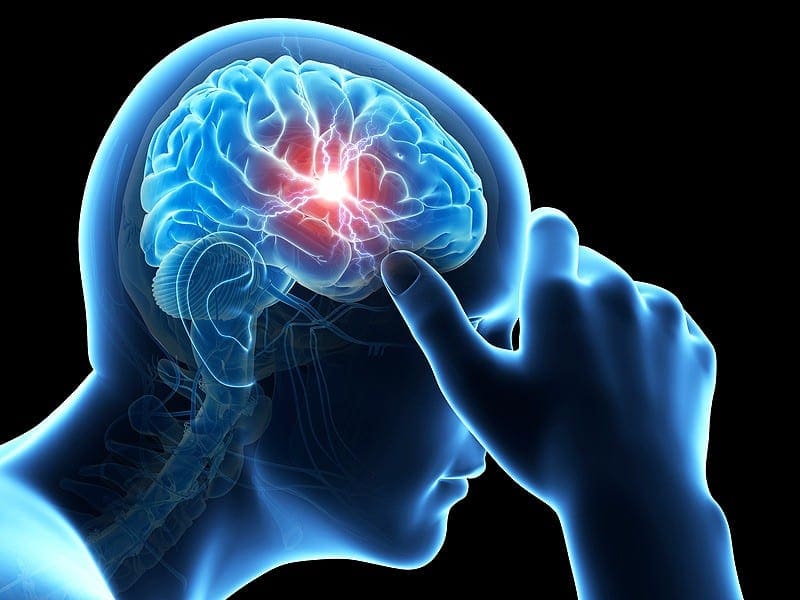
Pain is the human body’s natural response to injury or illness, and it is often a warning that something is wrong. Once the problem is healed, we generally stop experiencing this painful symptoms, however, what happens when the pain continues long after the cause is gone? Chronic pain is medically defined as persistent pain that lasts 3 to 6 months or more. Chronic pain is certainly a challenging condition to live with, affecting everything from the individual’s activity levels and their ability to work as well as their personal relationships and psychological conditions. But, are you aware that chronic pain may also be affecting the structure and function of your brain? It turns out these brain changes may lead to both cognitive and psychological impairment.


Chronic pain doesn’t just influence a singular region of the mind, as a matter of fact, it can result in changes to numerous essential areas of the brain, most of which are involved in many fundamental processes and functions. Various research studies over the years have found alterations to the hippocampus, along with reduction in grey matter from the dorsolateral prefrontal cortex, amygdala, brainstem and right insular cortex, to name a few, associated with chronic pain. A breakdown of a few of the structure of these regions and their related functions might help to put these brain changes into context, for a lot of individuals with chronic pain. The purpose of the following article is to demonstrate as well as discuss the structural and functional brain changes associated with chronic pain, particularly in the case where those reflect probably neither damage nor atrophy.


Structural Brain Changes in Chronic Pain Reflect Probably Neither Damage Nor Atrophy


Abstract


Chronic pain appears to be associated with brain gray matter reduction in areas ascribable to the transmission of pain. The morphological processes underlying these structural changes, probably following functional reorganisation and central plasticity in the brain, remain unclear. The pain in hip osteoarthritis is one of the few chronic pain syndromes which are principally curable. We investigated 20 patients with chronic pain due to unilateral coxarthrosis (mean age 63.25�9.46 (SD) years, 10 female) before hip joint endoprosthetic surgery (pain state) and monitored brain structural changes up to 1 year after surgery: 6�8 weeks, 12�18 weeks and 10�14 month when completely pain free. Patients with chronic pain due to unilateral coxarthrosis had significantly less gray matter compared to controls in the anterior cingulate cortex (ACC), insular cortex and operculum, dorsolateral prefrontal cortex (DLPFC) and orbitofrontal cortex. These regions function as multi-integrative structures during the experience and the anticipation of pain. When the patients were pain free after recovery from endoprosthetic surgery, a gray matter increase in nearly the same areas was found. We also found a progressive increase of brain gray matter in the premotor cortex and the supplementary motor area (SMA). We conclude that gray matter abnormalities in chronic pain are not the cause, but secondary to the disease and are at least in part due to changes in motor function and bodily integration.


Introduction


Evidence of functional and structural reorganization in chronic pain patients support the idea that chronic pain should not only be conceptualized as an altered functional state, but also as a consequence of functional and structural brain plasticity [1], [2], [3], [4], [5], [6]. In the last six years, more than 20 studies were published demonstrating structural brain changes in 14 chronic pain syndromes. A striking feature of all of these studies is the fact that the gray matter changes were not randomly distributed, but occur in defined and functionally highly specific brain areas � namely, involvement in supraspinal nociceptive processing. The most prominent findings were different for each pain syndrome, but overlapped in the cingulate cortex, the orbitofrontal cortex, the insula and dorsal pons [4]. Further structures comprise the thalamus, dorsolateral prefrontal cortex, basal ganglia and hippocampal area. These findings are often discussed as cellular atrophy, reinforcing the idea of damage or loss of brain gray matter [7], [8], [9]. In fact, researchers found a correlation between brain gray matter decreases and duration of pain [6], [10]. But the duration of pain is also linked to the patient�s age, and the age dependent global, but also regionally specific decline of gray matter is well documented [11]. On the other hand, these structural changes could also be a decrease in cell size, extracellular fluids, synaptogenesis, angiogenesis or even due to blood volume changes [4], [12], [13]. Whatever the source is, for our interpretation of such findings it is important to see these morphometric findings in the light of a wealth of morphometric studies in exercise dependant plasticity, given that regionally specific structural brain changes have been repeatedly shown following cognitive and physical exercise [14].


It is not understood why only a relatively small proportion of humans develop a chronic pain syndrome, considering that pain is a universal experience. The question arises whether in some humans a structural difference in central pain transmitting systems may act as a diathesis for chronic pain. Gray matter changes in phantom pain due to amputation [15] and spinal cord injury [3] indicate that the morphological changes of the brain are, at least in part, a consequence of chronic pain. However, the pain in hip osteoarthritis (OA) is one of the few chronic pain syndrome which is principally curable, as 88% of these patients are regularly free of pain following total hip replacement (THR) surgery [16]. In a pilot study we have analysed ten patients with hip OA before and shortly after surgery. We found decreases of gray matter in the anterior cingulated cortex (ACC) and insula during chronic pain before THR surgery and found increases of gray matter in the corresponding brain areas in the pain free condition after surgery [17]. Focussing on this result, we now expanded our studies investigating more patients (n?=?20) after successful THR and monitored structural brain changes in four time intervals, up to one year following surgery. To control for gray matter changes due to motor improvement or depression we also administered questionnaires targeting improvement of motor function and mental health.


Materials and Methods


Volunteers


The patients reported here are a subgroup of 20 patients out of 32 patients published recently who were compared to an age- and gender-matched healthy control group [17] but participated in an additional one year follow-up investigation. After surgery 12 patients dropped out because of a second endoprosthetic surgery (n?=?2), severe illness (n?=?2) and withdrawal of consent (n?=?8). This left a group of twenty patients with unilateral primary hip OA (mean age 63.25�9.46 (SD) years, 10 female) who were investigated four times: before surgery (pain state) and again 6�8 and 12�18 weeks and 10�14 months after endoprosthetic surgery, when completely pain free. All patients with primary hip OA had a pain history longer than 12 months, ranging from 1 to 33 years (mean 7.35 years) and a mean pain score of 65.5 (ranging from 40 to 90) on a visual analogue scale (VAS) ranging from 0 (no pain) to 100 (worst imaginable pain). We assessed any occurrence of minor pain events, including tooth-, ear- and headache up to 4 weeks prior to the study. We also randomly selected the data from 20 sex- and age matched healthy controls (mean age 60,95�8,52 (SD) years, 10 female) of the 32 of the above mentioned pilot study [17]. None of the 20 patients or of the 20 sex- and age matched healthy volunteers had any neurological or internal medical history. The study was given ethical approval by the local Ethics committee and written informed consent was obtained from all study participants prior to examination.


Behavioural Data


We collected data on depression, somatization, anxiety, pain and physical and mental health in all patients and all four time points using the following standardized questionnaires: Beck Depression Inventory (BDI) [18], Brief Symptom Inventory (BSI) [19], Schmerzempfindungs-Skala (SES?=?pain unpleasantness scale) [20] and Health Survey 36-Item Short Form (SF-36) [21] and the Nottingham Health Profile (NHP). We conducted repeated measures ANOVA and paired two-tailed t-Tests to analyse the longitudinal behavioural data using SPSS 13.0 for Windows (SPSS Inc., Chicago, IL), and used Greenhouse Geisser correction if the assumption for sphericity was violated. The significance level was set at p<0.05.


VBM – Data Acquisition


Image acquisition. High-resolution MR scanning was performed on a 3T MRI system (Siemens Trio) with a standard 12-channel head coil. For each of the four time points, scan I (between 1 day and 3 month before endoprosthetic surgery), scan II (6 to 8 weeks after surgery), scan III (12 to 18 weeks after surgery) and scan IV (10�14 months after surgery), a T1 weighted structural MRI was acquired for each patient using a 3D-FLASH sequence (TR 15 ms, TE 4.9 ms, flip angle 25�, 1 mm slices, FOV 256�256, voxel size 1�1�1 mm).


Image Processing and Statistical Analysis


Data pre-processing and analysis were performed with SPM2 (Wellcome Department of Cognitive Neurology, London, UK) running under Matlab (Mathworks, Sherborn, MA, USA) and containing a voxel-based morphometry (VBM)-toolbox for longitudinal data, that is based on high resolution structural 3D MR images and allows for applying voxel-wise statistics to detect regional differences in gray matter density or volumes [22], [23]. In summary, pre-processing involved spatial normalization, gray matter segmentation and 10 mm spatial smoothing with a Gaussian kernel. For the pre-processing steps, we used an optimized protocol [22], [23] and a scanner- and study-specific gray matter template [17]. We used SPM2 rather than SPM5 or SPM8 to make this analysis comparable to our pilot study [17]. as it allows an excellent normalisation and segmentation of longitudinal data. However, as a more recent update of VBM (VBM8) became available recently (dbm.neuro.uni-jena.de/vbm/), we also used VBM8.


Cross-Sectional Analysis


We used a two-sample t-test in order to detect regional differences in brain gray matter between groups (patients at time point scan I (chronic pain) and healthy controls). We applied a threshold of p<0.001 (uncorrected) across the whole brain because of our strong a priory hypothesis, which is based on 9 independent studies and cohorts showing decreases in gray matter in chronic pain patients [7], [8], [9], [15], [24], [25], [26], [27], [28], that gray matter increases will appear in the same (for pain processing relevant) regions as in our pilot study (17). The groups were matched for age and sex with no significant differences between the groups. To investigate whether the differences between groups changed after one year, we also compared patients at time point scan IV (pain free, one year follow-up) to our healthy control group.


Longitudinal Analysis


To detect differences between time points (Scan I�IV) we compared the scans before surgery (pain state) and again 6�8 and 12�18 weeks and 10�14 months after endoprosthetic surgery (pain free) as repeated measure ANOVA. Because any brain changes due to chronic pain may need some time to recede following operation and cessation of pain and because of the post surgery pain the patients reported, we compared in the longitudinal analysis scan I and II with scan III and IV. For detecting changes that are not closely linked to pain, we also looked for progressive changes over all time intervals. We flipped the brains of patients with OA of the left hip (n?=?7) in order to normalize for the side of the pain for both, the group comparison and the longitudinal analysis, but primarily analysed the unflipped data. We used the BDI score as a covariate in the model.


Results


Behavioral Data


All patients reported chronic hip pain before surgery and were pain free (regarding this chronic pain) immediately after surgery, but reported rather acute post-surgery pain on scan II which was different from the pain due to osteoarthritis. The mental health score of the SF-36 (F(1.925/17.322)?=?0.352, p?=?0.7) and the BSI global score GSI (F(1.706/27.302)?=?3.189, p?=?0.064) showed no changes over the time course and no mental co-morbidity. None of the controls reported any acute or chronic pain and none showed any symptoms of depression or physical/mental disability.


Before surgery, some patients showed mild to moderate depressive symptoms in BDI scores that significantly decreased on scan III (t(17)?=?2.317, p?=?0.033) and IV (t(16)?=?2.132, p?=?0.049). Additionally, the SES scores (pain unpleasantness) of all patients improved significantly from scan I (before the surgery) to scan II (t(16)?=?4.676, p<0.001), scan III (t(14)?=?4.760, p<0.001) and scan IV (t(14)?=?4.981, p<0.001, 1 year after surgery) as pain unpleasantness decreased with pain intensity. The pain rating on scan 1 and 2 were positive, the same rating on day 3 and 4 negative. The SES only describes the quality of perceived pain. It was therefore positive on day 1 and 2 (mean 19.6 on day 1 and 13.5 on day 2) and negative (n.a.) on day 3 & 4. However, some patients did not understand this procedure and used the SES as a global �quality of life� measure. This is why all patients were asked on the same day individually and by the same person regarding pain occurrence.


In the short form health survey (SF-36), which consists of the summary measures of a Physical Health Score and a Mental Health Score [29], the patients improved significantly in the Physical Health score from scan I to scan II (t(17)?=??4.266, p?=?0.001), scan III (t(16)?=??8.584, p<0.001) and IV (t(12)?=??7.148, p<0.001), but not in the Mental Health Score. The results of the NHP were similar, in the subscale �pain� (reversed polarity) we observed a significant change from scan I to scan II (t(14)?=??5.674, p<0.001, scan III (t(12)?=??7.040, p<0.001 and scan IV (t(10)?=??3.258, p?=?0.009). We also found a significant increase in the subscale �physical mobility� from scan I to scan III (t(12)?=??3.974, p?=?0.002) and scan IV (t(10)?=??2.511, p?=?0.031). There was no significant change between scan I and scan II (six weeks after surgery).


Structural Data


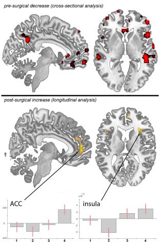
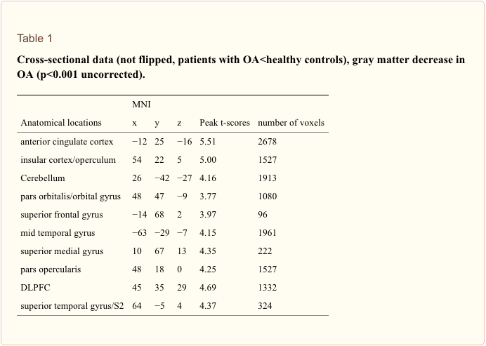
Cross-sectional analysis. We included age as a covariate in the general linear model and found no age confounds. Compared to sex and age matched controls, patients with primary hip OA (n?=?20) showed pre-operatively (Scan I) reduced gray matter in the anterior cingulate cortex (ACC), the insular cortex, operculum, dorsolateral prefrontal cortex (DLPFC), right temporal pole and cerebellum (Table 1 and Figure 1). Except for the right putamen (x?=?31, y?=??14, z?=??1; p<0.001, t?=?3.32) no significant increase in gray matter density was found in patients with OA compared to healthy controls. Comparing patients at time point scan IV with matched controls, the same results were found as in the cross-sectional analysis using scan I compared to controls.


Figure 1 Statistical Parametric Maps

Figure 1: Statistical parametric maps demonstrating the structural differences in gray matter in patients with chronic pain due to primary hip OA compared to controls and longitudinally compared to themselves over time. Significant gray matter changes are shown superimposed in color, cross-sectional data is depicted in red and longitudinal data in yellow. Axial plane: the left side of the picture is the left side of the brain. top: Areas of significant decrease of gray matter between patients with chronic pain due to primary hip OA and unaffected control subjects. p<0.001 uncorrected bottom: Gray matter increase in 20 pain free patients at the third and fourth scanning period after total hip replacement surgery, as compared to the first (preoperative) and second (6�8 weeks post surgery) scan. p<0.001 uncorrected Plots: Contrast estimates and 90% Confidence interval, effects of interest, arbitrary units. x-axis: contrasts for the 4 timepoints, y-axis: contrast estimate at ?3, 50, 2 for ACC and contrast estimate at 36, 39, 3 for insula.


Table 1 Cross-Sectional Data


Flipping the data of patients with left hip OA (n?=?7) and comparing them with healthy controls did not change the results significantly, but for a decrease in the thalamus (x?=?10, y?=??20, z?=?3, p<0.001, t?=?3.44) and an increase in the right cerebellum (x?=?25, y?=??37, z?=??50, p<0.001, t?=?5.12) that did not reach significance in the unflipped data of the patients compared to controls.


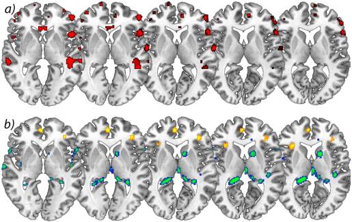
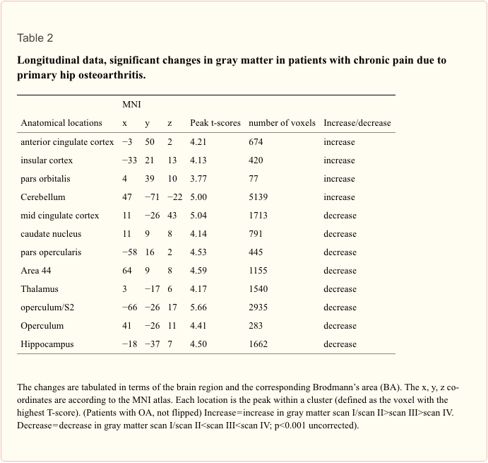
Longitudinal analysis. In the longitudinal analysis, a significant increase (p<.001 uncorrected) of gray matter was detected by comparing the first and second scan (chronic pain/post-surgery pain) with the third and fourth scan (pain free) in the ACC, insular cortex, cerebellum and pars orbitalis in the patients with OA (Table 2 and Figure 1). Gray matter decreased over time (p<.001 whole brain analysis uncorrected) in the secondary somatosensory cortex, hippocampus, midcingulate cortex, thalamus and caudate nucleus in patients with OA (Figure 2).


Figure 2 Increases in Brain Gray Matter

Figure 2: a) Significant increases in brain gray matter following successful operation. Axial view of significant decrease of gray matter in patients with chronic pain due to primary hip OA compared to control subjects. p<0.001 uncorrected (cross-sectional analysis), b) Longitudinal increase of gray matter over time in yellow comparing scan I&IIscan III>scan IV) in patients with OA. p<0.001 uncorrected (longitudinal analysis). The left side of the picture is the left side of the brain.


Table 2 Longitudinal Data


Flipping the data of patients with left hip OA (n?=?7) did not change the results significantly, but for a decrease of brain gray matter in the Heschl�s Gyrus (x?=??41, y?=??21, z?=?10, p<0.001, t?=?3.69) and Precuneus (x?=?15, y?=??36, z?=?3, p<0.001, t?=?4.60).


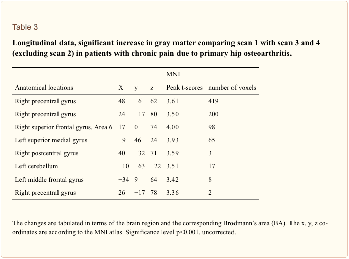
By contrasting the first scan (presurgery) with scans 3+4 (postsurgery), we found an increase of gray matter in the frontal cortex and motor cortex (p<0.001 uncorrected). We note that this contrast is less stringent as we have now less scans per condition (pain vs. non-pain). When we lower the threshold we repeat what we have found using contrast of 1+2 vs. 3+4.


By looking for areas that increase over all time intervals, we found changes of brain gray matter in motor areas (area 6) in patients with coxarthrosis following total hip replacement (scan I<scan II<scan III<scan IV)). Adding the BDI scores as a covariate did not change the results. Using the recently available software tool VBM8 including DARTEL normalisation (dbm.neuro.uni-jena.de/vbm/) we could replicate this finding in the anterior and mid-cingulate cortex and both anterior insulae.


We calculated the effect sizes and the cross-sectional analysis (patients vs. controls) yielded a Cohen�s d of 1.78751 in the peak voxel of the ACC (x?=??12, y?=?25, z?=??16). We also calculated Cohen�s d for the longitudinal analysis (contrasting scan 1+2 vs. scan 3+4). This resulted in a Cohen�s d of 1.1158 in the ACC (x?=??3, y?=?50, z?=?2). Regarding the insula (x?=??33, y?=?21, z?=?13) and related to the same contrast, Cohen�s d is 1.0949. Additionally, we calculated the mean of the non-zero voxel values of the Cohen�s d map within the ROI (comprised of the anterior division of the cingulate gyrus and the subcallosal cortex, derived from the Harvard-Oxford Cortical Structural Atlas): 1.251223.


Dr-Jimenez_White-Coat_01.png

Dr. Alex Jimenez’s Insight

Chronic pain patients can experience a variety of health issues over time, aside from their already debilitating symptoms. For instance, many individuals will experience sleeping problems as a result of their pain, but most importantly, chronic pain can lead to various mental health issues as well, including anxiety and depression. The effects that pain can have on the brain may seem all too overwhelming but growing evidence suggests that these brain changes are not permanent and can be reversed when chronic pain patients receive the proper treatment for their underlying health issues. According to the article, gray matter abnormalities found in chronic pain do not reflect brain damage, but rather, they are a reversible consequence which normalizes when the pain is adequately treated. Fortunately, a variety of treatment approaches are available to help ease chronic pain symptoms and restore the structure and function of the brain.


Discussion


Monitoring whole brain structure over time, we confirm and expand our pilot data published recently [17]. We found changes in brain gray matter in patients with primary hip osteoarthritis in the chronic pain state, which reverse partly when these patients are pain free, following hip joint endoprosthetic surgery. The partial increase in gray matter after surgery is nearly in the same areas where a decrease of gray matter has been seen before surgery. Flipping the data of patients with left hip OA (and therefore normalizing for the side of the pain) had only little impact on the results but additionally showed a decrease of gray matter in the Heschl�s gyrus and Precuneus that we cannot easily explain and, as no a priori hypothesis exists, regard with great caution. However, the difference seen between patients and healthy controls at scan I was still observable in the cross-sectional analysis at scan IV. The relative increase of gray matter over time is therefore subtle, i.e. not sufficiently distinct to have an effect on the cross sectional analysis, a finding that has already been shown in studies investigating experience dependant plasticity [30], [31]. We note that the fact that we show some parts of brain-changes due to chronic pain to be reversible does not exclude that some other parts of these changes are irreversible.


Interestingly, we observed that the gray matter decrease in the ACC in chronic pain patients before surgery seems to continue 6 weeks after surgery (scan II) and only increases towards scan III and IV, possibly due to post-surgery pain, or decrease in motor function. This is in line with the behavioural data of the physical mobility score included in the NHP, which post-operatively did not show any significant change at time point II but significantly increased towards scan III and IV. Of note, our patients reported no pain in the hip after surgery, but experienced post-surgery pain in surrounding muscles and skin which was perceived very differently by patients. However, as patients still reported some pain at scan II, we also contrasted the first scan (pre-surgery) with scans III+IV (post-surgery), revealing an increase of gray matter in the frontal cortex and motor cortex. We note that this contrast is less stringent because of less scans per condition (pain vs. non-pain). When we lowered the threshold we repeat what we have found using contrast of I+II vs. III+IV.


Our data strongly suggest that gray matter alterations in chronic pain patients, which are usually found in areas involved in supraspinal nociceptive processing [4] are neither due to neuronal atrophy nor brain damage. The fact that these changes seen in the chronic pain state do not reverse completely could be explained with the relatively short period of observation (one year after operation versus a mean of seven years of chronic pain before the operation). Neuroplastic brain changes that may have developed over several years (as a consequence of constant nociceptive input) need probably more time to reverse completely. Another possibility why the increase of gray matter can only be detected in the longitudinal data but not in the cross-sectional data (i.e. between cohorts at time point IV) is that the number of patients (n?=?20) is too small. It needs to be pointed out that the variance between brains of several individuals is quite large and that longitudinal data have the advantage that the variance is relatively small as the same brains are scanned several times. Consequently, subtle changes will only be detectable in longitudinal data [30], [31], [32]. Of course we cannot exclude that these changes are at least partly irreversible although that is unlikely, given the findings of exercise specific structural plasticity and reorganisation [4], [12], [30], [33], [34]. To answer this question, future studies need to investigate patients repeatedly over longer time frames, possibly years.


We note that we can only make limited conclusions regarding the dynamics of morphological brain changes over time. The reason is that when we designed this study in 2007 and scanned in 2008 and 2009, it was not known whether structural changes would occur at all and for reasons of feasibility we chose the scan dates and time frames as described here. One could argue that the gray matter changes in time, which we describe for the patient group, might have happened in the control group as well (time effect). However, any changes due to aging, if at all, would be expected to be a decrease in volume. Given our a priori hypothesis, based on 9 independent studies and cohorts showing decreases in gray matter in chronic pain patients [7], [8], [9], [15], [24], [25], [26], [27], [28], we focussed on regional increases over time and therefore believe our finding not to be a simple time effect. Of note, we cannot rule out that the gray matter decrease over time that we found in our patient group could be due to a time effect, as we have not scanned our control group in the same time frame. Given the findings, future studies should aim at more and shorter time intervals, given that exercise dependant morphometric brain changes may occur as fast as after 1 week [32], [33].


In addition to the impact of the nociceptive aspect of pain on brain gray matter [17], [34] we observed that changes in motor function probably also contribute to the structural changes. We found motor and premotor areas (area 6) to increase over all time intervals (Figure 3). Intuitively this may be due to improvement of motor function over time as the patients were no more restricted in living a normal life. Notably we did not focus on motor function but an improvement in pain experience, given our original quest to investigate whether the well-known reduction in brain gray matter in chronic pain patients is in principle reversible. Consequently, we did not use specific instruments to investigate motor function. Nevertheless, (functional) motor cortex reorganization in patients with pain syndromes is well documented [35], [36], [37], [38]. Moreover, the motor cortex is one target in therapeutic approaches in medically intractable chronic pain patients using direct brain stimulation [39], [40], transcranial direct current stimulation [41], and repetitive transcranial magnetic stimulation [42], [43]. The exact mechanisms of such modulation (facilitation vs. inhibition, or simply interference in the pain-related networks) are not yet elucidated [40]. A recent study demonstrated that a specific motor experience can alter the structure of the brain [13]. Synaptogenesis, reorganisation of movement representations and angiogenesis in motor cortex may occur with special demands of a motor task. Tsao et al. showed reorganisation in the motor cortex of patients with chronic low back pain that seem to be back pain-specific [44] and Puri et al. observed a reduction in left supplemental motor area gray matter in fibromyalgia sufferers [45]. Our study was not designed to disentangle the different factors that may change the brain in chronic pain but we interpret our data concerning the gray matter changes that they do not exclusively mirror the consequences of constant nociceptive input. In fact, a recent study in neuropathic pain patients pointed out abnormalities in brain regions that encompass emotional, autonomic, and pain perception, implying that they play a critical role in the global clinical picture of chronic pain [28].


Figure 3 Statistical Parametric Maps

Figure 3: Statistical parametric maps demonstrating a significant increase of brain gray matter in motor areas (area 6) in patients with coxarthrosis before compared to after THR (longitudinal analysis, scan I<scan II<scan III<scan IV). Contrast estimates at x?=?19, y?=??12, z?=?70.


Two recent pilot studies focussed on hip replacement therapy in osteoarthritis patients, the only chronic pain syndrome which is principally curable with total hip replacement [17], [46] and these data are flanked by a very recent study in chronic low back pain patients [47]. These studies need to be seen in the light of several longitudinal studies investigating experience-dependent neuronal plasticity in humans on a structural level [30], [31] and a recent study on structural brain changes in healthy volunteers experiencing repeated painful stimulation [34]. The key message of all these studies is that the main difference in the brain structure between pain patients and controls may recede when the pain is cured. However, it must be taken into account that it is simply not clear whether the changes in chronic pain patients are solely due to nociceptive input or due to the consequences of pain or both. It is more than likely that behavioural changes, such as deprivation or enhancement of social contacts, agility, physical training and life style changes are sufficient to shape the brain [6], [12], [28], [48]. Particularly depression as a co-morbidity or consequence of pain is a key candidate to explain the differences between patients and controls. A small group of our patients with OA showed mild to moderate depressive symptoms that changed with time. We did not find the structural alterations to covary significantly with the BDI-score but the question arises how many other behavioural changes due to the absence of pain and motor improvement may contribute to the results and to what extent they do. These behavioural changes can possibly influence a gray matter decrease in chronic pain as well as a gray matter increase when pain is gone.


Another important factor which may bias our interpretation of the results is the fact that nearly all patients with chronic pain took medications against pain, which they stopped when they were pain free. One could argue that NSAIDs such as diclofenac or ibuprofen have some effects on neural systems and the same holds true for opioids, antiepileptics and antidepressants, medications which are frequently used in chronic pain therapy. The impact of pain killers and other medications on morphometric findings may well be important (48). No study so far has shown effects of pain medication on brain morphology but several papers found that changes in brain structure in chronic pain patients are neither solely explained by pain related inactivity [15], nor by pain medication [7], [9], [49]. However, specific studies are lacking. Further research should focus the experience-dependent changes in cortical plasticity, which may have vast clinical implications for the treatment of chronic pain.


We also found decreases of gray matter in the longitudinal analysis, possibly due to reorganisation processes that accompany changes in motor function and pain perception. There is little information available about longitudinal changes in brain gray matter in pain conditions, for this reason we have no hypothesis for a gray matter decrease in these areas after the operation. Teutsch et al. [25] found an increase of brain gray matter in the somatosensory and midcingulate cortex in healthy volunteers that experienced painful stimulation in a daily protocol for eight consecutive days. The finding of gray matter increase following experimental nociceptive input overlapped anatomically to some degree with the decrease of brain gray matter in this study in patients that were cured of long-lasting chronic pain. This implies that nociceptive input in healthy volunteers leads to exercise dependant structural changes, as it possibly does in patients with chronic pain, and that these changes reverse in healthy volunteers when nociceptive input stops. Consequently, the decrease of gray matter in these areas seen in patients with OA could be interpreted to follow the same fundamental process: exercise dependant changes brain changes [50]. As a non-invasive procedure, MR Morphometry is the ideal tool for the quest to find the morphological substrates of diseases, deepening our understanding of the relationship between brain structure and function, and even to monitor therapeutic interventions. One of the great challenges in the future is to adapt this powerful tool for multicentre and therapeutic trials of chronic pain.


Limitations of this Study


Although this study is an extension of our previous study expanding the follow-up data to 12 months and investigating more patients, our principle finding that morphometric brain changes in chronic pain are reversible is rather subtle. The effect sizes are small (see above) and the effects are partly driven by a further reduction of regional brain gray matter volume at the time-point of scan 2. When we exclude the data from scan 2 (directly after the operation) only significant increases in brain gray matter for motor cortex and frontal cortex survive a threshold of p<0.001 uncorrected (Table 3).


Table 3 Longitudinal Data


Conclusion


It is not possible to distinguish to what extent the structural alterations we observed are due to changes in nociceptive input, changes in motor function or medication consumption or changes in well-being as such. Masking the group contrasts of the first and last scan with each other revealed much less differences than expected. Presumably, brain alterations due to chronic pain with all consequences are developing over quite a long time course and may also need some time to revert. Nevertheless, these results reveal processes of reorganisation, strongly suggesting that chronic nociceptive input and motor impairment in these patients leads to altered processing in cortical regions and consequently structural brain changes which are in principle reversible.


Acknowledgments


We thank all volunteers for the participation in this study and the Physics and Methods group at NeuroImage Nord in Hamburg. The study was given ethical approval by the local Ethics committee and written informed consent was obtained from all study participants prior to examination.


Funding Statement


This work was supported by grants from the DFG (German Research Foundation) (MA 1862/2-3) and BMBF (The Federal Ministry of Education and Research) (371 57 01 and NeuroImage Nord). The funders had no role in study design, data collection and analysis, decision to publish, or preparation of the manuscript.


Endocannabinoid System | El Paso, TX Chiropractor


The Endocannabinoid System: The Essential System You�ve Never Heard Of


In case you haven’t heard of the endocannabinoid system, or ECS, there’s no need to feel embarrassed. Back in the 1960’s, the investigators that became interested in the bioactivity of Cannabis eventually isolated many of its active chemicals. It took another 30 years, however, for researchers studying animal models to find a receptor for these ECS chemicals in the brains of rodents, a discovery which opened a whole world of inquiry into the ECS receptors existence and what their physiological purpose is.


We now know that most animals, from fish to birds to mammals, possess an endocannabinoid, and we know that humans not only make their own cannabinoids that interact with this particular system, but we also produce other compounds that interact with the ECS, those of which are observed in many different plants and foods, well beyond the Cannabis species.


As a system of the human body, the ECS isn’t an isolated structural platform like the nervous system or cardiovascular system. Instead, the ECS is a set of receptors widely distributed throughout the body which are activated through a set of ligands we collectively know as endocannabinoids, or endogenous cannabinoids. Both verified receptors are just called CB1 and CB2, although there are others which were proposed. PPAR and TRP channels also mediate some functions. Likewise, you will find just two well-documented endocannabinoids: anadamide and 2-arachidonoyl glycerol, or 2-AG.


Moreover, fundamental to the endocannabinoid system are the enzymes that synthesize and break down the endocannabinoids. Endocannabinoids are believed to be synthesized in an as-needed foundation. The primary enzymes involved are diacylglycerol lipase and N-acyl-phosphatidylethanolamine-phospholipase D, which respectively synthesize 2-AG and anandamide. The two main degrading enzymes are fatty acid amide hydrolase, or FAAH, which breaks down anandamide, and monoacylglycerol lipase, or MAGL, which breaks down 2-AG. The regulation of these two enzymes may increase or decrease the modulation of the ECS.


What is the Function of the ECS?


The ECS is the principal homeostatic regulatory system of the body. It may readily be viewed as the body’s internal adaptogenic system, always working to maintain the balance of a variety of function. Endocannabinoids broadly work as neuromodulators and, as such, they regulate a broad range of bodily processes, from fertility to pain. Some of those better-known functions from the ECS are as follows:


Nervous System


From the central nervous system, or the CNS, general stimulation of the CB1 receptors will inhibit the release of glutamate and GABA. In the CNS, the ECS plays a role in memory formation and learning, promotes neurogenesis in the hippocampus, also regulates neuronal excitability. The ECS also plays a part in the way the brain will react to injury and inflammation. From the spinal cord, the ECS modulates pain signaling and boosts natural analgesia. In the peripheral nervous system, in which CB2 receptors control, the ECS acts primarily in the sympathetic nervous system to regulate functions of the intestinal, urinary, and reproductive tracts.


Stress and Mood


The ECS has multiple impacts on stress reactions and emotional regulation, such as initiation of this bodily response to acute stress and adaptation over time to more long-term emotions, such as fear and anxiety. A healthy working endocannabinoid system is critical to how humans modulate between a satisfying degree of arousal compared to a level that is excessive and unpleasant. The ECS also plays a role in memory formation and possibly especially in the way in which the brain imprints memories from stress or injury. Because the ECS modulates the release of dopamine, noradrenaline, serotonin, and cortisol, it can also widely influence emotional response and behaviors.


Digestive System


The digestive tract is populated with both CB1 and CB2 receptors that regulate several important aspects of GI health. It’s thought that the ECS might be the “missing link” in describing the gut-brain-immune link that plays a significant role in the functional health of the digestive tract. The ECS is a regulator of gut immunity, perhaps by limiting the immune system from destroying healthy flora, and also through the modulation of cytokine signaling. The ECS modulates the natural inflammatory response in the digestive tract, which has important implications for a wide range of health issues. Gastric and general GI motility also appears to be partially governed by the ECS.


Appetite and Metabolism


The ECS, particularly the CB1 receptors, plays a part in appetite, metabolism, and regulation of body fat. Stimulation of the CB1 receptors raises food-seeking behaviour, enhances awareness of smell, also regulates energy balance. Both animals and humans that are overweight have ECS dysregulation that may lead this system to become hyperactive, which contributes to both overeating and reduced energy expenditure. Circulating levels of anandamide and 2-AG have been shown to be elevated in obesity, which might be in part due to decreased production of the FAAH degrading enzyme.


Immune Health and Inflammatory Response


The cells and organs of the immune system are rich with endocannabinoid receptors. Cannabinoid receptors are expressed in the thymus gland, spleen, tonsils, and bone marrow, as well as on T- and B-lymphocytes, macrophages, mast cells, neutrophils, and natural killer cells. The ECS is regarded as the primary driver of immune system balance and homeostasis. Though not all the functions of the ECS from the immune system are understood, the ECS appears to regulate cytokine production and also to have a role in preventing overactivity in the immune system. Inflammation is a natural part of the immune response, and it plays a very normal role in acute insults to the body, including injury and disease ; nonetheless, when it isn’t kept in check it can become chronic and contribute to a cascade of adverse health problems, such as chronic pain. By keeping the immune response in check, the ECS helps to maintain a more balanced inflammatory response through the body.


Other areas of health regulated by the ECS:


  • Bone health
  • Fertility
  • Skin health
  • Arterial and respiratory health
  • Sleep and circadian rhythm


How to best support a healthy ECS is a question many researchers are now trying to answer. Stay tuned for more information on this emerging topic.


In conclusion,�chronic pain has been associated with brain changes, including the reduction of gray matter. However, the article above demonstrated that chronic pain can alter the overall structure and function of the brain. Although chronic pain may lead to these, among other health issues, the proper treatment of the patient’s underlying symptoms can reverse brain changes and regulate gray matter. Furthermore, more and more research studies have emerged behind the importance of the endocannabinoid system and it’s function in controlling as well as managing chronic pain and other health issues. Information referenced from the National Center for Biotechnology Information (NCBI).�The scope of our information is limited to chiropractic as well as to spinal injuries and conditions. To discuss the subject matter, please feel free to ask Dr. Jimenez or contact us at�915-850-0900�.


Curated by Dr. Alex Jimenez

Green-Call-Now-Button-24H-150x150-2-3.png

Additional Topics: Back Pain

Back pain is one of the most prevalent causes for disability and missed days at work worldwide. As a matter of fact, back pain has been attributed as the second most common reason for doctor office visits, outnumbered only by upper-respiratory infections. Approximately 80 percent of the population will experience some type of back pain at least once throughout their life. The spine is a complex structure made up of bones, joints, ligaments and muscles, among other soft tissues. Because of this, injuries and/or aggravated conditions, such as herniated discs, can eventually lead to symptoms of back pain. Sports injuries or automobile accident injuries are often the most frequent cause of back pain, however, sometimes the simplest of movements can have painful results. Fortunately, alternative treatment options, such as chiropractic care, can help ease back pain through the use of spinal adjustments and manual manipulations, ultimately improving pain relief.


blog picture of cartoon paperboy big news


EXTRA IMPORTANT TOPIC: Low Back Pain Management


MORE TOPICS: EXTRA EXTRA:�Chronic Pain & Treatments


Blank
References
1.�Woolf CJ, Salter MW (2000)�Neuronal plasticity: increasing the gain in pain.�Science288: 1765�1769.[PubMed]
2.�Flor H, Nikolajsen L, Staehelin Jensen T (2006)�Phantom limb pain: a case of maladaptive CNS plasticity?Nat Rev Neurosci7: 873�881.�[PubMed]
3.�Wrigley PJ, Gustin SM, Macey PM, Nash PG, Gandevia SC, et al. (2009)�Anatomical changes in human motor cortex and motor pathways following complete thoracic spinal cord injury.�Cereb Cortex19: 224�232.�[PubMed]
4.�May A (2008)�Chronic pain may change the structure of the brain.�Pain137: 7�15.�[PubMed]
5.�May A (2009) Morphing voxels: the hype around structural imaging of headache patients. Brain.[PubMed]
6.�Apkarian AV, Baliki MN, Geha PY (2009)�Towards a theory of chronic pain.�Prog Neurobiol87: 81�97.�[PMC free article][PubMed]
7.�Apkarian AV, Sosa Y, Sonty S, Levy RM, Harden RN, et al. (2004)�Chronic back pain is associated with decreased prefrontal and thalamic gray matter density.�J Neurosci24: 10410�10415.�[PubMed]
8.�Rocca MA, Ceccarelli A, Falini A, Colombo B, Tortorella P, et al. (2006)�Brain gray matter changes in migraine patients with T2-visible lesions: a 3-T MRI study.�Stroke37: 1765�1770.�[PubMed]
9.�Kuchinad A, Schweinhardt P, Seminowicz DA, Wood PB, Chizh BA, et al. (2007)�Accelerated brain gray matter loss in fibromyalgia patients: premature aging of the brain?J Neurosci27: 4004�4007.[PubMed]
10.�Tracey I, Bushnell MC (2009)�How neuroimaging studies have challenged us to rethink: is chronic pain a disease?J Pain10: 1113�1120.�[PubMed]
11.�Franke K, Ziegler G, Kloppel S, Gaser C (2010)�Estimating the age of healthy subjects from T1-weighted MRI scans using kernel methods: exploring the influence of various parameters.�Neuroimage50: 883�892.�[PubMed]
12.�Draganski B, May A (2008)�Training-induced structural changes in the adult human brain.�Behav Brain Res192: 137�142.�[PubMed]
13.�Adkins DL, Boychuk J, Remple MS, Kleim JA (2006)�Motor training induces experience-specific patterns of plasticity across motor cortex and spinal cord.�J Appl Physiol101: 1776�1782.�[PubMed]
14.�Duerden EG, Laverdure-Dupont D (2008)�Practice makes cortex.�J Neurosci28: 8655�8657.�[PubMed]
15.�Draganski B, Moser T, Lummel N, Ganssbauer S, Bogdahn U, et al. (2006)�Decrease of thalamic gray matter following limb amputation.�Neuroimage31: 951�957.�[PubMed]
16.�Nikolajsen L, Brandsborg B, Lucht U, Jensen TS, Kehlet H (2006)�Chronic pain following total hip arthroplasty: a nationwide questionnaire study.�Acta Anaesthesiol Scand50: 495�500.�[PubMed]
17.�Rodriguez-Raecke R, Niemeier A, Ihle K, Ruether W, May A (2009)�Brain gray matter decrease in chronic pain is the consequence and not the cause of pain.�J Neurosci29: 13746�13750.�[PubMed]
18.�Beck AT, Ward CH, Mendelson M, Mock J, Erbaugh J (1961)�An inventory for measuring depression.�Arch Gen Psychiatry4: 561�571.�[PubMed]
19.�Franke G (2002) Die Symptom-Checkliste nach L.R. Derogatis – Manual. G�ttingen Beltz Test Verlag.
20.�Geissner E (1995) The Pain Perception Scale�a differentiated and change-sensitive scale for assessing chronic and acute pain. Rehabilitation (Stuttg) 34: XXXV�XLIII.�[PubMed]
21.�Bullinger M, Kirchberger I (1998) SF-36 – Fragebogen zum Gesundheitszustand. Hand-anweisung. G�ttingen: Hogrefe.
22.�Ashburner J, Friston KJ (2000)�Voxel-based morphometry�the methods.�Neuroimage11: 805�821.[PubMed]
23.�Good CD, Johnsrude IS, Ashburner J, Henson RN, Friston KJ, et al. (2001)�A voxel-based morphometric study of ageing in 465 normal adult human brains.�Neuroimage14: 21�36.�[PubMed]
24.�Baliki MN, Chialvo DR, Geha PY, Levy RM, Harden RN, et al. (2006)�Chronic pain and the emotional brain: specific brain activity associated with spontaneous fluctuations of intensity of chronic back pain.�J Neurosci26: 12165�12173.�[PMC free article][PubMed]
25.�Lutz J, Jager L, de Quervain D, Krauseneck T, Padberg F, et al. (2008)�White and gray matter abnormalities in the brain of patients with fibromyalgia: a diffusion-tensor and volumetric imaging study.�Arthritis Rheum58: 3960�3969.�[PubMed]
26.�Wrigley PJ, Gustin SM, Macey PM, Nash PG, Gandevia SC, et al. (2008)�Anatomical Changes in Human Motor Cortex and Motor Pathways following Complete Thoracic Spinal Cord Injury.�Cereb Cortex19: 224�232.�[PubMed]
27.�Schmidt-Wilcke T, Hierlmeier S, Leinisch E (2010) Altered Regional Brain Morphology in Patients With Chronic Facial Pain. Headache.�[PubMed]
28.�Geha PY, Baliki MN, Harden RN, Bauer WR, Parrish TB, et al. (2008)�The brain in chronic CRPS pain: abnormal gray-white matter interactions in emotional and autonomic regions.�Neuron60: 570�581.�[PMC free article][PubMed]
29.�Brazier J, Roberts J, Deverill M (2002)�The estimation of a preference-based measure of health from the SF-36.�J Health Econ21: 271�292.�[PubMed]
30.�Draganski B, Gaser C, Busch V, Schuierer G, Bogdahn U, et al. (2004)�Neuroplasticity: changes in grey matter induced by training.�Nature427: 311�312.�[PubMed]
31.�Boyke J, Driemeyer J, Gaser C, Buchel C, May A (2008)�Training-induced brain structure changes in the elderly.�J Neurosci28: 7031�7035.�[PubMed]
32.�Driemeyer J, Boyke J, Gaser C, Buchel C, May A (2008)�Changes in gray matter induced by learning�revisited.�PLoS ONE3: e2669.�[PMC free article][PubMed]
33.�May A, Hajak G, Ganssbauer S, Steffens T, Langguth B, et al. (2007)�Structural brain alterations following 5 days of intervention: dynamic aspects of neuroplasticity.�Cereb Cortex17: 205�210.�[PubMed]
34.�Teutsch S, Herken W, Bingel U, Schoell E, May A (2008)�Changes in brain gray matter due to repetitive painful stimulation.�Neuroimage42: 845�849.�[PubMed]
35.�Flor H, Braun C, Elbert T, Birbaumer N (1997)�Extensive reorganization of primary somatosensory cortex in chronic back pain patients.�Neurosci Lett224: 5�8.�[PubMed]
36.�Flor H, Denke C, Schaefer M, Grusser S (2001)�Effect of sensory discrimination training on cortical reorganisation and phantom limb pain.�Lancet357: 1763�1764.�[PubMed]
37.�Swart CM, Stins JF, Beek PJ (2009)�Cortical changes in complex regional pain syndrome (CRPS).�Eur J Pain13: 902�907.�[PubMed]
38.�Maihofner C, Baron R, DeCol R, Binder A, Birklein F, et al. (2007)�The motor system shows adaptive changes in complex regional pain syndrome.�Brain130: 2671�2687.�[PubMed]
39.�Fontaine D, Hamani C, Lozano A (2009)�Efficacy and safety of motor cortex stimulation for chronic neuropathic pain: critical review of the literature.�J Neurosurg110: 251�256.�[PubMed]
40.�Levy R, Deer TR, Henderson J (2010)�Intracranial neurostimulation for pain control: a review.�Pain Physician13: 157�165.�[PubMed]
41.�Antal A, Brepohl N, Poreisz C, Boros K, Csifcsak G, et al. (2008)�Transcranial direct current stimulation over somatosensory cortex decreases experimentally induced acute pain perception.�Clin J Pain24: 56�63.�[PubMed]
42.�Teepker M, Hotzel J, Timmesfeld N, Reis J, Mylius V, et al. (2010)�Low-frequency rTMS of the vertex in the prophylactic treatment of migraine.�Cephalalgia30: 137�144.�[PubMed]
43.�O�Connell N, Wand B, Marston L, Spencer S, Desouza L (2010)�Non-invasive brain stimulation techniques for chronic pain. A report of a Cochrane systematic review and meta-analysis.�Eur J Phys Rehabil Med47: 309�326.�[PubMed]
44.�Tsao H, Galea MP, Hodges PW (2008)�Reorganization of the motor cortex is associated with postural control deficits in recurrent low back pain.�Brain131: 2161�2171.�[PubMed]
45.�Puri BK, Agour M, Gunatilake KD, Fernando KA, Gurusinghe AI, et al. (2010)�Reduction in left supplementary motor area grey matter in adult female fibromyalgia sufferers with marked fatigue and without affective disorder: a pilot controlled 3-T magnetic resonance imaging voxel-based morphometry study.�J Int Med Res38: 1468�1472.�[PubMed]
46.�Gwilym SE, Fillipini N, Douaud G, Carr AJ, Tracey I (2010) Thalamic atrophy associated with painful osteoarthritis of the hip is reversible after arthroplasty; a longitudinal voxel-based-morphometric study. Arthritis Rheum.�[PubMed]
47.�Seminowicz DA, Wideman TH, Naso L, Hatami-Khoroushahi Z, Fallatah S, et al. (2011)�Effective treatment of chronic low back pain in humans reverses abnormal brain anatomy and function.�J Neurosci31: 7540�7550.�[PubMed]
48.�May A, Gaser C (2006)�Magnetic resonance-based morphometry: a window into structural plasticity of the brain.�Curr Opin Neurol19: 407�411.�[PubMed]
49.�Schmidt-Wilcke T, Leinisch E, Straube A, Kampfe N, Draganski B, et al. (2005)�Gray matter decrease in patients with chronic tension type headache.�Neurology65: 1483�1486.�[PubMed]
50.�May A (2009)�Morphing voxels: the hype around structural imaging of headache patients.�Brain 132(Pt6): 1419�1425.�[PubMed]
Close Accordion
Biochemistry Of Pain

Biochemistry Of Pain

Biochemistry of Pain:�All pain syndromes have an inflammation profile. An inflammatory profile can vary from person to person and can also vary in one person at different times. The treatment of pain syndromes is to understand this inflammation profile. Pain syndromes are treated medically, surgically or both. The goal is to inhibit/suppress the production of inflammatory mediators. And a successful outcome is one that results in less inflammation and of course less pain.

Biochemistry Of Pain

Objectives:

  • Who are the key players
  • What are the biochemical mechanisms?
  • What are the consequences?

Inflammation Review:

Key Players

biochemistry of pain el paso tx.

biochemistry of pain el paso tx.

biochemistry of pain el paso tx.

biochemistry of pain el paso tx.Why Does My Shoulder Hurt? A Review Of The Neuroanatomical & Biochemical Basis Of Shoulder Pain
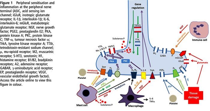
ABSTRACT

If a patient asks �why does my shoulder hurt?� the conversation will quickly turn to scientific theory and sometimes unsubstantiated conjecture. Frequently, the clinician becomes aware of the limits of the scientific basis of their explanation, demonstrating the incompleteness of our understanding of the nature of shoulder pain. This review takes a systematic approach to help answer fundamental questions relating to shoulder pain, with a view to providing insights into future research and novel methods for treating shoulder pain. We shall explore the roles of (1) the peripheral receptors, (2) peripheral pain processing or �nociception�, (3) the spinal cord, (4) the brain, (5) the location of receptors in the shoulder and (6) the neural anatomy of the shoulder. We also consider how these factors might contribute to the variability in the clinical presentation, the diagnosis and the treatment of shoulder pain. In this way we aim to provide an overview of the component parts of the peripheral pain detection system and central pain processing mechanisms in shoulder pain that interact to produce clinical pain.

INTRODUCTION: A VERY BRIEF HISTORY OF PAIN SCIENCE ESSENTIAL FOR CLINICIANS

The nature of pain, in general, has been a subject of much controversy over the past century. In the 17th century Descartes� theory1 proposed that the intensity of pain was directly related to the amount of associated tissue injury and that pain was processed in one distinct pathway. Many earlier theories relied upon this so-called �dualist� Descartian philosophy, seeing pain as the consequence of the stimulation of a �specific� peripheral pain receptor in the brain. In the 20th century a scientific battle between two opposing theories ensued, namely specificity theory and pattern theory. The Descartian �specificity theory� saw pain as a specific separate modality of sensory input with its own apparatus, while �pattern theory� felt that pain resulted from the intense stimulation of non-specific receptors.2 In 1965, Wall and Melzack�s 3 gate theory of pain provided evidence for a model in which pain perception was modulated by both sensory feedback and the central nervous system. Another huge advance in pain theory at around the same time saw the discovery of the specific mode of actions of the opioids.4 Subsequently, recent advances in neuroimaging and molecular medicine have vastly expanded our overall understanding of pain.

So how does this relate to shoulder pain?�Shoulder pain is a common clinical problem, and a robust understanding of the way in which pain is processed by the body is essential to best diagnose and treat a patient�s pain. Advances in our knowledge of pain processing promise to explain the mismatch between pathology and the perception of pain, they may also help us explain why certain patients fail to respond to certain treatments.

BASIC BUILDING BLOCKS OF PAIN

Peripheral sensory receptors: the mechanoreceptor and the �nociceptor�

There are numerous types of peripheral sensory receptors present in the human musculoskeletal system. 5 They may be classified based on their func�tion (as mechanoreceptors, thermoreceptors or nociceptors) or morphology (free nerve endings or different types of encapsulated receptors).5 The dif�ferent types of receptor can then be further subclas�sified based on the presence of certain chemical markers. There are significant overlaps between dif�ferent functional classes of receptor, for example

Peripheral Pain Processing: �Nociception�

Tissue injury involves a variety of inflammatory mediators being released by damaged cells including bradykinin, histamine, 5-hydroxytryptamine, ATP, nitric oxide and certain ions (K+ and H+). The activation of the arachidonic acid pathway leads to the production of prostaglandins, thromboxanes and leuko- trienes. Cytokines, including the interleukins and tumor necrosis factor ?, and neurotrophins, such as nerve growth factor (NGF), are also released and are intimately involved in the facilitation of inflammation.15 Other substances such as excitatory amino acids (glutamate) and opioids (endothelin-1) have also been implicated in the acute inflammatory response.16 17 Some of these agents may directly activate nociceptors, while others bring about the recruitment of other cells which then release further facilitatory agents.18 This local process resulting in the increased responsiveness of nociceptive neurons to their normal input and/or the recruitment of a response to normally subthreshold inputs is termed �peripheral sensitization�.�Figure 1 summarizes some of the key mechanisms involved.

biochemistry of pain el paso tx.NGF and the transient receptor potential cation channel subfamily V member 1 (TRPV1) receptor have a symbiotic relationship when it comes to inflammation and nociceptor sensitization. The cytokines produced in inflamed tissue result in an increase in NGF production.19 NGF stimulates the release of histamine and serotonin (5-HT3) by mast cells, and also sensitizes nociceptors, possibly altering the properties of A? fibers such that a greater proportion become nociceptive. The TRPV1 receptor is present in a subpopulation of primary afferent fibers and is activated by capsaicin, heat and protons. The TRPV1 receptor is synthesized in the cell body of the afferent fibre, and is transported to both the peripheral and central terminals, where it contributes to the sensitivity of nociceptive afferents. Inflammation results in NGF production peripherally which then binds to the tyrosine kinase receptor type 1 receptor on the nociceptor terminals, NGF is then transported to the cell body where it leads to an up regulation of TRPV1 transcription and consequently increased nociceptor sensitivity.19 20 NGF and other inflammatory mediators also sensitize TRPV1 through a diverse array of secondary messenger pathways. Many other receptors including cholinergic receptors, ?-aminobutyric acid (GABA) receptors and somatostatin receptors are also thought to be involved in peripheral nociceptor sensitivity.
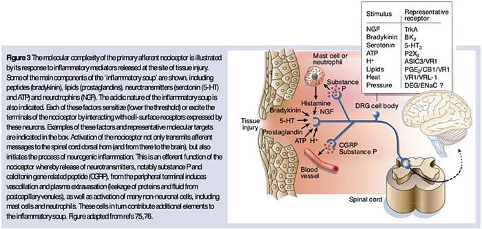
A large number of inflammatory mediators have been specifically implicated in shoulder pain and rotator cuff disease.21�25 While some chemical mediators directly activate nociceptors, most lead to changes in the sensory neuron itself rather than directly activating it. These changes may be early post- translational or delayed transcription dependent. Examples of the former are changes in the TRPV1 receptor or in voltage- gated ion channels resulting from the phosphorylation of membrane-bound proteins. Examples of the latter include the NGF-induced increase in TRV1 channel production and the calcium-induced activation of intracellular transcription factors.

Molecular Mechanisms Of Nociception

The sensation of pain alerts us to real or impending injury and triggers appropriate protective responses. Unfortunately, pain often outlives its usefulness as a warning system and instead becomes chronic and debilitating. This transition to a chronic phase involves changes within the spinal cord and brain, but there is also remarkable modulation where pain messages are initiated � at the level of the primary sensory neuron. Efforts to determine how these neurons detect pain-producing stimuli of a thermal, mechanical or chemical nature have revealed new signaling mechanisms and brought us closer to understanding the molecular events that facilitate transitions from acute to persistent pain.

biochemistry of pain el paso tx.The Neurochemistry Of Nociceptors

Glutamate is the predominant excitatory neurotransmitter in all nociceptors. Histochemical studies of adult DRG, however, reveal two broad classes of unmyelinated C fiber.

Chemical Transducers To Make The Pain Worse

As described above, injury heightens our pain experience by increasing the sensitivity of nociceptors to both thermal and mechanical stimuli. This phenomenon results, in part, from the production and release of chemical mediators from the primary sensory terminal and from non-neural cells (for example, fibroblasts, mast cells, neutrophils and platelets) in the environment36 (Fig. 3). Some components of the inflammatory soup (for example, protons, ATP, serotonin or lipids) can alter neuronal excitability directly by inter- acting with ion channels on the nociceptor surface, whereas others (for example, bradykinin and NGF) bind to metabotropic receptors and mediate their effects through second-messenger signaling cascades11. Considerable progress has been made in understanding the biochemistry basis of such modulatory mechanisms.

Extracellular Protons & Tissue Acidosis

Local tissue acidosis is a hallmark physiological response to injury, and the degree of associated pain or discomfort is well correlated with the magnitude of acidification37. Application of acid (pH 5) to the skin produces sustained discharges in a third or more of polymodal nociceptors that innervate the receptive field 20.

biochemistry of pain el paso tx.Cellular & Molecular Mechanisms Of Pain
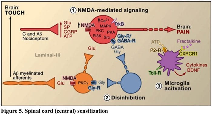
Abstract

The nervous system detects and interprets a wide range of thermal and mechanical stimuli as well as environmental and endogenous chemical irritants. When intense, these stimuli generate acute pain, and in the setting of persistent injury, both peripheral and central nervous system components of the pain transmission pathway exhibit tremendous plasticity, enhancing pain signals and producing hypersensitivity. When plasticity facilitates protective reflexes, it can be beneficial, but when the changes persist, a chronic pain condition may result. Genetic, electrophysiological, and pharmacological studies are elucidating the molecular mechanisms that underlie detection, coding, and modulation of noxious stimuli that generate pain.

Introduction: Acute Versus Persistent Pain

biochemistry of pain el paso tx.

biochemistry of pain el paso tx.Figure 5. Spinal Cord (Central) Sensitization

  1. Glutamate/NMDA receptor-mediated sensitization.�Following intense stimulation or persistent injury, activated C and A? nociceptors release a variety of neurotransmitters including dlutamate, substance P, calcitonin-gene related peptide (CGRP), and ATP, onto output neurons in lamina I of the superficial dorsal horn (red). As a consequence, normally silent NMDA glutamate receptors located in the postsynaptic neuron can now signal, increase intracellular calcium, and activate a host of calcium dependent signaling pathways and second messengers including mitogen-activated protein kinase (MAPK), protein kinase C (PKC), protein kinase A (PKA) and Src. This cascade of events will increase the excitability of the output neuron and facilitate the transmission of pain messages to the brain.
  2. Disinhibition.�Under normal circumstances, inhibitory interneurons (blue) continuously release GABA and/or glycine (Gly) to decrease the excitability of lamina I output neurons and modulate pain transmission (inhibitory tone). However, in the setting of injury, this inhibition can be lost, resulting in hyperalgesia. Additionally, disinhibition can enable non-nociceptive myelinated A? primary afferents to engage the pain transmission circuitry such that normally innocuous stimuli are now perceived as painful. This occurs, in part, through the disinhibition of excitatory PKC? expressing interneurons in inner lamina II.
  3. Microglial activation.�Peripheral nerve injury promotes release of ATP and the chemokine fractalkine that will stimulate microglial cells. In particular, activation of purinergic, CX3CR1, and Toll-like receptors on microglia (purple) results in the release of brain-derived neurotrophic factor (BDNF), which through activation of TrkB receptors expressed by lamina I output neurons, promotes increased excitability and enhanced pain in response to both noxious and innocuous stimulation (that is, hyperalgesia and allodynia). Activated microglia also release a host of cytokines, such as tumor necrosis factor ? (TNF?), interleukin-1? and 6 (IL-1?, IL-6), and other factors that contribute to central sensitization.

The Chemical Milieu Of Inflammation

Peripheral sensitization more commonly results from inflammation-associated changes in the chemical environment of the nerve fiber (McMahon et al., 2008). Thus, tissue damage is often accompanied by the accumulation of endogenous factors released from activated nociceptors or non-neural cells that reside within or infiltrate into the injured area (including mast cells, basophils, platelets, macrophages, neutrophils, endothelial cells, keratinocytes, and fibroblasts). Collectively. these factors, referred to as the �inflammatory soup�, represent a wide array of signaling molecules, including neurotransmitters, peptides (substance P, CGRP, bradykinin), eicosinoids and related lipids (prostaglandins, thromboxanes, leukotrienes, endocannabinoids), neurotrophins, cytokines, and chemokines, as well as extracellular proteases and protons. Remarkably, nociceptors express one or more cell surface receptors capable of recognizing and responding to each of these pro-inflammatory or pro-algesic agents (Figure 4). Such interactions enhance excitability of the nerve fiber, thereby heightening its sensitivity to temperature or touch.

Unquestionably the most common approach to reducing inflammatory pain involves inhibiting the synthesis or accumulation of components of the inflammatory soup. This is best exemplified by non-steroidal anti-inflammatory drugs, such as aspirin or ibuprofen, which reduce inflammatory pain and hyperalgesia by inhibiting cyclooxygenases (Cox-1 and Cox-2) involved in prostaglandin synthesis. A second approach is to block the actions of inflammatory agents at the nociceptor. Here, we highlight examples that provide new insight into cellular mechanisms of peripheral sensitization, or which form the basis of new therapeutic strategies for treating inflammatory pain.

NGF is perhaps best known for its role as a neurotrophic factor required for survival and development of sensory neurons during embryogenesis, but in the adult, NGF is also produced in the setting of tissue injury and constitutes an important component of the inflammatory soup (Ritner et al., 2009). Among its many cellular targets, NGF acts directly on peptidergic C fiber nociceptors, which express the high affinity NGF receptor tyrosine kinase, TrkA, as well as the low affinity neurotrophin receptor, p75 (Chao, 2003; Snider and McMahon, 1998). NGF produces profound hypersensitivity to heat and mechanical stimuli through two temporally distinct mechanisms. At first, a NGF-TrkA interaction activates downstream signaling pathways, including phospholipase C (PLC), mitogen-activated protein kinase (MAPK), and phosphoinositide 3-kinase (PI3K). This results in functional potentiation of target proteins at the peripheral nociceptor terminal, most notably TRPV1, leading to a rapid change in cellular and behavioral heat sensitivity (Chuang et al., 2001).

Irrespective of their pro-nociceptive mechanisms, interfering with neurotrophin or cytokine signaling has become a major strategy for controlling inflammatory disease or resulting pain. The main approach involves blocking NGF or TNF-? action with a neutralizing antibody. In the case of TNF-?, this has been remarkably effective in the treatment of numerous autoimmune diseases, including rheumatoid arthritis, leading to dramatic reduction in both tissue destruction and accompanying hyperalgesia (Atzeni et al., 2005). Because the main actions of NGF on the adult nociceptor occur in the setting of inflammation, the advantage of this approach is that hyperalgesia will decrease without affecting normal pain perception. Indeed, anti-NGF antibodies are currently in clinical trials for treatment of inflammatory pain syndromes (Hefti et al., 2006).

Glutamate/NMDA Receptor-Mediated Sensitization

Acute pain is signaled by the release of glutamate from the central terminals of nociceptors, generating excitatory post-synaptic currents (EPSCs) in second order dorsal horn neurons. This occurs primarily through activation of postsynaptic AMPA and kainate subtypes of ionotropic glutamate receptors. Summation of sub-threshold EPSCs in the postsynaptic neuron will eventually result in action potential firing and transmission of the pain message to higher order neurons.

Other studies indicate that changes in the projection neuron, itself, contribute to the dis- inhibitory process. For example, peripheral nerve injury profoundly down-regulates the K+- Cl- co-transporter KCC2, which is essential for maintaining normal K+ and Cl- gradients across the plasma membrane (Coull et al., 2003). Downregulating KCC2, which is expressed in lamina I projection neurons, results in a shift in the Cl- gradient, such that activation of GABA-A receptors depolarize, rather than hyperpolarize the lamina I projection neurons. This would, in turn, enhance excitability and increase pain transmission. Indeed, pharmacological blockade or siRNA-mediated downregulation of KCC2 in the rat induces mechanical allodynia.

Share Ebook

Sources:

Why does my shoulder hurt? A review of the neuroanatomical and biochemical basis of shoulder pain

Benjamin John Floyd Dean, Stephen Edward Gwilym, Andrew Jonathan Carr

Cellular and Molecular Mechanisms of Pain

Allan I. Basbaum1, Diana M. Bautista2, Gre?gory Scherrer1, and David Julius3

1Department of Anatomy, University of California, San Francisco 94158

2Department of Molecular and Cell Biology, University of California, Berkeley CA 94720 3Department of Physiology, University of California, San Francisco 94158

Molecular mechanisms of nociception

David Julius* & Allan I. Basbaum�

*Department of Cellular and Molecular Pharmacology, and �Departments of Anatomy and Physiology and W. M. Keck Foundation Center for Integrative Neuroscience, University of California San Francisco, San Francisco, California 94143, USA (e-mail: julius@socrates.ucsf.edu)

Overview of the Pathophysiology of Neuropathic Pain

Overview of the Pathophysiology of Neuropathic Pain

Neuropathic pain is a complex, chronic pain condition that is generally accompanied by soft tissue injury. Neuropathic pain is common in clinical practice and also poses a challenge to patients and clinicians alike. With neuropathic pain, the nerve fibers themselves may be either damaged, dysfunctional or injured. Neuropathic pain is the result of damage from trauma or disease to the peripheral or central nervous system, where the lesion may occur at any site. As a result, these damaged nerve fibers can send incorrect signals to other pain centers. The effect of a nerve fiber injury consists of a change in neural function, both at the region of the injury and also around the injury. Clinical signs of neuropathic pain normally include sensory phenomena, such as spontaneous pain, paresthesias and hyperalgesia.


Neuropathic pain, as defined by the International Association of the Study of Pain or the IASP, is pain initiated or caused by a primary lesion or dysfunction of the nervous system. It could result from damage anywhere along the neuraxis: peripheral nervous system, spinal or supraspinal nervous system. Traits that distinguish neuropathic pain from other kinds of pain include pain and sensory signs lasting beyond the recovery period. It’s characterized in humans by spontaneous pain, allodynia, or the experience of non-noxious stimulation as painful, and causalgia, or persistent burning pain. Spontaneous pain includes sensations of “pins and needles”, burning, shooting, stabbing and paroxysmal pain, or electric-shock like pain, often associated with dysesthesias and paresthesias. These sensations not only alter the patient’s sensory apparatus, but also the patient’s well-being, mood, attention and thinking. Neuropathic pain is made up of both “negative” symptoms, such as sensory loss and tingling sensations, and “positive” symptoms, such as paresthesias, spontaneous pain and increased feeling of pain.


Conditions frequently related to neuropathic pain can be classified into two major groups: pain due to damage in the central nervous system and pain because of damage to the peripheral nervous system. Cortical and sub-cortical strokes, traumatic spinal cord injuries, syringo-myelia and syringobulbia, trigeminal and glossopharyngeal neuralgias, neoplastic and other space-occupying lesions are clinical conditions that belong to the former group. Nerve compression or entrapment neuropathies, ischemic neuropathy, peripheral polyneuropathies, plexopathies, nerve root compression, post-amputation stump and phantom limb pain, postherpetic neuralgia and cancer-related neuropathies are clinical conditions that belong to the latter group.


Pathophysiology of Neuropathic Pain


The pathophysiologic processes and concepts underlying neuropathic pain are multiple. Prior to covering these processes, a review of ordinary pain circuitry is critical. Regular pain circuitries involve activation of a nociceptor, also known as the pain receptor, in response to a painful stimulation. A wave of depolarization is delivered to the first-order neurons, together with sodium rushing in via sodium channels and potassium rushing out. Neurons end in the brain stem in the trigeminal nucleus or in the dorsal horn of the spinal cord. It is here where the sign opens voltage-gated calcium channels in the pre-synaptic terminal, allowing calcium to enter. Calcium allows glutamate, an excitatory neurotransmitter, to be released into the synaptic area. Glutamate binds to NMDA receptors on the second-order neurons, causing depolarization.


These neurons cross through the spinal cord and travel until the thalamus, where they synapse with third-order neurons. These then connect to the limbic system and cerebral cortex. There is also an inhibitory pathway that prevents pain signal transmission from the dorsal horn. Anti-nociceptive neurons originate in the brain stem and travel down the spinal cord where they synapse with short interneurons in the dorsal horn by releasing dopamine and norepinephrine. The interneurons modulate the synapse between the first-order neuron as well as the second-order neuron by releasing gamma amino butyric acid, or GABA, an inhibitory neurotransmitter. Consequently, pain cessation is the result of inhibition of synapses between first and second order neurons, while pain enhancement might be the result of suppression of inhibitory synaptic connections.


Pathophysiology of Neuropathic Pain Diagram | El Paso, TX Chiropractor


The mechanism underlying neuropathic pain, however, aren’t as clear. Several animal studies have revealed that lots of mechanisms may be involved. However, one has to remember that what applies to creatures may not always apply to people. First order neurons may increase their firing if they’re partially damaged and increase the amount of sodium channels. Ectopic discharges are a consequence of enhanced depolarization at certain sites in the fiber, resulting in spontaneous pain and movement-related pain. Inhibitory circuits might be diminished in the level of the dorsal horn or brain stem cells, as well as both, allowing pain impulses to travel unopposed.


In addition, there might be alterations in the central processing of pain when, because of chronic pain and the use of some drug and/or medications, second- and third-order neurons can create a “memory” of pain and become sensitized. There’s then heightened sensitivity of spinal neurons and reduced activation thresholds. Another theory demonstrates the concept of sympathetically-maintained neuropathic pain. This notion was demonstrated by analgesia following sympathectomy from animals and people. However, a mix of mechanics can be involved in many chronic neuropathic or mixed somatic and neuropathic pain conditions. Among those challenges in the pain field, and much more so as it pertains to neuropathic pain, is the capability to check it. There is a dual component to this: first, assessing quality, intensity and advancement; and second, correctly diagnosing neuropathic pain.


There are, however, some diagnostic tools that may assist clinicians in evaluating neuropathic pain. For starters, nerve conduction studies and sensory-evoked potentials may identify and quantify the extent of damage to sensory, but not nociceptive, pathways by monitoring neurophysiological responses to electrical stimuli. Additionally, quantitative sensory testing steps perception in reaction to external stimuli of varying intensities by applying stimulation to the skin. Mechanical sensitivity to tactile stimuli is measured with specialized tools, such as von Frey hairs, pinprick with interlocking needles, as well as vibration sensitivity together with vibrameters and thermal pain with thermodes.


It is also extremely important to perform a comprehensive neurological evaluation to identify motor, sensory and autonomic dysfunctions. Ultimately, there are numerous questionnaires used to distinguish neuropathic pain in nociceptive pain. Some of them include only interview queries (e.g., the Neuropathic Questionnaire and ID Pain), while others contain both interview questions and physical tests (e.g., the Leeds Assessment of Neuropathic Symptoms and Signs scale) and the exact novel tool, the Standardized Evaluation of Pain, which combines six interview questions and ten physiological evaluations.


Neuropathic Pain Diagram | El Paso, TX Chiropractor


Treatment Modalities for Neuropathic Pain


Pharmacological regimens aim at the mechanisms of neuropathic pain. However, both pharmacologic and non-pharmacologic treatments deliver complete or partial relief in just about half of patients. Many evidence-based testimonials suggest using mixtures of drugs and/or medications to function for as many mechanisms as possible. The majority of studies have researched mostly post-herpetic neuralgia and painful diabetic neuropathies but the results may not apply to all neuropathic pain conditions.


Antidepressants


Antidepressants increase synaptic serotonin and norepinephrine levels, thereby enhancing the effect of the descending analgesic system associated with neuropathic pain. They’ve been the mainstay of neuropathic pain therapy. Analgesic actions might be attributable to nor-adrenaline and dopamine reuptake blockade, which presumably enhance descending inhibition, NMDA-receptor antagonism and sodium-channel blockade. Tricyclic antidepressants, such as TCAs; e.g., amitriptyline, imipramine, nortriptyline and doxepine, are powerful against continuous aching or burning pain along with spontaneous pain.


Tricyclic antidepressants have been proven significantly more effective for neuropathic pain than the specific serotonin reuptake inhibitors, or SSRIs, such as fluoxetine, paroxetine, sertraline and citalopram. The reason may be that they inhibit reuptake of serotonin and nor-epinephrine, while SSRIs only inhibit serotonin reuptake. Tricyclic antidepressants can have unpleasant side effects, including nausea, confusion, cardiac conduction blocks, tachycardia and ventricular arrhythmias. They can also cause weight gain, a reduced seizure threshold and orthostatic hypotension. Tricyclics have to be used with care in the elderly, who are particularly vulnerable to their acute side effects. The drug concentration in the blood should be monitored to avoid toxicity in patients who are slow medication metabolizers.


Serotonin-norepinephrine reuptake inhibitors, or SNRIs, are a new class of antidepressants. Like TCAs, they seem to be more effective than SSRIs for treating neuropathic pain because they also inhibit reuptake of both nor-epinephrine and dopamine. Venlafaxine is as effective against debilitating polyneuropathies, such as painful diabetic neuropathy, as imipramine, in the mention of TCA, and the two are significantly greater than placebo. Like the TCAs, the SNRIs seem to confer benefits independent of their antidepressant effects. Side effects include sedation, confusion, hypertension and withdrawal syndrome.


Antiepileptic Drugs


Antiepileptic drugs can be utilized as first-line treatment especially for certain types of neuropathic pain. They act by modulating voltage-gated calcium and sodium channels, by improving the inhibitory effects of GABA and by inhibiting excitatory glutaminergic transmission. Anti-epileptic medications have not been demonstrated to be effective for acute pain. In chronic pain cases, antiepileptic drugs seem to be effective only in trigeminal neuralgia. Carbamazepine is routinely employed for this condition. Gabapentin, which functions by inhibiting calcium channel function through agonist actions at the alpha-2 delta subunit of the calcium channel, is also known to be effective for neuropathic pain. However, gabapentin acts centrally and it might cause fatigue, confusion and somnolence.


Non-Opioid Analgesics


There is a lack of strong data supporting using non-steroidal anti inflammatory medications, or NSAIDs, in the relief of neuropathic pain. This may be due to the lack of an inflammatory component in relieving pain. But they have been utilized interchangeably with opioids as adjuvants in treating cancer pain. There have been reported complications, though, especially in severely debilitated patients.


Opioid Analgesics


Opioid analgesics are a subject of much debate in relieving neuropathic pain. They act by inhibiting central ascending pain impulses. Traditionally, neuropathic pain has been previously observed to be opioid-resistant, in which opioids are more suitable methods for coronary and somatic nociceptive types of pain. Many doctors prevent using opioids to treat neuropathic pain, in large part because of concerns about drug abuse, addiction and regulatory issues. But, there are many trials that have found opioid analgesics to succeed. Oxycodone was superior to placebo for relieving pain, allodynia, improving sleep and handicap. Controlled-release opioids, according to a scheduled basis, are recommended for patients with constant pain to encourage constant levels of analgesia, prevent fluctuations in blood glucose and prevent adverse events associated with higher dosing. Most commonly, oral preparations are used because of their greater ease of use and cost-effectiveness. Trans-dermal, parenteral and rectal preparations are generally used in patients who cannot tolerate oral drugs.


Local Anesthetics


Nearby acting anesthetics are appealing because, thanks to their regional action, they have minimal side effects. They act by stabilizing sodium channels at the axons of peripheral first-order neurons. They work best if there is only partial nerve injury and excess sodium channels have collected. Topical lidocaine is the best-studied representative of the course for neuropathic pain. Specifically, the use of this 5 percent lidocaine patch for post-herpetic neuralgia has caused its approval by the FDA. The patch seems to work best when there is damaged, but maintained, peripheral nervous system nociceptor function from the involved dermatome demonstrating as allodynia. It needs to be set directly on the symptomatic area for 12 hours and eliminated for another 12 hours and may be used for years this way. Besides local skin reactions, it is often well tolerated by many patients with neuropathic pain.


Miscellaneous Drugs


Clonidine, an alpha-2-agonist, was shown to be effective in a subset of patients with diabetic peripheral neuropathy. Cannabinoids have been found to play a role in experimental pain modulation in animal models and evidence of the efficacy is accumulating. CB2-selective agonists suppress hyperalgesia and allodynia and normalize nociceptive thresholds without inducing analgesia.


Interventional Pain Management


Invasive treatments might be considered for patients who have intractable neuropathic pain. These treatments include epidural or perineural injections of local anesthetics or corticosteroids, implantation of epidural and intrathecal drug delivery methods and insertion of spinal cord stimulators. These approaches are reserved for patients with intractable chronic neuropathic pain who have failed conservative medical management and also have experienced thorough psychological evaluation. In a study by Kim et al, it was shown that a spinal cord stimulator was effective in treating neuropathic pain of nerve root origin.


Dr-Jimenez_White-Coat_01.png

Dr. Alex Jimenez’s Insight

With neuropathic pain, chronic pain symptoms occur due to the nerve fibers themselves being damaged, dysfunctional or injured, generally accompanied by tissue damage or injury. As a result, these nerve fibers can begin to send incorrect pain signals to other areas of the body. The effects of neuropathic pain caused by nerve fiber injuries includes modifications in nerve function both at the site of injury and at areas around the injury. Understanding the pathophysiology of neuropathic pain has been a goal for many healthcare professionals, in order to effectively determine the best treatment approach to help manage and improve its symptoms. From the use of drugs and/or medications, to chiropractic care, exercise, physical activity and nutrition, a variety of treatment approaches may be used to help ease neuropathic pain for each individual’s needs.


Additional Interventions for Neuropathic Pain


Lots of patients with neuropathic pain pursue complementary and alternative treatment options to treat neuropathic pain. Other well-known regimens used to treat neuropathic pain include acupuncture, percutaneous electrical nerve stimulation, transcutaneous electrical nerve stimulation, cognitive behavioral treatment, graded motor imagery and supportive treatment, and exercise. Among these however, chiropractic care is a well-known alternative treatment approach commonly utilized to help treat neuropathic pain. Chiropractic care, along with physical therapy, exercise, nutrition and lifestyle modifications can ultimately offer relief for neuropathic pain symptoms.


Chiropractic Care


What is known is that a comprehensive management application is crucial to combat the effects of neuropathic pain. In this manner, chiropractic care is a holistic treatment program that could be effective in preventing health issues associated with nerve damage. Chiropractic care provides assistance to patients with many different conditions, including those with neuropathic pain. Sufferers of neuropathic pain often utilize non-steroidal-anti-inflammatory medications, or NSAIDs, such as ibuprofen, or heavy prescription painkillers to help ease neuropathic pain. These may provide a temporary fix but need constant use to manage the pain. This invariably contributes to harmful side effects and in extreme situations, prescription drug dependence.


Chiropractic care can help improve symptoms of neuropathic pain and enhance stability without these downsides. An approach such as chiropractic care offers an individualized program designed to pinpoint the root cause of the issue. Through the use of spinal adjustments and manual manipulations, a chiropractor can carefully correct any spinal misalignments, or subluxations, found along the length of the spine, which could lower the consequences of nerve wracking via the realigning of the backbone. Restoring spinal integrity is essential to keeping a high-functioning central nervous system.


A chiropractor can also be a long-term treatment towards enhancing your overall well-being. Besides spinal adjustments and manual manipulations, a chiropractor may offer nutritional advice, such as prescribing a diet rich in antioxidants, or they may design a physical therapy or exercise program to fight nerve pain flair-ups. A long-term condition demands a long-term remedy, and in this capacity, a healthcare professional who specializes in injuries and/or conditions affecting the musculoskeletal and nervous system, such as a doctor of chiropractic or chiropractor, may be invaluable as they work to gauge favorable change over time.


Physical therapy, exercise and movement representation techniques have been demonstrated to be beneficial for neuropathic pain treatment. Chiropractic care also offers other treatment modalities which may be helpful towards the management or improvement of neuropathic pain. Low level laser therapy, or LLLT, for instance, has gained tremendous prominence as a treatment for neuropathic pain. According to a variety of research studies, it was concluded that LLLT had positive effects on the control of analgesia for neuropathic pain, however, further research studies are required to define treatment protocols that summarize the effects of low level laser therapy in neuropathic pain treatments.


Chiropractic care also includes nutritional advice, which can help control symptoms associated with diabetic neuropathy. During a research study, a low fat plant-based diet was demonstrated to improve glycemic control in patients with type 2 diabetes. After about 20 weeks of the pilot study, the individuals involved reported changes in their body weight and electrochemical skin conductance in the foot was reported to have improved with the intervention. The research study suggested a potential value in the low-fat plant-based diet intervention for diabetic neuropathy. Moreover, clinical studies found that the oral application of magnesium L-threonate is capable of preventing as well as restoring memory deficits associated with neuropathic pain.


Chiropractic care can also offer additional treatment strategies to promote nerve regeneration. By way of instance, enhancing the regeneration of axons has been suggested to help improve functional recovery after peripheral nerve injury. Electrical stimulation, together with exercise or physical activities, was found to promote nerve regeneration after delayed nerve repair in humans and rats, according to recent research studies. Both electrical stimulation and exercise were ultimately determined to be promising experimental treatments for peripheral nerve injury which seem ready to be transferred to clinical use. Further research studies may be needed to fully determine the effects of these in patients with neuropathic pain.


Conclusion


Neuropathic pain is a multifaceted entity with no particular guidelines to take care of. It’s best managed using a multidisciplinary approach. Pain management requires ongoing evaluation, patient education, ensuring patient follow-up and reassurance. Neuropathic pain is a chronic condition that makes the option for the best treatment challenging. Individualizing treatment involves consideration of the impact of the pain on the individual’s well-being, depression and disabilities together with continuing education and evaluation. Neuropathic pain studies, both on the molecular level and in animal models, is relatively new but very promising. Many improvements are anticipated in the basic and clinical fields of neuropathic pain hence opening the doorways to improved or new treatment modalities for this disabling condition. The scope of our information is limited to chiropractic as well as to spinal injuries and conditions. To discuss the subject matter, please feel free to ask Dr. Jimenez or contact us at�915-850-0900�.


Curated by Dr. Alex Jimenez


Green-Call-Now-Button-24H-150x150-2-3.png


Additional Topics: Back Pain


Back pain is one of the most prevalent causes for disability and missed days at work worldwide. As a matter of fact, back pain has been attributed as the second most common reason for doctor office visits, outnumbered only by upper-respiratory infections. Approximately 80 percent of the population will experience some type of back pain at least once throughout their life. The spine is a complex structure made up of bones, joints, ligaments and muscles, among other soft tissues. Because of this, injuries and/or aggravated conditions, such as herniated discs, can eventually lead to symptoms of back pain. Sports injuries or automobile accident injuries are often the most frequent cause of back pain, however, sometimes the simplest of movements can have painful results. Fortunately, alternative treatment options, such as chiropractic care, can help ease back pain through the use of spinal adjustments and manual manipulations, ultimately improving pain relief.


blog picture of cartoon paperboy big news


EXTRA IMPORTANT TOPIC: Low Back Pain Management


MORE TOPICS: EXTRA EXTRA:�Chronic Pain & Treatments


Sleep Loss Increases Risk of Obesity

Sleep Loss Increases Risk of Obesity

Losing sleep increases the risk of becoming obese, according to a Swedish study. Researchers from Uppsala University say a lack of sleep affects energy metabolism by disrupting sleep patterns and affecting the body’s response to food and exercise.

Although several studies have found a connection between sleep deprivation and weight gain, the cause has been unclear.

Dr. Christian Benedict and his colleagues have conducted a number of human studies to investigate how sleep loss may affect energy metabolism. These studies have measured and imaged behavioral, physiological, and biochemical responses to food following acute sleep deprivation.

The behavioral data reveal that metabolically healthy, sleep-deprived human subjects prefer larger portions of food, seek more calories, show signs of increased impulsivity related to food, and expend less energy.

The group’s physiological studies indicate that sleep loss shifts the hormonal balance from hormones that promote fullness (satiety), such as GLP-1, to those that promote hunger, such as ghrelin. Sleep restriction also increased levels of endocannabinoids, which are known to stimulate appetite.

In addition, their research showed that acute sleep loss alters the balance of gut bacteria, which has been widely implicated as key for maintaining a healthy metabolism. The same study also found reduced sensitivity to insulin after sleep loss.

“Since perturbed sleep is such a common feature of modern life, these studies show it is no surprise that metabolic disorders, such as obesity are also on the rise,” said Benedict.

“My studies suggest that sleep loss favors weight gain in humans,” he said. “It may also be concluded that improving sleep could be a promising lifestyle intervention to reduce the risk of future weight gain.”

Not only is a lack of sleep adding pounds, other research has discovered that too much light while you sleep can also increase your risk for obesity. A British study of 113,000 women found that the more light they were exposed to during sleeping hours, the greater their risk of being fat. Light disrupts the body’s circadian rhythm, which affects sleep and wake patterns, and also affects metabolism.

But getting exposure to light in the early waking hours might help keep weight in check. A study from Northwestern University found that people who got most of their exposure to sunlight, even if it’s overcast, early in the day had a lower body mass index (BMI) than those who got their sun exposure later in the day, regardless of physical activity, caloric intake, or age.