Chiropractic Care and Faster Healing from Hand Numbness
Find out about effective chiropractic care options for addressing hand numbness and enhancing your quality of life.
Contents
Understanding Hand Numbness and Carpal Tunnel Syndrome: How Chiropractic Care Offers Natural Relief
Hand numbness and tingling sensations affect millions of people worldwide, disrupting daily activities and diminishing quality of life. These uncomfortable symptoms often signal nerve compression issues, with carpal tunnel syndrome being the most common culprit. While many individuals immediately think surgery is their only option, research increasingly demonstrates that conservative, non-surgical approaches—particularly chiropractic care—can provide significant relief and lasting results. This comprehensive guide explores the causes, symptoms, and clinical rationale for using chiropractic treatment to address hand numbness and carpal tunnel syndrome. We’ll examine how environmental factors contribute to nerve compression, the critical connection between spinal health and hand symptoms, and evidence-based conservative treatments that can help you avoid surgery.
Understanding Hand Numbness: Causes and Symptoms
Hand numbness represents a sensory dysfunction involving the loss of normal sensation, including pain, temperature, touch, or vibratory perception. The severity varies considerably among individuals, ranging from mild intermittent tingling to constant numbness that significantly impairs hand function.
Common Symptoms of Hand Numbness
Individuals experiencing hand numbness typically report a constellation of symptoms that may include:
- Paresthesia: The medical term for abnormal sensations, paresthesia manifests as numbness with loss of touch or temperature sensation. Some people describe feeling like they’re wearing gloves when they aren’t, while others experience gait and balance problems when numbness affects their ability to feel the ground beneath their feet.
- Tingling and “Pins and Needles”: Often described as the sensation of limbs “falling asleep,” this symptom frequently occurs in the thumb, index, middle, and sometimes the ring finger. The tingling may start intermittently but can progress to become constant.
- Burning Sensations: Many patients report a burning feeling along the affected nerve pathway, which can extend from the fingertips up through the hand and into the forearm.
- Pain: Sharp, stabbing, or shooting pain often accompanies numbness, particularly at night when symptoms tend to worsen. This pain may radiate from the wrist up the forearm and sometimes as far as the shoulder.
- Weakness: Muscle weakness accompanies numbness in the same location, making it difficult to grip objects, hold tools, or perform fine motor tasks like buttoning clothing.
- Loss of Coordination: Decreased finger dexterity and hand clumsiness can make everyday activities challenging, from typing on a keyboard to opening jars.
What Causes Hand Numbness?
Hand numbness occurs when there is pressure, irritation, or damage to the nerves that supply sensation to the hands. The causes are varied and understanding the underlying mechanism is crucial for effective treatment:
- Peripheral Neuropathy: This condition affects the very ends of nerves in the hands and feet. Diabetes is the most common cause of peripheral neuropathy, but alcoholism, vitamin deficiencies (especially B12), autoimmune conditions, liver or kidney disorders, and exposure to toxins can also damage peripheral nerves.
- Nerve Compression Syndromes: Pressure on a nerve anywhere along its course from the neck to the fingertips can cause numbness. Common compression sites include the carpal tunnel at the wrist (carpal tunnel syndrome), the cubital tunnel at the elbow (cubital tunnel syndrome), and the cervical spine in the neck.
- Cervical Radiculopathy: Compression or irritation of nerve roots exiting the cervical spine can send radiating pain, numbness, and weakness down through the shoulder, arm, and hand. This occurs when herniated discs, bone spurs, or degenerative changes put pressure on the nerve roots.
- Thoracic Outlet Syndrome: Compression of nerves and blood vessels between the collarbone and first rib can cause symptoms similar to carpal tunnel syndrome.
- Trauma and Injuries: Bone dislocations, fractures, and crushing injuries can cause swelling or direct nerve damage, resulting in numbness.
- Double Crush Syndrome: This phenomenon occurs when a nerve is compressed at two distinct locations along its pathway—typically at both the cervical spine and the wrist. Compression at one site makes the nerve more vulnerable to symptoms from compression at a second site.
What is Carpal Tunnel Syndrome?
Carpal tunnel syndrome represents the most common peripheral nerve entrapment condition, affecting approximately one in ten adults at some point in their lifetime. For individuals with diabetes, the lifetime risk increases dramatically to 84 percent.
Anatomical Overview
The carpal tunnel is a narrow passageway in the wrist formed by the transverse carpal ligament at its upper boundary and the carpal bones at its lower boundary. This confined space accommodates nine flexor tendons and the median nerve, which must traverse through it to reach the hand.
The median nerve originates from nerve roots C5-T1 in the cervical spine and travels through the brachial plexus, down the arm, through the forearm, and ultimately through the carpal tunnel. The nerve provides both motor function (allowing movement) and sensory function (providing feeling) to the thumb, index finger, middle finger, and the thumb-side of the ring finger.
How Carpal Tunnel Syndrome Develops
Carpal tunnel syndrome develops when elevated pressure within the carpal tunnel compresses the median nerve. Normal pressure within the carpal tunnel ranges from 2 to 10 mmHg. However, extension or flexion of the wrist causes pressure to increase eight to ten times the normal level.
The pathophysiology involves a combination of mechanisms:
- Mechanical Trauma: Repetitive compression and friction damage the nerve over time.
- Increased Pressure: Elevated intracarpal pressure restricts blood flow to the endoneurial capillary system, causing ischemic damage to nerve tissue.
- Inflammation: Swelling of the tendons and surrounding tissues within the confined space further compresses the median nerve.
- Demyelination: Repeated compression can lead to demyelination (loss of the protective nerve covering) at the site of compression, impairing nerve signal transmission.
Symptoms Specific to Carpal Tunnel Syndrome
While carpal tunnel syndrome shares many symptoms with general hand numbness, it has distinctive characteristics:
- Distribution Pattern: Numbness, tingling, and pain specifically affect the thumb, index, middle, and lateral half of the ring finger. The little finger is typically spared because it receives sensation from the ulnar nerve rather than the median nerve.
- Nocturnal Symptoms: Symptoms frequently manifest or worsen at night while lying down. Many patients wake up shaking their hands to restore sensation—a phenomenon so common it’s considered pathognomonic for carpal tunnel syndrome.
- Progressive Nature: Initially, symptoms come and go and tend to improve during the daytime. Over time, most patients begin to encounter symptoms during the day, particularly when engaged in repetitive activities such as typing, driving, or holding a phone.
- Thenar Atrophy: In advanced cases, the muscles at the base of the thumb (thenar eminence) can atrophy and weaken, causing a flattened appearance and inability to oppose the thumb effectively.
- Positive Provocative Tests: Clinical examination reveals positive Phalen’s test (symptoms reproduced by flexing the wrists for 60 seconds) and Tinel’s sign (tapping over the median nerve at the wrist reproduces symptoms).
Environmental and Occupational Risk Factors
Carpal tunnel syndrome is a multifactorial condition arising from a combination of patient-specific, occupational, social, and environmental factors. Understanding these risk factors is essential for both prevention and treatment.
Personal and Medical Risk Factors
- Obesity: Being obese or overweight significantly increases carpal tunnel syndrome risk. Each unit rise in body mass index (BMI) increases the risk by approximately 7.4 percent. The association can be explained by accumulation of fat tissue inside the carpal tunnel or by increased hydrostatic pressure causing swelling that compresses the median nerve.
- Diabetes Mellitus: Diabetes is strongly associated with carpal tunnel syndrome, with prevalence estimates suggesting that 60-70 percent of people with diabetes have mild to severe neuropathy. Diabetic polyneuropathy may render the median nerve more prone to entrapment, exemplifying the “double crush” phenomenon.
- Thyroid Disorders: Hypothyroidism increases the risk of carpal tunnel syndrome with an odds ratio of 3.70. Thyroid disease was present in 7.8 percent of participants who developed acute carpal tunnel syndrome complicating distal radius fractures.
- Pregnancy: Hormonal fluctuations and fluid retention during pregnancy commonly cause temporary carpal tunnel syndrome, which typically resolves after delivery.
- Rheumatoid Arthritis and Inflammatory Conditions: Autoimmune diseases like rheumatoid arthritis, lupus, and Guillain-Barré syndrome increase susceptibility to nerve compression.
- Age and Gender: Carpal tunnel syndrome is more common in women than men for unclear reasons, and incidence increases with age, particularly affecting individuals aged 45 to 64.
- Genetics: Carpal tunnel syndrome tends to run in families, suggesting a genetic component. Certain physical characteristics like wrist shape (a square wrist ratio exceeding 0.7) increase risk.
Workplace and Environmental Factors
- Repetitive Hand Movements: Occupations involving frequent repetitive hand and wrist activities significantly elevate carpal tunnel syndrome risk. Workers who assemble products, particularly in meat and poultry processing (incidence as high as 15 percent) and automobile manufacturing (affecting up to 10 percent of workers), face exceptionally high risk.
- Forceful Exertion: Time spent in forceful exertion can be a greater risk factor for carpal tunnel syndrome than even obesity if job exposure is high. Research demonstrates that working with forceful exertion 20-60 percent of the time increases risk nearly threefold, while exertion more than 60 percent of the time increases risk nearly twentyfold.
- Vibrating Tools and Equipment: Workers using hand-held vibratory tools such as rock drills, chainsaws, and power tools in quarry drilling and forestry operations face elevated risk. Hand-arm vibration syndrome can cause tingling and numbness that persist even after vibration stops.
- Non-Neutral Wrist Postures: Positions of wrist flexion and extension during work activities increase carpal tunnel pressure and nerve compression risk.
- Cold Temperature Exposure: Work performed in cold environments while performing repetitive wrist movements or using vibrating equipment significantly increases risk.
- Computer and Keyboard Use: While traditionally associated with carpal tunnel syndrome, the evidence implicating computer use as a major cause is actually weak. Mouse use shows some association with carpal tunnel syndrome, but keyboard typing alone has not been definitively linked to the condition.
- Psychosocial Workplace Factors: Job strain, intense deadlines, poor social work environment, and low job satisfaction are major contributors to carpal tunnel pain beyond just physical factors.
Chemical Exposure
Emerging research suggests that workers exposed to neurotoxic chemicals face increased carpal tunnel syndrome risk. Chemicals like n-hexane have potential neurotoxic effects, and frequent biomechanical and chemical co-exposure may create synergistic effects. Exposure to chemicals may generate diffuse subtle nerve damage, rendering the median nerve more prone to entrapment at the carpal tunnel—particularly when combined with biomechanical wrist stressors.
The Clinical Anatomy: How Nerve Compression Occurs
Understanding the anatomical pathway of the median nerve from the cervical spine through the carpal tunnel illuminates why symptoms can arise from compression at multiple sites and why addressing spinal health is crucial for treating hand numbness.
The Median Nerve Pathway
The median nerve begins its journey from nerve roots C5-T1 in the cervical spine. The anterior rami of these nerve roots merge to form the lateral and medial cords of the brachial plexus, which unite to create the median nerve proper.
- Upper Arm Course: The median nerve descends through the arm lateral to the brachial artery, then crosses the artery (usually in front) to lie on its medial side at the elbow.
- Forearm Course: At the elbow, the median nerve passes between the two heads of the pronator teres muscle and descends beneath the flexor digitorum superficialis. In the forearm, the median nerve supplies motor innervation to most flexor muscles including the pronator teres, palmaris longus, flexor digitorum superficialis, flexor carpi radialis, and through its anterior interosseous branch, the flexor pollicis longus and pronator quadratus.
- Wrist Approach: Approximately 5 cm above the wrist, the median nerve becomes more superficial, lying between the tendons of the flexor digitorum superficialis and flexor carpi radialis. At this point, it gives off the palmar cutaneous branch, which passes over (not through) the carpal tunnel to provide sensation to the palm.
- Carpal Tunnel Transit: The median nerve enters the carpal tunnel under the transverse carpal ligament, traveling alongside nine flexor tendons in this confined space. The median nerve is the most superficial structure within the carpal tunnel.
- Hand Distribution: After exiting the carpal tunnel, the median nerve gives off the recurrent thenar motor branch to innervate the abductor pollicis brevis, opponens pollicis, and superficial head of the flexor pollicis brevis. It then divides into digital branches providing sensation to the palmar surface of the thumb, index, middle, and lateral half of the ring finger, while also innervating the first and second lumbrical muscles.
Multiple Compression Sites and Double Crush Syndrome
Nerve compression can occur at any point along the median nerve’s pathway from the cervical spine to the fingertips. The “double crush” hypothesis, formalized by Upton and McComas, suggests that compression of an axon at one location makes it more sensitive to effects of compression at another location because of impaired axoplasmic flow.
- Cervical Spine Compression: Misalignments in the cervical vertebrae, herniated discs, bone spurs, or degenerative changes can compress nerve roots as they exit the spinal cord. A forward head posture can increase strain on the brachial plexus, and tight scalene or pectoralis minor muscles may compress nerves along their path.
- Thoracic Outlet: Dysfunction in the thoracic outlet—located between the collarbone and first rib—can mimic or worsen carpal tunnel symptoms.
- Elbow (Pronator Syndrome): The median nerve can be compressed at the elbow as it passes between the two heads of the pronator teres muscle.
- Wrist (Carpal Tunnel): Finally, compression occurs at the carpal tunnel itself, the most common site of median nerve entrapment.
The double crush phenomenon is particularly relevant because in approximately 10 percent of carpal tunnel cases, there is also a cervical radiculopathy. Studies show that 65-75 percent of chronic lower arm injuries have a neck component, and treating the neck often produces much better and quicker results.
The clinical implication is profound: treating only the wrist may result in residual symptoms from uncorrected cervical compression, while addressing both sites of impingement offers the best outcomes.
Double Crush Syndrome: The Neck-Wrist Connection
Many patients diagnosed with carpal tunnel syndrome actually experience nerve compression originating not primarily at the wrist but at the cervical spine or multiple locations simultaneously. This concept—known as double crush syndrome—has important implications for treatment selection and outcomes.
Understanding Double Crush Physiology
Double crush syndrome occurs when a nerve is compressed at two distinct points along its pathway. The theory proposes that compression at one site renders the nerve more susceptible to dysfunction from compression at a second site, even when neither compression alone would produce significant symptoms.
Several mechanisms explain this increased vulnerability:
- Impaired Axoplasmic Flow: Compression at one location disrupts the transport of nutrients and sustaining compounds along the length of the nerve, compromising overall nerve health.
- Immune-Mediated Attacks: Compression may trigger immune responses affecting sensory nerve cell centers (dorsal root ganglion).
- Ion Channel Deregulation: Compression can disrupt the ion channels integral to the nerve’s ability to carry information to and from the spinal cord.
- Restricted Nerve Mobility: Nerves normally glide along openings in the neck, muscles, and around joints during movement. Compression at one location may compromise this movement, creating increased pressure and tension in other parts of the nerve.
Clinical Presentation and Diagnosis
Patients with double crush syndrome often present with symptoms that extend beyond typical carpal tunnel distributions. They may experience:
-
Numbness and tingling not only in the first three-and-a-half fingers but also radiating up the forearm, past the elbow, into the upper arm, shoulder, and neck
-
Persistent symptoms despite conservative wrist-focused treatments
-
Bilateral symptoms (affecting both hands)
-
Associated neck pain, cervical stiffness, or limited cervical range of motion
-
Positive cervical spine examination findings including hyperreflexia, sensory deficits, or motor weakness
Chiropractors and other clinicians trained in differential diagnosis can identify double crush syndrome through comprehensive examination that includes cervical spine assessment, postural evaluation, orthopedic testing at multiple sites, and neurological screening.
The Importance of Treating Both Sites
In double crush syndromes, recognizing and treating both compression sites is essential. Research demonstrates that addressing cervical spine dysfunction can completely resolve carpal tunnel symptoms in many cases—even without direct wrist treatment.
One case report documented complete resolution of carpal tunnel syndrome after improving cervical spine posture to remove the “first crush,” suggesting that treatment should be aimed at restoring normal cervical spine alignment. Another study found that when chronic carpal tunnel or arm pain cases failed to respond to traditional one-site treatments including physical therapy, chiropractic care, or even surgery, addressing the neck component led to successful resolution.
Discovering the Benefits of Chiropractic Care- Video
Clinical Rationale for Chiropractic Care
Chiropractic care offers a comprehensive, evidence-based approach to treating hand numbness and carpal tunnel syndrome by addressing the root causes of nerve compression rather than merely masking symptoms.
The Chiropractic Philosophy
Chiropractors recognize that the spine and nervous system are deeply interconnected. Misalignments in the spine—particularly in the cervical region—can interfere with nerve function throughout the body, including the median nerve that passes through the carpal tunnel.
Unlike conventional treatments that often focus on localized wrist pain, chiropractors take a holistic, full-body approach. They investigate and treat compression of nerves anywhere in the body, understanding that issues in the spine and musculoskeletal system can profoundly influence nerve function.
How Chiropractic Adjustments Address Nerve Compression
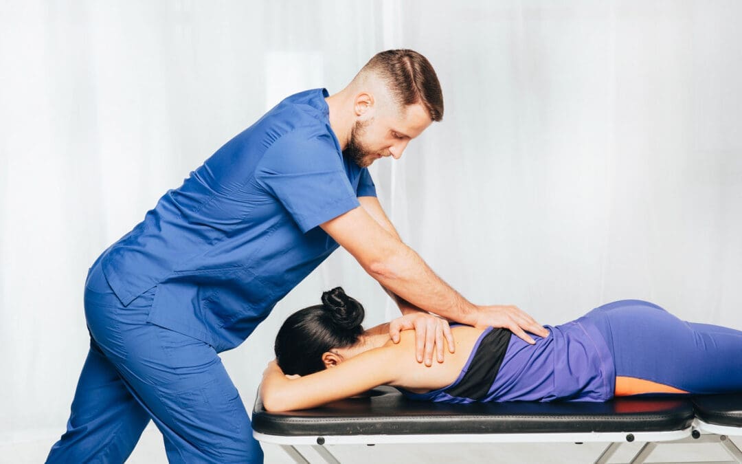
- Spinal Realignment: Chiropractic adjustments gradually restore proper alignment of the cervical, thoracic, and lumbar spine. This realignment releases compression within nerve roots exiting the spinal cord, allowing nerve signals to flow normally to the extremities.
- Improved Nerve Communication: By correcting spinal misalignments (subluxations), chiropractors restore proper nerve communication between the brain and body. When the upper cervical spine is properly aligned, nerve function improves, reducing pressure on nerves and restoring sensation and function to the hands.
- Reduced Inflammation: Chiropractic care helps decrease inflammation around compressed nerves, reducing swelling that contributes to carpal tunnel pressure.
- Enhanced Blood Flow: Adjustments promote improved circulation to nerve tissues, supporting healing and reducing ischemic damage.
- Improved Biomechanics: Correcting postural dysfunctions like forward head carriage and protracted shoulders reduces strain on the brachial plexus and median nerve pathway.
Evidence Supporting Chiropractic for Carpal Tunnel Syndrome
Research increasingly supports the effectiveness of chiropractic care for carpal tunnel syndrome and related nerve compression conditions:
- Manual Therapy Effectiveness: A 2024 systematic review and meta-analysis comparing manual therapy versus surgery found that manual therapy was more effective for short-term pain relief at one and three months compared with surgery. At six to twelve months, surgical intervention provided greater improvements, but quality-of-life improvements were similar in both groups. The researchers concluded that manual therapy offers effective short-term relief for mild to moderate carpal tunnel syndrome, making it a viable first-line option.
- Conservative Treatment Success: A comprehensive 2018 European review of ten studies comparing surgery versus non-surgical care found that while results favored non-surgical approaches at three months and surgery at six months, there was no difference in outcome one year later. The research team concluded that conservative treatment should be preferred unless otherwise indicated.
- Cochrane Review Findings: A Cochrane systematic review of exercise and mobilization interventions found that nerve mobilization, carpal bone mobilization, yoga, and chiropractic treatment provided symptom improvement for patients with carpal tunnel syndrome. While acknowledging limited evidence quality, the review supported these approaches as valid non-surgical treatment options.
- Case Study Evidence: Multiple published case reports document successful chiropractic management of nerve compression syndromes. One case involving a 41-year-old woman with ulnar nerve compression demonstrated complete symptom resolution after 11 treatments consisting of chiropractic manipulation, myofascial therapy, and elastic therapeutic taping. Another case documented identification and successful treatment of cervical myelopathy by a chiropractor, leading to complete symptom resolution.
- Comparison with Traditional Treatments: A 2003 Cochrane review found that chiropractic care and medical treatment provided similar short-term improvement in mental distress, vibrometry, hand function, and finger sensation. Importantly, chiropractic care achieved these results without medications or their associated side effects.
What Chiropractic Treatment Involves
Chiropractic care for carpal tunnel syndrome typically includes multiple treatment modalities:
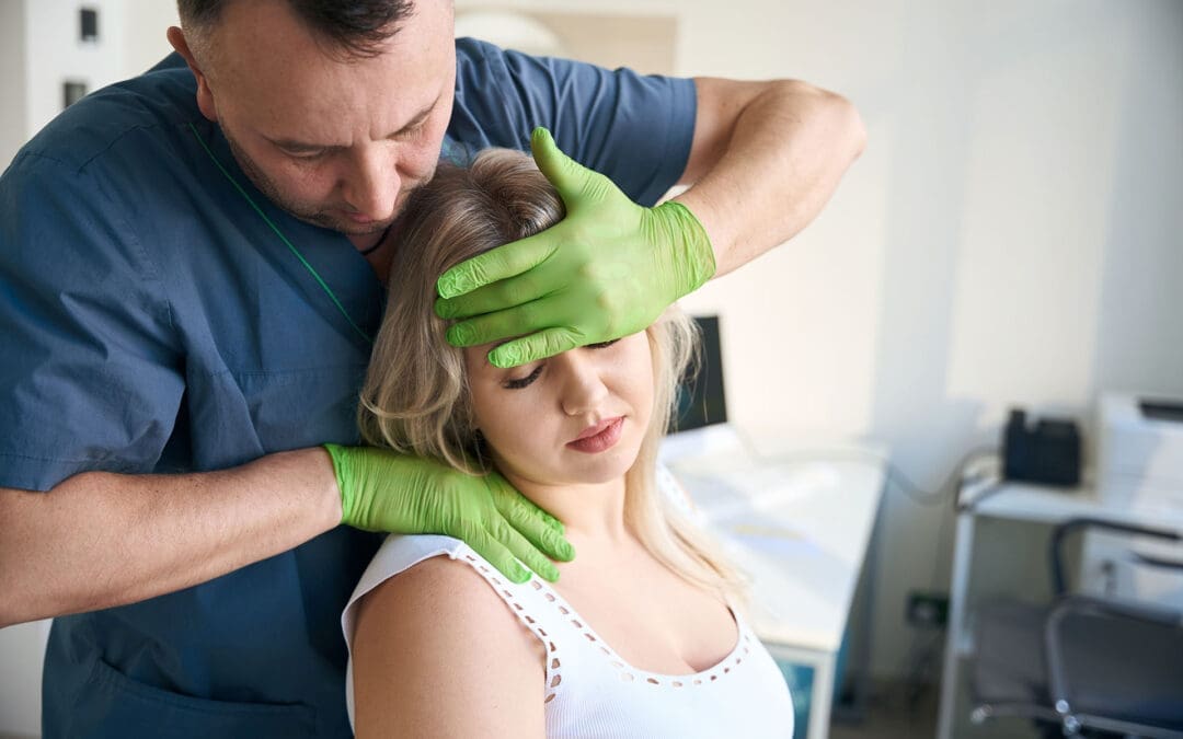
- Cervical Spine Adjustments: Gentle manipulations realign the neck to relieve pressure on nerve roots, improve posture, reduce forward head carriage, and restore proper nerve communication to the arm and hand.
- Wrist and Hand Adjustments: Specific adjustments restore joint mobility in the carpal bones, reduce inflammation, increase circulation, and address biomechanical imbalances from overuse or improper motion.
- Elbow and Shoulder Adjustments: Treatments resolve radial nerve entrapment, release restrictions in the shoulder girdle affecting nerve flow, and address thoracic outlet compression.
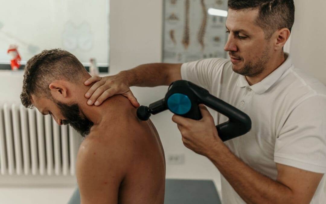
- Myofascial Release: Soft tissue techniques ease tension in the forearm and hand muscles, target trigger points that radiate pain, and break up adhesions and scar tissue using active release technique or instrument-assisted mobilization.
- Nerve Gliding Exercises: Patient education on specific exercises that help the median nerve move freely within surrounding tissues, reduce entrapment, and prevent scar tissue buildup.
- Ergonomic Education: Guidance on proper workstation setup, posture correction, activity modification, and techniques to minimize repetitive stress.
- Therapeutic Modalities: Additional treatments may include ultrasound therapy to reduce inflammation, cold laser therapy to accelerate healing, electrical stimulation, and massage therapy.
Dr. Alexander Jimenez’s Clinical Approach
Dr. Alexander Jimenez, DC, APRN, FNP-BC, represents a unique dual-credentialed practitioner who combines advanced medical expertise as a board-certified Family Practice Nurse Practitioner with specialized chiropractic training. His integrative approach exemplifies the evolution of conservative care for conditions like carpal tunnel syndrome and hand numbness.
Dual-Scope Practice Model
Operating El Paso’s premier wellness and injury care clinic, Dr. Jimenez offers comprehensive assessment and treatment capabilities that bridge traditional medical diagnosis with natural, non-invasive chiropractic interventions. As both a Doctor of Chiropractic and Advanced Practice Registered Nurse Practitioner, he can perform detailed clinical evaluations, order and interpret advanced imaging and diagnostic tests, and provide evidence-based treatment protocols inspired by integrative medicine principles.
Clinical Assessment Methodology
Dr. Jimenez’s approach to patients presenting with hand numbness or carpal tunnel symptoms includes:
- Comprehensive Health History: Detailed evaluation of symptom onset, progression, aggravating and relieving factors, occupational exposures, medical conditions, and family history.
- Functional Medicine Assessment: Utilizing the Institute for Functional Medicine’s assessment programs, Dr. Jimenez evaluates personal history, current nutrition, activity behaviors, environmental exposures to toxic elements, psychological and emotional factors, and genetics.
- Advanced Imaging: When clinically indicated, Dr. Jimenez correlates patient injuries and symptoms with advanced imaging studies including X-rays, MRI, nerve conduction studies, and electrodiagnostic testing.
- Physical Examination: Thorough orthopedic, neurological, and musculoskeletal examination assessing the cervical spine, thoracic outlet, shoulder, elbow, wrist, and hand.
- Postural Analysis: Evaluation of forward head posture, shoulder protraction, and other biomechanical dysfunctions that contribute to nerve compression.
Individualized Treatment Plans
Dr. Jimenez emphasizes that treatment must be personalized based on each patient’s unique presentation, underlying causes, and health goals. His treatment protocols may include:
- Chiropractic Adjustments: Targeted spinal and extremity manipulations to restore proper alignment and relieve nerve compression.
- Functional Medicine Interventions: Root-cause analysis incorporating nutrition, lifestyle modifications, and environmental factor correction.
- Acupuncture and Electro-Acupuncture: Traditional and modern techniques to reduce inflammation and promote healing.
- Rehabilitation Programs: Customized flexibility, agility, and strength programs tailored for all age groups and abilities.
- Nutritional Support: Personalized nutrition plans to optimize health, reduce inflammation, and support nerve function.
Collaborative Care Philosophy
A distinguishing feature of Dr. Jimenez’s practice is his commitment to collaborative care. When he believes another specialist is better suited for a patient’s condition, he refers to appropriate providers, ensuring patients receive the highest standard of care. He has established partnerships with top surgeons, medical specialists, and rehabilitation experts to bring comprehensive treatment options to his patients.
Focus on Non-Invasive Protocols
Dr. Jimenez’s practice prioritizes natural recovery, avoiding unnecessary surgeries or medications whenever possible. His treatments focus on what works for the patient, using the body’s inherent ability to heal rather than introducing harmful chemicals, controversial hormone replacement, unnecessary surgery, or addictive drugs.
Through his unique functional health approach to healing, Dr. Jimenez continues to be voted the best chiropractor in El Paso by reviewing sites, clinical specialists, researchers, and readers. This recognition reflects his compassionate, patient-centered approach and commitment to addressing the root causes of health issues through integrative care.
Non-Surgical Treatments and Conservative Management
Numerous non-surgical interventions have demonstrated effectiveness for carpal tunnel syndrome and hand numbness, offering patients alternatives to surgical intervention while providing significant symptom relief and functional improvement.
1. Wrist Splinting and Bracing
Wrist splints represent one of the most commonly prescribed and effective conservative treatments for carpal tunnel syndrome.
- Mechanism of Action: Splints maintain the wrist in a neutral position, which results in the lowest carpal tunnel pressure compared with flexion or extension positions. Neutral positioning minimizes compression on the median nerve and prevents the excessive wrist flexion that commonly occurs during sleep—a primary contributor to nocturnal symptoms.
- Optimal Splint Design: Recent research indicates that wrist splints incorporating the metacarpophalangeal (MCP) joints are more effective than traditional wrist-only splints. Active finger flexion causes lumbrical muscles to intrude into the carpal tunnel, elevating pressure and compressing the median nerve. Splints that limit both wrist and MCP joint motion yield better outcomes, with improvements persisting even after six months of intervention.
- Wearing Schedule: Most doctors recommend wearing splints primarily at night, as symptoms like numbness and tingling tend to worsen during sleep when wrists naturally assume flexed positions. During the day, wearing the brace for a few hours while performing repetitive wrist movements can reduce strain on the median nerve. However, continuous wear is not recommended as overuse can lead to stiffness and weakness.
- Evidence: A randomized controlled trial of 83 participants found that subjects wearing a soft hand splint at night for four weeks had decreased self-reported carpal tunnel symptoms and functional limitations compared to untreated controls. Another study comparing splinting with surgery found that while both groups improved, the differences at one-year follow-up were not statistically significant.
2. Therapeutic Ultrasound
Ultrasound therapy represents an evidence-based non-invasive treatment that has shown effectiveness for carpal tunnel syndrome relief.
- Mechanism: Therapeutic ultrasound uses high-frequency sound waves (typically 1 MHz) to penetrate deep into wrist tissues, reducing inflammation, improving circulation, and promoting healing. The treatment creates gentle vibrations that increase blood flow, reduce swelling, help release pressure on the median nerve, and soften scar tissue in chronic cases.
- Treatment Protocol: Effective protocols typically involve 20 sessions of ultrasound treatment (1 MHz, 1.0 W/cm², pulsed mode 1:4, 15 minutes per session) applied to the area over the carpal tunnel. Initial treatments are performed daily (five sessions per week), followed by twice-weekly treatments for five weeks.
- Evidence: A landmark randomized, double-blind, sham-controlled trial found that ultrasound treatment had good short-term effectiveness and satisfying medium-term effects in patients with mild to moderate idiopathic carpal tunnel syndrome. At the end of treatment, 68 percent of wrists treated with active ultrasound showed satisfactory improvement or complete remission compared to 38 percent receiving sham treatment. At six-month follow-up, 74 percent of actively treated wrists maintained improvement compared to only 20 percent of sham-treated wrists. Both subjective symptoms and electroneurographic variables (motor distal latency and sensory nerve conduction velocity) showed significant improvement with active treatment.
- Anti-Inflammatory Effect: Ultrasound therapy induces an anti-inflammatory effect that provides relief of carpal tunnel symptoms by enhancing blood flow, increasing membrane permeability, altering connective tissue extensibility, and affecting nerve conduction through thermal effects.
3. Low-Level Laser Therapy (Cold Laser)
Low-level laser therapy (LLLT), also called cold laser therapy, offers a non-invasive treatment option that has gained support from multiple systematic reviews and meta-analyses.
- Mechanism: LLLT uses focused light at specific wavelengths and low intensities to stimulate healing without heating tissue. The light energy penetrates tissue and interacts with intracellular biomolecules to increase biochemical energy production, enhance oxygenated blood supply, increase collagen supply for tissue elasticity, accelerate nerve regeneration, and reduce swelling and inflammation.
- Treatment Application: During treatment, low-intensity laser diodes are placed directly on the skin over the carpal tunnel and affected areas. Patients typically feel a warming sensation at the treatment site, and treatment is virtually painless with relief often experienced immediately.
- Evidence: A 2016 meta-analysis of seven randomized clinical trials involving 531 participants found that LLLT improved hand grip strength, visual analog scale pain scores, and sensory nerve action potential after three months of follow-up for mild to moderate carpal tunnel syndrome. The researchers concluded that LLLT was more effective than placebo for both short-term and long-term symptom improvement.
- Limitations: A 2017 Cochrane review noted that while some studies showed benefit, the risk of bias was moderate to low across studies, and more high-quality research using standardized laser intervention protocols is needed to confirm effects.
4. Nerve Gliding and Tendon Gliding Exercises
Nerve gliding (also called nerve flossing) and tendon gliding exercises help mobilize the median nerve and flexor tendons, improving their movement through the carpal tunnel and reducing compression.
- Nerve Gliding Technique: Basic median nerve glides involve extending the affected arm straight out with the elbow extended and palm facing up, then bending the wrist downward toward the floor while tilting the head away from the arm. This position is held for two to five seconds, then released. More advanced versions involve extending the arm to the side, bending the wrist upward while tilting the head away, then bending the wrist downward while tilting the head toward the arm.
- Tendon Gliding Exercises: These exercises involve sequential finger movements designed to glide the flexor tendons through the carpal tunnel. Starting with the wrist neutral and fingers straight, patients flex fingers at different joints in specific sequences, performing approximately 20 repetitions of each pattern.
- Benefits: Nerve gliding improves median nerve mobility, reduces adhesions and tension along the nerve pathway, relieves symptoms associated with nerve compression (pain, tingling, numbness), enhances flexibility and range of motion, and supports the rehabilitation process. When combined with other conservative treatments, nerve gliding exercises significantly enhance outcomes.
- Evidence: Studies incorporating nerve gliding as part of multi-component interventions have shown symptom improvement, though the independent effect of nerve gliding alone requires further research.
5. Oral Medications
Several oral medications have been studied for carpal tunnel syndrome treatment, with varying levels of evidence supporting their use.
- Oral Corticosteroids: Short-term oral steroid treatment has demonstrated significant improvement in symptoms. Pooled data from randomized trials showed that two-week oral steroid treatment resulted in significant symptom improvement (weighted mean difference -7.23), with benefits maintained at four weeks. However, long-term use of steroids carries significant side effects and is not recommended.
- NSAIDs (Non-Steroidal Anti-Inflammatory Drugs): Despite their anti-inflammatory properties and common prescription, NSAIDs have not demonstrated significant benefit compared to placebo for carpal tunnel syndrome in randomized trials.
- Vitamin B6: The use of vitamin B6 (pyridoxine) for carpal tunnel syndrome remains controversial. While some early studies and clinical observations suggested benefit, the largest and most comprehensive study found no correlation between vitamin B6 status and carpal tunnel syndrome. A University of Michigan study of 125 workers found that 32 percent reported carpal tunnel symptoms and 8 percent had vitamin B6 deficiency, but there was no relationship between the deficiency, symptoms, or impaired nerve function. Vitamin B6 at doses less than 200 mg daily is unlikely to cause adverse effects, but excessive doses (200 mg or more) can be neurotoxic and cause sensory nerve damage.
- Diuretics: Diuretics have not demonstrated significant benefit for carpal tunnel syndrome when compared to placebo.
6. Acupuncture
Acupuncture and electroacupuncture represent traditional and modern approaches to treating carpal tunnel syndrome that have shown promise in research studies.
- Mechanism: Acupuncture involves inserting needles at specific points on the wrist, forearm, and hand. The needles are typically left in place for 15 to 30 minutes, with multiple sessions needed to alleviate pain.
- Evidence: A 2013 study on acupuncture-evoked response in carpal tunnel syndrome found that electroacupuncture applied at local acupoints on the affected wrist and at distal acupoints on the contralateral ankle both produced reduced pain and paresthesia. Brain response to acupuncture in prefrontal cortex and other regions correlated with pain reduction following stimulation.
A multicenter randomized controlled trial examining acupuncture with complementary and integrative medicine modalities for chemotherapy-induced peripheral neuropathy (which shares mechanisms with carpal tunnel-related numbness) found significant improvement in hand numbness, tingling, discomfort, and physical functioning.
7. Yoga and Stretching
Yoga has been investigated as a treatment for carpal tunnel syndrome based on the theory that stretching may relieve compression in the carpal tunnel, better joint posture may decrease nerve compression, and improved blood flow may benefit the median nerve.
Evidence: A randomized trial involving 51 participants found that yoga significantly reduced pain after eight weeks when compared with wrist splinting alone. The yoga program focused on upper body postures, breathing, and relaxation techniques designed to improve strength, flexibility, and awareness in the joints from the shoulder to the hand.
8. Ergonomic Modifications
Activity and workstation modifications aim to position the wrist in a neutral position, provide maximum space within the carpal tunnel, and avoid forceful and repeated movements central to occupations associated with increased carpal tunnel risk.
- Principles: Effective ergonomic interventions include adjusting chair height so feet rest flat with knees level with hips, positioning monitors at eye level to avoid neck strain, using ergonomic keyboards or mice to reduce wrist strain, ensuring proper wrist positioning during typing (wrists held up in line with backs of hands rather than resting), and investing in chairs with lumbar support.
- Workplace Interventions: Research on ergonomic keyboards compared to controls has demonstrated equivocal results for pain and function. However, comprehensive ergonomic programs that include workstation modifications, job rotation, frequent microbreaks, and worker education show promise for preventing repetitive strain injuries including carpal tunnel syndrome.
Practical Tips and Home Remedies
In addition to professional treatment, numerous self-care strategies can help manage carpal tunnel symptoms and prevent progression.
Daily Hand Care Practices
- Frequent Breaks: When performing repetitive hand activities, take breaks every 30-45 minutes to stretch and rest your hands. Set a timer as a reminder to prevent prolonged repetitive motions without rest.
- Gentle Hand Shaking: When numbness occurs, particularly at night, gently shake your hands to restore circulation and sensation. Many carpal tunnel patients instinctively do this, and it can provide temporary relief.
- Temperature Therapy: Some patients find relief alternating between cold and warm compresses on the wrist. Cold reduces inflammation, while warmth improves circulation.
- Avoid Sleeping on Hands: Sleeping with hands under pillows or in bent positions increases carpal tunnel pressure. Try to maintain neutral wrist positions during sleep, and consider wearing wrist splints at night.
Hand Strengthening Exercises
- Grip Strengthening: Use a stress ball or therapy putty to strengthen hand muscles. Compress the ball with your affected hand and repeat 10 times.
- Wrist Curls: Hold a light weight (1-2 pounds) in your hand with your palm facing up. Curl your wrist up, then release and let the weight fall back down. Repeat 10 times.
- Finger Opposition: Touch the tip of your thumb to the base of each finger on the same hand, moving from index finger to pinky. Repeat 10 times. This exercise helps maintain thenar muscle function.
- Finger Abduction: Hold your hand out with fingers together. Slowly spread your fingers apart, then release and let them come back together. Repeat 10 times.
Stretching Exercises
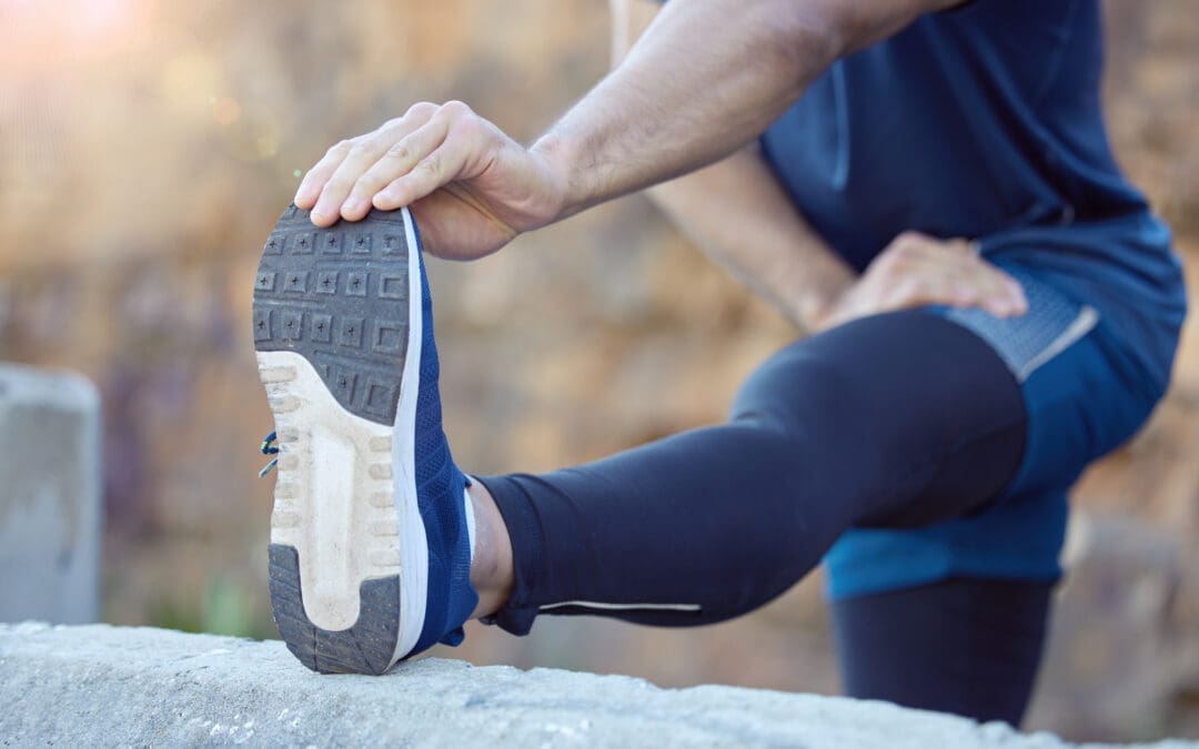
- Prayer Stretch: Place your hands together in front of your chest in a prayer position. Keeping palms together, slowly lower them toward your waist until you feel a moderate stretch in your wrists and forearms. Hold for 20-30 seconds and repeat 2-4 times.
- Wrist Flexor Stretch: Extend your affected arm straight in front of you with your palm facing down. Bend your wrist back, pointing your fingers upward toward the ceiling. Use your opposite hand to gently pull the fingers back until you feel a stretch. Hold for 20-30 seconds and repeat 3 times.
- Wrist Extensor Stretch: Extend your arm with palm facing down, then bend your wrist so fingers point toward the floor. Gently pull down with your opposite hand until you feel a stretch on top of your forearm. Hold for 20-30 seconds.
- Thumb Stretch: Using your opposite hand, gently push your thumb backward until you feel a gentle stretch. Hold for 20 seconds and repeat 3-4 times.
Activity Modifications
- Modify Grip: When possible, use tools and utensils with larger handles that require less grip force. Avoid pinch grips when a whole-hand grip will suffice.
- Reduce Force: Type gently rather than pounding keys. Use a light touch on computer mice and avoid death-gripping steering wheels, tools, or phones.
- Neutral Wrist Position: Keep wrists in neutral alignment rather than flexed or extended during activities. Use wrist rests appropriately—they’re for resting between typing, not supporting your wrists while typing.
- Hand Position Variation: Alternate hand positions and tasks throughout the day to avoid sustained postures. If possible, switch between different types of work to vary the stress on your hands.
Nutritional Considerations
- Anti-Inflammatory Diet: While specific dietary interventions for carpal tunnel syndrome lack extensive research, adopting an anti-inflammatory diet rich in omega-3 fatty acids, colorful fruits and vegetables, and whole grains may help reduce systemic inflammation.
- Adequate Hydration: Proper hydration supports tissue health and may help reduce swelling that contributes to carpal tunnel pressure.
- Limiting Pro-Inflammatory Foods: Reducing intake of processed foods, excess sugar, and trans fats may help minimize inflammation.
- Vitamin B6 Consideration: While evidence is controversial, some practitioners recommend moderate vitamin B6 supplementation (50-100 mg daily) with zinc support. However, consult with a healthcare provider before starting supplements, as excessive B6 (over 200 mg daily) can cause nerve damage.
Lifestyle Modifications and Ergonomic Strategies
Preventing carpal tunnel syndrome progression and reducing symptoms requires addressing the lifestyle and environmental factors that contribute to nerve compression.
Workstation Ergonomics
- Computer Setup: Position your monitor directly in front of you at arm’s length, with the top of the screen at or slightly below eye level. This prevents excessive neck flexion that contributes to cervical spine dysfunction and double crush syndrome.
- Keyboard and Mouse Placement: Keep your keyboard directly in front of you at a height that allows your elbows to rest comfortably at a 90-degree angle. Position your mouse close to your keyboard at the same height to avoid reaching. Consider an ergonomic mouse that’s moved with finger motion rather than wrist motion.
- Chair Adjustment: Select a chair with good lumbar support and adjust the height so your feet rest flat on the floor with knees at hip level. Armrests should support your elbows without elevating your shoulders.
- Document Holder: If you frequently reference documents while typing, use a document holder positioned at the same height and distance as your monitor to avoid repetitive neck turning and flexion.
Posture Correction
- Forward Head Posture: One of the most common postural dysfunctions contributing to upper extremity nerve compression is forward head carriage. For every inch your head moves forward from neutral alignment, it effectively weighs an additional 10 pounds, increasing strain on cervical structures. Conscious correction of forward head posture, combined with strengthening exercises for deep neck flexors and stretching of chest muscles, can significantly reduce nerve compression.
- Shoulder Position: Protracted (rounded forward) shoulders contribute to thoracic outlet compression and brachial plexus tension. Regularly performing scapular retraction exercises (shoulder blade squeezes) helps maintain proper shoulder positioning.
- Overall Spinal Alignment: Maintaining neutral spinal curves throughout the day reduces stress on the nervous system. Regular breaks from sitting, standing desks used intermittently, and conscious attention to posture all contribute to better spinal health.
Activity Management
- Job Rotation: If your work involves repetitive hand motions, advocate for job rotation that allows you to alternate between different types of tasks throughout the day. This variation prevents sustained stress on the same structures.
- Microbreaks: Taking frequent short breaks (30-60 seconds every 20-30 minutes) to stretch and change position is more effective than infrequent long breaks. Use these microbreaks to perform wrist rotations, finger stretches, and shoulder rolls.
- Pacing: Avoid marathon sessions of repetitive activities. Break large projects into smaller segments with rest periods between.
- Tool Selection: When possible, choose ergonomically designed tools that require less grip force and allow neutral wrist positioning. Power tools with anti-vibration features reduce transmission of harmful vibrations to hands and wrists.
Weight Management
Obesity significantly increases carpal tunnel syndrome risk, with each BMI unit increase raising risk by approximately 7.4 percent. Weight loss through balanced nutrition and regular physical activity can reduce pressure within the carpal tunnel and improve symptoms.
Management of Underlying Conditions
- Diabetes Control: Maintaining optimal blood glucose levels through medication, diet, and exercise helps protect nerves from diabetic neuropathy and reduces carpal tunnel risk.
- Thyroid Management: If you have hypothyroidism, ensuring proper thyroid hormone replacement and regular monitoring can help reduce carpal tunnel risk.
- Blood Pressure Control: Managing hypertension may reduce carpal tunnel syndrome risk, as arterial hypertension shows strong association with the condition.
Stress Management
Psychosocial factors including job strain, stress, and low job satisfaction contribute significantly to carpal tunnel symptoms. Incorporating stress management techniques such as mindfulness, meditation, adequate sleep, and work-life balance can help reduce overall symptom burden.
Conclusion and Disclaimer
Hand numbness and carpal tunnel syndrome affect millions of people, significantly impacting quality of life and functional capacity. However, as this comprehensive review demonstrates, numerous conservative treatment options—particularly chiropractic care—offer effective alternatives to surgery for many patients experiencing these conditions. The clinical rationale for chiropractic treatment is grounded in understanding the median nerve’s complete pathway from the cervical spine through the carpal tunnel. By recognizing that nerve compression can occur at multiple sites and that spinal dysfunction often contributes to hand symptoms through the double crush phenomenon, chiropractors can address root causes rather than merely treating symptoms. This whole-body approach, combined with specific adjustments, soft tissue therapy, patient education, and ergonomic guidance, provides comprehensive care that has been validated by research demonstrating effectiveness comparable to or exceeding more invasive interventions in the short to medium term. Dr. Alexander Jimenez’s integrative model, combining advanced nurse practitioner medical training with chiropractic expertise, exemplifies the evolution toward comprehensive, patient-centered care that utilizes advanced diagnostics while prioritizing conservative, natural treatment approaches. His emphasis on functional medicine assessment, personalized treatment plans, and collaborative care ensures patients receive optimal management tailored to their unique circumstances. The extensive non-surgical treatment options reviewed—including wrist splinting, therapeutic ultrasound, low-level laser therapy, nerve gliding exercises, acupuncture, and ergonomic modifications—provide patients and practitioners with an evidence-based toolkit for managing carpal tunnel syndrome and hand numbness. Combined with lifestyle modifications addressing underlying risk factors such as obesity, diabetes, and occupational exposures, these interventions offer real hope for symptom resolution without surgical intervention.
Important Medical Disclaimer
This article is intended for educational and informational purposes only and should not be construed as medical advice, diagnosis, or treatment recommendation. The information presented here represents a synthesis of current research and clinical practice patterns but does not replace individualized medical evaluation and care. If you are experiencing hand numbness, tingling, weakness, or other concerning symptoms, you should seek immediate evaluation by a qualified healthcare provider. These symptoms may indicate serious conditions including but not limited to nerve compression syndromes, peripheral neuropathy, cervical spine disorders, vascular insufficiency, or other medical conditions requiring prompt diagnosis and treatment. The treatments and interventions described in this article—including chiropractic care, physical therapy, exercises, and complementary approaches—should only be undertaken under the supervision and guidance of licensed healthcare professionals who can properly diagnose your specific condition, assess for contraindications, and monitor your progress. Not all treatments are appropriate for all patients, and individual results will vary based on numerous factors including symptom severity, underlying causes, patient compliance, and individual health status. Carpal tunnel syndrome and related nerve compression conditions can progress to cause permanent nerve damage if left untreated or if treatment is delayed. While conservative approaches are often successful, some cases require surgical intervention. Failure to seek appropriate medical evaluation or delaying necessary treatment can result in irreversible complications including permanent sensory loss, chronic pain, and loss of hand function. Dr. Alexander Jimenez and the practitioners mentioned in this article provide clinical services within their scope of practice and licensure. References to specific practitioners are for informational and illustrative purposes and do not constitute an endorsement or guarantee of specific outcomes. Patients should verify credentials, licensure, and appropriateness of care providers for their individual needs. Decisions regarding your healthcare should be made in consultation with qualified medical professionals who have personally examined you, reviewed your complete medical history, conducted appropriate diagnostic testing, and can provide individualized recommendations based on your specific circumstances. This article does not establish a doctor-patient relationship, and readers should not rely solely on the information presented here for making healthcare decisions. By reading and using the information in this article, you acknowledge that you have read and understood this disclaimer and agree to seek appropriate professional medical evaluation and treatment rather than relying solely on self-diagnosis or self-treatment based on information obtained from this or any other educational resource.
References
- American Academy of Family Physicians. (2012). Best treatment approaches for carpal tunnel syndrome. American Family Physician. https://www.aafp.org/pubs/afp/issues/2012/0315/p546.html
- American Society for Surgery of the Hand. (n.d.). Numbness in hands: Causes & treatment. https://www.assh.org/handcare/condition/numbness-in-hands
- Ebenbichler, G. R., Resch, K. L., Nicolakis, P., Wiesinger, G. F., Uhl, F., Ghanem, A. H., & Fialka, V. (1998). Ultrasound treatment for treating the carpal tunnel syndrome: Randomised “sham” controlled trial. BMJ, 316(7133), 731-735. https://pubmed.ncbi.nlm.nih.gov/9529407/
- Donati, D., Boccolari, P., & Tedeschi, R. (2024). Manual therapy vs. surgery: Which is best for carpal tunnel syndrome relief? Life, 14(10), 1286. https://pubmed.ncbi.nlm.nih.gov/39459587/
- Genova, A., Dix, O., Saefan, A., Thakur, M., & Hassan, A. (2020). Carpal tunnel syndrome: A review of literature. Cureus, 12(3), e7333. https://pubmed.ncbi.nlm.nih.gov/32313774/
- Illes, J. D., & Johnson, T. L., Jr. (2013). Chiropractic management of a patient with ulnar nerve compression symptoms: A case report. Journal of Chiropractic Medicine, 12(2), 66-73. https://pubmed.ncbi.nlm.nih.gov/24294148/
- Jimenez, A. (n.d.). El Paso’s premier wellness and injury care clinic. https://dralexjimenez.com/
- Page, M. J., O’Connor, D., Pitt, V., & Massy-Westropp, N. (2012). Exercise and mobilisation interventions for carpal tunnel syndrome. Cochrane Database of Systematic Reviews, 2012(6), CD009899. https://pubmed.ncbi.nlm.nih.gov/22696387/
- Schmid, A. B., Brunner, F., Luomajoki, H., Held, U., Bachmann, L. M., Künzer, S., & Coppieters, M. W. (2009). Reliability of clinical tests to evaluate nerve function and mechanosensitivity of the upper limb peripheral nervous system. BMC Musculoskeletal Disorders, 10, 11.
- Sevy, J. O., Sina, R. E., & Varacallo, M. A. (2023). Carpal tunnel syndrome. In StatPearls. StatPearls Publishing. https://pubmed.ncbi.nlm.nih.gov/28846321/
- Zhang, C., & Murrell, G. A. C. (2025). Prevalence of hand paresthesia and numbness in painful shoulders: A narrative review. Annals of Joint, 10, 6. https://pubmed.ncbi.nlm.nih.gov/39981434/


