Curcumin is the main active ingredient found in turmeric, a bright yellow aromatic powder obtained from the rhizome of a plant in the ginger family commonly utilized for flavoring and coloring in a variety of Asian cuisines, as well as a very strong antioxidant which additionally has powerful anti-inflammatory effects. While turmeric has been consumed for generations due to its medicinal properties, it’s curcumin content is actually not that high. Therefore, if you want to experience its full benefits, it may be best to take a supplement that contains significant amounts of curcumin, or better yet, a supplement specially formulated to bring out the benefits of this naturally occurring chemical compound.
Curcumin is ultimately found in turmeric, the key ingredient in curry, and as a powerful antioxidant that offers numerous health benefits, curcumin helps support joint health and cardiovascular function, and it helps maintain a normal inflammatory reaction in the human body. Individuals throughout Asia have benefited from the healthful effects of curcumin for centuries.�Whether taken as a nutritional supplement or from meals, however, curcumin is usually poorly absorbed into the bloodstream.
Fortunately, global experts in botanical extract technologies have uncovered the key to curcumin absorption: phytosome technology. Meriva curcumin phytosome is an exceptional curcumin extract that’s significantly better absorbed than other curcumin extracts. Curcumin nutrients which have previously been remarkable in “test tube” experiments are extremely poorly absorbed when taken orally by people. The product Curcumin Phytosome comprising Meriva� unites curcumin with another bioactive nutrient, thereby dramatically improving its absorption and releasing its overall health and wellness potential.�Meriva is a unique, patented, time-release curcumin product.
Meriva Product Overview
Phytosomes are plant extracts bound to phosphatidylcholine, or PC, an important component of individual cells. The human body can create PC, or phosphatidylcholine, but it can even be obtained from food and/or supplements. When taken orally, PC is very well absorbed.�PC is a significant building block for membranes, the most metabolically dynamic zones of our cells. PC most likely transfers the curcumins in the bloodstream until they arrive in cell membranes. Researchers found a way to attach a curcumin extract to PC for superior absorption, leading to Meriva.�Curcumin phytosome is a curcumin molecule bonded to a molecule of PC. Unlike curcumin alone, PC is excellently absorbed orally.
When consumers utilize Meriva curcumin phytosome, the body absorbs the PC as well as the attached curcumin, leading to more curcumin reaching the cells which can benefit more from it. After the curcumin phytosomes are accepted as a dietary supplement, PC protects its connected curcumin and economically transports it across the intestinal lining and into the circulating blood. Curcumins at phytosomes are over 29 times better absorbed than curcumin alone. PC itself has proven benefits for the liver, intestines, and lungs, and its existence in the Meriva curcumin phytosome amplifies the benefits available from the curcumin alone.
Dr. Alex Jimenez’s Insight
Turmeric contains four major curcuminoids: curcumin, demethoxycurcumin, bisdemethoxycurcumin and cyclocurcumin. These chemical compounds have been demonstrated to have tremendous benefits, which is why turmeric has been a fundamental addition in Asian cooking. However, curcumin, as well as all the other curcuminoids, are poorly absorbed by the human body when taken orally. Researchers developed Meriva curcumin phytosome, a curcumin compound which is bonded to the molecule phosphatidylcholine, or PC, which promotes the proper absorption of curcumin. Meriva curcumin phytosome can provide a variety of health benefits, including vision and eye health, joint health and even prostrate benefits, among other medicinal properties.
Meriva Curcumin Phytosome Research
Curcumin phytosome with Meriva� is a powerful antioxidant which protects cells and tissues by fighting free radicals. It’s also known to support healthy joint function, eye health and small vessel circulatory health. The curcumin is extracted from turmeric and provides a curcumin complex including, curcumin, bisdemethoxy curcumin and demethoxy curcumin. Meriva� curcumin phytosome is made utilizing the patented Phytosome� technology that enhances the absorption of those curcumins. Comprehensive research over the previous quarter of a century suggests that curcumin helps counter toxins, in addition to influencing cell survival through other means.
Although curcumins are poorly absorbed, Phytosome� technology helps improve bioavailability. The technology evolves every curcumin molecule with one or more atoms of the nutrient phosphatidylcholine, or PC, to create molecular complexes called phytosomes. Curcumin molecules at the Meriva� phytosome complexes are more stable against breakdown and combine far better into water in order for them to be better absorbed when taken orally. The PC molecules in the phytosomes serve as a “delivery vehicle” or even “chaperone”, which not only chemically protects the curcumin molecules but also markedly increases their absorption. A human absorption research discovered that the curcumins were absorbed when taken as Meriva� curcumin phytosomes, in comparison to their basic molecular forms.
The numerous beneficial effects attributed to turmeric root are linked to the antioxidant properties of the curcumins it contains. This antioxidant activity can neutralize free radicals which threaten the integrity of mobile structures. Free radicals may lead to “oxidative stress,” a process that can activate cell damage. In addition, oxidative stress is considered to play a role in the development of a variety of diseases. Because curcumins are poorly absorbed when taken orally, this can tremendously restrict their biological actions. The Phytosome� technology used with Meriva� curcumin phytosome creates phytosome complexes which are proven to be absorbed better.
A 2007 study published in the journal, Cancer Chemotherapy and Pharmacology, revealed Meriva’s exceptional bioavailability in contrast to ordinary curcumin. Liver levels of curcumin were also higher with Meriva supplementation. A recent human research study also demonstrated that Meriva curcumin phytosome has 29-times higher bioavailability. Meriva supplementation also resulted in higher plasma concentrations of curcumin from a much smaller dose compared to ordinary curcumin. In a recent eight-month human trial, Meriva was shown to encourage a healthy inflammatory reaction. People experienced improvements in clinical and biochemical end-points associated with joint health.
Several studies have shown curcumin’s hepatoprotective effects, causing researchers to suggest its use in protecting the liver from exogenous insults due to environmental toxins. Curcumin also has the capacity to increase both the circulation and solubility of bile. Curcumin’s hepatoprotective effects are due in part to lead free-radical scavenging; however, curcumin also enhances the body’s natural antioxidant system, which raises glutathione levels, thus aiding in hepatic detoxification and inhibiting nitrosamine formation. Furthermore, research studies have demonstrated several other health benefits of Meriva curcumin phytosome, including its support in joint health, eye health, vision and circulation, as well as several prostrate benefits, among others.
Supports Joint Health – Two Clinical Trials
In two double-blind trials with subjects having knee problems, Meriva, taken at about 1,000 mg/day, enhanced treadmill walk space by 345 percent after eight months. It improved joint pain, stiffness, swelling and joint work. Intakes of over-the-counter medications fell 63 percent and their adverse effects dropped by 67 percent. The subjects’ medical prices fell 45 percent after 8 months. C-Reactive Protein, or CRP, levels were reduced into a more healthy variety. After eight months, the Karnofsky Scale of well-being moved into “able to perform ordinary activity.”
Supports Eye Health, Vision, Circulation – Three Clinical Trials
One frequent eye problem is a really persistent redness that keeps coming back even when it is cleared. At a clinical trial, 106 patients obtained Meriva, taken at about 1,200 mg/day, for a single year. There were 86 percent fewer returns of this reddening, and in 82 percent of these patients, approximately 87 from 106, it didn’t return at all. In another study, Meriva, taken at about 1,000 mg/day, improved retinal blood circulation, retinal swelling, and eyesight. In only four weeks, visual sharpness improved from an initial average of 20/122-155 to 20/32-78. Meriva also improved circulation elsewhere in the body. In areas with foot blood circulation issues, it substantially reduced immunity, increased oxygen consumption, and enhanced nerve control over the capillary networks. The subjects’ Karnofsky Score of well-being improved, from “normal activity, with effort” into “normal activity.”
Breakthrough Prostate Benefits with Meriva
In a clinical trial on middle-aged men with prostate troubles, Meriva, taken at 1,000 mg/day, significantly enhanced: sense of incomplete bladder emptying, urination frequency, flow stop/start, feeble stream, straining and getting up at night, urinary block, and PSA levels. Quality of life and sexual activity were also considerably improved. The Meriva curcumin phytosome breakthrough guarantees that curcumins are effectively delivered to human cells, in functional mix with PC, or phosphatidylcholine. Multiple effective clinical trials have established curcumin phytosome comprising of Meriva� as the “gold standard” one of curcumin supplements.
In conclusion,�Meriva curcumin phytosome is bound to phosphatidylcholine, or PC, for increased absorption and bioavailability, ultimately protecting DNA against damage by free radicals and reactive oxygen species, among providing a variety of other health benefits. With its enhanced absorption through an improved curcumin absorb delivery system, Meriva curcumin phytosome is clinically documented to promote joint health, eye health, vision and circulation, and even has prostrate benefits. The scope of our information is limited to chiropractic as well as to spinal injuries and conditions. To discuss the subject matter, please feel free to ask Dr. Jimenez or contact us at�915-850-0900�.
Curated by Dr. Alex Jimenez
Additional Topics: Back Pain
Back pain is one of the most prevalent causes for disability and missed days at work worldwide. As a matter of fact, back pain has been attributed as the second most common reason for doctor office visits, outnumbered only by upper-respiratory infections. Approximately 80 percent of the population will experience some type of back pain at least once throughout their life. The spine is a complex structure made up of bones, joints, ligaments and muscles, among other soft tissues. Because of this, injuries and/or aggravated conditions, such as herniated discs, can eventually lead to symptoms of back pain. Sports injuries or automobile accident injuries are often the most frequent cause of back pain, however, sometimes the simplest of movements can have painful results. Fortunately, alternative treatment options, such as chiropractic care, can help ease back pain through the use of spinal adjustments and manual manipulations, ultimately improving pain relief.
Vegan and vegetarian diet have long been touted as some of the healthiest diets for people of all walks of life. There is a lot of confusion and many misconceptions, though, about the two.
What most people don�t realize is that there are many sub-category diets that fall under vegetarianism, some of which include animal by-products, seafood, and even meat. Understanding veganism and vegetarianism as well as knowing the benefits of these diets can help chiropractic patients stay healthier, get more active, and stave off many chronic illnesses.
Is There A Difference Between Vegan & Vegetarian Diets?
There are distinct differences between veganism and vegetarianism although many books and even food companies fail to clearly distinguish the two. While it is true that they are very similar diets, the differences are significant.
Vegans do not eat any animal by-products or animal flesh at all. This includes seafood. They also do not use products that use animal testing or that are made from animals including leather products.
On the other hand, vegetarians typically abstain from eating animal flesh, but do often eat animal by-products such as dairy products and eggs. Some also eat seafood including fish, shrimp, and crab. They are also a little more relaxed when it comes to the products they purchase as well.
What Is A Vegetarian Diet?
The vegetarian diet comes in several varieties. The standard vegetarian diet consists of eggs and dairy products but excludes fish and meat. This is also called a �lacto ovo vegetarian.� However, some vegetarians include dairy products but exclude eggs while others include the eggs and exclude the dairy. A �demi-vegetarian� includes seafood.
There are also what are known as �semi-vegetarians� although there is some controversy over whether they should be classified as vegetarian or not.
Pollo Vegetarian � includes poultry but excludes other meats and seafood.
Pesco Vegetarian � includes fish and chicken but excludes red meat.
Flexitarian � includes any kind of meat whenever they want but adhere mainly to a standard vegetarian diet.
What Is A Vegan Diet?
A vegan diet excludes all animal by products and meats. True vegans do not use or wear anything derived from animals (leather, snakeskin, etc.) or that is tested on animals. Most vegans tend to avoid sodas, sugary sweets, processed foods, and ready-meals, while choosing organic, high quality foods.
Still, there are a few variations of veganism too. For instance, a raw vegan combines a vegan diet with a raw diet meaning that they exclude all products and foods of animal origins as well as food that has been cooked at a temperature greater than 118 degrees Fahrenheit.
The Paleo vegan is defined by the types of foods that are eaten. It is still meat and animal product free, but the foods that are chosen are selected from the Paleo diet.
This type of diet centers on certain types of foods that are believed to have been consumed during the Paleolithic era. It includes fish, meat, fruit, and vegetables but excludes grain and dairy products. Of course, for vegan purposes, the meat and fish are excluded.
What Are The Benefits Of Vegan & Vegetarian Diets?
Vegetarian and vegan diets have many health benefits. The tend to be much higher in vitamins C and E, magnesium, folic acid, and potassium. The fat content tends to be more unsaturated.
Vegan diets typically have less cholesterol and unsaturated fat that vegetarian diets and they contain more fiber. This translates to lower blood pressure, lower serum cholesterol, less risk of diabetes and heart disease, and much lower incidence of obesity. While some of these diets may require certain vitamin supplements such as calcium, vitamin D, vitamin B-12, and omega 3 fatty acids, most doctors agree that vegan and vegetarian diets are very healthy when done correctly.
Injury Medical Clinic: Migraine Treatment & Recovery
Depression is one of the most common mental health issues in the United States. Current research suggests that depression results from a combination of genetic, biological, ecological, and psychological aspects. Depression is a major psychiatric disorder worldwide with a significant economic and psychological strain on society. Fortunately, depression, even the most severe cases, may be treated. The earlier that treatment can begin, the more effective it is.
As a result, however, there’s a need for robust biomarkers that will aid in improving diagnosis in order to accelerate the drug and/or medication discovery process for each patient with the disorder. These are objective, peripheral physiological indicators which presence can be used to predict the probability of onset or existence of depression, stratify according to severity or symptomatology, indicate predict and prognosis or monitor response to therapeutic interventions. The purpose of the following article is to demonstrate recent insights, current challenges and future prospects regarding the discovery of a variety of biomarkers for depression and how these can help improve diagnosis and treatment.
Biomarkers for Depression: Recent Insights, Current Challenges and Future Prospects
Abstract
A plethora of research has implicated hundreds of putative biomarkers for depression, but has not yet fully elucidated their roles in depressive illness or established what is abnormal in which patients and how biologic information can be used to enhance diagnosis, treatment and prognosis. This lack of progress is partially due to the nature and heterogeneity of depression, in conjunction with methodological heterogeneity within the research literature and the large array of biomarkers with potential, the expression of which often varies according to many factors. We review the available literature, which indicates that markers involved in inflammatory, neurotrophic and metabolic processes, as well as neurotransmitter and neuroendocrine system components, represent highly promising candidates. These may be measured through genetic and epigenetic, transcriptomic and proteomic, metabolomic and neuroimaging assessments. The use of novel approaches and systematic research programs is now required to determine whether, and which, biomarkers can be used to predict response to treatment, stratify patients to specific treatments and develop targets for new interventions. We conclude that there is much promise for reducing the burden of depression through further developing and expanding these research avenues.
Keywords:mood disorder, major depressive disorder, inflammation, treatment response, stratification, personalized medicine
Introduction
Challenges in Mental Health and Mood Disorders
Although psychiatry has a disease-related burden greater than any single other medical diagnostic category,1 a disparity of esteem is still apparent between physical and mental health across many domains including research funding2 and publication.3 Among the difficulties that mental health faces is a lack of consensus surrounding classification, diagnosis and treatment that stems from an incomplete understanding of the processes underlying these disorders. This is highly apparent in mood disorders, the category which comprises the single largest burden in mental health.3 The most prevalent mood disorder, major depressive disorder (MDD), is a complex, heterogeneous illness in which up to 60% of patients may experience some degree of treatment resistance that prolongs and worsens episodes.4 For mood disorders, and in the broader field of mental health, treatment outcomes would likely be improved by the discovery of robust, homogeneous subtypes within (and across) diagnostic categories, by which treatments could be stratified. In recognition of this, global initiatives to delineate functional subtypes are now in progress, such as the research domain criteria.5 It has been posited that biologic markers are priority candidates for subtyping mental disorders.6
Improving Response to Treatments for Depression
Despite an extensive range of treatment options for major depression, only approximately a third of patients with MDD achieve remission even when receiving optimal antidepressant treatment according to consensus guidelines and using measurement-based care, and rates of treatment response appear to fall with each new treatment.7 Furthermore, treatment-resistant depression (TRD) is associated with increased functional impairment, mortality, morbidity and recurrent or chronic episodes in the long term.8,9 Thus, obtaining improvements in treatment response at any clinical stage would afford wider benefits for overall outcomes in depression. Despite the substantial burden attributable to TRD, research in this area has been sparse. Definitions of TRD are not standardized, in spite of previous attempts:4 some criteria require only one treatment trial that fails to achieve a 50% symptom score reduction (from a validated measure of depression severity), while others require non-achievement of full remission or nonresponse to at least two adequately trialed antidepressants of different classes within an episode to be considered TRD.4,10 Furthermore, the staging and prediction of treatment resistance is improved by adding the key clinical features of severity and chronicity to the number of failed treatments.9,11 Nevertheless, this inconsistency in definition renders interpreting the research literature on TRD an even more complex task.
In order to improve response to treatments, it is clearly helpful to identify predictive risk factors of nonresponse. Some general predictors of TRD have been characterized, including a lack of full remission after previous episodes, comorbid anxiety, suicidality and early onset of depression, as well as personality (particularly low extraversion, low reward dependence and high neuroticism) and genetic factors.12 These findings are corroborated by reviews synthesizing the evidence separately for pharmacologic13 and psychological14 treatment for depression. Antidepressants and cognitive-behavioral therapies show approximately comparable efficacy,15 but due to their differing mechanisms of action might be expected to have different predictors of response. While early-life trauma has long been associated with poorer clinical outcomes and reduced responses to treatment,16 early indications suggest that people with a history of childhood trauma might respond better to psychological than pharmacologic therapies.17 Despite this, uncertainty prevails and little personalization or stratification of treatment has reached clinical practice.18
This review focuses on the evidence supporting the utility of biomarkers as potentially useful clinical tools to enhance treatment response for depression.
Biomarkers: Systems and Sources
Biomarkers provide a potential target for identifying predictors of response to various interventions.19 The evidence to date suggests that markers reflecting the activity of inflammatory, neurotransmitter, neurotrophic, neuroendocrine and metabolic systems may be able to predict mental and physical health outcomes in currently depressed individuals, but there is much inconsistency between findings.20 In this review, we focus on these five biologic systems.
To attain a full understanding of molecular pathways and their contribution in psychiatric disorders, it is now considered important to assess multiple biologic �levels�, in what is popularly referred to as an �omics� approach.21 Figure 1 provides a depiction of the different biologic levels at which each of the five systems can be assessed, and the potential sources of markers on which these assessments can be undertaken. However, note that while each system can be inspected at each omics level, the optimal sources of measurement clearly vary at each level. For example, neuroimaging provides a platform for indirect assessment of brain structure or function, while protein examinations in blood directly assess markers. Transcriptomics22 and metabolomics23 are increasingly popular, offering assessment of potentially huge numbers of markers, and the Human Microbiome Project is now attempting to identify all microorganisms and their genetic composition within humans.24 Novel technologies are enhancing our ability to measure these, including through additional sources; for example, hormones such as cortisol can now be assayed in hair or fingernails (providing a chronic indication) or sweat (providing a continuous measurement),25 as well as in blood, cerebrospinal fluid, urine and saliva.
Given the number of putative sources, levels and systems involved in depression, it is not surprising that the scale of biomarkers with translational potential is extensive. Particularly, when interactions between markers are considered, it is perhaps unlikely that examining single biomarkers in isolation will yield findings fruitful for improving clinical practice. Schmidt et al26 proposed the use of biomarker panels and, subsequently, Brand et al27 outlined a draft panel based on prior clinical and preclinical evidence for MDD, identifying 16 �strong� biomarker targets, each of which is rarely a single marker. They comprise reduced gray matter volume (in hippocampal, prefrontal cortex and basal ganglia regions), circadian cycle changes, hypercortisolism and other representations of hypothalamic�pituitary�adrenal (HPA) axis hyperactivation, thyroid dysfunction, reduced dopamine, noradrenaline or 5-hydroxyindoleacetic acid, increased glutamate, increased superoxide dismutase and lipid peroxidation, attenuated cyclic adenosine 3?,5?-monophosphate and mitogen-activated protein kinase pathway activity, increased proinflammatory cytokines, alterations to tryptophan, kynurenine, insulin and specific genetic polymorphisms. These markers have not been agreed by consensus and could be measured in various ways; it is clear that focused and systematic work must address this enormous task in order to prove their clinical benefits.
Aims of this Review
As a deliberately broad review, this article seeks to determine the overall needs for biomarker research in depression and the extent to which biomarkers hold real translational potential for enhancing response to treatments. We begin by discussing the most important and exciting findings in this field and direct the reader to more specific reviews pertaining to relevant markers and comparisons. We outline the current challenges faced in light of the evidence, in combination with needs for reducing the burden of depression. Finally, we look ahead to the important research pathways for meeting current challenges and their implications for clinical practice.
Recent Insights
The search for clinically useful biomarkers for people with depression has generated extensive investigation over the last half a century. The most commonly used treatments were conceived from the monoamine theory of depression; subsequently, neuroendocrine hypotheses gained much attention. In more recent years, the most prolific research has surrounded the inflammatory hypothesis of depression. However, a large number of relevant review articles have focused across all five systems; see Table 1 and below for a collection of recent insights across biomarker systems. While measured at many levels, blood-derived proteins have been examined most widely and provide a source of biomarker that is convenient, cost-effective and may be closer to translational potential than other sources; thus, more detail is given to biomarkers circulating in blood.
In a recent systematic review, Jani et al20 examined peripheral blood-based biomarkers for depression in association with treatment outcomes. Of only 14 studies included (searched up until early 2013), 36 biomarkers were studied of which 12 were significant predictors of mental or physical response indices in at least one investigation. Those identified as potentially representing risk factors for nonresponse included inflammatory proteins: low interleukin (IL)-12p70, ratio of lymphocyte to monocyte count; neuroendocrine markers (dexamethasone nonsuppression of cortisol, high circulating cortisol, reduced thyroid-stimulating hormone); neurotransmitter markers (low serotonin and noradrenaline); metabolic (low high-density lipoprotein cholesterol) and neurotrophic factors (reduced S100 calcium-binding protein B). Further to this, other reviews have reported on associations between additional biomarkers and treatment outcomes.19,28�30 A brief description of putative markers in each system is outlined in the subsequent sections and in Table 2.
Inflammatory Findings in Depression
Since Smith�s seminal paper outlining the macrophage hypothesis,31 this established literature has found increased levels of various proinflammatory markers in depressed patients, which have been reviewed widely.32�37 Twelve inflammatory proteins have been evaluated in meta-analyses comparing depressed and healthy control populations.38�43
IL-6 (P<0.001 in all meta-analyses; 31 studies included) and CRP (P<0.001; 20 studies) appear frequently and reliably elevated in depression.40 Elevated tumor necrosis factor alpha (TNF?) was identified in early studies (P<0.001),38 but substantial heterogeneity rendered this inconclusive when accounting for more recent investigations (31 studies).40 IL-1? is even more inconclusively associated with depression, with meta-analyses suggesting higher levels in depression (P=0.03),41 high levels only in European studies42 or no differences from controls.40 Despite this, a recent article suggested particular translational implications for IL-1?,44 supported by an extremely significant effect of elevated IL-1? ribonucleic acid predicting a poor response to antidepressants;45 other findings above pertain to circulating blood-derived cytokines. The chemokine monocyte chemoattractant protein-1 has shown elevations in depressed participants in one meta-analysis.39 Interleukins IL-2, IL-4, IL-8, IL-10 and interferon gamma were not significantly different between depressed patients and controls at a meta-analytic level, but have nonetheless demonstrated potential in terms of altering with treatment: IL-8 has been reported as elevated in those with severe depression prospectively and cross-sectionally,46 different patterns of change in IL-10 and interferon gamma during treatment have occurred between early responders versus nonresponders,47 while IL-4 and IL-2 have decreased in line with symptom remission.48 In meta-analyses, small decreases alongside treatment have been demonstrated for IL-6, IL-1?, IL-10 and CRP.43,49,50 Additionally, TNF? may only reduce with treatment in responders, and a composite marker index may indicate increased inflammation in patients who subsequently do not respond to treatment.43 It is notable, however, that almost all of the research examining inflammatory proteins and treatment response utilize pharmacologic treatment trials. Thus, at least some inflammatory alterations during treatment are likely attributable to antidepressants. The precise inflammatory effects of different antidepressants have not yet been established, but evidence using CRP levels suggests individuals respond differently to specific treatments based on baseline inflammation: Harley et al51 reported elevated pretreatment CRP predicting a poor response to psychological therapy (cognitive�behavioral or interpersonal psychotherapy), but a good response to nortriptyline or fluoxetine; Uher et al52 replicated this finding for nortriptyline and identified the opposite effect for escitalopram. In contrast, Chang et al53 found higher CRP in early responders to fluoxetine or venlafaxine than nonresponders. Furthermore, patients with TRD and high CRP have responded better to the TNF? antagonist infliximab than those with levels in the normal range.54
Together, the evidence suggests that even when controlling for factors such as body mass index (BMI) and age, inflammatory responses appear aberrant in approximately one-third of patients with depression.55,56 The inflammatory system, however, is extremely complex, and there are numerous biomarkers representing different aspects of this system. Recently, additional novel cytokines and chemokines have yielded evidence of abnormalities in depression. These include: macrophage inhibitory protein 1a, IL-1a, IL-7, IL-12p70, IL-13, IL-15, eotaxin, granulocyte macrophage colony-stimulating factor,57 IL-5,58 IL-16,59 IL-17,60 monocyte chemoattractant protein-4,61 thymus and activation-regulated chemokine,62 eotaxin-3, TNFb,63 interferon gamma-induced protein 10,64 serum amyloid A,65 soluble intracellular adhesion molecule66 and soluble vascular cell adhesion molecule 1.67
Growth Factor Findings in Depression
In light of the potential importance of non-neurotrophic growth factors (such as those relating to angiogenesis), we refer to neurogenic biomarkers under the broader definition of growth factors.
Brain-derived neurotrophic factor (BDNF) is the most frequently studied of these. Multiple meta-analyses demonstrate attenuations of the BDNF protein in serum, which appear to increase alongside antidepressant treatment.68�71 The most recent of these analyses suggests that these BDNF aberrations are more pronounced in the most severely depressed patients, but that antidepressants appear to increase the levels of this protein even in the absence of clinical remission.70 proBDNF has been less widely studied than the mature form of BDNF, but the two appear to differ functionally (in terms of their effects on tyrosine receptor kinase B receptors) and recent evidence suggests that while mature BDNF may be reduced in depression, proBDNF may be overproduced.72 Nerve growth factor assessed peripherally has also been reported as lower in depression than in controls in a meta-analysis, but may not be altered by antidepressant treatment despite being most attenuated in patients with more severe depression.73 Similar findings have been reported in a meta-analysis for glial cell line-derived neurotrophic factor.74
Vascular endothelial growth factor (VEGF) has a role in promoting angiogenesis and neurogenesis along with other members of the VEGF family (eg, VEGF-C, VEGF-D) and has promise for depression.75 Despite inconsistent evidence, two meta-analyses have recently indicated elevations of VEGF in blood of depressed patients compared to controls (across 16 studies; P<0.001).76,77 However, low VEGF has been identified in TRD78 and higher levels have predicted nonresponse to antidepressant treatment.79 It is not understood why the levels of VEGF protein would be elevated, but it may partly be attributable to proinflammatory activity and/or increases in blood�brain barrier permeability in depressed states that causes reduced expression in cerebrospinal fluid.80 The relationship between VEGF and treatment response is unclear; a recent study found no relationship between either serum VEGF or BDNF with response or depression severity, despite decreases alongside antidepressant treatment.81 Insulin-like growth factor-1 is an additional factor with neurogenic functions that may be increased in depression, reflecting an imbalance in neurotrophic processes.82,83 Basic fibroblast growth factor (or FGF-2) is a member of the fibroblast growth factor family and appears higher in depressed than control groups.84 However, reports are not consistent; one found that this protein was lower in MDD than healthy controls, but reduced further alongside antidepressant treatment.85
Further growth factors that have not been sufficiently explored in depression include tyrosine kinase 2 and soluble fms-like tyrosine kinase-1 (also termed sVEGFR-1) which act in synergy with VEGF, and tyrosine kinase receptors (that bind BDNF) may be attenuated in depression.86 Placental growth factor is also part of the VEGF family, but has not been studied in systematically depressed samples to our knowledge.
Metabolic Biomarker Findings in Depression
The main biomarkers associated with metabolic illness include leptin, adiponectin, ghrelin, triglycerides, high-density lipoprotein (HDL), glucose, insulin and albumin.87 The associations between many of these and depression have been reviewed: leptin88 and ghrelin89 appear lower in depression than controls in the periphery and may increase alongside antidepressant treatment or remission. Insulin resistance may be increased in depression, albeit by small amounts.90 Lipid profiles, including HDL-cholesterol, appear altered in many patients with depression, including those without comorbid physical illness, though this relationship is complex and requires further elucidation.91 Additionally, hyperglycemia92 and hypoalbuminemia93 in depression have been reported in reviews.
Investigations of overall metabolic states are becoming more frequent using metabolomics panels of small molecules with the hope of finding a robust biochemical signature for psychiatric disorders. In a recent study using artificial intelligence modeling, a set of metabolites illustrating increased glucose�lipid signaling was highly predictive of an MDD diagnosis,94 supportive of previous studies.95
Neurotransmitter Findings in Depression
While the attention paid to monoamines in depression has yielded relatively successful treatments, no robust neurotransmitter markers have been identified to optimize treatment based on the selectivity of monoamine targets of antidepressants. Recent work points toward the serotonin (5-hydroxytryptamine) 1A receptor as potentially important for both diagnosis and prognosis of depression, pending new genetic and imaging techniques.96 There are new potential treatments targeting 5-hydroxytryptamine; for example, using a slow-release administration of 5-hydroxytryptophan.97 Increased transmission of dopamine interacts with other neurotransmitters to improve cognitive outcomes such as decision making and motivation.98 Similarly, the neurotransmitters glutamate, noradrenaline, histamine and serotonin may interact and activate as part of a depression-related stress response; this might decrease 5-hydroxytryptamine production through �flooding�. A recent review sets out this theory and suggests that in TRD, this could be reversed (and 5-HT restored) through multimodal treatment targeting multiple neurotransmitters.99 Interestingly, increases in serotonin do not always occur conjunctively with therapeutic antidepressant benefits.100 Despite this, neurotransmitter metabolites such as 3-methoxy-4-hydroxyphenylglycol, of noradrenaline, or homovanillic acid, of dopamine, have often been found to increase alongside reduction in depression with antidepressant treatment101,102 or that low levels of these metabolites predict a better response to SSRI treatment.102,103
Neuroendocrine Findings in Depression
Cortisol is the most common HPA axis biomarker to have been studied in depression. Numerous reviews have focused on the various assessments of HPA activity; overall, these suggest that depression is associated with hypercortisolemia and that the cortisol awakening response is often attenuated.104,105 This is supported by a recent review of chronic cortisol levels measured in hair, supporting the hypothesis of cortisol hyperactivity in depression but hypoactivity in other illnesses such as panic disorder.106 Furthermore, particularly, elevated cortisol levels may predict a poorer response to psychological107 and antidepressant108 treatment. Historically, the most promising neuroendocrine marker of prospective treatment response has been the dexamethasone suppression test, where cortisol nonsuppression following dexamethasone administration is associated with a lower likelihood of subsequent remission. However, this phenomenon has not been considered sufficiently robust for clinical application. Related markers corticotrophin-releasing hormone and adrenocorticotropin hormone as well as vasopressin are inconsistently found to be overproduced in depression and dehydroepiandrosterone is found to be attenuated; the ratio of cortisol to dehydroepiandrosterone may be elevated as a relatively stable marker in TRD, persisting after remission.109 Neuroendocrine hormone dysfunctions have long been associated with depression, and hypothyroidism may also play a causal role in depressed mood.110 Furthermore, thyroid responses can normalize with successful treatment for depression.111
Within the above, it is important also to consider signaling pathways across systems, such as glycogen synthase kinase-3, mitogen-activated protein kinase and cyclic adenosine 3?,5?-monophosphate, involved in synaptic plasticity112 and modified by antidepressants.113 Further potential biomarker candidates that span biologic systems particularly are measured using neuroimaging or genetics. In response to the lack of robust and meaningful genomic differences between depressed and nondepressed populations,114 novel genetic approaches such as polygenic scores115 or telomere length116,117 could prove more useful. Additional biomarkers gaining popularity are examining circadian cycles or chronobiologic biomarkers utilizing different sources. Actigraphy can provide an objective assessment of sleep and wake activity and rest through an accelerometer, and actigraphic devices can increasingly measure additional factors such as light exposure. This may be more useful for detection than commonly used subjective reports of patients and could provide novel predictors of treatment response.118 The question of which biomarkers are the most promising for translational use is a challenging one, which is expanded upon below.
Current Challenges
For each of these five neurobiological systems reviewed, the evidence follows a similar narrative: there are many biomarkers that exist that are associated in some respects with depression. These markers are frequently interrelated in a complex, difficult-to-model fashion. The evidence is inconsistent, and it is likely that some are epiphenomena of other factors and some are important in only a subset of patients. Biomarkers are likely to be useful through a variety of routes (eg, those that predict subsequent response to treatment, those indicating specific treatments as more likely to be effective or those that alter with interventions regardless of clinical improvements). Novel methods are required to maximize consistency and clinical applicability of biologic assessments in psychiatric populations.
Biomarker Variability
Variation of biomarkers over time and across situations pertains more to some types (eg, proteomics) than others (genomics). Standardized norms for many do not exist or have not been widely accepted. Indeed, the influence of environmental factors on markers frequently depends on genetic composition and other physiologic differences between people that cannot all be accounted for. This makes the assessment of biomarker activity, and identifying biologic abnormalities, difficult to interpret. Due to the number of potential biomarkers, many have not been measured widely or in a complete panel alongside other relevant markers.
Many factors have been reported to alter the protein levels across biologic systems in patients with affective disorders. Along with research-related factors such as duration and conditions of storage (which may cause degradation of some compounds), these include time of day measured, ethnicity, exercise,119 diet (eg, microbiome activity, especially provided that most blood biomarker studies do not require a fasting sample),120 smoking and substance use,121 as well as health factors (such as comorbid inflammatory, cardiovascular or other physical illnesses). For example, although heightened inflammation is observed in depressed but otherwise healthy individuals compared to nondepressed groups, depressed individuals who also have a comorbid immune-related condition frequently have even higher levels of cytokines than either those without depression or illness.122 Some prominent factors with probable involvement in the relationship between biomarkers, depression and treatment response are outlined below.
Stress. Both endocrine and immune responses have well-known roles in responding to stress (physiologic or psychological), and transient stress at the time of biologic specimen collection is rarely measured in research studies despite the variability of this factor between individuals that may be accentuated by current depressive symptoms. Both acute and chronic psychological stressors act as an immune challenge, accentuating inflammatory responses in the short and longer term.123,124 This finding extends to the experience of early-life stress, which has been associated with adult inflammatory elevations that are independent of stress experienced as an adult.125,126 During childhood traumatic experience, heightened inflammation has also been reported only in those children who were currently depressed.127 Conversely, people with depression and a history of childhood trauma may have blunted cortisol responses to stress, compared to those with depression and no early-life trauma.128 Stress-induced HPA axis alterations appear interrelated with cognitive function,129 as well as depression subtype or variation in HPA-related genes.130 Stress also has short- and long-term impairing effects on neurogenesis131 and other neural mechanisms.132 It is unclear precisely how childhood trauma affects biologic markers in depressed adults, but it is possible that early-life stress predisposes some individuals to enduring stress reactions in adulthood that are amplified psychologically and/or biologically.
Cognitive functioning. Neurocognitive dysfunctions occur frequently in people with affective disorders, even in unmedicated MDD.133 Cognitive deficits appear cumulative alongside treatment resistance.134 Neurobiologically, the HPA axis129 and neurotrophic systems135 are likely to play a key role in this relationship. Neurotransmitters noradrenaline and dopamine are likely important for cognitive processes such as learning and memory.136 Elevated inflammatory responses have been linked with cognitive decline, and likely affect cognitive functioning in depressive episodes,137 and in remission, through a variety of mechanisms.138 Indeed, Krogh et al139 proposed that CRP is more closely related to cognitive performance than to the core symptoms of depression.
Age, gender and BMI. The absence or presence, and direction of biologic differences between men and women has been particularly variable in the evidence to date. Neuroendocrine hormone variation between men and women interacts with depression susceptibility.140 A review of inflammation studies reported that controlling for age and gender did not affect patient-control differences in inflammatory cytokines (although the association between IL-6 and depression reduced as age increased, which is consistent with theories that inflammation generally heightens with age).41,141 VEGF differences between patients and controls are larger in studies assessing younger samples, while gender, BMI and clinical factors did not affect these comparisons at a meta-analytic level.77 However, the lack of adjustment for BMI in previous examinations of inflammation and depression appears to confound highly significant differences reported between these groups.41 Enlarged adipose tissue has been definitively demonstrated to stimulate cytokine production as well as being closely linked to metabolic markers.142 Because psychotropic medications may be associated with weight gain and a higher BMI, and these have been associated with treatment resistance in depression, this is an important area to examine.
Medication. Many biomarker studies in depression (both cross-sectional and longitudinal) have collected baseline specimens in unmedicated participants to reduce heterogeneity. However, many of these assessments are taken after a wash-out period from medication, which leaves the potentially significant confounding factor of residual changes in physiology, exacerbated by the extensive range of treatments available that may have had differing effects on inflammation. Some studies have excluded psychotropic, but not other medication use: in particular, the oral contraceptive pill is frequently permitted in research participants and not controlled for in analyses, which has recently been indicated to increase hormone and cytokine levels.143,144 Several studies indicate that antidepressant medications have effects on the inflammatory response,34,43,49,145�147 HPA-axis,108 neurotransmitter,148 and neurotrophic149 activity. However, the numerous potential treatments for depression have distinct and complex pharmacologic properties, suggesting there may be discrete biologic effects of different treatment options, supported by current data. It has been theorized that in addition to monoamine effects, specific serotonin-targeting medications (ie, SSRIs) are likely to target Th2 shifts in inflammation, and noradrenergic antidepressants (eg, SNRIs) effect a Th1 shift.150 It is not yet possible to determine the effects of individual or combination medications on biomarkers. These are likely mediated by other factors including the length of treatment (few trials assess long-term medication use), sample heterogeneity and not stratifying participants by response to treatment.
Heterogeneity
Methodological. As alluded to above, differences (between and within studies) in terms of which treatments (and combinations) the participants are taking and have taken previously are bound to introduce heterogeneity into research findings, particularly in biomarker research. In addition to this, many other design and sample characteristics vary across studies, thus augmenting the difficulty with interpreting and attributing findings. These include biomarker measurement parameters (eg, assay kits) and methods of collecting, storing, processing and analyzing markers in depression. Hiles et al141 examined some sources of inconsistency in the literature on inflammation and found that accuracy of depression diagnosis, BMI and comorbid illnesses were most important to account for in assessing peripheral inflammation between depressed and nondepressed groups.
Clinical. The extensive heterogeneity of depressed populations is well documented151 and is a critical contributor to contrasting findings within the research literature. It is probable that even within diagnoses, abnormal biologic profiles are confined to subsets of individuals that may not be stable over time. Cohesive subgroups of people suffering with depression may be identifiable through a combination of psychological and biologic factors. Below, we outline the potential for exploring subgroups in meeting the challenges that biomarker variability and heterogeneity pose.
Subtypes within Depression
Thus far, no homogenous subgroups within depression episodes or disorders have been reliably able to distinguish between patients based on symptom presentations or treatment responsiveness.152 The existence of a subgroup in whom biologic aberrations are more pronounced would help to explain the heterogeneity between previous studies and could catalyze the path toward stratified treatment. Kunugi et al153 have proposed a set of four potential subtypes based on the role of different neurobiological systems displaying clinically relevant subtypes in depression: those with hypercortisolism presenting with melancholic depression, or hypocortisolism reflecting an atypical subtype, a dopamine-related subset of patients who may present prominently with anhedonia (and could respond well to, eg, aripiprazole) and an inflammatory subtype characterized by elevated inflammation. Many articles focusing on inflammation have specified the case for the existence of an �inflammatory subtype� within depression.55,56,154,155 Clinical correlates of elevated inflammation are as yet undetermined and few direct attempts have been made to discover which participants may comprise this cohort. It has been proposed that people with atypical depression could have higher levels of inflammation than the melancholic subtype,156 which is perhaps not in line with findings regarding the HPA axis in melancholic and atypical subtypes of depression. TRD37 or depression with prominent somatic symptoms157 has also been posited as a potential inflammatory subtype, but neurovegetative (sleep, appetite, libido loss), mood (including low mood, suicidality and irritability) and cognitive symptoms (including affective bias and guilt)158 all appear related to biologic profiles. Further potential candidates for an inflammatory subtype involve the experience of sickness behavior-like symptoms159,160 or a metabolic syndrome.158
The propensity toward (hypo) mania may distinguish biologically between patients suffering from depression. Evidence now suggests that bipolar illnesses are a multifaceted group of mood disorders, with bipolar subsyndromal disorder found more prevalently than was previously recognized.161 Inaccurate and/or delayed detection of bipolar disorder has recently been highlighted as a major problem in clinical psychiatry, with the average time to correct diagnosis frequently exceeding a decade162 and this delay causing greater severity and cost of overall illness.163 With the majority of patients with bipolar disorder presenting initially with one or more depressive episodes and unipolar depression being the most frequent misdiagnosis, the identification of factors that might differentiate between unipolar and bipolar depression has substantial implications.164 Bipolar spectrum disorders likely have been undetected in some previous MDD biomarker investigations, and smatterings of evidence have indicated differentiation of HPA axis activity109 or inflammation165,166 between bipolar and unipolar depression. However, these comparisons are scarce, possess small sample sizes, identified nonsignificant trend effects or recruited populations that were not well characterized by diagnosis. These investigations also do not examine the role of treatment responsiveness in these relationships.
Both bipolar disorders167 and treatment resistance168 are not dichotomous constructs and lie on continua, which increases the challenge of subtype identification. Apart from subtyping, it is worth noting that many biologic abnormalities observed in depression are similarly found in patients with other diagnoses. Thus, transdiagnostic examinations are also potentially important.
Biomarker Measurement Challenges
Biomarker selection. The large number of potentially useful biomarkers presents a challenge for psychobiology in determining which markers are implicated in which way and for whom. To increase the challenge, relatively few of these biomarkers have been subject to sufficient investigation in depression, and for most, their precise roles in healthy and clinical populations are not well understood. Despite this, a number of attempts have been made to propose promising biomarker panels. In addition to Brand et al�s 16 sets of markers with strong potential,27 Lopresti et al outline an additional extensive set of oxidative stress markers with potential for improving treatment response.28 Papakostas et al defined a priori a set of nine serum markers spanning biologic systems (BDNF, cortisol, soluble TNF? receptor type II, alpha1 antitrypsin, apolipoprotein CIII, epidermal growth factor, myeloperoxidase, prolactin and resistin) in validation and replication samples with MDD. Once combined, a composite measure of these levels was able to distinguish between MDD and control groups with 80%�90% accuracy.169 We propose that even these do not cover all potential candidates in this field; see Table 2 for a nonexhaustive delineation of biomarkers with potential for depression, containing both those with an evidence base and promising novel markers.
Technology. Due to technologic advances, it is now possible (indeed, convenient) to measure a large array of biomarkers simultaneously at a lower cost and with higher sensitivity than has been the case previously. At present, this capability to measure numerous compounds is ahead of our ability to effectively analyze and interpret the data,170 something that will continue with the rise in biomarker arrays and new markers such as with metabolomics. This is largely due to a lack of understanding about the precise roles of and the interrelationships between markers, and an insufficient grasp of how related markers associate across different biologic levels (eg, genetic, transcription, protein) within and between individuals. Big data using new analytical approaches and standards will assist with addressing this, and new methodologies are being proposed; one example is the development of a statistical approach grounded in flux-based analysis to discover new potential metabolic markers based on their reactions between networks and integrate gene expression with metabolite data.171 Machine learning techniques are already being applied and will assist with models using biomarker data to predict treatment outcomes in studies with big data.172
Aggregating biomarkers. Examining an array of biomarkers simultaneously is an alternative to inspecting isolated markers that could provide a more accurate viewpoint into the complex web of biologic systems or networks.26 Also, to assist with disentangling contrasting evidence in this literature to date (particularly, where biomarker networks and interactions are well understood), biomarker data can then be aggregated or indexed. One challenge is in identifying the optimum method of conducting this, and it may require enhancements in technology and/or novel analytical techniques (see the �Big data� section). Historically, ratios between two distinct biomarkers have yielded interesting findings.109,173 Few attempts have been made to aggregate biomarker data on a larger scale, such as those using principal component analysis of proinflammatory cytokine networks.174 In a meta-analysis, proinflammatory cytokines have been converted into a single-effect size score for each study, and overall showed significantly higher inflammation before antidepressant treatment, predicting subsequent nonresponse in outpatient studies. Composite biomarker panels are both a challenge and opportunity for future research to identify meaningful and reliable findings that can be applied to improve treatment outcomes.43 A study by Papakostas et al took an alternative approach, selecting a panel of heterogeneous serum biomarkers (of inflammatory, HPA axis and metabolic systems) that had been indicated to differ between depressed and control individuals in a previous study and composited these into a risk score which differed in two independent samples and a control group with >80% sensitivity and specificity.169
Big data. The use of big data is probably necessary for addressing the current challenges outlined surrounding heterogeneity, biomarker variability, identifying the optimal markers and bringing the field toward translational, applied research in depression. However, as outlined above, this brings technological and scientific challenges.175 The health sciences have only recently begun using big data analytics, a decade or so later than in the business sector. However, studies such as iSPOT-D152 and consortia such as the Psychiatric Genetics Consortium176 are progressing with our understanding of biologic mechanisms in psychiatry. Machine-learning algorithms have, in very few studies, started to be applied to biomarkers for depression: a recent investigation pooled data from >5,000 participants of 250 biomarkers; after multiple imputation of data, a machine-learning boosted regression was conducted, indicating 21 potential biomarkers. Following further regression analyses, three biomarkers were selected as associating most strongly with depressive symptoms (highly variable red blood cell size, serum glucose and bilirubin levels). The authors conclude that big data can be used effectively for generating hypotheses.177 Larger biomarker phenotyping projects are now underway and will help to advance our journey into the future of the neurobiology of depression.
Future Prospects
Biomarker Panel Identification
The findings in the literature to date require replication in large-scale studies. This is particularly true for novel biomarkers, such as the chemokine thymus and activation-regulated chemokine and the growth factor tyrosine kinase 2 which, to our knowledge, have not been investigated in clinically depressed and healthy control samples. Big data studies must assay comprehensive biomarker panels and use sophisticated analysis techniques to fully ascertain the relationships between markers and those factors which modify them in clinical and nonclinical populations. Additionally, large-scale replications of principal component analysis might establish highly correlated groups of biomarkers and could also inform the use of �composites� in biologic psychiatry, which may enhance the homogeneity of future findings.
Discovery of Homogenous Subtypes
Regarding biomarker selection, multiple panels may be required for different potential pathways that research could implicate. Taken together, the current evidence indicates that biomarker profiles are assuredly, but abstrusely altered in a subpopulation of individuals currently suffering from depression. This may be established within or across diagnostic categories, which would account for some inconsistency of findings that can be observed in this literature. Quantifying a biologic subgroup (or subgroups) may most effectively be facilitated by a large cluster analysis of biomarker network panels in depression. This would illustrate within-population variability; latent class analyses could exhibit distinct clinical characteristics based on, for example, inflammation.
Specific Treatment Effects on Inflammation and Response
All commonly prescribed treatments for depression should be comprehensively assessed for their specific biologic effects, also accounting for the effectiveness of treatment trials. This may enable constructs relating to biomarkers and symptom presentations to predict outcomes to a variety of antidepressant treatments in a more personalized fashion, and may be possible in the context of both unipolar and bipolar depression. This is likely to be useful for new potential treatments as well as currently indicated treatments.
Prospective Determination of Treatment Response
Use of the above techniques is likely to result in an improved ability to forecast treatment resistance prospectively. More authentic and persistent (eg, long-term) measures of treatment response may contribute to this. Assessment of other valid measures of patient well-being (such as quality of life and everyday functioning) could provide a more holistic assessment of treatment outcome that may associate more closely with biomarkers. While biologic activity alone might not be able to distinguish treatment responders from nonresponders, concurrent measurement of biomarkers with psychosocial or demographic variables could be integrated with biomarker information in developing a predictive model of insufficient treatment response. If a reliable model is developed to predict response (either for the depressed population or a subpopulation) and is validated retrospectively, a translational design can establish its applicability in a large controlled trial.
Toward Stratified Treatments
At present, patients with depression are not systematically directed to receive an optimized intervention program. If validated, a stratified trial design could be employed to test a model to predict nonresponse and/or to determine where a patient needs to be triaged in a stepped care model. This could be useful in both standardized and naturalistic treatment settings, across different types of intervention. Ultimately, a clinically viable model could be developed to provide individuals with the most appropriate treatment, to recognize those who are likely to develop refractory depression and supply enhanced care and monitoring to these patients. Patients identified as being at risk for treatment resistance may be prescribed a concomitant psychological and pharmacologic therapy or combination pharmacotherapy. As a speculative example, participants with no proinflammatory cytokine elevations might be indicated to receive psychological rather than pharmacologic therapy, while a subset of patients with particularly high inflammation could receive an anti-inflammatory agent in augmentation to standard treatment. Similar to stratification, personalized treatment-selection strategies may be possible in the future. For example, a particular depressed individual might have markedly high TNF? levels, but no other biologic abnormalities, and could benefit from short-term treatment with a TNF? antagonist.54 Personalized treatment may also entail monitoring biomarker expression during treatment to inform possible intervention changes, the length of continuation therapy required or to detect early markers of relapse.
Novel Treatment Targets
There are a huge number of potential treatments that could be effective for depression, which have not been adequately examined, including novel or repurposed interventions from other medical disciplines. Some of the most popular targets have been in anti-inflammatory medications such as celecoxib (and other cyclooxygenase-2 inhibitors), TNF? antagonists etanercept and infliximab, minocycline or aspirin. These appear promising.178 Antiglucocorticoid compounds, including ketoconazole179 and metyrapone,180 have been investigated for depression, but both have drawbacks with their side effect profile and the clinical potential of metyrapone is uncertain. Mifepristone181 and the corticosteroids fludrocortisone and spironolactone,182 and dexamethasone and hydrocortisone183 may also be effective in treating depression in the short term. Targeting glutamate N-methyl-d-aspartate receptor antagonists, including ketamine, might represent efficacious treatments in depression.184 Omega-3 polyunsaturated fatty acids influence inflammatory and metabolic activity and appear to demonstrate some effectiveness for depression.185 It is possible that statins may have antidepressant effects186 through relevant neurobiological pathways.187
In this way, the biochemical effects of antidepressants (see the �Medication� section) have been utilized for clinical benefits in other disciplines: particularly gastroenterological, neurologic and nonspecific symptom illnesses.188 Anti-inflammatory effects of antidepressants may represent part of the mechanism for these benefits. Lithium has also been suggested to reduce inflammation, critically through glycogen synthase kinase-3 pathways.189 A focus on these effects could prove informative for a depression biomarker signature and, in turn, biomarkers could represent surrogate markers for novel drug development.
Dr. Alex Jimenez’s Insight
Depression is a mental health disorder characterized by severe symptoms which affect mood, including loss of interest in activities. Recent research studies, however, have found that it may be possible to diagnose depression using more than just a patient’s behavioral symptoms. According to the researchers, identifying easily obtainable biomarkers which could more accurately diagnose depression is fundamental towards improving a patient’s overall health and wellness. By way of instance, clinical findings suggest that individuals with major depressive disorder, or MDD, have lower levels of the molecule acetyl-L-carnitine, or LAC, in their blood than healthy controls. Ultimately, establishing biomarkers for depression could potentially help better determine who is at risk of developing the disorder as well as help healthcare professionals determine the best treatment option for a patient with depression.
Conclusion
The literature indicates that approximately two-thirds of patients with depression do not achieve remission to an initial treatment and that the likelihood of nonresponse increases with the number of treatments trialed. Providing ineffective therapies has substantial consequences for individual and societal cost, including persistent distress and poor well-being, risk of suicide, loss of productivity and wasted health care resources. The vast literature in depression indicates a huge number of biomarkers with the potential to improve treatment for people with depression. In addition to neurotransmitter and neuroendocrine markers which have been subject to widespread study for many decades, recent insights highlight the inflammatory response (and the immune system more generally), metabolic and growth factors as importantly involved in depression. However, excessive contrasting evidence illustrates that there are a number of challenges needing to be tackled before biomarker research can be applied in order to improve the management and care of people with depression. Due to the sheer complexity of biologic systems, simultaneous examinations of a comprehensive range of markers in large samples are of considerable benefit in discovering interactions between biologic and psychological states across individuals. Optimizing the measurement of both neurobiological parameters and clinical measures of depression is likely to facilitate greater understanding. This review also highlights the importance of examining potentially modifying factors (such as illness, age, cognition and medication) in gleaning a coherent understanding of the biology of depression and mechanisms of treatment resistance. It is likely that some markers will show most promise for predicting treatment response or resistance to specific treatments in a subgroup of patients, and the concurrent measurement of biologic and psychological data may enhance the ability to prospectively identify those at risk for poor treatment outcomes. Establishing a biomarker panel has implications for boosting diagnostic accuracy and prognosis, as well as for individualizing treatments at the earliest practicable stage of depressive illness and developing effective novel treatment targets. These implications may be confined to subgroups of depressed patients. The pathways toward these possibilities complement recent research strategies to link clinical syndromes more closely to underlying neurobiological substrates.6 Apart from reducing heterogeneity, this may facilitate a shift toward parity of esteem between physical and mental health. It is clear that although much work is needed, establishment of the relationship between relevant biomarkers and depressive disorders has substantial implications for reducing the burden of depression at an individual and societal level.
Acknowledgments
This report represents independent research funded by the National Institute for Health Research (NIHR) Biomedical Research Centre at South London and Maudsley NHS Foundation Trust and King�s College London. The views expressed are those of the authors and not necessarily those of the NHS, the NIHR or the Department of Health.
Footnotes
Disclosure. AHY has in the last 3 years received honoraria for speaking from Astra Zeneca (AZ), Lundbeck, Eli Lilly, Sunovion; honoraria for consulting from Allergan, Livanova and Lundbeck, Sunovion, Janssen; and research grant support from Janssen and UK funding agencies (NIHR, MRC, Wellcome Trust). AJC has in the last 3 years received honoraria for speaking from Astra Zeneca (AZ), honoraria for consulting from Allergan, Livanova and Lundbeck, and research grant support from Lundbeck and UK funding agencies (NIHR, MRC, Wellcome Trust).
The authors report no other conflicts of interest in this work.
In conclusion,�while numerous research studies have found hundreds of biomarkers for depression, not many have established their role in depressive illness or how exactly biologic information could be utilized to enhance diagnosis, treatment and prognosis. However, the article above reviews the available literature on the biomarkers involved during other processes and compares the clinical findings to that of depression. Furthermore, new findings on biomarkers for depression may help better diagnose depression in order to follow up with better treatment. Information referenced from the National Center for Biotechnology Information (NCBI).�The scope of our information is limited to chiropractic as well as to spinal injuries and conditions. To discuss the subject matter, please feel free to ask Dr. Jimenez or contact us at�915-850-0900�.
Curated by Dr. Alex Jimenez
Additional Topics: Back Pain
Back pain is one of the most prevalent causes for disability and missed days at work worldwide. As a matter of fact, back pain has been attributed as the second most common reason for doctor office visits, outnumbered only by upper-respiratory infections. Approximately 80 percent of the population will experience some type of back pain at least once throughout their life. The spine is a complex structure made up of bones, joints, ligaments and muscles, among other soft tissues. Because of this, injuries and/or aggravated conditions, such as herniated discs, can eventually lead to symptoms of back pain. Sports injuries or automobile accident injuries are often the most frequent cause of back pain, however, sometimes the simplest of movements can have painful results. Fortunately, alternative treatment options, such as chiropractic care, can help ease back pain through the use of spinal adjustments and manual manipulations, ultimately improving pain relief.
1.�Prince M, Patel V, Saxena S, et al. No health without mental health.�Lancet.�2007;370(9590):859�877.[PubMed]
2.�Kingdon D, Wykes T. Increased funding needed for mental health research.�BMJ.�2013;346:f402.[PubMed]
3.�Vivekanantham S, Strawbridge R, Rampuri R, Ragunathan T, Young AH. Parity of publication for psychiatry.�Br J Psychiatry.�2016;209(3):257�261.�[PubMed]
4.�Fava M. Diagnosis and definition of treatment-resistant depression.�Biol Psychiatry.�2003;53(8):649�659.�[PubMed]
5.�Insel T, Cuthbert B, Garvey M, et al. Research domain criteria (RDoC): toward a new classification framework for research on mental disorders.�Am J Psychiatry.�2010;167(7):748�751.�[PubMed]
6.�Kapur S, Phillips AG, Insel TR. Why has it taken so long for biological psychiatry to develop clinical tests and what to do about it.�Mol Psychiatry.�2012;17(12):1174�1179.�[PubMed]
7.�Gaynes BN, Warden D, Trivedi MH, Wisniewski SR, Fava M, Rush JA. What did STAR*D teach us? Results from a large-scale, practical, clinical trial for patients with depression.�Psychiatr Serv.�2009;60(11):1439�1445.�[PubMed]
8.�Fekadu A, Rane LJ, Wooderson SC, Markopoulou K, Poon L, Cleare AJ. Prediction of longer-term outcome of treatment-resistant depression in tertiary care.�Br J Psychiatry.�2012;201(5):369�375.[PubMed]
9.�Fekadu A, Wooderson SC, Markopoulo K, Donaldson C, Papadopoulos A, Cleare AJ. What happens to patients with treatment-resistant depression? A systematic review of medium to long term outcome studies.�J Affect Disord.�2009;116(1�2):4�11.�[PubMed]
10.�Trivedi M. Treatment strategies to improve and sustain remission in major depressive disorder.�Dialogues Clin Neurosci.�2008;10(4):377.�[PMC free article]�[PubMed]
11.�Fekadu A, Wooderson SC, Markopoulou K, Cleare AJ. The Maudsley Staging Method for treatment-resistant depression: prediction of longer-term outcome and persistence of symptoms.�J Clin Psychiatry.�2009;70(7):952�957.�[PubMed]
12.�Bennabi D, Aouizerate B, El-Hage W, et al. Risk factors for treatment resistance in unipolar depression: a systematic review.�J Affect Disord.�2015;171:137�141.�[PubMed]
13.�Serretti A, Olgiati P, Liebman MN, et al. Clinical prediction of antidepressant response in mood disorders: linear multivariate vs. neural network models.�Psychiatry Res.�2007;152(2�3):223�231.[PubMed]
14.�Driessen E, Hollon SD. Cognitive behavioral therapy for mood disorders: efficacy, moderators and mediators.�Psychiatr Clin North Am.�2010;33(3):537�555.�[PMC free article]�[PubMed]
15.�Cleare A, Pariante C, Young A, et al. Members of the Consensus Meeting Evidence-based guidelines for treating depressive disorders with antidepressants: a revision of the 2008 british association for Psychopharmacology guidelines.�J Psychopharmacol.�2015;29(5):459�525.�[PubMed]
16.�Tunnard C, Rane LJ, Wooderson SC, et al. The impact of childhood adversity on suicidality and clinical course in treatment-resistant depression.�J Affect Disord.�2014;152�154:122�130.�[PubMed]
17.�Nemeroff CB, Heim CM, Thase ME, et al. Differential responses to psychotherapy versus pharmacotherapy in patients with chronic forms of major depression and childhood trauma.�Proc Natl Acad Sci U S A.�2003;100(24):14293�14296.�[PMC free article]�[PubMed]
18.�Nierenberg AA. Predictors of response to antidepressants general principles and clinical implications.�Psychiatr Clin North Am.�2003;26(2):345�352.�[PubMed]
19.�Thase ME. Using biomarkers to predict treatment response in major depressive disorder: evidence from past and present studies.�Dialogues Clin Neurosci.�2014;16(4):539�544.�[PMC free article]�[PubMed]
20.�Jani BD, McLean G, Nicholl BI, et al. Risk assessment and predicting outcomes in patients with depressive symptoms: a review of potential role of peripheral blood based biomarkers.�Front Hum Neurosci.�2015;9:18.�[PMC free article]�[PubMed]
21.�Suravajhala P, Kogelman LJ, Kadarmideen HN. Multi-omic data integration and analysis using systems genomics approaches: methods and applications in animal production, health and welfare.�Genet Sel Evol.�2016;48(1):1.�[PMC free article]�[PubMed]
22.�Menke A. Gene expression: Biomarker of antidepressant therapy?�Int Rev Psychiatry.�2013;25(5):579�591.�[PubMed]
23.�Peng B, Li H, Peng X-X. Functional metabolomics: from biomarker discovery to metabolome reprogramming.�Protein Cell.�2015;6(9):628�637.�[PMC free article]�[PubMed]
24.�Aagaard K, Petrosino J, Keitel W, et al. The Human Microbiome Project strategy for comprehensive sampling of the human microbiome and why it matters.�FASEB J.�2013;27(3):1012�1022.[PMC free article]�[PubMed]
25.�Sonner Z, Wilder E, Heikenfeld J, et al. The microfluidics of the eccrine sweat gland, including biomarker partitioning, transport, and biosensing implications.�Biomicrofluidics.�2015;9(3):031301.[PMC free article]�[PubMed]
26.�Schmidt HD, Shelton RC, Duman RS. Functional biomarkers of depression: diagnosis, treatment, and pathophysiology.�Neuropsychopharm.�2011;36(12):2375�2394.�[PMC free article]�[PubMed]
27.�J Brand S, Moller M, H Harvey B. A review of biomarkers in mood and psychotic disorders: a dissection of clinical vs. preclinical correlates.�Curr Neuropharmacol.�2015;13(3):324�368.[PMC free article]�[PubMed]
28.�Lopresti AL, Maker GL, Hood SD, Drummond PD. A review of peripheral biomarkers in major depression: the potential of inflammatory and oxidative stress biomarkers.�Prog Neuropsychopharmacol Biol Psychiatry.�2014;48:102�111.�[PubMed]
29.�Fu CH, Steiner H, Costafreda SG. Predictive neural biomarkers of clinical response in depression: a meta-analysis of functional and structural neuroimaging studies of pharmacological and psychological therapies.�Neurobiol Dis.�2013;52:75�83.�[PubMed]
30.�Mamdani F, Berlim M, Beaulieu M, Labbe A, Merette C, Turecki G. Gene expression biomarkers of response to citalopram treatment in major depressive disorder.�Transl Psychiatry.�2011;1(6):e13.[PMC free article]�[PubMed]
31.�Smith RS. The macrophage theory of depression.�Med Hypotheses.�1991;35(4):298�306.�[PubMed]
32.�Irwin MR, Miller AH. Depressive disorders and immunity: 20 years of progress and discovery.�Brain Behav Immun.�2007;21(4):374�383.�[PubMed]
33.�Maes M, Leonard B, Myint A, Kubera M, Verkerk R. The new �5-HT� hypothesis of depression: cell-mediated immune activation induces indoleamine 2,3-dioxygenase, which leads to lower plasma tryptophan and an increased synthesis of detrimental tryptophan catabolites (TRYCATs), both of which contribute to the onset of depression.�Prog Neuropsychopharmacol Biol Psychiatry.�2011;35(3):702�721.[PubMed]
34.�Miller AH, Maletic V, Raison CL. Inflammation and its discontents: The role of cytokines in the pathophysiology of major depression.�Biol Psychiatry.�2009;65(9):732�741.�[PMC free article]�[PubMed]
35.�Miller AH, Raison CL. The role of inflammation in depression: from evolutionary imperative to modern treatment target.�Nat Rev Immun.�2016;16(1):22�34.�[PMC free article]�[PubMed]
36.�Raison CL, Capuron L, Miller AH. Cytokines sing the blues: inflammation and the pathogenesis of depression.�Trends Immun.�2006;27(1):24�31.�[PMC free article]�[PubMed]
37.�Raison CL, Felger JC, Miller AH. Inflammation and treatment resistance in major depression: The perfect storm.�Psychiatr Times.�2013;30(9)
38.�Dowlati Y, Herrmann N, Swardfager W, et al. A meta-analysis of cytokines in major depression.�Biol Psychiatry.�2010;67(5):446�457.�[PubMed]
39.�Eyre HA, Air T, Pradhan A, et al. A meta-analysis of chemokines in major depression.�Prog Neuropsychopharmacol Biol Psychiatry.�2016;68:1�8.�[PMC free article]�[PubMed]
40.�Haapakoski R, Mathieu J, Ebmeier KP, Alenius H, Kivim�ki M. Cumulative meta-analysis of interleukins 6 and 1?, tumour necrosis factor ? and C-reactive protein in patients with major depressive disorder.�Brain Behav Immun.�2015;49:206�215.�[PMC free article]�[PubMed]
41.�Howren MB, Lamkin DM, Suls J. Associations of depression with C-reactive protein, IL-1, and IL-6: a meta-analysis.�Psychosom Med.�2009;71(2):171�186.�[PubMed]
42.�Liu Y, Ho RC-M, Mak A. Interleukin (IL)-6, tumour necrosis factor alpha (TNF-?) and soluble interleukin-2 receptors (sIL-2R) are elevated in patients with major depressive disorder: a meta-analysis and meta-regression.�J Affect Disord.�2012;139(3):230�239.�[PubMed]
43.�Strawbridge R, Arnone D, Danese A, Papadopoulos A, Herane Vives A, Cleare AJ. Inflammation and clinical response to treatment in depression: A meta-analysis.�Eur Neuropsychopharmacol.�2015;25(10):1532�1543.�[PubMed]
44.�Farooq RK, Asghar K, Kanwal S, Zulqernain A. Role of inflammatory cytokines in depression: Focus on interleukin-1? (Review)�Biomed Rep.�2017;6(1):15�20.�[PMC free article]�[PubMed]
45.�Cattaneo A, Ferrari C, Uher R, et al. Absolute measurements of macrophage migration inhibitory factor and interleukin-1-? mRNA levels accurately predict treatment response in depressed patients.�Int J Neuropsychopharmacol.�2016;19(10):pyw045.�[PMC free article]�[PubMed]
46.�Baune B, Smith E, Reppermund S, et al. Inflammatory biomarkers predict depressive, but not anxiety symptoms during aging: the prospective Sydney memory and aging study.�Psychoneuroendocrinol.�2012;37(9):1521�1530.�[PubMed]
47.�Fornaro M, Rocchi G, Escelsior A, Contini P, Martino M. Might different cytokine trends in depressed patients receiving duloxetine indicate differential biological backgrounds.�J Affect Disord.�2013;145(3):300�307.�[PubMed]
48.�Hernandez ME, Mendieta D, Martinez-Fong D, et al. Variations in circulating cytokine levels during 52 week course of treatment with SSRI for major depressive disorder.�Eur Neuropsychopharmacol.�2008;18(12):917�924.�[PubMed]
49.�Hannestad J, DellaGioia N, Bloch M. The effect of antidepressant medication treatment on serum levels of inflammatory cytokines: a meta-analysis.�Neuropsychopharmacology.�2011;36(12):2452�2459.[PMC free article]�[PubMed]
50.�Hiles SA, Attia J, Baker AL. Changes in interleukin-6, C-reactive protein and interleukin-10 in people with depression following antidepressant treatment: A meta-analysis.�Brain Behav Immun; Presented at: 17th Annual Meeting of the PsychoNeuroImmunology Research Society PsychoNeuroImmunology: Crossing Disciplines to Combat Disease; 2012. p. S44.
51.�Harley J, Luty S, Carter J, Mulder R, Joyce P. Elevated C-reactive protein in depression: A predictor of good long-term outcome with antidepressants and poor outcome with psychotherapy.�J Psychopharmacol.�2010;24(4):625�626.�[PubMed]
52.�Uher R, Tansey KE, Dew T, et al. An inflammatory biomarker as a differential predictor of outcome of depression treatment with escitalopram and nortriptyline.�Am J Psychiatry.�2014;171(2):1278�1286.[PubMed]
53.�Chang HH, Lee IH, Gean PW, et al. Treatment response and cognitive impairment in major depression: Association with C-reactive protein.�Brain Behav Immun.�2012;26(1):90�95.�[PubMed]
54.�Raison CL, Rutherford RE, Woolwine BJ, et al. A randomized controlled trial of the tumor necrosis factor antagonist infliximab for treatment-resistant depression: the role of baseline inflammatory biomarkers.�JAMA Psychiatry.�2013;70(1):31�41.�[PMC free article]�[PubMed]
55.�Krishnadas R, Cavanagh J. Depression: an inflammatory illness?�J Neurol Neurosurg Psychiatry.�2012;83(5):495�502.�[PubMed]
56.�Raison CL, Miller AH. Is depression an inflammatory disorder?�Curr Psychiatry Rep.�2011;13(6):467�475.�[PMC free article]�[PubMed]
57.�Simon N, McNamara K, Chow C, et al. A detailed examination of cytokine abnormalities in Major Depressive Disorder.�Eur Neuropsychopharmacol.�2008;18(3):230�233.�[PMC free article]�[PubMed]
58.�Dahl J, Ormstad H, Aass HC, et al. The plasma levels of various cytokines are increased during ongoing depression and are reduced to normal levels after recovery.�Psychoneuroendocrinol.�2014;45:77�86.�[PubMed]
59.�Stelzhammer V, Haenisch F, Chan MK, et al. Proteomic changes in serum of first onset, antidepressant drug-na�ve major depression patients.�Int J Neuropsychopharmacol.�2014;17(10):1599�1608.�[PubMed]
60.�Liu Y, HO RCM, Mak A. The role of interleukin (IL)-17 in anxiety and depression of patients with rheumatoid arthritis.�Int J Rheum Dis.�2012;15(2):183�187.�[PubMed]
61.�Diniz BS, Sibille E, Ding Y, et al. Plasma biosignature and brain pathology related to persistent cognitive impairment in late-life depression.�Mol Psychiatry.�2015;20(5):594�601.�[PMC free article][PubMed]
62.�Janelidze S, Ventorp F, Erhardt S, et al. Altered chemokine levels in the cerebrospinal fluid and plasma of suicide attempters.�Psychoneuroendocrinol.�2013;38(6):853�862.�[PubMed]
63.�Powell TR, Schalkwyk LC, Heffernan AL, et al. Tumor necrosis factor and its targets in the inflammatory cytokine pathway are identified as putative transcriptomic biomarkers for escitalopram response.�Eur Neuropsychopharmacol.�2013;23(9):1105�1114.�[PubMed]
64.�Wong M, Dong C, Maestre-Mesa J, Licinio J. Polymorphisms in inflammation-related genes are associated with susceptibility to major depression and antidepressant response.�Mol Psychiatry.�2008;13(8):800�812.�[PMC free article]�[PubMed]
65.�Kling MA, Alesci S, Csako G, et al. Sustained low-grade pro-inflammatory state in unmedicated, remitted women with major depressive disorder as evidenced by elevated serum levels of the acute phase proteins C-reactive protein and serum amyloid A.�Biol Psychiatry.�2007;62(4):309�313.�[PMC free article][PubMed]
66.�Schaefer M, Sarkar S, Schwarz M, Friebe A. Soluble intracellular adhesion molecule-1 in patients with unipolar or bipolar affective disorders: results from a pilot trial.�Neuropsychobiol.�2016;74(1):8�14.[PubMed]
67.�Dimopoulos N, Piperi C, Salonicioti A, et al. Elevation of plasma concentration of adhesion molecules in late-life depression.�Int J Geriatr Psychiatry.�2006;21(10):965�971.�[PubMed]
68.�Bocchio-Chiavetto L, Bagnardi V, Zanardini R, et al. Serum and plasma BDNF levels in major depression: a replication study and meta-analyses.�World J Biol Psychiatry.�2010;11(6):763�773.�[PubMed]
69.�Brunoni AR, Lopes M, Fregni F. A systematic review and meta-analysis of clinical studies on major depression and BDNF levels: implications for the role of neuroplasticity in depression.�Int J Neuropsychopharmacol.�2008;11(8):1169�1180.�[PubMed]
70.�Molendijk M, Spinhoven P, Polak M, Bus B, Penninx B, Elzinga B. Serum BDNF concentrations as peripheral manifestations of depression: evidence from a systematic review and meta-analyses on 179 associations.�Mol Psychiatry.�2014;19(7):791�800.�[PubMed]
71.�Sen S, Duman R, Sanacora G. Serum brain-derived neurotrophic factor, depression, and antidepressant medications: meta-analyses and implications.�Biol Psychiatry.�2008;64(6):527�532.�[PMC free article][PubMed]
72.�Zhou L, Xiong J, Lim Y, et al. Upregulation of blood proBDNF and its receptors in major depression.�J Affect Disord.�2013;150(3):776�784.�[PubMed]
73.�Chen Y-W, Lin P-Y, Tu K-Y, Cheng Y-S, Wu C-K, Tseng P-T. Significantly lower nerve growth factor levels in patients with major depressive disorder than in healthy subjects: a meta-analysis and systematic review.�Neuropsychiatr Dis Treat.�2014;11:925�933.�[PMC free article]�[PubMed]
74.�Lin PY, Tseng PT. Decreased glial cell line-derived neurotrophic factor levels in patients with depression: a meta-analytic study.�J Psychiatr Res.�2015;63:20�27.�[PubMed]
75.�Warner-Schmidt JL, Duman RS. VEGF as a potential target for therapeutic intervention in depression.�Curr Op Pharmacol.�2008;8(1):14�19.�[PMC free article]�[PubMed]
76.�Carvalho AF, K�hler CA, McIntyre RS, et al. Peripheral vascular endothelial growth factor as a novel depression biomarker: a meta-analysis.�Psychoneuroendocrinol.�2015;62:18�26.�[PubMed]
77.�Tseng P-T, Cheng Y-S, Chen Y-W, Wu C-K, Lin P-Y. Increased levels of vascular endothelial growth factor in patients with major depressive disorder: A meta-analysis.�Eur Neuropsychopharmacol.�2015;25(10):1622�1630.�[PubMed]
78.�Carvalho L, Torre J, Papadopoulos A, et al. Lack of clinical therapeutic benefit of antidepressants is associated overall activation of the inflammatory system.�J Affect Disord.�2013;148(1):136�140.�[PubMed]
79.�Clark-Raymond A, Meresh E, Hoppensteadt D, et al. Vascular endothelial growth factor: Potential predictor of treatment response in major depression.�World J Biol Psychiatry.�2015:1�11.�[PubMed]
80.�Isung J, Mobarrez F, Nordstr�m P, �sberg M, Jokinen J. Low plasma vascular endothelial growth factor (VEGF) associated with completed suicide.�World J Biol Psychiatry.�2012;13(6):468�473.�[PubMed]
81.�Buttensch�n HN, Foldager L, Elfving B, Poulsen PH, Uher R, Mors O. Neurotrophic factors in depression in response to treatment.�J Affect Disord.�2015;183:287�294.�[PubMed]
82.�Szcz?sny E, ?lusarczyk J, G?ombik K, et al. Possible contribution of IGF-1 to depressive disorder.�Pharmacol Rep.�2013;65(6):1622�1631.�[PubMed]
83.�Tu KY, Wu MK, Chen YW, et al. Significantly higher peripheral insulin-like growth factor-1 levels in patients with major depressive disorder or bipolar disorder than in healthy controls: a meta-analysis and review under Guideline of PRISMA.�Med.�2016;95(4):e2411.�[PMC free article]�[PubMed]
84.�Wu CK, Tseng PT, Chen YW, Tu KY, Lin PY. Significantly higher peripheral fibroblast growth factor-2 levels in patients with major depressive disorder: A preliminary meta-analysis under MOOSE guidelines.�Med.�2016;95(33):e4563.�[PMC free article]�[PubMed]
85.�He S, Zhang T, Hong B, et al. Decreased serum fibroblast growth factor-2 levels in pre-and post-treatment patients with major depressive disorder.�Neurosci Lett.�2014;579:168�172.�[PubMed]
86.�Dwivedi Y, Rizavi HS, Conley RR, Roberts RC, Tamminga CA, Pandey GN. Altered gene expression of brain-derived neurotrophic factor and receptor tyrosine kinase B in postmortem brain of suicide subjects.�Arch Gen Psychiatry.�2003;60(8):804�815.�[PubMed]
87.�Srikanthan K, Feyh A, Visweshwar H, Shapiro JI, Sodhi K. Systematic review of metabolic syndrome biomarkers: A panel for early detection, management, and risk stratification in the West Virginian population.�Int J Med Sci.�2016;13(1):25.�[PMC free article]�[PubMed]
88.�Lu X-Y. The leptin hypothesis of depression: a potential link between mood disorders and obesity?�Curr Op Pharmacol.�2007;7(6):648�652.�[PMC free article]�[PubMed]
89.�Wittekind DA, Kluge M. Ghrelin in psychiatric disorders�A review.�Psychoneuroendocrinol.�2015;52:176�194.�[PubMed]
90.�Kan C, Silva N, Golden SH, et al. A systematic review and meta-analysis of the association between depression and insulin resistance.�Diabetes Care.�2013;36(2):480�489.�[PMC free article]�[PubMed]
91.�Liu X, Li J, Zheng P, et al. Plasma lipidomics reveals potential lipid markers of major depressive disorder.�Anal Bioanal Chem.�2016;408(23):6497�6507.�[PubMed]
92.�Lustman PJ, Anderson RJ, Freedland KE, De Groot M, Carney RM, Clouse RE. Depression and poor glycemic control: a meta-analytic review of the literature.�Diabetes Care.�2000;23(7):934�942.�[PubMed]
93.�Maes M. Evidence for an immune response in major depression: a review and hypothesis.�Prog NeuroPsychopharmacol Biol Psychiatry.�1995;19(1):11�38.�[PubMed]
94.�Zheng H, Zheng P, Zhao L, et al. Predictive diagnosis of major depression using NMR-based metabolomics and least-squares support vector machine.�Clinica Chimica Acta.�2017;464:223�227.[PubMed]
95.�Xia Q, Wang G, Wang H, Xie Z, Fang Y, Li Y. Study of metabolism of glucose and lipid in patients of first-episode depressioon.�J Clin Psychiatry.�2009;19:241�243.
96.�Kaufman J, DeLorenzo C, Choudhury S, Parsey RV. The 5-HT 1A receptor in major depressive disorder.�Eur Neuropsychopharmacology.�2016;26(3):397�410.�[PMC free article]�[PubMed]
98.�Salamone JD, Correa M, Yohn S, Cruz LL, San Miguel N, Alatorre L. The pharmacology of effort-related choice behavior: Dopamine, depression, and individual differences.�Behav Processes.�2016;127:3�17.�[PubMed]
99.�Coplan JD, Gopinath S, Abdallah CG, Berry BR. A neurobiological hypothesis of treatment-resistant depression�mechanisms for selective serotonin reuptake inhibitor non-efficacy.�Front Behav Neurosci.�2014;8:189.�[PMC free article]�[PubMed]
100.�Popa D, Cerdan J, Rep�rant C, et al. A longitudinal study of 5-HT outflow during chronic fluoxetine treatment using a new technique of chronic microdialysis in a highly emotional mouse strain.�Eur J Pharmacol.�2010;628(1):83�90.�[PubMed]
101.�Atake K, Yoshimura R, Hori H, et al. Duloxetine, a selective noradrenaline reuptake inhibitor, increased plasma levels of 3-methoxy-4-hydroxyphenylglycol but not homovanillic acid in patients with major depressive disorder.�Clin Psychopharmacol Neurosci.�2014;12(1):37�40.�[PMC free article][PubMed]
102.�Ueda N, Yoshimura R, Shinkai K, Nakamura J. Plasma levels of catecholamine metabolites predict the response to sulpiride or fluvoxamine in major depression.�Pharmacopsychiatry.�2002;35(05):175�181.[PubMed]
103.�Yamana M, Atake K, Katsuki A, Hori H, Yoshimura R. Blood biological markers for prediction of escitalopram response in patients with major depressive disorder: preliminary study.�J Depress Anxiety.�2016;5:222.
104.�Parker KJ, Schatzberg AF, Lyons DM. Neuroendocrine aspects of hypercortisolism in major depression.�Horm Behav.�2003;43(1):60�66.�[PubMed]
105.�Stetler C, Miller GE. Depression and hypothalamic-pituitary-adrenal activation: a quantitative summary of four decades of research.�Psychosom Med.�2011;73(2):114�126.�[PubMed]
106.�Herane Vives A, De Angel V, Papadopoulos A, et al. The relationship between cortisol, stress and psychiatric illness: New insights using hair analysis.�J Psychiatr Res.�2015;70:38�49.�[PubMed]
107.�Fischer S, Strawbridge R, Vives AH, Cleare AJ. Cortisol as a predictor of psychological therapy response in depressive disorders: systematic review and meta-analysis.�Br J Psychiatry.�2017;210(2):105�109.�[PubMed]
108.�Anacker C, Zunszain PA, Carvalho LA, Pariante CM. The glucocorticoid receptor: pivot of depression and of antidepressant treatment?�Psychoneuroendocrinology.�2011;36(3):415�425.�[PMC free article][PubMed]
109.�Markopoulou K, Papadopoulos A, Juruena MF, Poon L, Pariante CM, Cleare AJ. The ratio of cortisol/DHEA in treatment resistant depression.�Psychoneuroendocrinol.�2009;34(1):19�26.�[PubMed]
110.�Joffe RT, Pearce EN, Hennessey JV, Ryan JJ, Stern RA. Subclinical hypothyroidism, mood, and cognition in older adults: a review.�Int J Geriatr Psychiatry.�2013;28(2):111�118.�[PMC free article][PubMed]
111.�Duval F, Mokrani MC, Erb A, et al. Chronobiological hypothalamic�pituitary�thyroid axis status and antidepressant outcome in major depression.�Psychoneuroendocrinol.�2015;59:71�80.�[PubMed]
112.�Marsden W. Synaptic plasticity in depression: molecular, cellular and functional correlates.�Prog Neuropsychopharmacol Biol Psychiatry.�2013;43:168�184.�[PubMed]
113.�Duman RS, Voleti B. Signaling pathways underlying the pathophysiology and treatment of depression: novel mechanisms for rapid-acting agents.�Trends Neurosci.�2012;35(1):47�56.[PMC free article]�[PubMed]
114.�Ripke S, Wray NR, Lewis CM, et al. A mega-analysis of genome-wide association studies for major depressive disorder.�Mol Psychiatry.�2013;18(4):497�511.�[PMC free article]�[PubMed]
115.�Mullins N, Power R, Fisher H, et al. Polygenic interactions with environmental adversity in the aetiology of major depressive disorder.�Psychol Med.�2016;46(04):759�770.�[PMC free article]�[PubMed]
116.�Lewis S. Neurological disorders: telomeres and depression.�Nat Rev Neurosci.�2014;15(10):632.[PubMed]
117.�Lindqvist D, Epel ES, Mellon SH, et al. Psychiatric disorders and leukocyte telomere length: underlying mechanisms linking mental illness with cellular aging.�Neurosci Biobehav Rev.�2015;55:333�364.�[PMC free article]�[PubMed]
118.�McCall WV. A rest-activity biomarker to predict response to SSRIs in major depressive disorder.�J Psychiatr Res.�2015;64:19�22.�[PMC free article]�[PubMed]
119.�Schuch FB, Deslandes AC, Stubbs B, Gosmann NP, da Silva CTB, de Almeida Fleck MP. Neurobiological effects of exercise on major depressive disorder: a systematic review.�Neurosci Biobehav Rev.�2016;61:1�11.�[PubMed]
120.�Foster JA, Neufeld K-AM. Gut�brain axis: how the microbiome influences anxiety and depression.�Trends Neurosci.�2013;36(5):305�312.�[PubMed]
121.�Quattrocki E, Baird A, Yurgelun-Todd D. Biological aspects of the link between smoking and depression.�Harv Rev Psychiatry.�2000;8(3):99�110.�[PubMed]
122.�Maes M, Kubera M, Obuchowiczwa E, Goehler L, Brzeszcz J. Depression�s multiple comorbidities explained by (neuro)inflammatory and oxidative and nitrosative stress pathways.�Neuro Endocrinol Lett.�2011;32(1):7�24.�[PubMed]
123.�Miller G, Rohleder N, Cole SW. Chronic interpersonal stress predicts activation of pro- and anti-inflammatory signaling pathways six months later.�Psychosom Med.�2009;71(1):57.�[PMC free article][PubMed]
124.�Steptoe A, Hamer M, Chida Y. The effects of acute psychological stress on circulating inflammatory factors in humans: a review and meta-analysis.�Brain Behav Immun.�2007;21(7):901�912.�[PubMed]
125.�Danese A, Moffitt TE, Harrington H, et al. Adverse childhood experiences and adult risk factors for age-related disease: depression, inflammation, and clustering of metabolic risk markers.�Arch Pediatr Adolesc Med.�2009;163(12):1135�1143.�[PMC free article]�[PubMed]
126.�Danese A, Pariante CM, Caspi A, Taylor A, Poulton R. Childhood maltreatment predicts adult inflammation in a life-course study.�Proc Natl Acad Sci U S A.�2007;104(4):1319�1324.�[PMC free article][PubMed]
127.�Danese A, Caspi A, Williams B, et al. Biological embedding of stress through inflammation processes in childhood.�Mol Psychiatry.�2011;16(3):244�246.�[PMC free article]�[PubMed]
128.�Suzuki A, Poon L, Kumari V, Cleare AJ. Fear biases in emotional face processing following childhood trauma as a marker of resilience and vulnerability to depression.�Child Maltreat.�2015;20(4):240�250.�[PubMed]
129.�Strawbridge R, Young AH. HPA axis and cognitive dysregulation in mood disorders. In: McIntyre RS, Cha DS, editors.�Cognitive Impairment in Major Depressive Disorder: Clinical Relevance, Biological Substrates, and Treatment Opportunities.�Cambridge: Cambridge University Press; 2016. pp. 179�193.
130.�Keller J, Gomez R, Williams G, et al. HPA axis in major depression: cortisol, clinical symptomatology and genetic variation predict cognition.�Mol Psychiatry.�2016 Aug 16;�Epub.�[PMC free article]�[PubMed]
131.�Hanson ND, Owens MJ, Nemeroff CB. Depression, antidepressants, and neurogenesis: a critical reappraisal.�Neuropsychopharmacol.�2011;36(13):2589�2602.�[PMC free article]�[PubMed]
132.�Chen Y, Baram TZ. Toward understanding how early-life stress reprograms cognitive and emotional brain networks.�Neuropsychopharmacol.�2015;41(1):197�206.�[PMC free article]�[PubMed]
133.�Porter RJ, Gallagher P, Thompson JM, Young AH. Neurocognitive impairment in drug-free patients with major depressive disorder.�Br J Psychiatry.�2003;182:214�220.�[PubMed]
134.�Gallagher P, Robinson L, Gray J, Young A, Porter R. Neurocognitive function following remission in major depressive disorder: potential objective marker of response?�Aust N Z J Psychiatry.�2007;41(1):54�61.�[PubMed]
135.�Pittenger C, Duman RS. Stress, depression, and neuroplasticity: a convergence of mechanisms.�Neuropsychopharmacol.�2008;33(1):88�109.�[PubMed]
136.�B�ckman L, Nyberg L, Lindenberger U, Li SC, Farde L. The correlative triad among aging, dopamine, and cognition: current status and future prospects.�Neurosci Biobehav Rev.�2006;30(6):791�807.�[PubMed]
137.�Allison DJ, Ditor DS. The common inflammatory etiology of depression and cognitive impairment: a therapeutic target.�J Neuroinflammation.�2014;11:151.�[PMC free article]�[PubMed]
138.�Rosenblat JD, Brietzke E, Mansur RB, Maruschak NA, Lee Y, McIntyre RS. Inflammation as a neurobiological substrate of cognitive impairment in bipolar disorder: Evidence, pathophysiology and treatment implications.�J Affect Disord.�2015;188:149�159.�[PubMed]
139.�Krogh J, Benros ME, J�rgensen MB, Vesterager L, Elfving B, Nordentoft M. The association between depressive symptoms, cognitive function, and inflammation in major depression.�Brain Behav Immun.�2014;35:70�76.�[PubMed]
140.�Soares CN, Zitek B. Reproductive hormone sensitivity and risk for depression across the female life cycle: a continuum of vulnerability?�J Psychiatry Neurosci.�2008;33(4):331.�[PMC free article]�[PubMed]
141.�Hiles SA, Baker AL, de Malmanche T, Attia J. A meta-analysis of differences in IL-6 and IL-10 between people with and without depression: exploring the causes of heterogeneity.�Brain Behav Immun.�2012;26(7):1180�1188.�[PubMed]
142.�Fontana L, Eagon JC, Trujillo ME, Scherer PE, Klein S. Visceral fat adipokine secretion is associated with systemic inflammation in obese humans.�Diabetes.�2007;56(4):1010�1013.�[PubMed]
143.�Divani AA, Luo X, Datta YH, Flaherty JD, Panoskaltsis-Mortari A. Effect of oral and vaginal hormonal contraceptives on inflammatory blood biomarkers.�Mediators Inflamm.�2015;2015:379501.[PMC free article]�[PubMed]
144.�Ramsey JM, Cooper JD, Penninx BW, Bahn S. Variation in serum biomarkers with sex and female hormonal status: implications for clinical tests.�Sci Rep.�2016;6:26947.�[PMC free article]�[PubMed]
145.�Eyre H, Lavretsky H, Kartika J, Qassim A, Baune B. Modulatory effects of antidepressant classes on the innate and adaptive immune system in depression.�Pharmacopsychiatry.�2016;49(3):85�96.[PMC free article]�[PubMed]
146.�Hiles SA, Baker AL, de Malmanche T, Attia J. Interleukin-6, C-reactive protein and interleukin-10 after antidepressant treatment in people with depression: a meta-analysis.�Psychol Med.�2012;42(10):2015�2026.�[PubMed]
147.�Janssen DG, Caniato RN, Verster JC, Baune BT. A psychoneuroimmunological review on cytokines involved in antidepressant treatment response.�Hum Psychopharmacol.�2010;25(3):201�215.�[PubMed]
148.�Artigas F. Serotonin receptors involved in antidepressant effects.�Pharmacol Ther.�2013;137(1):119�131.�[PubMed]
149.�Lee B-H, Kim Y-K. The roles of BDNF in the pathophysiology of major depression and in antidepressant treatment.�Psychiatry Investig.�2010;7(4):231�235.�[PMC free article]�[PubMed]
150.�Hashimoto K. Inflammatory biomarkers as differential predictors of antidepressant response.�Int J Mol Sci.�2015;16(4):7796�7801.�[PMC free article]�[PubMed]
151.�Goldberg D. The heterogeneity of �major depression��World Psychiatry.�2011;10(3):226�228.[PMC free article]�[PubMed]
152.�Arnow BA, Blasey C, Williams LM, et al. Depression subtypes in predicting antidepressant response: a report from the iSPOT-D trial.�Am J Psychiatry.�2015;172(8):743�750.�[PubMed]
153.�Kunugi H, Hori H, Ogawa S. Biochemical markers subtyping major depressive disorder.�Psychiatry Clin Neurosci.�2015;69(10):597�608.�[PubMed]
154.�Baune B, Stuart M, Gilmour A, et al. The relationship between subtypes of depression and cardiovascular disease: a systematic review of biological models.�Transl Psychiatry.�2012;2(3):e92.[PMC free article]�[PubMed]
155.�Vogelzangs N, Duivis HE, Beekman AT, et al. Association of depressive disorders, depression characteristics and antidepressant medication with inflammation.�Transl Psychiatry.�2012;2:e79.[PMC free article]�[PubMed]
156.�Lamers F, Vogelzangs N, Merikangas K, De Jonge P, Beekman A, Penninx B. Evidence for a differential role of HPA-axis function, inflammation and metabolic syndrome in melancholic versus atypical depression.�Mol Psychiatry.�2013;18(6):692�699.�[PubMed]
157.�Penninx BW, Milaneschi Y, Lamers F, Vogelzangs N. Understanding the somatic consequences of depression: biological mechanisms and the role of depression symptom profile.�BMC Med.�2013;11(1):1.[PMC free article]�[PubMed]
158.�Capuron L, Su S, Miller AH, et al. Depressive Symptoms and Metabolic Syndrome: Is Inflammation the Underlying Link?�Biol Psychiatry.�2008;64(10):896�900.�[PMC free article]�[PubMed]
159.�Dantzer R, O�Connor JC, Freund GG, Johnson RW, Kelley KW. From inflammation to sickness and depression: when the immune system subjugates the brain.�Nat Rev Neurosci.�2008;9(1):46�56.[PMC free article]�[PubMed]
160.�Maes M, Berk M, Goehler L, et al. Depression and sickness behavior are Janus-faced responses to shared inflammatory pathways.�BMC Med.�2012;10:66.�[PMC free article]�[PubMed]
161.�Merikangas KR, Jin R, He J-P, et al. Prevalence and correlates of bipolar spectrum disorder in the world mental health survey initiative.�Arch Gen Psychiatry.�2011;68(3):241�251.�[PMC free article][PubMed]
162.�Hirschfeld RM, Lewis L, Vornik LA. Perceptions and impact of bipolar disorder: how far have we really come? Results of the national depressive and manic-depressive association 2000 survey of individuals with bipolar disorder.�J Clin Psychiatry.�2003;64(2):161�174.�[PubMed]
163.�Young AH, MacPherson H. Detection of bipolar disorder.�Br J Psychiatry.�2011;199(1):3�4.[PubMed]
164.�V�hringer PA, Perlis RH. Discriminating between bipolar disorder and major depressive disorder.�Psychiatr Clin North Am.�2016;39(1):1�10.�[PubMed]
165.�Becking K, Spijker AT, Hoencamp E, Penninx BW, Schoevers RA, Boschloo L. Disturbances in hypothalamic-pituitary-adrenal axis and immunological activity differentiating between unipolar and bipolar depressive episodes.�PLoS One.�2015;10(7):e0133898.�[PMC free article]�[PubMed]
166.�Huang TL, Lin FC. High-sensitivity C-reactive protein levels in patients with major depressive disorder and bipolar mania.�Prog NeuroPsychopharmacol Biol Psychiatry.�2007;31(2):370�372.�[PubMed]
167.�Angst J, Gamma A, Endrass J. Risk factors for the bipolar and depression spectra.�Acta Psychiatr Scand.�2003;418:15�19.�[PubMed]
168.�Fekadu A, Wooderson S, Donaldson C, et al. A multidimensional tool to quantify treatment resistance in depression: the Maudsley staging method.�J Clin Psychiatry.�2009;70(2):177.�[PubMed]
169.�Papakostas G, Shelton R, Kinrys G, et al. Assessment of a multi-assay, serum-based biological diagnostic test for major depressive disorder: a pilot and replication study.�Mol Psychiatry.�2013;18(3):332�339.�[PubMed]
170.�Fan J, Han F, Liu H. Challenges of big data analysis.�Natl Sci Rev.�2014;1(2):293�314.[PMC free article]�[PubMed]
171.�Li L, Jiang H, Qiu Y, Ching WK, Vassiliadis VS. Discovery of metabolite biomarkers: flux analysis and reaction-reaction network approach.�BMC Syst Biol.�2013;7(Suppl 2):S13.�[PMC free article][PubMed]
172.�Patel MJ, Khalaf A, Aizenstein HJ. Studying depression using imaging and machine learning methods.�NeuroImage Clin.�2016;10:115�123.�[PMC free article]�[PubMed]
173.�Lanquillon S, Krieg JC, Bening-Abu-Shach U, Vedder H. Cytokine production and treatment response in major depressive disorder.�Neuropsychopharmacol.�2000;22(4):370�379.�[PubMed]
174.�Lindqvist D, Janelidze S, Erhardt S, Tr�skman-Bendz L, Engstr�m G, Brundin L. CSF biomarkers in suicide attempters�a principal component analysis.�Acta Psychiatr Scand.�2011;124(1):52�61.�[PubMed]
175.�Hidalgo-Mazzei D, Murru A, Reinares M, Vieta E, Colom F. Big data in mental health: a challenging fragmented future.�World Psychiatry.�2016;15(2):186�187.�[PMC free article]�[PubMed]
176.�Consortium C-DGotPG Identification of risk loci with shared effects on five major psychiatric disorders: a genome-wide analysis.�Lancet.�2013;381(9875):1371�1379.�[PMC free article]�[PubMed]
177.�Dipnall JF, Pasco JA, Berk M, et al. Fusing data mining, machine learning and traditional statistics to detect biomarkers associated with depression.�PLoS One.�2016;11(2):e0148195.�[PMC free article][PubMed]
178.�K�hler O, Benros ME, Nordentoft M, et al. Effect of anti-inflammatory treatment on depression, depressive symptoms, and adverse effects: a systematic review and meta-analysis of randomized clinical trials.�JAMA Psychiatry.�2014;71(12):1381�1391.�[PubMed]
179.�Wolkowitz OM, Reus VI, Chan T, et al. Antiglucocorticoid treatment of depression: double-blind ketoconazole.�Biol Psychiatry.�1999;45(8):1070�1074.�[PubMed]
180.�McAllister-Williams RH, Anderson IM, Finkelmeyer A, et al. Antidepressant augmentation with metyrapone for treatment-resistant depression (the ADD study): a double-blind, randomised, placebo-controlled trial.�Lancet Psychiatry.�2016;3(2):117�127.�[PubMed]
181.�Gallagher P, Young AH. Mifepristone (RU-486) treatment for depression and psychosis: A review of the therapeutic implications.�Neuropsychiatr Dis Treat.�2006;2(1):33�42.�[PMC free article]�[PubMed]
182.�Otte C, Hinkelmann K, Moritz S, et al. Modulation of the mineralocorticoid receptor as add-on treatment in depression: a randomized, double-blind, placebo-controlled proof-of-concept study.�J Psychiatr Res.�2010;44(6):339�346.�[PubMed]
183.�Ozbolt LB, Nemeroff CB. HPA axis modulation in the treatment of mood disorders.�Psychiatr Disord.�2013;51:1147�1154.
184.�Walker AK, Budac DP, Bisulco S, et al. NMDA receptor blockade by ketamine abrogates lipopolysaccharide-induced depressive-like behavior in C57BL/6J mice.�Neuropsychopharmacol.�2013;38(9):1609�1616.�[PMC free article]�[PubMed]
185.�Lesp�rance F, Frasure-Smith N, St-Andr� E, Turecki G, Lesp�rance P, Wisniewski SR. The efficacy of omega-3 supplementation for major depression: a randomized controlled trial.�J Clin Psychiatry.�2010;72(8):1054�1062.�[PubMed]
186.�Kim S, Bae K, Kim J, et al. The use of statins for the treatment of depression in patients with acute coronary syndrome.�Transl Psychiatry.�2015;5(8):e620.�[PMC free article]�[PubMed]
187.�Shishehbor MH, Brennan M-L, Aviles RJ, et al. Statins promote potent systemic antioxidant effects through specific inflammatory pathways.�Circulation.�2003;108(4):426�431.�[PubMed]
188.�Mercier A, Auger-Aubin I, Lebeau J-P, et al. Evidence of prescription of antidepressants for non-psychiatric conditions in primary care: an analysis of guidelines and systematic reviews.�BMC Family Practice.�2013;14(1):55.�[PMC free article]�[PubMed]
189.�Freland L, Beaulieu J-M. Inhibition of GSK3 by lithium, from single molecules to signaling networks.�Front Mol Neurosci.�2012;5:14.�[PMC free article]�[PubMed]
190.�Horowitz MA, Zunszain PA. Neuroimmune and neuroendocrine abnormalities in depression: two sides of the same coin.�Ann N Y Acad Sci.�2015;1351(1):68�79.�[PubMed]
191.�Juruena MF, Cleare AJ. Overlap between atypical depression, seasonal affective disorder and chronic fatigue syndrome.�Rev Bras Psiquiatr.�2007;29:S19�S26.�[PubMed]
192.�Castr�n E, Kojima M. Brain-derived neurotrophic factor in mood disorders and antidepressant treatments.�Neurobiol Dis.�2017;97(Pt B):119�126.�[PubMed]
193.�Pan A, Keum N, Okereke OI, et al. Bidirectional association between depression and metabolic syndrome a systematic review and meta-analysis of epidemiological studies.�Diabetes Care.�2012;35(5):1171�1180.�[PMC free article]�[PubMed]
194.�Carvalho AF, Rocha DQ, McIntyre RS, et al. Adipokines as emerging depression biomarkers: a systematic review and meta-analysis.�J Psychiatric Res.�2014;59:28�37.�[PubMed]
195.�Wise T, Cleare AJ, Herane A, Young AH, Arnone D. Diagnostic and therapeutic utility of neuroimaging in depression: an overview.�Neuropsychiatr Dis Treat.�2014;10:1509�1522.[PMC free article]�[PubMed]
196.�Tamatam A, Khanum F, Bawa AS. Genetic biomarkers of depression.�Indian J Hum Genet.�2012;18(1):20.�[PMC free article]�[PubMed]
197.�Yoshimura R, Nakamura J, Shinkai K, Ueda N. Clinical response to antidepressant treatment and 3-methoxy-4-hydroxyphenylglycol levels: mini review.�Prog Neuropsychopharmacol Biol Psychiatry.�2004;28(4):611�616.�[PubMed]
198.�Pierscionek T, Adekunte O, Watson S, Ferrier N, Alabi A. Role of corticosteroids in the antidepressant response.�ChronoPhys Ther.�2014;4:87�98.
199.�Hage MP, Azar ST. The link between thyroid function and depression.�J Thyroid Res.�2012;2012:590648.�[PMC free article]�[PubMed]
200.�Dunn EC, Brown RC, Dai Y, et al. Genetic determinants of depression: recent findings and future directions.�Harv Rev Psychiatry.�2015;23(1):1.�[PMC free article]�[PubMed]
201.�Yang C-C, Hsu Y-L. A review of accelerometry-based wearable motion detectors for physical activity monitoring.�Sensors.�2010;10(8):7772�7788.�[PMC free article]�[PubMed]
There has been a lot in the media lately about alkalinity and acidity in the body, but finding solid, straightforward information isn�t always easy. In short, acidity can cause a number of health issues. There are many benefits of bringing your body into balance.
What Is High Acidity?
The term acidity describes a condition where the body is affected by the excess production of gastric acids. Under normal conditions, hydrochloric acid is secreted by the stomach, aiding in the digestion and breakdown of food.
However, when this normal process is triggered in such a way that it causes overproduction of the acid, it can result in health problems. Acidity can be caused by irregular eating patterns, fad diets, alcohol consumption, stress, smoking, an unhealthy diet, and a sedentary lifestyle. Symptoms can include:
Indigestion
Burning in the stomach
Belching
Sour taste
Burning in the throat
Constipation
Nausea
Restlessness
What Are The Dangers Of High Acidity In The Body?
When the body is acidic, it can affect everything from immunity to neurological function to bone health. The Japanese have linked acidity to degenerative diseases like arthritis, cancer, and osteoporosis.
An acidic body is also a very hospitable environment for bacteria and viruses to thrive meaning the person will often get sick more often. When the body is out of balance it becomes susceptible to conditions as simple as dandruff and as complex as diabetes. Interestingly, many people have reversed or gone into remission by simply bringing their body into balance.
What Is Alkalinity?
In order to understand alkalinity, you need to understand pH levels. This is the measure used to determine how alkaline or acid something is. A pH of 0 is at the acidic end of the scale and means the thing being measured is completely acidic. At the other end of the scale, a pH of 14 is totally alkaline. The neutral point is a pH of 7.
Different parts of the body have different pH levels, meaning that some parts are more acidic while others are more alkaline. For instance, blood typically has a pH that is between 7.35 and 7.45, making it slightly alkaline. The stomach, on the other hand, is highly acidic, registering a pH of 3.5 or lower. Making the body more alkaline is not about making it completely alkaline � you need some acidity, it is necessary for digestion and other processes � it is more about bringing the body into balance.
What Are The Benefits Of Alkalinity?
When the body has increased alkalinity, bringing it into better pH balance, it is healthier and has a decreased risk of chronic illness. There is also less likelihood of illness. When the body is in a better pH balance it can result in many benefits including:
More energy
Improved cognitive function
Slowed aging process
Weight loss
Lower cancer risk
Decreased risk of chronic illness
Increased immunity
How Can You Bring Your Body Into Balance?
The best way to bring your body into better pH balance is by modifying your diet. As a rule of thumb, animal based foods like meat, eggs, and dairy tend to be more acidic. A vegetarian diet rich in plant-based foods like vegetables and fruits tend to be more alkaline. While the body does need a diet that includes both acidic and alkaline foods, a diet of processed foods and foods high in fat and sugar can cause too much acidity. By adjusting the diet, eliminating processed foods, and maintaining a healthier, more vegetarian based diet, you can bring your body into balance and enjoy better health as a result.
Injury Medical Clinic: Elderly & Geriatric Fitness
Biomarkers (short for biological markers) are biological measurements of a biological condition. By definition, a biomarker is “a characteristic that is objectively measured and evaluated as an indicator of normal biological processes, pathogenic processes or pharmacological responses to a therapeutic intervention” Biomarkers are the measurements utilized to perform a clinical evaluation like blood pressure or cholesterol level and therefore are used to monitor and forecast health conditions in individuals or across populations so that appropriate treatment options could be proposed. Biomarkers may be used by itself or in combination to assess the health or disease state of an individual.
Variety of Biomarkers
A wide selection of biomarkers are used now. Every biological system, such as the cardiovascular system, metabolic system or the immune system, has its own specific biomarkers. Many of these biomarkers are rather easy to quantify and form part of regular medical examinations. By way of instance, a general health check may include assessment of blood pressure, heart rate, cholesterol, triglycerides and fasting glucose levels. Body dimensions such as weight, body mass index, or BMI, and waist-to-hip ratio are routinely used for assessing conditions like obesity and metabolic disorders, among others. These varieties in biomarkers can ultimately be useful in the diagnosis of a variety of health issues.
Attributes of a Perfect Biomarker
An ideal biomarker has particular characteristics that make it suitable for assessing a particular disease or condition. Ideally, an ideal marker should possess the following features, as it should be:
Safe and simple to measure
Cost effective to follow up
Modifiable with treatment
Consistent across gender and cultural groups
Biomarkers as Health and Disease Predictors
Biomarkers are used to predict significant ailments like diabetes and cardiovascular disease, among others. Each individual biomarker indicates whether there’s a disease or health condition and can be combined to offer a thorough demonstration of how healthy an individual is and whether further diagnosis needs to be made. Biomarkers ultimately serve as health and disease predictors, capable of determining a potential onset of disease or illness, such as that of cancer.
Biomarkers in Cancer Detection and Drug Development
The principles of biomarkers in disease have been applied to the discovery, screening, diagnosis, treatment and monitoring of cancer. Traditionally, anti-cancer drugs and/or medications were agents that eliminated both cancer cells and healthy cells. However, more targeted therapies have now been developed that can be instructed to kill cancer cells only, while sparing healthy cells. The evaluation of a typical biomarker in cancer will help in the development of therapies that may target the biomarker. This can minimize the risk of toxicity and reduce the cost of treatment. In cancer research, genetic studies are valuable because genetic abnormalities so often underlie the evolution of cancer. Certain DNA or RNA markers might therefore help in the treatment and detection of specific cancers. The purpose of the following article, however, is to demonstrate the biomarkers involved in low back pain, disc degeneration and other chronic pain health issues, such as neuropathic pain.
Inflammatory Biomarkers of Low Back Pain and Disc Degeneration: a Review
Abstract
Biomarkers are biological characteristics that can be used to indicate health or disease. This paper reviews studies on biomarkers of low back pain (LBP) in human subjects. LBP is the leading cause of disability, caused by various spine-related disorders, including intervertebral disc degeneration, disc herniation, spinal stenosis, and facet arthritis. The focus of these studies is inflammatory mediators, because inflammation contributes to the pathogenesis of disc degeneration and associated pain mechanisms. Increasingly, studies suggest that the presence of inflammatory mediators can be measured systemically in the blood. These biomarkers may serve as novel tools for directing patient care. Currently, patient response to treatment is unpredictable with a significant rate of recurrence, and, while surgical treatments may provide anatomical correction and pain relief, they are invasive and costly. The review covers studies performed on populations with specific diagnoses and undefined origins of LBP. Since the natural history of LBP is progressive, the temporal nature of studies is categorized by duration of symptomology/disease. Related studies on changes in biomarkers with treatment are also reviewed. Ultimately, diagnostic biomarkers of LBP and spinal degeneration have the potential to shepherd an era of individualized spine medicine for personalized therapeutics in the treatment of LBP.
Biomarkers for Chronic Neuropathic Pain and their Potential Application in Spinal Cord Stimulation: a Review
Abstract
This review was focused on understanding which substances inside the human body increase and decrease with increasing neuropathic pain. We reviewed various studies, and saw correlations between neuropathic pain and components of the immune system (this system defends the body against diseases and infections). Our findings will especially be useful for understanding ways to reduce or eliminate the discomfort, chronic neuropathic pain brings with it. Spinal cord stimulation (SCS) procedure is one of the few fairly efficient remedial treatments for pain. A follow-up study will apply our findings from this review to SCS, in order to understand the mechanism, and further optimize efficaciousness.
Chronic neuropathic pain disorders represent a common long-term disability in the United States, as well as, globally. They affect 1 in 4 Americans. Chronic neuropathic pain treatment has limited success because of poor understanding of the mechanisms that lead to the initiation and maintenance. Additionally, the effectiveness of neuropathic pain management regimens and procedures has been difficult to determine in the past, due to the subjectivity related to pain perception, and a lack of standardized assessment of neuropathic pain. However, one of the most effective management strategies in recent times is spinal cord stimulation (SCS). The main goals of spinal cord stimulation are to improve physical function and quality of life in the patients afflicted with chronic neuropathic pain associated with complex regional pain syndrome (CRPS), failed back syndrome, and other chronic neuropathic pain syndromes [1�2]. Despite limited knowledge of how people benefit from SCS, more than 20,000 stimulators are implanted each year, with more than a half-billion dollars in revenue [3]. While it is generally believed that spinal cord stimulation inhibits pain transmission in the dorsal horn, the exact mechanisms by which SCS relieves neuropathic pain is unknown. Chronic neuropathic pain is caused often by inflammation and/or nerve injury. The advances have shown that inflammation and nerve injury produce changes in the expression of cytokines, neurotransmitters, and structural proteins [4]. It is very likely that there are changes in the body�s serum biomarkers of neuropathic pain before SCS and after SCS. Such a study would contribute greatly to the field of neuromodulation, as objective quantifiers of neuropathic pain control before and after SCS have not yet been investigated. Such definitive data regarding the effectiveness of SCS in relieving neuropathic pain and improving function will be important in future use of SCS.
In preparation for the launching of this study, the authors� goal with this transcript is to provide a review of the literature regarding known biomarkers for chronic neuropathic pain, and then prepare a role for biomarker analysis in the prediction of therapy success in SCS.
Data
The expression of certain genes is altered under chronic pain conditions. This alteration has helped provide an insight into the identification of potential biomarkers [5]. Current advanced research suggests that genetic expression of cytokines, positively or negatively, correlates with the experience of chronic pain. This negative or positive correlation primarily depends on the nature of the cytokine. Cytokines are signaling proteins which mediate the activation, differentiation, and proliferation of immune cells. They can be pro-inflammatory or anti-inflammatory. A misaligned balance between pro-inflammatory and anti-inflammatory cytokines has been common in most of the studies conducted (Table 1). Pro-inflammatory cytokines such as IL-1?, IL-6, IL-2, IL-33, CCL3, CXCL1, CCR5, and TNF-?, have been found to play significant roles in the amplification of chronic pain states. In studies involving discogenic pain, Complete Freund�s adjuvant (CFA)-induced discogenic pain in animal models has been observed to coincide with a sustained up-regulation of the above named cytokines [6]. In a more recent study, chronic constriction injury (CCI)-induced rats (neuropathic pain induction) were shown to have increased serum levels of CCL3 and CCR5. Even more interesting, an intrathecal injection of the anti-inflammatory cytokine, IL-4, and the CCL3-neutralizing antibody, reduced the CCI-induced neuropathic pain, estimated by a plantar test [7]. Other studies have also shown that the selective genetic impairment of the highlighted pro-inflammatory cytokines attenuated nerve-injury induced pain behavior, observed in neuropathic pain models [8]. Particularly, Zarpelon et al. revealed that CCI-induced rats showed reduced mechanical hyperalgesia when the IL-33 receptor gene, IL-33R (ST2), was knocked out, compared to the wild-type mice [9].
On the other hand, one study showed that blood levels of anti-inflammatory cytokines (such as IL-10 and IL-4) of Complex Regional Pain Syndrome (CRPS) patients were lower compared to the control [10]. A recent study also shows a distinction of the significant increases of pro-inflammatory cytokines based on the part of the back affected. There were more significant elevations (p<0.05, student�s t test) of pro-inflammatory cytokines in the plasma of lower back pain patients than in upper back patients, when compared to controls [11]. There has also been a study focusing on the levels of the aforementioned cytokines in patients with painful neuropathy in contrast with painless neuropathy and healthy control subjects. Patients with a painful neuropathy had about double the level of IL-2 expression (p = 0.001), TNF expression (p < 0.0001) and protein levels (p = 0.009) than the controls. The study further indicated there was about double the IL-2 and TNF level expression (p = 0.03; p = 0.001) and protein levels in painful neuropathy (p = 0.04; p = 0.04) than patients with painless neuropathy. On the contrary, levels of mRNA expression of the anti-inflammatory cytokines, IL-10 and IL-4 were considerably lower in patients with painful neuropathy than in patients with painless neuropathy (p =0.001) [12].
Several other studies, focused on the antagonist and agonist effects of some drugs targeting pro-inflammatory and anti-inflammatory cytokines, also have pointed out their significance with pain. Certain known analgesics were seen to reduce the levels of pro-inflammatory cytokines in the studies reviewed. There was a study about (CCI)-induced rats, in which case, this induced injury significantly, elevated the levels of pro-inflammatory cytokines, and decreased the serum levels of anti-inflammatory cytokines. Omeprazole, a known remedy for stomach pain, was observed to reduce the levels of pro-inflammatory cytokines (TNF-?, IL-1?, and IL-6) to normal, compared with the CCI control. It is important to note that this was while curbing the CCI-induced neuropathic pain, measured with the paw withdrawal latency [13]. Zhou et al. also highlighted the significance of certain drugs in determining the correlation between cytokines and neuropathic pain. CCI was again, used to induce neuropathic pain on rat models; and in turn, Paenoflorin, an established analgesic [14] was administered. Once Paenoflorin was introduced, significant decreases in serum levels of pro-inflammatory cytokines of CCI-induced rats (IL-1?, IL-6, TNF-?, and CXCL1) were observed, compared to the CCI-control [15]. The cytokines identified here, are the ones that showed correlation in various studies reviewed.
Even though cytokines are the key chronic pain biomarkers, according to the studies reviewed, there are still other proteins and nucleotides that have been observed to associate with chronic pain disorder. Two studies laid emphasis on regulatory microRNAs (miRNAs), which are small non-coding RNA molecules involved in post-transcriptional gene regulation. miRNAs achieve this by binding to mRNAs and degrading them or repressing their functions. Orlova et al. showed that 60% of CRPS patients in their study showed a significant down-regulation of 18 different miRNAs. The rest of the patients, however, showed variable (contradicting) miRNA levels. miRNA levels show variability, depending on the gene being regulated [5]. Tao et al. revealed that an increased inflammatory stimulation by the cytokine IL-1? in normal and osteoarthritis chondrocytes produced a significant down-regulation of the miRNA, miR-558, and a significant up-regulation of miR-21 in DRG neurons. A connection between IL-1? and miR-21 was attributed to AP-1, which is a transcription factor located in the promoter site of the mRNA, and is activated by IL-1? [4]. siRNAs have the same features as miRNAs, in the sense, that they are regulatory nucleotides. They also show variability, depending on the gene being regulated. SIRT1, a deacetylase, functions in regulating various pathways, including inflammation. It was observed that an intrathecal injection of SRT170, an SIRT1 agonist, reduced serum levels of NF-?B, a transcription factor for pro-inflammatory cytokines, in CCI-induced rat models. When SRT170-siRNA (a regulator of the regulator) was administered before SRT170, there was no agonistic effect [16].
Dr. Alex Jimenez’s Insight
A biomarker is most accurately defined as any measurement which demonstrates an interaction between a biological system and the possibility of a chemical, physical or biological hazard. However, biomarkers are often most commonly associated with medicine. In this setting, these can be utilized to determine the effects a particular treatment may have on a patient as well as for determining the risk a patient may have of developing certain health issues. An example of a diagnostic use of biomarkers includes the measurement of biomarkers in blood to evaluate the severity of a heart attack. In the same manner, blood samples can be analyzed and biomarkers can be measured in the instance of chronic pain.
Discussion
Chronic neuropathic pain affects a tremendous amount of the population. There are few effective therapies. However, outcomes are hard to determine due to the subjective nature of pain. We would like to devise a strategy that would establish objectivity of pain assessment. After review of various studies relating to pain biomarkers, we found that serum levels of pro-inflammatory cytokines and chemokines, such as IL-1?, IL-6, IL-2, IL-33, CCL3, CXCL1, CCR5, and TNF-?, were significantly up-regulated during chronic pain experience. On the other hand, anti-inflammatory cytokines such as IL-10 and IL-4 were found to show significant down-regulation during chronic pain state. Regulatory miRNAs, siRNAs, and deacetylases that coincide with these cytokines, also showed negative correlation, corresponding to the cytokine they were regulating.
The authors would like to apply this knowledge to SCS, a therapy for chronic neuropathic pain, in an attempt to develop a biomarker profile to help predict success. This study will be a prospective study including patients scheduled for SCS. One month before SCS surgery, patients will complete a survey assessing their subjective level of pain on the visual analog scale and subjective level of function. Patients will also have venipuncture performed, and serum will be analyzed for levels of pain biomarkers. After SCS surgery, patients will be followed at 6 more time points: 2 weeks, 1 month, 3 months, 6 months, 1 year, and 2 years. At each time point, the survey will be re-administered and blood work will be repeated. By evaluating patients pre-operatively and post-operatively, we will be able to assess subjective and objective levels of pain, allowing us to analyze trends in pain biomarkers in the context of patient-reported pain measurement. The duration of this study will be 4 years. Each subject will participate in this study for a total period of 25 months, which will allow us to follow these patients for 2 years postoperatively.
Conclusion
The review of various studies relating to inflammation- and/or nerve injury-induced chronic pain disorder led us to hypothesize that the application of the spinal cord stimulation procedure should relatively reduce serum pro-inflammatory cytokines, and relatively increase serum levels of anti-inflammatory cytokines. This should, in turn, help us understand the mechanism of spinal cord stimulation, thereby optimizing the efficaciousness of the procedure, and perhaps allow us to make predictions regarding therapy success. A follow-up prospective study regarding serum biomarker profile in SCS patients is currently being undertaken.
Footnotes
Author Disclosure:The authors declare no conflicts of interest.
Disclosure of Funding:This work was supported by grants from the University of Medicine and Dentistry of New Jersey and the National Institutes of Health, Bethesda, Maryland (grant numbers: NS072206, HL117684, and DA033390).
In conclusion,�diagnostic biomarkers have the potential of leading new personalized therapeutics in the treatment of chronic pain health issues, such as low back pain, disc degeneration and neuropathic pain. Several research studies like the ones above have been established to ultimately help healthcare professionals understand better ways to reduce or eliminate the pain and discomfort associated with these chronic pain problems. Furthermore, biomarkers can be essential diagnostic tools for the evaluation and treatment of a variety of health issues. Information referenced from the National Center for Biotechnology Information (NCBI).�The scope of our information is limited to chiropractic as well as to spinal injuries and conditions. To discuss the subject matter, please feel free to ask Dr. Jimenez or contact us at�915-850-0900�.
Curated by Dr. Alex Jimenez
Additional Topics: Back Pain
Back pain is one of the most prevalent causes for disability and missed days at work worldwide. As a matter of fact, back pain has been attributed as the second most common reason for doctor office visits, outnumbered only by upper-respiratory infections. Approximately 80 percent of the population will experience some type of back pain at least once throughout their life. The spine is a complex structure made up of bones, joints, ligaments and muscles, among other soft tissues. Because of this, injuries and/or aggravated conditions, such as herniated discs, can eventually lead to symptoms of back pain. Sports injuries or automobile accident injuries are often the most frequent cause of back pain, however, sometimes the simplest of movements can have painful results. Fortunately, alternative treatment options, such as chiropractic care, can help ease back pain through the use of spinal adjustments and manual manipulations, ultimately improving pain relief.
2.�Kumar K, Hunter G, Demeria D. Spinal Cord Stimulation in Treatment of Chronic Benign Pain: Challenges in Treatment Planning and Present Status, a 22-year Experience.�Neurosurgery.�2006;58:481�491.�[PubMed]
3.�Vallejo R, Kramer J, Benjamin R. Neuromodulation of the Cervical Spinal Cord in the Treatment of Chronic Intractable Neck and Upper Extremity Pain: A Case Series and Review of the Literature.�Pain Physician.�2007;10:305�311.�[PubMed]
4.�Lutz BM, Bekker A, Tao YX. Non-coding RNAs: New Players in Chronic Pain.�Anesthesiology.�2014 doi: 10.1097/ALN.0000000000000265.�[PMC free article]�[PubMed]�[Cross Ref]
5.�Orlova IA, Alexander GM, Qureshi RA, Sacan A, et al. MicroRNA Modulation in Complex Regional Pain Syndrome.�Journal of Translational Medicine.�2011;9:195.�[PMC free article]�[PubMed]
6.�Jung WW, Kim HS, Shon JR, et al. Intervertebral Disc Degeneration-induced Expression of Pain-Related Molecules: Glial Cell-derived Neurotropic Factor as a Key Factor.�J Neurosurg Anesthesiol.�2011;23:329�334.�[PubMed]
7.�Sun S, Chen D, Lin F, Chen M, Yu H, Hou L, Li C. Role of Interleukin-4, the Chemokine CCL3 and its Receptor CCR5 in Neuropathic Pain.�Mol Immunol.�2016;77:184�192.�[PubMed]
8.�Clark AK, Old EA, Malcangio M. Neuropathic Pain and Cytokines: Current Perspectives.�Journal of Pain Research.�2013;6:803�814.�[PMC free article]�[PubMed]
10.�Parkitny L, McAuley JH, Pietro FD, Stanton TR, O�Connell NE, Marinus J, van Hilten JJ, Moseley GL. Inflammation in Complex Regional Pain Syndrome.�Neurology.�2013;80:106�117.�[PMC free article][PubMed]
11.�Li Y, Liu J, Liu ZZ, Duan DP. Inflammation in Low Back may be detected from the Peripheral Blood: Suggestions for Biomarker.�Bioscience Reports.�2016 doi: 10.1042/BSR20160187.�[PMC free article][PubMed]�[Cross Ref]
12.�Uceyler N, Rogausch JP, Toyka KV, Sommer C. Differential Expression of Cytokines in Painful and Painless Neuropathies.�Neurology.�2007;69:42�49.�[PubMed]
13.�Chanchal SK, Mahajan UB, Siddharth S, Reddy N, Goyal SN, Patil PH, Bommanahalli BP, Kundu CN, Patil CR, Ojha S. In Vivo and In Vitro Protective Effects of Omeprazole against Neuropathic Pain.�Sci Rep.�2016;6:30007.�[PMC free article]�[PubMed]
14.�Yin D, Liu YY, Wang TX, Hu ZZ, Qu WM, Chen JF, Cheng NN, Huang ZL. Paeoniflorin exerts analgesic and hypnotic effects via adenosine A1 receptors in a mouse neuropathic pain model.�Psychopharmacology (Berl)�2016 Jan;233(2):281�93.�[PubMed]
15.�Zhou J, Wang L, Wang J, Wang C, Yang Z, Wang C, Zhu Y, Zhang J. Evid Based Complement.�Alternat Med.�2016;2016:8082753.�[PMC free article]�[PubMed]
16.�Lv C, Hu HY, Zhao L, Zheng H, Luo XZ, Zhang J. Intrathecal SRT1720, a SIRT1 agonist, exerts anti-hyperalgesic and anti-inflammatory effects on chronic constriction injury-induced neuropathic pain in rats.�Int J Clin Exp Med.�2015;8(5):7152�7159.�[PMC free article]�[PubMed]
17.�Kraychete DC, et al. Serum Cytokine Levels in Patients with Chronic Low Back Pain due to Herniated Disc: Analytical Cross-Sectional Study.�Sao Paulo Med J.�2010;128(5):259�292.�[PubMed]
18.�Bazzichi L, et al. Cytokine Patterns in Fibromyalgia and Their Correlation with Clinical Manifestations.�Clinical and Experimental Rheumatology.�2007;25:225�230.�[PubMed]
19.�Iannuccelli C, et al. Pathophysiology of Fibromyalgia: A Comparison with the Tension-Type Headache, a Localized Pain Syndrome.�Ann NY Acad Sci.�2010;1193:78�83.�[PubMed]
20.�Backonja MM, Coe CL, Muller DA, Schell K. Altered Cytokine Levels in the Blood and Cerebrospinal Fluid of Chronic Pain Patients.�Journal of Neuroimmunology.�2008;195:157�163.�[PubMed]
21.�Empl M1, Renaud S, Erne B, Fuhr P, Straube A, Schaeren-Wiemers N, Steck AJ. TNF-Alpha Expression in Painful and Non-painful Neuropathies.�Neurology.�2001;56(10):1371�7.�[PubMed]
22.�Della Vedova C, et al. Peripheral Interleukin-1? Levels are elevated in Chronic Tension-Type Headache Patients.�Pain Res Manag.�2013;18(6):301�306.�[PMC free article]�[PubMed]
23.�Ko�er A, et al. Interleukin-6 Levels in Tension Headache Patients.�Clin J Pain.�2010;26:690�693.[PubMed]
24.�Bekker A, Haile M, Kline R, Didehvar S, Babu R, Martiniuk F, Urban M. The effect of intraoperative infusion of dexmedetomidine on the quality of recovery after major spinal surgery.�J Neurosurg Anesthesiol.�2013;25(1):16�24.�[PMC free article]�[PubMed]
25.�Dominques RB, Duarte H, Rocha NP, Teixeira AL. Increased Serum Levels of Interleukin-8 in Patients with Tension-Type Headache.�Cephalagia.�2014;0(0):1�6.
The Nervous System: At its very foundation chiropractic helps improve�nervous system function by making adjustments to the spine. Over time, more and more studies are proving that it is extremely effective and as a result many health conditions are improved and even healed completely.
Function Of The Nervous System
The nervous system is a complex network of nerves, spinal cord, and brain that reach and affect every part of the body. The core of the nervous system is the spinal cord which receives and transmits information in the body.
It is responsible for organ function, movement, and experiencing the senses sight, sound, touch, taste, and smell. The brain receives the information that is carried by the nervous system. It processes the information and helps the body react appropriately.
When you look at a cat, the image is transmitted through the nervous system, processed in the brain, and translated to the image of a cat as you understand it. It happens so quickly; there is no pause or lapse of time in a healthy nervous system. The transmission of the message is instantaneous.
How Chiropractic Helps Improve The Nervous System
When the spine is not aligned or the body is out of balance it can impact the function of the nervous system, causing chronic pain, difficulty in mobility, and a wide range of health conditions. Chiropractic adjustments open the pathways of the nervous system allowing the information to flow unobstructed.
It goes far beyond just the spine though. The chiropractor may adjust the arms, legs, neck, and hips in addition to the spine which all work together to provide a healthy, functioning nervous system. Regular chiropractic care can help keep the nervous system unimpeded and working as it should.
Improved Nervous System Means Improved Immune & Endocrine Systems
The nervous system, immune system, and endocrine system are inextricably linked so when one is impacted, the others are impacted as well. This occurs because all three systems share certain molecules that carry message between them, allowing them to communicate or work together. It is this connection that makes chiropractic an effective treatment for immunity.
While the immune system protects the body from disease, the endocrine system is responsible for producing certain hormones. These hormones are responsible for regulating tissue function, sleep, metabolism, sexual function, mood, growth and development, as well as other vital functions.
This is why chiropractic treatment can often help with depression and insomnia; it is even an effective treatment for infertility. When you look at the nervous system from this perspective it is easy to see how far reaching it is and how much it impacts the entire body.
Benefits Of An Improved Nervous System
There are many chronic and even degenerative health conditions that are impacted by the nervous system. Studies have shown that chiropractic is a very effective treatment for numerous neurological conditions including:
Vertigo
Cerebral palsy
Ataxia
Multiple sclerosis
Parkinson�s
Epilepsy
Tourette�s Syndrome
Autism
This makes chiropractic more of a global treatment in many cases. A patient may seek treatment for pain or limited mobility but will often experience many other benefits due to the positive effects that the treatment has on the nervous system.
When there is a communication breakdown between the brain and the body�s tissues, organs, and cells it can lead to a variety of health problems. Chiropractic provides a safe, effective treatment for improved nervous system function.
In some cases, a patient may need regular chiropractic treatments that may be once a week or several times a week. The type, frequency, and intensity of the treatment is dependent upon the patient and their condition. Chiropractic treatment positively affects the nervous system and as a result, positively affects the entire body.
Injury Medical Clinic: Fibromyalgia Care & Treatment
Bobby Gomez describes how each visit with Dr. Alex Jimenez and to PUSH Fitness with Daniel Alvarado has resulted in great improvements in the stability of his shoulders as well as in the placement of his hips. Although Bobby Gomez’s recovery has been progressing gradually, he discusses the tremendous changes he has experienced mentally, emotionally and physically. Bobby Gomez highly recommends Dr. Alex Jimenez as the non-surgical choice for neck and back pain, as well as shoulder and hip pain.
Shoulder Pain Treatment
Cerebral palsy (commonly known as CP) affects ordinary motion in various areas of the human body and has many degrees of severity. CP causes problems with posture, gait, muscle tone and coordination of movement. Some children with CP have coexisting conditions, such as eyesight and hearing impairment. These disorders are brought on by brain damage and aren’t a direct result of cerebral palsy. Cerebral palsy does not affect life expectancy. Based on the way in which the condition is handled, motor abilities can improve or decrease over time. While severity and symptoms vary, most individuals with this condition go on to direct a rich, fulfilling life.
We are blessed to present to you�El Paso�s Premier Wellness & Injury Care Clinic.
As El Paso�s Chiropractic Rehabilitation Clinic & Integrated Medicine Center,�we passionately are focused treating patients after frustrating injuries and chronic pain syndromes. We focus on improving your ability through flexibility, mobility and agility programs tailored for all age groups and disabilities.
If you have enjoyed this video and/or we have helped you in any way please feel free to subscribe and share us.
Arthritis pain is a complex phenomenon involving intricate neurophysiological processing at all levels of the pain pathway. The treatment options available to alleviate joint pain are fairly limited, and most arthritis patients report only modest pain relief with current treatments. A better understanding of the neural mechanisms responsible for musculoskeletal pain and identifying new targets will help develop future pharmacological therapies. This article reviews some of the latest research into factors that contribute to joint pain and covers areas such as cannabinoids, proteinase-activated receptors, sodium channels, cytokines, and transient receptor potential channels. The emerging hypothesis that osteoarthritis may have a neuropathic component is also discussed.
Introduction
The world health organization ranks musculoskeletal disorders as the most frequent cause of disability in the modern world, affecting one in three adults [1]. Even more alarming is that the prevalence of these diseases is rising while our knowledge of their underlying causes is fairly rudimentary.
Fig. 1 A schematic illustrating some of the targets known to modulate joint pain. Neuromodulators can be released from nerve terminals as well as mast cells and macrophages to alter afferent mechanosensitivity. Endovanilloids, acid, and noxious heat can activate transient receptor potential vanilloid type 1 (TRPV1) ion channels leading to the release of algogenic substance P (SP), which subsequently binds to neurokinin-1 (NK1) receptors. Proteases can cleave and stimulate protease-activated receptors (PARs). Thus far, PAR2and PAR4have been shown to sensitize joint primary afferents. The endocannabinoid anandamide (AE) is produced on demand and synthesized from N-arachidonoyl phosphatidylethanolamine (NAPE) under the enzymatic action of phospholipases. A portion of AE then binds to cannabinoid-1 (CB1) receptors leading to neuronal desensitization. Unbound AE is rapidly taken up by an anandamide membrane transporter (AMT)before being broken down by a fatty acid amide hydrolase (FAAH)into ethanolamine (Et) and arachidonic acid (AA). The cytokines tumor necrosis factor-?(TNF-?), interleukin-6 (IL-6) and interleukin1-beta (IL-1?) Can bind to their respective receptors to enhance pain transmission. Finally, tetrodotoxin (TTX)-resistant sodium channels (Nav1.8) are involved in neuronal sensitization.
Patients yearn for their chronic pain to disappear; however, currently prescribed analgesics are largely ineffective and are accompanied by a wide range of unwanted side effects. As such, millions of people worldwide are suffering from the debilitating effects of joint pain, for which there is no satisfactory treatment [2].
More than 100 different forms of arthritis have osteoarthritis (OA) being the most common. OA is a progressively degenerative joint disease that causes chronic pain and loss of function. Commonly, OA is the inability of the joint to repair damage effectively in response to excessive forces being placed on it. The biological and psychosocial factors that comprise chronic OA pain are not well understood, although ongoing research unravels the complex nature of disease symptoms [2]. Current therapeutics, such as non-steroidal anti-inflammatory drugs (NSAIDs), provide some symptomatic relief, reducing the pain for short periods of time, but do not alleviate pain across the patient’s lifespan. Furthermore, high-dose NSAIDs cannot be taken repeatedly over many years, as this can lead to renal toxicity and gastrointestinal bleeding.
Traditionally, arthritis research has focused largely on the articular cartilage as a primary target for the therapeutic development of novel OA drugs for disease modification. This chondrogenic focus has shed new light on the intricate biochemical and biomechanical factors that influence chondrocyte behavior in diseased joints. However, as the articular cartilage is aneural and avascular, this tissue is unlikely to be the source of OA pain. This fact, coupled with the findings that there is no correlation between the damage of articular cartilage and pain in OA patients [3,4] or preclinical models of OA [5], has caused a shift in focus to develop drugs for effective pain control. This article will review the latest findings in joint pain research and highlight some of the emerging targets that may be the future of arthritis pain management (summarized in Fig. 1)
Cytokines
The actions of various cytokines in joint neurophysiology studies have featured quite prominently recently. Interleukin-6 (IL-6), for example, is a cytokine that typically binds to the membrane-bound IL-6 receptor (IL-6R). IL-6 can also signal by binding with a soluble IL-6R (SIL-6R) to produce an IL-6/sIL-6R complex. This IL-6/sIL-6R complex subsequent lybinds to a transmembrane glycoprotein subunit 130(gp130), thereby allowing IL-6 to signal in cells that do not constitutively express membrane-bound IL-6R [25,26]. IL-6 and SIL-6R are key players in systemic inflammation and arthritis, as upregulation of both has been found in RA patients’ serum and synovial fluid [27,29]. Recently, Vazquez et al.observed that co-administration of IL-6/sIL-6R into rat knees caused inflammation-evoked pain, as revealed by an increase in the response of spinal dorsal horn neurons to mechanical stimulation of the knee and other parts of the hindlimb [30]. Spinal neuron hyperexcitability was also seen when IL-6/sIL-6R was applied locally to the spinal cord. Spinal application of soluble gp130 (which would mop up IL-6/sIL-6R complexes, thereby reducing trans-signaling) inhibited IL-6/sIL-6R-induced central sensitization. However, acute application of soluble gp130 alone did not reduce the neuronal responses to already established joint inflammation.
The transient receptor potential (TRP) channels are non-selective cation channels that act as integrators of various physiological and pathophysiological processes. In addition to thermosensation, chemosensation, and mechanosensation, TRP channels are involved in the modulation of pain and inflammation. For example, TRP vanilloid-1 (TRPV1) ion channels have been shown to contribute to joint inflammatory pain as thermal hyperalgesia was not evocable in TRPV1 mono arthritic mice [31]. Similarly, TRP ankyrin-1 (TRPA1)ion channels are involved in arthritic mechano hypersensitivity as blockade of the receptor with selective antagonists attenuated mechanical pain in the Freunds complete adjuvant model inflammation [32,33]. Further evidence thatTRPV1 may be involved in the neurotransmission of OA pain comes from studies in which neuronal TRPV1 expression is elevated in the sodium monoiodoacetate model of OA [34]. In addition, systemic administration of the TRPV1 antagonist A-889425 reduced the evoked and spontaneous activity of spinal-wide dynamic range and nociception-specific neurons in the monoiodoacetate model [35]. These data suggest that endovanilloids could be involved in central sensitization processes associated with OA pain.
There are currently known to be at least four polymorphisms in the gene that encodes TRPV1, leading to an alteration in the structure of the ion channel and impaired function. One particular polymorphism (rs8065080) alters the sensitivity of TRPV1 to capsaicin, and individuals carrying this polymorphism are less sensitive to thermal hyperalgesia [36]. A recent study examined whether OA patients with the rs8065080 polymorphism experienced altered pain perception based on this genetic anomaly. The research team found that patients with asymptomatic knee OA were more likely to carry the rs8065080 gene than patients with painful joints [37]. This observation indicates that OA patients with normal functioning; TRPV1 channels have an increased risk of joint pain and re-affirms the potential involvement of TRPV1 in OA pain perception.
Conclusion
While the hurdle of treating arthritis pain effectively remains, great leaps are being made in our understanding of the neurophysiological processes responsible for the generation of joint pain. New targets are being discovered continually, while the mechanisms behind known pathways are being further defined and refined. Targeting one specific receptor or ion channel is unlikely to be the solution to normalizing joint pain, but rather a polypharmacy approach is indicated in which various mediators are used in combination during specific phases of the disease. Unraveling the functional circuitry at each level of the pain pathway will also improve our knowledge of how joint pain is generated. For example, identifying the peripheral mediators of joint pain will allow us to control nociception within the joint and likely avoid the central side effects of systemically administered pharmacotherapeutics.
FACETOGENIC PAIN
FACET SYNDROME & FACETOGENIC PAIN
Facet syndrome is an articular disorder related to the lumbar facet joints and their innervations and produces both local and radiating facetogenic pain.
Excessive rotation, extension, or flexion of the spine (repeated overuse) can result in degenerative changes to the joint’s cartilage. In addition, itt may involve degenerative changes to other structures, including the intervertebral disc.
CERVICAL FACET SYNDROME & FACETOGENIC PAIN
Axial neck pain (rarely radiating past the shoulders), most common unilaterally.
Pain with and/or limitation of extension and rotation
Tenderness upon palpation
Radiating facetogenic pain locally or into the shoulders or upper back, and rarely radiate in the front or down an arm or into the fingers as a herniated disc might.
LUMBAR FACET SYNDROME & FACETOGENIC PAIN
Pain or tenderness in the lower back.
Local tenderness/stiffness alongside the spine in the lower back.
Pain, stiffness, or difficulty with certain movements (such as standing up straight or getting up from a chair.
Pain upon hyperextension
Referred pain from upper lumbar facet joints can extend into the flank, hip, and upper lateral thigh.
Referred pain from lower lumbar facet joints can penetrate deep into the thigh, laterally and/or posteriorly.
L4-L5 and L5-S1 facet joints can refer to pain extending into the distal lateral leg, and in rare instances, to the foot
EVIDENCE-BASED MEDICINE
Evidence-based Interventional Pain Medicine according to Clinical Diagnoses
12. Pain Originating from the Lumbar Facet Joints
Abstract
Although the existence of a facet syndrome had long been questioned, it is now generally accepted as a clinical entity. Depending on the diagnostic criteria, the zygapophysial joints account for between 5% and 15% of cases of chronic, axial low back pain. Most commonly, facetogenic pain results from repetitive stress and/or cumulative low-level trauma, leading to inflammation and stretching of the joint capsule. The most frequent complaint is axial low back pain with referred pain perceived in the flank, hip, and thigh. No physical examination findings are pathognomonic for diagnosis. The strongest indicator for lumbar facetogenic pain is pain reduction after anesthetic blocks of the rami mediales (medial branches) of the rami dorsales that innervate the facet joints. Because false-positive and, possibly, false-negative results may occur, results must be interpreted carefully. In patients with injection-confirmed zygapophysial joint pain, procedural interventions can be undertaken in the context of a multidisciplinary, multimodal treatment regimen that includes pharmacotherapy, physical therapy, and regular exercise, and, if indicated, psychotherapy. Currently, the gold standard for treating facetogenic pain is radiofrequency treatment (1 B+). The evidence supporting intra-articular corticosteroids is limited; hence, this should be reserved for those who do not respond to radiofrequency treatment (2 B1).
Facetogenic Pain emanating from the lumbar facet joints is a common cause of low back pain in the adult population. Goldthwaite was the first to describe the syndrome in 1911, and Ghormley is generally credited with coining the term �facet syndrome� in 1933. Facetogenic pain is defined as pain that arises from any structure that is part of the facet joints, including the fibrous capsule, synovial membrane, hyaline cartilage, and bone.35
More commonly, it is the result of repetitive stress and/or cumulative low-level trauma. This leads to inflammation, which can cause the facet joint to be filled with fluid and swell, resulting in stretching of the joint capsule and subsequent pain generation.27 Inflammatory changes around the facet joint can also irritate the spinal nerve via foraminal narrowing, resulting in sciatica. In addition, Igarashi et al.28 found that inflammatory cytokines released through the ventral joint capsule in patients with zygapophysial joint degeneration may be partially responsible for the neuropathic symptoms in individuals with spinal stenosis. Predisposing factors for zygapophysial joint pain include spondylolisthesis/lysis, degenerative disc disease, and advanced age.5
I.C ADDITIONAL TESTS
The prevalence rate of pathological changes in the facet joints on radiological examination depends on the mean age of the subjects, the radiological technique used, and the definition of abnormality. Degenerative facet joints can be best visualized via computed tomography (CT) examination.49
NEUROPATHIC PAIN
Pain initiated or caused by a primary lesion or dysfunction in the somatosensory nervous system.
Neuropathic pain is usually chronic, difficult to treat, and often resistant to standard analgesic management.
Abstract
Neuropathic pain is caused by a lesion or disease of the somatosensory system, including peripheral fibers (A?, A? and C fibers) and central neurons, and affects 7-10% of the general population. Multiple causes of neuropathic pain have been described. Its incidence is likely to increase due to the aging global population, increased diabetes mellitus, and improved survival from cancer after chemotherapy. Indeed, imbalances between excitatory and inhibitory somatosensory signaling, alterations in ion channels, and variability in how pain messages are modulated in the central nervous system all have been implicated in neuropathic pain. Furthermore, the burden of chronic neuropathic pain seems to be related to the complexity of neuropathic symptoms, poor outcomes, and difficult treatment decisions. Importantly, quality of life is impaired in patients with neuropathic pain due to increased drug prescriptions and visits to health care providers and the morbidity from the pain itself and the inciting disease. Despite challenges, progress in understanding the pathophysiology of neuropathic pain is spurring the development of new diagnostic procedures and personalized interventions, which emphasize the need for a multidisciplinary approach to the management of neuropathic pain.
PATHOGENESIS OF NEUROPATHIC PAIN
PERIPHERAL MECHANISMS
After a peripheral nerve lesion, neurons become more sensitive and develop abnormal excitability and elevated sensitivity to stimulation.
This is known as…Peripheral Sensitization!
CENTRAL MECHANISMS
As a consequence of ongoing spontaneous activity in the periphery, neurons develop an increased background activity, enlarged receptive fields, and increased responses to afferent impulses, including normal tactile stimuli. This is known as…Central Sensitization!
Chronic neuropathic pain is more frequent in women (8% versus 5.7% in men) and in patients >50 years of age (8.9% versus 5.6% in those <49 years of age), and most commonly affects the lower back and lower limbs, neck and upper limbs24. Lumbar and cervical painful radiculopathies are probably the most frequent cause of chronic neuropathic pain. Consistent with these data, a survey of >12,000 patients with chronic pain with both nociceptive and neuropathic pain types, referred to pain specialists in Germany, revealed that 40% of all patients experienced at least some characteristics of neuropathic pain (such as burning sensations, numbness, and tingling); patients with chronic back pain and radiculopathy were particularly affected25.
The contribution of clinical neurophysiology to the comprehension of the tension-type headache mechanisms.
Abstract
So far, clinical neurophysiological studies on tension-type headache (TTH) have been conducted with two main purposes: (1) to establish whether some neurophysiological parameters may act as markers of TTH, and (2) to investigate the physiopathology of TTH. Regarding the first point, the present results are disappointing since some abnormalities found in TTH patients may also be frequently observed in migraineurs. On the other hand, clinical neurophysiology has played an important role in the debate about the pathogenesis of TTH. Studies on the exteroceptive suppression of the temporalis muscle contraction have detected a dysfunction of the brainstem excitability and suprasegmental control. A similar conclusion has been reached using trigeminocervical reflexes, whose abnormalities in TTH have suggested a reduced inhibitory activity of brainstem interneurons, reflecting abnormal endogenous pain control mechanisms. Interestingly, the neural excitability abnormality in TTH seems to be a generalized phenomenon, not limited to the cranial districts. Defective DNIC-like mechanisms have indeed been evidenced also in somatic districts by nociceptive flexion reflex studies. Unfortunately, most neurophysiological studies on TTH are marred by serious methodological flaws, which should be avoided in future research to clarify the TTH mechanisms better.
References:
Neurophysiology of arthritis pain. McDougall JJ1 Linton P.
Pain originating from the lumbar facet joints. van Kleef M1,Vanelderen P,Cohen SP,Lataster A,Van Zundert J,Mekhail N.
Neuropathic painLuana Colloca,1Taylor Ludman,1Didier Bouhassira,2Ralf Baron,3Anthony H. Dickenson,4David Yarnitsky,5Roy Freeman,6Andrea Truini,7Nadine Attal, Nanna B. Finnerup,9Christopher Eccleston,10,11Eija Kalso,12David L. Bennett,13Robert H. Dworkin,14and Srinivasa N. Raja15
The contribution of clinical neurophysiology to the comprehension of the tension-type headache mechanisms. Rossi P1, Vollono C, Valeriani M, Sandrini G.
Heel Spur: Blanca, born and raised in El Paso, TX, has been suffering from heel spurs for about two years. As a registered nurse, her symptoms significantly affected her ability to work and her overall quality of life. Determined to improve her health, Blanca considered chiropractic care. Once she started treatment with Dr. Alex Jimenez, however, Blanca experienced tremendous relief from her heel spurs, almost instantly. Blanca highly recommends chiropractic care with Dr. Alex Jimenez as the non-surgical choice for treatment of heel spurs.
Chiropractic Heel Spur Treatment
A heel spur is a calcium residue resulting in a bony protrusion on the bottom of the heel bone. Although heel spurs are often painless, they can lead to heel pain. They are often associated with plantar fasciitis, a painful inflammation of the fibrous band of connective tissue (plantar fascia) that runs across the bottom of the foot and also connects the heel bone to the ball of the foot. Heel spurs are usually caused by strains on foot muscles and ligaments, stretching of the plantar fascia, and repeated tearing of the membrane which covers the heel bone. Heel spurs are particularly common among athletes.
We are blessed to present to you�El Paso�s Premier Wellness & Injury Care Clinic.
As El Paso�s Chiropractic Rehabilitation Clinic & Integrated Medicine Center,�we passionately are focused treating patients after frustrating injuries and chronic pain syndromes. We focus on improving your ability through flexibility, mobility and agility programs tailored for all age groups and disabilities.
If you have enjoyed this video and/or we have helped you in any way please feel free to subscribe and share us.
Truide Torres recibi� atenci�n quiropr�ctica con la Dra. Alex Jim�nez debido al dolor que experiment� a lo largo de su espalda baja, caderas y piernas. Ella fue diagnosticada con dolor en el nervio ci�tico, com�nmente conocida como ci�tica. La ci�tica puede ocurrir cuando los discos intervertebrales, que se encuentran a lo largo de la columna vertebral, comprimen o afectan el nervio ci�tico en la parte inferior de la espalda. Truide Torres experiment� un alivio tremendo de sus s�ntomas una vez que recibi� atenci�n quiropr�ctica con la Dra. Alex Jimenez y pudo regresar a sus actividades cotidianas. Truide Torres recomienda altamente la atenci�n quiropr�ctica para el dolor del nervio ci�tico.
Tratamiento de Ci�tica
Los doctores en Quiropr�ctica (DC) regularmente tratan la ci�tica. La ci�tica se caracteriza por un dolor que se origina en la parte inferior de la espalda o las nalgas, que se desplaza hacia una o ambas piernas. El dolor del nervio ci�tico var�a en intensidad y frecuencia. La ci�tica generalmente es provocada por la compresi�n del nervio ci�tico. Los trastornos conocidos por causar ci�tica incluyen subluxaciones de la columna lumbar (cuerpos vertebrales desalineados), discos herniados o abultados (discos deslizados), embarazo y parto, tumores y dolencias no espinales como diabetes, estre�imiento o estar sentado en el bolsillo trasero. El dolor a menudo es sordo, doloroso, agudo, como un diente, alfileres y agujas o similar a las descargas el�ctricas. Otros s�ntomas relacionados con la ci�tica incluyen sensaci�n de ardor, entumecimiento y hormigueo.tor.
Tenemos la bendici�n de presentarle la Cl�nica Premier de bienestar y lesiones de El Paso.
Como Cl�nica de Rehabilitaci�n Quiropr�ctica y Centro de Medicina Integrada de El Paso, nos enfocamos apasionadamente en tratar pacientes despu�s de lesiones frustrantes y s�ndromes de dolor cr�nico. Nos enfocamos en mejorar su capacidad a trav�s de programas de flexibilidad, movilidad y agilidad dise�ados para todos los grupos de edad y discapacidades.
Si ha disfrutado de este video y / o le hemos ayudado de alguna manera, no dude en suscribirse y compartirnos.
IFM's Find A Practitioner tool is the largest referral network in Functional Medicine, created to help patients locate Functional Medicine practitioners anywhere in the world. IFM Certified Practitioners are listed first in the search results, given their extensive education in Functional Medicine


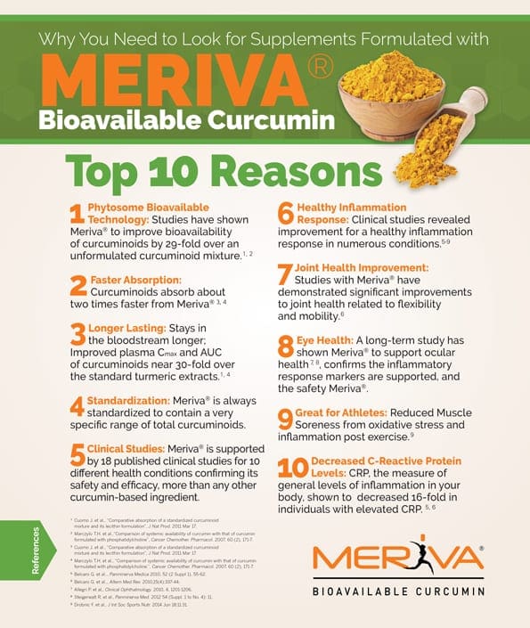


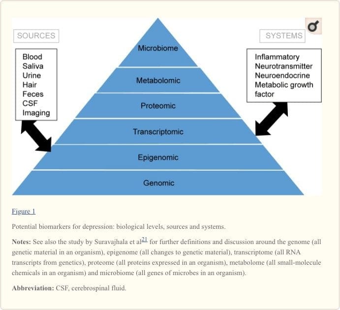
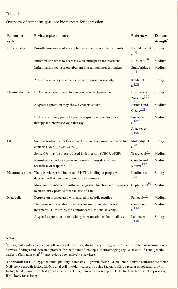
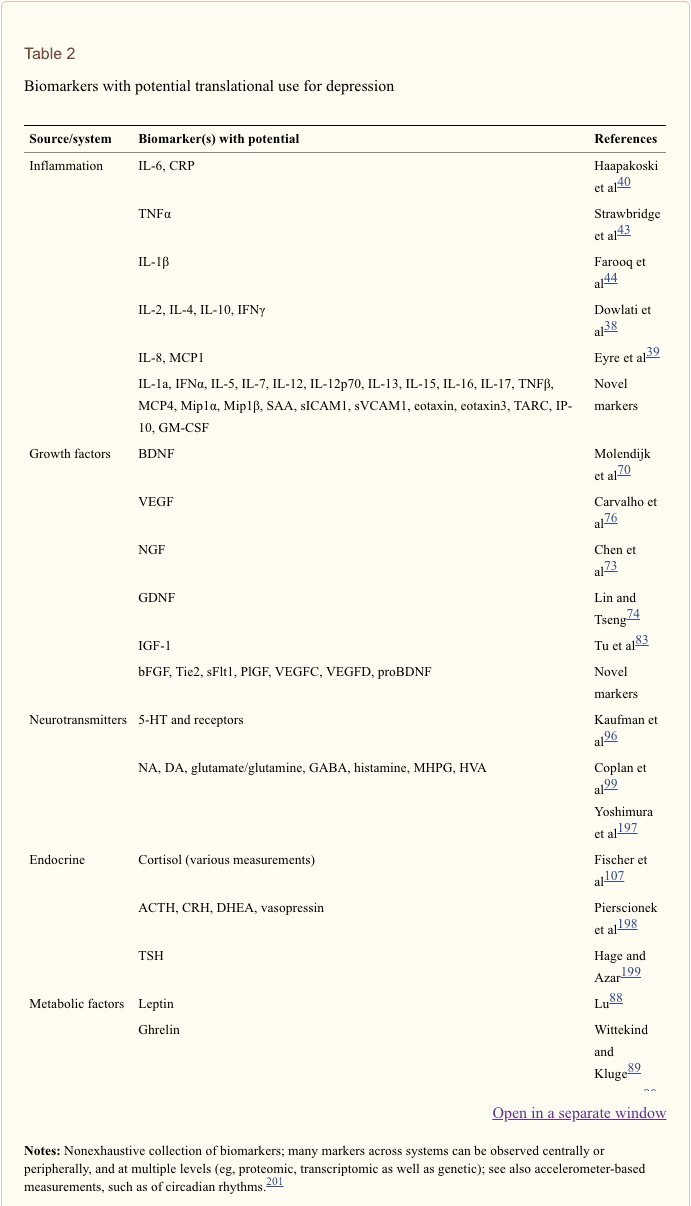


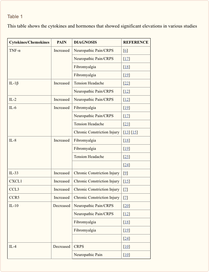


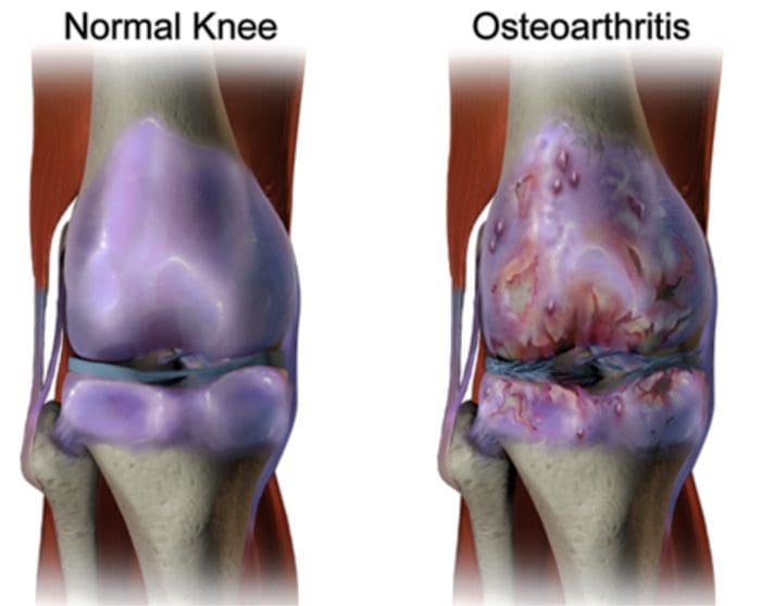
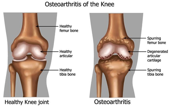
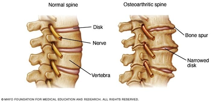
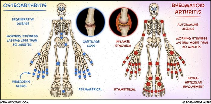 Abstract